36
TECHNICAL MONOGRAPH A FULLY MOLDABLE allograft with VIABLE CELLS ®

TECHNICAL MONOGRAPH
A FULLY MOLDABLE allograft with VIABLE CELLS
TECH
NIC
AL M
ON
OG
RA
PH®
TT-1409 © Orthofix Holdings, Inc. 2014 – 5000
1.888.298.5700www.orthof ix .com
®
INTRODUCTION 1
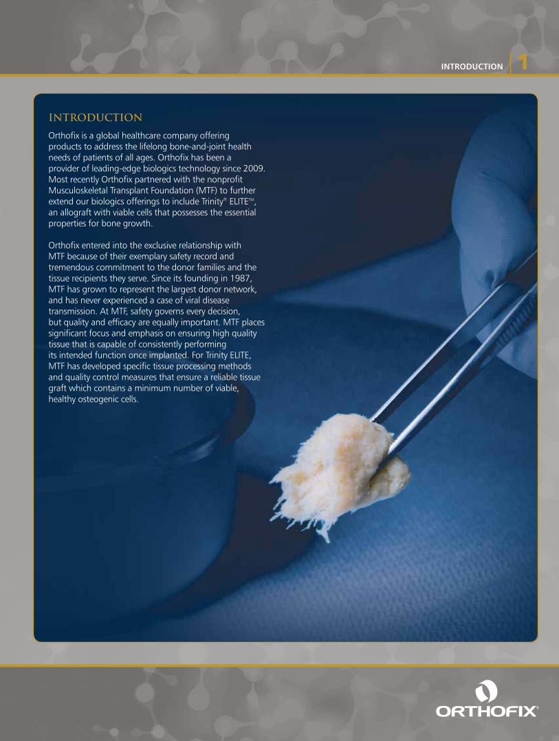
INTRODUCTION
Orthofix is a global healthcare company offering products to address the lifelong bone-and-joint health needs of patients of all ages. Orthofix has been a provider of leading-edge biologics technology since 2009. Most recently Orthofix partnered with the nonprofit Musculoskeletal Transplant Foundation (MTF) to further extend our biologics offerings to include Trinity® ELITETM, an allograft with viable cells that possesses the essential properties for bone growth. Orthofix entered into the exclusive relationship with MTF because of their exemplary safety record and tremendous commitment to the donor families and the tissue recipients they serve. Since its founding in 1987, MTF has grown to represent the largest donor network, and has never experienced a case of viral disease transmission. At MTF, safety governs every decision, but quality and efficacy are equally important. MTF places significant focus and emphasis on ensuring high quality tissue that is capable of consistently performing its intended function once implanted. For Trinity ELITE, MTF has developed specific tissue processing methods and quality control measures that ensure a reliable tissue graft which contains a minimum number of viable, healthy osteogenic cells.

2005 - Trinity MatrixFirst Generation Tissue Form
2009 - Trinity EvolutionSecond Generation Tissue Form
2013 - Trinity ELITEThird Generation Tissue Form
Orthofix – Osiris Therapeutics Orthofix – MTF Orthofix – MTF
INTRODUCTION2
Trinity® ELITE™ brings an enhanced handling experience to your surgery
When choosing the optimal bone graft to meet surgical demands, Trinity® ELITETM, a third generation allograft with viable cells, provides a unique alternative to autograft, long considered the standard for bone grafting. Exclusively processed for Orthofix by our premier partner, MTF, Trinity ELITE builds on the exemplary safety profile of more than 75,000 Trinity Evolution procedures to date. Trinity ELITE offers an enhanced handling experience, while providing a viable grafting alternative supplying the three physiologic and essential components for robust bone formation:
Osteogenic cells, including adult mesenchymal stem cells (MSCs) and osteoprogenitor cells (OPCs), are native to the cancellous matrix and capable of responding to their environment to differentiate into a variety of cell types as needed. This makes them an ideal osteogenic component of viable allograft. Adult MSCs are non-immunogenic since they do not present the Class II HLA antigens that trigger a T-cell response. Trinity ELITE undergoes a proprietary and selective process that depletes hematopoetic stem cells (HSCs) while maintaining viable osteogenic cells within the bone matrix.
Osteoinductive potential is derived from the demineralized cortical bone component which has been shown to possess active components of BMP-2, BMP-4, BMP-7, VEGF, TGF-ß, and other essential growth factors.1, 2, 3
Osteoconductive scaffold is provided by the cancellous bone component that serves as a natural trabecular bone matrix. It provides a porous, interconnected scaffold for bone ingrowth.
Trinity ELITE contains a minimum of 500,000 cells per cc; 100,000 of which are validated to be adult mesenchymal stem cells (MSCs) and/or osteoprogenitor cells (OPCs).*
* Data on file MTF
SCIENCE SCIEN
CE
SCIEN
CE

CHARACTERISTICS OF BONE GRAFTING MATERIALS
Bone graft material is often used to facilitate new bone formation. Successful bone healing and bony fusion can be influenced by the selection of a suitable bone graft material. Bone graft materials are typically characterized by the following definitions:
Osteogenic materials are capable of forming new bone from living cells. These materials contain MSCs and OPCs that can proliferate and differentiate into osteoblasts (bone-building cells) and eventually into osteocytes (mature bone cells). These cells represent the osteogenic potential of the graft.4
Osteoinductive materials have the potential to promote chemotaxis, mitogenesis and the differentiation of MSCs into OPCs that have osteogenic capacity (as described above).8
Osteoinductive materials will form bone when implanted into tissues which would not otherwise form new bone.9
Osteoconductive materials allow for fibrovascular tissue development and osteoprogenitor cell infiltration of a porous structure. This material then acts as a temporary scaffold which will be replaced with newly formed bone.8
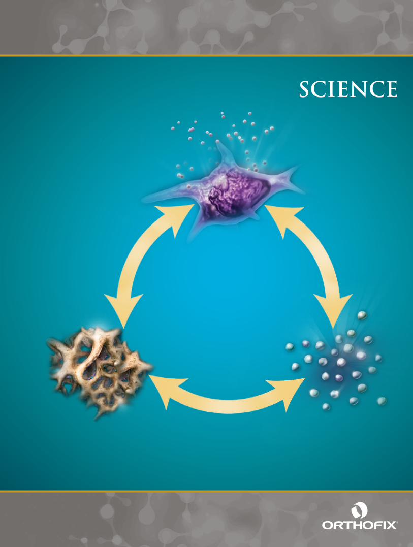
BONE HEALING AND GRAFT INCORPORATION
Along with an adequate blood supply, three components are necessary for bone healing and/or bone graft incorporation: the presence of cells capable of forming new bone (osteogenesis), a signal that triggers the differentiation of these cells into bone forming cells (osteoinductivity), and a scaffold or matrix on which the new bone can form (osteoconductivity).4
Mesenchymal stem cells (MSCs) have been characterized as types of adult stem cells that are multipotent and have the capacity to differentiate into bone-forming cells.5, 6 These cells can also produce cytokine signals that can recruit cells from the surrounding tissue to a target area.7 During the cascade of events that occurs during bone injury and subsequent healing, MSCs play a key role in creating a solid bone mass at the fracture or fusion site.
When bone is damaged, there is loss of mechanical integrity of the bone and disruption to the blood supply. The healing cascade begins immediately. The three phases are: inflammation, repair and remodeling. Inflammation is the process by which cells remove debris from the injured site, prepare the local matrix into a site which can support cell growth, and enable new bone to be formed. MSCs are also recruited to the site during the inflammation phase as revascularization occurs, which is required for new bone to form. Repair includes the differentiation of the MSCs first into OPCs and then bone-forming cells, which in turn produce new bone at the injured site. Lastly, remodeling is the resorption of immature or extraneous bone coupled with reorientation of bone along the direction of mechanical loading to provide adequate structural support. All of these phases (Figure 1) are regulated by the release of cytokine signals from the cells in and around the area of the injury.
Figure 1: Example of the healing cascade in fracture repair. The three phases of fracture repair include A) the inflammatory phase, B) the reparative phase, and C) the remodeling phase.4
MacrophagesMonocytesFibroblasts
GranulationTissue
Debris RemovalMacrop
MonoFibrob
GranulationTi
Debris R lphages
ocytesblasts
Tissue Removal
Vascular Ingrowth
A
BOsteogenic
Differentiationof MSC’s
OsteoblastsOsteoclasts
Mature,Integrated Bone
Osteoid
Healing Bone
IntramembraneousOssification
C
Recruitment of MSC’s
INFLAMMATORY PHASE
REPARATIVE PHASE
REMODELING PHASE
SCIENCE4
SCIE
NC
E

SCIENCE 5
HOW TRINITY ELITE WORKS
Trinity ELITE provides the three essential components for robust new bone formation in addition to desirable handling properties. These elements work in concert to elicit a rapid, reliable bone healing response.
• Trinity ELITE is a putty-like allograft that contains viable MSCs and OPCs residing within the graft material. Trinity ELITE also supplies an optimal environment to encourage these osteogenic cells to differentiate into osteoblasts and to secrete cytokines that recruit other bone-forming cells. Subsequently, the cells may actively contribute to new bone formation and later mature into osteocytes (Figure 3).
• Osteoinductive signals from the growth factors in the demineralized bone component promote bone growth by stimulating the bone-forming activity of these endogenous cells as well as recruiting the patient’s own cells to the site of repair.
• The cancellous bone matrix acts as a three-dimensional scaffold that is remodeled and then serves to facilitate the growth of new bone and support vascularization.
AUTOGRAFT BONE FOR BONE GRAFTING
Autograft cancellous bone has been widely used and is the traditional gold standard for bone grafting.10,11 Autograft bone is often recovered from the iliac crest of the patient at the time of surgery and then placed into the site where new bone formation is desired.11 The autograft bone has osteoconductive, osteoinductive, and osteogenic properties and contains the essential ingredients to create new bone including viable MSCs that reside within the tissue, endogenous growth factors, and a three-dimensional cell-friendly scaffold through which healing can occur (Figure 2).
Figure 2: Schematic depiction of the three critical elements that lead to bone healing and repair.
Figure 3: A diagram showing the sequential differentiation of an adult mesenchymal stem cell: first into a committed osteoprogenitor cell and then into a mature osteoblast under the influence of osteoinductive signals from the cellular microenvironment.
ADULT MESENCHYMALSTEM CELL
BMP’s
Adult MSC
IGF’s TGF-ßBMP’s
IGF’s TGF-ßBMP’s
OSTEOBLAST
COMMITED OSTEOPROGENITOR
CELL
Chondrocyte
Fibroblast
Adipocyte
Muscle

SCIENCE6
RATIONALE FOR AN ALTERNATIVE TO AUTOGRAFT
While there is an abundance of evidence that supports the ability of autograft bone to facilitate bone healing, there are a number of factors that may make autograft a less than ideal solution for the patient.10, 12 For example, there is only a limited supply of autograft bone, and using autograft bone may require a second surgical site (to procure bone) which has associated morbidity risks, can extend healing time, and can lead to pain and other lingering complications.13-15 Autograft bone grafts may also not possess the desired handling characteristics for certain surgical applications. In addition, there may be a large variability in the quantity and quality of the components of the autograft bone that can lead to unreliable and non-uniform bone healing.16 From one patient to another, there may be substantial differences in the number and health of the patient’s own stem cells and in the amount of osteoinductive signals within the tissue.17, 18 These elements can be affected by:
Patient age Systemic disease Osteoporosis Vascularity Presence of infection Use of anti-inflammatory drugs
TRINITY ELITE AS A PUTTY-LIKE AUTOGRAFT SUBSTITUTE
The benefits of an autograft substitute can only be realized if the alternative bone grafting material contains all of the essential components for new bone formation. Thus, this substitute needs to possess the healing “trinity”: osteogenic, osteoinductive and osteoconductive properties. Trinity ELITE is an allograft with viable cells that, not only retains these three attributes, but also possesses putty-like handling properties that may be desirable for specific surgical applications.19 Trinity ELITE represents a practical alternative to autograft bone and has some of its key advantages but none of its drawbacks. Although some synthetic materials and non-viable demineralized bone grafts are moldable and are formulated to be pastes or putties, none of these bone graft materials possess viable cells and rely solely on osteogenic cells from the host. Synthesized bone morphogenetic proteins (BMPs), while highly osteoinductive, also do not contain viable osteogenic cells.
For Trinity ELITE, the osteogenic cells are present within the bone matrix and remain adherent to the tissue until the time of implantation. Recent studies have shown that there may be as many as 65-fold more multipotent cells within trabecular bone compared to bone marrow aspirate.20 Thus, the matrix-adherent cells may represent the majority of the MSC population within bone.
In addition to a reliable number of viable osteogenic cells, Trinity ELITE contains other allogeneic components which give rise to its ability to stimulate and support new bone formation. These include demineralized cortical bone with verified osteoinductive potential and cancellous bone chips which serve as an osteoconductive scaffold.

SCIENCE 7
CONFIRMATION OF OSTEOGENIC CELLS IN TRINITY ELITE
Cells from samples of Trinity ELITE have been explanted onto culture dishes and shown to exhibit cell morphology that is characteristic of MSCs and to have the potential to exhibit osteogenic behavior (Figure 4A-D). In addition, cells from the viable cancellous component of the graft have been shown to be capable of differentiating into adipogenic and chondrogenic lineages (Figure 4E-F). To confirm their osteogenic capacity, the cells were cultured under specific culture conditions and demonstrated the ability to produce alkaline phosphatase (ALP), an enzyme secreted by cells during bone formation. The ability of the cells within trabecular bone to undergo multilineage differentiation is consistent with numerous previous studies of mesenchymal stem cells in culture.21-24
Figure 4: Adherent cells from a sample of Trinity ELITE that (A) have attached to a tissue culture dish and taken on a MSC-like cellmorphology; cells subsequently showing expression of ALP (B) and positivestaining for mineralization using von Kossa staining (C) and Alizarin Red staining (D) under osteogenic culture conditions; (E) cells exhibiting positive staining for lipids by oil-red O staining in an adipogenic environment; and (F) cells demonstrating proteoglycan production using Safranin-O staining under chondrogenic culture conditions.
Cells from the cancellous bone component of Trinity ELITE have also been evaluated in order to determine their MSC and OPC identities by staining for protein markers that are specific to these types of cells. In particular, MSCs have been detected by positive brown staining for CD166, and OPCs have been identified by positive brown staining for osteocalcin (Figure 5). These two stains represent common methods for classifying MSC and OPC cell types.25-27
Figure 5: Histology sections of the cancellous bone component of Trinity ELITE showing (A) cells that exhibit positive staining for the MSC marker, CD166 (brown color); and (B) cells that show expression of osteocalcin (brown color), which is characteristic of OPCs.
Adherent MSCs
Adipogenesis
Osteogenesis
Osteogenesis
Chondrogenesis
Osteogenesis
OsteocalcinPositive
(stain for OPC)
CD166 Positive
(MSC marker)A
A B
C
E
B
D
F

PROCESSING & HANDLING
PRO
CESSIN
G&
HA
ND
LING
PRO
CESSIN
G&
HA
ND
LING
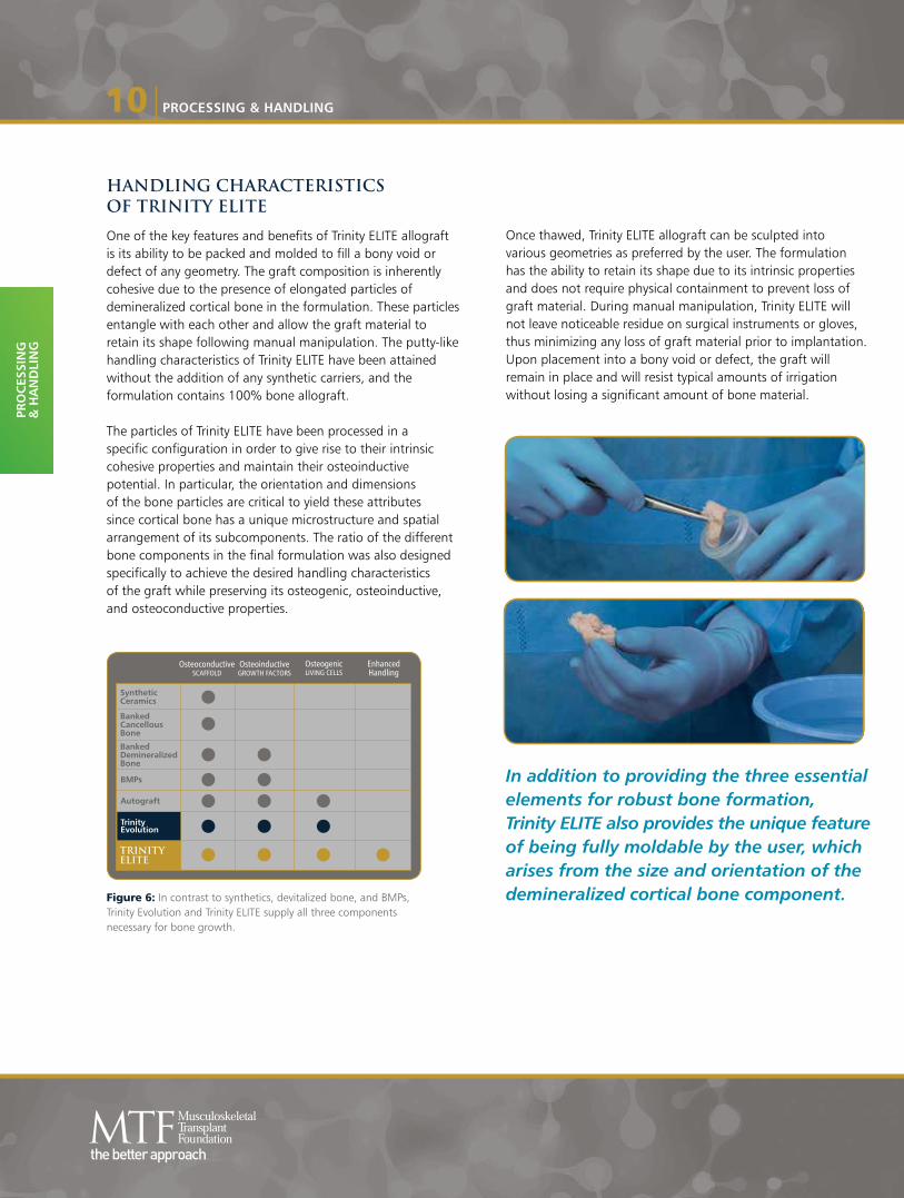
HANDLING CHARACTERISTICS OF TRINITY ELITE
One of the key features and benefits of Trinity ELITE allograft is its ability to be packed and molded to fill a bony void or defect of any geometry. The graft composition is inherently cohesive due to the presence of elongated particles of demineralized cortical bone in the formulation. These particles entangle with each other and allow the graft material to retain its shape following manual manipulation. The putty-like handling characteristics of Trinity ELITE have been attained without the addition of any synthetic carriers, and the formulation contains 100% bone allograft.
The particles of Trinity ELITE have been processed in a specific configuration in order to give rise to their intrinsic cohesive properties and maintain their osteoinductive potential. In particular, the orientation and dimensions of the bone particles are critical to yield these attributes since cortical bone has a unique microstructure and spatial arrangement of its subcomponents. The ratio of the different bone components in the final formulation was also designed specifically to achieve the desired handling characteristics of the graft while preserving its osteogenic, osteoinductive, and osteoconductive properties.
Figure 6: In contrast to synthetics, devitalized bone, and BMPs, Trinity Evolution and Trinity ELITE supply all three components necessary for bone growth.
Once thawed, Trinity ELITE allograft can be sculpted into various geometries as preferred by the user. The formulation has the ability to retain its shape due to its intrinsic properties and does not require physical containment to prevent loss of graft material. During manual manipulation, Trinity ELITE will not leave noticeable residue on surgical instruments or gloves, thus minimizing any loss of graft material prior to implantation. Upon placement into a bony void or defect, the graft will remain in place and will resist typical amounts of irrigation without losing a significant amount of bone material.
In addition to providing the three essential elements for robust bone formation, Trinity ELITE also provides the unique feature of being fully moldable by the user, which arises from the size and orientation of the demineralized cortical bone component.
OsteoconductiveSCAFFOLD GROWTH FACTORS LIVING CELLS
Osteoinductive Osteogenic Enhanced Handling
SyntheticCeramics
BankedCancellousBone
BankedDemineralizedBone
BMPs
Autograft
TRINITYELITE
TrinityEvolution
PROCESSING & HANDLING10
PRO
CES
SIN
G&
HA
ND
LIN
G

PROCESSING OF ALLOGRAFT TISSUE INTO TRINITY ELITE
Trinity ELITE is formulated from allograft donor bone using a proprietary process that has been specifically designed to preserve its key components, including the health of the resident bone-forming cells. Specific component ratios are maintained, as well as a consistent particle orientation and size.
• Processing of the donor tissue begins within 72 hours post-mortem of death once serological testing is complete. This timeline is critical to preserve cell viability and functionality.
• Cancellous bone chips are isolated and then processed with minimal manipulation to retain and preserve native mesenchymal stem cells and osteoprogenitor cells within the matrix. The retention of viable cells within the cancellous bone component of Trinity ELITE has been confirmed by live/dead cell staining (Figure 7).
• Cortical bone from the same donor is milled into elongated particles of specific dimensions and then demineralized, thereby resulting in bone that is inherently cohesive and possesses osteoinductive potential.
• The components of Trinity ELITE are subsequently combined in a defined ratio to yield the desired handling characteristics.
PROCESSING & HANDLING 11
CRYOPRESERVATION AND STORAGE OF TRINITY ELITE
In order to maintain cell health following cryopreservation, MTF has developed a process that protects the viable cells within the matrix during freezing and has established stringent storage conditions for the tissue once it is frozen to ensure that the cells are not compromised.
• Prior to cryopreservation, a nutrient-rich, base medium specific to MSCs is added, as well as a cryoprotectant. A slow, controlled-rate freezing process of 1ºC per min is then used to help minimize cell damage from osmotic membrane imbalance and reduce ice crystal formation within the cell (Figure 8).
• Cell metabolism stops at -130ºC and lower storage temperatures are known to increase the viable storage period.18, 19 Therefore, Trinity ELITE is stored in vapor phase liquid nitrogen tanks at -185ºC after cryopreservation until it is shipped to the hospital or surgery center.
Figure 7: A microscopic image of a sample of the viable cancellous bone component of Trinity ELITE showing fluorescently-stained live cells (represented by bright green dots).
Figure 8: Illustration demonstrates the effects of fast versus slow cooling rates during the cryopreservation of living cells and shows less intracellular ice formation during con-trolled-rate freezing.
FASTCOOLING
RATE
SLOWCOOLING
RATE
More Intracellular Ice Less Intracellular Ice
Figure 4: A schematic depiction of the effects of fast versus slow cooling rates on intracellular ice formation during the cryopreservation of living cells.


SAFETY
SAFETY

SAFE
TY
SAFETY 15
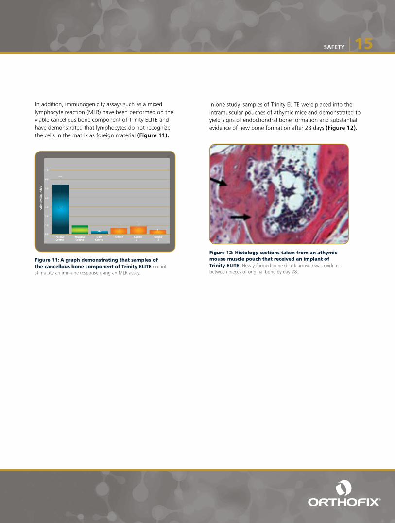
In addition, immunogenicity assays such as a mixed lymphocyte reaction (MLR) have been performed on the viable cancellous bone component of Trinity ELITE and have demonstrated that lymphocytes do not recognize the cells in the matrix as foreign material (Figure 11).
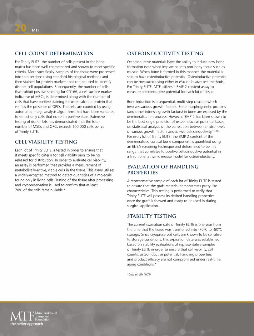
In one study, samples of Trinity ELITE were placed into the intramuscular pouches of athymic mice and demonstrated toyield signs of endochondral bone formation and substantial evidence of new bone formation after 28 days (Figure 12).
PositiveControl
NegativeControl
DBMControl
Sample1
Sample2
Sample3
Stim
ulat
ion
Inde
x
5.0
6.0
7.0
2.0
3.0
4.0
1.0
0.0
Figure 11: A graph demonstrating that samples of the cancellous bone component of Trinity ELITE do not stimulate an immune response using an MLR assay.
Figure 12: Histology sections taken from an athymic mouse muscle pouch that received an implant of Trinity ELITE. Newly formed bone (black arrows) was evident between pieces of original bone by day 28.


MTF

COMMITMENT TO QUALITY
MTF is dedicated to excellence in tissue quality and strives to ensure that the biologic properties and handling characteristics of each lot of Trinity ELITE meet the expectations of the user.Cell health is preserved during every phase in the processing of Trinity ELITE. Each step has been carefully planned and evaluated to maximize the number of viable, bone-forming cells while only minimal manipulation of the bone tissue occurs. Rigorous validation and comprehensive testing has proven that safe, high-quality tissue is obtained from these process steps. Many factors contribute to the high quality of Trinity ELITE including:
• Strict donor selection and screening criteria• Donor age• Careful processing and storage• Quality control• Stability testing
STRICT DONOR CRITERIA
At MTF, quality and safety standards consistently meet or exceed industry standards*, as well as the guidelines for screening and testing of tissue donors set forth by the US Food and Drug Administration (FDA). Minimum guidelines are set by regulatory agencies to ensure safety of tissue. Potential MTF donors must pass through an extensive quality assurance process.
Screening begins with a comprehensive medical and social history that includes the cause of death. Tissue and blood samples are tested for infectious diseases, including hepatitis, HIV and syphilis. A team of medical/ technical specialists from the infectious disease and tissue banking fields evaluates all information, including test results, before the donor is released for processing. (Figure 13) lists those areas in which MTF voluntarily defers donors for safety or quality reasons, even when not required by the FDA or industry standards.
*Generally recognized industry standards, although some tissue banks may vary
Figure 14: Less than 3% of donors accepted. MTF rejects 97% of donors for such conditions as cancer, illegal drug use, infection, high-risk behavior, age, osteoporosis, steroid use and more.
Figure 13: Strict Donor Criteria
MTF Donor Deferral Rate
> 97% deferred
< 3% accepted
SCREENING CRITERIA MTF INDUSTRY* FDA
Hepatitis B virus X X XHepatitis C virus X X XHIV 1/2 X X XMalaria X X XSepsis X X XSyphilis X X XTransmission spongiform encephalopathy (TSE) X X XVaccinia X X XWest Nile Virus (WNV) X X XClinically significant metabolic bone disease X XGonorrhea (clinically active) X X Illicit drug use, injected drugs X XLeprosy (Hansen’s disease) X X Polyarteritis nodosa X XRabies X XRheumatoid arthritis** X XSarcoidosis X XSystemic lupus erythematosus X XSystemic mycosis X XTuberculosis (clinically active) X XActive genital herpes XAcute infectious/septic illness XAnkylosing spondylitis XAntiphospholipid syndrome XAutoimmune hemolytic anemia XAutoimmune lymphoproliferative syndrome XAutoimmune thrombocytopenic purpura XAutoimmune vasculitis XCancer (see chart inside) XChagas disease XEnd stage renal disease/chronic dialysis** XEpstein Barr virus (clinically symptomatic mononucleosis) XClostridium difficile infection XCold agglutinin disease XEncephalitis (clinically active) XEndocarditis (clinically active) XGuillain-Barre syndrome (clinically active) XHigh risk behavior XIllicit drug use, non-injected drugs XMeningitis (clinically active) XMethicillin resistant staphylococcus aureus (MRSA) XMixed connective tissue disease XMultiple sclerosis X Myasthenia gravis XOsteoporosis, clinically diagnosed** XPeritonitis XPoliomyelitis XPyelonephritis XReactive arthritis (Reiter’s syndrome) XRheumatic fever XSteroid use/Treatment Chronic XVancomycin resistant enterococcus (VRE) XVaricella zoster XWegener’s granulomatosis X
*Generally recognized industry standards, although some tissue banks may vary**Not an automatic rule-out for skin donors
MTF18
MTF

MTF20
CELL COUNT DETERMINATION
For Trinity ELITE, the number of cells present in the bone matrix has been well-characterized and shown to meet specific criteria. More specifically, samples of the tissue were processed into thin sections using standard histological methods and then stained for protein markers that can be used to identify distinct cell populations. Subsequently, the number of cells that exhibit positive staining for CD166, a cell surface marker indicative of MSCs, is determined along with the number of cells that have positive staining for osteocalcin, a protein that verifies the presence of OPCs. The cells are counted by using automated image analysis algorithms that have been validated to detect only cells that exhibit a positive stain. Extensive testing of donor lots has demonstrated that the total number of MSCs and OPCs exceeds 100,000 cells per cc of Trinity ELITE.
CELL VIABILITY TESTING
Each lot of Trinity ELITE is tested in order to ensure that it meets specific criteria for cell viability prior to being released for distribution. In order to evaluate cell viability, an assay is performed that provides a measurement of metabolically-active, viable cells in the tissue. This assay utilizes a widely-accepted method to detect quantities of a molecule found only in living cells. Testing of the tissue after processing and cryopreservation is used to confirm that at least 70% of the cells remain viable.*
OSTEOINDUCTIVITY TESTING
Osteoinductive materials have the ability to induce new bone formation even when implanted into non-bony tissue such as muscle. When bone is formed in this manner, the material is said to have osteoinductive potential. Osteoinductive potential can be measured using either in vivo or in vitro test methods. For Trinity ELITE, MTF utilizes a BMP-2 content assay to measure osteoinductive potential for each lot of tissue.
Bone induction is a sequential, multi-step cascade which involves various growth factors. Bone morphogenetic proteins (and other intrinsic growth factors) in bone are exposed by the demineralization process. However, BMP-2 has been shown to be the best single predictor of osteoinductive potential based on statistical analysis of the correlation between in vitro levels of various growth factors and in vivo osteoinductivity.15,16
For every lot of Trinity ELITE, the BMP-2 content of the demineralized cortical bone component is quantified using an ELISA screening technique and determined to be in a range that correlates to positive osteoinductive potential in a traditional athymic mouse model for osteoinductivity.
EVALUATION OF HANDLING PROPERTIES
A representative sample of each lot of Trinity ELITE is tested to ensure that the graft material demonstrates putty-like characteristics. This testing is performed to verify that Trinity ELITE will possess its desired handling properties once the graft is thawed and ready to be used in during surgical application.
STABILITY TESTING
The current expiration date of Trinity ELITE is one year from the time that the tissue was transferred into -70ºC to -80ºC storage. Since cryopreserved cells are known to be sensitive to storage conditions, this expiration date was established based on stability evaluations of representative samples of Trinity ELITE in order to ensure that cell viability, cell counts, osteoinductive potential, handling properties, and product efficacy are not compromised under real-time aging conditions.*
*Data on file (MTF)

THAW & PREPARATION
THA
W &
PREPA
RA
TION
THA
W &
PREPA
RA
TION

PREPARATION OF TRINITY ELITE FOR USAGE
The thawing of cryopreserved cells is a critical step in their rejuvenation, and such cells are known to be sensitive to environmental conditions during this process.36 With this in mind, MTF has developed a specific procedure for thawing Trinity ELITE in order to minimize cell damage and optimize cell viability.
1. Peel back the lid of the outer tray. Grasp the top and bottom of the jar by placing fingers in the open area provided to remove jar from the outer tray. Pass it onto the sterile field.
3. Use sterile gauze or the optional strainer to decant the cryopreservation solution into a waste container.
5. Decant 5% Dextrose in Lactated Ringer’s Solution (DL5R) prior to use.
2. Place the jar containing allograft tissue and cryopreservation solution in a sterile basin containing a warm (35°C to 39°C; 95°F to 102.2°F) sterile irrigant (Normal saline or 5% Dextrose in Lactated Ringer’s Solution). The jar containing the allograft should remain in this solution until the contents of the jar flows freely upon inversion. The jar should be removed from the warm solution once free-flowing.
Note: The allograft tissue should NOT be implanted prior to thawing.
6. Implant within 2 hours of thawing.
Note: Trinity ELITE has been validated for a 2-hour window post-thaw for optimal cell viability.
4. Add 5% Dextrose in Lactated Ringer’s Solution to the indicated fill line to immerse the allograft tissue. Replace the cap and invert twice to suspend tissue until ready for use.
Items you will need:
2 sterile basins: 1 for thawing, 1 for the allograft tissue
Warm (37ºC ±2º) sterile saline
5% Dextrose in Lactated Ringer’s Solution (D5LR)
Forceps to remove the tissue Gauze (4x4) or optional strainer cap
Part Number Sizes410001 Small410002 Medium410003 Large410004 Extra-Large
TRINITY ELITEThawing: Trinity ELITE thaw times are approximately 25-35 minutes
THAW & PREPARATION22
THA
W &
PREP
AR
ATI
ON

FAQ
FAQ
GENERAL INFORMATION
What is Trinity ELITE?Trinity ELITE is a cryopreserved, fully moldable allograft that consists of viable cancellous bone containing adult mesenchymal stem cells (MSCs), osteoprogenitor cells (OPCs), and a demineralized cortical bone component.
What are the indications for Trinity ELITE?Trinity ELITE is an allograft intended for the treatment of musculoskeletal defects. Refer to package insert for contraindications.
What is the FDA Regulatory status for Trinity ELITE?Trinity ELITE is classified as, and subject to regulation by the FDA as a human allograft, per 21 C.F.R. § 1271 (alternatively Section 361 of the Public Health Service Act). Additionally, Trinity ELITE is an allograft tissue pursuant to American Association of Tissue Banks (AATB) guidelines, and its processing conforms to recommendations as set forth by Tissue Reference Group (TRG).
How does Trinity ELITE work to repair bone?Trinity ELITE contributes to new bone formation by providing osteogenic, osteoinductive, and osteoconductive elements to the bony defect site.
Cancellous bone in the Trinity ELITE tissue matrix serves as a three-dimensional osteoconductive structure, or scaffold, to support and facilitate the growth of bone and its blood supply. Growth factors provided by Trinity ELITE signal and promote bone growth (osteoinduction) by recruiting the patient’s own cells to the site and stimulating the bone-forming activity of these endogenous cells. Trinity ELITE contributes to osteogenesis by the direct formation of bone from adult mesenchymal stem cells and osteoprogenitor cells. These cells differentiate into osteoblasts, contribute to matrix formation, and later mature into osteocytes. The only other material that contributes to osteogenesis is autograft material (harvested bone or bone marrow aspirate).
What did MTF add to Trinity ELITE to make it handle like a putty?MTF changed the shape and size of the demineralized cortical bone component. Entanglement of the elongated bone particles in the Trinity ELITE formulation give rise to its cohesive, putty-like handling. NO carrier was added.
How did MTF validate the higher number of viable osteogenic cells in Trinity ELITE?MTF has identified and utilized enhanced tissue processing coupled with improved characterization methods of the tissue form.
Do the cells in Trinity ELITE wash away in a surgical, or blood soaked environment? Similar to Trinity Evolution, the osteogenic cells in Trinity ELITE are adherent to cancellous matrix, remain attached during implantation and will not wash away during standard rinse to the operative site.
TISSUE PREPARATION, USE AND STORAGE
What is the shelf life of Trinity ELITE and storage temperatures? Trinity ELITE has a 1 year shelf life when stored -70° to -80°C at the local hospital or surgery center.
What are the thawing procedures?Thaw tissue 25-35 min in basin containing warmed saline.Use gauze/strainer to decant the cryoprotectant.
Add 5% dextrose in Lactated Ringers (D5LR) solution.Rinse the D5LR through gauze/strainer and tissue is ready for use. (Additional gauze can be used to soak-up excess solution as desired.)
Please reference the Thaw and Preparation for Uses section or the package insert for more details.
Can Trinity ELITE be combined with common antibiotics during implantation?Studies performed at MTF have demonstrated that Bacitracin can be added to Trinity ELITE, up to limited concentrations,(50,000 Units/L) without an adverse effect on cell viability.
Can Trinity ELITE be re-frozen and/or re-used?No. Once the Trinity ELITE jar is thawed, opened and or rinsed, the cells begin to resume their normal function and metabolic role. The process by which Trinity ELITE is processed, cryopreserved and stored, is time- and temperature-sensitive and should not be replicated.
FAQ24
FAQ
FAQ 25
BONE GROWTHHow does Trinity ELITE promote osteogenesis compared to autograft?Osteogenesis is the regeneration of bone through the action of osteoblasts. Historically, autograft taken from the patient’s iliac crest has been the gold standard in bone grafting. However, the drawbacks of autograft include donor site morbidity, quality and quantity of cells recovered, and an additional surgical site to heal. Trinity ELITE provides the scaffold, the growth factors and the stem cells needed for osteoconduction, osteoinduction, and osteogenesis, without the potential complications of autograft.
What prompts Trinity ELITE to form bone cells rather than other tissues, such as cartilage or adipose? Signals from the microenvironment, including growth factors and other stimulatory cues, are expressed by the surrounding tissue.
SAFETYWhat is the liquid in the Trinity ELITE jar?The liquid is a basal medium containing a cryoprotectant agent, Dimethyl sulfoxide (DMSO), which ensures the cryopreservation process proceeds efficiently while reducing apoptosis. Dimethyl sulfoxide (DMSO) is a chemical commonly used as a cryoprotectant to preserve living cells. When mixed with water, it lowers the freezing point of the solution and helps to prevent cell damage by reducing overall ice crystal formation that can disrupt cellular structure.
Why does Trinity ELITE not stimulate an immune response?Mesenchymal stem cells do not express human leukocyte antigens (HLA Class II) or T-cell activation molecules (CD40, CD80, CD86). Without HLA antigens and the molecules that provoke T-cell activation, mesenchymal cells do not cause immune responses. Studies using mixed lymphocyte reaction (MLR) assays have verified lack of immune response to the mesenchymal cellular matrix. In addition, clinical trials have shown that high doses of mesenchymal cells can be given by infusion to patients with normal immune systems without triggering immune response or adverse reactions.
Is Trinity ELITE STERILE? Trinity ELITE is prepared aseptically for clinical use. The process is validated for microbial treatment of the tissue and every lot must pass USP <71> sterility standards.
RECOVERY & PROCESSING (MTF)What is the advantage of processing at MTF?Stringent donor selection criteria, younger donors, and careful processing ensure cell health.
How is cell health preserved? • Stringent donor screening standards
• Time-sensitive processing and controlled rate of freezing for optimal cell viability
• Cryopreservation and storage in vapor-phase liquid nitrogen at -185° C limits cell metabolism
• Expiration dating, which reflects real time testing and must pass MTF’s stringent release criteria
What are the donor screening criteria?MTF has strict criteria for donor selection–including stringent policies on non-IV drug use and cancer.
MTF never accepts donors deferred by other tissue banks for medical reasons.
Each donor is screened using medical and social history questionnaires and serology testing.
(Please see MTF & Safety Section for more information)

PACKAGEINSERT
PAC
KA
GE
INSER
T
TRINITY® ELITE™INSTRUCTIONS FOR USE
READ BEFORE USING DONATED HUMAN TISSUE
THIS TISSUE WAS RECOVERED FROM A DECEASED DONOR FROM WHOM LEGAL AUTHORIZATION OR CONSENT HAS BEEN OBTAINED. THIS RECOVERY WAS PERFORMED USING ASEPTIC TECHNIQUES. PROCESSING AND PACKAGING WERE PERFORMED UNDER ASEPTIC CONDITIONS. TERMINAL STERILIZATION AGENTS WERE NOT USED IN THE PROCESS.
Description and Indication for UseMUSCULOSKELETAL TRANSPLANT FOUNDATION (MTF) tissues are supplied in a variety of standard sized units designed for surgical use by qualified health care professionals (e.g., physicians, dentists, and/or podiatrists). Processed human bone and soft tissue have been used in a variety of surgical applications and in combination with prosthetic devices.
The amount and size of allograft necessary for a surgical procedure is based upon an individual surgeon’s preference and the size and type of defect. The description of the tissue, serial number, expiration date, product code, size and/or amount, and additional information are printed on the allograft container label.
CautionsTrace amounts of Acetic Acid, Dimethyl Sulfoxide, Polysorbate-80, Ethanol, Polyoxyethylene (10) Phenol Ether and Hydrogen Peroxide may be present. Caution should be exercised if the patient is allergic to any of these substances.
NOTE: No ß-lactam antibiotics are used during the processing of tissue.
Caution should be used for the following conditions:• Severe vascular or neurological disease• Fever• Uncontrolled diabetes• Pregnancy• Hypercalcemia• Renal-compromised patients• History of or active Pott’s disease• Osteomyelitis at the surgical site• Sepsis in or around the surgical site• Inability to cooperate with and/or comprehend post-operative instructions
PrecautionsExtensive medical screening procedures have been used in the selection of all tissue donors for MTF. Transmission of infectious diseases such as HIV or Hepatitis, as well as a theoretical risk of the Creutzfeldt-Jako (CJD) agent, may occur in spite of careful donor selection and serological testing. Bacterial infection at the site of grafting may occur. Within the United States: Adverse outcomes attributable to the tissue must be promptly reported to MTF. Outside of the United States: Adverse outcomes attributable to the tissue must be promptly reported to your local representative.
Adverse EffectsPossible adverse effects of using human tissues include but are not limited to:• Infection of soft tissue and/or bone (osteomyelitis)• Fever• Deformity of the bone at the site• Incomplete bone ingrowth, delayed union or non-union• Fracture of the newly formed bone• Disease transmission and undesirable immune response
Aseptically ProcessedALL ALLOGRAFTS ARE FOR SINGLE PATIENT USE ONLY. The allografts are not terminally sterilized. Each allograft is asepticallyprocessed and the finished product passes USP <71> Sterility Tests. Do not subject allografts to additional sterilization procedures. Do notuse portions of an allograft from one container on multiple patients.Dispose of excess or unused tissue in accordance with recognizedprocedures for discarding regulated medical waste materials.
This allograft must not be used under any of the following conditions:• If the container seal is damaged, or not intact.• If the container has any physical damage.• If the container label or identifying bar code is severely damaged, not readable or is missing.• If the expiration date shown on the container label has passed.• If the vial is received thawed.• If not used within 2 hours after thawing or has been stored at a temperature not recommended.
Donor Screening and TestingPrior to donation, the donor’s medical/social history is screened formedical conditions or disease processes that would contraindicate thedonation of tissues in accordance with current policies and proceduresapproved by the MTF Medical Board of Trustees.Donor blood samples taken at the time of recovery were tested by a CLIAlicensed facility for:• Hepatitis B surface antigen • HIV-1/2 antibody• Hepatitis B core antibody • Syphilis• Hepatitis C antibody • HIV -1 (NAT)• HCV (NAT)
The results of all serological testing were negative. This allograft tissuehas been determined to be suitable for transplantation.
The infectious disease test results, consent, current donor medical history interview, physical assessment, available relevant medical records to include previous medical history, laboratory test results, autopsy and coroner reports, if performed, and information obtained from any source or records which may pertain to donor suitability, have been evaluated by an MTF physician and are sufficient to indicate that donor suitability criteria current at the time of procurement, have been met. This tissue is suitable for transplantation. The donor suitability criteria used to screen this donor are in compliance with the FDA regulations published in 21 CFR Part 1271 Human Cells, Tissues, and Cellular and Tissue Based Products, as applicable. All procedures for donor screening, serologic and microbiologic testing meet or exceed current standards established by the American Association of Tissue Banks.
Cryopreserved TissueTissue prepared by cryopreserved processes has been stored in MTFat -185ºC in Vapor Phase Liquid Nitrogen until time of shipping and areshipped on dry ice.
StorageThe recommended storage temperature is -70 to -80 degrees C. Shortterm storage of -58 to -70 degrees C for up to 2 weeks is acceptable.Tissues stored at -58 to -70 degrees C may be placed back into therecommended storage environment of -70 to -80 degrees C at any timeduring that period. This short-term storage temperature would also allowfor any internal temperature fluctuations between -58 to -70 degrees Cthat may occur during long-term storage at -70 to -80 degrees due tocycling or opening freezer doors. It is the responsibility of the transplantfacility or clinician to maintain the tissue intended for transplantation in the appropriate recommended storage conditions prior to transplant.
PACKAGE INSERT28
PAC
KA
GE
INSE
RT

ORDERINGINFORMATION
OR
DER
ING
INFO
RM
ATIO
N

Part Number Sizes410001 Small410002 Medium410003 Large410004 Extra-Large
TRINITY ELITE
This tissue is provided as a generous gift from a donor and the donor’s family.
ORDERING INFORMATION
For more information contact your local representative or call:
1.888.298.5700 Orthofix1.800.946.9008 MTF
ORDERING INFORMATION30
OR
DER
ING
INFO
RM
ATI
ON
REFERENCES
REFER
ENC
ES
1. Bostrom MPG, and Seigerman DA. The clinical use of allografts, demineralized bone matrices, synthetic bone graft substitutes and osteoinductive growth factors: A survey study. 2005. HSS Journal 1(1):9-18.
2. Pacaccio DJ, and Stern SF. Demineralized bone matrix: Basic science and clinical applications. 2005. Clin Podiatr Med Surg 22:599-606.
3. Wildermann B, Kadow-Romacker A, Haas NP, Schmidmaier G. Quantification of various growth factors in different demineralized bone matrix preparations. 2007. J Biomed Mater Res A 81(2):437-442.
4. Mehta, S. and C. Collings, Orthobiologics: Improving Fracture Care Through Science. 2007: Lippincott Williams & Wilkins.
5. Bruder, S.P., D.J. Fink, and A.I. Caplan, Mesenchymal stem cells in bone development, bone repair, and skeletal regeneration therapy. J Cell Biochem, 1994. 56(3): p. 283-94.
6. Caplan, A.I., Mesenchymal stem cells. J Orthop Res, 1991. 9(5): p. 641-50.
7. Caplan, A.I., Why are MSCs therapeutic? New data: new insight. J Pathol, 2009. 217(2): p. 318-24.
8. Mouch, C. and T.A. Einhorn, Bone Morphogenetic Proteins and Other Growth Factors to Enhance Fracture Healing and Treatment of Nonunions, in Bone Regeneration and Repair: Biology and Clinical Applications, J. Lieberman and G. Friedlander, Editors. 2005, Humana Press.
9. Boyan, B.D. and J. McMillan, Bone Graft Substitutes: Basic Information for Successful Clinical Use with Special Focus on Synthetic Graft Substitutes, in Bone Graft Substitutes, C.T. Laurecin, Editor. 2003, ASTM. p. 231-259.
10. Seiler, J.G., 3rd and J. Johnson, Iliac crest autogenous bone grafting: donor site complications. J South Orthop Assoc, 2000. 9(2): p. 91-7.
11. Sandhu, H.S., H.S. Grewal, and H. Parvataneni, Bone grafting for spinal fusion. Orthop Clin North Am, 1999. 30(4): p. 685-98.
12. Boone, D.W., Complications of iliac crest graft and bone grafting alternatives in foot and ankle surgery. Foot Ankle Clin, 2003. 8(1): p. 1-14.
13. Silber, J.S., et al., Donor site morbidity after anterior iliac crest bone harvest for single-level anterior cervical discectomy and fusion. Spine (Phila Pa 1976), 2003. 28(2): p. 134-9.
14. Nocini, P.F., et al., Fractures of the iliac crest following anterior and posterior bone graft harvesting. Review of the literature and case presentation. Minerva Stomatol, 2003. 52(10): p. 441-8, 448-52.
15. Heary, R.F., et al., Persistent iliac crest donor site pain: independent outcome assessment. Neurosurgery, 2002. 50(3): p. 510-6; discussion 516-7.
16. Muschler, G.F., et al., Age- and gender-related changes in the cellularity of human bone marrow and the prevalence of osteoblastic progenitors. J Orthop Res, 2001. 19(1): p. 117-25.
17. Hernigou, P., et al., Percutaneous autologous bone-marrow grafting for nonunions. Influence of the number and concentration of progenitor cells. J Bone Joint Surg Am, 2005. 87(7): p. 1430-7.
18. D’Ippolito, G., et al., Age-related osteogenic potential of mesenchymal stromal stem cells from human vertebral bone marrow. J Bone Miner Res, 1999. 14(7): p. 1115-22.
19. Rush, S.M., Trinity evolution. Foot Ankle Spec, 2010. 3(3): p. 144-7.
20. Jones, E., et al., Large-scale extraction and characterization of CD271+ multipotential stromal cells from trabecular bone in health and osteoarthritis: implications for bone regeneration strategies based on uncultured or minimally cultured multipotential stromal cells. Arthritis Rheum, 2010. 62(7): p. 1944-54.
REFERENCES32
REF
EREN
CES
REFERENCES 33
21. Barry, F.P. and J.M. Murphy, Mesenchymal stem cells: clinical applications and biological characterization. Int J Biochem Cell Biol, 2004. 36(4):p. 568-84.
22. Jaiswal, N., et al., Osteogenic differentiation of purified, culture-expanded human mesenchymal stem cells in vitro. J Cell Biochem, 1997. 64(2):p. 295-312.
23. Marie, P.J. and O. Fromigue, Osteogenic differentiation of human marrow-derived mesenchymal stem cells. Regen Med, 2006. 1(4):p. 539-48.
24. Pittenger, M.F., et al., Multilineage potential of adult human mesenchymal stem cells. Science, 1999. 284(5411): p. 143-7.
25. Risbud, M.V., et al., Osteogenic potential of adult human stem cells of the lumbar vertebral body and the iliac crest. Spine(Phila Pa 1976), 2006. 31(1): p. 83-9.
26. Malaval, L., et al., Kinetics of osteoprogenitor proliferation and osteoblast differentiation in vitro. J Cell Biochem, 1999. 74(4):p. 616-27.
27. Claros, S., et al., Selection and induction of rat skeletal muscle-derived cells to the chondro-osteogenic lineage. Cell Mol Biol(Noisy-le-grand), 2008. 54(1): p. 1-10.
28. Le Blanc, K., et al., HLA expression and immunologic properties of differentiated and undifferentiated mesenchymal stem cells.Exp Hematol, 2003. 31(10): p. 890-6.
29. Niemeyer, P., et al., Mesenchymal stem cell-based HLA-independent cell therapy for tissue engineering of bone and cartilage.Curr Stem Cell Res Ther, 2006. 1(1): p. 21-7.
30. Ryan, J.M., et al., Mesenchymal stem cells avoid allogeneic rejection. J Inflamm (Lond), 2005. 2: p. 8.
31. Aggarwal, S. and M.F. Pittenger, Human mesenchymal stem cells modulate allogeneic immune cell responses. Blood, 2005. 105(4):p. 1815-22.
32. Sethe, S., A. Scutt, and A. Stolzing, Aging of mesenchymal stem cells. Ageing Res Rev, 2006. 5(1): p. 91-116.
33. Caplan, A.I., The mesengenic process. Clin Plast Surg, 1994. 21(3): p. 429-35.
34. Rush, S.M., Trinity Evolution: mesenchymal stem cell allografting in foot and ankle surgery. Foot Ankle Spec, 2010. 3(3): p. 140-3.
35. Stolzing, A., et al., Age-related changes in human bone marrow-derived mesenchymal stem cells: consequences for cell therapies. Mech AgeingDev, 2008. 129(3): p. 163-73.
36. Seo, B.M., et al., Recovery of stem cells from cryopreserved periodontal ligament. J Dent Res, 2005. 84(10): p. 907-12.
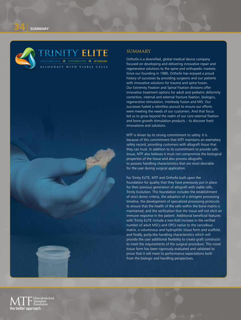
SUMMARY34
SUMMARY
Orthofix is a diversified, global medical device company focused on developing and delivering innovative repair and regenerative solutions to the spine and orthopedic markets. Since our founding in 1980, Orthofix has enjoyed a proud history of successes by providing surgeons and our patients with innovative solutions for trauma and spine fusion. Our Extremity Fixation and Spinal Fixation divisions offer innovative treatment options for adult and pediatric deformity correction, internal and external fracture fixation, biologics, regenerative stimulation, interbody fusion and MIS. Our successes fueled a relentless pursuit to ensure our efforts were meeting the needs of our customers. And that focus led us to grow beyond the realm of our core external fixation and bone growth stimulation products – to discover fresh innovations and solutions.
MTF is driven by its strong commitment to safety. It is because of this commitment that MTF maintains an exemplary safety record, providing customers with allograft tissue that they can trust. In addition to its commitment to provide safe tissue, MTF also believes it must not compromise the biological properties of the tissue and also process allografts to possess handling characteristics that are most desirable for the user during surgical application.
For Trinity ELITE, MTF and Orthofix built upon the foundation for quality that they have previously put in place for their previous generation of allograft with viable cells, Trinity Evolution. This foundation includes the establishment of strict donor criteria, the adoption of a stringent processing timeline, the development of specialized processing protocols to ensure that the health of the cells within the bone matrix is maintained, and the verification that the tissue will not elicit an immune response in the patient. Additional beneficial features with Trinity ELITE include a two-fold increase in the verified number of adult MSCs and OPCs native to the cancellous matrix, a voluminous and hydrophilic tissue form and scaffold, and finally, putty-like handling characteristics which will provide the user additional flexibility to create graft constructs to meet the requirements of the surgical procedure. This novel tissue form has been rigorously evaluated and validated to prove that it will meet its performance expectations both from the biologic and handling perspectives.
®

TECHNICAL MONOGRAPH
A FULLY MOLDABLE allograftwith VIABLE CELLS
®
TECH
NIC
AL M
ON
OG
RA
PH®
TT-1614 © Orthofix Holdings, Inc. 09/2016
1.888.298.5700www.orthof ix .com


