63
The Molecular Pathology of Neurodegeneration Dr Claire Troakes
The Molecular Pathology of
Neurodegeneration
Dr Claire Troakes
Objectives To understand the concept of neurodegenerative
proteinopathies - the basis of neuropathologicalclassification of neurodegenerative diseases
To understand the basic histological and biochemical characteristics of tau, -amyloid, α-synuclein, TDP-43 and FUS in neurodegenerative disorders
To understand some pathogenic implications of abnormal protein aggregates
To understand the basic molecular and pathological features of the cascade of events in Alzheimer’s disease as an example of neurodegeneration
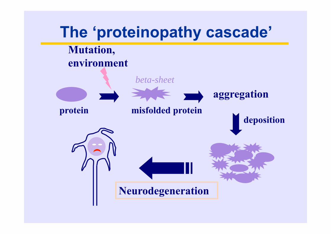
The ‘proteinopathy cascade’
protein misfolded protein
Mutation, environment
aggregation
deposition
Neurodegeneration
beta-sheet
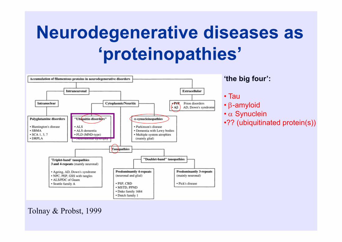
Neurodegenerative diseases as ‘proteinopathies’
Tolnay & Probst, 1999
‘the big four’:
• Tau• -amyloid• Synuclein•?? (ubiquitinated protein(s))
Alzheimer’s disease: theories of pathogenesis
• Tau hypothesis• Amyloid hypothesis

Tau
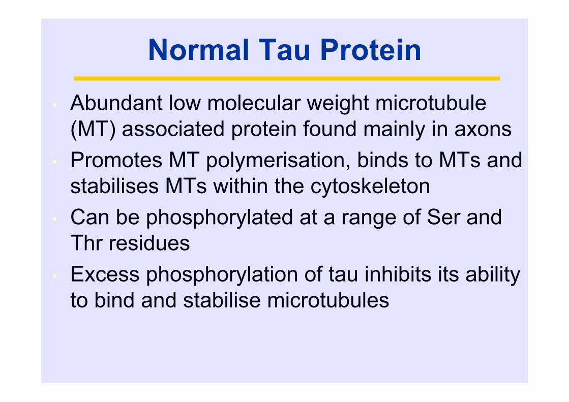
Normal Tau Protein• Abundant low molecular weight microtubule
(MT) associated protein found mainly in axons• Promotes MT polymerisation, binds to MTs and
stabilises MTs within the cytoskeleton• Can be phosphorylated at a range of Ser and
Thr residues• Excess phosphorylation of tau inhibits its ability
to bind and stabilise microtubules
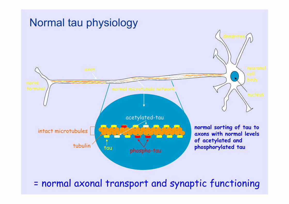
nerve terminal
axon
dendrites
neuronalcell body
nucleusnormal microtubule network
tau phospho-tau
acetylated-tau
tubulin
intact microtubules normal sorting of tau to axons with normal levels of acetylated and phosphorylated tau
Normal tau physiology
= normal axonal transport and synaptic functioning

Human brain Tau isoforms generated by alternative splicingThere are six isoforms of human CNS tau
3 repeat tau
4 repeat tau
PRD = proline-rich domain
M1-M4 = C-terminal repeat microtubule binding domains
Figure from Hanger DP, Anderton BH, Noble W. Trends Mol Med. 2009 Mar;15(3):112-9
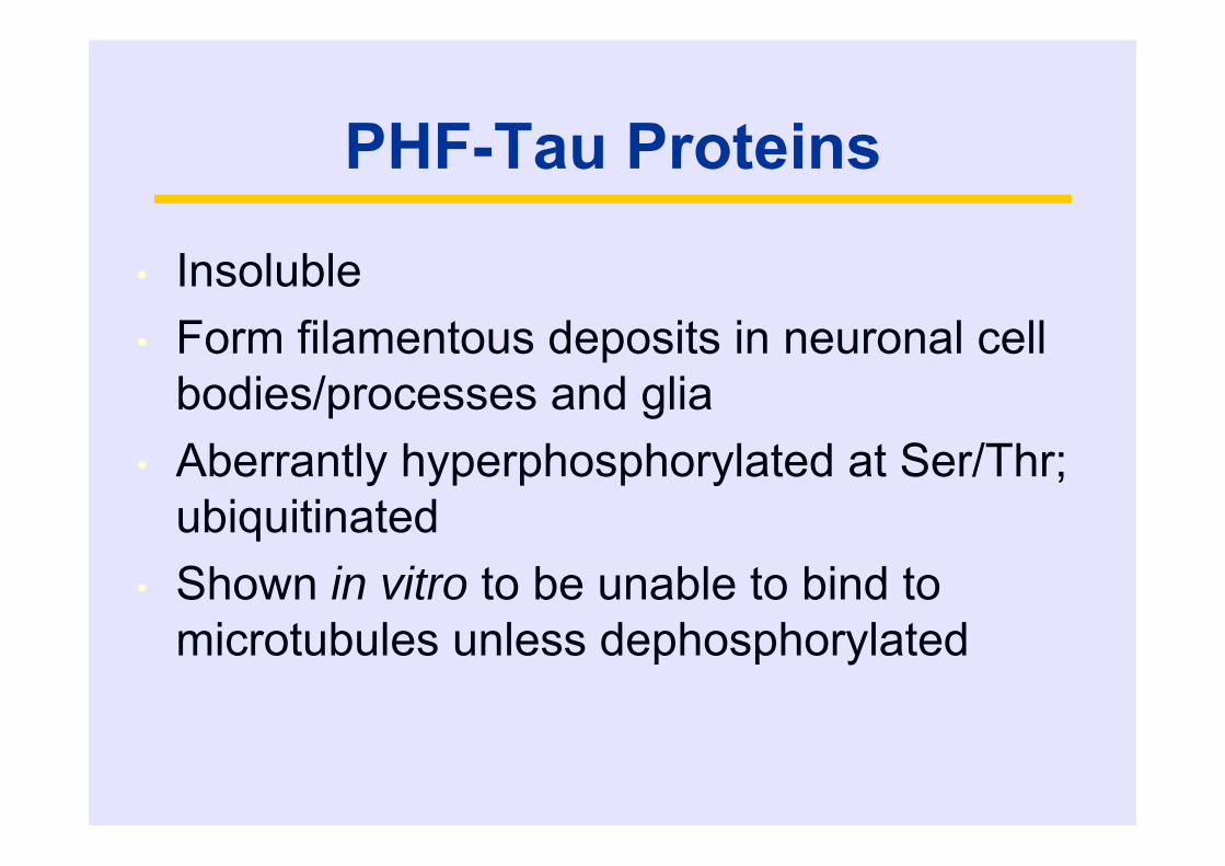
PHF-Tau Proteins
• Insoluble• Form filamentous deposits in neuronal cell
bodies/processes and glia• Aberrantly hyperphosphorylated at Ser/Thr;
ubiquitinated• Shown in vitro to be unable to bind to
microtubules unless dephosphorylated
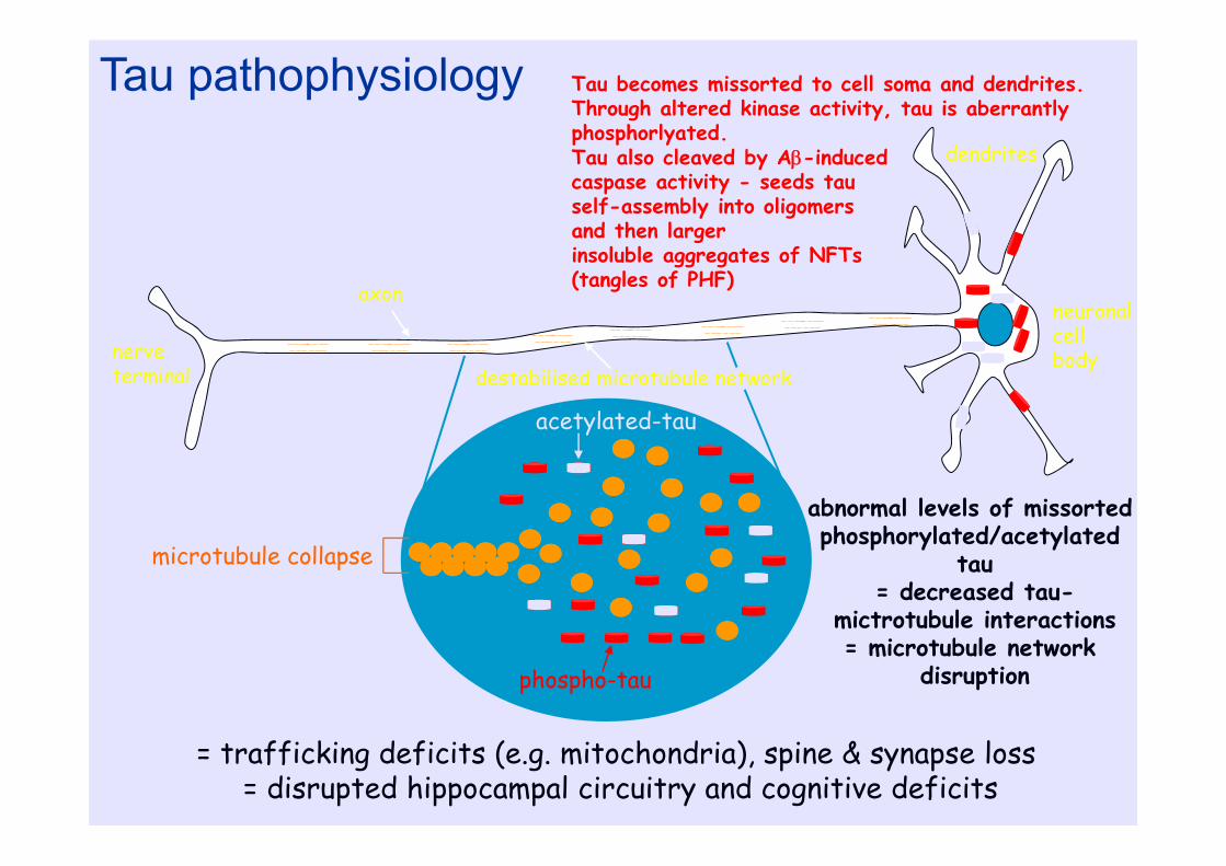
nerve terminal
axon
dendrites
neuronalcell body
destabilised microtubule network
phospho-tau
acetylated-tau
microtubule collapse
abnormal levels of missortedphosphorylated/acetylated
tau= decreased tau-
mictrotubule interactions= microtubule network
disruption
Tau pathophysiology
= trafficking deficits (e.g. mitochondria), spine & synapse loss = disrupted hippocampal circuitry and cognitive deficits
Tau becomes missorted to cell soma and dendrites. Through altered kinase activity, tau is aberrantly phosphorlyated.Tau also cleaved by A-induced caspase activity - seeds tau self-assembly into oligomers and then larger insoluble aggregates of NFTs (tangles of PHF)
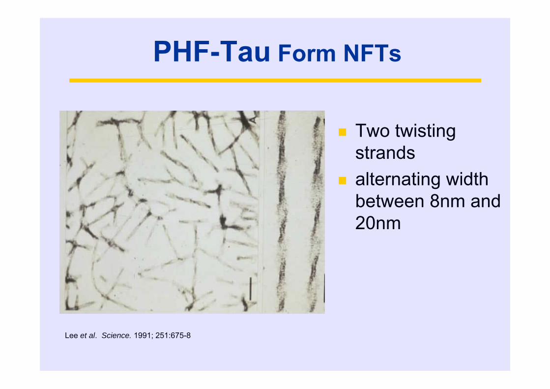
PHF-Tau Form NFTs
Two twisting strands
alternating width between 8nm and 20nm
Lee et al. Science. 1991; 251:675-8
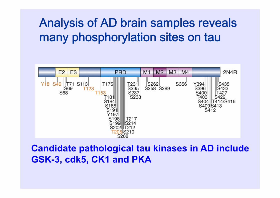
Analysis of AD brain samples revealsmany phosphorylation sites on tau
Candidate pathological tau kinases in AD include GSK-3, cdk5, CK1 and PKA
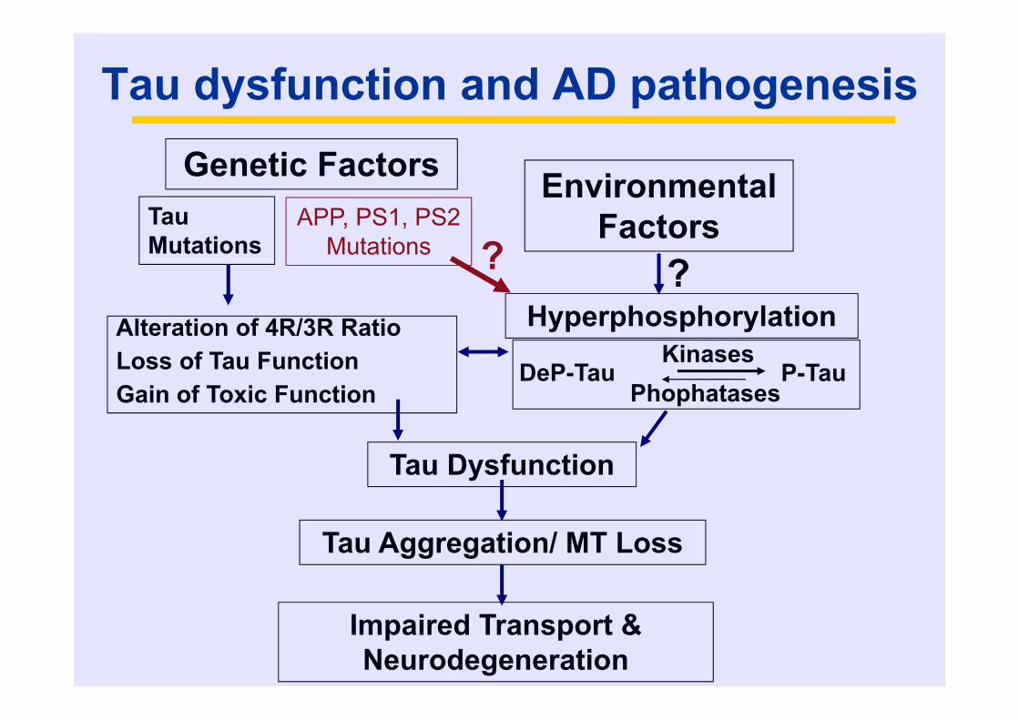
Tau dysfunction and AD pathogenesisGenetic Factors
Tau Mutations
APP, PS1, PS2Mutations
Environmental Factors
Alteration of 4R/3R RatioLoss of Tau FunctionGain of Toxic Function
Hyperphosphorylation
DeP-TauKinases
PhophatasesP-Tau
Tau Dysfunction
Tau Aggregation/ MT Loss
Impaired Transport & Neurodegeneration
? ?
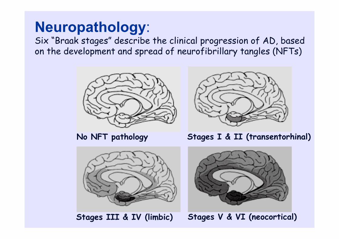
Stages I & II (transentorhinal)
Stages III & IV (limbic) Stages V & VI (neocortical)
No NFT pathology
Neuropathology:Six “Braak stages” describe the clinical progression of AD, based on the development and spread of neurofibrillary tangles (NFTs)
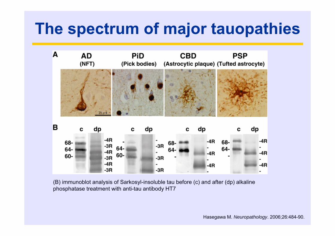
The spectrum of major tauopathies
(B) immunoblot analysis of Sarkosyl-insoluble tau before (c) and after (dp) alkaline phosphatase treatment with anti-tau antibody HT7
Hasegawa M. Neuropathology. 2006;26:484-90.
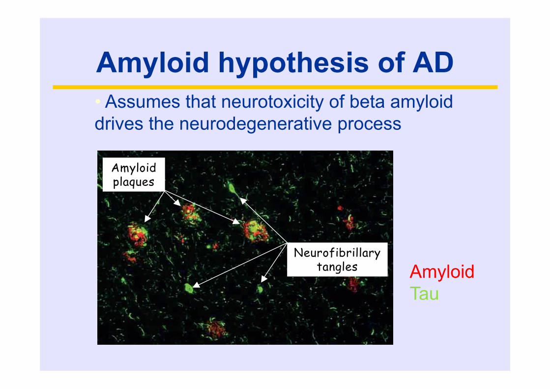
Amyloid hypothesis of AD
Neurofibrillary tangles
Amyloid plaques
Neurofibrillary tangles
Amyloid plaques
• Assumes that neurotoxicity of beta amyloiddrives the neurodegenerative process
AmyloidTau
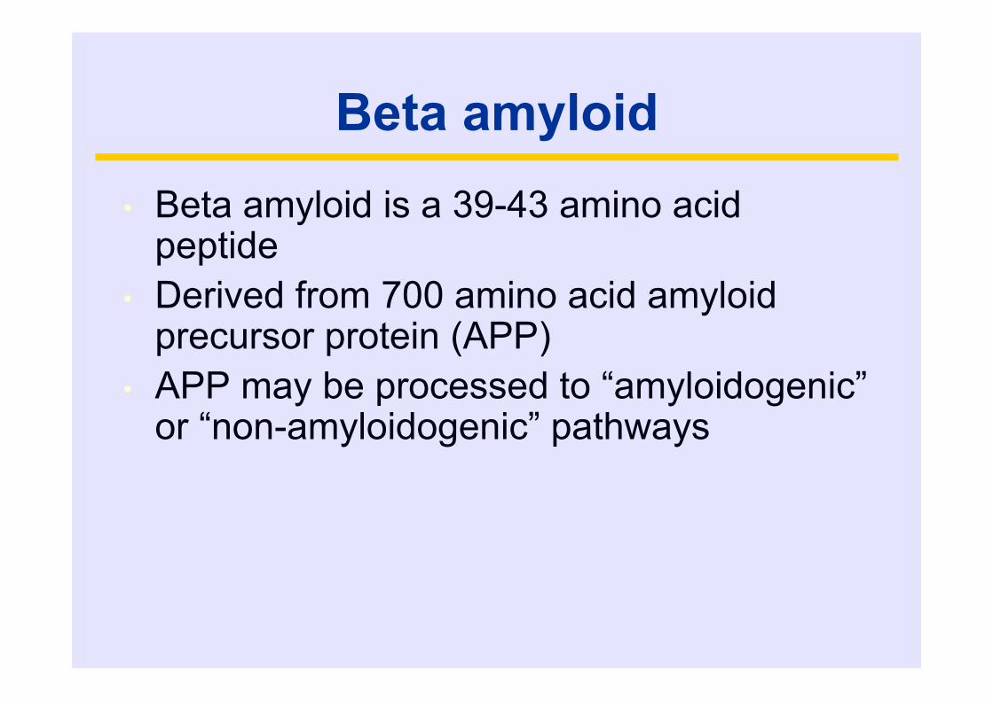
Beta amyloid • Beta amyloid is a 39-43 amino acid
peptide• Derived from 700 amino acid amyloid
precursor protein (APP)• APP may be processed to “amyloidogenic”
or “non-amyloidogenic” pathways

Gandy, J Clin Invest, 2005
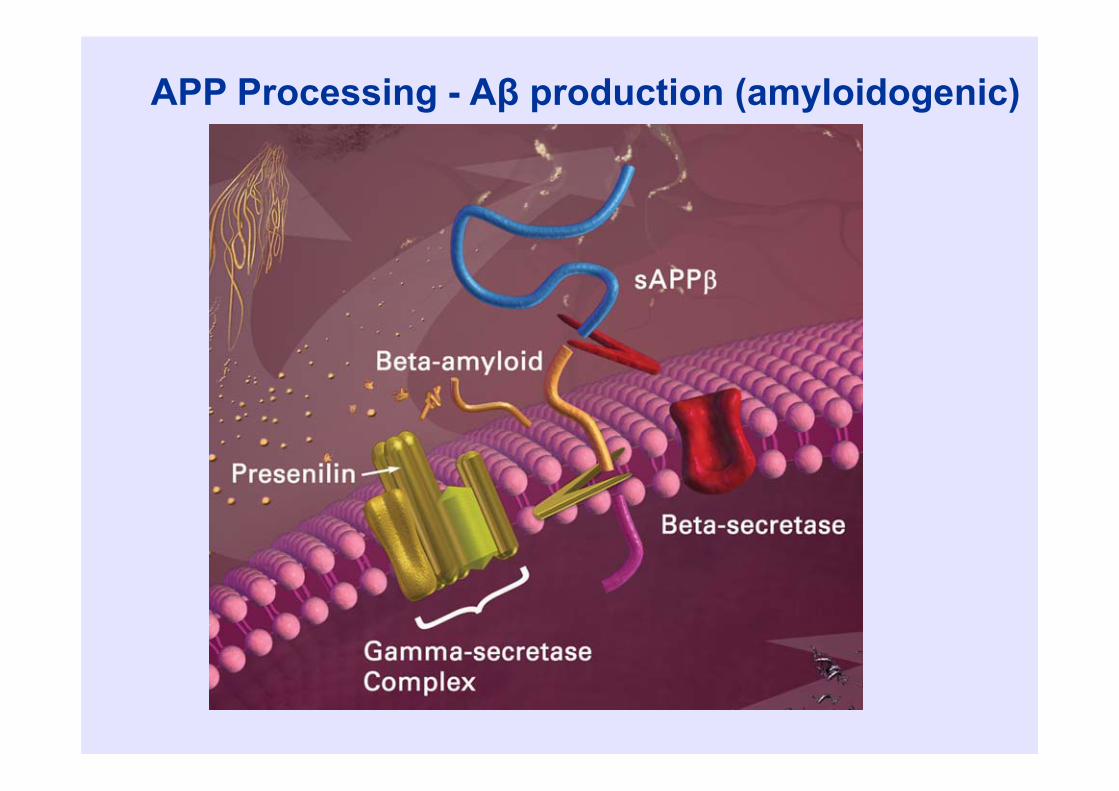
APP Processing - Aβ production (amyloidogenic)
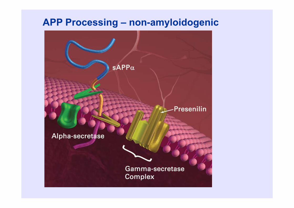
APP Processing – non-amyloidogenic
beta alpha gamma
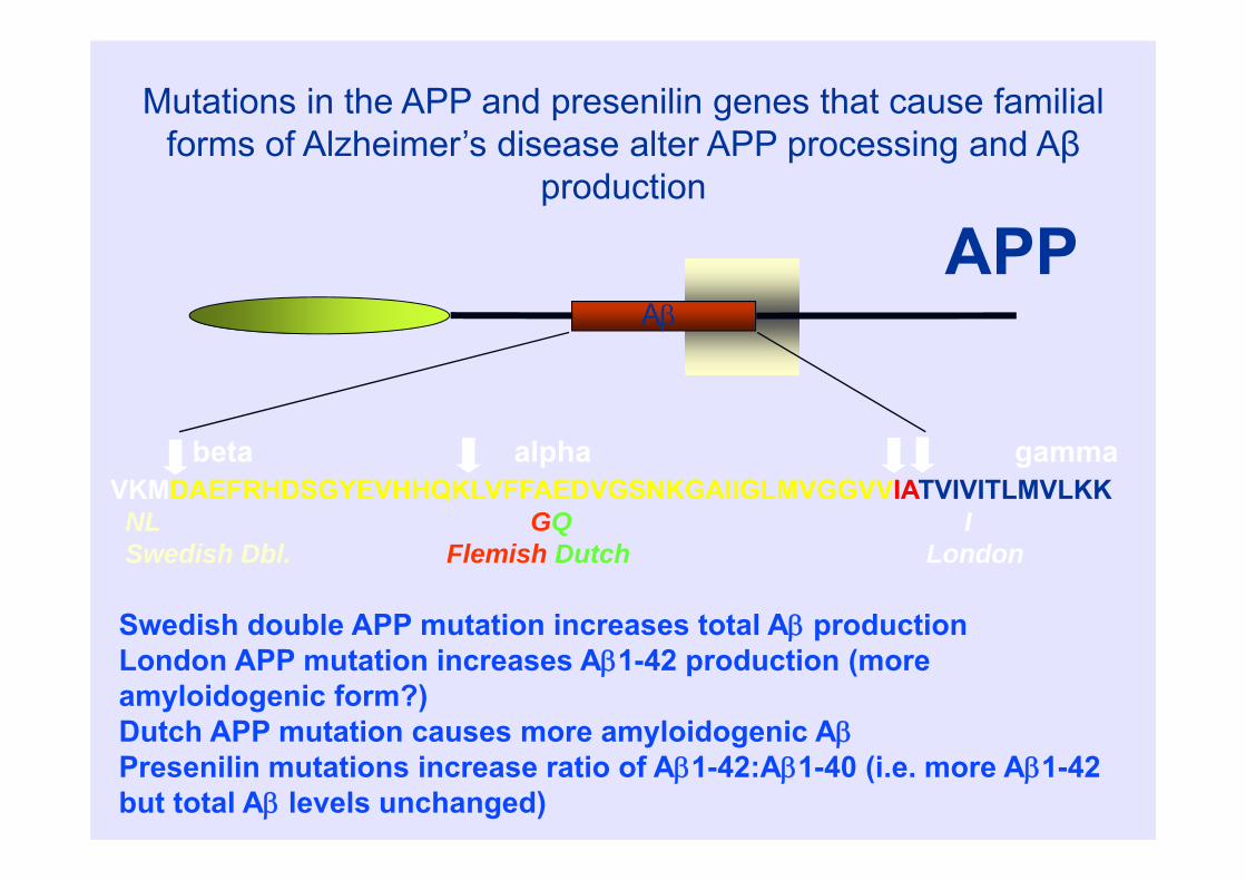
Mutations in the APP and presenilin genes that cause familial forms of Alzheimer’s disease alter APP processing and Aβ
production
Swedish double APP mutation increases total A productionLondon APP mutation increases A1-42 production (more amyloidogenic form?)Dutch APP mutation causes more amyloidogenic APresenilin mutations increase ratio of A1-42:A1-40 (i.e. more A1-42 but total A levels unchanged)
VKMDAEFRHDSGYEVHHQKLVFFAEDVGSNKGAIIGLMVGGVVIATVIVITLMVLKKNL GQ ISwedish Dbl. Flemish Dutch London
APPA
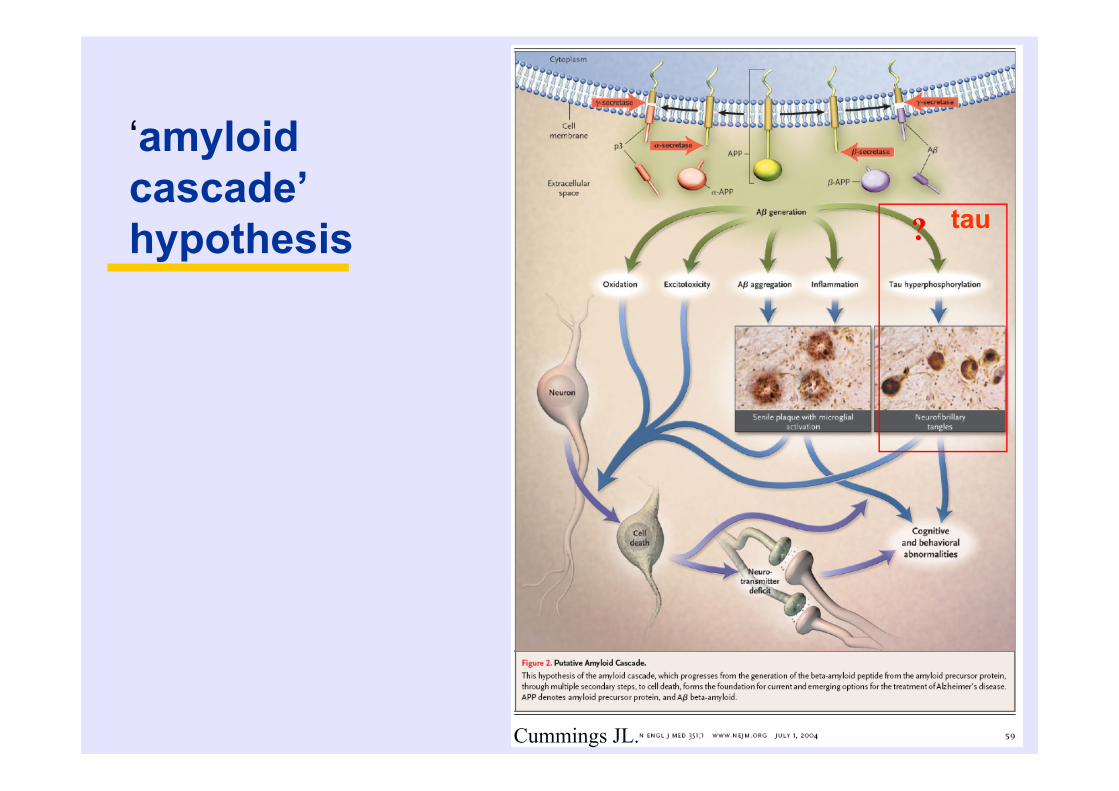
‘amyloid cascade’ hypothesis
Cummings JL.
? tau
Evaluating the amyloid hypothesis- for and against
• Clinicopathologic correlation• Genetics of AD• Cell culture studies• Animal studies
Pathologic correlates of dementia severity
• Amyloid plaques: poor• Neurofibrillary tangles: better• Neuronal loss: as tangles• Synaptic density: best
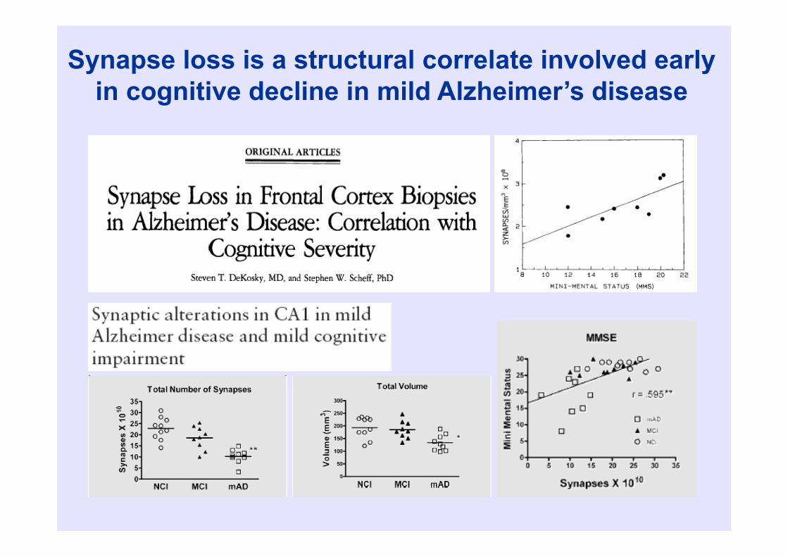
Synapse loss is a structural correlate involved early in cognitive decline in mild Alzheimer’s disease
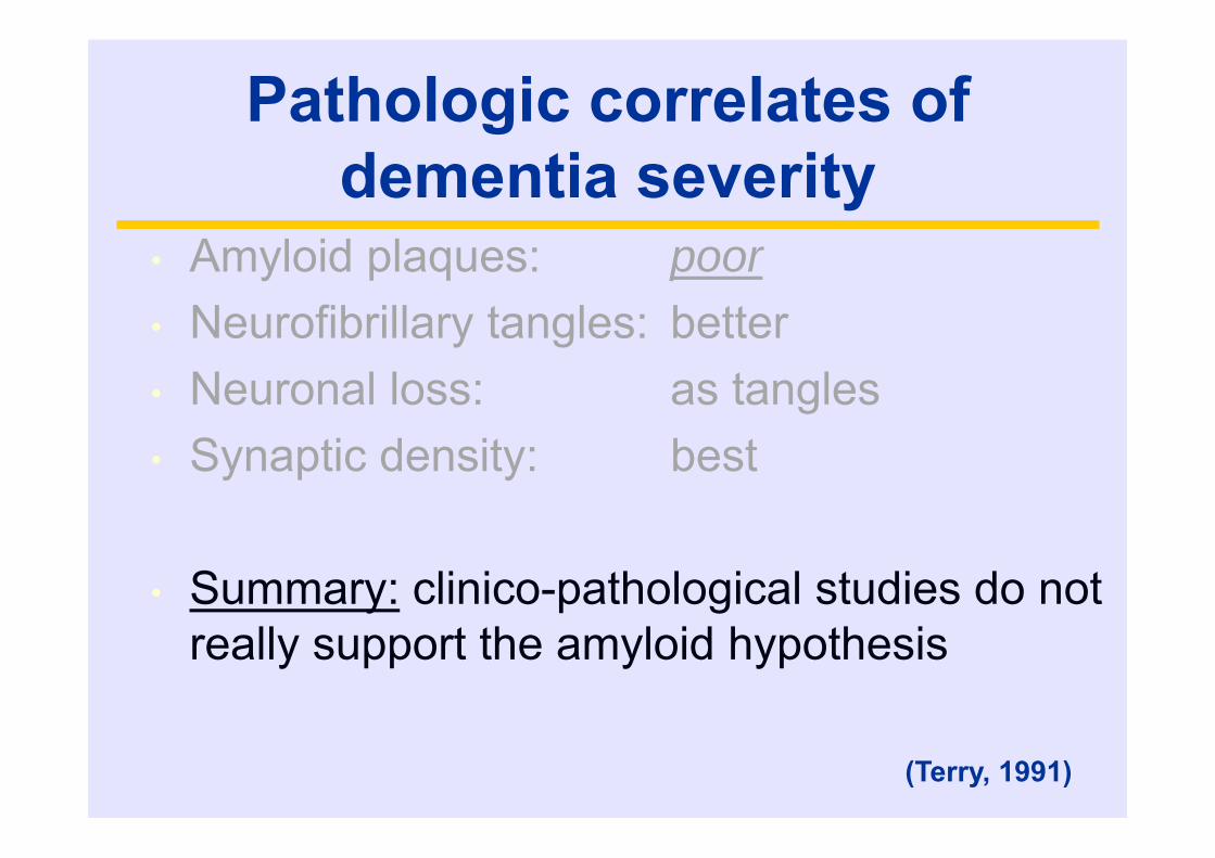
Pathologic correlates of dementia severity
• Amyloid plaques: poor• Neurofibrillary tangles: better• Neuronal loss: as tangles• Synaptic density: best
• Summary: clinico-pathological studies do not really support the amyloid hypothesis
(Terry, 1991)
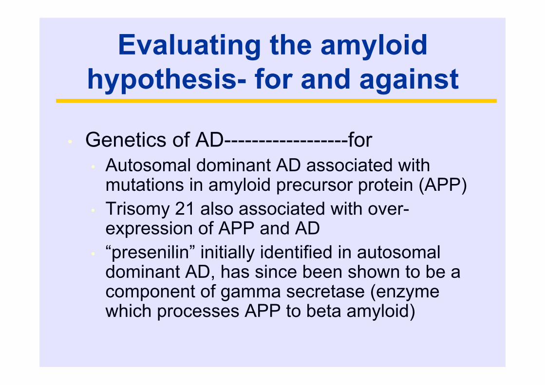
Evaluating the amyloid hypothesis- for and against
• Genetics of AD------------------for• Autosomal dominant AD associated with
mutations in amyloid precursor protein (APP)• Trisomy 21 also associated with over-
expression of APP and AD• “presenilin” initially identified in autosomal
dominant AD, has since been shown to be a component of gamma secretase (enzyme which processes APP to beta amyloid)
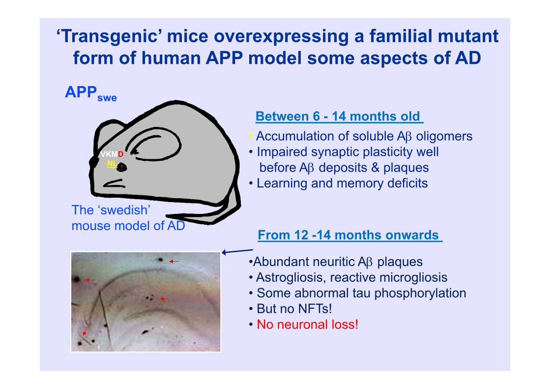
‘Transgenic’ mice overexpressing a familial mutant form of human APP model some aspects of AD
• Accumulation of soluble A oligomers• Impaired synaptic plasticity well
before A deposits & plaques• Learning and memory deficits
•Abundant neuritic A plaques• Astrogliosis, reactive microgliosis• Some abnormal tau phosphorylation• But no NFTs!• No neuronal loss!
Between 6 - 14 months old APPswe
VKMDNL
The ‘swedish’mouse model of AD From 12 -14 months onwards
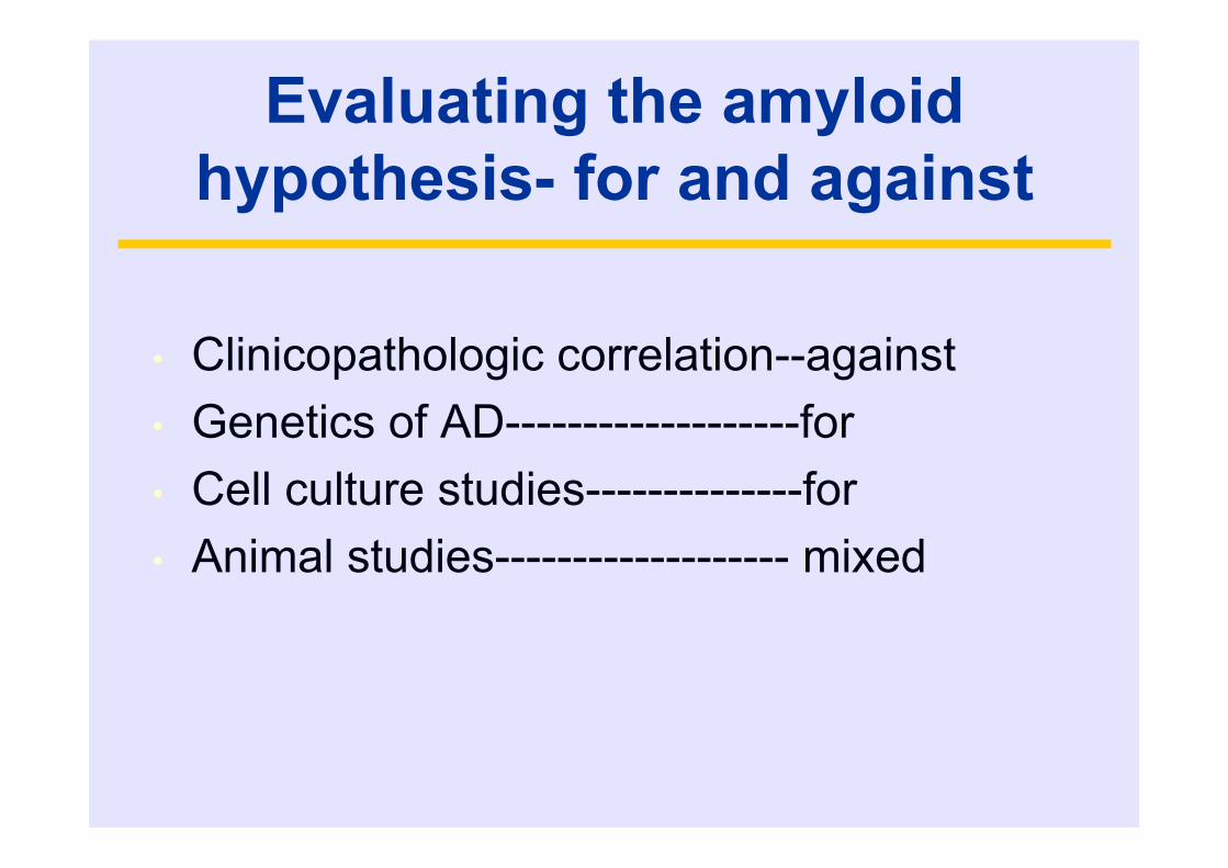
Evaluating the amyloid hypothesis- for and against
• Clinicopathologic correlation--against• Genetics of AD-------------------for• Cell culture studies--------------for• Animal studies------------------- mixed
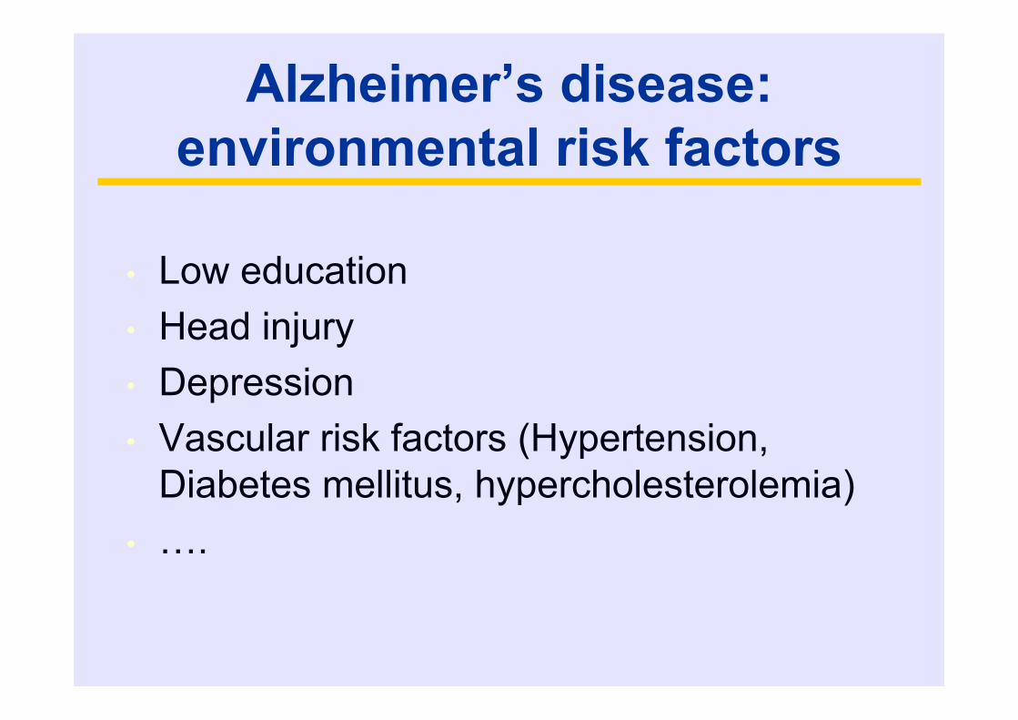
Alzheimer’s disease: environmental risk factors
• Low education• Head injury• Depression• Vascular risk factors (Hypertension,
Diabetes mellitus, hypercholesterolemia)• ….
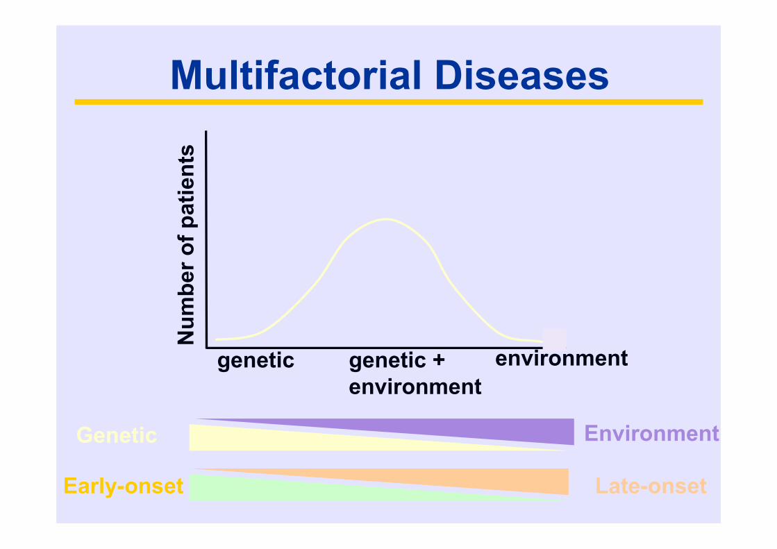
Multifactorial Diseases
Num
ber o
f pat
ient
s
genetic environmentgenetic +environment
Genetic Environment
Early-onset Late-onset
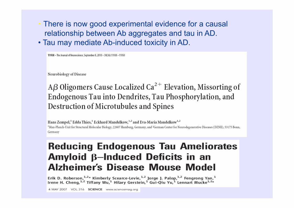
• There is now good experimental evidence for a causalrelationship between Ab aggregates and tau in AD.
• Tau may mediate Ab-induced toxicity in AD.
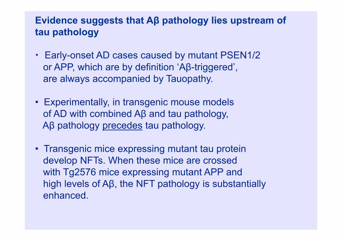
Evidence suggests that Aβ pathology lies upstream of tau pathology
• Early-onset AD cases caused by mutant PSEN1/2 or APP, which are by definition ‘Aβ-triggered’, are always accompanied by Tauopathy.
• Experimentally, in transgenic mouse models of AD with combined Aβ and tau pathology,Aβ pathology precedes tau pathology.
• Transgenic mice expressing mutant tau proteindevelop NFTs. When these mice are crossed with Tg2576 mice expressing mutant APP andhigh levels of Aβ, the NFT pathology is substantially enhanced.
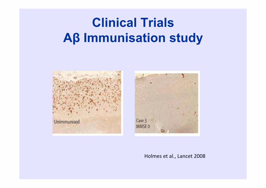
Clinical TrialsAβ Immunisation study
Holmes et al., Lancet 2008
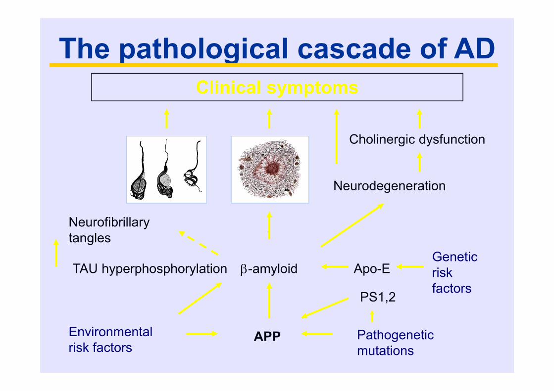
The pathological cascade of ADClinical symptoms
Neurodegeneration
Neurofibrillary tangles
-amyloid
Environmental risk factors
Genetic risk factors
Apo-E
Pathogenetic mutations
APP
PS1,2
Cholinergic dysfunction
TAU hyperphosphorylation
α-synuclein
Normal α-Synuclein
An abundant synaptic protein, present to a lesser extent in cell body and axons, but also in oligodendroglia
Other members of the synuclein family of synaptic proteins include β and -synuclein
Function is unknown but may play roles in synaptic transmission
Is a phosphoprotein, but role of α-synuclein phosphorylation in its normal function is unknown
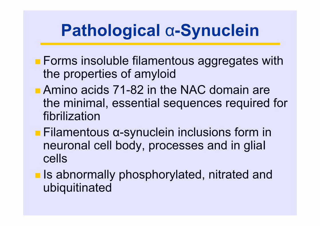
Pathological α-SynucleinForms insoluble filamentous aggregates with
the properties of amyloidAmino acids 71-82 in the NAC domain are
the minimal, essential sequences required for fibrilization
Filamentous α-synuclein inclusions form in neuronal cell body, processes and in gliaI cells
Is abnormally phosphorylated, nitrated and ubiquitinated
Major synucleinopathies• Parkinson’s disease - familial and sporadic
• Dementia with Lewy bodies
• Multiple system atrophy
• Neurodegeneration with brain iron accumulation-1 (formerly Hallevorden-Spatz disease)
• Pure autonomic failure
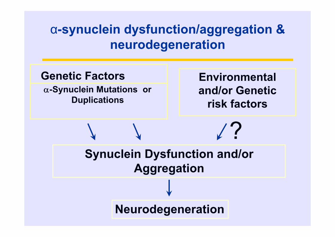
Environmental and/or Genetic
risk factors
Synuclein Dysfunction and/or Aggregation
Genetic Factors-Synuclein Mutations or
Duplications
Neurodegeneration
?
α-synuclein dysfunction/aggregation & neurodegeneration
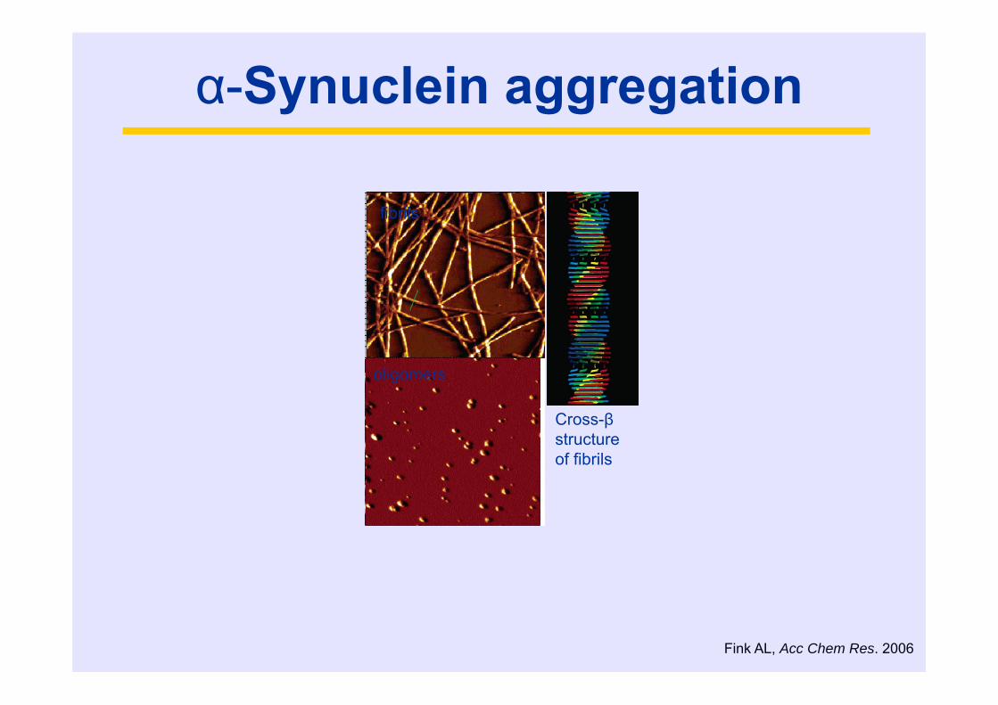
α-Synuclein aggregation
Fink AL, Acc Chem Res. 2006
fibrils
oligomers
Cross-βstructure of fibrils
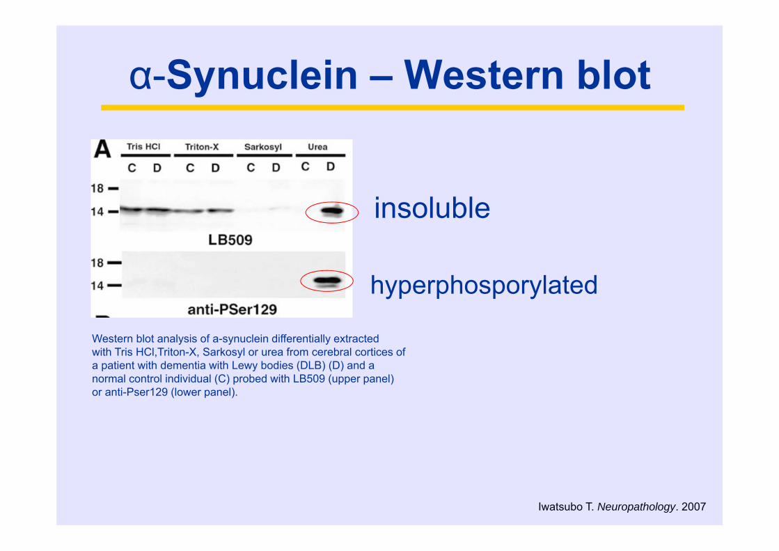
α-Synuclein – Western blot
Iwatsubo T. Neuropathology. 2007
Western blot analysis of a-synuclein differentially extractedwith Tris HCl,Triton-X, Sarkosyl or urea from cerebral cortices ofa patient with dementia with Lewy bodies (DLB) (D) and anormal control individual (C) probed with LB509 (upper panel)or anti-Pser129 (lower panel).
insoluble
hyperphosporylated
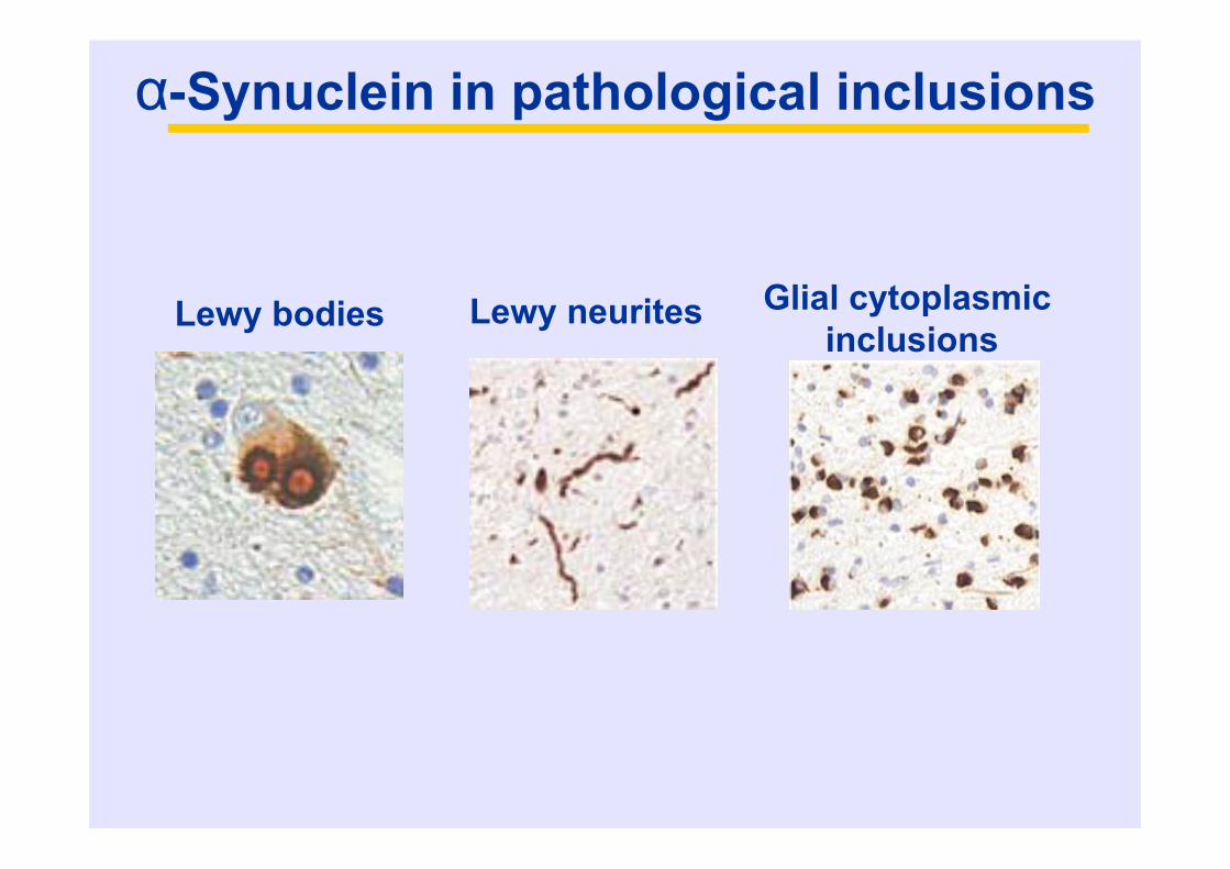
α-Synuclein in pathological inclusions
Lewy neuritesLewy bodies Glial cytoplasmicinclusions

Neurodegenerative diseases as ‘proteinopathies’
Tolnay & Probst, 1999
‘the big four’:• Tau• amyloid• Synuclein•?? (ubiquitinated protein)
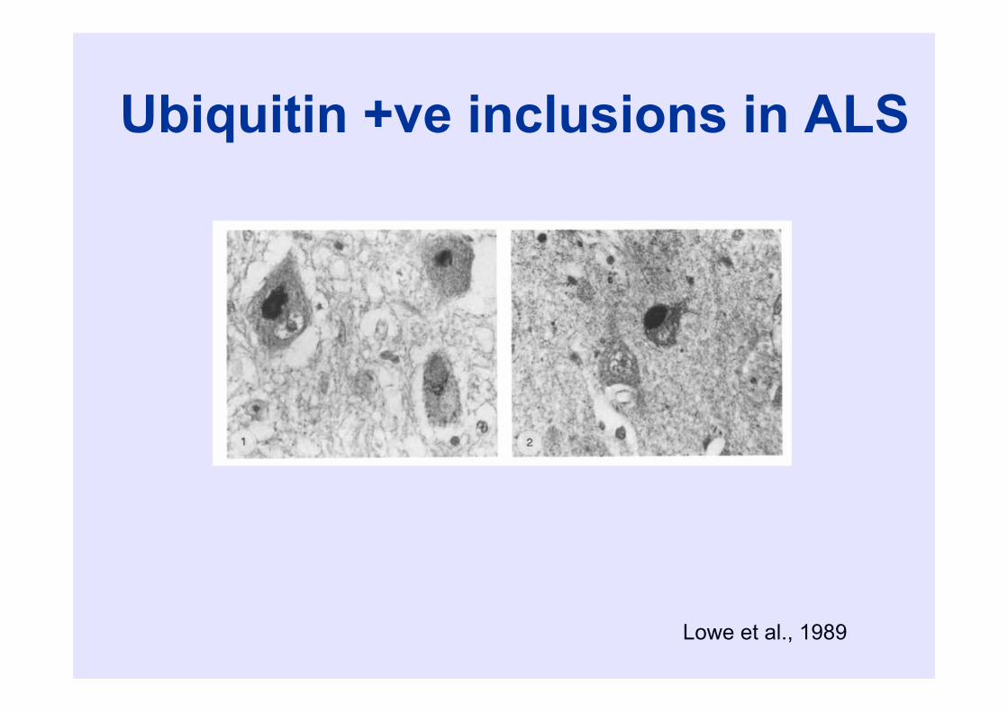
Ubiquitin +ve inclusions in ALS
Lowe et al., 1989
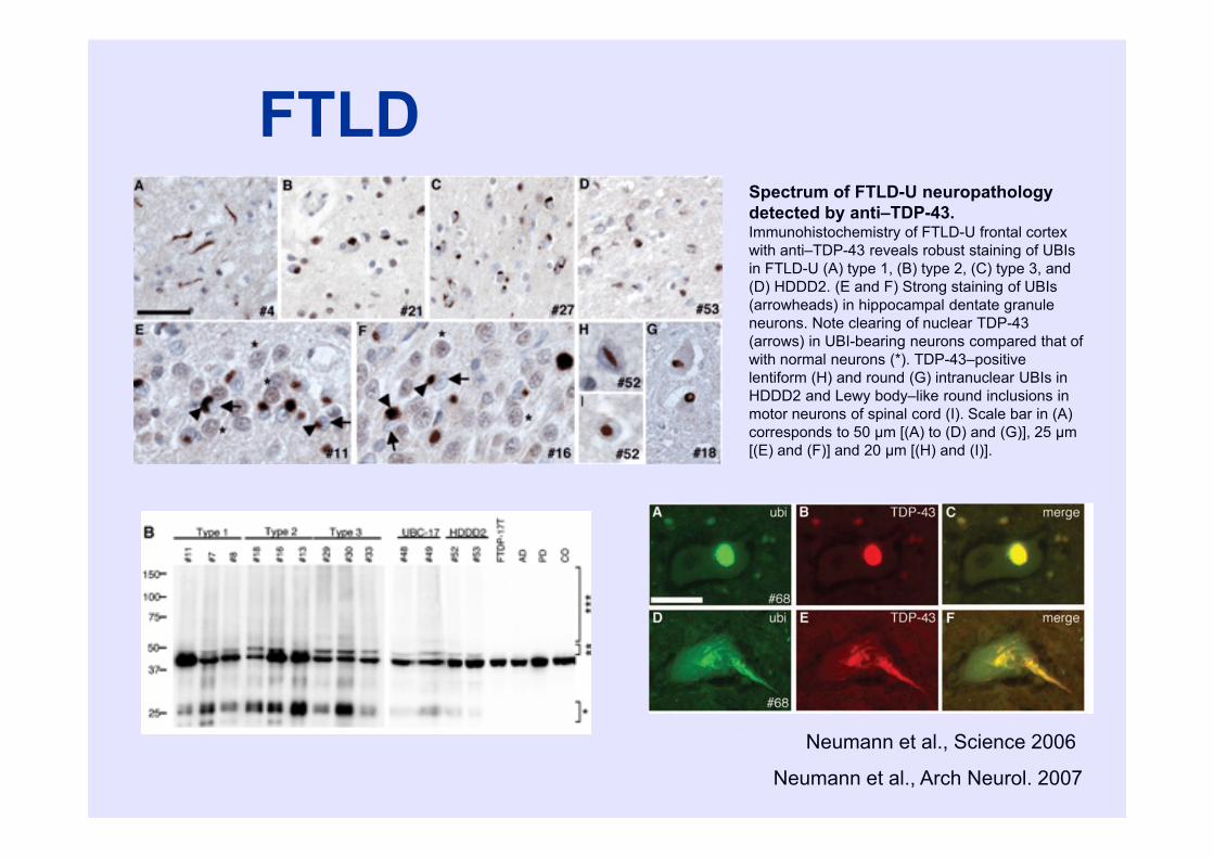
FTLD
Neumann et al., Science 2006
Spectrum of FTLD-U neuropathology detected by anti–TDP-43. Immunohistochemistry of FTLD-U frontal cortex with anti–TDP-43 reveals robust staining of UBIsin FTLD-U (A) type 1, (B) type 2, (C) type 3, and (D) HDDD2. (E and F) Strong staining of UBIs(arrowheads) in hippocampal dentate granule neurons. Note clearing of nuclear TDP-43 (arrows) in UBI-bearing neurons compared that of with normal neurons (*). TDP-43–positive lentiform (H) and round (G) intranuclear UBIs in HDDD2 and Lewy body–like round inclusions in motor neurons of spinal cord (I). Scale bar in (A) corresponds to 50 μm [(A) to (D) and (G)], 25 μm[(E) and (F)] and 20 μm [(H) and (I)].
Neumann et al., Arch Neurol. 2007
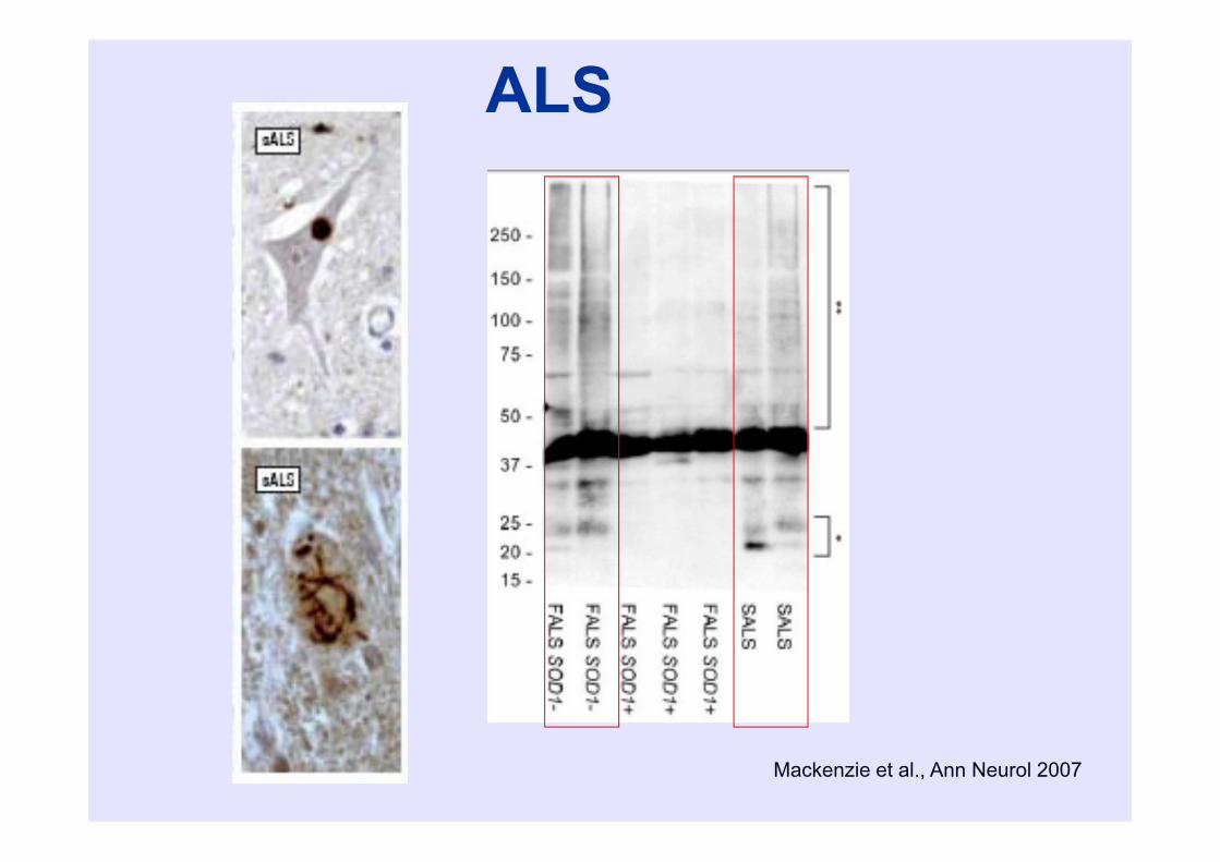
Mackenzie et al., Ann Neurol 2007
ALS
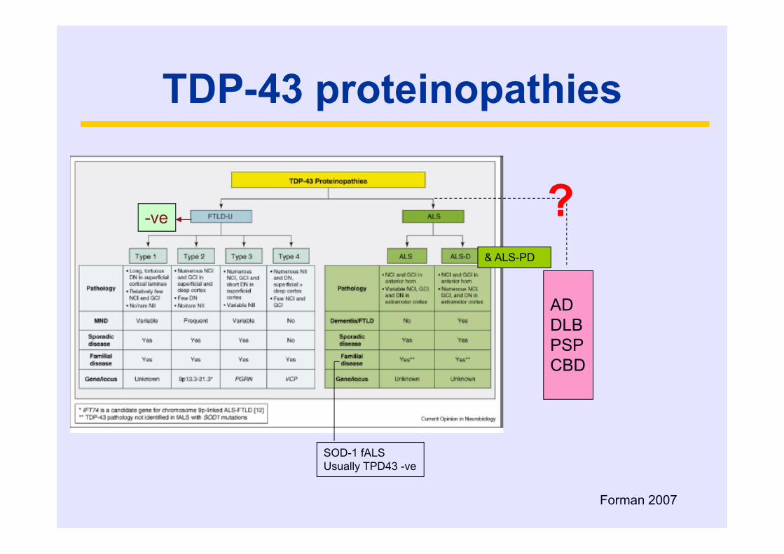
Forman 2007
-ve
& ALS-PD
ADDLBPSPCBD
SOD-1 fALS Usually TPD43 -ve
?
TDP-43 proteinopathies

FUS
• Ubiquitin +-ve• TDP-43 –ve (!)
Implicated in ALS and FTLD pathogenesis

FUS/TLS 526 amino acids; 63 kDa first identified in human myxoid and round cell
liposarcomas as an oncogenic fusion protein closely related to Ewing's sarcoma (EWS) protein component of the heterogeneous nuclear
ribonucleoprotein (hnRNP) complex involved in pre-mRNA splicing and transport of processed mRNA to the cytoplasm
involved in transcriptional activation and interacts with the RNA polymerase
C-terminal half of FUS/TLS contains several structural motifs involved in RNA binding (RRM, RGG, Zn-finger)
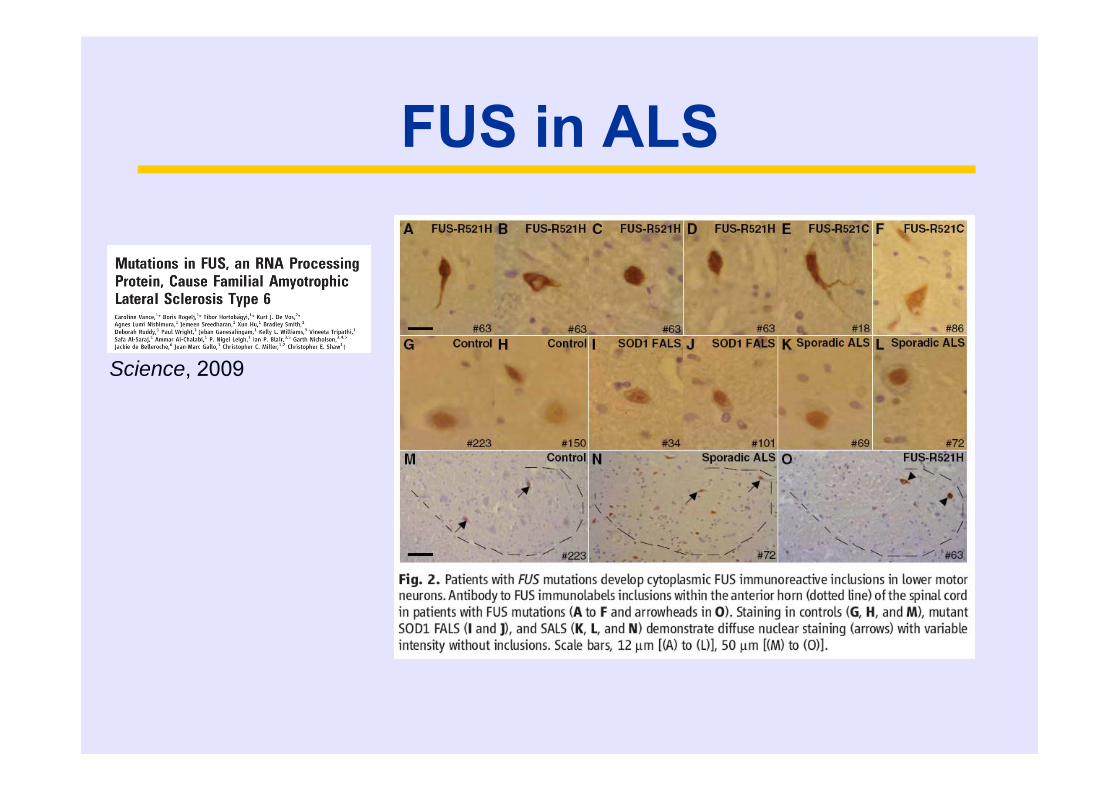
FUS in ALS
Science, 2009
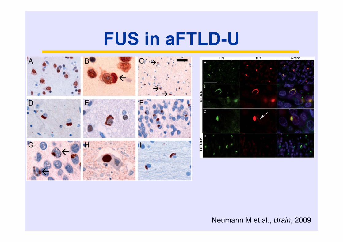
FUS in aFTLD-U
Neumann M et al., Brain, 2009
FUS – a unifying protein in TDP-43 negative
FTLD-U cases
aFTLD-U = FTLD-FUS
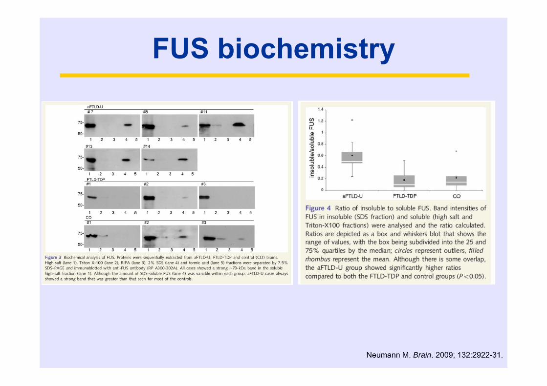
FUS biochemistry
Neumann M. Brain. 2009; 132:2922-31.
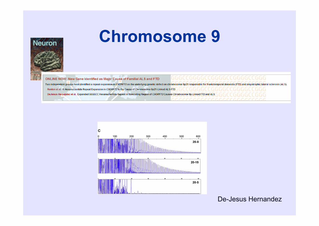
Chromosome 9
De-Jesus Hernandez
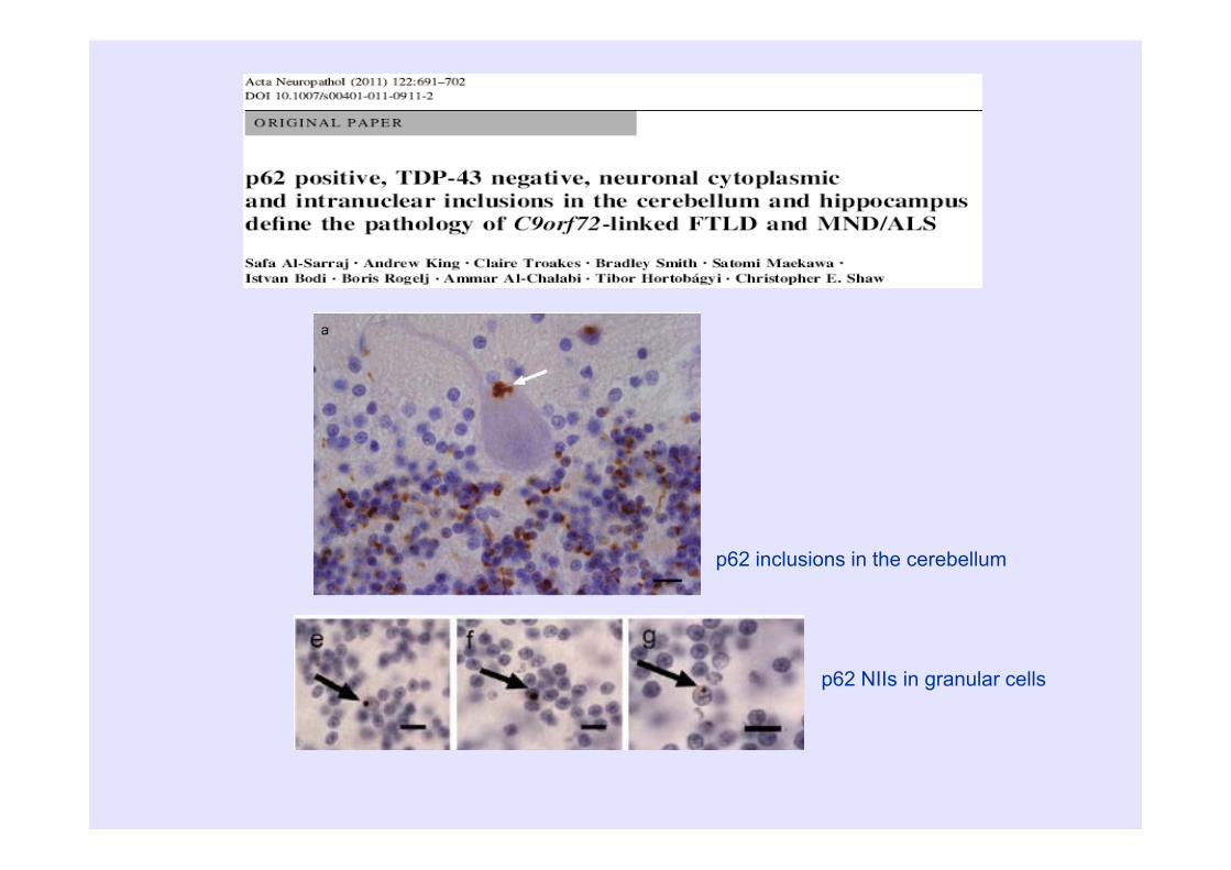
p62 inclusions in the cerebellum
p62 NIIs in granular cells
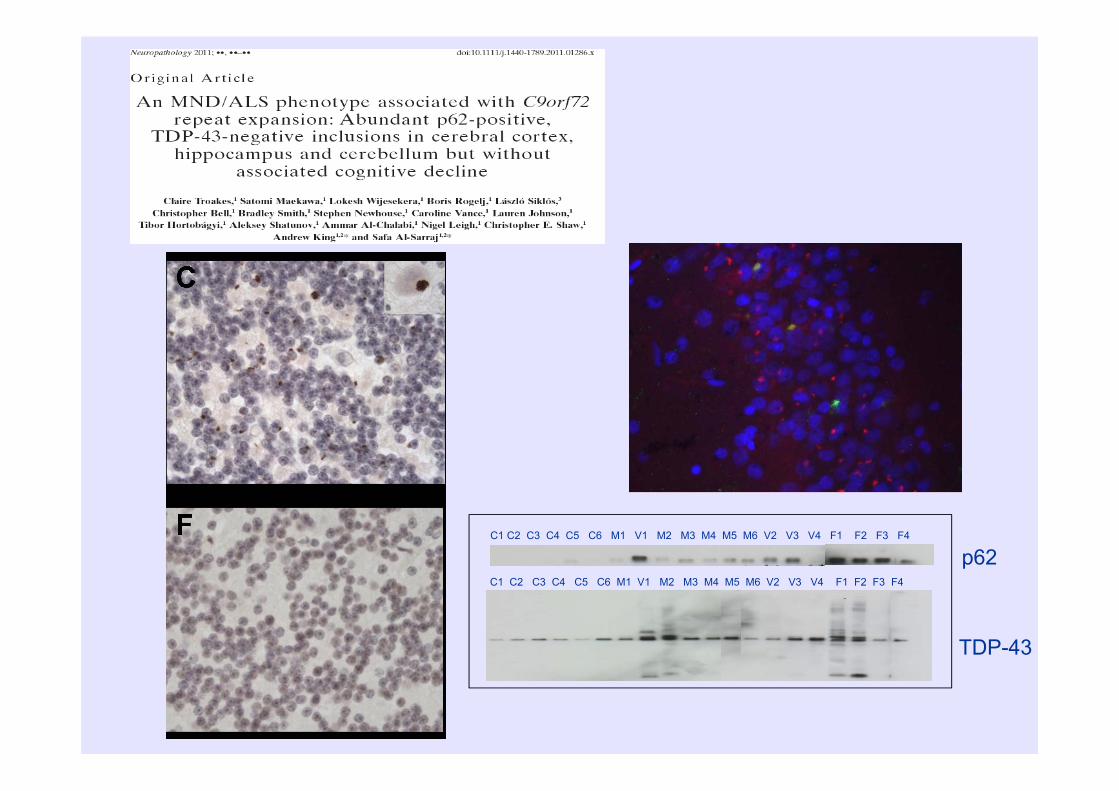
C1 C2 C3 C4 C5 C6 M1 V1 M2 M3 M4 M5 M6 V2 V3 V4 F1 F2 F3 F4
C1 C2 C3 C4 C5 C6 M1 V1 M2 M3 M4 M5 M6 V2 V3 V4 F1 F2 F3 F4
TDP-43
p62

Recommended reading Hasegawa M. Biochemistry and molecular biology of tauopathies.
Neuropathology. 2006; 26:484-90 Crews L. Molecular mechanisms of neurodegeneration in Alzheimer’s Disease.
Hum. Mol. Genet. 2010; 19:R12-20 Hardy J. The amyloid hypothesis for Alzheimer’s disease: a critical reappraisal. J.
Neurochem. 2009; 110(4): 1129-34 Cookson MR. The biochemistry of Parkinson’s disease. Annu Rev Biochem.
2005; 74:29-52 Fink AL. The aggregation and fibrillation of α-synuclein. Acc Chem Res. 2006;
39:628-34 Iwatsubo T. Pathological biochemistry of α-synucleinopathy. Neuropathology.
2007; 27:474-8 Sreedharan J.TDP-43 mutations in familial and sporadic ALS. Science. 2008;
319:1668-1672 Vance C. Mutations in FUS, an RNA processing protein, cause familial ALS type
6. Science. 2009; 323:1208-11 Neumann M. A new subtype of frontotemporal lobar degeneration with FUS
pathology. Brain. 2009; 132:2922-31. Neumann M. The molecular basis of frontotemporal dementia. Expert Rev Mol
Med. 2009; 11:e23
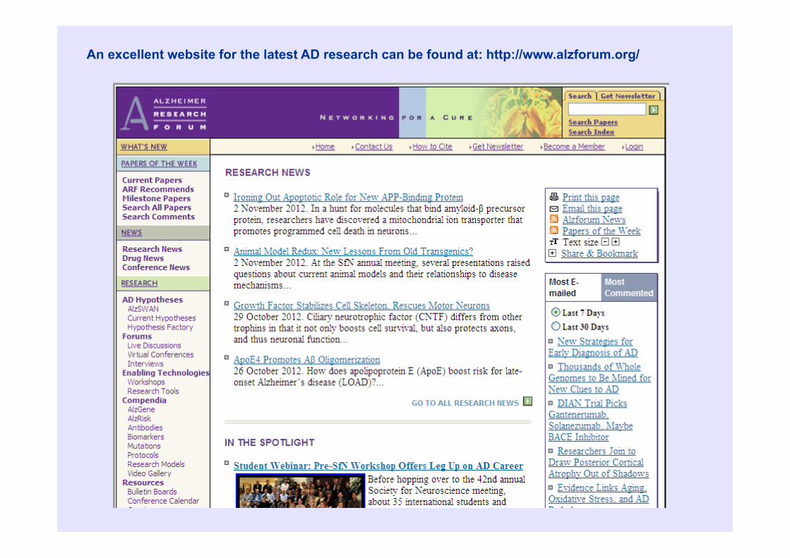
An excellent website for the latest AD research can be found at: http://www.alzforum.org/


