1 AOGD SECRETARIAT AOGD SECRETARIAT Room No 712, 7 th Floor, Private Ward, MCH Block Department of Obstetrics and Gynecology Guru Teg Bahadur Hospital & University College of Medical Sciences Dilshad Garden, Delhi-110095, India [email protected], [email protected]www.aogd.org Volume 17; Issue No.3; July 2017 Price: ` 30 only AOGD BULLETIN AOGD BULLETIN Issue: Surgery for Benign Gynecological Conditions AOGD Theme 2017-18 AOGD Theme 2017-18 ‘Optimizing Women’s Health Through ‘Optimizing Women’s Health Through Enhanced Skills and Best Practices’ Enhanced Skills and Best Practices’ AOGD BULLETIN AOGD BULLETIN
AOGD Theme 2017-18AOGD Theme 2017-18‘Optimizing Women’s Health Through ‘Optimizing Women’s Health Through Enhanced Skills and Best Practices’Enhanced Skills and Best Practices’
Public Relations & HospitalityDr Rashmi GuptaDr Seema Prakash
AOGD Executive Council Members 2017-2018Dr Abha SinghDr Achala BatraDr Amita SaxenaDr Amita SunejaDr Anjali TempeDr B K GoelDr Gita RadhakrishnanDr Harsha KhullarDr Kuldeep JainDr Malvika SabharwalDr Nalini MahajanDr Neerja BhatlaDr Nirmala AgarwalDr Puneeta MahajanDr Pushpa SinghDr Renu MishraDr S N BasuDr Sabhyata GuptaDr Sangeeta GuptaDr Sonia MalikDr Sumita Mehta
AOGD SecretariatRoom No 712, 7th Floor, Private Ward, MCH BlockDepartment of Obstetrics & GynaecologyGuru Teg Bahadur Hospital & University College of Medical Sciences Delhi-110 095, Indiawww.aogd.org
AOGD BULLETINVolume 17-3, July 2017
AOGD Executive Committee 2017-18
DisclaimerThe advertisements in this bulletin are not a warranty, endorsement or approval of the products or services. The statements and opinions contained in the articles of the AOGD Bulletin are solely those of the individual authors and contributors, and do not necessarily refl ect the opinions or recommendations of the publisher. The publisher disclaims responsibility of any injury to persons or property resulting from any ideas or products referred to in the articles or advertisements.
Plagiarism DisclaimerAny plagiarism in the articles will be the sole responsibility of the authors and the editorial board or publisher will not be responsible for this.
Publisher/Printer/EditorDr Rashmi on behalf of Association of Obstetricians & Gynecologists of Delhi.
Published fromAOGD Offi ce, Room No 712, 7th Floor, Private Ward, MCH Block, Department of Obstetrics & Gynaecology, Guru Teg Bahadur Hospital & University College of Medical Sciences, Delhi-110 095, India
PatronsDr S N MukherjeeDr S K DasDr Urmil SharmaDr Kamal BucksheeDr Neera AgarwalAdvisorsDr Chitra RaghunandanDr Gauri DeviDr Indrani GanguliDr N B VaidDr Neerja GoelDr S S TrivediDr Shakti Bhan KhannaDr Sharda JainDr Suneeta MittalDr Swaraj BatraScientifi c AdvisorsDr Gita RadhakrishnanDr Amita Suneja
Ex Offi cio ExecutivePast PresidentsDr P K Malkani (1962-66)Dr L V Pathak (1966-72)Dr Anusuya Das (1972-78)Dr S N Mukherjee (1978-81)Dr V Hingorani (1981-88)Dr S K Das (1988-90)Dr P Chadha (1990-94)Dr Neera Agarwal (1994-97)Dr Maya Sood (1997-99)Dr D Takkar (1999-2001)Dr Sudha Salhan (2001-03)Dr Swaraj Batra (2003-05)Dr N B Vaid (2005-06)Dr S S Trivedi (2006-07)Dr Suneeta Mittal (2007-08)Dr I Ganguli (2008-09)Dr Shashi Prateek (2009-10)Dr U Manaktala (2010-11)Dr Neerja Goel (2011-12)Dr C Raghunandan (2012-13)Dr Alka Kriplani (2013-14)Dr U P Jha (2014-15)Dr Pratima Mittal (2015-16)
Immediate Past PresidentDr Sudha Prasad (2016-17)
Immediate Past SecretaryDr Ashok Kumar (2016-17)
President ElectDr Abha Singh (2018-19)
Immediate Past President FOGSIDr Alka Kriplani
ChairpersonsAOGD Sub-CommitteesDr Achla BatraDr Amita JainDr Anjali TempeDr Ashok KumarDr Jyotsna SuriDr K D NayarDr Mala SrivastavaDr Nalini MahajanDr Renu MisraDr Rupinder SekhonDr Shakuntala KumarDr Sunita MalikDr Vatsla Dadhwal
Total number of pages = 56
ContentsMinimally Invasive Surgery in Benign Gynecological Conditions
9
Vidushi Kulshrestha, Varnit Toshyan
SOP: Hysteroscopic Myomectomy 13Rashmi
Robotic Surgery in Benign Gynecologic Conditions 15Sarika Gupta
Contained Morcellation: Current technique and future directions
18
BB Dash, Rupali KhuranaMIND, BODY & SOULPost Operative Recovery with Meditation
21
Rashmi
Quiz Time 23Compiled by Bindiya Gupta
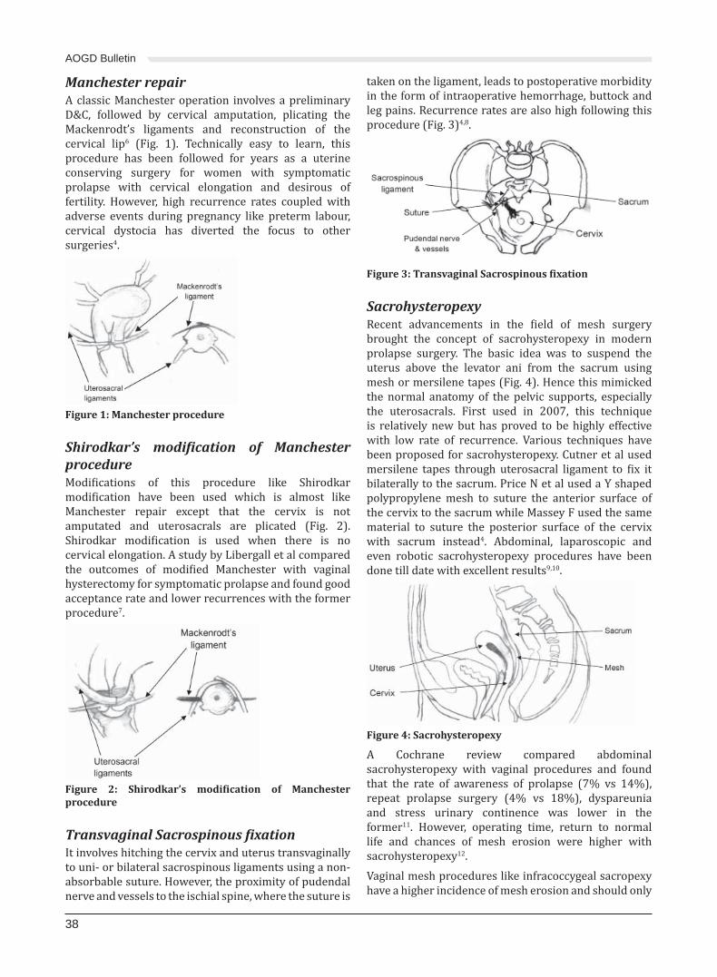
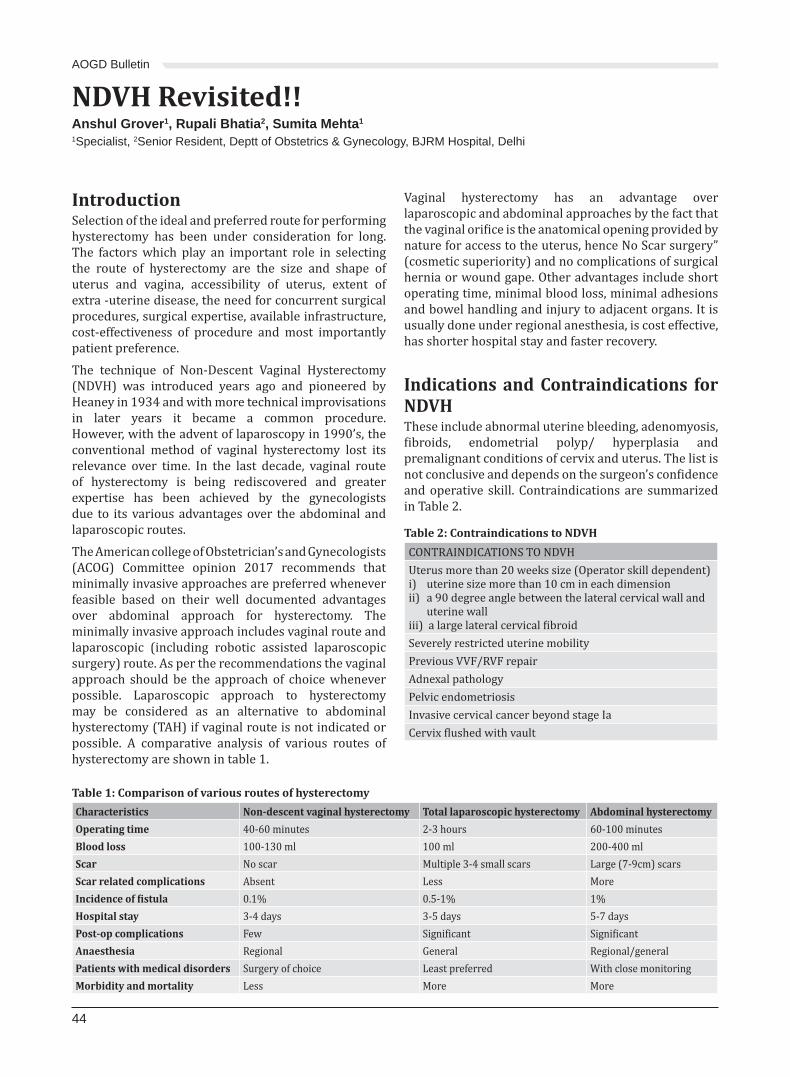
Uterine Conserving Surgeries for Genital Prolapse 37Alpana Singh, Varsha Priyadarshani
Retrograde Hysterectomy for Benign Gynaecologic Conditions
Dr Amita SunejaDr Gita RadhakrishnanScientifi c Advisors
Dr Alpana SinghTreasurer
Dr Sandhya JainDr Himshweta Srivastava Dr Archana Chaudhary Co TreasurerJoint Secretaries
Clinical Secretaries
Dr Richa Sharma Dr Bhanu PriyaDr Sanjeeta BeheraCoordinators Skill Workshops
Dr Shalini Rajaram President
Dr Kiran GuleriaVice President
Volume 17-3; July 2017
5
President’s Message
Dear Friends
June has seen the passing on of a pillar and senior AOGD member, Dr. SK Das, a warm, humble and beautiful soul. A prayer meeting was held in Safdarjang Hospital on 13th June and members spoke fondly of her life and contributions. AOGD pays homage and respect to this noble personality with a full page ‘Obituary’ in this bulletin.
June was very eventful too with a number of activities, yoga day celebrations and a wonderful clinical meeting at Army Hospital, R&R, Dhaula Kuan organised by Dr. BK Goel. Despite a terrible downpour, water clogged roads and bumper to bumper traf ic, the auditorium was full. Doctor’s Day was celebrated in its full glory with balloons and buntings and cake cutting by the senior most member of AOGD Dr. SN Mukherjee.
The last bulletin on ‘Near Miss’ was well received and commendation on content by Dr. Alka Kriplani and other members was gratifying. This issue on ‘Surgery for Benign Gynecologic conditions’ is an attempt to equip our readers with skills and best practices of various surgical procedures. The old adage ‘Practice makes a man perfect’ holds true for honing surgical skills and adapting to changing practice with superior patient outcomes are needed. Robotics are ideal for pelvic surgery with high quality 3D vision, ine dissection and endo-wrist technology. Wonderful surgical precision! The bane of laparoscopic surgery has been ‘morcellation’ and the inal word is yet to be said but calls for caution. This issue carries useful articles on robotics
and laparoscopy. Finally despite the modern world of gizmos one should be able to do a vaginal hysterectomy, the most non-invasive of all techniques! Enjoy the content and post your feedback.
I once again urge all AOGD members to support Dr. Sudha Prasad for the post of Vice-President FOGSI, North Zone. Ballots will be reaching soon so don’t forget to cast your valuable vote on time. She is a true leader and visionary and will be a perfect choice to represent the North zone.
By the time this issue reaches you two interesting academic activities under the AOGD banner namely ‘Basics of Endoscopy’, 21st July, GTB Hospital and Challenges in the management of preterm labour’ at Army Hospital R&R on 23rd July 2017 will be underway. Take the opportunity of attending either or both. The mega ‘BOH- The Triology’ conference organised by FOGSI in collaboration with AOGD will be held on 19th & 20th August 2017 at The Leela, Gurgaon. All members are invited to attend this academic extravaganza!
Cheers and Enjoy the Monsoons!
Shalini RajaramPresident, AOGD (2017-18)
AOGD Bulletin
6
Dear friends
As monsoon hits Delhi, our AOGD team rollout yet another issue of bulletin dedicated to surgery for benign gynaecological conditions. So please sit back, relax and enjoy reading this with a cup of tea and ‘pakoras’ in this rainy season.
The surgical approach to benign gynaecological conditions has been discussed in the light of the relative bene its and hazards. These bene its and hazards seem to be dependent on surgical expertise and this may in luence the decision. There is a plethora of surgical options available these days. eg. Hysterectomy can be vaginal (VH), abdominal (AH), laparoscopic (LH) or robotic (RH). VH may be the best option, but if not feasible, LH may avoid the need for AH, but LH is associated with more urinary tract injuries. There is no evidence that RH is of bene it in this population. So, a physician needs to fully assess the patients to determine which surgery is best for their needs, as each case is unique. Preferably then, the surgical approach to hysterectomy should be decided by the woman in discussion with her surgeon.
I take this opportunity to invite you once again to the forthcoming conference “BOH-The Triology”; a joint venture of FOGSI & AOGD. The scienti ic programme has been designed carefully keeping minutest details in mind, is comprehensive and likely to bene it all obstetricians. A galaxy of star speakers will deliberate upon the various subjects. Additional attractions are endoscopy videos, neonatal and maternal resuscitation sessions and free papers & e-posters by young obstetricians and trainees. We promise you a great academic, social and gourmet feast along with a great ambience at “The Leela Ambience, Delhi” on 19th-20th August.
Please join us in large numbers to make this conference a huge success.
Cheers!
Kiran GuleriaVice President AOGD (2017-18)
Vice President’s Message
Volume 17-3; July 2017
7
Dear friends,
Monsoon Greetings!
Moving ahead in the year, we bring to you new ideas, developments and challenges in this issue of the bulletin. The current issue features a compilation of articles on surgeries for benign gynecological conditions. New developments viz. minimally invasive endoscopic procedures, retrograde hysterectomy, robotic surgery and controversies like morcellation are especially being highlighted.
Next in the series of Skill development workshops is “Basics of Endoscopy in Gynaecology” which will be held on 21st July again at GTB Hospital. After a successful CME on Ante-natal Care, we have planned another CME on “Post-natal Services - Continuum of Care”, in early August at GTB Hospital.
FOGSI in association with AOGD is organizing “BOH–The Triology” on 19th and 20th August in Delhi which will be enlightening; so hurry up and register! Do block your dates for our AOGD Annual Conference on 19th and 20th November at IHC. Ensure you a great academic feast!
I request you all to vote for Dr Sudha Prasad as Vice-President FOGSI, North Zone (2019). Ballot papers will reach you in the irst week of July 2017. Delhi must ensure its representation in FOGSI and Dr Prasad is our best bet!
Finally, do enjoy the monsoon glory; but protect yourself from diseases gory!
Abha SharmaSecretary AOGD (2017-18)
From the Secretary’s Desk.....
Monthly Clinical Meet Monthly Clinical Meet will be held at All India Institutes of Medical Sciences, New Delhi
on 28th July, 2017 from 4:00-5:00pm.
AOGD Bulletin
8
Respected Seniors & Dear Friends,
Greetings from the editorial team. In the month of Monsoons, we bring to you an issue on the topic close to every gynecologist’s heart i.e. Surgeries for Benign Gynecological Conditions. The thrill of holding the scalpel for irst time and giving irst incision, irst hysterectomy, irst vaginal surgery always remains fresh in one’s minds.
We as surgeons are in a unique and privileged position to meet hopes and expectations of our patients by picking up our knives and applying our surgical skills. Nothing is more exciting and satisfying than a surgery well performed. Last few decades have seen so many advances and changing scenarios in operative gynecology. The endoscopic surgeries have revolutionized the ways we operate with advantages of better visualization and minimal invasion. As if that was not enough, Robotics have made an entry into our lives. On the other hand, we the gynecologists have the vaginal route to our advantage & NDVH has stood the test of time with boundaries being pushed. Indian contributions to these ields is being acknowledged internationally.
Considering all this, we have tried to include various topics, not to forget the importance of mental well-being, pain management & infection prevention for optimal outcome in any surgical procedure. Hope the bulletin offers something interesting, knowledgeable and insightful to each and every one. Feel free to respond as your feedbacks are always welcome.
With warm regards,
The Editorial TeamAOGD (2017-18)
From the Editorial Board
Volume 17-3; July 2017
9
Due to its inherent advantages over conventional laparotomy, ‘minimally invasive surgery’(MIS) has largely taken over in the surgical management of gynaecological conditions. According to American College of Obstetricians and Gynaecologists, June 2017 recommendations, MIS in gynaecology includes vaginal procedures, laparoscopy, hysteroscopy as well as robotic surgeries. Spectrum of surgeries possible by laparoscopy and hysteroscopy for various benign gynaecological conditions are being described in this article. Vaginal hysterectomy and robotic surgery are discussed in subsequent articles in the bulletin.Laparoscopy offers obvious advantages to the patients as well as surgeons. Patients have shorter hospital stay, lesser pain, early recovery, smaller cosmetic scar; and advantages for surgeons include magni ication, improved anatomic visualization of the peritoneal cavity facilitating dissection, improved hemostasis, ease of approach to otherwise technically dif icult sites such as pouch of Douglas, presacral space, and space of Retzius.Hence, laparoscopyis usually preferred to open surgery, whenever feasible. The basic surgical principles remain the same as with open surgeries.
HysterectomyHysterectomy is one of the most frequently performed surgeries for a variety of benign conditions like abnormal uterine bleeding (AUB), symptomatic uterine ibroids, adenomyosis not responding to medical management or as de initive treatment in women > 40 years with completed family. Hysterectomy is the standard treatment for atypical endometrial hyperplasia, and is indicated in hyperplasia without atypia if repeat histopathology after 3-6 months shows persistence or progression.Laparoscopy allows surgeons with limited experience in vaginal surgery to remove uterus without abdominal incision in presence of adhesions, endometriosis, adnexal disease. Advantages of total laparoscopic hysterectomy over vaginal route include good vault support and less risk of vault granulation. However, a recent systematic review of surgical approaches to hysterectomy for benign gynaecological conditions reported that vaginal route is superior to laparoscopic and abdominal due to faster return to normal activities. According to the statement by AAGL, most hysterectomies for benign disease should be performed either vaginally or laparoscopically and that continued
efforts should be taken to facilitate these approaches. Surgeons without requisite training and skills required for the safe performance of VH or LH should enlist the aid of colleagues who do or should refer patients requiring hysterectomy to such individuals for their surgical care.
MyomectomyMyomectomy is the conservative surgical treatment indicated for symptomatic myomas causing AUB or other symptoms when medical treatment fails and patient desires to preserve fertility. In infertile women, myomectomy is indicated for submucosal myomas, intramural myomas >4 cm even without cavity distortion as they negatively in luence fertility and foreven smaller ones after multiple IVF failures. Myomectomy is also indicated in infertility and history of poor prior reproductive outcome after excluding other contributory causes. A good imaging, ideally with MRI, for mapping of myomas is an important pre-requisite to con irm the number, size and location of myomas. This helps in decidingthe route of myomectomy and ensures removal of all mapped ibroids. The best candidates for hysteroscopic route are FIGO PALM-COEIN Type-0 (entirely intracavitary) and less than 4 cm type-1(> 50% intracavitary) myomas. FIGO type 2 to 6 myomas, multiple myomas and >4cm submucosal myomas are best managed with abdominal route. Traditional criteria for laparoscopy had been uterine size ≤ 14 weeks after at least 3 doses of GnRH analogue injection, no single myoma > 7 cm, no myoma near uterine artery or tubal ostia if fertility is desired and atleast 50% of myoma to be subserosal thus permitting adequate repair through laparoscope. But currently, in expert hands, there are no limiting factors in terms of size, number and location. Any size and number can be removed laparoscopically by an experienced surgeon. Current evidence suggests there is no signi icant difference between the laparoscopic and open approach regarding fertility performance, although the former is associated with better delineation of planes under magni ication and principles of microsurgery, lesser postoperative morbidity, less postoperative adhesions, rapid post-operative recovery. GnRH agonists given prior to myomectomy in bigger myomas improves general condition, corrects anaemia, reduces myoma size facilitating endoscopic surgery and reduces intraoperative blood loss; however dissecting planes might be dif icult after GnRH.
Minimally Invasive Surgery in Benign Gynecological ConditionsVidushi Kulshrestha1, Varnit Toshyan2
1Assistant Professor, 2Senior Resident, Department of Obstetrics & Gynaecology, All India Institute of Medical Sciences, New Delhi
A good surgeon operates with his hand, not with his heart - Alexandre Dumas Pere
AOGD Bulletin
10
Recently, laparoscopic myomectomy has been a subject of debate due to risks of morcellation which includes risks inherent to surgery such as viscera/major vessels injury, risk of disseminated leiomyomatosis and upstaging of occult leiomyosarcoma due to tissue dissemination. In-bag morcellation should be done within specimen retrieval pouches for containment.
Uterine sparing surgeries for adenomyosis (Adenomyomectomy, Cytoreductive surgery)These are indicated in symptomatic women desiring future fertility and in infertile women with failed IVF. Focal adenomyosis is managed with adenomyomectomy i.e. complete resection of adenomyoma. Diffuse adenomyosis is managed with cytoreductive surgeries where clinically recognizable non-microscopic lesion is partially removed maximizing adenomyotic tissue excision, still maintaining uterine wall integrity to minimize risk of rupture in future pregnancy. Various techniques have been described in literature such as Osada’s Triple lap method, Tacheshi’s overlapping laps, wedge resection etc.Traditionally, laparotomy has been used for these surgeries because of dif iculty in delineating the margins/extension within myometrium and dif iculty in suturing the remaining uterine wedges after excision. Laparotomy offers the advantage of palpation and recognition of adenomyotic lesions intraoperatively. However, with availability of MRI, accurate mapping of lesion is possible; hence laparoscopy is feasible for adenomyosis also, and with better ergonomics of laparoscopic instruments, suturing also poses no more dif iculty. Use of transtrocar ultrasound has been proposed to aid intraoperative recognition of adenomyotic lesion. In laparoscopy, morcellator is used to remove the mass.Other minimally invasive techniques reported for conservative management include nonexcisional methods such as laparoscopic electrocoagulation of myometrium and uterine artery ligation; hysteroscopic nonexcisional techniques such as rollerball endometrial ablation, transcervical resection of the endometrium (TCRE), endomyometrial resection; and laparoscopically assisted vaginal excision.
EndometriosisFor endometriosis, laparoscopy is both a diagnostic and therapeutic tool. Diagnostic laparoscopy involves systematic inspection of uterus, adnexa, uterosacrals, peritoneum especially over ovarian fossae, uterovesical fold, pouch of Douglas and other intra-abdominal organs including bowel. Laparoscopy provides superior visualization of cul-de-sac and allows a high degree of magni ication of peritoneal surfaces, which aids in the identi ication of subtle disease. ESHRE recommends
per-operative speculum examination and palpation of vagina and cervix under laparoscopic control, to check for ‘buried’ nodules. Besides visualization, tissue sample can simultaneously be taken for histopathology, which is the gold standard for de initive diagnosis, though a negative biopsy does not rule out endometriosis. If performed by an experienced surgeon, a negative diagnostic laparoscopy can reassuringly exclude endometriosis, except deep retroperitoneal disease or disease outside peritoneal cavity.Operative laparoscopy is indicated for infertility, when pain or other symptoms do not respond to hormonal treatment, when there is >3 cm endometrioma on imaging, sudden enlargement in the cyst size, or when malignancy cannot be ruled out. ESHRE-2013 recommends surgical treatment in moderate to severe endometriosis and in minimal or mild endometriosis in patients undergoing laparoscopy. It advocates laparoscopic ablation or excision of endometriotic lesions, adhesiolysis and laparoscopic cystectomy in ovarian endometriom as in endometrioma > 3 cm when it is associated with pain or inaccessibility of follicles. Endometrioma drainage is not recommended. Operative laparoscopy increase spontaneous pregnancy rates even in stage III/IV endometriosis compared to expectant management.Another surgical treatment for pelvic pain is laparoscopic uterosacral nerve ablation (LUNA) and presacral neurectomy (PSN) which disrupts the neural pathways. Radical surgery for endometriosis includes hysterectomy, bilateral oophorectomy, and removal of all endometriotic implants. Lapraoscopic hysterectomy may be dif icult in grade III-IV disease; hence retrograde technique can be used in this condition.Laparoscopy is the preferred choice for above mentioned procedures after considering surgeon’s skill, equipment availablility and extent of excision required. Laparotomy may be preferred in cases where extensive adhesiolysis or removal of large endometriomas is to be performed. Laparoscopic cystectomy: Laparoscopy should be considered as a method of choice for removal of benign ovarian cysts. However, laparoscopic approach could result in chemical peritonitis in dermoid cyst caused by the spilled contents of a ruptured cyst. Removing cysts in an endobag signi icantly reduces both operating time and spillage.
Fertility enhancing surgeriesLaparoscopy is indispensable for evaluation of infertile women as besides being a diagnostic and therapeutic tool, it also assess the prognosis. Various fertility enhancing surgeries include tuboplasty, adhesiolysis, ovarian drilling and tubal clipping for hydrosalpinx prior to ART. Others include myomectomy, adenomyomectomy and surgery for endometriosis as described above.
Volume 17-3; July 2017
11
Though role of these surgeries is gradually decreasing due to increased availability and utilization of ART, these surgeries still are indispensable in developing country like ours due to unaffordability of ART by most couples.Laparoscopic ovarian drilling is the second-line treatment after clomiphene failure and/or clomiphene resistant PCOS. Electrocautery, using an insulated unipolar needle electrode with a non-insulated distal end measuring 1-2 cm, is the most commonly used method. The number of punctures is empirically chosen depending on the ovarian size. Most surgeons perform four punctures per ovary, each for 4 seconds at 40 Watts (rule of 4), delivering 640 Joules of energy per ovary (the lowest effective dose recommended).Laparoscopic tuboplasty is indicated for distal tubal disease. In imbrial phimosis where distal tubal opening is narrowed by adhesions, Bruhat’s imbrioplasty is performed by deagglutination and dilatation of imbria which are then everted and sutured to tubal serosa to maintain patency. Severe phimosis may require a cuff salpingostomy. These procedures carry good prognosis if the adnexal adhesions are limsy, tubal dilatation is < 3 cm, tubal walls are thin and pliable and mucosal folds are preserved. Complete distal tubal occlusion leading to hydrosalpinges may require neosalpingostomy.Hysteroscopic uterine septal resection and hysteroscopic adhesiolysis improve pregnancy outcome in infertile women. Hysteroscopic cannulation of cornual end of tube is indicated in tubal factor infertility due to cornual block. Ideally, procedure is performed under laparoscopic guidance to exclude coexistent distal tubal block, ibrosed /beaded tubes which are contraindications for cannulation
Laparoscopy for emergency surgeriesEctopic pregnancyLaparoscopy has become the standard approach for managing ectopic pregnancy surgically, if adequate expertise and equipment are available and patient is hemodynamically stable. Unruptured tubal pregnancies can be laparoscopically treated with either linear salpingostomy or salpingectomy depending on patient’s desire for future fertility andcondition of tube. For interstitial pregnancies laparoscopic cornuostomy, cornual excision or resection can be performed.
Ovarian TorsionLaparoscopic detorsion or untwisting the ovary restores ovarian function in >90% cases regardless of the dusky blue-black or necrotic appearance of twisted ischemic ovary. If there is an associated ovarian cyst, cystectomy is also performed. Surgical management to prevent recurrence includes plication of utero-ovarian ligament where the ligament is shortened with a running stitch; ovariopexy, where ovary is sutured to posterior aspect
of the uterus or lateral pelvic wall; and oophoropexy, where the utero-ovarian ligament is sutured either to the posterior aspect of the uterus or lateral pelvic wall.
Laparoscopic tubal recanalization (Laparoscopic Tubal Anastomosis)Laparoscopy ful ils all microsurgical principles like magni ication, minimal tissue handling and haemostasis. Laparoscopic route also avoids tissue trauma associated with packing and retractors.Surgical steps remain the same as laparotomy: i.e distention of the proximal segment of the tube by trans-cervical chromotubation to localize site of block, excision of ibrosed tubal segment, minimal damage to mesosalpinx,
ensuring that the cut tubal ends are right-angled and of similar size for better alignment & approximation. End to end anastomosis is performed in two layers. Patients with inal tubal length of <4 cm and with marked luminal discrepancy have lower success rates.
Laparoscopic surgeries for uterovaginal prolapse and stress urinary incontinenceLaparoscopic access has been applied to most abdominal-route and numerous vaginal-route surgical procedures for treatment of urinary incontinence and pelvic organ prolapse with advantage of improved anatomic visualization of presacral space, and space of Retzius. The indications for laparoscopic repair of cystocele, enterocele and vaginal apical prolapse and vault prolapse are identical to those for vaginal and abdominal routes.
Laparoscopic sacral colpopexyor sacro-hysteropexySuspension of vagina in vault prolapse / uterus in uterovaginal prolapse to the anterior longitudinal presacral ligament at sacral promontory using an intervening polypropylenemesh has been shown to be an effective treatment. ‘Y’ shaped mesh is preferred for post hysterectomy vault prolapse and ‘L’ shaped mesh for sacrohysteropexy. Potential intraoperative complications include injury to bowel, bladder, ureter, and infection. Hemorrhage, especially from presacral vessels, can be life-threatening and hemostasis can be dif icult because damaged presacral vessels tend to retract beneath the bony surface. Mesh erosion is the most common long-term complication.
Laparoscopic Burch ColposuspensionIn surgical management of stress incontinence, the choice of laparoscopic versus open retropubic colposuspension depends on factors such as previous pelvic/incontinence surgery; history of abdominopelvic infection, known extensive abdominopelvic adhesions;
There is no better surgeon than a man with many scars.
AOGD Bulletin
12
patient preference; and operator experience and preference. Most laparoscopic colposuspensions have been reported for only primary stress incontinence because of dif iculty in dissecting retropubic adhesions.Complications include urinary tract and bowel injury, inferior epigastric or other major vessel injury, blood loss requiring transfusion, and abscess in space of Retzius. Long-term problems include failure of the procedure requiring resuspension, new-onset urethral intrinsic sphincter de iciency, de novo detrusor overactivity, urinary retention, voiding pain, vesicovaginal istula, ureteral obstruction, posterior and apical compartment compensatory defects requiring surgery, small bowel obstruction in a postoperative peritoneal defect, and incisional hernias.
Laparoscopy in Mullerian AnomaliesLaparoscopic metroplasty for bicornuate uterus was irst reported in 2006 and is indicated if there is recurrent pregnancy loss or in whom no other etiologic factor has been identi ied for BOH. Laparoscopic surgeries for vaginal agenesis includes the modi ied laparoscopic Vecchietti procedure which involves placement of an acrylic 2-cm olive-shaped bead onto the vaginal dimple that is gradually pulled superiorly by threads placed laparoscopically that are then connected to the traction device placed on the patient’s abdomen. Laparoscopic Davydov technique uses patient’s own pelvic peritoneum to line the neovagina. It involves dissection of the perineum to create a neovaginal space while laparoscopically mobilizing the peritoneum which is then sutured to the introitus and a purse-string suture closes the cranial end of the neovagina. Postoperative care remains like the open route. Laparoscopic assisted uterovaginal anastomosis is performed for congenital atresia of uterine cervix with reported successful cyclic menstruation and pregnancy, though maintenance of patent genital tract remains a challenge.
Hysteroscopic Procedures in benign Gynaecologic ConditionsOf ice hysteroscopy is the gold standard for diagnosing intrauterine lesions. Diagnostic hysteroscopy is indicated abnormal uterine bleeding, intermenstrual bleeding, postmenopausal bleeding, recurrent pregnancy loss and suspected intracavitary pathology such as septum, polyps, submucosal myomas, intrauterine adhesions or foreign body. Contraindications to hysteroscopy include acute pelvic in lammation, active herpes infection, profuse uterine bleeding, recent uterine perforation, pregnancy, medically unstable patient as in cardiovascular disease. Operative hysteroscopy: Hysteroscopic surgery has been evolving rapidly with the development of hysteroscopic morcellator, global endometrial ablation
systems, and hysteroscopic tubal sterilization. Besides procedures described above additional surgeries that can be performed hysteroscopically include polypectomy, uterine ablation and TCRE in abnormal uterine bleeding. Hysteroscopy is also invaluable for removal of foreign bodies e.g. bone chips, displaced intrauterine device. Monitoring ongoing luid balance is very important when performing operative hysteroscopy to minimize complications of luid overload.Despite many advantages of MIS, there are certain limitations in terms of longer learning curve, expensive instruments, infrastructure etc. Moreover, use of laparoscopy in medically high risk patients, use of morcellation and occult risk of leiomysarcoma further pose challenges. To conclude, surgical route and procedure depends upon patient factors, surgeon factors, and hospital factors. Surgeon acceptance, increase in training modules and a rapid evolution of instrumentation have enabled increased use of laparoscopy in recent years. There is a considerable shift in the use of laparoscopic surgery for approach of benign gynecologic conditions.
Suggested Reading1. Aarts JW, Nieboer TE, Johnson N, Tavender E, Garry R,
Mol BW, Kluivers KB. Surgicalapproach to hysterectomy for benigngynaecological disease.Cochrane Database Syst Rev. 2015 Aug 12;(8):CD003677. doi: 10.1002/14651858.CD003677
2. AAGL advancing Minimally Invasive Gynecology worldwide. AAGL position statement: Route of hysterectomy to treat benign uterine disease. J Minim Invasive Gynecol 2011;18:1
3. Sinha R, Hegde A, Mahajan C, Dubey N, Sundaram M. Laparoscopic myomectomy: do size, number, and location of the myomas form limiting factors for laparoscopic myomectomy? J Minim Invasive Gynecol. 2008;15(3):292-300.
4. Grimbizis GF, Mikos T, Tarlatzis B. Uterus-sparing operative treatment for adenomyosis. Fertil Steril. 2014;101(2):472-87.
5. Wykes CB, Clark TJ and Khan KS. Accuracy of laparoscopy in the diagnosis of endometriosis: a systematic quantitative review. BJOG 2004; 111:1204–1212.
6. Hou HY, Chen YQ, Li TC, Hu CX, Chen X, Yang ZH. Outcome of laparoscopy-guided hysteroscopic tubal catheterization for infertility due to proximal tubal obstruction. J Minim Invasive Gynecol. 2014;21(2):272-8.
7. Lee A. Richter, Andrew I. Sokol. Pelvic Organ Prolapse---Vaginal and Laparoscopic Mesh: The Evidence.Obstet Gynecol Clin N Am 43 (2016) 83–92.
8. Daniilidis A, Balaouras D, Chitzios D, Athanssiadis A. Advances in Hysteroscopy - Where are we now? In Studd J, Tan SL, Chervenak FA Eds, Current Progress in Obstetrics & Gynaecology, volume 4, P192-204.
Volume 17-3; July 2017
13
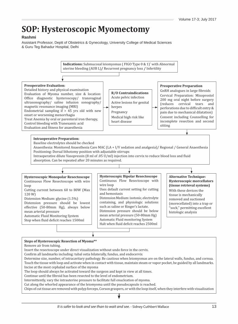
SOP: Hysteroscopic MyomectomyRashmiAssistant Professor, Deptt of Obstetrics & Gynecology, University College of Medical Sciences& Guru Teg Bahadur Hospital, Delhi
Indications: Submucosal leiomyomas ( FIGO Type 0 & 1)* with Abnormal uterine bleeding (AUB L)/ Recurrent pregnancy loss / Infertility
Preoperative Evaluation:Detailed history and physical examinationEvaluation of Myoma number, size & location: Of ice diagnostic hysteroscopy/ transvaginal ultrasonography/ saline infusion sonography/ magnetic resonance imaging (MRI) Endometrial sampling if > 45 yrs old with new onset or worsening menorrhagiaTreat Anemia by oral or parenteral iron therapy,Control bleeding with Tranexamic acid Evaluation and itness for anaesthesia
Hysteroscopic Monopolar Resectoscope Continuous Flow Resectoscope with wire loopCutting current between 60 to 80W (Max 120 W)Distension Medium: glycine (1.5%)Distension pressure should be lowest effective (50-80mm Hg), always below mean arterial pressureAutomatic Fluid Monitoring SystemStop when luid de icit reaches 1500ml
Hysteroscopic Bipolar ResectoscopeContinuous Flow Resectoscope with wire loopUses default current setting for cutting and hemostasisDistension Medium: isotonic, electrolyte containing, and physiologic solutions such as saline or Ringer’s lactate.Distension pressure should be below mean arterial pressure (50-80mm Hg)Automatic Fluid monitoring SystemHalt when luid de icit reaches 2500ml
Alternative Technique: Hysteroscopic morcellators (tissue retrieval systems)With these devices the tissue is mechanically removed and suctioned (morecellated) into a trap or ‘‘sock,’’ permitting excellent histologic analysis
Preoperative PreparationGnRH analogues in large ibroidsCervical Preparation: Misoprostol 200 mg oral night before surgery (reduces cervical tears and perforations due to dif icult entry & pain due to mechanical dilatation)Consent including Counselling for incomplete resection and second sitting
R/O ContraindicationsAcute pelvic infectionActive lesions for genital herpesPregnancyMedical high risk like heart disease
Intraoperative Preparation:Baseline electrolytes should be checkedAnaesthesia: Monitored Anaesthesia Care MAC (LA + I/V sedation and analgesia)/ Regional / General AnaesthesiaPositioning: Dorsal lithotomy position with adjustable stirrupsIntraoperative dilute Vasopressin (8 ml of .05 U/ml) injection into cervix to reduce blood loss and luid absorption. Can be repeated after 20 minutes as required.
Steps of Hysteroscopic Resection of Myoma**Remove air from tubing. Insert the resectoscope under direct visualization without undo force in the cervix. Con irm all landmarks including: tubal ostia bilaterally, fundus, and endocervixDetermine size, number, of intracavitary pathology. Be cautious when leiomyomas are on the lateral walls, fundus, and cornua.Touch the tissue with loop and activate when in contact with tissue, maintain steam or vapor pocket, be guided by all landmarks.Incise at the most cephalad surface of the myomaThe loop should always be activated toward the surgeon and kept in view at all times.Continue until the ibroid has been resected to the level of endometrium.Intermittently, vary the intrauterine pressure to facilitate full enucleation of myoma.Cut along the whorled appearance of the leiomyoma until the pseudocapsule is reached.Chips of cut tissue are removed with polyp forceps, Corson graspers, or with the loop itself, when they interfere with visualization
It is safer to look and see than to wait and see. - Sidney Cuthbert Wallace
AOGD Bulletin
14
Post operative Care- NSAIDS/ Acetaminophen/ Low dose Opioid Analgesics - Resume normal activity and return to work in 24 to 48 hours- resume sexual activity in 1 week.- No role of routine prophylactic antibiotics- Mild serosanguinous vaginal discharge may persist for up to 4 weeks- Patients with fever, malaise, worsening pain, or escalating pain
medication requirements should be carefully evaluated for bowel injury, bladder injury, and endometritis.
- Pregnancy should be attempted after 3 months
**Adjunctive StepsHysteroscopic scissors can be used for small pedunculated ibroids Mass Vaporizing Electrodes may be used to desiccate larger leiomyomas, available for monopolar and bipolar hysteroscope.
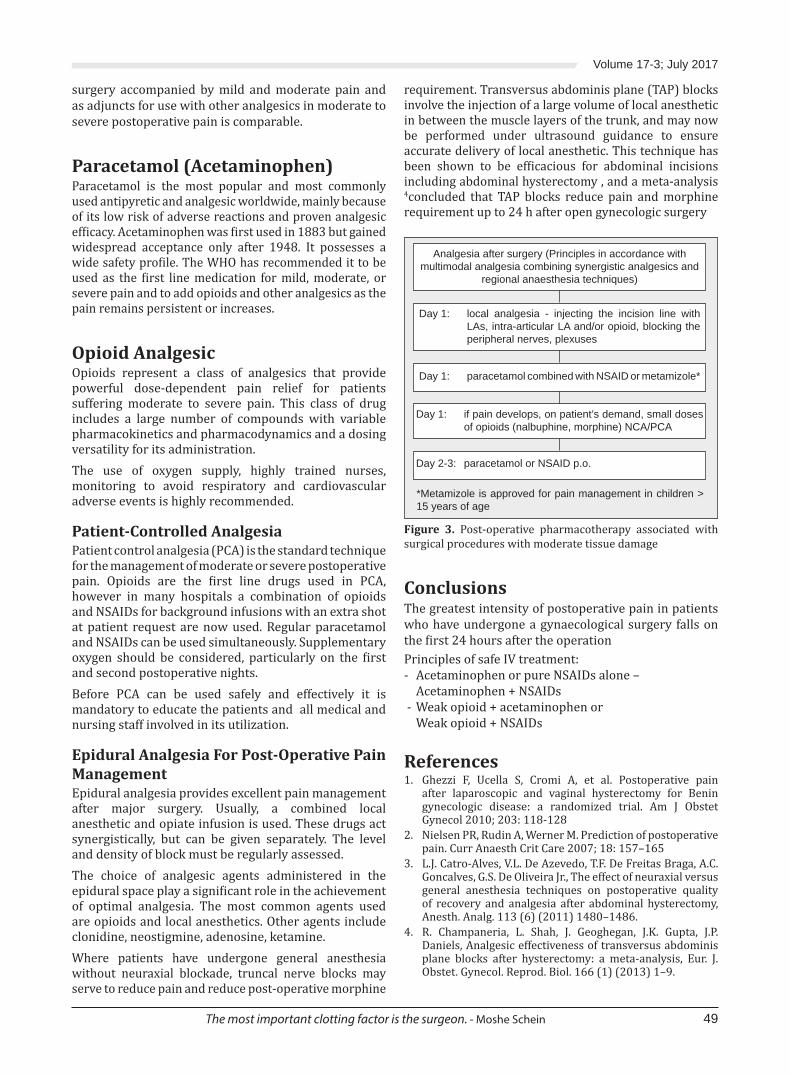
*The FIGO Classi ication of submucosal ibroids: Type 0 - Pedunculated intracavitary ibroid without intramural extension; Type 1 - <50% intramural; Type 2 - >50% intramural extension
Watch for Intraoperative complications
Uterine PerforationSigns: loss of visualization or intrauterine pressure, quick rise in luid de icit, increased bleeding or
hemodynamic instabilityManagement: Terminate the procedureUltrasound to con irm intraperitoneal luid/ blood if doubt
Laparoscopy/ Laparotomy if increased bleeding/ perforation with electrosurgical instrumentClose monitoring postoperatively
Excessive BleedingCauses: cervical or vaginal lacerations/ uterine perforation/ my from myoma bedManagement: Evaluate for cause and rule out perforation.If no perforation, control bleeding with electrocautery.If bleeding continues from myoma bed, place intrauterine foley’s tamponade.
Hyponatremia and HypervolemiaSymptoms include confusion, mental status changes, nausea, vomiting, visual disturbances, and headache, GA may mask these symptomsManagement: Stop the procedure when max luid de icit limits are reached or s/s developImmediately measure serum electrolytesHyponatremia: Oxygen, Inj Frusemide 40-60mg I/V, 0.9% salineIn severe cases ventilator support, Inj Frusemide 1mg/Kg 4-6hrly, Hypertonic saline 3%
Suggeste d reading • Valentine LN1, Bradley LD. Hysteroscopy for Abnormal
Uterine Bleeding and Fibroids. Clin Obstet Gynecol 2017 Jun;60(2):231-244.
• Lucy Whitake r, Hilary O.D.Critchley. Abnormal uterine bleeding. Best Practice & Research Clinical Obstetrics & Gynaecology; Volume 34, July 2016, Pages 54-65
Volume 17-3; July 2017
15
IntroductionThe utilization of robotics in various benign and malignant gynecologic procedures has rapidly risen and revolutionized minimal invasive surgery. The robot has empowered the surgeon to operate while sitting and further fascinated the patient with cutting edge technology and swifter recovery. Robotic platform has bridged most of the challenges of conventional laparoscopic approach (CLS) like 2-dimension depth perception, camera instability, limited range of motion, poor ergonomics and tedious endo-suturing. It is particularly advantageous for surgeries that require iner dissection and exhaustive endo-suturing like
myomectomies, tubal reconstruction, extensive endometriosis and gynecologic cancers.Robotic surgery was introduced in India in 2001. Data and experience has accumulated and evolved during this time. Till May 2017, 52 robots were functioning in the country and more than 2000 gynecological procedures were accomplished robotically. As per Vattikuti foundation data, 61% of robotic procedures were done for benign pathology and 39% were for gynecologic cancers.
Overview of TechnologyThe Da Vinci Robotic surgical system includes a console and a robotic cart. After 4-5 trocar placements, patient is positioned and docked. The surgeon sits un-scrubbed at the console. The camera, all 3 robotic arms and energy sources are under charge of the surgeon. The freedom of movement of the wristed instruments and intuitive human interface control of instruments makes dissection and suturing iner and easier. Fatigue of the surgeon is undeniably less while sitting. Typically, 2 bedside assistants, help in the introduction of robotic instruments and uterine manipulation. Newer generations of Da Vinci robots with enhanced surgical performance are getting innovated. Cumbersome large robots that used to invade vaginal and tableside assistants’ territory are no longer a concern with the Xi system. The Xi system is remarkably sleeker and its overhead boom can pivot in any direction to make complex multi-quadrant surgery easier for the console surgeon and assistants. The assembly of the instruments and camera are less complicated and more intuitive in the newer robots.Certainly, more affordable robots would be launched in
the competitive market that would change the dynamics of robotic surgery soon. Future foresees cost effective compact robots, faster assembly, better ergonomics, 3 D imaging for assistants, haptic feedback, sleeker snake like arms and camera to operate in small corners, integrated table motion and specimen retrieval systems.
Robotic HysterectomiesHysterectomy is the most common major gynecologic procedure. More than ifteen thousand hysterectomies have been performed robotically in India. Almost 50% of these hysterectomies were performed for benign indications like ibroid, abnormal uterine bleeding, endometriosis and uterine prolapse (Vattikuti database)The comparison of effectiveness of robotic versus CLS for benign disease has been studied in 4 RCTs and multiple retrospective studies. Indication for hysterectomy in these studies was mainly myomas or abnormal uterine bleeding. Insigni icant differences in surgical complication and pain scores were reported between the robotic and CLS arms. The duration of robotic surgery was longer than CLS, however the length of stay was comparatively shorter in robotic arm. Of note, the enrolment of these studies occurred early, during learning phase of robotics and results are likely to be biased and evidence might change with further research.Laparoscopic and robotic hysterectomy is associated with higher incidence of vaginal cuff dehiscence relative to open or vaginal hysterectomy. Ucella et al (2012) did a retrospective study on 12,398 hysterectomies.The incidence of vault dehiscence was reported to be 0.13 % for vaginal approach, 0.2% abdominal approach and 0.64% for laparoscopic approach and 1.64 % for robotic approach. Drudi et al (2013) prospectively evaluated 441 robotic hysterectomies for gynecologic cancers to delineate risk factors for vault dehiscence. In this study, the risk factors for vault dehiscence were adjuvant chemotherapy and/or brachytherapy, early resumption of sexual activity and low body mass index. Even, review of operative videos did not reveal a detectable etiologic factor, such as excessive cautery damage to vaginal cuff or shallow tissue sutured or suturing by staff or trainees.The safety of use of barbed suture in minimal invasive hysterectomy is also debated. A randomized controlled trial on 90 women who underwent robotic hysterectomy, evaluated the long term outcome for different vaginal cuff closure techniques (Tsafrir et al 2017). The authors concluded that barbed suture is equally safe as un-
Robotic Surgery in Benign Gynecologic ConditionsSarika GuptaConsultant, Gynecologic Oncology & Robotic Gynecology, Indraprasth Apollo Hospital, Sarita Vihar, New Delhi
Abdominal wall closure: if it looks all right, it’s too tight - if it looks too loose, it’s all right. - Matt Oliver
AOGD Bulletin
16
barbed suture. Similarly, a recent systematic review and meta-analysis on use of barbed suture in minimal invasive gynecology (by Bogliolo S 2015 et al) concluded that there was signi icant reduction in operative time with barbed suture with no difference in major or minor complications as compared to vicryl sutures.Robotic hysterectomy is also reported to be associated with higher risk of pneumonia in a few studies. Rosero et al 2013 compared matched cohort of 7788 patients undergoing robotic assisted hysterectomy and 7788 patients undergoing CLS hysterectomy. The surgical complications were comparable in between the groups. However, robotic arm patients were two times more likely to experience postoperative pneumonia (RR=2.2; 95% CI 1.243.78; p=0.0005).Health care cost is an important facet in the decision of any surgical procedure. Robotic surgery is often criticized for its high cost and longer operative time. Early data suggest that operative times and cost are optimized with greater surgeon experience and high volume centers. It anticipated that with time, technology would make the equipment more cost effective. The average cost of robotic hysterectomy in Delhi ranges from 2.5 to 3.5 lakhs. Both the surgeon and the team in the operating room get trained with number of cases. Various studies have shown that the cost of surgery decreases with the experience of the team. A recent retrospective case series by Moawad G (2017) compared the cost and operative outcomes of robotic hysterectomy to laparoscopic hysterectomy after strati ication of cases by uterine weights. The author’s concluded that robot is cost effective for uteri weighing more than 750 gm.
Robotic MyomectomyMost surgeons prefer robotic myomectomy in lieu of CLS, as it typically overcomes challenges confronted with laparoscopic suturing. Multiple retrospective series reiterate feasibility and safety of robotic myomectomy. Barakat et al (2011) compared 89 robotic myomectomies to 93 laparoscopic myomectomies and 393 abdominal myomectomies done in Cleveland clinic from 1995-2009. The author’s described a hybrid technique where myoma was partially excised by laparoscopic instruments, followed by docking. The dissection of the myoma from bed, hemostasis and suturing were performed robotically. This hybrid approach overcomes robotic limitation of quadrantic surgery and laparoscopic limitation of tedious suturing. Laparoscopic instruments can better maneuver the uterus in all quadrants and laparoscopic myoma screw can effectively stabilize heavy uterus during enucleation of ibroid. Robotic suturing can oppose the myometrium effectively. It was observed that both robotic and laparoscopic myomectomies were associated with less blood loss, shorter length of stay, and longer operative times. However, the mean weight
of myoma removed robotically was signi icantly higher than laparoscopic group. Review of literature still lacks randomized control comparison between CLS and robotic myomectomies. However, a meta-analyses of 8 retrospective studies, compares 412 laparoscopic myomectomies to 458 robotic myomectomies (Iavazzo et al 2016). This meta-analysis showed statistically insigni icant difference between laparoscopic and robotic techniques regarding blood loss, need for transfusion, duration of the operation, postoperative pain, postoperative fertility, complications, and hospital stay or recovery time. The cost of robotic myomectomy is higher as compared to open and CLS.Robotic myomectomy has similar fertility rates compared to laparoscopy and open route. There is no data demonstrating superiority of robotic approach over standard laparoscopy for myomectomy in terms of scar dehiscence after conception.
Sacro-colpopexyLaparoscopic sacro-colpopexy and robotic sacro-colpopexy have similar outcomes. However, laparoscopic sacro-colpopexy is technically challenging and is usually performed by expert laparoscopist. Two RCT’s have been published on comparison of robotic sacro-colpopexy to CLS. Both studies observed signi icantly longer operative room time, higher post-operative pain score and cost after robotic surgery as compared to CLS. Surgeons in these studies were required to have done 10 or more robotic surgeries only. This might be a source of bias in the study.These two RCTs in addition to one prospective cohort and six retrospective cohorts were meta-analyzed by DGD Sa M et al.in 2016. It concluded that the pelvic loor distress inventory, POP stage, rate of adverse effects and recurrent prolapse 1 year postoperatively were no different in between groups. Hence, robotic sacro-colpopexy may be de ined ergonomically superior with similar results and higher cost.
EndometriosisResection of deep endometriosis is one indication that might bene it most with magni ication, camera stability and dexterity of robotic instruments. However, as per Indian data only 3.1 % robotic gynecologic surgeries are done for this indication (Vattikuti data 2017). Optimal resection of deep endometriosis might effect short term and long-term outcome of patients in terms of fertility and relief of symptoms.Multiple retrospective series and a meta-analysis have reported no difference in the length of stay or complications with robotic surgery. No study has yet compared the long term outcome of robotic versus CLS and initiation of such studies is urgently needed to
Volume 17-3; July 2017
17
demonstrate the role of robotic surgery in endometriosis resection. Siesto et al 2014 published a retrospective series that included 19 bowel resections, 23 RVS nodules removals and ive bladder resections, alone or in combination in patients with deep in iltrating endometriosis. No intra-operative complications or conversion to laparotomy occurred. One anastomotic leakage was recorded. They concluded that resection of deep endometriosis is feasible and safe through robotic route. Few other case reports demonstrate organ preservation with robotic surgery as surgeon can better de ine anatomy and dissect precisely.
Tubal reconstructive surgeryTubal reconstructive surgeries are a bit obsolete in the world of IVF. However, tubal reconstruction can be effective in reversal of tubal ligation. There have been very few studies comparing laparoscopic tubal reconstruction with robotic reconstruction. The largest study analyzed the robotic tubal re-anastomosis outcomes of 97 consecutive patients (Caillet et al 2010). The overall pregnancy and birth rates were 71% and 62%, respectively, which were similar to prior laparoscopy and robotics results.
Other SurgeriesSmall case series and reports of robotic uro-gynecologic procedures indicate safety and ef icacy of robot in vesico-vaginal repair, recto-vaginopexy and Burch colpo-suspension.
ConclusionThe advent of robotic surgery in gynecology looks quite promising. Most of the RCT’s and retrospective studies conclude that the surgical outcome of surgery is not inferior to laparoscopy with the advantage of better ergonomics to surgeon and lesser pain and shorter hospital stay to the patient. The major obstacle to its popularity is its higher cost that might be mitigated in the future with more experience and newer generation of robots.ACOG published committee opinion (Number 628), in March 2015 to offer practice recommendations for robotic assisted gynecologic procedures. This is in congruence to AAGL position statement (2012) and Cochrane review on robotic gynecologic surgeries (2012)• Well-designed RCT’s are needed to determine bene it
of robotic surgery.• In addition to didactic and hands-on training, ongoing
quality assurance is essential to ensure patient safety.• Adequate informed consent with discussion of
indication for surgery and bene its associated with
robotic technique with approaches to the patient.• Surgeons should be skilled at abdominal and
laparoscopic approaches for a speci ic procedure before undertaking robotic procedure
• Surgeons training and quality metrics should be developed at the institutional level.
• Registry of robotic gynecology procedures and associated adverse events.
Falcone T. Robotic-assisted, laparoscopic, and abdominal myomectomy: a comparison of surgical outcomes. Obstet Gynecol. 2011;117(2 Pt 1):256-65.
• Bogliolo S, Musacchi V, Dominoni M, Cassani C, Gaggero CR, De Silvestri A, et al. Barbed suture in minimally invasive hysterectomy: a systematic review and meta-analysis. Arch Gynecol Obstet. 2015;292(3):489-97.
• Caillet M, Vandromme J, Rozenberg S, Paesmans M, Germay O, Degueldre M. Robotically assisted laparoscopic microsurgical tubal reanastomosis: a retrospective study. Fertil Steril. 2010;94(5):1844-7.
• De Gouveia De Sa M, Claydon LS, Whitlow B, Artahona MA. Robotic versuslaparoscopic sacrocolpopexy for treatment of prolapse of the apical segmentof the vagina: a systematic review and meta-analysis. Int Urogynecol J 2016;27:355–366.
• Drudi L, Press JZ, Lau S, Gotlieb R, How J, Eniu I, et al. Vaginal vault dehiscence after robotic hysterectomy for gynecologic cancers: search for risk factors and literature review. Int J Gynecol Cancer. 2013;23(5):943-50.
• Iavazzo C, Mamais I, Gkegkes ID. Robotic assisted vs laparoscopic and/or open myomectomy: systematic review and meta-analysis of the clinical evidence. Arch Gynecol Obstet. 2016;294(1):5-17.
• Moawad GN, Abi Khalil ED, Tyan P, Shu MK, Samuel D, Amdur R, et al. Comparison of cost and operative outcomes of robotic hysterectomy compared to laparoscopic hysterectomy across different uterine weights. J Robot Surg. 2017 Jan 31. doi: 10.1007/s11701-017-0674-4. [Epub ahead of print]
• Rosero EB, Kho KA, Joshi GP, Giesecke M, Schaffer JI. Comparison of robotic and laparoscopic hysterectomy for benign gynecologic disease. Obstet Gynecol. 2013;122(4):778-86.
• Siesto G, Ieda N, Rosati R, Vitobello D. Robotic surgery for deep endometriosis: a paradigm shift. Int J Med Robot. 2014;10(2):140-6.
• Tsafrir Z, Palmer M, Dahlman M, Nawfal AK, Aoun J, Taylor A, et al. Long-term outcomes for different vaginal cuff closure techniques in robotic-assisted laparoscopic hysterectomy: A randomized controlled trial. Eur J Obstet Gynecol Reprod Biol 2017;210:7-12.
• Uccella S, Ceccaroni M, Cromi A, Malzoni M, Berretta R, De Iaco P, et al. Vaginal cuff dehiscence in a series of 12,398 hysterectomies: effect of different types of colpotomy and vaginal closure. Obstet Gynecol 2012;120(3):516-23.
• Zeybek B, Hill A, Menderes G, Borahay MA, Azodi M, Kilic GS. Robot-Assisted abdominal cerclage during pregnancy. JSLS. 2016;20(4). pii: e2016.00072.
Two things a surgeon fears the most are God and Peritonitis. - Henri Mondor
AOGD Bulletin
18
IntroductionMinimally invasive surgery (MIS) has dramatically changed the vision of modern surgical approach. Advantages of MIS in the treatment of a diverse range of gynecological conditions have been well demonstrated by several studies in the last few decades.Along with the signi icant technological advances and expanded applications, MIS brought an inherent challenge, the safe extraction of surgical specimen. To accomplish the operation overcoming this challenge, an additional process named morcellation has been described. Morcellation procedure is de ined as fragmentation of the large tissue specimen into smaller pieces to facilitate the specimen extraction1.
Impact of morcellation techniques on MISMorcellators were introduced initially in 1973 as a hand-activated device for laparoscopic tissue removal. The irst electromechanical or power morcellator was made
available by Steiner in 1993. In 1995, FDA approved the irst laparoscopic power morcellator with a gynecologic
indication for use. Severe complications mostly involving bowel and vascular structures caused by the spinning blade of the morcellator were reported2. Performing intracorporeal power morcellation (PM) can also lead to scattering of benign tissues such as leiomyoma and endometriosis. Dispersed tissue fragments may implant on abdominal organ surfaces leading to in lammation, infection, and intestinal obstruction, which require additional surgical interventions and treatments2.However, among all concerns the most formidable one that brought this technology under scrutiny is unintentional dissemination of malignant cells, which can lead to severe consequences such as worsening the prognosis by upstaging occult cancer. In October 2013, a Boston-based anesthesiologist Amy J Reed had undergone minimally invasive surgery with power morcellation at Brigham and Women’s Hospital to ind out postoperatively that the mass was cancerous, igniting a debate about the true risk-bene it pro ile of power morcellation because of iatrogenic dissemination of unexpected malignancies such as sarcomas and adenocarcinomas.On April 17, 2014, the US Food and Drug Administration (FDA) published a safety communication discouraging “the use of laparoscopic power morcellation during
hysterectomy or myomectomy for the treatment of women with uterine ibroids”3. In an Italian survey, Mandato et al. reported that 58.7% of gynecologists declared that they would change their surgical practice after FDA safety communication only to prevent legal litigation.In another recent study from US, comparing the number of cases performed during 8 months before and after FDA warning statement, authors reported 5.8% and 19% decrease in the number of minimally invasive hysterectomy and myomectomy procedures respectively4. More concerning, Harris et al. reported that the number of major surgical complications and hospital readmissions were signi icantly increased with the decreasing numbers of minimally invasive hysterectomy. These results, from a statewide surgical cohort study, can be translated to an additional $23 million burden to annual health care costs. In response to these negative impacts on surgical outcomes and health care costs, surgeons embarked on a quest to overcome the challenges and eliminate tissue dissemination during morcellation.
Types of morcellation techniquesOverview of options for specimen removal available in minimally invasive gynaecologic surgery
Mini-laparotomy
Power morcellation
with or without a bag through culpotomy
or culdotomy
• with or without a bag• extending trocar
incision or another site• ± Alexis retractor• laparoendoscopic
single site morcellation
• with or without a bag
Vaginal morcellation
Abdominal Approach to Contained Tissue ExtractionIn 2014, Einarsson et al. described “Sydney in bag morcellation technique” as a contained abdominal morcellation method for multiport laparoscopic surgery. In this technique, using two different tissue retrieval systems- a 15-mm EndoCatch bag (Covidien, Mans ield, MA, USA) and Anchor TRS-200 tissue retrieval system (Anchor Surgical, Addison, IL, USA) -authors placed the surgical specimen inside the retrieval bag and inserted a 12-mm trocar into the bag through the umbilicus followed by insuf lation. Subsequently, a 5-mm balloon-
Contained Morcellation: Current technique and future directionsBB Dash, Rupali KhuranaRejoice, Infertility & Gyne Endoscopy Training Centre, New Delhi
Volume 17-3; July 2017
19
tip Kii trocar (Applied Medical, Rancho Santa Margarita, CA, USA) pierced into the insuf lated bag with the specimen inside. Power morcellator was introduced through the umbilicus incision and morcellation performed under optic visualization. Once morcellation has been completed, the isolation bag was removed through the umbilical port after de lation of trocar balloon. (Fig.1)
Figure 1: Bag used in Sydney in bag morcellation
The Sydney in-bag morcellation technique presents a novel approach for contained PM. However, it poses a potential risk of tissue dissemination arising from the piercing of the isolation bag. Penetrating the bag inside the abdominal cavity jeopardizes the bag integrity and may result in tissue leakage. Therefore, an innovative method for enclosed morcellation inspired by the topological shape of the surgical glove was developed by Rimbach et al. (More-Cell-Safe, A.M.I., Austria). It was aimed to create an isolated space with protrusions that can be manipulated through the ports on abdominal wall. The bag has two openings, larger opening was used for placement of the uterine tissue inside the bag and introduction of the power morcellator as well, while the smaller opening was used to insert the laparoscope5. Recently, Paul et al. described the use of a specially designed isolation bag using a similar bag shape (MorSafe; Veol Technologies, Mumbai, India) for two-port morcellation method (Fig.2)6.
Figure 2: MorSafe bag
Morcellation of uterine specimen within an insuf lated isolation bag during single-site laparoscopic surgery was also recently described. Using a cordless electric morcellator, the LiNA Xcise (LiNA Medical, Glostrup, Denmark), introduced through a 5-mm trocar, uterine specimens of 12 patients were morcellated in a contained fashion without any complications (Fig.3)7. As it requires neither bag penetration nor piercing with trocar, this technique appears as a reliable approach.
Figure 3: LiNA Xcise
Contained PM of uterine specimen in multiport laparoscopy was associated with 20–26 min longer operative time when compared with those without PM. However, no signi icant differences related to estimated blood loss, length of hospital stay, and perioperative complications were recorded. On the other hand, Venturella et al. reported operative times less in-bag manual morcellation without PM. These reported variations in operative times are possibly due to the differences in morcellation technique, tissue size, and surgical experience. The current body of evidence suggests that contained PM is a time ef icient and feasible method in laparoscopic surgery.To evaluate the safety of contained PM in terms of tissue dissemination, Cohen et al. conducted both in vivo and in vitro studies. Although investigators detected dye leakages in a few trials, PM in an isolation bag was suggested as a feasible method with the need of further studies to con irm the safety of current techniques and materials.
Vaginal Approach to Contained Tissue ExtractionVaginal channel is a natural ori ice through which an abdominal specimen can be easily removed after creation of colpotomy or culdotomy incision. Vaginal route can provide removal of entire uterus without morcellation especially in oncological cases. However, in cases with severe vaginal atrophy, narrow pelvic arch, nulliparity, and bulky uterus, the tissue removal can be challenging. When uterine specimen is too large to be extracted, intact morcellation can be performed through vagina. Vaginal morcellation is relatively faster and simple to learn and perform. Commonly used vaginal morcellation techniques include bivalving, wedge resection, coring, myomectomy, and recently described paper roll method8. Moreover, vaginal morcellation can be performed within a containment bag to prevent tissue dissemination. After either abdominal or vaginal insertion of containment bag in various types, authors accomplished vaginal morcellation in an enclosed fashion9.Besides its ef iciency, vaginal tissue extraction should be performed in experienced hands as it carries the risks of bladder, rectum, and vaginal lacerations. Therefore,
The best doctor gives the least medicines - Benjamin Franklin
AOGD Bulletin
20
meticulous inspection of the surgical ield should be done to exclude possible injuries after removal of the specimen. Although contained vaginal morcellation may prevent the risk of tissue dissemination, Solima et al. reported that in 4 of 12 cases, the containment bags were found to be ruptured after illing up with methylene dye. Demonstrating potential risk of tissue dissemination even in contained vaginal morcellation, authors addressed the importance of development of new, resistant, and durable materials and devices.
Conclusion and future directions Uterine sarcomas are very rare with an incidence of 7–8% of all uterine cancers and there is no currently available method for an accurate preoperative diagnosis. Although the occurrence of uterine sarcomas is very rare in women younger than 40 years, its risk factors are not well understood. They are usually discovered postoperatively and, regardless of the tumor stage, the prognosis is very poor with only 40–66% survival at 5 years. The risk of unanticipated uterine sarcoma in patients undergoing a uterine morcellation was 0.22% in a retrospective cohort study.
As the main concern with morcellation is the spillage of malignant cells, it should be realized that cellular dissemination is most likely initialized by the surgical procedure itself. Spindle cells after myomectomy have been detected in peritoneal washings even in the absence of the morcellation. Although its clinical signi icance is still unclear, patients should be informed that there is an inherent risk of cellular dissemination during myomectomy procedure regardless of morcellation. In this point, we believe that an advanced innovative surgical method providing an enclosed space not only for the morcellation procedure but also for the preceding myomectomy procedure can be developed in the future. This futuristic view may provide an insight for the future developments of new surgical methods and devices.A simple bag system with the mouth exteriorised and the tumour, morcellator and laparoscope within the containment bag is already in use and a more sophisticated system using an activated mesh and bag system to surround the tumour during morcellation is now undergoing evaluation.
In conclusion, it should be emphasized that there is currently no available method for tissue extraction that completely eliminates the risk of cellular dissemination. Therefore, further investigations and technological developments are needed to improve morcellation technique. Collaboration between surgeons, device manufacturers, and designers should also be encouraged to ind innovative solutions.
3. US Food and Drug Administration. Laparoscopic uterine power morcellation in hysterectomy and myomectomy: FDA Safety Communication. April 17, 2014. Washington, DC: US Food and Drug Administration. In: US Food and Drug Administration, ed April 17 2014
4. Barron KI, Richard T, Robinson PS, Lamvu G. Association of the U.S. Food and Drug Administration morcellation warning with rates of minimally invasive hysterectomy and myomectomy. Obstet Gynecol (2015) 126:1174–80.
5. Rimbach S, Holzknecht A, Schmedler C, Nemes C, Offner F. First clinical experiences using a new in-bag morcellation system during laparoscopic hysterectomy. Arch Gynecol Obstet (2016) 294:83–93.
6. Paul PG, Thomas M, Das T, Patil S, Garg R. Contained morcellation for laparoscopic myomectomy within a specially designed bag. J Minim Invasive Gynecol (2016) 23:257–60.
7. Aoki Y, Matsuura M, Matsuno T, Yamamoto T. Single-site in-bag morcellation achieved via direct puncture of the pneumoperitoneum cap, a cordless electric morcellator, and a 5-mm lexible scope. Eur J Obstet Gynecol Reprod Biol (2016) 201:126–30.
8. Wong WS, Lee TC, Lim CE. Novel vaginal “paper roll” uterine morcellation technique for removal of large (>500 g) uterus. J Minim Invasive Gynecol (2010) 17:374–8.
9. Günthert AR, Christmann C, Kostov P, Mueller MD. Safe vaginal uterine morcellation following total laparoscopic hysterectomy. Am J Obstet Gynecol (2015) 212.
Volume 17-3; July 2017
21
“Mind, Body & Soul”“Mind, Body & Soul”Post Operative Recovery with MeditationRashmiAssistant Professor, Deptt of Obstetrics & Gynecology, University College of Medical Sciences& Guru Teg Bahadur Hospital, Delhi
Undergoing surgery is a stressful time for the patient and it involves lot of stress and anxiety starting before surgery and continuing in the postoperative period. After surgery there is an added problem of pain - or more accurately, anticipation of pain. Other psychological stresses are fear of an inability to return to life the way the patient was used to living it, loss of an organ (e.g. hysterectomy), prognosis of the disease (like endometriosis), partial treatment (like in advanced malignancy). Results from even the best performed surgeries can fail to give optimal outcome if the postoperative and preoperative period is not well managed. Anxiety and stress further aggravate the perception of pain.It is well known that stress and anxiety cause secretion of in lammatory cytokines and stress hormones into the blood stream. These can affect the immune system resulting in slow recovery after the surgical procedure and wound healing may also get delayed. Compromised immune system makes the postoperative patient for more prone to infections also which can further make the recovery more stressful. A signi icant number of studies suggest preoperative anxiety is associated with increased postoperative pain, slower recovery, increased risk of infection, and other negative outcomes. One study of 230 adults showed that those with negative attitudes like fear, distress, or hostility before radiological treatments, were more likely to experience unstable heart rate and blood pressure during the procedure, and had an increased risk of postoperative bleeding. Traditionally these problems are dealt with antianxiety medications (with their own side effects) and narcotic analgesics (with addiction problems).Alternative approaches like music therapy, acupressure, relaxation, yoga and meditation are being tried and studied to tackle the stressful response in perioperative period. These therapies decrease the release of pro-in lammatory cytokines and may result in better post -operative recovery.
Meditation can help the post-operative process in several waysMeditation has been shown to play an important role in strengthening the body’s immune system and regulating moods, due to its powers to elicit the “relaxation response” in the brain and body. Dr. Herbert Benson
of Mind-Body Institute, part of Harvard University’s Medical School has found that the relaxation response triggers increased activity in the parasympathetic nervous system, which is responsible for the body’s “rest and digest” functions; further, meditation decreases activity in the sympathetic nervous system, the body’s “ ight or light” response. When the body is in the “rest and digest” mode, it can concentrate its efforts on healing at the cellular level, by regenerating cells, repairing others, and restoring normal functioning to the area.Meditation elicits positive mental states in people like feelings of well-being, trust, love and increase in peace. Thinking positive thoughts releases oxytocin, the feel good hormone. This aids in the healing process by allowing a patient to remain calm in a stressful situation (such as post-operative recovery). Meditating before surgery can help make the patient calmer and more receptive to healing. The programs generally include guided meditation, deep relaxation techniques and pranayama. Guided meditation after surgery can elicit the relaxation response needed to allow the body to heal itself, and to manage a patient’s pain and anxiety.
What is Guided Meditation?Guided meditation is a process by which one meditates in response to the guidance provided by a trained practitioner or teacher, either in person or via a written text, sound recording, video, or audiovisual media comprising music or verbal instruction or both. This process often leads to the participant engaging in visualization and generating mental imagery that may simulate or re-create the sensory perception of sights, sounds, tastes, smells, movements, and images associated with touch, such as texture, temperature, and pressure, as well as imagined mental content de ies conventional sensory categories. The generating of such mental imagery can precipitate or accompany strong emotions or feelings.In a study from University of Houston, guided meditation proved most effective for pain management during breast biopsies. Meditation has been shown to improve survival and decrease readmissions in cardiac patients after acute events. Meditation has also been shown to help in post stroke rehabilitation.There is an ongoing randomized study at Mount Sinai
The aim is to operate only when necessary but not to delay a necessary operation. - Moshe Schein
AOGD Bulletin
22
Hospital in New York City, to test whether the meditation technique can lessen pain in spine-surgery patients and reduce the need for opioid painkillers, which can be highly addictive. The randomized trial trains patients in a simple form of meditation and asks them to practice it starting two weeks before their surgery and for six weeks after, using audiotapes to guide them.To conclude, these mind body techniques may not cure the disease, but they may blunt the increased physiological harm associated with the stress, anxiety, or depression that often come with medical treatments and serious diagnoses. Uncovering these connections between mind and body can help us understand the
importance of such therapies to address mental health when undergoing medical treatment including post - operative recovery. These techniques help not only the patient but the doctor too. Dr. Devi Shetty, the famous cardiac surgeon in Bangalore, had said that “while a doctor’s body needs to endure long hours of standing to perform an operation, his mind has to be equally or more it to endure the stressful long hours. A surgeon getting into pressure and anxiety does not help the patient and so meditation helps keep the mind calm and relaxed so that we are able to support the patient in turn.”
Surgeries For Benign Lesions
I am the queen of Women EmpireLiving with pride in the world entireThe path is rosy with millions of chasersBut I select the best and fastest swimmerTo give mankind it’s most beautiful creationA prince or a princess for next generation
But when a benign lesion invades my wellbeingI cry silently as I am peace lovingI only have the language of pain & bleedingTo refer to misery and my heart pleadingRuthless enemies are fi broid, lumps & ovarian torsion Endometriosis, aden omyosis and PALM- COEIN notation
Medicines are essential for fi rst time management But defi nitive options remain the surgical treatment Polypectomy, myomectomy, hysterectomy are safe & sure Laparoscopic & robotic surgeries are new methods of cure Benign tumors of ovary are removed with ease & simplicity Unless there are issues of adhesions & fecundity
Tricky neighbors are bladder, bowel and uretersWithout consent and caution they can be traitorsSurgery may sound simple for benign lesion All depends on the surgeons’ skill and selectionTo give women new life, happiness and rejuvenationGynecologists stand united with will and dedication.
Dr Neerja Goel, Dr Sandhya
Volume 17-3; July 2017
23
Q.1 Give two examples of static sling surgeries for pelvic organ prolapse …………………...................………
Q.2 The best candidates for hysteroscopic removal of ibroids are:
a) FIGO PALM-COEIN Type-0 b) less than 3 cm type-1c) a+bd) FIGO type 2
Q.3 Operative laparoscopy is indicated in endometriosisa) For infertilityb) To evaluate response to hormonal treatmentc) >5 cm endometrioma on imaging d) Mild –minimal endometriosis for staging
Q.4 Ovariopexy is de ined as …............................……
Q.5. Boundaries of Litta’s triangle in laparoscopic hysterectomy are ……….................…………….
Q.6. List any two contraindications for Non descent vaginal hysterectomy
Q.10 Write True/ False: Hair in the surgical site should always
be removed using razors or electric clippers
Prophylactic antibiotics should be repeated for any surgical procedure lasting longer than 2–3 hours or when substantial blood loss (greater than 1,500 mL) occurs
Uterosacral cardinal ligament complex is level one De Lancey support
Vaginal route is superior to laparoscopic and abdominal for hysterectomy
Quiz Time: Tick it, Fill it, Click it, Whatsapp/ Email itCompiled by Bindiya GuptaAsstt Professor, Department of Obstetrics & Gynecology, University College of Medical Sciences & Guru Teg Bahadur Hospital, Delhi
Tick the MCQs and ill in the blanks.Click a pic and whatsapp or email to usWhatsapp Nos.: 9810645212, 9810719002Email: [email protected]
Heartiest Congratulations to the Winners of Quiz of June Issue
1. Dr Anita Rajhoria 2. Dr Usha Upreti 3. Dr Sweta Prasad
Answers Key to Quiz - June Issue1. C 2. A 3. hypotension or blood lactate concentration ≥ 4 mmol/L persisting after initial isotonic crystalloid luid challenge of 30mls/kg 4. C 5. MEOWS, SOS 6. Grade 2 7. C 8. D 9. C 10. C 11. SMO 12. A 13. Belfort-Dildy Obstetrical Tamponade System 14. Chattisgarh balloon 15. Mushroom bag, pelvic bleeding 16. 27th March 17. C 18. C 19. C 20. C
Who learns his anatomy from books should operate on books only.
AOGD Bulletin
24
Calendar of AOGD Skill Workshops (2017-2018)AOGD & The Department of Obstetrics and Gynecology, UCMS & GTBH plan to hold skill workshop series in the year April 2017 to March 2018.
Basics of Endoscopy in Gynaecology 21st July Venue
Email id: ........................................................................... Mobile no.: ................................................ Tel no.: .....................................................
Filled Form to be sent to: AOGD Offi ce,7th Floor MCH Block, Department of Obstetrics & Gynecology
Dr SK DasDr SK DasA great human being, a dedicated gynaecologist & a cherished teacher!
Dr S K Das served Association of Obstetricians and Gynaecologists of Delhi (AOGD) comprising of more than 2500 gynaecologists of Delhi as President, Advisor and Patron. She dedicated her life to the noble profession of medicine, patient care and was a highly skilled surgeon. She worked in Safdarjung Hospital for two decades and retired as professor and head of the department. After retirement she worked for cancer patients, developed the speciality of ‘Gynaecologic Oncology’ in Delhi. Early detection of cancer by colposcopy was her passion and she regularly conducted workshops to train budding gynaecologists & authored two books on the same subject.
She was awarded WHO fellowship in 1976 for MTP training program; USAID fellowship for design and implementation of child survival in 1988; Certifi cate of excellence in surgical contraception in the community by National Association for Voluntary Sterilization and Family Welfare of India; Bhartiya Chikitsa Ratan award by Council of Educational Research & Development, Pune in 2008; Bhartiya Chikitsa Ratan award by Institute of Economic Study, Delhi in 2009; Indira Gandhi Priyadarshni award at National Unity conference in New Delhi, 2010; Life Time Achievement award presented by Association of Cervical Pathology and Colposcopy in Kolkata, January 2014; Life Time Achievement award for work in Gynae oncology presented by Samita Misra Memorial Foundation for Cancer Research in April 2014; Felicitated by Indian Menopause Society in May 2016 for her outstanding contribution in fi eld of Gynaecology Oncology And fi nally, only recently, she received two prestigious awards, lifetime achievement award by the Association of Gynaecologic Oncologists of India (November 2016) and lifetime achievement award by Indian Society of Colposcopy and Cervical Pathology (March 2017).
Dr Das was a teacher of teachers and a role model for everyone. Her tremendous capacity to work, magnanimous heart, pleasing smile, polite behavior and leadership qualities are all laudable. She was a woman of substance, showed the path of righteousness and worked till the end. May God rest her soul in peace.
AOGD will fondly cherish memories of the legend DR. SK DAS !!
Your life was a blessing.... Your memory a treasure... You are loved beyond words... And missed beyond measure...
AOGD (2017-18)
AOGD Bulletin
26
Events Held in June 2017• IUI Workshop conducted on 2nd June by Aarogya Hospital at IMA East Delhi.
• Lunch CME on laparoscopic techniques was organized by Sunrise Hospital in association with IMA SDB on 10th june saturday 1.30 pm onwards at IHC.
CME on laparoscopic techniques was organized by Sunrise Hospital at IHC
• Scienti ic session on ‘Letrazole in ovulation induction’ on 21st June, 21-4.30pm at Hotel Piccadily by Gynae Forum Dwarka under the aegis of IFS and AOGD infertility Subcommittee
Scienti ic session on ‘Letrazole in ovulation induction’
IUI workshop by Aarogya Hospital at IMA East Delhi
Volume 17-3; July 2017
27
• Yoga day celebrations at GTB Hospital on 21st June. Great enthusiasm amongst faculty and the participants. Dr Sujata from AYUSH Department also joined.
• AOGD monthly clinical meeting was held at Army Hospital (R & R) on 30th June with interesting case presentations. Doctor’s day was celebrated by cutting cake with great enthusiasm.
Yoga day celebrations at GTB Hospital
AOGD monthly clinical meeting at Army Hospital (R & R)
AOGD Bulletin
28
Volume 17-3; July 2017
29
AOGD Bulletin
30
Cold CoagulationAblation of cervical lesions by „boiling“ of tissue
TREATMENT THERAPY PROBES with 8 different versionsOF CIN 1-3
KRITICARE SOLUTIONSDwarka, New Delhi-110075 Email: [email protected] Mobile: 9958366200, 9810504326
Volume 17-3; July 2017
31
CentoNIPT®Expertise you can trust
100% safe for mother & childFast & reliable resultsTest as early as possibleMaximum certainty
Centogene India Private Limited 107 Wegman‘s Business Park, Knowledge Park III Greater NOIDA - 201308, Uttar Pradesh, India Tel: +91-85273-17888 E-mail: [email protected] www.centogene.com
The non-invasive prenatal testing to screen for Trisomy 21,
Trisomy 18, Trisomy 13 and sex chromosome aneuploidies
AOGD Bulletin
32
Dr. Rishma PaiPresident FOGSI 2017
FOGSI BOH Organising Team
Na onal ConvenorDr. Alka Kriplani
Dr. Jaideep Malhotra
Organising Secretaries
Jt. Organising Secretaries
Scien fi c Co-chairpersons
North Zone ConvenorDr. Manju Verma
North Zone Co-ordinatorsDr. Maninder Ahuja Dr. Pra ma Mi al
Dr. Ranjana KhannaVice President FOGSI 2017
Organising Chairperson+91 9335106867
Dr. Shakuntla Kumar+91 9811445853
Dr. Tarini Taneja
Dr. Susheela GuptaDr. Amita Tripathi
Dr. Archana VermaDr. Kavita Agarwal
Dr. Sadhana Gupta Dr. Ar Gupta
Dr. Kiran Guleria+91 9811142329
Dr. Shalini RajaramPresident AOGD
Dr. Mala AroraScien fi c Chairperson
Volume 17-3; July 2017
33
7:30
am
onw
ards
Regi
stra
tion
08:0
0am
- 09
:00a
mFr
ee C
omm
unic
atio
ns (H
alls
)
Ha
ll A
Ha
ll B
09:0
0 am
- 10
:00a
mU
nd
ers
tan
din
g P
ree
cla
mp
sia
Pred
icto
rs o
f pre
ecla
mps
ia :
From
Ben
ch to
bed
side
Late
ons
et p
reec
lam
psia
: Is
the
path
ogen
esis
diff
eren
t?M
anag
emen
t of a
cute
ons
et s
ever
e pr
e-ec
lam
psia
Emer
ging
dru
gs fo
r pre
ecla
mps
ia: t
arge
ting
the
endo
thel
ium
Dis
cuss
ion
Re
surr
ec
tio
n o
f th
e C
on
tra
cep
tiv
e B
ask
et
Ant
ara
and
Chay
a: O
ld w
ine
in a
new
bot
tleEm
erge
ncy
Cont
race
ptio
n: E
xpan
ding
ind
icat
ions
Prog
este
rone
vag
inal
Rin
g an
d Si
no im
plan
t II
Men
stru
al M
oksh
aD
iscu
ssio
n
10:0
0am
- 10
:30a
mTe
a
Ha
ll A
& H
all
B
10:3
0am
- 11
:00a
mAO
GD
Pre
side
nt’s
Ora
tion
11:0
0am
- 11
:20a
mKe
y N
ote
Addr
ess:
Pas
t, Pr
esen
t and
Fut
ure
of R
obot
ic S
urge
ry
11:2
0am
- 12
:20p
mPa
nel D
iscu
ssio
n: A
ddre
ssin
g an
d Ra
tiona
lisin
g Ri
sing
Ces
area
n se
ctio
n ra
tes
12:2
0pm
- 01
:00p
mIn
augu
ratio
n
01:0
0pm
- 02
:00p
mLu
nch
& P
oste
r Vie
win
g
Ha
ll A
Ha
ll B
02:0
0pm
- 03
:00p
mPa
nel D
iscu
ssio
n: M
inim
ally
Inva
sive
Sur
gery
in G
ynec
olog
ic O
ncol
ogy:
Saf
e Pr
actic
e
03:0
0pm
- 04
:00p
mH
igh
Ris
k O
bst
etr
ics
Preg
nanc
y af
ter B
aria
tric
sur
gery
Jaun
dice
in p
regn
ancy
: Min
imis
ing
mor
bidi
ty &
Mor
talit
yCe
rebr
o-pl
acen
tal r
atio
in p
reve
ntin
g st
ill b
irths
in F
GR
Une
xpla
ined
recu
rren
t mis
carr
iage
An
appr
oach
to a
cas
e w
ith O
ligoa
mni
osD
iscu
ssio
n
Infe
rtil
ty:
Ch
an
gin
g p
rac
tice
Biom
arke
rs fo
r ova
rian
rese
rve:
wha
t is
best
?Pr
e-im
plan
tatio
n ge
netic
scr
eeni
ng: S
houl
d th
e pr
actic
e co
ntin
ue?
Ova
rian
agin
g: C
an it
be
stop
ped?
Lute
al s
uppo
rt: W
hat,
Whe
n an
d fo
r how
long
?M
axim
isin
g su
cces
ful i
mpl
anta
ton:
Adv
ance
s in
end
omet
rial r
ecep
tivity
Dis
cuss
ion
04:0
0 - 0
5:00
pmV
ide
o S
ess
ion
: O
bst
etr
ics
Retr
ogra
de H
yste
rect
omy
for p
lace
nta
prae
via/
accr
eta
Lase
r abl
atio
n in
TTT
S12
wee
k sc
an: N
T, n
asal
bon
e, a
nom
alie
sLo
w c
ost i
nnov
atio
n in
PPH
man
agem
ent:
Chat
tisga
rh b
allo
onLa
paro
scop
ic E
ncer
clag
e
Vid
eo
Se
ssio
n:
Gy
ne
colo
gy
Endo
scop
ic S
entin
el n
ode
diss
ectio
nLe
For
t’s p
roce
dure
: Sim
plic
ity p
erso
nifi e
d!M
ini s
ling
for S
UI
Clito
ropl
asty
Spec
imen
retr
ieva
l tec
hniq
ues
in L
apar
osco
py
05:0
0pm
- 5:
30pm
Tea
39th
Annu
al C
onfe
renc
e of
AO
GD 2
017
Sc
ien
tifi
c P
rog
ram
me
Da
y 1
: S
atu
rda
y,
18
th N
ov
em
be
r 2
01
7
AOGD Bulletin
34
07:3
0am
onw
ards
Regi
stra
tion
08:0
0am
- 09
:00a
mFr
ee C
omm
unic
atio
ns
Ha
ll A
Ha
ll B
09:0
0am
- 10
:00a
mF
eta
l m
ed
icin
eN
IPT:
Has
it re
plac
ed tr
aditi
onal
scr
eeni
ng fo
r fet
al a
neup
loid
y?Rh
Isoi
mm
unis
atio
n/Fe
tal a
nem
ia: W
hen
to re
fer,
wha
t to
do?
Opt
ions
bey
ond
lase
r in
com
plic
ated
twin
pre
gnan
cyG
row
th p
robl
ems,
Mon
itorin
g an
d Ti
min
g D
eliv
ery
in M
ultip
les
Dis
cuss
ion
Ra
tio
na
l U
se o
f H
orm
on
es:
Wh
ich
, wh
en
, ho
w m
uch
an
d h
ow
lo
ng
?Th
reat
ened
abo
rtio
nAU
B Ad
oles
cent
End
omet
riosi
sM
enop
ausa
l HT
Dis
cuss
ion
10:0
0am
- 10
:30a
mTe
a &
Exh
ibiti
on
Ha
ll A
& H
all
B
10:3
0am
- 11
:00a
mBr
igad
ier K
hann
a O
ratio
n: D
r. M
ario
Lei
tao,
Dire
ctor
, Rob
otic
surg
ery,
Mem
oria
l Slo
an K
ette
ring
Hos
pita
l, N
ew Yo
rk, M
anag
emen
t of E
ndom
etria
l Can
cer:
MSK
CC P
ract
ice
Gu
est
Le
ctu
res
11:0
0am
- 11
:15a
m
ABC
of B
reas
t Hea
lth
11:1
5am
- 11
:30a
mH
PV V
acci
natio
n: A
dec
ade
of e
xper
ienc
e
11.3
0am
- 12
:00p
mU
nder
stan
ding
Gen
etic
Test
s in
Gyn
ecol
ogic
al C
ance
rs: C
ase
Stud
ies
12:0
0pm
- 01
:30p
mCo
mpe
titio
n Pa
pers
01:3
0pm
- 02
:30p
mLu
nch
& P
oste
r Vie
win
g
Ha
ll A
Ha
ll B
02:3
0pm
- 03
:30p
mD
eb
ate
s:
All
Fibr
oids
see
n du
ring
cesa
rean
sec
tion
mus
t be
rem
oved
Cesa
rean
on
dem
and
is th
e rig
ht o
f a p
regn
ant m
othe
rSo
il an
d Se
ed a
re ri
pe fo
r ute
rine
tran
spla
ntat
ion
in In
dia
Egg
Free
zing
for a
ll be
fore
30:
sur
e sh
ot w
ay o
f ach
ievi
ng fu
ture
pre
gnan
cy
Issu
es
in G
yn
eco
log
yCo
ntro
vers
ies
in L
eiom
yom
a m
anag
emen
tIV
F vs
. rev
ersa
l of l
igat
ion
post
ste
riliz
atio
nM
anag
emen
t of A
deno
myo
sis
in y
oung
wom
enD
ilem
mas
in m
anag
emen
t of E
ctop
ic p
regn
ancy
03:3
0pm
- 04
:30p
mB
est
of
20
16
-17
: E
vid
en
ce B
ase
d P
rac
tice
in
Pre
gn
an
cy
Be
st o
f 2
01
6-1
7:
Ev
ide
nce
Ba
sed
Pra
cti
ce i
n G
yn
eco
log
y
Pre
sen
tati
on
s &
Dis
cu
ssio
nP
rese
nta
tio
ns
& D
isc
uss
ion
CHIP
S (C
ontr
ol o
f Hyp
erte
nsio
n in
Pre
gnan
cy s
tudy
)ET
IP: E
xerc
ise
trai
ning
and
wei
ght g
ain
in o
bese
pre
gnan
t wom
enEl
ectiv
e de
liver
y ve
rsus
exp
ecta
nt m
anag
emen
t fo
r pr
e-ec
lam
psia
: M
eta
anal
ysis
of R
CT’
sPr
eter
m b
irth
prev
entio
n in
Tw
in P
regn
ancy
: Sys
tem
atic
Rev
iew
& M
etaa
naly
sis
Vita
min
D a
nd R
isk
of p
rete
rm b
irth:
RC
TsD
iscu
ssio
n
Trea
tmen
t Str
ateg
ies f
or W
HO
Type
II an
ovul
atio
n: Sy
stem
atic
revi
ew an
d m
etaa
naly
sisH
ydro
salp
inx:
Tub
al S
urge
ry o
r in
vitr
o fe
rtili
satio
n: A
n ev
erla
stin
g D
ilem
ma
Ute
rine
arte
ry e
mbo
lizat
ion
vs.h
yste
rect
omy
in th
e tr
eatm
ent o
f sym
ptom
atic
ut
erin
e fi b
roid
s: E
MM
Y tr
ial
Targ
eted
The
rapy
in O
varia
n Ca
ncer
: Sys
tem
atic
Rev
iew
Mor
cella
tion
in F
ibro
ids:
Ris
ks a
nd c
urre
nt p
ract
ice
wor
ldw
ide
Dis
cuss
ion
04:3
0pm
- 05
:00p
mVa
ledi
ctor
y
05:0
0pm
- 05
:30p
mTe
a
39th
Annu
al C
onfe
renc
e of
AO
GD 2
017
Sc
ien
tifi
c P
rog
ram
me
Da
y 2
: S
un
da
y,
19
th N
ov
em
be
r 2
01
7
Volume 17-3; July 2017
35
18th - 19th November, 2017
Pre-conference Workshops: 17th November 2017
Venue: India Habitat Centre, Lodhi Road, New Delhi
REGISTRATION FORM
39th Annual Conference of Associationof
Obstetricians and Gynecologists of Delhi
Name ....................................................................................................................... Institution .........................................................................................................
Department .......................................................................................................... Designation ....................................................................................................
Address ................................................................................................................................ City ............................................. Pin Code ........................................
Mobile No .......................................................... Landline No .......................................................... Email ..................................................................................
ACCOMPANYING PERSON’S DETAILS
Name ............................................................................................................................................................................................ Age .................................................
All DD/Cheque payable at New Delhi & should be made in favour of “AOGD CONFERENCE 2017”
• Write your Name and Contact No. at the back of DD/Cheque
Registration for the conference is mandatory in order to register for the pre conference workshops.
Payment by Cheque or Draft Only.
Online payment through payment gateway can be made only if Registration form is fi lled online.
PAYMENT DETAILS
Please fi nd enclosed herewith Cash/DD/Cheque No ...................................................................................................... Dated .......................................
Drawn on (Name of the Bank) ................................................................................................................................... Branch .....................................................
For Rs. ........................................................... (In words) .....................................................................................................................................................................
REGISTRATION FEES
Conference Workshop
Registration Category Upto 30th Sept ‘17 Upto 30th Oct ‘17 Spot Registration Upto to 30th Sept ’17 Upto to 30th Oct ’17 Spot Registration
Genital Prolapse is a common condition for women worldwide and almost a ifth of vaginal hysterectomies are done for pelvic organ prolapse according to a survey in rural India1.Patients may seek care for a myriad of symptoms like dif iculty to walk/sit, urinary/bowel complaints or bleeding or foul-smelling discharge coming from the prolapsed organ. Many times, the complaint may be simply the awareness of something coming out per vaginum. Despite the symptoms, a lot of women neglect the condition for a long time, spanning years, even decades owing to social issues and because the symptoms do not interfere much with day to day activities.For successful surgery, its important to understand the anatomical supports of uterus and the defects resulting in prolapse.
Anatomical Supports of uterusThe anatomical supports of uterus have been divided into three levels by DeLancey 2
posteriorly to perineal body and laterally to levator ani margins
Attenuation or detachment of level 1 support causes apical prolapse in form of uterine descent and enterocoele, level 2 support causes cystocoele and rectocoele and level 3 support causes perineal defect2.
Surgeries for Genital ProlapseVaginal hysterectomy with pelvic loor reconstruction has been the standard of care for high grade prolapse for a long time now. This is because generally these women belong to the peri and postmenopausal age group, not
desirous of fertility and removal of uterus makes it easy to repair the prolapse. Uterine conservation was reserved for women who desired fertility. However, opponents of this management say that since the pathophysiology of prolapse lies in the supports of the uterus and vagina, removal of uterus is unnecessary and focus should be shifted to strengthening and restoring the normal uterine and vaginal anatomy. Also, as high as 33% women may undergo repeat surgery after vaginal hysterectomy for prolapse. Many a times, hysterectomy may pose greater morbidity for the woman than the prolapse itself3. Concerns about alterations in ovarian blood supply and its function eventually following hysterectomy have also been voiced4. Hence in the past few years, a consensus regarding preservation of uterus for treatment of symptomatic prolapse is beginning to form, even in women who do not desire fertility and in postmenopausal women. Uterine conserving surgeries have added bene its of preserving the menstrual function (deemed important by a lot of women) and reproductive desirability, avoids the complications of vaginal hysterectomy and cultural and social issues associated with removal of uterus. Also, there has been a better understanding of the anatomy and pathophysiology of genital organs and their supports combined with advances in surgical techniques like use of mesh and mersilene tapes, leading to an increase in the number of women opting for uterus conservation for prolapse5.
Uterus Conserving Surgeries for ProlapseThe current goals of uterus conserving surgeries are to restore the strength of supports of the uterus at all three levels, improve symptoms and decrease risk of recurrence. An ideal uterine conserving surgery would be technically easy to learn, maintain the normal anatomy, have low rate of recurrence and should not interfere with course of pregnancy and labor.Various surgical options available are:1. Manchester repair2. Shirodkar’s modi ication of Manchester procedure3. Vaginal sacrospinous ixation4. Abdominal sacrohysteropexy5. Abdominal sling surgeries
Uterine Conserving Surgeries for Genital ProlapseAlpana Singh1, Varsha Priyadarshani2
1Associate Professor, 23rd Year Postgraduate, Department of Obstetrics & Gynaecology, University College of Medical Sciences & Guru Teg Bahadur Hospital, Delhi
There is an inverse relationship between the surgeon’s ability and the frequency he asks for more muscle relaxants.
AOGD Bulletin
38
Manchester repairA classic Manchester operation involves a preliminary D&C, followed by cervical amputation, plicating the Mackenrodt’s ligaments and reconstruction of the cervical lip6 (Fig. 1). Technically easy to learn, this procedure has been followed for years as a uterine conserving surgery for women with symptomatic prolapse with cervical elongation and desirous of fertility. However, high recurrence rates coupled with adverse events during pregnancy like preterm labour, cervical dystocia has diverted the focus to other surgeries4.
Figure 1: Manchester procedure
Shirodkar’s modi ication of Manchester procedureModi ications of this procedure like Shirodkar modi ication have been used which is almost like Manchester repair except that the cervix is not amputated and uterosacrals are plicated (Fig. 2). Shirodkar modi ication is used when there is no cervical elongation. A study by Libergall et al compared the outcomes of modi ied Manchester with vaginal hysterectomy for symptomatic prolapse and found good acceptance rate and lower recurrences with the former procedure7.
Figure 2: Shirodkar’s modi ication of Manchester procedure
Transvaginal Sacrospinous ixationIt involves hitching the cervix and uterus transvaginally to uni- or bilateral sacrospinous ligaments using a non-absorbable suture. However, the proximity of pudendal nerve and vessels to the ischial spine, where the suture is
taken on the ligament, leads to postoperative morbidity in the form of intraoperative hemorrhage, buttock and leg pains. Recurrence rates are also high following this procedure (Fig. 3)4,8.
Figure 3: Transvaginal Sacrospinous ixation
SacrohysteropexyRecent advancements in the ield of mesh surgery brought the concept of sacrohysteropexy in modern prolapse surgery. The basic idea was to suspend the uterus above the levator ani from the sacrum using mesh or mersilene tapes (Fig. 4). Hence this mimicked the normal anatomy of the pelvic supports, especially the uterosacrals. First used in 2007, this technique is relatively new but has proved to be highly effective with low rate of recurrence. Various techniques have been proposed for sacrohysteropexy. Cutner et al used mersilene tapes through uterosacral ligament to ix it bilaterally to the sacrum. Price N et al used a Y shaped polypropylene mesh to suture the anterior surface of the cervix to the sacrum while Massey F used the same material to suture the posterior surface of the cervix with sacrum instead4. Abdominal, laparoscopic and even robotic sacrohysteropexy procedures have been done till date with excellent results9,10.
Figure 4: Sacrohysteropexy
A Cochrane review compared abdominal sacrohysteropexy with vaginal procedures and found that the rate of awareness of prolapse (7% vs 14%), repeat prolapse surgery (4% vs 18%), dyspareunia and stress urinary continence was lower in the former11. However, operating time, return to normal life and chances of mesh erosion were higher with sacrohysteropexy12.Vaginal mesh procedures like infracoccygeal sacropexy have a higher incidence of mesh erosion and should only
Volume 17-3; July 2017
39
be done by highly specialized surgeons under special circumstances for audit or research purposes.
Sling surgery Sling surgeries have been introduced by various Indian authors, all pioneers in the ield of uterine conserving surgeries for pelvic organ prolapse as around 1.5-2.0% of genital prolapse in India are attributed to nulliparous women while 7-8% of women with prolapse have just 1 or 2 issues. The concerns with classic operations like Manchester, i.e. risk of recurrence and interference with antenatal and labor events coupled with the simplicity and effectiveness of sling surgeries has led to the growing popularity of the latter. Initially native tissues like rectus sheath were used to support the uterus. A popular procedure described by Purandare uses a dynamic sling of rectus sheath to hitch the anterior surface of the cervix to the abdominal wall. However, as many women with young prolapse also have intrinsic tissue weakness, these procedures have been superseded by use of inert sling materials like mersilene tape that can give lifelong support with minimal tissue reaction13.Various modi ications of synthetic sling procedures have been described that differ basically in the points of attachment of cervix to various anatomical landmarks. For example, in Shirodkar sling operation (Fig. 5), the posterior cervix is hitched to the sacrum via mersilene tape. Khanna et al, on the other hand, anchored anterior aspect of isthmus to the anterior superior iliac spine. Virkud’s procedure anchors the posterior part of cervix to sacral promontory on the right side and anterior abdominal wall on the left side together with plication of uterosacral ligament. Joshi’s sling procedure suspends cervix to the pectineal ligament on both sides. Soonawala et al anchored uterosacral ligament to the anterior longitudinal ligament on the right side of sacrum14.
Figure 5: Shirodkar’s sling surgery
The selection criteria for women suitable for sling operations include young women with 2nd/3rd degree prolapse, normal cervical length and absent to minimal cystocoele/rectocoele. If a large cystocoele/rectocoele is present, they should be vaginally repaired before proceeding with the sling operations. These procedures can also be performed endoscopically with equal results.
Very few articles have been published and there are no published randomized controlled trial so far that compares the outcomes of abdominal sling operations vs other conservative surgeries. Cutner et al have also described a novel procedure of suspending uterus using mersilene tapes laparoscopically14. However, there is still paucity of literature and randomized controlled trials are needed with long term follow up in order to fully understand the success of sling operations.In conclusion, various surgeries have been described, some well tested but with apparent shortcomings thus causing the advent of newer surgical techniques that address to the pathophysiology of genital prolapse more closely than the earlier ones. However, suf icient studies and long term follow up on these novel procedures is still lacking and nothing conclusively can be said as to which procedure should be preferred. The type of procedure used should be individualized depending upon the patient’s pro ile and the surgeon’s experience and more research is still needed in this area.
References1. Chhabra S, Ramteke M, Mehta S, Bhole N, Yadav Y. Trends
in hysterectomy for genital prolapse: rural experience. Clin Med Insights Reprod Health 2013;7:11-6.
2. Zimmerman CW. Pelvic organ prolapse: basic principles. In: Jones HW III, Rock JA, editors. Te Linde’s Operative Gynecology, 11th ed. Philadelphia: Lippincott Williams & Wilkins; 2015:824-845.
3. Glazener C, Constable L, Hemming C, Breeman S, Elders A, Cooper K, et al. Two parallel, pragmatic, UK multicentre, randomised controlled trials comparing surgical options for upper compartment (vault or uterine) pelvic organ prolapse (the VUE Study): study protocol for a randomised controlled trial. Trials 2016;17(1):441.
4. Khan A, Jaleel R, Nasrullah FD. Sacrohysteropexy performed as uterus conserving surgery for pelvic organ prolapse: Review of case iles. Pak J Med Sci 2016;32(5):1174-1178.
5. Khunda A, Vashisht A, Cutner A. New procedures for uterine prolapse. Best Pract Res Clin Obstet Gynaecol 2013;27(3):363-79.
6. Tolstrup CK, Lose G, Klarskov N. The Manchester procedure versus vaginal hysterectomy in the treatment of uterine prolapse: a review. Int Urogynecol J 2017;28(1):33-40.
7. Liebergall-Wischnitzer M, Ben-Meir A, Sarid O, Cwikel J, Lavy Y. Women’s well-being after Manchester procedure for pelvic reconstruction with uterine preservation: a follow-up study. Arch Gynecol Obstet 2012;285(6):1587-92.
8. Api M, Kayatas S, Boza A, Nazik H, Aytan H. Laparoscpic Sacral Uteropexy with Cravat Technique- experience and results. Int Braz J Urol 2014;40:526–532.
9. Chen Y, Hua K. Medium-term outcomes of laparoscopic sacrocolpopexy or sacrohysteropexy versus vaginal sacrospinous ligament ixation for middle compartment prolapse. Int J Gynaecol Obstet 2017;137(2):164-169.
Surgery is not a trade or an art but a way of life - Alexander A Artemeiv
AOGD Bulletin
40
Long-term follow-up and quality of life after robot assisted sacrohysteropexy. Eur J Obstet Gynecol Reprod Biol. 2016; 206:27-31.
11. Maher C, Feiner B, Baessler K, Schmid C. Surgical management of pelvic organ prolapse in women. Cochrane Database Syst Rev 2013;(4):CD004014.
12. Maher C, Baessler K, Glazener CM, Adams EJ, Hagen S. Surgical management of pelvic organ prolapse in women:
a short version Cochrane review. Neurourol Urodyn 2008; 27(1):3-12.
13. Virkud A. Conservative Operations in Genital Prolapse. J Obstet Gynaecol India. 2016;66(3):144-8.
14. Cutner A, Kearney R, Vashisht A. Laparoscopic uterine sling suspension: a new technique of uterine suspension in women desiring surgical management of uterine prolapse with uterine conservation. BJOG 2007;114(9):1159-62.
Calendar of Monthly Clinical Meetings 2017-2018Months Name of the Institute28th July 2017 AIIMS25th August 2017 VMMC & Safdarjung Hospital29th September 2017 Hindu Rao Hospital27th October 2017 ESI Hospital, Basaidarapur24th November 2017 MAMC & LN Hospital29th December 2017 Sir Ganga Ram Hospital19th January 2018 Dr RML Hospital23rd February 2018 Lady Hardinge Medical College23rd March 2018 UCMS & GTB Hospital27th April 2018 Apollo Hospital, Sarita Vihar
Forthcoming Events• CME on Urogynecology- An evolving Concept on 8th July, 1:00pm - 3:00pm at Pind Balluchi Restaurant by
Urogynecology Subcommittee arranged by Fortis Escorts Kidney and Urology Institute. Kindly con irm your presence in advance to Mr. Dheeraj (9990460965) as there is limitation of seats.
• Skill Workshop on ‘Basics of Endoscopy in Gynecology’ on 21st July, 11:00am - 5:00pm, 7th Floor MCH Block, GTB Hospital.
• Challenges in Management of Preterm Labour on 23rd July 2017 in Army Hospital by Dr. BK Goyal and team.• CME on ‘Post Natal Services-continuum of Care’ at GTB Hospital on 4th August, 2017.• The Reproductive Endocrinology Committee of AOGD under chairmanship of Dr. Nalini Mahajan is
organizing a CME on ‘Reproductive Health Dilemma’s - Adolescence to Menopause’ from 2:00pm - 5:00pm on the 12th August 2017 at the India Habitat Centre (Magnolia Hall).
• “BOH- The Trilogy 2017” on 19th and 20th August focused on current practices, breakthrough and current dilemmas on BOH patients by the FOGSI BOH Organizing Team in association with AOGD at Hotel Leela Ambience, New Delhi
• DGES (Delhi Gynecological Endoscopists Society) Annual Conference & IAGE (Indian Association of Gynecological Endoscopists- NZ) in association with AOGD on 25th-27th August, 2017 at Indian Habitat Centre, Lodhi Road; PreCongress workshop on 25th August, 2017.
• 39th AOGD Annual Conference on 18th and 19th November, 2017 at Indian Habitat Centre; Pre-conference workshops on 17th November, 2017.
Volume 17-3; July 2017
41
Total hysterectomy for benign uterine conditions is the most frequent gynaecologic surgical procedure1. With the mounting growth in endoscopic surgery, laparoscopic hystrectomy(LH) has diffusely penetrated the realms of most gynaecologists. It was irst demonstrated by Reich et al2 in 1989 and has come a long way since then with a lot of modi ications done for better outcomes and reduced rates of injuries. Urological injuries are often encountered during hystrectomy with an incidence of 0.04 – 0.4 %3 in open and 0.65 – 1.39 %4 in laparoscopic hysterectomy. While bladder injuries are relatively straightforward, ureteral injuries are dif icult to recognize and insiduous in nature. In a Cochrane review by Neiboer et al5, all the bene its of LH came at a cost of a higher risk of urinary tract injuries compared to open technique (OR 2.41). Leonard et al6 evaluated 1300 cases of benign laparoscopic hysterectomies and suggested that the complications of the laparoscopic technique can be signi icantly reduced by adequate training of surgeons, education about electrosurgical principles, adequate exposure, correct knowledge of anatomical landmarks and proper selection of patients and the route of hysterectomy. Consideration to basic principles during surgery like isolation of the uterine artery before coagulation, development of the paravesical space, use of a uterine manipulator to stretch the uterine pedicle, and especially restoration of the normal anatomy in case of deeply in iltrating endometriosis and/or in the presence of severe adhesions help in achieving a successful outcome. But complications occur when it is not possible to follow these basic rules due to extremely challenging conditions. The most common technique for TLH involves intrafascial dissection of vascular pedicles and the use of a vaginal manipulator for the mobilization of the uterus. In the classic technique, the broad ligament is entirely dissected or fenestrated. Thus, in case of bleeding while dissecting the uterine vessels, the course of the ureter must be detected to safely execute the hemostasis, and this can be accomplished by pulling the posterior leaf of broad ligament. The time required could result in a delay of hemostasis and may increase the risk of direct (thermal or mechanic by suture) or indirect (thermal spread) ureteral injuries. Uterine artery identi ication and coagulation can be sometimes dif icult, due to a
wide variety of conditions like severe endometriosis, broad ligament ibroids, sequelae of infections, prior pelvic surgery. Ureteral injury is usually associated with thermal spread from coagulation devices or with suture ligation during uterine artery occlusion and vaginal cuff closure. To minimize the damage in such challenging conditions Litta et al7 described a modi ied approach for LH that we called ‘‘total reverse laparoscopic hysterectomy.’’ The name was derived from the steps, the irst of which is the dissection of the vesicouterine fold and pubocervical fascia followed by the coagulation and incision of the round ligament. The second step is the opening of the anterior fold of the broad ligament between the lateral (round ligament incision) and central (pubocervical fascia incision) landmarks. This way better visualization of the uterine vascular pedicle is obtained, and a real plane, represented by the posterior layer of the broad ligament, is created that permits the identi ication of the ureter until it crosses the uterine artery, thus creating a ‘‘safe triangle’’, a triangular area whose sides are represented medially by the uterine vessels (ascending branch) and laterally by the ureter. The apex points downward and corresponds to the crossing of the ureter and the uterine artery (Litta’s triangle). The consecutive coagulation and dissection of uterine vessels, at least 1 cm above the anterior incision of the pubocervical fascia ( irst landmark), is of relevance for the management of potential bleeding. The uterine pedicle is resected, leaving a suf icient caudal part that can be easily seen and quickly identi ied, and in the case of bleeding or retraction after the dissection, this area can be immediately and safely sealed without injuring the ureter. Coagulating the uterine vessels 1 cm above the pubocervical fascia is already well away from the ureter. Circular colpotomy after exposure of the anterior vaginal fornix is performed. Paul et al8 described injection of dilute vasopressin into the myometrium close to bowel adhesions or reduction in vascularity and facilitation of dissection by raising the serosa from the myometrium at the site of adhesion. Retrograde hystrectomy is described in a similar way and circumferential dissection of the vagina is continued to completely separate the cervix and the vagina. The rectal adhesions to the posterior vaginal wall are released by application of traction to the anterior and subsequently posterior lip of the cervix. The posterior lip of the cervix is pulled away from posterior rectal
Retrograde Hysterectomy for Benign Gynaecologic ConditionsKanika Batra Modi1, Rupinder Sekhon2
1Fellow, 2Senior Consultant, Gynae Oncology, Rajiv Gandhi Cancer Institute & Research Centre, Delhi
A surgeon operates as good as his assistant permits.
AOGD Bulletin
42
adhesions, and by maintaining traction on the cervix, sharp adhesiolysis is done.Another techique as described by Nezhat et al9, is adequately developing the vesicovaginal plane and mobilizing the urinary bladder. This step is relatively straightforward in patients with no vesicouterine adhesions. In cases with prior cesarean deliveries, the plane between the bladder and the lower uterine segment and cervix can be scarred with dense adhesions. These adhesions create distorted anatomy, resulting in dif icult dissections, prolonged operating time, and possible inadvertent injury to surrounding structures, speci ically the urinary bladder. In a study by Rooney et al10 an odds ratio of 7.5 was reported for incidental cystotomy at the time of laparoscopically assisted vaginal hysterectomy in women with a history of cesarean delivery. With the cesarean delivery birth rate approaching 33%, these adhesions are being encountered more frequently. The traditional method of mobilizing the bladder involves a superior to inferior dissection. In using the superior to inferior dissection technique, the scarring can distort the planes and the dissection can be dif icult, occasionally resulting in bladder injury. Development of the vesicovaginal space is done by creating a space lateral and caudal to the vesicovaginal space and then using a reverse caudal to cephalad dissection, facilitates complete mobilization of the bladder. The partially developed vesicovaginal space is entered inferiorly from the lateral dissection. The development of the avascular space occurs using blunt dissection with inferior to superior sweeping motions. This is performed bilaterally until both sides are connected inferiorly, while superiorly, the vesicouterine adhesions are present. The space in the vesicovaginal plane is bordered medially by the vesicocervical–vaginal ligament or bladder pillar, distally by the bladder, anteriorly by the remaining anterior leaf of the broad ligament, laterally by the inner aspect of the obliterated umbilical artery, and posteriorly by the parametrium where the ureter traverses toward the bladder. The described space is present bilaterally and is termed as the “new space” by them. The complete mobilization of the bladder is then proceeded using sharp dissection so that the remaining scarred bladder can be entirely dissected off the cervicouterine junction. Mobilization of the bladder is done inferiorly, 3 cm past the leading edge of the cervicovaginal junction, to allow adequate vaginal tissue to suture the cuff. The technique of open retrograde hysterectomy is an excellent alternative in the case of an elongated cervix or obliterated cul de sac, as described in Te Linde’s.11 In this method, an anterior colpotomy is created 0.5 to 1.0 cm below the cervicovaginal junction using the ESU. An empty spongestick placed transvaginally and elevated against the anterior cervicovaginal junction for adequate anatomical landmark and sereves as the initiation for colpotomy. The cervicovaginal junction is
circumferentially incised by placing a series of curved Heaney clamps, with each pedicle being clamped, divided, and secured with a Heaney trans ixion stitch of 1-0 delayed absorbable suture in sequence (i.e., each pedicle is tied before the next clamp is placed). The ties from each pedicle are held long and used to provide upward traction on the vaginal cuff, optimizing exposure for placement of the next clamp. The inal two clamps are placed across the posterior proximal vagina and the specimen excised. A simple method for closing the vaginal cuff consists of placing a horizontal mattress stitch of 1-0 delayed absorbable suture on either side of the vaginal cuff, working from posterior to anterior, below the tip of the clamp. Injuries occur even in the hands of experts and the expertise lies in timely identi ication and repair of those injuries. Routine use of cystoscopy and identi ication of ureters at the end of surgery can prevent inadvertent situations. Retrograde hysterectomy is an excellent tool in the right hands for dealing with dif icult pelvic conditions like endometriosis and adhesions.
The irst cervical landmark: the vesicouterine folder and pubocervical fascia are resected transversely for approximately 2 cm.
Figure 1: Photograph by Litta et al7, describing the irst step of retrograde hysterectomy.
The “safe triangle”: the apex of the cross between the urethra and the uterine artery; the lateral side is represented by the ureter, and the medial side is represented by the ascending branch of the uterine vessels.
Figure 2: Photograph by Litta et al7, describing the safe triangle of dissection
MJ, Peterson HB, DeStefano F, Rubin GL, Ory HW. Complications of abdominal and vaginal hysterectomy among women of reproductive age in the United States.
Volume 17-3; July 2017
43
The Collaborative Review of Sterilization. Am J Obstet Gynecol 1982;144:841– 848.
2. Reich H, De Caprio J, McGlynn F. Laparoscopic hysterectomy. J Gynecol Surg 1989; 5:213– 216.
3. H€arkki-Sir_en P, Sj€oberg J, Tiitinen A. Urinary tract injuries after hysterectomies. Obstet Gynecol. 1998;92: 113–118.
4. Garry R, Fountain J, Brown J, et al. EVALUATE hysterectomy trial: a multicentre randomised trial comparing abdominal, vaginal and laparoscopicmethods of hysterectomy. Health Technol Assess. 2004;8:1–154.
5. Nieboer TE, Johnson N, Lethaby A, Tavender E, Curr E, Garry R, van Voorst S, Mol BW, Kluivers KB. Surgical approach to hysterectomy for benign gynaecological disease. Cochrane database syst rev. 2009 Jul 8;3(8).
6. Léonard F, Fotso A, Borghese B, Chopin N, Foulot H, Chapron C. Ureteral complications from laparoscopic hysterectomy indicated for benign uterine pathologies: a 13-year experience in a continuous series of 1300 patients. Human reproduction. 2007 May 8;22(7):2006-11.
7. Litta P, Saccardi C, Conte L, Florio P. Reverse hysterectomy: another technique for performing a laparoscopic hysterectomy. Journal of minimally invasive gynecology. 2013 Oct 31;20(5):631-6.
8. Paul PG, Shabnam K, Bhosale SA, Kaur H, Talwar P, Thomas T. Laparoscopic hysterectomy in frozen pelvis—an alternative technique of retrograde adhesiolysis. Gynecological Surgery. 2013 Nov 1;10(4):285-90.
9. Nezhat C, Grace LA, Razavi GM, Mihailide C, Bamford H. Reverse vesicouterine fold dissection for laparoscopic hysterectomy after prior cesarean deliveries. Obstetrics & Gynecology. 2016 Sep 1;128(3):629-33.
10. Rooney CM, Crawford AT, Vassallo BJ, Kleeman SD, Karram MM. Is previous cesarean section a risk for incidental cystotomy at the time of hysterectomy? A case-controlled study. Am J Obstet Gynecol 2005;193:2041–4.
11. Bristow, R. (n.d.). Te Linde’s atlas of gynecologic surgery. 10th ed.
AOGD MONTHLY CLINICAL MEETING28th July 2017
Venue: Conference Hall AIIMS
Time: 1600-1700 h
Contact us: Dr JB Sharma: 9868397309, Dr Seema Singhal 9818291001
Hosted by Dept of Obstetrics and Gynecology
All India Institute of Medical Sciences, New Delhi.
Cases to be presented1. Laparoscopic management of uterovesical fi stula2. Unusual cause of primary infertility: Transverse vaginal septum3. Saving fetuses of Rh-isoimmunised pregnancies: the challenge
continues......
When there’s no place for the scalpel, words are the surgeon’s only tool. - Paul Kalanith
AOGD Bulletin
44
IntroductionSelection of the ideal and preferred route for performing hysterectomy has been under consideration for long. The factors which play an important role in selecting the route of hysterectomy are the size and shape of uterus and vagina, accessibility of uterus, extent of extra -uterine disease, the need for concurrent surgical procedures, surgical expertise, available infrastructure, cost-effectiveness of procedure and most importantly patient preference. The technique of Non-Descent Vaginal Hysterectomy (NDVH) was introduced years ago and pioneered by Heaney in 1934 and with more technical improvisations in later years it became a common procedure. However, with the advent of laparoscopy in 1990’s, the conventional method of vaginal hysterectomy lost its relevance over time. In the last decade, vaginal route of hysterectomy is being rediscovered and greater expertise has been achieved by the gynecologists due to its various advantages over the abdominal and laparoscopic routes. The American college of Obstetrician’s and Gynecologists (ACOG) Committee opinion 2017 recommends that minimally invasive approaches are preferred whenever feasible based on their well documented advantages over abdominal approach for hysterectomy. The minimally invasive approach includes vaginal route and laparoscopic (including robotic assisted laparoscopic surgery) route. As per the recommendations the vaginal approach should be the approach of choice whenever possible. Laparoscopic approach to hysterectomy may be considered as an alternative to abdominal hysterectomy (TAH) if vaginal route is not indicated or possible. A comparative analysis of various routes of hysterectomy are shown in table 1.
Vaginal hysterectomy has an advantage over laparoscopic and abdominal approaches by the fact that the vaginal ori ice is the anatomical opening provided by nature for access to the uterus, hence No Scar surgery” (cosmetic superiority) and no complications of surgical hernia or wound gape. Other advantages include short operating time, minimal blood loss, minimal adhesions and bowel handling and injury to adjacent organs. It is usually done under regional anesthesia, is cost effective, has shorter hospital stay and faster recovery.
Indications and Contraindications for NDVHThese include abnormal uterine bleeding, adenomyosis, ibroids, endometrial polyp/ hyperplasia and
premalignant conditions of cervix and uterus. The list is not conclusive and depends on the surgeon’s con idence and operative skill. Contraindications are summarized in Table 2.
Table 2: Contraindications to NDVHCONTRAINDICATIONS TO NDVHUterus more than 20 weeks size (Operator skill dependent)i) uterine size more than 10 cm in each dimension ii) a 90 degree angle between the lateral cervical wall and
uterine wall iii) a large lateral cervical ibroidSeverely restricted uterine mobilityPrevious VVF/RVF repairAdnexal pathologyPelvic endometriosisInvasive cervical cancer beyond stage IaCervix lushed with vault
Table 1: Comparison of various routes of hysterectomyCharacteristics Non-descent vaginal hysterectomy Total laparoscopic hysterectomy Abdominal hysterectomyOperating time 40-60 minutes 2-3 hours 60-100 minutesBlood loss 100-130 ml 100 ml 200-400 mlScar No scar Multiple 3-4 small scars Large (7-9cm) scarsScar related complications Absent Less MoreIncidence of istula 0.1% 0.5-1% 1%Hospital stay 3-4 days 3-5 days 5-7 daysPost-op complications Few Signi icant Signi icantAnaesthesia Regional General Regional/generalPatients with medical disorders Surgery of choice Least preferred With close monitoringMorbidity and mortality Less More More
Volume 17-3; July 2017
45
Challenging SituationsCommon reasons for hesitancy in performing NDVH by gynecologists include a narrow vaginal ori ice, absence of signi icant uterine descent, presence of uterine enlargement, prior abdominal surgery and need for concomitant oophorectomy.The following points are in favour of NDVH as the preferred surgery even in the above situations:Narrow vaginal ori ice: Tohic et al and Agostine et al in separate studies have documented 92-96% success in performing NDVH even in nulliparous patients with irmer tissues. It is suggested that in the presence of a mobile uterus less than 12 weeks size without adnexal pathology, vaginal approach to hysterectomy is always possible. Absence of signi icant uterine descent: Clamping of the uterosacrals and cardinal ligaments placed close to the vault produces descent of the uterus to 1st degree, thus making the surgery technically easier. Also, there is some amount of uterine descent under anesthesia.Presence of uterine enlargement: In the presence of ibriods or uterine enlargement, a pre- operative sonographic assessment of the uterine volume is suggested for deciding the route of surgery. NDVH can easily be performed without complications in uteri upto 12 weeks or 250-300cm3 in volume. According to the surgeon’s expertise and with the help of debulking procedures studies have reported successful outcomes in uterus upto 20 weeks (Unger et al). Sheth SS in his study of more than 3000 hysterectomies performed only for AUB reported removal of uterus as large as 20 - 22 weeks and uterine volume greater than 500-600cm3. The following debulking techniques can be useda) Morcellation: It can be used in cases involving
uterine enlargement, uterine ixation, or limited vaginal exposure. It should not be performed if the uterine arteries cannot be secured or if malignancy is suspected.
b) Intramyometrial coring: It is accomplished by circumferentially incising the outer myometrium beneath the uterine serosa with a scalpel while placing the cervix on traction. The incision should be kept as close to the uterine serosa as possible. The enlarged uterus is delivered as an elongated mass inverting the uterine fundus. This procedure is useful in case of adenomyosis.
c) Uterine bisection: It is performed by cutting the cervix and the uterine fundus in the sagittal plane. This technique is often combined with myomectomy or wedge morcellation to reduce the bulk of the uterine halves so that the tubo-ovarian vessels can be ligated.
d) Myomectomy: When myoma is seen bulging after bisection, myomectomy can be performed. Larger myomas may have to be removed piecemeal.
e) Slicing: In cases of adenomyosis, no plane of cleavage can be found making enucleation dif icult. In such
cases the myometrial tissue is sliced off the uterus layer by layer and once the bulk is decreased, uterus can be removed.
f) Pryor’s technique: removal of multiple uterine wedges help in debulking and reducing uterine volume.
Prior Abdominal surgeries: Previous abdominal surgeries like caesarean section, or myomectomy are not absolute contraindications. Techniques like “lateral window” approach, sharp dissection etc.can help access the vesicouterine space. Concomitant oophorectomy: Performing bilateral oophorectomy along with hysterectomy is not a deterrent for choosing the vaginal approach. The success of the procedure performed vaginally varies from 65- 97.5% in various studies. We now have newer methods in the form of endoloops, endostapler and vessel sealing system which help in accessing the ovaries. In case of dif iculty in performing oophorectomy, it is suggested to seek laparoscopic assistance.Other adjuvant techniques like use of aqua dissection, newer vessel sealing systems like Electro-surgical Bipolar Vessel Sealing system (EBVS) help in facilitating hysterectomy. The latter is associated with reduced thermal spread in comparison with unipolar cautery and is relatively faster compared with suture ligation as the current delivered to achieve haemostasis takes between 2 and 7 seconds.
ConclusionNon-availability or non-accessibility of laparoscopic surgeons and costly equipment specially in resource constraint countries like India, VH proves to be the most economical and safe procedure over other routes of hysterectomy. Newer advances such as bipolar vessel sealing systems further reduces the operating time, blood loss and surgical morbidity compared to use of conventional sutures in vaginal hysterectomy. Vaginal route will be used more frequently in the future as it gives a natural route, is scarless and gives operative results better than abdominal route.
Suggested Reading1. American College of Obstriticians and Gynaecologists,
Committee Opinion number 701, June 2017.2. Sheth SS. Vaginal Hysterectomy. Best Practice and Research
- Clinical Obstetrics and Gynaecology. Edited by Prof. S. Arulkumaran, ( Guest Editor R. Thakar & I. Myonda) USA. Elsevier Ltd. 2005;Vol.19(3), 307-32.
3. Tohic AL, Dhainaut C, Yazbeck C, et al. Hysterectomy for benign uterine pathology among women without previous vaginal delivery. Obstet gynecol, 2008 Apr; 111(4): 829-37
4. Agostine A, Bretelle F, Carvello L et al. Vaginal Hysterectomy in nulliparous women without prolapse: a prospective comparitive study. Int J Obstet Gynecol 2003; 110.515-518
Have plenty of assistance but not many assistants - Augustus C. Bernays
AOGD Bulletin
46
The irst ever Global guidelines for the prevention of surgical site infection were published on 3 November 2016 by WHO. They include a list of concrete recommendations distilled by 20 of the world’s leading experts from 26 reviews of the latest evidence.
Preoperative measures• It is good clinical practice for patients to bathe
or shower prior to surgery, using pain soap or antimicrobial soap.
• The patients undergoing cardiothoracic and orthopaedic surgery with known nasal carriage of S. aureus should receive perioperative intranasal applications of mupirocin 2% ointment with or without a combination of CHG body wash. This may be considered for other surgeries also.
• SAP should be administered prior to the surgical incision when indicated (depending on the type of operation). Administration of SAP is recommended within 120 minutes before incision, while considering the half-life of the antibiotic.
• Preoperative oral antibiotics combined with mechanical bowel preparation should be used to reduce the risk of SSI in adult patients undergoing elective colorectal surgery.
• Before surgical procedure, hair should either not be removed or, if absolutely necessary, it should be removed only with a clipper. Shaving is strongly discouraged at all times, whether preoperatively or in the OR.
• Alcohol-based antiseptic solutions based on CHG is recommended for surgical site skin preparation in patients undergoing surgical procedures.
• Antimicrobial sealants should not be used after surgical site skin preparation for the purpose of reducing SSI.
• Surgical hand preparation should be performed by scrubbing with either a suitable antimicrobial s oap and water or using a suitable alcohol-based handrub before donning sterile gloves.
Preoperative and/or intraoperative measures• Administration of oral or enteral multiple nutrient-
enhanced nutritional formulas should be considered for the purpose of preventing SSI in underweight
patients who undergo major surgical operations.• It is not suggested to discontinue immunosuppressive
medication prior to surgery for the purpose of preventing SSI.
• Adult patients undergoing general anaesthesia with endotracheal intubation for surgical procedures should receive an 80% fraction of inspired oxygen intraoperatively and, if feasible, in the immediate postoperative period for 2-6 hours to reduce the risk of SSI.
• Use of warming devices in the OR and during the surgical procedure is suggested for patient body warming with the purpose of reducing SSI.
• Use of protocols for intensive perioperative blood glucose control for both diabetic and non-diabetic adult patients undergoing surgical procedures is suggested, but recommendation on targeted perioperative blood sugar levels is not given due to lack of evidence.
• Use of goal-directed luid therapy is suggested intraoperatively.
• Either sterile, disposable non-woven or sterile, reusable woven drapes and gowns can be used during surgical operations. The panel suggests not to use plastic adhesive incise drapes with or without antimicrobial properties for the purpose of preventing SSI.
• It is suggested to consider the use of wound protector devices in clean-contaminated, contaminated and dirty abdominal surgical procedures.
• It is suggested to consider the use of irrigation of the incisional wound with an aqueous PVP-I solution before closure for the purpose of preventing SSI, particularly in clean and clean-contaminated wounds.
• The use of prophylactic negative pressure wound therapy is recommended in adult patients on primarily closed surgical incisions in high-risk wounds, of resources are available.
• No recommendation due to the lack of evidence to assess whether double gloving or a change of gloves during the operation or the use of speci ic types of gloves are more effective in reducing the risk of SSI.
• Use of triclosan-coated sutures is suggested for the purpose of reducing the risk of SSI, independent of the type of surgery.
• Laminar air low ventilation systems should not be
Global Guidelines on the Prevention of Surgical Site Infection
Volume 17-3; July 2017
47
used to reduce the risk of SSI for patients undergoing total arthroplasty surgery.
• No recommendation formed on use of fans/ cooling devices or natural/ mechanical ventilation.
Postoperative measures• The panel recommends against the prolongation of
SAP after completion of the operation.• It is recommended not to use any type of advanced
dressing over a standard dressing on primarily closed surgical wounds.
• Preoperative antibiotic prophylaxis should not be continued in the presence of a wound drain for the purpose of preventing SSI.
• The wound drain should be removed when clinically indicated. No evidence was found to allow making a recommendation on the optimal timing of wound drain removal.
Day of the Month: DOCTORS’ DAYDoctor’s Day in India was established by Government of India in 1991 to be recognized and celebrated every year on 1st of July. It is celebrated to commemorate the birth and death anniversary of the most famous physician and renowned educationist of India Dr. Bidhan Chandra Roy (Dr. B C Roy). A recipient of Bharat Ratna award, he was a freedom ighter and later became the Chief Minister of West Bengal. Dr. B. C. Roy National Award was instituted in his honor in the year 1976 to pay respect and tribute.
The annual celebration of this awareness campaign helps common public to get aware about the roles, importance and responsibilities of doctors. It helps promote medical professionals to come closer and follow the responsibilities of their profession very dedicatedly. National doctors’ day celebration has been marked as the day of respect for entire professional doctors who have made great efforts in saving the life of their patients. It is a day to say a vote of thanks for their love, affection and priceless care. Staff of healthcare organizations organize variety of events and activities for the campaign celebration which include free medical checkup camps, discussions, screening test camps, health counseling, health nutrition talks, chronic diseases awareness programs and free blood tests to name a few.
AOGD wishes all the members a very happy Doctors Day!!
Best clinician is who has physician’s mind and surgeon’s hand.
AOGD Bulletin
48
Gynaecological surgeries are performed on women of all ages. The choice of the surgery method is dependent on the disease entity, its progression, the location of lesions, the patient’s age and the surgical approach to the operated organ. Pain, as de ined by the International Association for the Study of Pain, is an unpleasant sensory and emotional experience associated with actual or potential tissue damage, or described in terms of such damagePain is a subjective sensation and in order to assess it, one needs to have the information on the following: the time and duration of the patient’s pain, its intensity, location, nature as well as factors that intensify or relieve it and the patient’s expectations towards the analgesic treatment. Postoperative pain is a severe pain experienced after the anaesthetic wears off with both physical (nociception) and emotional components (suffering). The intensity of postoperative pain is directly related to the extent of the surgical intervention, regardless of the patient’s individual features, her genetic predispositions or social and environmental conditions. The duration of pain normally should not exceed two or three days after the surgery. The correct selection of postoperative analgesia reduces the hormonal and metabolic stress, minimises the risk of complications, decreases the cost of the treatment and facilitates recovery.The sensation of pain is an individual characteristic of every human being. Women, as compared to men, suffer more from acute postoperative pain and handle chronic pain worse. Postoperative pain is an acute pain accompanied by hypersensitivity. A patient feels it after the intraoperative analgesia wears off – it consists of an unpleasant sensory and emotional sensation. It reaches its highest intensity during the irst and the second day after the surgery, whereas on days 3 and 4 after the operation, the intensity drops signi icantly1. The aim of analgesic treatment after gynaecological operations is the subjective improvement of the patient’s somatic and mental well-being. The details of an analgesic therapy should be established on an individual basis in each case, taking into consideration factors such as: the intensity and duration of preoperative pain, the location of the pain, the duration and extent of the surgery, the patient’s age and body mass, the data on possible drug abuse and tolerance for opioids, cultural and psychological factors. Proper analgesia decreases pain and improves the patient’s quality of life. Analgesic postoperative treatment in gynaecology is based on the algorithm developed by the specialists from the Polish Association for the Study of Pain and the Polish Gynaecological Society 2,3
Mechanistic Approach to Pain TreatmentThe different types of pain may be divided into two categories: nociceptive and neurogenic. Acute postoperative pain falls in the nociceptive category where there is a transitory response to nociceptive stimuli that could be mechanical, thermal or chemicalPostoperative pain management is based on a number of principles. Pain prevention is preferable to, and more ef icacious than, treatment of established pain. Multimodal analgesia is a rational approach to pain management and is more effective. The aim of multimodal analgesia combinations is to reduce postoperative pain. Theoretically, multimodal analgesia is achieved by a combination of opioids, regional blocks, which attenuate the pain-related signals in the central nervous system, and nonsteroidal antiin lammatory drugs, which act mainly in the periphery to inhibit the initiation of pain signals.Hysterectomy is amongst the most common procedures associated with pain in the gynecologic population. Whether it is an open or laparoscopic procedure, abdominal or vaginal approach, the clinician has many options for providing multimodal analgesia: patient control analgesia, epidural analgesia, intrathecal opioids, wound in iltration, nonsteroidal anti-in lammatory drugs and adjuvant drugs.
Pre-Emptive AnalgesiaIt aims at minimising or eliminating the increased afferent nociceptive stimulation of the central nervous system that can develop during surgery, which limits the development of peripheral and central sensitisation. This type of management is known as pre-emptive analgesia.To induce the effect of pre-emptive analgesia, it is recommended to use various groups of drugs and various methods, e.g., block anaesthesia, gabapentinoids (gabapentin, pregabalin), opioids, non-steroidal anti-in lammatory drugs, paracetamol, metamizole, NMDA receptor antagonists (ketamine, dexmedetomidine), alpha-2 receptor agonists (clonidine, dexmedetomidine), tricyclic antidepressants (e.g., doxepin) or cytokine activation modulators (i.v. lidocaine).
Non-opioid Analgesics Non-Steroidal Anti-In lammatory Drugs (NSAIDS)This group of drugs includes COX-1 and COX-2 inhibitors. The ef icacy of these two types of COX inhibitors after
Post Operative Pain Management after Gynaecological Surgeries –A simple approachChhavi SharmaSpecialist, Department of Anaesthesia, University College of Medical Sciences & Guru Teg Bahadur Hospital, Delhi
Volume 17-3; July 2017
49
surgery accompanied by mild and moderate pain and as adjuncts for use with other analgesics in moderate to severe postoperative pain is comparable.
Paracetamol (Acetaminophen)Paracetamol is the most popular and most commonly used antipyretic and analgesic worldwide, mainly because of its low risk of adverse reactions and proven analgesic ef icacy. Acetaminophen was irst used in 1883 but gained widespread acceptance only after 1948. It possesses a wide safety pro ile. The WHO has recommended it to be used as the irst line medication for mild, moderate, or severe pain and to add opioids and other analgesics as the pain remains persistent or increases.
Opioid AnalgesicOpioids represent a class of analgesics that provide powerful dose-dependent pain relief for patients suffering moderate to severe pain. This class of drug includes a large number of compounds with variable pharmacokinetics and pharmacodynamics and a dosing versatility for its administration. The use of oxygen supply, highly trained nurses, monitoring to avoid respiratory and cardiovascular adverse events is highly recommended.
Patient-Controlled AnalgesiaPatient control analgesia (PCA) is the standard technique for the management of moderate or severe postoperative pain. Opioids are the irst line drugs used in PCA, however in many hospitals a combination of opioids and NSAIDs for background infusions with an extra shot at patient request are now used. Regular paracetamol and NSAIDs can be used simultaneously. Supplementary oxygen should be considered, particularly on the irst and second postoperative nights. Before PCA can be used safely and effectively it is mandatory to educate the patients and all medical and nursing staff involved in its utilization.
Epidural Analgesia For Post-Operative Pain ManagementEpidural analgesia provides excellent pain management after major surgery. Usually, a combined local anesthetic and opiate infusion is used. These drugs act synergistically, but can be given separately. The level and density of block must be regularly assessed.The choice of analgesic agents administered in the epidural space play a signi icant role in the achievement of optimal analgesia. The most common agents used are opioids and local anesthetics. Other agents include clonidine, neostigmine, adenosine, ketamine.Where patients have undergone general anesthesia without neuraxial blockade, truncal nerve blocks may serve to reduce pain and reduce post-operative morphine
requirement. Transversus abdominis plane (TAP) blocks involve the injection of a large volume of local anesthetic in between the muscle layers of the trunk, and may now be performed under ultrasound guidance to ensure accurate delivery of local anesthetic. This technique has been shown to be ef icacious for abdominal incisions including abdominal hysterectomy , and a meta-analysis 4concluded that TAP blocks reduce pain and morphine requirement up to 24 h after open gynecologic surgery
Analgesia after surgery (Principles in accordance with multimodal analgesia combining synergistic analgesics and
regional anaesthesia techniques)
Day 1: local analgesia - injecting the incision line with LAs, intra-articular LA and/or opioid, blocking the peripheral nerves, plexuses
Day 1: paracetamol combined with NSAID or metamizole*
Day 1: if pain develops, on patient’s demand, small doses of opioids (nalbuphine, morphine) NCA/PCA
Day 2-3: paracetamol or NSAID p.o.
*Metamizole is approved for pain management in children > 15 years of age
Figure 3. Post-operative pharmacotherapy associated with surgical procedures with moderate tissue damage
ConclusionsThe greatest intensity of postoperative pain in patients who have undergone a gynaecological surgery falls on the irst 24 hours after the operationPrinciples of safe IV treatment:- Acetaminophen or pure NSAIDs alone – Acetaminophen + NSAIDs - Weak opioid + acetaminophen or Weak opioid + NSAIDs
References1. Ghezzi F, Ucella S, Cromi A, et al. Postoperative pain
after laparoscopic and vaginal hysterectomy for Benin gynecologic disease: a randomized trial. Am J Obstet Gynecol 2010; 203: 118-128
2. Nielsen PR, Rudin A, Werner M. Prediction of postoperative pain. Curr Anaesth Crit Care 2007; 18: 157–165
3. L.J. Catro-Alves, V.L. De Azevedo, T.F. De Freitas Braga, A.C. Goncalves, G.S. De Oliveira Jr., The effect of neuraxial versus general anesthesia techniques on postoperative quality of recovery and analgesia after abdominal hysterectomy, Anesth. Analg. 113 (6) (2011) 1480–1486.
4. R. Champaneria, L. Shah, J. Geoghegan, J.K. Gupta, J.P. Daniels, Analgesic effectiveness of transversus abdominis plane blocks after hysterectomy: a meta-analysis, Eur. J. Obstet. Gynecol. Reprod. Biol. 166 (1) (2013) 1–9.
The most important clotting factor is the surgeon. - Moshe Schein
AOGD Bulletin
50
Journal ScanSruthi BhaskaranAssistant Professor, Department of Obstetrics and Gynecology, University College of Medical Sciences & Guru Teg Bahadur Hospital, Delhi
1. The Journal of minimally invasive surgery 2017; 24(5):797–80
Does Ulipristal Acetate Affect Surgical Experience at Laparoscopic Myomectomy?Lea Luketic, Lindsay Shirreff, Sari Kives, Grace Liu, Ramadan El Sugy, Nicholas Leyland, Meir Jonathon Solnik, Ally Murji
Study ObjectiveTo compare surgical experience of laparoscopic/robotic myomectomy in premenopausal patients pretreated with ulipristal acetate (UPA) with women not hormonally pretreated.
Design & SettingA retrospective, multicenter cohort study of laparoscopic/robotic myomectomy procedure videos (Canadian Task Force Classi ication III) in multiple university-af iliated tertiary care hospitals.
PatientsFifty- ive premenopausal women who underwent laparoscopic/robotic myomectomy for intramural myomas and were either pretreated with 3 months of UPA or had no hormonal pretreatment.
InterventionsLaparoscopic/robotic myomectomy surgical videos were independently reviewed by 2 gynecologists blinded to whether or not patients received pretreatment with UPA. Each procedure was scored using a novel 22-point surgical global rating tool containing 6 subscales: depth of myometrial incision, ease of myoma-myometrium cleavage plane identi ication, ease of myoma detachment, blood loss during myoma detachment, myometrial blood loss after myoma detachment, and myoma consistency.
Measurements and Main ResultsParticipating surgeons submitted 55 videos of laparoscopic/robotic myomectomy procedures recorded over a 3-year period (2012–2015). Fifty met the inclusion
criteria (25 UPA-treated patients and 25 patients without hormonal pretreatment). Patients treated with UPA were more likely to be older than patients with no medical pretreatment (mean age = 33.5 vs 38.3 years, p = .002). There were no statistically signi icant differences regarding other baseline characteristics such as the largest diameter of myoma removed, the number of myomas removed, or the estimated blood loss. There was no difference in the physician assessors’ mean global rating score for patients with UPA pretreatment versus no pretreatment (12.4 vs 13.4, p = .23). Within the 6 subscales, no differences were observed between the 2 groups. Each video was graded independently by 2 assessors, and there was high inter-rater agreement for the total score and each subscale.
ConclusionThere was no difference in surgical experience for myomectomies of patients pre-treated with UPA versus those without medical pre-treatment.
Editor’s noteWhen Ulipristal acetate (UPA) was initially introduced, its primary use was for pre-surgical treatment of ibroid. Since the initial trials, more than 300,000 women have been treated with UPA for ibroids in over 50 countries. With a direct action on both ibroids and uterine tissue through the progesterone receptor, there was concern that UPA like GnRH may alter ibroid consistency and surgical planes at myomectomy affecting surgical dif iculty. Despite proven ef icacy for ibroid symptoms, data on the effect of UPA at myomectomy is limited.
Introduction and hypothesisTo determine if laparoscopic sacral colpopexy (LSC) offers better apical support with a lower exposure rate than transvaginal mesh surgery with Elevate™.
MethodsThis was a retrospective cohort study comparing
patients with apical prolapse (POP-Q point C ≥ -1) who underwent Elevate™ mesh repair (n = 146) with patients who underwent laparoscopic sacral colpopexy (n = 267).
ResultsThe sacral colpopexy group had a mean age of 59 years
2. Int Urogynecol J. 2017 Jun 15. doi: 10.1007/s00192-017-3379-3.
Evidence to justify retention of transvaginal mesh: comparison between laparoscopic sacral colpopexy and transvaginal Elevate™ meshTo V, Hengrasmee P, Lam A, Luscombe G, Lawless A, Lam J
Volume 17-3; July 2017
51
ObjectivesInfected pelvic hematoma is a relatively common complication of vaginal hysterectomy, manifesting with postoperative pain and fever which often necessitate surgical drainage. We aimed to assess the effect of the surgical technique for vaginal cuff closure on the incidence of this complication.
Study designUntil March 31, 2010, our surgical protocol for vaginal hysterectomy included complete vaginal cuff closure. After this date, all surgeries were performed using another technique, by which a patent tract was left at the vaginal cuff for drainage of blood, secretions and debris. We reviewed medical records of all women who underwent vaginal hysterectomy for pelvic organ prolapse in our institution between January 2006 and November 2015, including demographic, clinical and surgical data. We compared the incidence of postoperative infected pelvic hematomas before and after March 31, 2010.
ResultsWe identi ied 325 women who underwent vaginal hysterectomy during the irst time period (group I) and 243 women who underwent this procedure during the second time period (group II). While demographic and
Vaginal cuff closure technique and the risk for infected pelvic hematoma after vaginal hysterectomyFeferkorn I, Schmidt M, Segev Y, Zilberlicht A, Auslender R, Abramov Y.
clinical data were not signi icantly different between the two groups, the incidence of infected pelvic hematomas necessitating hospitalization was signi icantly lower in group II (3.8% vs. 13.5%, p<0.0001).
ConclusionsA signi icant reduction in the incidence of infected pelvic hematoma following vaginal hysterectomy was noted using a surgical technique that allows for drainage of blood and debris through the vaginal cuff.
Editor’s noteVaginal route of hysterectomy is one of the preferred routes particularly for women with pelvic organ prolapse and even non descent for certain benign indications. This route has the advantage of faster recovery, fewer complications, shorter hospital stay and reduced costs in comparison to the abdominal route. One of the complications associated with VH is pelvic hematomas (incidence 25-98%, infected-6-9%) formation which requires prolonged antibiotics and surgical drainage. Vaginal cuff closure during abdominal hysterectomy has been widely debated with some surgeons supporting leaving the vaginal cuff open to heal secondarily. However, there is paucity of data regarding the cuff closure technique in VH and this study highlights a technique which may reduce the incidence of pelvic hematomas and its associated morbidity.
and a BMI of 25.7. Patients in the Elevate™ group were older, with a mean age of 63 and a BMI of 26.3. Most of the patients of both groups presented with pelvic organ prolapse stage III (LSC 73.8% and Elevate™ 87.0%) and their mean POP-Q point C were not significantly different (LSC 1.4 vs Elevate™ 1.2 cm). Operative time was longer in the LSC group (113 vs 91 min, p < 0.001), but estimated blood loss was lower (75 cm3 vs 137 cm3, p < 0.001). No difference in mesh exposure rate could be found between the two groups at one year (Elevate™ 0.7% vs LSC 2.6%, OR 0.26, 95% CI 0.03 to 2.10, p = 0.21). One-year objective cure rate, de ined as no descent beyond the hymen, was 97.0% in the LSC group and 96.6% in the Elevate™ group (p = .81). The overall recurrence (objective, subjective recurrence or reoperation) was also not different between the groups (LSC 4.5% vs Elevate 4.8%, p = 0.89).
ConclusionTransvaginal Elevate™ mesh delivers comparable apical support with a low exposure rate similar to that of laparoscopic sacral colpopexy.
Editor’s noteSacral colpopexy has long been considered the gold standard for the surgical treatment of apical prolapse and both laparoscopic and robot assisted sacral colpopexy have delivered similar success rates, with less blood loss, shorter hospital stay, and lower morbidity compared with the open abdominal approach. However, it demands intensive laparoscopic skills and steep learning curve. Over the last decade, great interest in transvaginal mesh kits arose when published evidence suggested comparable apical anatomical results, a shorter operative time, and lower morbidity with use of the vaginal approach. However, reports of mesh related complications led to its ever decreasing use in vaginal surgeries. As the debate regarding transvaginal mesh continues, it becomes clear that techniques maximizing success while reducing complications and morbidity are sought after and this study has demonstrated that, in experienced hands and a carefully selected patient population, laparoscopic sacral colpopexy and vaginal mesh surgery are equally safe and effective minimally invasive approaches.
A good chief has always been a good assistant - Charles F. M. Saint
AOGD Bulletin
52
Proceedings of AOGD Monthly Clinical Meet
AOGD Monthly Clinical Meeting was held at Army Hospital (R & R), Delhi Cantt. on 30th June, 2017, 4.00-5.00pm. Four interesting presentations were made.
A Case of 46 XY MotherVenus DeshwalA 25 years old married lady presented now with complaints of secondary amenorrhoea and infertility. She reported to a gynaecologist at 18 years of age with complaints of primary amenorrhea and after she was put on hormonal therapy, she achieved menstruation. On examination she was 180 cm tall, average built, breast and phobic hair were tanner stage 4, axillary hair normal. External genitalia and internal genitalia normal. Ultrasound revealed non visualization of RT ovary, Lt ovary and uterus were normal. Serum FSH and LH were elevated, 65.60 and 25.49 mIU/ml suggestive of hypergonadotropic hypogonadism. other hormonal essays were normal. Karyotyping showed 46XY. A diagnosis of Swyer’s syndrome was made. During hysterolaparoscopy, right streak gonad was excised Left gonad was biopsied and histopathology was consistent with streak gonads. Couple was counseled and subjected to IVF with donor egg, conceived six months later. She delivered twins through LSCS at term (1st twin - non vertex). Lt gonad was removed at the time of LSCS. Breastfeeding was well established.
Young girl with Vaginal Atresia and HematocolposVikram Yadav
Case HistoryA 12 year old girl presented with a complaint of pain lower abdomen for last 3 months. Patient had not yet attained menarche and pain was cyclical in nature which got relieved on its own after few days each time. There was a history of urethroplasty and left side nephrectomy for hydronephrosis in 2008. There was no history of fever/trauma/bleeding pervaginum / bladder bowel discomfort. On investigating, USG and MRI showed hematocolpos and mild hydronephrosis in right kidney. Local examination showed blind vagina with cystic swelling behind it on PR s/o hematocolpos. Patient taken up for vaginal re-canalization and hematocolpos drainage under GA on 22 Mar 2017. After an uneventful post op period patient discharged with advice to manually dilate the newly formed vagina with ingers daily for at least next 6 months.
Patient came back after 2 months with complaints of similar pain abdomen and still not had her periods yet. Patient was reexamined and evaluated, found to have
again developed hematocolpos and the vagina had got obliterated. Patient was taken up for repeat surgery. This time 22 F Foleys was kept in the newly formed vagina for 5 days post surgery. After 5 days with uneventful post op period Foleys was removed and was discharged with a specially designed vaginal mould given with advice to keep it in the vagina for next 6 months. Patient came for follow up in June and was comfortably and regularly using the vaginal mould. She had her irst period on 20 June 2017.
Discussion Transverse vaginal septum is a relatively rare entity with incidence 1:2000 to 1:72000. In younger patients it is very challenging surgically. But results are very gratifying with counseling to regularly dilate the newly formed vagina.
Management of a case of a seven year old girl with large ovarian cystsGargi Vikas Sharma
Case HistoryA seven year old girl was referred with insidious onset progressively increasing size of her lower abdomen. Her mother also noticed enlargement of her breasts. On examination, she was well oriented and intelligent, 105 cm tall (short statured), weighed 21.3 kg, with thyromegaly and breasts Tanner stage 2, with no evidence of adrenarche, hyperandrogenism, insulin resistance or striae. She had bilateral, nontender, cystic, abdomino-pelvic masses palpable per abdomen. Initial ultrasonography revealed bilateral large thin walled multicystic ovarian masses without any solid areas with an infantile uterus and normal adrenals. Her MRI abdomen did not show any lymphadenopathy or ascitis and con irmed the sonographic indings. Tumour markers were normal. TSH was 213.2 μIU/ml, FSH & LH within normal prepubertal range. MR brain revealed diffuse pitutary enlargement. Diagnosis of Van Wyk Grumbach syndrome was made.She was managed with thyroxin supplementation. Repeat sonography and MRI after 3 months of treatment showed regression of her ovarian enlargement and pituitary enlargement. TSH after 6 months is normal.
DiscussionIn 1960, Van Wyk and Grumbach found association between long-standing primary hypothyroidism,
Volume 17-3; July 2017
53
isosexual precocious pseudopuberty and multicystic enlarged ovaries. Elevated TSH mimics FSH and stimulates FSH receptors in the ovary to cause ovarian enlargement. Thyroxin replacement resolves symptoms of hypothyroidism and also symptoms of ovarian enlargement. Diagnosing this syndrome is important as unnecessary investigations and surgery for cystic enlarged ovaries can be avoided along with prevention of short stature and life threatening complications like OHSS.
Successful pregnancy with surgical sperm retrieval in a case of non obstructive AzoospermiaPraveen Kumar, Pankaj Talwar, VSM
CaseAn infertile couple with 45 years old male having non obstructive azoospermia (high serum FSH level of 18.25 mIU/dl, b/l small testes and low serum testosterone level of 263 ng/dl) and female of 35 years with low ovarian reserve (AMH – 1.26 ng/dl), bicornuate uterus and h/o tuberculosis (9 months ATT for cervical lymphadenopathy) underwent IVF- ICSI and ET following surgical sperm retrieval with TESE. Ovum pick up was performed under anaesthesia (TIVA) and 3 metaphase II oocytes were retrieved for ICSI. TESE was performed under same setting in which few
immotile spermatozoa were retrieved after mechanical treatment of seminiferous tubules in buffered medium and centrifugation. The viable spermatozoa were picked up for ICSI after treatment with theophyline. Two grade I embryos formed after ICSI and culture in cleavage stage media for 48 hours. Embryo transfer was performed subsequently at 4 cell stage and a viable single intrauterine pregnancy was achieved.
DiscussionIntroduction of ICSI in 1992 was a major breakthrough in the treatment of male infertility. Successful use of epididymal and testicular sperm a few years later, revolutionized the treatment of the azoospermic man. Azoospermia is de ined as the absence of spermatozoa in the ejaculate after assessment of centrifuged semen (3000 X g or greater for 15 minutes) on at least two occasions. Observed in 1% of the general population and in 10–15% of infertile men.Clinically, azoospermia is classi ied as obstructive (OA) and non obstructive (NOA). Obstructive (OA) constitutes 40% of azoospermia where surgical sperm retrieval is almost 100%. Procedure of choice for OA is MESA or PESA. It has better fertilization and clinical pregnancy rates. Non obstructive azoospermia (NOA) constitutes 60% of Azoospermia where sperm retrieval rate is 50%. Procedure of choice is TESE or micro TESE. It has lesser fertilization and clinical pregnancy rates.
AOGD MembershipMembership Form can be downloaded from AOGD website www.aogd.org
Membership Fee:Life Membership: ` 11,000/-New Annual Membership*: ` 2,000/-Renewal of Old Membership+: ` 1,200/- - Enclose/attach two photocopies of all degrees and two photographs * - Annual Membership is for the calendar year January to December.+ - In case of renewal, mention old membership number
Send completed membership form along with cheque (drawn in favour of Association of Obstetricians & Gynaecologists of Delhi) to AOGD Secretariat
Surgery is not a trade or an art but a way of life. - Alexander A. Artemiev
AOGD Bulletin
54
Chairperson: Dr Nirmala Agarwal: ([email protected] / 9811888732)Treasurer
Dr Anjila AnejaVice Chairperson
Dr Anita KaulHon. Secretary
Dr Arbinder Dang
Royal College of Obstetricians & Gynaecologists - AICC- Northern Zone India
Website: www.aiccrcognzindia.com
announcing next courses / conferenceThe RCOG UK MRCOG Part II Final Preparation Course
The Enhanced Revision Programme is a 15 week revision programme organized by RCOG UK, to prepare you for the Part 2 MRCOG examination• E-lectures live from UK. Small group tuition in a dedicated learning environment • Virtual interactive weekly classroom sessions live direct from UK to your home
Important Dates• Last date for registration 21st August 2017• Online classrooms start 3rd September 2017- 3rd December 2017• MRCOG Part 2 Revision Course (written) (duration 3 days-Dates to be announced later)
Course Fee: £300.00
Registration Guidelines (Online registration available on website)
A centralized booking system for the ERP via which the candidates can directly book through the RCOG website. Below is the link that the candidates needs to use to book their place.https://www.rcog.org.uk/en/departmental-catalog/Departments/revision-courses/1896---enhanced-revision-programme---september-2017/-Registration is done via the RCOG.
Simms Black Travelling Professorship ProgramPrestigious fellowship initiated by RCOG UK,
to spread knowledge and encourage training in India
Organized by RCOG North Zone India & Chandigarh Obstetrics & Gynaecology SocietyTopics: Early assessment and fi rst trimester screening, Algorithms to manage IUGR & twin pregnancies. Dates: 8th September 2017Speaker: Dr Pranav PandyaDirector of Fetal Medicine, UCLH London UK, Chair of Fetal Anomaly Screening Programme Advisory Group, NSC. Program- 2:30pm to 5:00pm (PGI Chandigarh)Convener- Prof. Dr Rashmi Bagga (Department of OBG PGI Chandigarh) E-mail: [email protected] 6:00pm onwards. (Venue to be announced later)Convener- Dr Nirja Chawla (President, Chandigarh Obstetrics & Gynaecology Society) E-mail: [email protected]
AICC RCOG North Zone Conveners:Dr Nirmala Agarwal (Chairperson North Zone AICC RCOG) Mobile No: +919811888732, E-mail: [email protected] Anita Kaul (Vice Chairperson North Zone AICC RCOG) Mobile No: +919811100511, E-mail: [email protected]
31st Annual AICC RCOG Conference
AICC RCOG in association with FOGSI & MOGSPre Congress Workshops - Friday 1st September at The IMA Hall Juhu, MumbaiCongress Workshops - on Saturday 2nd September and Sunday 3rd September, 2017 at Hotel Trident, Nariman Point, MumbaiFor Online Registration visit: www.aiccrcog2017.com