Version 11 Last Updated 11 April 2019 Instructions for Use For the rapid, sensitive and accurate measurement of glutathione peroxidase activity in various samples. View kit datasheet: www.abcam.com/ab102530 (use www.abcam.cn/ab102530 for China, or www.abcam.co.jp/ab102530 for Japan) This product is for research use only and is not intended for diagnostic use. ab102530 Glutathione Peroxidase Assay Kit (Colorimetric)
Transcript
Version 11 Last Updated 11 April 2019
Instructions for Use
For the rapid, sensitive and accurate measurement of glutathione peroxidase activity in various samples.
View kit datasheet: www.abcam.com/ab102530(use www.abcam.cn/ab102530 for China, or www.abcam.co.jp/ab102530 for Japan)
This product is for research use only and is not intended for diagnostic use.
Glutathione Peroxidase Assay Kit (Colorimetric) (ab102530), GPx reduces cumene hydroperoxide while oxidizing GSH to GSSG. The generated GSSG is reduced to GSH with consumption of NADPH by GR. The decrease of NADPH (easily measured at 340 nm) is proportional to GPx activity. The assay can be used to measure all of the glutathione dependent peroxidases in plasma, erythrocyte lysates, tissue homogenates, and cell lysates with a detection sensitivity of ~0.5 mU/ml of GPx in samples.
The glutathione peroxidase (GPx) family of enzymes plays an important role in the protection of organisms from oxidative damage. GPx converts reduced glutathione (GSH) to oxidized glutathione (GSSG) while reducing lipid hydroperoxides to their corresponding alcohols or free hydrogen peroxide to water. Several isozymes have been found in different cellular locations and with different substrate specificity. Low levels of GPx have been correlated with free radical related disorders.
Discover more at www.abcam.com 4
INTRODUCTION
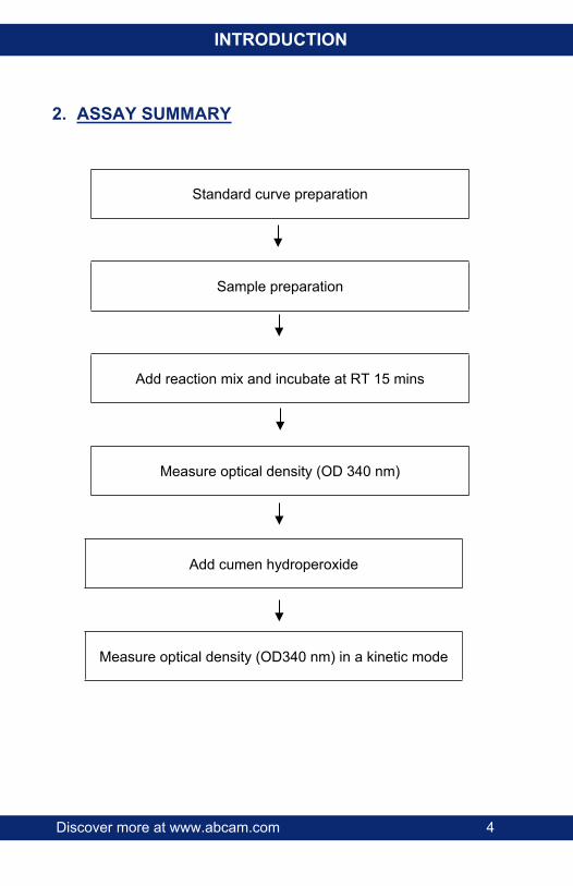
2. ASSAY SUMMARY
Standard curve preparation
Sample preparation
Add reaction mix and incubate at RT 15 mins
Measure optical density (OD 340 nm)
Add cumen hydroperoxide
Measure optical density (OD340 nm) in a kinetic mode
Discover more at www.abcam.com 5
GENERAL INFORMATION
3. PRECAUTIONSPlease read these instructions carefully prior to beginning the assay.All kit components have been formulated and quality control tested to function successfully as a kit. Modifications to the kit components or procedures may result in loss of performance.
4. STORAGE AND STABILITYStore kit at -20ºC in the dark immediately upon receipt. Kit has a storage time of 1 year from receipt, providing components have not been reconstituted.Refer to list of materials supplied for storage conditions of individual components. Observe the storage conditions for individual prepared components in section 5.Aliquot components in working volumes before storing at the recommended temperature. Reconstituted components are stable for 2 months.
Discover more at www.abcam.com 6
GENERAL INFORMATION
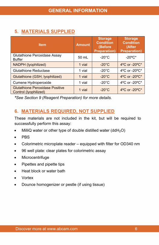
5. MATERIALS SUPPLIED
Item AmountStorage
Condition(Before
Preparation)
StorageCondition
(After Preparation)
Glutathione Peroxidase Assay Buffer 50 mL -20°C -20ºC*
NADPH (lyophilized) 1 vial -20°C 4ºC or -20ºC*Glutathione Reductase 1 vial -20°C 4ºC or -20ºC*Glutathione (GSH; lyophilized) 1 vial -20°C 4ºC or -20ºC*Cumene Hydroperoxide 1 vial -20°C 4ºC or -20ºC*Glutathione Peroxidase Positive Control (lyophilized) 1 vial -20°C 4ºC or -20ºC*
*See Section 9 (Reagent Preparation) for more details.
6. MATERIALS REQUIRED, NOT SUPPLIEDThese materials are not included in the kit, but will be required to successfully perform this assay:
MilliQ water or other type of double distilled water (ddH2O)
PBS
Colorimetric microplate reader – equipped with filter for OD340 nm
96 well plate: clear plates for colorimetric assay
Microcentrifuge
Pipettes and pipette tips
Heat block or water bath
Vortex
Dounce homogenizer or pestle (if using tissue)
Discover more at www.abcam.com 7
GENERAL INFORMATION
7. LIMITATIONS Assay kit intended for research use only. Not for use in diagnostic
procedures.
Do not use kit or components if it has exceeded the expiration date on the kit labels.
Do not mix or substitute reagents or materials from other kit lots or vendors. Kits are QC tested as a set of components and performance cannot be guaranteed if utilized separately or substituted.
Discover more at www.abcam.com 8
GENERAL INFORMATION
8. TECHNICAL HINTS This kit is sold based on number of tests. A ‘test’ simply
refers to a single assay well. The number of wells that contain sample, control or standard will vary by product. Review the protocol completely to confirm this kit meets your requirements. Please contact our Technical Support staff with any questions.
Keep enzymes, heat labile components and samples on ice during the assay.
Make sure all buffers and solutions are at room temperature before starting the experiment.
Samples generating values higher than the highest standard should be further diluted in the appropriate sample dilution buffers.
Avoid foaming or bubbles when mixing or reconstituting components.
Avoid cross contamination of samples or reagents by changing tips between sample, standard and reagent additions.
Ensure plates are properly sealed or covered during incubation steps.
Ensure complete removal of all solutions and buffers from tubes or plates during wash steps.
Make sure you have the right type of plate for your detection method of choice.
Make sure the heat block/water bath and microplate reader are switched on.
Discover more at www.abcam.com 9
ASSAY PREPARATION
9. REAGENT PREPARATION Briefly centrifuge small vials at low speed prior to opening.
9.1 Glutathione Peroxidase Assay Buffer:Ready to use as supplied. Warm to room temperature before use. Store at -20ºC.
9.2 NADPH Standard:Reconstitute with 500 µL of dH2O to get a 40 mM NADPH standard solution. Aliquot standard so that you have enough to perform the desired number of assays Store at -20°C for 1 month or at 4ºC for 1 week.
9.3 Glutahione Reductase:Dilute with 220 µL of Assay Buffer. Aliquot enzyme so that you have enough to perform the desired number of assays. Store at -20°C for 1 month or at 4ºC for 1 week. Keep on ice during use.
9.4 Glutathione (GSH):Reconstitute with 220 µL of Assay Buffer. Aliquot GSH so that you have enough to perform the desired number of assays. Store at -20°C for 1 month or at 4ºC for 1 week.
9.5 Cumene Hydroperoxide:Dilute with 1.25 mL of Assay Buffer. Aliquot so that you have enough to perform the desired number of assays. Store at -20°C for 1 month or at 4ºC for 1 week.
9.6 Glutathione Peroxidase (Positive Control):Reconstitute with 100 µL of Assay Buffer. Aliquot positive control so that you have enough to perform the desired number of assays Store at -20°C for 1 month or at 4ºC for 1 week. Keep on ice during use.
ASSAY PRE
Discover more at www.abcam.com 10
ASSAY PREPARATION
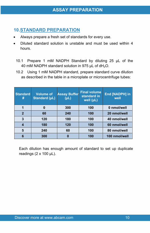
10.STANDARD PREPARATION Always prepare a fresh set of standards for every use.
Diluted standard solution is unstable and must be used within 4 hours.
10.1 Prepare 1 mM NADPH Standard by diluting 25 µL of the 40 mM NADPH standard solution in 975 µL of dH2O.
10.2 Using 1 mM NADPH standard, prepare standard curve dilution as described in the table in a microplate or microcentrifuge tubes:
Each dilution has enough amount of standard to set up duplicate readings (2 x 100 µL).
ASSAY PRE
Discover more at www.abcam.com 11
ASSAY PREPARATION
11.SAMPLE PREPARATIONGeneral Sample information:
We recommend performing several dilutions of your sample to ensure the readings are within the standard value range.
We recommend that you use fresh samples. If you cannot perform the assay at the same time, we suggest that you complete the Sample Preparation step before storing the samples. Alternatively, if that is not possible, we suggest that you snap freeze cells or tissue in liquid nitrogen upon extraction and store the samples immediately at -80°C. When you are ready to test your samples, thaw them on ice. Be aware however that this might affect the stability of your samples and the readings can be lower than expected.
11.1 Cell (adherent or suspension) samples:11.1.1 Harvest the amount of cells necessary for each assay (initial
recommendation = 2 x 10⁶ cells).11.1.2 Wash cells with cold PBS.11.1.3 Resuspend cells in 200 µL of cold Assay Buffer.11.1.4 Homogenize cells quickly by pipetting up and down a few
times, on ice.11.1.5 Centrifuge 15 minutes at 4°C at 10,000g using a cold
microcentrifuge to remove any insoluble material.11.1.6 Collect supernatant and transfer to a clean tube.11.1.7 Keep on ice.
11.2 Tissue samples:11.2.1 Harvest the amount of tissue necessary for each assay
(initial recommendation = 100 mg).11.2.2 Wash tissue in cold PBS.11.2.3 Resuspend tissue in 200 µL of cold Assay Buffer.
ASSAY PRE
Discover more at www.abcam.com 12
ASSAY PREPARATION
11.2.4 Homogenize tissue with a Dounce homogenizer sitting on ice, with 10 – 15 passes.
11.2.5 Centrifuge 15 minutes at 4°C at 10,000g using a cold microcentrifuge to remove any insoluble material.
11.2.6 Collect supernatant and transfer to a clean tube.11.2.7 Keep on ice.
Buffer.11.3.2 Centrifuge 15 minutes at 4°C at 10,000 x g using a cold
microcentrifuge to remove any insoluble material.11.3.3 Collect supernatant and transfer to a clean tube.11.3.4 Keep on ice.
11.4 Plasma and serum samples: Serum samples can be tested directly by adding sample to the microplate wells. Samples can be stored at -80ºC.
NOTE: We suggest using different volumes of sample to ensure readings are within the Standard Curve range.
Discover more at www.abcam.com 13
ASSAY PROCEDURE and DETECTION
12.ASSAY PROCEDURE and DETECTION● Equilibrate all materials and prepared reagents to room
temperature prior to use.● It is recommended to assay all standards, controls and
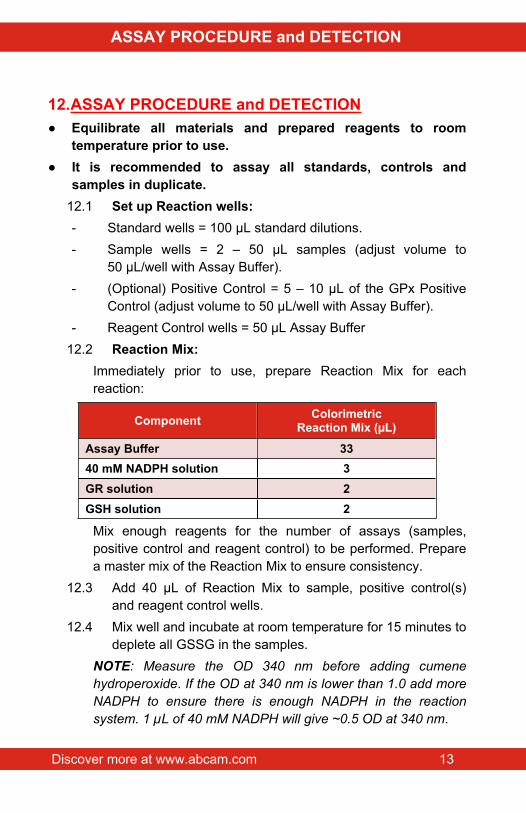
samples in duplicate.12.1 Set up Reaction wells:- Standard wells = 100 µL standard dilutions.- Sample wells = 2 – 50 µL samples (adjust volume to
50 µL/well with Assay Buffer).- (Optional) Positive Control = 5 – 10 µL of the GPx Positive
Control (adjust volume to 50 µL/well with Assay Buffer).- Reagent Control wells = 50 µL Assay Buffer
12.2 Reaction Mix:Immediately prior to use, prepare Reaction Mix for each reaction:
Mix enough reagents for the number of assays (samples, positive control and reagent control) to be performed. Prepare a master mix of the Reaction Mix to ensure consistency.
12.3 Add 40 µL of Reaction Mix to sample, positive control(s) and reagent control wells.
12.4 Mix well and incubate at room temperature for 15 minutes to deplete all GSSG in the samples.
NOTE: Measure the OD 340 nm before adding cumene hydroperoxide. If the OD at 340 nm is lower than 1.0 add more NADPH to ensure there is enough NADPH in the reaction system. 1 µL of 40 mM NADPH will give ~0.5 OD at 340 nm.
12.5 Add 10 µL cumene hydroperoxide solution, to the sample, positive control and reagent control wells only, to start the glutathione peroxidase (GPx) reaction. Mix well.
12.6 Measure output (A1) on a microplate reader at OD340 nm at T1.
12.7 Incubate at 25ºC for 5 min (or longer if the GPx activity is low). Protect from light.
12.8 Measure output (A2) on a microplate reader at OD340 nm at T2.
NOTE: If A1 reading is too low (<0.7), it means either too much GPx or too much GSSG is present in the sample. You may need to dilute the samples, or remove GSSG from your sample using methods, such as dialyzing the sample or using spin filters (ab93349) to remove GSSG.
NOTE: It is essential to read A1 and A2 in the reaction linear range. It will be more accurate if you read the reaction kinetics. Then choose A1 and A2 in the reaction linear range.
Discover more at www.abcam.com 15
DATA ANALYSIS
13.CALCULATIONS Samples producing signals greater than that of the highest
standard should be further diluted in appropriate buffer and reanalyzed, then multiplying the concentration found by the appropriate dilution factor.
For statistical reasons, we recommend each sample should be assayed with a minimum of two replicates (duplicates).
13.1 Average the duplicate reading for each standard and sample.
13.2 Subtract the mean absorbance value of the blank (Standard #1) from all standard and sample readings. This is the corrected absorbance.
13.3 Plot the corrected absorbance values for each standard as a function of the final concentration of NADPH.
13.4 Draw the best smooth curve through these points to construct the standard curve. Most plate reader software or Excel can plot these values and curve fit. Calculate the trendline equation based on your standard curve data (use the equation that provides the most accurate fit).
13.5 Extrapolate sample readings from the standard curve plotted using the following equation:
13.6 Apply the Δ_A340nm to the NAPDH standard curve to get NADPH amount B:
𝐵 = (Δ𝐴_340𝑛𝑚 ‒ 𝑖𝑛𝑡𝑒𝑟𝑐𝑒𝑝𝑡𝑆𝑙𝑜𝑝𝑒 )
13.7 Concentration of GPx in the test samples is calculated as (nmol/min/mL = mU/mL):
𝐺𝑃𝑥 𝐴𝑐𝑡𝑖𝑣𝑖𝑡𝑦 = ( 𝐵(𝑇2 ‒ 𝑇1) ∗ 𝑉) ∗ 𝐷
Discover more at www.abcam.com 16
DATA ANALYSIS
Where:B = NADPH amount that was decreased between T1 and T2
(in nmol). T1 = Time of the first reading (A1) (minutes). T2 = Time of second reading (A2) (minutes).V = Pretreated sample volume added into the reaction well (mL).D = Sample dilution factor.
Unit Definition: One unit is defined as the amount of enzyme that will cause the oxidation of 1.0 µmol of NADPH to NADP+ under the assay kit condition per minute at 25°C.
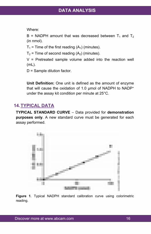
14.TYPICAL DATATYPICAL STANDARD CURVE – Data provided for demonstration purposes only. A new standard curve must be generated for each assay performed.
Figure 1. Typical NADPH standard calibration curve using colorimetric reading.
Discover more at www.abcam.com 17
DATA ANALYSIS
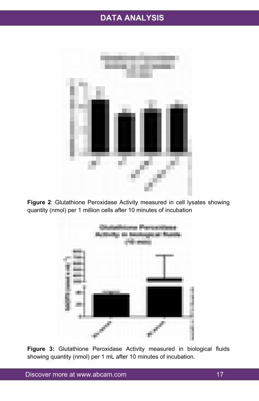
Figure 2: Glutathione Peroxidase Activity measured in cell lysates showing quantity (nmol) per 1 million cells after 10 minutes of incubation
Figure 3: Glutathione Peroxidase Activity measured in biological fluids showing quantity (nmol) per 1 mL after 10 minutes of incubation.
Discover more at www.abcam.com 18
RESOURCES
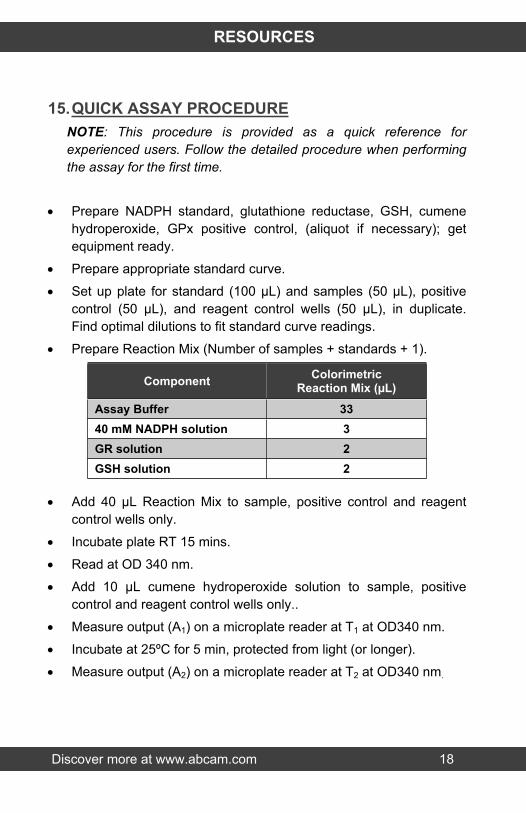
15.QUICK ASSAY PROCEDURENOTE: This procedure is provided as a quick reference for experienced users. Follow the detailed procedure when performing the assay for the first time.
Prepare NADPH standard, glutathione reductase, GSH, cumene hydroperoxide, GPx positive control, (aliquot if necessary); get equipment ready.
Prepare appropriate standard curve.
Set up plate for standard (100 µL) and samples (50 µL), positive control (50 µL), and reagent control wells (50 µL), in duplicate. Find optimal dilutions to fit standard curve readings.
Prepare Reaction Mix (Number of samples + standards + 1).
Add 40 µL Reaction Mix to sample, positive control and reagent control wells only.
Incubate plate RT 15 mins.
Read at OD 340 nm.
Add 10 µL cumene hydroperoxide solution to sample, positive control and reagent control wells only..
Measure output (A1) on a microplate reader at T1 at OD340 nm.
Incubate at 25ºC for 5 min, protected from light (or longer).
Measure output (A2) on a microplate reader at T2 at OD340 nm.
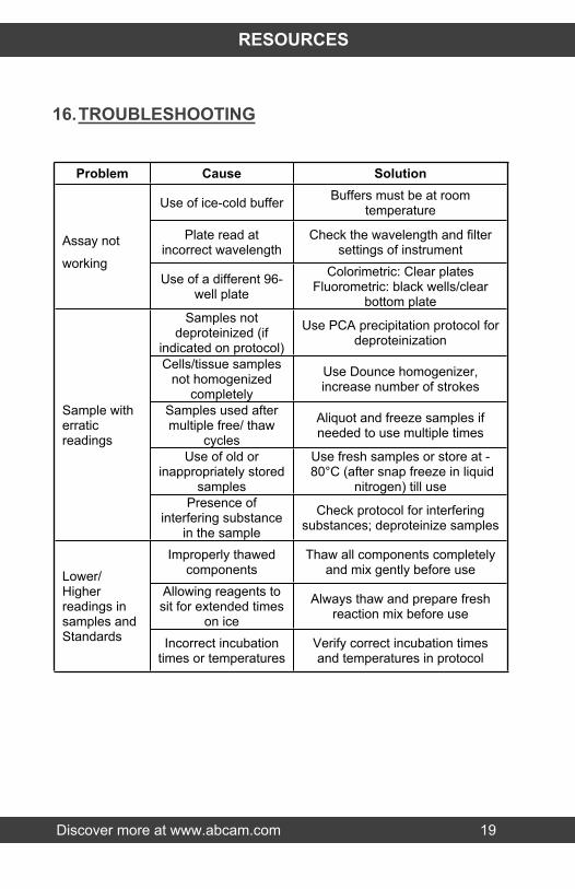
Use of ice-cold buffer Buffers must be at room temperature
Plate read at incorrect wavelength
Check the wavelength and filter settings of instrument
Assay not
workingUse of a different 96-
well plate
Colorimetric: Clear platesFluorometric: black wells/clear
bottom plateSamples not
deproteinized (if indicated on protocol)
Use PCA precipitation protocol for deproteinization
Cells/tissue samples not homogenized
completely
Use Dounce homogenizer, increase number of strokes
Samples used after multiple free/ thaw
cycles
Aliquot and freeze samples if needed to use multiple times
Use of old or inappropriately stored
samples
Use fresh samples or store at - 80°C (after snap freeze in liquid
nitrogen) till use
Sample with erratic readings
Presence of interfering substance
in the sample
Check protocol for interfering substances; deproteinize samples
Improperly thawed components
Thaw all components completely and mix gently before use
Allowing reagents to sit for extended times
on ice
Always thaw and prepare fresh reaction mix before use
Lower/ Higher readings in samples and Standards Incorrect incubation
times or temperaturesVerify correct incubation times and temperatures in protocol
Discover more at www.abcam.com 20
RESOURCES
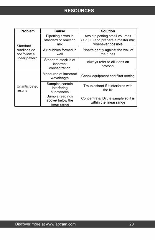
Problem Cause SolutionPipetting errors in
standard or reaction mix
Avoid pipetting small volumes (< 5 µL) and prepare a master mix
whenever possibleAir bubbles formed in
wellPipette gently against the wall of
the tubes
Standard readings do not follow a linear pattern Standard stock is at
incorrect concentration
Always refer to dilutions on protocol
Measured at incorrect wavelength Check equipment and filter setting
Samples contain interfering
substances
Troubleshoot if it interferes with the kit
Unanticipated results
Sample readings above/ below the
linear range
Concentrate/ Dilute sample so it is within the linear range
Discover more at www.abcam.com 21
RESOURCES
17.FAQWhat is the minimum detection of this kit?The assay has a detection sensitivity of 0.5 mU/mL of glutathione peroxidase in samples.
Can this kit be used with plasma and whole blood?The protocol contains instructions for erythrocytes. Whole blood can be processed similarly. Plasma can be diluted over a range and then the dilution that gives readings within the linear range of the standard curve can be used for the assay.
What is the activity level of the positive control? How can we increase its value to be comparable with our samples?The positive control is only a benchmark sample. As long as the values are within the range of the standard curve this is fine. The positive control is not be used to compare values with the samples. The positive control is provided to validate that the assay components are all working.
Discover more at www.abcam.com 22
RESOURCES
18.INTERFERENCESThese chemicals or biological materials will cause interferences in this assay causing compromised results or complete failure
SDS – will denature proteins and affect enzyme activity. RIPA buffer contains SDS.