129
| Date post: | 15-Jul-2015 |
| Category: |
Health & Medicine |
| Upload: | sherlau2013 |
| View: | 214 times |
| Download: | 2 times |

TransfusionFourth Edition
EDITED BY
Marcela Contreras, DBERoyal Free & University College Hospitals Medical School
London, UK
and
Blood Transfusion International
Contreras_FM.indd iContreras_FM.indd i 12/18/2008 1:33:34 Shobha12/18/2008 1:33:34 Shobha

This edition fi rst published 2009, © 2009 by Blackwell Publishing Ltd
Blackwell Publishing was acquired by John Wiley & Sons in February 2007. Blackwell's publishing program has been merged with Wiley's global Scientifi c, Technical and Medical business to form Wiley-Blackwell.
Registered offi ce: John Wiley & Sons Ltd, The Atrium, Southern Gate, Chichester, West Sussex, PO19 8SQ, UK
Editorial offi ces: 9600 Garsington Road, Oxford, OX4 2DQ, UKThe Atrium, Southern Gate, Chichester, West Sussex, PO19 8SQ, UK111 River Street, Hoboken, NJ 07030–5774, USA
For details of our global editorial offi ces, for customer services and for information about how to apply for permission to reuse the copyright material in this book please see our website at www.wiley.com/wiley-blackwell
The right of the author to be identifi ed as the author of this work has been asserted in accordance with the Copyright, Designs and Patents Act 1988.
All rights reserved. No part of this publication may be reproduced, stored in a retrieval system, or transmitted, in any form or by any means, electronic, mechanical, photocopying, recording or otherwise, except as permitted by the UK Copyright, Designs and Patents Act 1988, without the prior permission of the publisher.
Wiley also publishes its books in a variety of electronic formats. Some content that appears in print may not be available in electronic books.
Designations used by companies to distinguish their products are often claimed as trademarks. All brand names and product names used in this book are trade names, service marks, trademarks or registered trademarks of their respective owners. The publisher is not associated with any product or vendor mentioned in this book. This publication is designed to provide accurate and authoritative information in regard to the subject matter covered. It is sold on the understanding that the publisher is not engaged in rendering professional services. If professional advice or other expert assistance is required, the services of a competent professional should be sought.
The contents of this work are intended to further general scientifi c research, understanding, and discussion only and are not intended and should not be relied upon as recommending or promoting a specifi c method, diagnosis, or treatment by physicians for any particular patient. The publisher and the author make no representations or warranties with respect to the accuracy or completeness of the contents of this work and specifi cally disclaim all warranties, including without limitation any implied warranties of fi tness for a particular purpose. In view of ongoing research, equipment modifi cations, changes in governmental regulations, and the constant fl ow of information relating to the use of medicines, equipment, and devices, the reader is urged to review and evaluate the information provided in the package insert or instructions for each medicine, equipment, or device for, among other things, any changes in the instructions or indication of usage and for added warnings and precautions. Readers should consult with a specialist where appropriate. The fact that an organization or Website is referred to in this work as a citation and/or a potential source of further information does not mean that the author or the publisher endorses the information the organization or Website may provide or recommendations it may make. Further, readers should be aware that Internet Websites listed in this work may have changed or disappeared between when this work was written and when it is read. No warranty may be created or extended by any promotional statements for this work. Neither the publisher nor the author shall be liable for any damages arising herefrom.
Library of Congress Cataloging-in-Publication Data
ABC of transfusion / edited by Marcela Contreras. -- 4th ed. p. ; cm. -- (ABC series) Includes index. ISBN: 978-1-4051-5646-2 (alk. paper) 1. Blood--Transfusion. I. Contreras, Marcela. II. Series: ABC series (Malden, Mass.) [DNLM: 1. Blood Transfusion. WB 356 A134 2008] RM171.A23 2008 615.39--dc22 2007048168
A catalogue record for this book is available from the British Library.
Set in 9.25/12 pt Minion by Newgen Imaging Systems Pvt. Ltd, Chennai, IndiaPrinted in Singapore by Ho Printing Singapore Pte Ltd
1 2009
Contreras_FM.indd iiContreras_FM.indd ii 12/18/2008 1:33:35 Shobha12/18/2008 1:33:35 Shobha

Contents
Contributors, v
Introduction, vii
The Blood Donor: Demographics, Donor Selection and Tests on Donor Blood, 11 Liz Caffrey, Patricia Hewitt and John Barbara
Supply and Demand for Blood and Blood Components and Stock Management, 62 Judith Chapman, Peter Garwood and Sue Knowles
Compatibility Testing Before Transfusion; Blood Ordering and Administration, 103 Marcela Contreras and Aleksandar Mijovic
Red Cell Transfusion, 154 Mike Murphy and Jonathan Wallis
Platelet and Granulocyte Transfusions, 225 Modupe Elebute, Simon Stanworth and Cristina Navarrete
Haemolytic Disease of the Newborn and its Prevention, 276 Fiona Regan, Sailesh Kumar and Marcela Contreras
Fetal and Neonatal Transfusion, 337 Helen V. New and Sailesh Kumar
Plasma Products and Indications for Their Use, 408 Hannah Cohen and Trevor Baglin
Human Albumin Solutions and the Controversy of Crystalloids Versus Colloids , 489 Neil Soni
Treatment of Massive Haemorrhage in Surgery and Trauma, 5410 Tim Walsh
Immunological Complications of Blood Transfusion, 6111 Marcela Contreras and Cristina Navarrete
Infectious Complications of Blood Transfusion: Bacteria and Parasites, 6912 John Barbara and Marcela Contreras
Infectious Complications of Blood Transfusion: Viruses, 7413 John Barbara and Marcela Contreras
Variant Creutzfeldt–Jakob Disease and its Impact on the UK Blood Supply, 7914 Patricia Hewitt, James Ironside and Marcela Contreras
Risks of Transfusion in the Context of Haemovigilance: SHOT – the UK Haemovigilance System, 8315 Dorothy Stainsby, Hannah Cohen and Brian McClelland
Alternatives to Allogeneic Blood Transfusion, 8916 Dafydd Thomas and Beverley Hunt
iii
Contreras_FM.indd Sec1:iiiContreras_FM.indd Sec1:iii 12/18/2008 1:33:35 Shobha12/18/2008 1:33:35 Shobha

iv Contents
Blood Substitutes and Oxygen Therapeutics, 9517 Kenneth C. Lowe
Appropriate Use of Blood and Better Blood Transfusion, 9918 Mike Murphy
Stem Cell Transplantation and Cellular Therapies, 10419 Aleksandar Mijovic, Derwood Pamphilon and Suzanne Watt
Blood Transfusion in a Regulatory Environment and the EU Directives, 11020 Shubha Allard, Clare Taylor and Angela Robinson
Index, 117
Contreras_FM.indd Sec1:ivContreras_FM.indd Sec1:iv 12/18/2008 1:33:35 Shobha12/18/2008 1:33:35 Shobha

Shubha AllardConsultant HaematologistRoyal London HospitalNational Blood ServiceLondon, UK
Trevor BaglinConsultant HaematologistAddenbrooke’s NHS TrustCambridge, UK
John BarbaraEmeritus Microbiology Consultant to the NHSBTColindale Centre, LondonandVisiting Professor in Transfusion Microbiology at the University of West of EnglandBristol, UK
Liz CaffreyClinical Director – DonorsNational Blood ServiceCambridge, UK
Judith Chapman ManagerBlood Stocks Management SchemeLondon, UK
Hannah Cohen Consultant HaematologistUniversity College London Hospitals NHS Foundation TrustLondon, UK
Marcela ContrerasChairman of Blood Transfusion InternationalProfessor of Transfusion MedicineRoyal Free and University College Hospitals Medical Schooland retiredNational Director of DiagnosticsDevelopment and ResearchNational Blood ServiceLondon, UK
Modupe Elebute Consultant HaematologistSt George’s HospitalLondon, UK
Peter GarwoodManaging DirectorNational Blood ServiceBrentwood, UK
Patricia HewittConsultant Specialist in Transfusion MicrobiologyNational Blood ServiceElstree Gate, UK
Beverley HuntProfessor of Thrombosis and HaemostasisKing’s CollegeLondon, UKandDepartments of Haematology, Pathology and RheumatologyGuy’s and St Thomas’ Foundation TrustLondon, UK
James IronsideProfessor of Clinical NeuropathologyWestern General HospitalEdinburgh, UK
Sue KnowlesConsultant HaematologistEpsom and St Helier University Hospitals NHS TrustCarshalton, UK
Sailesh KumarConsultant Obstetrician and GynaecologistQueen Charlotte’s and Chelsea HospitalLondon, UK
Kenneth C. LoweAssociate Professor and Reader in BiotechnologySchool of Biology SciencesUniversity of NottinghamNottingham, UK
Brian McClellandScottish National Blood Transfusion ServiceEdinburgh, UK
Aleksandar MijovicConsultant in Transfusion MedicineKing’s College HospitalLondon, UK
v
Contributors
Contreras_FM.indd Sec2:vContreras_FM.indd Sec2:v 12/18/2008 1:33:35 Shobha12/18/2008 1:33:35 Shobha

vi Contributors
Mike Murphy Professor of Blood Transfusion MedicineConsultant HaematologistNational Blood ServiceJohn Radcliffe HospitalOxford, UK
Cristina NavarreteHead of Histocompatibility and ImmunogeneticsNational Blood ServiceLondon, UK
Helen V. NewConsultant in Paediatric Haematology and Transfusion MedicineSt Mary’s HospitalLondon, UK
Derwood Pamphilon Consultant Haematologist and Honorary Clinical ReaderNational Blood ServiceBristol, UK
Fiona ReganConsultant Haematologist/Transfusion Medicine SpecialistHammersmith Hospital and National Blood ServicesLondon, UK
Angela RobinsonRetired Medical DirectorNHSBTWatford, UK
Neil SoniConsultant in Intensive CareChelsea and Westminster HospitalLondon, UK
Dorothy StainsbyConsultant in Transfusion Medicine and SHOT National Medical CoordinatorNational Blood ServiceNewcastle, UK
Simon StanworthConsultant HaematologistNational Blood ServiceJohn Radcliffe HospitalOxford, UK
Clare TaylorConsultant HaematologistMedical Director, SHOTLondon, UK
Dafydd Thomas Consultant in Anaesthesia and Intensive TherapyMorriston HospitalSwansea, UK
Tim WalshConsultant Anaesthetist and Honorary ProfessorRoyal Infi rmary of EdinburghEdinburgh, UK
Jonathan WallisConsultant HaematologistFreeman HospitalNewcastle, UK
Suzanne WattHead of Stem Cells and ImmunotherapyNational Blood ServiceOxford, UK
Contreras_FM.indd Sec2:viContreras_FM.indd Sec2:vi 12/18/2008 1:33:35 Shobha12/18/2008 1:33:35 Shobha

Introduction
vii
In this era of super-specialization, it is diffi cult to fi nd experts writing clearly about the basics of their specialties, making their subjects accessible to other doctors and healthcare workers. I feel that my collaborators have covered the fundamentals of transfusion medicine in an admirable, easy-to-read, comprehensible way.
This fourth edition of ABC of Transfusion has been expanded, with changes to previous chapters and four additional chapters to cover topics which are now established in transfusion medicine, such as haemovigilance, variant CJD, blood stocks management, appropriate use of blood and alternatives to allogeneic trans-fusion, as well as the increasing involvement of the regulatory environment. The wider breadth of the subject shows that this is not an area devoted exclusively to haematologists, but to all those colleagues collecting, processing and screening blood, prescrib-ing blood components, preparing compatible safe blood for transfusion and administering it.
During my visits abroad in the last ten years, it has been reward-ing to learn about the many colleagues worldwide who have
encountered transfusion medicine for the fi rst time when read-ing previous editions of ABC of Transfusion. Some of them have become leaders in the fi eld as medical doctors, scientists, medical technologists, nurses, managers and marketers.
Blood transfusion continues to be life-saving in special situa-tions, such as massive surgical haemorrhage, post-partum haemor-rhage and severe malarial anaemia in young children. In addition, the safety of the blood supply and transfusion medicine have pro-gressed considerably in the last few years. However, as the message from this book shows, we should only transfuse when the ben-efi ts outweigh the risks, yet we are still lacking evidence that blood transfusion works, or that it is the best therapy in a number of the clinical situations in which it is used.
I am grateful to the many colleagues who have contributed to this updated edition of ABC of Transfusion; they have patiently awaited its long gestation. I have no doubt that they, as well as the readers, will be satisfi ed with the outcome.
Professor Dame Marcela Contreras
Contreras_FM.indd Sec2:viiContreras_FM.indd Sec2:vii 12/18/2008 1:33:35 Shobha12/18/2008 1:33:35 Shobha

Contreras_FM.indd Sec2:viiiContreras_FM.indd Sec2:viii 12/18/2008 1:33:35 Shobha12/18/2008 1:33:35 Shobha

1
CHAPTER 1
The Blood Donor: Demographics, Donor Selection and Tests on Donor Blood
Liz Caffrey, Patricia Hewitt and John Barbara
ABC of Transfusion, 4th edition, 2009. Edited by Marcela Contreras. © 2009
Blackwell Publishing, ISBN: 978-1-4051-5646-2.
Demographics
In the UK all cellular and fresh frozen blood components are sourced from donations made by voluntary unpaid blood donors. A suffi cient supply of components for transfusion to patients is therefore reliant upon these altruistic donors continu-ing to donate. Between 4% and 6% of the eligible adult popula-tion donate blood and, in 2005, 1.2 million English donors gave 2.1 million donations. The age range for regular whole blood donation is from 17 to 70 years. New donors are accepted up to their 66th birthday (Figure 1.1).
Donors come from all walks of life but are more commonly from social groups with stable, established lifestyles. Family tradition, peer pressure and personal or professional experience of transfu-sion are strong motivators.
In recent years it has become more diffi cult to maintain donor attendance at adequate levels to meet hospital demand. Donor numbers are falling despite heavy investment in recruitment and marketing activity. There are many reasons for this, but the pace of modern living and loss of community spirit are major factors.
Others include lack of time, inadequate opportunities to donate, inconvenient venues and/or opening times, fear of needles and simple apathy. Lack of general awareness of the constant need for blood to support routine medical and surgical treatments is another factor. Volunteers fl ock to donate at times of ‘emergency’ but tend not to continue once the perceived need is over.
Donor selection
The possibility that donations might present a risk from transfusion transmissible infections or other conditions is minimized through two essential, complementary steps:
Robust donor selection procedures to prevent unsuitable 1 donations from being collected.Routine testing of all donations for markers of infection.2
Decisions about donor acceptability and screening tests must take into account the characteristics of the donor population and the prevalence of infections transmissible by blood, the susceptibility of the recipient population, and any emerging risks. Two recent examples of the latter are variant Creutzfeldt–Jakob disease (vCJD) and West Nile virus.
Donor selection has two purposes: to protect the donor from harm and the recipient from any ill effects of transfusion. Potential donors should be provided with suffi cient information to allow
OVERVIEW
A safe and suffi cient blood supply depends upon the • recruitment and retention of volunteers who have a low risk of infection with blood-borne viruses and have the committment to make regular blood donations.
Most blood services world-wide are faced with a challenge in • maintaining adequate numbers of safe donors.
Donor selection is designed to select donors who present a low • risk of blood-borne infections and to detect any condition which might make donation hazardous to the volunteer.
Modern donation screening tests assure a high degree of safety • for blood transfusion recipients, but cannot detect all infected donors.
Increasingly stringent donor selection and donation testing lead • to a loss of donors and donations. Figure 1.1 Age profi le of English donors, January 2005.
017–20
0.062
0.195
0.247
0.135
0.112
0.254
0.128 0.126
0.192
0.096 0.096
0.051
0.0230.028
0.12
0.075
0.0370.035
21–30 31–40
Age range (years)
% o
f to
tal d
atab
ase
41–50 51–60 61–70
0.05
0.1
0.15
0.2
0.25
0.3
Total
Female
Male
Contreras_C001.indd 1Contreras_C001.indd 1 12/19/2008 12:25:14 PM12/19/2008 12:25:14 PM

2 ABC of Transfusion
them to exclude themselves; they are required to read essential material before each donation (Figure 1.2).
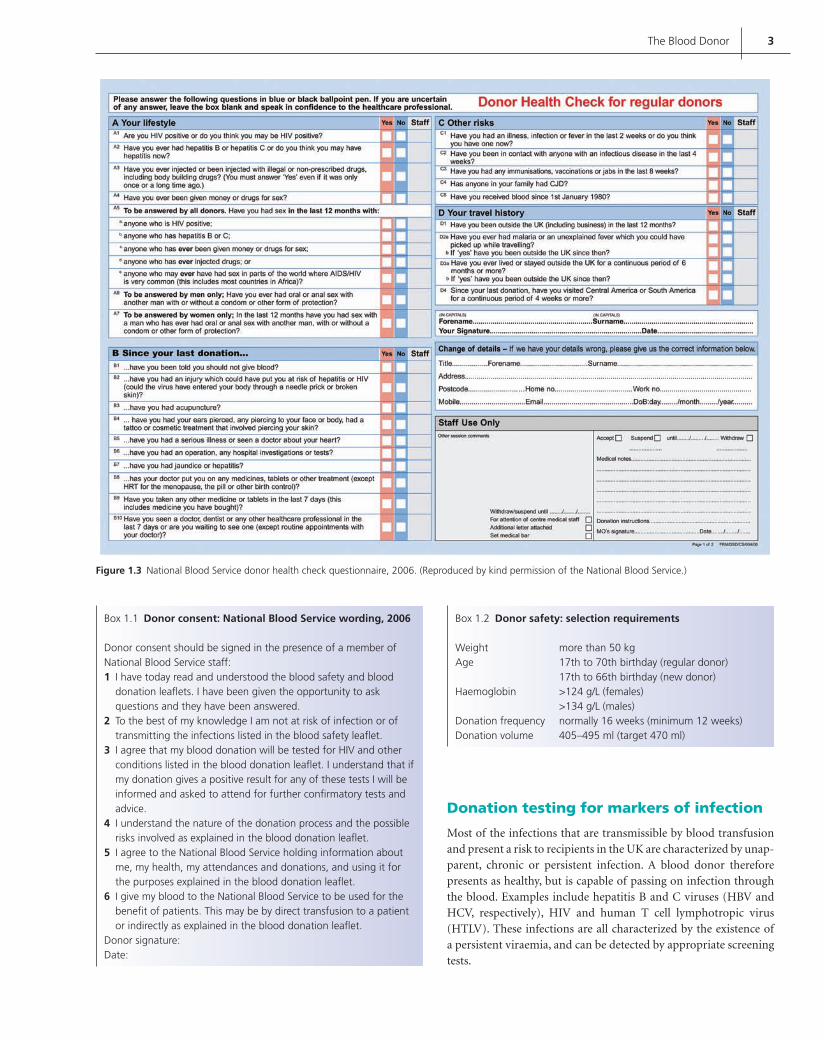
It is not practical to carry out a full medical examination on every volunteer. Therefore reliance is placed on simple visual assess-ment and answers to questions about general health, medical his-tory and medication. These are administered using a questionnaire (Figure 1.3) and face-to-face structured interview with a trained member of staff. Confi dentiality throughout this process is key to encouraging donors to provide truthful answers. All donors must give informed consent to donation and are required to sign to confi rm this before every donation (Box 1.1).
Donor selection criteriaThese have been developed and agreed throughout the UK for over 15 years. In November 2005, many selection criteria (particularly with respect to recipient safety) became legal requirements when the EU Blood Directive (2004/33/EC) was incorporated into UK statute (The Blood Safety and Quality Regulations 2005).
Donor safetyDonors must be in good health, within the permitted age range, and meet the minimum requirements for weight, donation volume, haemoglobin and donation frequency (Box 1.2).
Figure 1.2 National Blood Service blood safety leafl et. (Reproduced by kind permission of the National Blood Service.)
The weight and donation volume limits protect the donor from giving more than 13% of their circulating blood volume, to minimize the risk of vasovagal reactions. The minimum haemo-globin levels ensure that: (i) the recipient receives an adequate amount of haemoglobin (minimum 40 g per unit transfused); and (ii) the donor is not rendered anaemic. Before each dona-tion the haemoglobin level is assessed, usually by a simple, semiquantitative, gravimetric method using a drop of capillary blood introduced into a solution of copper sulphate of known specifi c gravity. This may be supplemented or replaced by the use of portable haemoglobinometers.
Where the potential donor’s medical history or medication indi-cate that the donor is not in good health or that their own health may be adversely affected as a result of donating, they are deferred either permanently (e.g. in cardiovascular disease) or temporarily (e.g. in pregnancy, anaemia or unexplained symptoms awaiting diagnosis).
Medications are rarely a cause per se to prevent donation but may indicate underlying pathology that requires the donor to be deferred.
Adverse effects of donationMost donors suffer no ill effects. The most commonly reported prob-lem is bruising and/or a painful arm. The overwhelming majority of these donors require only reassurance and simple fi rst aid, unless complicated by infection or nerve injury. Approximately one in 75 donors feels faint during or shortly after donation and 15% of these suffer syncope (rarely serious unless associated with physical injury or slow recovery). These vasovagal symptoms are more com-mon in younger, fi rst time and female donors. Some donors report fatigue in the days following donation. Iron depletion may also occur and blood donation should be considered in the differential diagnosis of unexplained iron defi ciency in regular donors.
Recipient safetyThe most important consideration in the selection of donors is to avoid the transmission of infectious agents. The voluntary, unpaid status of UK donors contributes to patient safety as there is no fi nancial incentive to conceal relevant details of medical or personal history. In addition, the fact that most UK blood donors are regular donors is an added safety factor.
Donors whose activities are known to be associated with an increased risk of acquiring infections are deferred temporar-ily for a period that exceeds the incubation period of the infec-tion or, if there is a screening test which is routinely performed, that exceeds the window period for detection by routine screen-ing tests. Deferral is permanent if the activities are ongoing or the infection is chronic, i.e. the volunteer is a carrier of a blood-borne agent. It is very important to exclude individuals whose behav-iours are associated with a high risk of acquiring human immuno-defi ciency virus (HIV), hepatitis B or hepatitis C, and all donors are asked about these sensitive, personal issues each time they donate (Figure 1.4).
In addition, selection criteria take account of other known infectious risks as well as the small (theoretical) risk that may be posed by diseases of unknown aetiology (Box 1.3).
Contreras_C001.indd 2Contreras_C001.indd 2 12/19/2008 12:25:16 PM12/19/2008 12:25:16 PM
The Blood Donor 3
Figure 1.3 National Blood Service donor health check questionnaire, 2006. (Reproduced by kind permission of the National Blood Service.)
Box 1.1 Donor consent: National Blood Service wording, 2006
Donor consent should be signed in the presence of a member of National Blood Service staff:
I have today read and understood the blood safety and blood 1 donation leafl ets. I have been given the opportunity to ask questions and they have been answered.To the best of my knowledge I am not at risk of infection or of 2 transmitting the infections listed in the blood safety leafl et.I agree that my blood donation will be tested for HIV and other 3 conditions listed in the blood donation leafl et. I understand that if my donation gives a positive result for any of these tests I will be informed and asked to attend for further confi rmatory tests and advice.I understand the nature of the donation process and the possible 4 risks involved as explained in the blood donation leafl et.I agree to the National Blood Service holding information about 5 me, my health, my attendances and donations, and using it for the purposes explained in the blood donation leafl et.I give my blood to the National Blood Service to be used for the 6 benefi t of patients. This may be by direct transfusion to a patient or indirectly as explained in the blood donation leafl et.
Donor signature:Date:
Box 1.2 Donor safety: selection requirements
Weight more than 50 kgAge 17th to 70th birthday (regular donor) 17th to 66th birthday (new donor)Haemoglobin >124 g/L (females) >134 g/L (males)Donation frequency normally 16 weeks (minimum 12 weeks)Donation volume 405–495 ml (target 470 ml)
Donation testing for markers of infection
Most of the infections that are transmissible by blood transfusion and present a risk to recipients in the UK are characterized by unap-parent, chronic or persistent infection. A blood donor therefore presents as healthy, but is capable of passing on infection through the blood. Examples include hepatitis B and C viruses (HBV and HCV, respectively), HIV and human T cell lymphotropic virus (HTLV). These infections are all characterized by the existence of a persistent viraemia, and can be detected by appropriate screening tests.
Contreras_C001.indd 3Contreras_C001.indd 3 12/19/2008 12:25:19 PM12/19/2008 12:25:19 PM

4 ABC of Transfusion
Currently, UK blood donations are screened for the presence of:hepatitis B surface antigen (HBsAg)• HIV infection, through the use of combined antibody/antigen • detection tests with supplementary genomic testing on pools of samples for HIV RNA in some areas
HCV infection, through the use of tests to detect antibody • supplemented by genomic testing for HCV RNA on pools of samplesHTLV, through testing for antibody on pools of samples• treponemal infection, through specifi c antibody detection assays.•
All these tests are mandatory, and must be performed on every donation using nationally validated assays, with national ‘working standard’ samples and full process control.
Additional tests may be indicated for certain donors in particu-lar circumstances. The necessity for these tests is usually decided after considering the epidemiology of the relevant infection and the risk presented from the local blood donor population. For instance, testing for antibodies to hepatitis B core (anti-HBc) is performed on donations in many developed countries, but it is not a rou-tine screening test in the UK. It is used, however, for donors who have a higher risk of recent exposure to HBV infection through, for instance, skin piercing. It is also indicated for donors with a his-tory of past HBV infection. A further example of such additional testing would be for evidence of malaria antibodies, as a marker of past exposure and possible continued infection. The decision whether to test depends upon a careful assessment of the poten-tial donor’s travel and residence history. A combination of his-tory taking, postponement of donation until some months after the last possible exposure, and a negative malarial antibody test should ensure that malaria is not transmitted by blood transfusion. A second parasitic infection, Chagas’ disease, is treated similarly.
There are other infections that may present a special risk to only a subset of transfusion recipients. An example is cytomegalovi-rus (CMV) infection, which is a particular hazard for immuno-suppressed recipients. Despite routine leucodepletion of all UK blood components, which would be expected to substantially reduce the risk of transmission of cell-associated agents such as CMV, screening of selected blood donations continues to be per-formed to provide a supply of CMV ‘safe’ blood components for susceptible recipients. In areas of the world where CMV seropreva-lence is very high, such a step would be impractical.
Despite careful blood donor selection and donation screening tests, infection may still be transmitted. Rarely, microbial agents that are not associated with persistent infection, and not therefore included in routine screening tests, can be transmitted by blood transfusion. This is usually because a donor gives blood during the incubation period, and examples have been reported for both hepatitis A and hepatitis E. Transmission of bacterial infection (unapparent donor bacteraemia) has also been reported on rare occasions but most bacterial transmissions are due to (exogenous) skin contaminants. Donation during the incubation period of an infection, i.e. during the ‘window period’ of infectivity, before reac-tive screening tests were developed, has also accounted for very small numbers of transmissions of those infections for which blood is now routinely screened, e.g. HIV.
Finally, there are infections for which there are no suitable screening tests; for the UK, vCJD is the most signifi cant example. As virtually the whole of the UK population has been at risk of vCJD infection through diet in the past, the development of suit-able blood tests and/or prion removal fi lters is proceeding (see Chapter 14). Thus, although blood transfusions in the UK are
Figure 1.4 UK high risk exclusions as detailed on the National Blood Safety Service blood safety leafl et. (Reproduced by kind permission of the National Blood Service.)
Box 1.3 Recipient safety: other exclusions
PermanentChronic infections, e.g. Chagas’ disease, brucellosis• History of malignant disease• Ulcerative colitis• Blood transfusion in UK since 1980 (vCJD risk)• Recipients of human pituitary hormones (CJD risk)• Recipients of corneal, scleral or dura mater grafts (vCJD risk)•
TemporarySkin piercing• Travel to malaria endemic countries• Surgery• Flexible endoscopy• Acute infectious disease• Immunization with live vaccines• Dentistry•
Contreras_C001.indd 4Contreras_C001.indd 4 12/19/2008 12:25:25 PM12/19/2008 12:25:25 PM
The Blood Donor 5
exceedingly safe, there still remains a very small risk of transmis-sion of infection, and this fact reinforces the need for testing to be combined with careful donor selection.
Serological testing
Serological tests are carried out on all donations to ascertain the blood group (A, B, AB or O) and for RhD typing; the results are checked against those previously obtained from that donor or by repeat typing with different batches of antibodies and test cells. Most UK centres also test for RhC, c, e, E and K antigens, and this information appears on the blood pack label. Blood units found negative for D antigen are labelled ‘RhD negative’. With the mono-clonal typing antibodies in current use, most weak and variant forms of D antigen are detected on direct testing. Those below the limit of detection with monoclonal anti-D are labelled as RhD negative since they are not considered to be immunogenic to a D-negative recipient. Extended testing to detect, for example, weak D or Du in donors is not universally carried out. A proportion of the units is also typed for Cw, Fya, Fyb, M, S, s, Jka and Jkb, thus making the phe-notyped red cell stocks readily available for alloimmunized patients in need of transfusion.
All donations are screened for clinically important red cell anti-bodies. Any donation found to have a high antibody titre should not be used for transfusion, although it may be a valuable source of red cell typing reagent. Low titres of antibodies should not auto-matically exclude a donation from therapeutic use as the antibody would be further diluted on direct transfusion. As well as this, about 90% of the plasma (and hence antibodies therein) from most donations is removed and the cells are resuspended in an additive solution such as saline adenine glucose mannitol (SAG-M); most of the remaining red cells just have most of the plasma removed (see Chapter 4). The comparatively unrefi ned antibody screening, pos-sible on automated blood grouping machines, is therefore accept-able in the testing of blood donations, although it is not acceptable
in the screening for antibodies of samples from potential recipients. An exception to this is the selection of blood for ‘massive’ transfu-sion of a neonate, when donor blood should be screened for anti-bodies using sensitive techniques.
Testing of group O blood for high titre haemolytic anti-A, anti-B and anti-AB is still carried out in some centres in the UK, so that plasma-rich components, such as platelet preparations, can be appropriately labelled. This practice should not be allowed to override the principle that a patient should receive blood of his/her own group and that group O donor blood (especially plasma-rich components) should not be given to patients of other groups except in an emergency.
In England, typing for human leucocyte antigen (HLA) or histo-compatibility antigens is carried out on regular plateletpheresis donors, to satisfy the demand for HLA-matched platelets. Such platelets are used in the treatment of a severely thrombocytopenic patient who, because of many exposures to blood components, has developed multispecifi c antibodies to HLA antigens and has become refractory to random platelet transfusions. Normally, HLA-compatible donors would provide one or two adult doses of platelets by means of plateletpheresis. Typing for human platelet antigens HPA-1a and HPA-5b is also performed on regular platelet-pheresis donors to supply compatible platelets for the transfusion of fetuses and infants affected by neonatal alloimmune thrombo-cytopenia. Occasionally, HPA-typed platelets are required for the transfusion of immunologically refractory patients with anti-HPA.
Further reading
Barbara JAJ, Regan F, Contreras M. Transfusion Microbiology. Cambridge
University Press, 2008.
Klein HG, Anstee DJ, Blood Transfusion in Clinical Medicine, 11th edn.
Blackwell Publishing Ltd, 2005.
Murphy MF, Pamphilon DH (Eds). Practical Transfusion Medicine. Blackwell
Publishing Ltd, 2001.
Contreras_C001.indd 5Contreras_C001.indd 5 12/19/2008 12:25:27 PM12/19/2008 12:25:27 PM

6
CHAPTER 2
Supply and Demand for Blood and Blood Components and Stock Management
Judith Chapman, Peter Garwood and Sue Knowles
ABC of Transfusion, 4th edition, 2009. Edited by Marcela Contreras. © 2009
Blackwell Publishing, ISBN: 978-1-4051-5646-2.
Ability to meet the demand for blood and blood components is a primary goal of blood services, and is achievable through the good will of voluntary donors, effective inventory management and the appropriate use of blood and its alternatives by clinicians. The blood supply chain (Figure 2.1) includes the voluntary blood donor, the blood services, the hospital laboratory, the prescribing clinician and the recipient of blood. It is the responsibility of the blood services to minimize production loss and wastage and to employ good inventory management practice in conjunction with the hospital laboratories, whilst clinicians are responsible for pre-scribing blood only when there are no alternative approaches and the benefi ts exceed the risks. Blood is a freely given resource and a collaborative approach along the chain is required to ensure that it is available and used for the maximum benefi t to patient care.
The blood supply
Volunteer donors are the source of the blood supply chain; how-ever, the donor base continues to fall despite recruitment efforts. Research in England and North Wales indicates that the active donor base is shrinking. Although the risk from transfusion-transmitted infections has never been lower, the demand for safety through additional screening tests has never been greater. All addi-tional testing for pathogens has the potential to lead to false posi-tive reactions, and further donor deferrals and disqualifi cations.
The appreciation that variant Creutzfeldt–Jakob disease (vCJD) is likely to be transmitted through the blood supply has led to the exclusion of donors who have themselves been transfused and who are therefore particularly motivated to donate. Increased foreign travel and its associated risks of disease (e.g. malaria and West Nile virus) have also had an impact on the blood supply.
Whole blood donations (450 ml 10% of blood) are pro-cessed and converted into concentrated red cells and, according to requirements, platelet concentrates, fresh frozen plasma and cryoprecipitate.
In many developed countries, blood undergoes universal leuco-depletion (Figure 2.2) for a number of reasons: to reduce the risk
OVERVIEW
This chapter covers the different elements of the blood supply • chain.
Blood supply and component preparation are examined.•
Infl uences on the demand for blood and blood components are • discussed.
Factors affecting blood inventory management practice are • outlined.
Finally, future infl uences on the blood supply chain are looked at.•
Donor
Hospital
Blood serviceRecipient
Figure 2.1 The blood supply chain.
Figure 2.2 The leucocyte depletion process showing red cells undergoing fi ltration for leucocyte depletion.
Contreras_C002.indd 6Contreras_C002.indd 6 12/19/2008 12:34:30 PM12/19/2008 12:34:30 PM
Supply and Demand for Blood and its Components 7
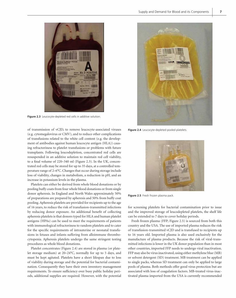
of transmission of vCJD, to remove leucocyte-associated viruses (e.g. cytomegalovirus or CMV), and to reduce other complications of transfusions related to the white cell content (e.g. the develop-ment of antibodies against human leucocyte antigen (HLA)) caus-ing refractoriness to platelet transfusions or problems with future transplants. Following leucodepletion, concentrated red cells are resuspended in an additive solution to maintain red cell viability, to a fi nal volume of 220–340 ml (Figure 2.3). In the UK, concen-trated red cells may be stored for up to 35 days, at a controlled tem-perature range of 2–6°C. Changes that occur during storage include loss of viability, changes in metabolism, a reduction in pH, and an increase in potassium levels in the plasma.
Platelets can either be derived from whole blood donations or by pooling buffy coats from four whole blood donations or from single donor apheresis. In England and North Wales approximately 50% of preparations are prepared by apheresis and 50% from buffy coat pooling. Apheresis platelets are provided for recipients up to the age of 16 years, to reduce the risk of transfusion-transmitted infections by reducing donor exposure. An additional benefi t of collecting apheresis platelets is that donors typed for HLA and human platelet antigens (HPAs) can be used to meet the requirements of patients with immunological refractoriness to random platelets and to cater for the specifi c requirements of intrauterine or neonatal transfu-sions in fetuses and infants suffering from alloimmune thrombo-cytopenia. Apheresis platelets undergo the same stringent testing procedures as whole blood donations.
Platelet concentrates (Figure 2.4) are stored in plasma (or plate-let storage medium) at 20–24°C, normally for up to 5 days, and must be kept agitated. Platelets have a short lifespan due to loss of viability during storage and the potential for bacterial contami-nation. Consequently they have their own inventory management requirements. To ensure suffi ciency over busy public holiday peri-ods, additional supplies are required. However, with the potential
for screening platelets for bacterial contamination prior to issue and the improved storage of leucodepleted platelets, the shelf life can be extended to 7 days to cover holiday periods.
Fresh frozen plasma (FFP; Figure 2.5) is sourced from both this country and the USA. The use of imported plasma reduces the risk of transfusion-transmitted vCJD and is transfused to recipients up to 16 years old. Imported plasma is also used exclusively for the manufacture of plasma products. Because the risk of viral-trans-mitted infections is lower in the UK donor population than in most other countries, imported FFP needs to undergo viral inactivation. FFP may also be virus inactivated, using either methylene blue (MB) or solvent detergent (SD) treatment. MB treatment can be applied to single packs, whereas SD treatment can only be applied to large pools of plasma. Both methods offer good virus protection but are associated with loss of coagulation factors. MB-treated virus-inac-tivated plasma imported from the USA is currently recommended
Figure 2.3 Leucocyte-depleted red cells in additive solution.
Figure 2.4 Leucocyte-depleted pooled platelets.
Figure 2.5 Fresh frozen plasma pack.
Contreras_C002.indd 7Contreras_C002.indd 7 12/19/2008 12:34:34 PM12/19/2008 12:34:34 PM

8 ABC of Transfusion
Figure 2.6 Demand for red cells in England and North Wales, 2000–2007.
2.22 2.19 2.17 2.14 2.02 1.93 1.871.60
1.70
1.80
1.90
2.00
2.10
2.20
2.30
2000\01 2000\02 2000\03 2000\04Date
2000\05 2000\06 2000\07
Mill
ion
red
cel
l un
its
for recipients up to the age of 16 years, while SD-treated plasma is reserved for patients with thrombotic thrombocytopenic purpura undergoing daily large volume plasma exchange.
Demand for blood components
Demand is diffi cult to forecast for many reasons, since it follows the net effect of changes in population demographics and the degree of uptake of blood conservation strategies. Tools such as environmen-tal scanning, mathematical modelling and trending can be used to try to forecast demand more accurately. The red cell demand fore-cast is used to calculate and set blood collection targets, which then form the basis for collection session planning.
There has been a decline in red cell demand in England over recent years and, despite an estimated blood donation rate of about 39 units per 1000 of the eligible donor population (the range for high Human Development Index (HDI) countries is 10.4–74.0), England and North Wales are self-suffi cient in red cells. All requests for red cells from hospitals have been fully met by the National Blood Service over the last 6 years. In England and North Wales the demand for red cells has fallen year on year since 2000. In 2000/01 red cell demand was 2.22 million, but in 2006/07 had fallen to 1.87 million, a fall of 15.8% in 6 years (Figure 2.6). Lower demand may be attributable to a number of reasons including the publication of the Health Service Circular (HSC) 2002/09 Better Blood Transfusion II (see Chapter 18), the year on year increase in red cell prices, concerns over possible blood shortages, and the establishment of the Blood Stocks Management Scheme (BSMS) (Figure 2.7).
The use of blood during surgery has fallen signifi cantly due to several factors, including improved surgical and anaesthetic tech-niques, treatment of correctable anaemias at pre-assessment clin-ics, the use of antifi brinolytic agents, intra- and postoperative cell salvage, and protocols for transfusion thresholds (see Chapter 16). However, successive audits still show considerable variability in the blood used for a given surgical procedure between different hospi-tals. An increasing proportion of red cells is transfused to medical and haemato-oncology patients, some of whom may be entirely transfusion-dependent. Erythropoietin may alleviate the anaemia
Figure 2.7 Blood Stocks Management Scheme website home page, www.bloodstocks.co.uk.
in some categories of patients (e.g. lymphoproliferative disorders or following chemotherapy for solid tumours), but in the absence of recommendations from the National Institute for Health and Clinical Excellence few trusts have opted to fund it. Nevertheless, erythropoietin may reduce the demand for blood in some medical patients with chronic anaemia.
Inventory management
Different factors infl uence inventory levels in hospitals and blood services. Blood services need to balance the need to have suffi cient stock to meet demand against having an excess of stock that leads to older red cells being issued and wastage due to time expiry. High red cell stock levels in the blood services leads to hospitals receiving blood that has a reduced shelf life, giving the hospital less time for the unit to circulate through the reserved/unreserved, stock/issue loop, thus increasing time expiry losses. The National Blood Service in England and North Wales has a policy of moving stock from centre to centre to ensure an equitable supply throughout the coun-try. However, because of the lack of cold chain validation for the whole supply chain including the hospital, stock cannot currently be moved back from the hospital to the blood centre.
Hospitals also need to balance their inventory levels in order to have suffi cient red cells to meet clinical demand but not an excess that leads to increased time expiry wastage. Several factors infl u-ence a hospital’s red cell inventory levels, including its size, the time taken for blood to arrive from the local blood centre, and the pres-ence of specialist clinical units including trauma and orthopaedics.
Laboratory policies may also have an impact on the inven-tory. The BSMS has noted that, for group O RhD-positive red cells, an increasing reservation period following crossmatching is associated with a higher inventory level and has demonstrated a signifi cant difference between a 24-hour reservation period and longer reservation periods of 48 or 72 hours. It has also shown that hospitals that have replaced the serological crossmatch between donor and recipient with electronic issue have lower red
Contreras_C002.indd 8Contreras_C002.indd 8 12/19/2008 12:34:38 PM12/19/2008 12:34:38 PM

Supply and Demand for Blood and its Components 9
cell issues from the National Blood Service. Electronic issue relies on an electronic check for blood group matching between the red cell unit and the recipient; the security is dependent on the valid-ity of each of these blood groups. The recipient must have been grouped twice with identical fi ndings and have a negative red cell antibody screen.
Hospitals using electronic issue are able to reduce their red cell inventory because less red cell units are tied up in crossmatch fridges and, therefore, stock available for issue can be used more effi -ciently. Effective hospital inventory management practice includes a 24-hour crossmatch reservation period, use of electronic issue, appropriate stock holding of group O RhD-negative and groups A and AB red cell units, and stock management training.
In addition to time expiry wastage, losses may occur through-out the supply chain for a number of reasons. Incomplete dona-tions may occur because of poor veins, and processing losses include faulty seals and repeat reactive microbiology screening tests (e.g. anti-human immunodefi ciency virus, anti-hepatitis C virus). Units may also be lost to the supply chain because they are sent for quality monitoring. Reasons for hospital losses include the unit being left out of a temperature-controlled environment for more than 30 minutes, if the unit is being returned to stock, or a breakdown of the storage fridge.
The future
The future of the blood supply remains dependent upon the altru-istic blood donor; the possibility of a fl u pandemic or the introduc-tion of a test for vCJD may have a signifi cant detrimental impact
upon its availability. Blood services and hospitals have prepared emergency blood management plans in order to try to ensure sup-ply continuity for those patients whose dependence upon blood transfusion cannot be avoided.
The increased use of erythropoietin could help bridge the gap in the event of a supply shortage for some patients. Effective red cell substitutes may provide another solution, but although these have been discussed over a number of years and some progress has been made, much work still needs to be carried out before those in devel-opment are brought into clinical use (see Chapter 17).
There is undoubtedly still much to be achieved in educating pre-scribers of blood to avoid unnecessary transfusions and to consider alternative strategies. Peer review of transfusion practice through national and regional audits of blood component usage and the benchmarking of blood use in common surgical procedures are ongoing, and are to be encouraged.
Further reading
Chapman JF, Cook R. The Blood Stocks Management Scheme, a partnership
venture between the National Blood Service of England and North
Wales and participating hospitals for maximising blood supply chain
management. Vox Sang 2005; 83: 239–46.
Prastacos GP. Blood inventory management: an overview of theory and
practice. Management Science 1984; 30: 777–800.
Thomas D, Thompson J, Ridler B. A Manual for Blood Conservation. tfm
Publishing, Shrewsbury, 2005.
Wallace EL. Monitoring the nation’s blood supply. Transfusion 2003; 43:
299–301.
www.bloodstocks.co.uk. For articles on blood inventory management.
Contreras_C002.indd 9Contreras_C002.indd 9 12/19/2008 12:34:39 PM12/19/2008 12:34:39 PM

10
CHAPTER 3
Compatiblity Testing Before Transfusion; Blood Ordering and Administration
Marcela Contreras and Aleksandar Mijovic
ABC of Transfusion, 4th edition, 2009. Edited by Marcela Contreras. © 2009
Blackwell Publishing, ISBN: 978-1-4051-5646-2.
Compatibility testing (crossmatching) using the recipient’s plasma/serum and the donor’s red cells has been the standard blood bank approach in preventing the serious haemolytic transfusion reac-tions that might ensue if the recipient has antibodies directed against antigens present on the donor’s cells. Clinical experience accumulated over years, the need to meet an increasing demand for blood in busy hospitals, as well as the impact of pre-transfusion testing on the workload in the blood bank, have led to a review of techniques used for compatibility testing. At the same time, it is mandatory that high safety standards are maintained.
Ordering blood components and compatibility tests
When the decision is made that a patient needs (or is likely to need) a transfusion, blood samples (anticoagulated with ethylene diaminetetra-acetic acid (EDTA)) should be sent to the blood bank or hospital transfusion laboratory (HTL). The sample must be accompanied by a request form, fi lled in either by hand or on the computer linked to a printer in the blood bank. Emergency requests can be made by phone: HTL staff must write down all the necessary details on the form specially designed for this purpose.
The request form must contain the name and contact number of the clinician responsible for the patient, and patient identify-ing details – full name, date of birth and unique hospital number (Figure 3.1). Clinical details should be included, such as the diag-nosis, or the surgical operation the patient is scheduled for, as well as the history of previous transfusions or pregnancies. The number of units of blood requested (if any) and the urgency of transfu-sion, as well as the reason for transfusion, should be stated. The HTL should not accept incomplete request forms. The percent-age of samples refused because of incomplete patient details is usually around 5–10%; ordering through electronic patient records is expected to reduce the number of these errors.
Most deaths associated with blood transfusion are the result of mistakes in identifi cation. Some of the mistakes occur at the stage of taking a blood sample from the potential recipient. In an inter-national survey, approximately one in 2000 samples contained blood from the wrong patient. Measures to avoid these errors are listed in Table 3.1.
Blood grouping and antibody screening
Blood groupingThe patient’s red cells are grouped for ABO and RhD and the plasma is tested for anti-A and anti-B to confi rm the patient’s group (‘reverse grouping’). This is essential because of the presence of haemolytic antibodies in the plasma of subjects who lack the corresponding ABO antigens. These antibodies can cause intra-vascular haemolytic transfusion reactions, and even death, in the case of major incompatibility between the donor and the recipient (e.g. recipient group O and the donor red cells group A, B or AB) (Tables 3.2 and 3.3).
OVERVIEW
If tests are done to ensure that the donor and recipient • belong to the same ABO and RhD groups, then – even if no other tests are done – the donor’s red cells will be compatible with the recipient’s plasma in more than 98% of cases.
Sampling and labelling errors can be avoided by scrupulous • attention to patient identifi cation and proper sampling procedure.
A negative antibody screen and confi rmed ABO and Rh groups • permit ‘electronic’ blood issue, reducing the time required for the provision of blood as well as the blood bank workload. A serological crossmatch must be performed for patients with red cell alloantibodies and those who have had organ transplantation.
A fi nal transfusion check matches the details on the blood bags • with those on the blood prescription chart and on the patient’s wristband. Electronic identifi cation systems further reduce the risk of transfusing the wrong blood.
Blood usage in surgery is declining, despite increasingly complex • procedures, due to lower transfusion triggers, wider use of blood conservation methods, and advances in surgical techniques.
A regular audit of blood ordering and usage is paramount in • maintaining good transfusion practice.
Contreras_C003.indd 10Contreras_C003.indd 10 12/10/2008 3:24:54 Shobha12/10/2008 3:24:54 Shobha

Compatibility Testing Before Transfusion 11
The RhD antigen is highly immunogenic; once anti-D is formed, it may destroy RhD-positive cells and may also cause severe haemolytic disease of the fetus/newborn. Thus it is important to type women of childbearing age, as well as girls, for RhD before transfusion. RhD-negative subjects are, as a rule, transfused with RhD-negative red cells, but if there is a shortage of such blood, unimmunized RhD-negative men and unimmunized women above the age of 60 may safely be given RhD-positive red cells.
Ideally, girls and women of childbearing age who are candi-dates for red cell transfusion should also be typed for the RhD and K antigens so that, if negative, they are given antigen-negative red cells, thus avoiding immunization and the risk of haemolytic disease in their offspring.
Some categories of transfusion-dependent patients, such as those with sickle cell anaemia, need to be fully phenotyped before starting transfusions, in order to anticipate potential alloantibody formation. They should be given red cells matched for the most immunogenic antigens, i.e. all Rh (CcDEe) and K antigens.
Antibody screeningTo detect clinically important red cell alloantibodies – those react-ing at 37°C and capable of destroying red cells that carry the
relevant antigen (e.g. Rh, Kell, Duffy or Kidd antibodies) – test-ing against two or three selected screening cells should be carried out on all patients’ plasma/serum samples. The screening cells are always group O and carry, between them, most of the antigens that stimulate the formation of clinically signifi cant antibodies (e.g. D, C, c, E, e, K, Fya, etc.) (Figure 3.2). When an atypical antibody (against antigens other than A or B) is detected, its specifi city must be defi ned by testing the patient’s plasma against a collection of fully typed red cells (‘identifi cation panel’). Testing for red cell antibodies should be done by the indirect antiglobulin test (IAT or indirect Coombs’ test), performed at 37°C.
Whenever possible, grouping and screening should be performed well in advance of transfusion, in order to allow time for additional tests and ordering cells of rare blood groups. If the patient has alloantibodies directed against a common antigen (e.g. anti-U), it may take a few days to fi nd compatible blood. In certain cases the national and international bank of frozen rare cells may be the only source of compatible cells.
If patients have received transfusions or been pregnant within 3 months samples should be taken within 48–72 hours; and if patients have received transfusions less than 10 days previously, sam-ples should be taken within 24 hours of the planned transfusion.
(a)
Figure 3.1a An example of the blood prescription chart. In addition to patient information and special requirements, it contains data on the blood component requested, and provides a column to affi x the label from the blood component, tracing it to the patient. Matching is checked by two authorized persons who sign the chart. The next columns are for recording observations during transfusion.
Contreras_C003.indd 11Contreras_C003.indd 11 12/10/2008 3:24:54 Shobha12/10/2008 3:24:54 Shobha

12 ABC of Transfusion
CrossmatchingCrossmatching means testing the red cells from the donor units against the patient’s plasma (or serum). If blood grouping has been carried out, and the antibody screen found negative, the main pur-pose of crossmatching is to confi rm the ABO compatibility between the donor and the recipient. This can be done by a quick test
(b)
Figure 3.1b A summary of the procedure for the collection and administration of the blood and instructions in case of a transfusion reaction.
(3–5 minutes) at room temperature (‘immediate spin’). A second reason for crossmatching by IAT at 37°C is to detect an antibody against a low/moderate incidence antigen that may be present on donor cells but not on the screening cells (e.g. Wra, Jsa, Cw or antibodies against subgroups of A and B antigens, e.g. anti-A1).
Is it necessary to perform all these tests? Studies conducted in the 1980s showed that if the crossmatch by IAT is omitted, the risk of missing an alloantibody is about one in 5500 samples. However, the risk of selecting an incompatible unit is between one in 10 000 and 50 000, depending on the frequency of the (missed) antibody and its antigen. Finally, a review of 1.3 million negative antibody screens and ‘immediate spin’ crossmatches in 20 US hospitals
Table 3.1 Measures to avoid sampling errors.
Tubes with blood samples must be clearly labelled with the patient’s • full name, hospital number and date of birth. Pre-printed labels (‘addressographs’) are not recommended in the UK, as they may be misfi led in the notesOne patient should be bled at a time; tubes should be labelled after they • are fi lled with bloodThe person who takes the blood must positively identify the patient by • checking the wristband, and whenever possible, speaking to the patientNo discrepancies are allowed between the information on the request • form and on the tubesFor patients with historical blood records current information must • be identical with previous ones (the exception is for patients after haemopoietic stem cell transplantation)
Table 3.2 The ABO blood group system.
Blood groups
O A B AB
Antigens on red (and other) cells None A B A BAntibody in serum Anti-AB Anti-B Anti-A NoneApproximate percentage in UK 47 42 8 3
Contreras_C003.indd 12Contreras_C003.indd 12 12/10/2008 3:24:59 Shobha12/10/2008 3:24:59 Shobha

Compatibility Testing Before Transfusion 13
Table 3.3 Frequency (as percentage) of population positive for some red cell antigens in various racial groups.
Antigen White Caucasian Black African Oriental
D 85 94 99K 9 1–2 0.02Jk(b) 74 49 –B 9–11 25–30 29
and screening procedures, robust and safe blood ordering poli-cies and reliable hospital computer systems. To be certain of the patient’s ABO group, it is mandatory to test the patient on at least two occasions before blood is issued. Testing two separate samples ensures the presence of a ‘historical group’ on the blood bank computer system, which is paramount for blood safety. Performing two tests on a single sample eliminates laboratory error, but not the more common ‘wrong blood in tube’ situation.
If clinically signifi cant antibodies are detected red cells negative for the corresponding antigen(s) should be selected, and a full sero-logical crossmatch by IAT before transfusion will be needed on all occasions.
From the blood bank to the patient
Once blood is issued from the blood bank, it is labelled and assigned for an individual patient (the exception is the O RhD-negative blood kept for use in ungrouped patients with life-threatening bleeding, the ‘fl ying squad’ blood). The fi nal step in the transfu-sion chain is the check, preferably performed by two people or with electronic aids, at the bedside or in the operating theatre, that the information on the compatibility form and the labels on the units of blood matches that on the transfusion prescription form, the patient’s notes and the patient’s wristband. Whenever possible the patient should be positively identifi ed. The checking procedure, if not computerized, must be documented and signed by the two people concerned. In case of any doubt transfusion must be with-held until the discrepancy is resolved. The compatibility form, con-taining the donation numbers, blood groups, expiry dates of the units transfused, and signatures of the person(s) who conducted the check should be fi led in the patient’s notes. This is to ensure that the ultimate destination of each unit of blood is known, as required by law in the countries of the European Union. In this manner, if an adverse reaction occurs, the transfusion service will have no dif-fi culty in tracing the donor. Increasingly, the compatibility form is being replaced by a unit tag with a peel-off label that contains the same set of data, and which can be stuck onto the transfusion prescription form, or in a designated place in the patient’s notes. Although currently not widely used due to signifi cant costs, com-puterized systems for patient/blood unit identifi cation offer addi-tional safety and will probably become the norm in the future.
Ordering blood for surgical operations
A large number of blood units are crossmatched for patients about to undergo elective surgery. Many of them will not be transfused, although compatible units of blood will have been reserved and kept unused during the perioperative period. Minimizing over-ordering is important, as it increases the available blood stock, and reduces wastage and the workload in the blood bank. For this rea-son, it is advisable that every hospital should analyse its usage of blood and establish the crossmatch to transfusion ratio for com-mon elective surgery. If the ratio is more than 2:1 it is considered that an excessive number of units are being crossmatched for that type of operation.
revealed only fi ve haemolytic transfusion reactions, i.e. one in 260 000 crossmatches.
These fi ndings have simplifi ed pre-transfusion testing, and have led to the concept of the ‘electronic’ crossmatch (ElXm). Implementation of ElXm requires, in addition to reliable grouping
Figure 3.2 Gel column agglutination. The cells at the top of the column denote a positive result and those at the bottom a negative result. (a) Negative reactions with anti-A (blue), anti-B (yellow) and two anti-Ds indicate blood group O, RhD negative. Reverse typing (pink) confi rms group O, showing positive reaction with A1 and B cells due to the presence of anti-A and anti-B in the patient’s plasma. (b) A positive reaction with anti-c and anti-e, and negative with anti-C, anti-E and anti-K, shows the patient is group O, cde/cde (rr), K negative.
(a)
(b)
Contreras_C003.indd 13Contreras_C003.indd 13 12/10/2008 3:25:03 Shobha12/10/2008 3:25:03 Shobha

14 ABC of Transfusion
After the crossmatch to transfusion ratios have been com-piled, all those concerned (surgeons, anaesthetists, physicians and haematologists) should agree a list of common operations for which blood should be crossmatched, with the number of units to be crossmatched for each operation. Procedures for which less than 30% of patients require blood do not need to have blood crossmatched, and are listed as ‘group and screen’ operations. As an example, an audit at King’s College Hospital, London, found that 27% of patients undergoing primary unilateral hip replace-ment were transfused. While crossmatching 2 units of blood was previously routinely requested for this operation, it has since been converted to ‘group and screen’. The use of such a plan, particu-lar to each hospital – referred to as the maximum surgical blood ordering schedule (MSBOS) (Table 3.4) – minimizes unnecessary crossmatching and helps the hospital to concentrate on providing blood for patients who really need it. These policies must not be rigid, and should be overruled if a patient is likely to need more blood than is stipulated, for example if the operation is expected to be more diffi cult than usual, or if the patient has a coagulation defect that is not readily correctable.
If the patient is found to have clinically signifi cant alloanti-bodies, antigen-negative blood should be obtained, crossmatched and reserved, even when chances that transfusion will be needed are small. The surgeon and anaesthetist should be warned that, if the antigen-negative blood is diffi cult to fi nd, it may be neces-sary to postpone the operation. This emphasizes the need for pre-transfusion testing to be done in pre-assessment clinics, well before admission for surgery.
In recent years there has been a trend towards reducing the number of blood units ordered and to convert many opera-tions to ‘group and screen’. The reasons for this are several: lower triggers for red cell transfusion have been widely accepted; the widespread use of blood conservation methods, like cell salvage; and novel techniques that reduce blood loss (e.g. ‘off-pump’ cor-onary artery bypass, see Chapter 16). Finally, EIXm allows the issue of group-specifi c blood within 10–15 minutes, even if this includes another ABO group check by the ‘immediate spin’ test. Conceivably, as the users’ confi dence in the blood bank’s abil-ity to supply blood grows, blood will need to be reserved only for a minority of operations, such as aortic aneurysm repair or orthotopic liver transplantation, where some blood will almost certainly be required in the course of surgery.
Adverse events
One of the main causes of serious morbidity and even morta lity attributable to transfusion is human error. Errors can occur at every stage of the blood transfusion chain – when taking samples from the patient, during laboratory testing, at collection of blood from the blood bank, or during administration of the blood. As shown in Chapter 18, an alarming incidence of non-compliance with procedures is reported year on year, leading to incompatible transfusions, haemolytic transfusion reactions and, in a few cases, death. Hospital transfusion committees are responsible for moni-toring, reporting and taking corrective action for adverse incidents of transfusion in hospitals.
Further reading
British Committee for Standards in Haematology, Blood Transfusion Task
Force Guidelines for compatibility procedures in blood transfusion
laboratories. Transfusion Medicine 2004; 14: 59–73.
Emily Cooley Lecture 2002: transfusion safety in the hospital. Transfusion
2003; 43: 1190–9.
Goodnough LT, Shander A, Brecher ME. Transfusion medicine: looking to the
future. Lancet 2003; 361: 161–9.
McCullough J. Transfusion Medicine, 2nd edn. Elsevier/Churchill Livingstone,
London, 2005.
Mollison PL, Engelfriet CP, Contreras M. Blood Transfusion in Clinical
Medicine, 10th edn. Blackwell Publishing, Oxford, 1997.
Table 3.4 An example of a maximum surgical blood ordering schedule (MSBOS).
Cholecystectomy G&S (group & save)Hernia repair G&SCaesarean section G&SHemicolectomy G&SLaparoscopy G&SSplenectomy 2 unitsCoronary artery bypass 2 unitsRadical prostatectomy 2–3 unitsAortofemoral bypass 6 units
Contreras_C003.indd 14Contreras_C003.indd 14 12/10/2008 3:25:09 Shobha12/10/2008 3:25:09 Shobha

15
CHAPTER 4
Red Cell Transfusion
Mike Murphy and Jonathan Wallis
ABC of Transfusion, 4th edition, 2009. Edited by Marcela Contreras. © 2009
Blackwell Publishing, ISBN: 978-1-4051-5646-2.
This chapter considers the red cell preparations currently available, the indications for their use, their administration, and potential harmful effects. Use of red cell concentrates has fallen by about 15% in the last 5 years in England, largely due to acceptance of lower transfusion triggers in elective surgery. This change in prac-tice has been due to concerns about the risks of transfusion, the cost of transfusion and developments of alternatives such as red cell salvage. These issues are described in more detail elsewhere in the book.
Recipients of red cell transfusions
In the UK most blood goes to older patients (median age of 65 years) (Figure 4.1). Only about 33% is now given for surgical indications. Haematological disease and gastrointestinal haemor-rhage are the leading medical indications (Figure 4.2).
Preparation of red cells
Each unit of red cells comes from a single donation of 470 ml (range 405–495 ml) whole blood taken into citrate anticoagulant, and con-tains 40 g haemoglobin (Hb) per unit (Figure 4.3). In the UK all red cell units originating from whole blood are fi ltered to remove 99.9% of white cells, leaving 5 106 leucocytes per unit. Filtration also removes all the platelets.
The collection of 2 units of red cells from donors with large blood volumes, by apheresis, at a single session is being piloted by the National Blood Service and may become more widespread with time.
Red cells in saline adenine glucose mannitol (SAG-M) are pro-cessed by the removal of 90% of the plasma and resuspension of the red cells in an approved additive solution made up of saline, adenine, glucose and mannitol for optimal red cell storage (fi nal volume 280 60 ml) (Figure 4.4). This is the standard product in the UK.
About 5% of units simply have some of the plasma removed to give plasma reduced cells with a similar fi nal volume. Washed red cells are prepared to remove nearly all the plasma (residual protein 0.5 g/unit) for specifi c patients with adverse reactions to plasma proteins or to remove anti-A or anti-B for mismatched solid organ transplants. Frozen and thawed red cells allow for the storage of rare donor or autologous red cells for patients with single or mul-tiple red cell antibodies that make the provision of matched donor blood diffi cult. Frozen units can be stored for up to 10 years.
Storage of red cell units
Red cell units are stored at 4 2°C, both to preserve red cell func-tion by reducing red cell metabolic requirements and to prevent bacterial growth from any chance contaminants. During storage intracellular potassium leaks out of red cells. Levels of extracellular K+ rise to as much as 50 mmol/L by 35 days (more rapidly in irradi-ated units) but as the extracellular fl uid volume is small the total K+ load is usually 5 mmol (Table 4.1). Intracellular energy supplies in the form of adenosine triphosphate (ATP) are reduced by about 50%, and 2,3-diphosphoglycerate (2,3-DPG), an intermediary in the glycolytic pathway, falls to near zero after 9 days. Membrane changes also occur with a reduction in deformability and there is some loss of lipid and protein as microvesicles. These membrane changes are partly or largely reversible and are less pronounced in leucocyte-depleted units.
After transfusion the red cell ATP level rises to above normal levels within 1 hour but the 2,3-DPG level rises more slowly. The 2,3-DPG concentration is one of the factors controlling the Hb oxygen affi nity curve. A low 2,3-DPG level signifi cantly increases the oxygen affi nity of Hb and leads to less oxygen off-loading in tissues. The recovery of 2,3-DPG after transfusion is shown in
OVERVIEW
Red cell transfusion is given to increase oxygen delivery from the • lungs to tissues and in particular to the myocardium.
A postoperative transfusion threshold of 7–8 g/dl is suitable for • healthy adults.
A postoperative threshold of 8–9 g/dl is suitable for patients • with known cardiovascular disease.
Pre-transfusion checks • must be carried out at the bedside.
In an emergency, uncrossmatched O RhD-negative blood is safe • to transfuse in 99% of patients.
Contreras_C004.indd 15Contreras_C004.indd 15 12/5/2008 5:59:26 PM12/5/2008 5:59:26 PM

16 ABC of Transfusion
Figure 4.5. Even the moderate reduction in 2,3-DPG that persists at 24 hours after transfusion is suffi cient in theory to have an adverse effect on oxygen delivery to tissues (Figure 4.6). However, there is little hard evidence for or against a clinically signifi cant effect. The red cell membrane defects acquired on storage are of uncertain clinical signifi cance.
In the UK, red cells are stored for up to 35 days before trans-fusion. This time limit is based on a required viability of 75% of transfused red cells 24 hours after transfusion. Leucocyte-depleted red cells in additive solution perform much better than this, with 90% survival 24 hours post-transfusion after 35 days storage.
Rationale for the use of red cell transfusions
The only true indication for red cell transfusions is a need to rapidly increase the delivery of oxygen to the tissues. The heart extracts 60–70% of oxygen from the blood fl owing through the coronary arteries (the next most oxygen-hungry organ, other than exercising skeletal muscle, is the brain, which takes about 30% of the oxygen from perfusing blood). Because the heart is already
off-loading the majority of oxygen from red cells in normal cir-cumstances, both increased cardiac output (increased cardiac work) and anaemia require an increase in coronary blood fl ow to supply adequate oxygen to the myocardium. Healthy coro-nary arteries can dilate, and so increase blood fl ow, by as much as
01–
45–
9
10–1
4
15–1
9
20–2
4
25–2
9
30–3
4
35–3
9
40–4
4
45–4
9
50–5
4
55–5
9
60–6
4
65–6
9
70–7
4
75–7
9
80–8
4
85–8
9
90+
0
250
500
750
1000Obstetrics and gynaecologySurgeryMedicine
Age (years)
Red
cel
l un
its
1250
Figure 4.1 Age distribution of transfusion recipients in the north of England in 2004, according to the specialty under which they were transfused.
Surgical blood use – 33% Orthopaedics 6.3%Trauma 5.8% Cardiothoracic 5.2%Gastro 5.5%Vascular 3.9%Urology 2.1%Transplant 1.4%Neurosurgery 1.1%Other surgical 1.7%
Medical blood use – 62%Haematology 18.2%Gastrointestinal bleed 13.9%Non-haematology cancer 8.8%Renal failure 3.9%Iron deficiency 2.3%Unknown cause 7.0%
Obstetric andGynaecology blood use – 5%
Medicine62%
Obstetricsand
gynaecology5%
Surgery33%
Figure 4.2 Use of red cells in the north of England in 2004.
Figure 4.3 A standard unit of leucocyte-depleted, plasma-reduced red cells resuspended in optimal additive solution.
Contreras_C004.indd 16Contreras_C004.indd 16 12/5/2008 5:59:26 PM12/5/2008 5:59:26 PM

Red Cell Transfusion 17
50 ml optimal additive solutionSaline, adenine, glucose and/mannitol (SAG-M)
130–200 ml packed red cells
15–25 ml residual plasma<1% residual white cells<1% residual platelets<10% of original citrateanticoagulant
Figure 4.4 A schematic unit of red cells in SAG-M showing the typical contents.
fi ve-fold. Unhealthy or stenosed coronary arteries may not be able to do so. If the coronary blood fl ow fails to deliver suffi cient oxygen the heart muscle becomes relatively hypoxic, and there will be a fall in cardiac output and a reduction in systemic blood fl ow that may lead to organ failure. Anaemia and coronary artery disease together place a particular stress on adequate myocardial oxygenation (Figure 4.7). Transfusion is therefore fi rst and foremost to maintain oxygen delivery to the heart, which in turn will maintain perfusion and oxygen supply to other organs.
Effects of anaemia
Retrospective studies in Jehovah’s Witnesses refusing transfusion have shown a marked rise in mortality and serious morbidity when the preoperative Hb is 5 g/dl. The relationship between mortality and preoperative Hb is shown in Figure 4.8, which emphasizes the fi nding that patients with pre-existing cardiovascular disease show a greater risk of death for the same degree of anaemia. Healthy, resting humans can tolerate a surprising degree of acute normo-volaemic anaemia, but patients are not always healthy and often not at rest. Transfusion triggers or thresholds will differ depend-ing on clinical circumstances. The situation in acute anaemia may be very different from that in chronic anaemia when patients will
often compensate well both physiologically and by modifying their lifestyle.
Appropriate transfusion thresholds for red cell transfusion
The following questions should be considered when deciding whether to transfuse red cells:
Is the patient symptomatic?• Is the anaemia acute or chronic?• Is the patient likely to resume physical activities immediately or • is he/she bedbound?Does the patient have a history of heart or respiratory disease?• Is the patient septic or already in heart failure?• Is the anaemia correctable with measures other than • transfusion?
Postoperative transfusion triggers of an Hb level of 7–8 g/dl for healthy adults and children have become widely accepted and appear to be safe. In older and frailer patients or those with car-diovascular disease, a threshold of 8–9 g/dl is acceptable. Similar thresholds are appropriate in critically ill patients without active bleeding.
Patients who present with well-compensated anaemia due to correctable haematinic defi ciencies will usually manage with-out transfusion unless they are markedly symptomatic. Patients with anaemia of chronic disease rarely require or benefi t from transfusion.
Patients with bone marrow failure typically become symptom-atic when the Hb falls below 9 g/dl. Transfusion thresholds vary between 7 and 10 g/dl in this group and should be based individ-ually on symptoms. Patients with renal failure who are failing to respond to erythropoietin should be transfused as for bone marrow failure.
There is evidence that an Hb of 11 g/dl, even if achieved by transfusion, improves the response of cervical cancer to radiotherapy.
Administration of red cells
In patients with active bleeding, red cell units should be given as quickly as required to keep up with blood loss. In a chronically anaemic patient each unit of red cells can safely be given over 90 minutes, and 4 units given over 6 hours. The infusion rate should be adjusted appropriately for small adults or children. There is
Table 4.1 Potassium levels in red cells stored in additive solution (data compiled by M. Wiltshire and S. Thomas, National Blood Service).
Day 0 Day 1 Day 7 Day 14 Day 21 Day 28 Day 35
Free K(mmol/L)1 1.19 0.23 1.40 0.32 16.8 6.57 26.9 7.86 33.2 6.23 40.5 4.31 44.0 8.12Free K (mmol/unit) 0.15 0.2 0.18 0.07 1.88 0.60 2.88 0.90 _ _ 4.67 0.95
Results are mean SD. n 20.
Contreras_C004.indd 17Contreras_C004.indd 17 12/5/2008 5:59:29 PM12/5/2008 5:59:29 PM

18 ABC of Transfusion
00.0
0.2
0.4
% H
aem
og
lob
in o
xyg
en s
atu
rati
on
0.6
0.8
1.0
50Partial pressure of oxygen (mmHg)
25 100 150
Figure 4.6 The haemoglobin–oxygen disassociation curve. A moderate reduction in red cell 2,3-DPG (blue curve) has a marked effect on oxygen unloading compared to normal (green curve). At a partial pressure of 25 mmHg oxygen, typical in the myocardium, oxygen unloading is reduced by a third from about 60% of bound oxygen to just 40%.
no need to use any routine premedication unless the patient has a history of:
repeated febrile reactions; treat with oral paracetamol 1 g 6-hourly, • and i.v. hydrocortisone 100 mg if severerecurrent urticaria associated with transfusion; treat with i.v. • chlorpheniramine 10 mg 6-hourlyheart failure; treat with frusemide 20–80 mg orally or intrave-• nously depending on the dose of any regular diuretics. If the patient is in heart failure then the rate of transfusion may be slowed, or only 1 or 2 units transfused at a time with a gap for recovery before further transfusion.
Drugs and other fl uids should never be mixed directly with the blood in the bag, and calcium-containing solutions or hypotonic solutions such as 5% dextrose should not be used to fl ush lines. Red cells should be administered using a standard drip set with a
170 m mesh fi lter. Bedside leucocyte depletion fi lters are super-fl uous if all red cell units are already leucocyte depleted, as in the UK. Large volumes of blood and other fl uids being administered rapidly should be warmed in a blood warmer at infusion to prevent hypothermia, which will worsen haemostasis.
Patients receiving red cell transfusions should be monitored by checking and recording pulse, blood pressure and temperature before, at 15 minutes and after every unit of blood. The patient should be observed particularly carefully for the fi rst 15 minutes of each transfusion. If the patient is suspected of having a seri-ous adverse event during a transfusion the transfusion should be stopped and appropriate investigations carried out.
Expected benefi ts of red cell transfusion
Normal red cells produced by the marrow have a lifespan of 110–120 days. Donor red cells are on average 60 days old when col-lected, therefore the lifespan of transfused red cells is on average 60 days. In a chronically anaemic adult, a typical 3 unit top-up transfu-sion will increase the Hb by 2–3 g/dl, say from 7 to 10 g/dl all told. In patients with little or no effective red cell production of their own, the Hb would fall to 7 g/dl again after 21 days. Chronically transfused patients typically report a delayed benefi t that is most noticeable by the second day after transfusion. This may relate to the recovery of red cell 2,3-DPG with the enhanced oxygen off-loading that this produces. Patients usually report 1 week, or at most 2 weeks, improved energy followed by increasing fatigue.
In patients with severe acute anaemia the benefi cial effects of transfusion are immediate, and may be marked. In acute bleeding a haematocrit of 30% (approximate Hb of 10 g/dl) has been shown to be associated with a reduced skin bleeding time. It has been suggested that this is due to the peripheral partitioning of platelets by the axial fl owing red cells within the blood vessels, so improving platelet–vessel wall interactions.
Figure 4.5 Recovery of 2,3-diphosphoglycerate (2,3-DPG) in transfused red cells. The three solid lines show the recovery of 2,3-DPG after the transfusion of red cells kept in three different storage media. The hashed lines show 2,3-DPG levels in the recipients’ own cells. There is an early and rapid recovery of about 50% of normal 2,3-DPG at 6 hours after transfusion, 66% recovery by 24 hours, and essentially complete recovery at 48 hours. (Reproduced with permission from Heaton et al. 1989.)
14
12
10
8
6
4
2
00 1 2 3 4 5 6 7
Time post–transfusion (h)
2,3–
DPG
(m
mo
les/
g H
gb
)
24 48 72
Contreras_C004.indd 18Contreras_C004.indd 18 12/5/2008 5:59:30 PM12/5/2008 5:59:30 PM

Red Cell Transfusion 19
Adverse effects of red cell transfusions
The UK haemovigilance scheme, Serious Hazards of Transfusion (SHOT), collects reports of adverse transfusion events (see Chapter 15). ABO incompatible red cell transfusion remains one of the most important serious hazards of transfusion and is a much greater risk than that of human immunodefi ciency virus (HIV) or hepatitis C virus (HCV) transmission by blood (Figure 4.9). Box 4.1 provides learning points for the avoidance of transfusion errors collated from the SHOT annual report for 2003. The follow-ing risks are those most often encountered in the UK:
ABO incompatible transfusions1 usually, though not always, pro-duce immediate intravascular haemolysis with pain at the site of infusion, an initial increase in blood pressure, loin pain and
haemoglobinuria, followed, in about 10–20% of patients, by hypotensive shock, disseminated intravascular coagulation (DIC) and renal failure. Fortunately, serious haemolytic trans-fusion reactions due to ABO incompatible transfusions are rare occurrences (around one in 100 000 transfused units), but several deaths are reported each year in the UK. They are due to errors at some stage in the transfusion process, such as collection of the blood sample for compatibility testing from the wrong patient or labelling it with the wrong patient’s identifi cation details; techni-cal and sample handling errors in the laboratory; collection of the wrong blood from the blood refrigerator; and administration of the blood to the wrong patient. Rapid recognition, diagnosis and prompt treatment are vital. If a severe transfusion reaction is suspected the transfusion should be stopped immediately; the blood unit and a new blood sample should be sent to the labora-tory for urgent investigation.Acute haemolytic reactions2 due to other red cell antibodies are rarer but may be missed if not considered. Delayed haemolytic reactions due to atypical red cell antibodies develop 3 days to 3 weeks after transfusion, and occasionally result in symptomatic extravascular haemolysis.Non-haemolytic febrile transfusion reactions3 remain common, although they have decreased signifi cantly following universal leucodepletion (one in 500 transfused units), but are rarely clini-cally important.Urticaria4 is a minor reaction and does not necessitate stopping of the transfusion unless it is part of anaphylaxis or a severe general allergic reaction.Bacterial infected units5 causing acute shock are very rare but should always be considered when a severe, early transfusion
No CVD
CVD
61
4
7
10
13
16
7 8Preoperative haemoglobin (g/dl)
Ad
just
ed o
dd
s ra
tio
9 10 11 12+
Figure 4.8 The odds ratio for death in Jehovah’s Witnesses presenting for surgery according to the level of preoperative anaemia. The likelihood of death rose moderately with increasing anaemia for those patients without cardiovascular disease, but much more rapidly for those patients with anaemia and cardiovascular disease. (Reproduced with permission from Carson et al. 1996.)
Anaemic blood is lessviscous and flows moreeasily, significantlyoff-setting reduced O2carriage per ml
Coronary arteryvasodilatation can increaseblood flow and myocardialoxygen delivery up to five-fold
In anaemia, red cell 2,3-DPGincreases over 6–12 hours toimprove oxygen off-loadingat any given partial pressureof O2
Increased cardiac output isneeded during exercise,particularly in the presenceof anaemia
Increased cardiac outputrequires increased cardiacwork
Coronary artery diseaserestricts increased coronaryblood flow
Inadequate coronary bloodflow leads to myocardialhypoxia and reducedcardiac output
Inadequate cardiac outputleads to dysfunction andpossibly damage ofperipheral organs
Red celltransfusion maybe required tomaintainadequatemyocardialoxygenation
Factors that amelioratethe effects of anaemia
Effects of anaemia andexercise on cardiac function
Increased cardiac workrequires increased O2 supply
The myocardium alreadyextracts about 60% of theO2 delivered by theperfusing blood;Increased O2 supply ismainly achieved byincreasing coronary bloodflow
Factors that compoundthe effects of anaemia
Figure 4.7 Anaemia, myocardial oxygenation and cardiac output. O2, oxygen.
Contreras_C004.indd 19Contreras_C004.indd 19 12/5/2008 5:59:30 PM12/5/2008 5:59:30 PM

20 ABC of Transfusion
Box 4.1 Learning points for the avoidance of transfusion errors collated from the Serious Hazards of Transfusion (SHOT) annual report 2003
The fi nal patient identity check prior to transfusion must be done • at the bedside against an identifi cation wristband or equivalent attached to the patient (Figure 4.10).Mislabelling of patient samples can result in potentially fatal ABO • incompatible transfusion. Samples should be labelled at the bedside.A decision to transfuse must take account of clinical as well • as laboratory results; unexpected laboratory results should be confi rmed.Errors occur more often when staff are under pressure and there • is a high level of stress, both at the bedside and in the laboratory. Safety must not be compromised in emergency situations.Laboratory staff working out of hours and/or under pressure must • be supported by robust procedures and technology, and should not be required to work beyond their level of competence or experience.Transfusion laboratory information technology systems should • alert staff to select appropriate blood components.Patients should be educated regarding their special requirements • and staff should take note of information from patients.Education and training are of key importance for safe and • effective transfusion practice.
IBCT (1451) 66.2% ATR (253) 11.5% DTR (228) 10.4%
PTP (44) 2% TRALI (150) 6.8% TA-GVHD (13) 0.6%
TTI (45) 2.1% Unclassified (7) 0.3%
Figure 4.9 A total of 2191 cumulative incidents from the UK reported to SHOT between 1996 and 2003 show that incorrect blood transfusion remains the most common transfusion error. ATR, acute transfusion reaction; DTR, delayed transfusion reaction; IBCT, incorrect blood component transfused; PTP, post-transfusion purpura; TA-GvHD, transfusion-associated graft-versus-host disease; TRALI, transfusion-related acute lung injury; and TTI, transfusion-transmitted infection.
Immunomodulation8 . There is no convincing evidence that leucocyte-depleted red cell transfusion has any signifi cant immunomodulatory effect leading either to an increased risk of infection or to increased tumour recurrence.Iron overload9 is an inevitable consequence of continued trans-fusion for chronic marrow failure or haemolysis or haemo-globinopathies without the lifelong use of iron chelation treatment (Figure 4.11). For transfusion-dependent patients in whom survival is likely to be long enough to develop complica-tions of iron overload chelation must be considered. For young patients with haemoglobinopathies or haemolysis, it should be planned in good time by the specialist centre responsible for their care. In older patients with aplastic anaemia or myelodysplasia the decision to start iron chelation depends on prognosis and acceptability. Iron chelation should be given active consideration after the transfusion of a total of 40 units of red cells.
10 Alloantibodies to red cells may develop in the chronically trans-fused. Multiple antibodies can make the provision of compatible red cells diffi cult. There is some evidence that routine match-ing for antigens such as Rh Cc/Ee (in addition to D) and Kell reduces immunization to other antigens. It is recommended that such matching is done for haemoglobinopathy patients, and some consider that it should be done for all chronically transfused patients.
Red cell transfusion in an emergency
Patients with acute bleeding in whom the blood group and anti-body screen results are not yet available can be transfused with group O Rh-negative blood. Very rarely (less than one in 200) the patient will have an atypical red cell alloantibody (e.g. anti-c) that will react with O RhD-negative blood. If the patient has an anti-body card or is known to have antibodies, discuss urgently with
Figure 4.10 Bedside administration errors are the most common cause of an ABO incompatible transfusion. Be sure to check the patient identity, both verbally and from the wristband, with the patient details on the compatibility label attached to the unit of blood, and a record in the notes according to local protocols.
reaction occurs. Blood samples from the patient and the blood unit should be sent for culture.Acute transfusion-related acute lung injury6 (TRALI) is uncom-mon with red cells in additive solution or plasma-reduced red cells transfused slowly, but may occur during rapid infusion for bleeding. This type of reaction should be suspected when there is acute-onset pulmonary oedema, especially with hypotension and frothy tracheal aspirate.Volume overload7 with cardiogenic pulmonary oedema is not common with red cell transfusions, but may be diffi cult to dis-tinguish from TRALI acutely.
Contreras_C004.indd 20Contreras_C004.indd 20 12/5/2008 5:59:31 PM12/5/2008 5:59:31 PM

Red Cell Transfusion 21
the laboratory but do not withhold transfusion for life-threat-ening bleeding. As soon as the patient’s blood group is known, red cells of the same ABO and Rh group may be given. In the case of male patients and women past childbearing age with no preformed anti-D, even if they are group O RhD-negative, group O RhD-positive red cells can be transfused safely. This policy will avoid depletion of stocks of group O Rh-negative red cells.
Further reading
British Committee for Standards in Haematology. Guidelines for the admin-
istration of blood components and the management of transfused patients.
Transfusion Medicine 1999; 9: 227–38.
British Committee for Standards in Haematology. Guidelines for the clinical
use of red cell transfusion. British Journal of Haematology 2001; 113:
24–31.
Carson JL, Duff A, Poses RM, Berlin JA, Spence RK, Trout R, Noveck H,
Strom BL. Effect of anaemia and cardiovascular disease on surgical mor-
tality and morbidity. Lancet 1996; 348: 1055–60.
Heaton A, Keegan T, Home S. In vivo regeneration of red cell 2,3diphos-
phoglycerate following transfusion of DPG-depleted AS-1, AS-3 and
CPD-1 red cells. British Journal of Haematology 1989; 71; 131–6.
Hébert PC, Wells G, Blajchman MA, et al. A multicenter, randomized, con-
trolled clinical trial of transfusion requirements in critical care. New
England Journal of Medicine 1999; 340: 409–17.
Serious Hazards of Transfusion (SHOT) scheme. Annual Report 2003. SHOT,
Manchester, 2006, www.shotuk.org.
Figure 4.11 A chronically transfused patient who has developed transfusion associated haemosiderosis. Note the greyish all-over tan due to iron deposition in the skin, and the subcutaneous needle in the right shoulder for infusion of the iron chelator, desferrrioxamine. The patient photo ID card is being used to check patient identity.
Contreras_C004.indd 21Contreras_C004.indd 21 12/5/2008 5:59:33 PM12/5/2008 5:59:33 PM

22
CHAPTER 5
Platelet and Granulocyte Transfusions
Modupe Elebute, Simon Stanworth and Cristina Navarrete
ABC of Transfusion, 4th edition, 2009. Edited by Marcela Contreras. © 2009
Blackwell Publishing, ISBN: 978-1-4051-5646-2.
Platelet transfusions are used for the treatment or prevention of life-threatening bleeding in patients with severe thrombocytopenia, most often caused by bone marrow failure. Thrombocytopenia may be the direct consequence of bone marrow infi ltration (for example, acute leukaemia), of bone marrow failure due to disease (aplastic anaemia) or of treatment with cytotoxic agents. Platelet trans-fusions have made potentially curative intensive cytotoxic treatment regimens, with or without stem cell transplantation, possible.
Interest in the use of granulocyte transfusions has been renewed due to the increase in numbers of patients with (or at high risk of) fungal infections arising from periods of prolonged neutro penia following more intensive chemotherapy regimens. Granulocyte transfusions also appear to be benefi cial in patients with
life-threatening infection and disorders of abnormal neutrophil function. The development of centrifugation cell separators and the availability of granulocyte colony-stimulating factor (G-CSF) has made possible the collection of greater numbers of granulo-cytes from donors. There is, however, little published literature on the clinical effi cacy of transfused granulocytes and well recognized potentially serious complications.
Preparation of platelet and granulocyte concentrates
Platelet concentrates can be prepared from units of blood collected from random donors into multiple blood collection packs or, alter-natively, a cell separator (apheresis machine; Figure 5.1) can be used to collect platelets from a single donor. In the United Kingdom all red cell and platelet concentrates have been leucodepleted since 1999 (see Chapter 14).
The properties of the different platelet concentrates available in the UK are outlined in Table 5.1. Platelets should be kept agitated at 22 2°C and have a shelf life of 5 days, although this can be extended to 7 days with appropriate bacteriological monitoring.
OVERVIEW
Platelet transfusions are invaluable for the prevention or • treatment of life-threatening bleeding in patients with severe thrombocytopenia and in patients with inherited disorders of platelet function.
Platelet transfusions are ineffective in acquired platelet • dysfunction and in autoimmune thrombocytopenic purpura but may be given to support life-threatening bleeding in these patients.
Platelet transfusions are contraindicated in patients with • thrombotic thrombocytopenic purpura, haemolytic uraemic syndrome or heparin-induced thrombocytopenia.
A major, potentially fatal, risk of platelet transfusions is acute • haemolytic transfusion reaction due to bacterial contamination because platelets are stored at room temperature, facilitating bacterial growth.
There has been renewed interest in the use of granulocyte • transfusions to support patients with prolonged neutropenia with serious infections.
Potentially serious adverse effects and lack of effi cacy of • granulocyte transfusions in the published literature limit their use to desperately ill patients with bacterial or fungal sepsis resulting from prolonged neutropenia.
Figure 5.1 Centrifugation blood cell separator machine (Trima® Accel™ Collection System) (courtesy of GAMBRO BCT).
Contreras_C005.indd 22Contreras_C005.indd 22 12/5/2008 6:00:00 PM12/5/2008 6:00:00 PM

Platelet and Granulocyte Transfusions 23
Granulocyte concentrates can be harvested as buffy coats pre-pared from 10 whole blood donations or by apheresis, either from unstimulated donors or from donors who have received corticosteroids and/or G-CSF shortly before the procedure. The properties of the different granulocyte concentrates available in the UK are outlined in Table 5.2. Granulocytes should be kept without agitation at 22 ± 2°C and transfused as soon as possible after collection/preparation; the product has a 24-hour expiry shelf life. Recent work in the UK has evaluated a different tech-nique for the preparation of granulocytes derived from whole blood, and this may represent an improvement on the existing buffy coat component.
In special circumstances there is a need for platelet concentrates to be collected from donors whose human leucocyte antigen (HLA) or human platelet antigen (HPA, platelet-specifi c) phenotype is compatible with the patient’s antibodies (see Chapter 11).
Platelet transfusions
Indications for platelet transfusionsPlatelet transfusions (Figure 5.2) may be used prophylactically to prevent bleeding in thrombocytopenic patients or therapeutically to stop haemorrhage. In the UK most platelet concentrates are
Table 5.1 Properties of platelet concentrates (from McClelland 2007).
Platelets, buffy coat derived, leucocyte depleted (random donor)
Platelets, apheresis, leucocyte depleted (single donor)
HLA/HPA typed apheresis platelets (see Chapter 11)
Crossmatched apheresis platelets (see Chapter 11)
Unit A pool derived from four buffy coats
One (adult) donation
Volume* Up to 300 ml of plasma from one of the donors involved
Mean 215 ml (range 180–300 ml)Check local product specifi cation
Content of platelets 240 109 per pool 240 109 per unit (one donation may contain 2 units)
Content of white cellsAdult dose
<5 106 One pool
5 106
One unit
Points to note when prescribing
Contain suffi cient volume (200–300 ml) of a single donor’s plasma to cause haemolysis if the donor has potent ABO antibodies. Donor should be ABO compatible for plasma and cells
* Typical volumes are given.
Table 5.2 Properties of different granulocyte concentrates (data provided by Rebecca Cardigan, Saber Bashir and Fred Goddard, Component Development Laboratory, NHSBT, UK).
Single buffy coat(n = 21) (mean, SD)
10 buffy coats (dose typically transfused for adults)
Unstimulated apheresis collection(n = 20) (mean, SD)
Stimulated apheresis collection(n = 5) (median, range)
Volume (ml) 59 (3) 590 279 (46) 299 (214–333)Neutrophils (1010/U) 0.105 (0.04) 1.05 0.54 (0.2) 6.37 (3.69–8.47)Haematocrit (%) 45 (6) 45 23 (7) 9 (7–20)Platelets (109/U) 70 (22) 700 111 (25) 160 (82–293)Red cells (1012/U) 0.27 (0.04) 2.70 0.71 (0.23) 3 (2.8–6.1)
Figure 5.2 Leucocyte-depleted platelet concentrate.
transfused prophylactically; the following are the major indications for platelet transfusion:
Bone marrow failure caused by disease, myelosuppressive treatment or following haematopoietic stem cell transplantationSevere bleeding in patients with bone marrow failure resulting from disease or caused by treatment may be prevented by maintaining
Contreras_C005.indd 23Contreras_C005.indd 23 12/5/2008 6:00:01 PM12/5/2008 6:00:01 PM

24 ABC of Transfusion
the platelet count above 10 109/L. Figure 5.3 shows the architec-ture of a normal bone marrow biopsy and a marrow in aplastic anaemia. An increased dose and frequency of transfusions may be required to support patients with increased platelet consumption due to haemorrhage, febrile episodes, abnormal coagulation (as in disseminated intravascular coagulopathy, DIC) or splenomegaly.
Abnormalities of platelet functionPlatelet transfusions are indicated in patients with inherited dis-orders of platelet function (e.g. Glanzmann’s thrombasthenia, Bernard–Soulier syndrome and platelet storage pool defi ciency) who develop serious haemorrhage or who require surgery. Platelet transfusions are ineffective in secondary or acquired platelet dysfunction due to myeloma, paraproteinaemia and uraemia. Abnormal platelet function caused by drugs (e.g. aspirin) may be irreversible until suffi cient unexposed new platelets have been formed, or may improve rapidly once the drug is discontinued (e.g. non-steroidal anti-infl ammatory analgesics).
Dilutional thrombocytopeniaThrombocytopenia due to massive or exchange transfusions should be treated with platelet concentrates if the platelet count is less than 50 109/L and the patient is bleeding. The threshold may be higher in special circumstances such as intracranial bleeding, neuro-surgery or in newborn infants.
Cardiopulmonary bypass surgeryPlatelet transfusions are not indicated routinely in cardiac surgery. Haemorrhage during or after cardiopulmonary bypass may be
caused by dilutional thrombocytopenia or by damage to platelets in the extracorporeal circulation. Platelet transfusions are helpful in both these situations. If possible all antiplatelet agents includ-ing aspirin and clopidogrel should be discontinued, or replaced by alternatives, at least 10 days before surgery to decrease the risk of bleeding.
Autoimmune thrombocytopenic purpuraTransfused platelets are destroyed by circulating autoantibod-ies in this condition and are therefore ineffective. However, platelet transfusions are given to support life-threatening bleeding in patients with severe, refractory autoimmune thrombocytopenic purpura.
Alloimmune thrombocytopeniaMaternal alloimmunization to paternal HPA is one of the main causes of thrombocytopenia in the newborn. Similarly, preformed HPA alloantibodies in adults can result in severe thrombocytopenia when transfused with blood components expressing the relevant antigen. In these cases transfusions with appropriate HPA-negative platelets may be indicated, either as prophylaxis or in the face of active bleeding
Administration of platelet concentratesPlatelet concentrates should be transfused over 30 minutes through a fresh platelet or standard blood administration set. One adult dose is equivalent either to one apheresis pack or to one buffy coat-derived platelet pool (see Figure 5.2); the paediatric dose is 10–15 ml/kg. ABO and RhD identical units should be used as far as is possible. To prevent acute haemolysis group O platelets should not be used, or used only as a last resort, for groups A, B or AB patients. In emergencies, group O platelets may be used for non-O recipients if the units have been tested and labelled as negative for high titre anti-A and anti-B. RhD-negative subjects given RhD-positive platelets may very rarely develop anti-D through sensitization by red cells present in the platelet concentrate. In girls and women of childbearing age this can be prevented by subcutaneous administration of 250 IU of anti-D immunoglob-ulin. Gamma-irradiated platelet concentrates should be given to immuno compromised patients, including all patients with Hodgkin’s disease, and to recipients of HLA-matched platelets and of platelet concentrates collected from fi rst degree relatives, in order to prevent transfusion-associated graft-versus-host disease (TA-GvHD) (see Chapter 11).
Platelet recoveryThe expected recovery of platelets 1 hour after transfusion is 50–80% and their half-life in the circulation is about 4 days. The effi cacy of each platelet transfusion should be checked by clinical observation, by monitoring the platelet count increment (either at 24 hours or at 1 hour) and, in some cases, by calculating the cor-rected platelet increment (referenced in many general textbooks of transfusion). In a clinically stable patient, the expected increment after one dose of platelets is 30–40 109/L.
Figure 5.3 Bone marrow trephine biopsy showing (a) the normal ratio of haemopoietic cells and fat spaces, and (b) a lack of haemopoietic cells in aplastic anaemia.
(a)
(b)
Contreras_C005.indd 24Contreras_C005.indd 24 12/5/2008 6:00:02 PM12/5/2008 6:00:02 PM

Platelet and Granulocyte Transfusions 25
Indications for antigen-matched or crossmatch compatible platelet transfusions
Immunological refractoriness to platelet transfusionsImmunological refractoriness may be defi ned as the failure of two consecutive ABO identical, fresh platelet transfusions to give a corrected increment of 7.5 109/L 1 hour after transfusion in the absence of fever, severe haemorrhage, splenomegaly or DIC. Refractoriness is caused by alloantibodies to HLA and, rarely, to platelet-specifi c antigens, stimulated by previous transfusion or pregnancy. Although 30–70% of recipients of multiple transfusions of platelet concentrates from random donors become alloimmu-nized to HLA, only some will become immunologically ‘refractory’. The presence of antibodies to HLA in the patient’s serum should be confi rmed. If necessary, the presence of anti-HPA should be investigated. HLA-matched or crossmatch compatible platelets are required for future support of immunologically refractory patients with confi rmed HLA- or HPA-specifi c antibodies.
Bleeding in patients who lack human platelet antigensPatients who lack the glycoproteins carrying HPAs, such as those with Glanzmann’s thrombasthenia or Bernard–Soulier syn-drome, may develop isoantibodies after transfusion or pregnancy. The current protocol for the transfusion support of such patients includes the use of HLA-matched platelets on the assumption that these will reduce the risk of alloimmunization in these transfusion-dependent patients.
Patients with preformed anti-HPA can develop post-transfusion purpura (PTP) when challenged by subsequent transfusions of blood components that contain platelets. Most PTP reactions are due to the presence of anti-HPA-1a, but antibodies against other HPA specifi cities have also been responsible. The current treatment of choice for these patients is the use of intravenous immunoglobulin (2 g/kg over 2–5 consecutive days). Transfusions of random or anti-gen-matched platelets are not very effective.
HPA-1a positive fetuses and infants of HPA-1a negative moth-ers with anti-HPA-1a may develop severe neonatal alloimmune thrombocytopenia (NAIT) following transplacental transfer of the antibody. Although anti-HPA-1a, and to a lesser extent anti-HPA-5b, are the main antibody specifi cities involved, antibodies against other HPA specifi cities have also been described. Similarly to the case of PTP, antibodies to other HPAs have also been found to be involved. HPA-1a (or the relevant antigen) negative platelets should be used for the treatment of thrombocytopenic patients with anti-HPA-1a and for fetuses and infants affected by NAIT due to anti-HPA-1a. If HPA-negative platelets are not available or in an emergency, random, ABO compatible platelet concentrates should be transfused.
Adverse effects of platelet transfusionsMost of the adverse effects common to other blood components may also occur following platelet transfusions (see Chapters 11–13). The most common adverse effect of platelet transfusion is the devel-opment of immunological reactions such as non-haemolytic febrile transfusion reactions (NHFTRs). Febrile reactions can be caused by white cell antibodies, mainly HLAs, present in the recipient
and reacting with donor leucocytes, or, rarely, by interleukins and tumour necrosis factor released by leucocytes present in the platelet concentrate during storage. Other adverse effects are platelet refrac-toriness, TA-GvHD and allergic or anaphylactic reactions to plasma proteins such as immunoglobulin A (see Chapter 11). In addition the possible transmission of viruses, bacteria and prions remains a serious risk for all transfusions of blood components.
Cytomegalovirus (CMV) persists in the leucocytes of some of the individuals who have had an infection in the past. Transmission of CMV by transfusion to immunocompromised recipients may cause a severe and sometimes fatal infection. Therefore, trans-fusion of platelets collected from CMV-negative donors or know-ingly leucodepleted platelet concentrates are indicated for these patients.
Bacterial contamination and endotoxic transmission are major risks of platelet transfusions because platelets are stored at room temperature, facilitating bacterial growth. Most of the cases of transfusion-transmitted bacterial infection reported to various haemovigilance programmes are caused by platelet transfusions.
Contraindications to platelet transfusionsPlatelet transfusions should not be given to patients with throm-botic thrombocytopenic purpura, haemolytic uraemic syndrome or heparin-induced thrombocytopenia.
Granulocyte transfusions
Indications for granulocyte transfusions
Bone marrow failure caused by disease or myelotoxic treatmentGranulocyte transfusions may be indicated for patients with severe neutropenia, defi ned as an absolute neutrophil count of 0.5 109/L, due to congenital or acquired bone marrow fail-ure syndromes with proven or highly probable fungal or bac-terial infection is unresponsive to appropriate antimicrobial therapy. Although therapeutic indications for infection form a major group of indications, some centres consider their use in patients for secondary prophylaxis.
Abnormal neutrophil function and persistent infectionGranulocyte transfusions may also be used to support patients with a known congenital disorder of neutrophil function (e.g. chronic granulomatous disease) with infections unresponsive to antimicro-bial therapy regardless of their neutrophil count.
Administration of granulocyte concentratesABO and RhD-specifi c granulocyte concentrates (Figure 5.4) should be used following red cell compatibility testing against the patient’s serum (because of the high red cell content of the prod-uct). Granulocytes should be transfused over 1–2 hours through a standard red cell giving set.
An adequate effective dose is generally considered to be a min-imum of 1 1010 granulocytes/m2 body surface area, but trans-fusion of larger numbers of cells results in higher increments in the absence of alloimmunization. Daily doses of buffy coats
Contreras_C005.indd 25Contreras_C005.indd 25 12/5/2008 6:00:04 PM12/5/2008 6:00:04 PM

26 ABC of Transfusion
(usually 10 donations per dose) may be indicated for larger children (30 kg) and adults, while unstimulated apheresis donations have suffi cient granulocytes to support only small children weighing 30 kg. Stimulated donations from relatives and friends contain considerably higher numbers of granulocytes (see Table 5.2) and may be required only 2–3 times per week. Although, arguably, apheresis collections of granulocytes from stimulated donors are the product of choice for adults, there are major logistic constraints on their collection in the UK. Additionally, G-CSF and corticoster-oids are known to cause side effects, and they should be used only in the context of an approved clinical protocol ensuring informed consent and safe donor selection.
Granulocyte transfusions should be discontinued when the neutrophil count recovers, the infection resolves, the patient’s condition worsens despite transfusions, or if they provoke severe reactions.
Adverse effects of granulocyte transfusionsAdverse effects of granulocyte transfusions are similar to those caused by platelet transfusions but the reactions are of higher magnitude and severity as they are in part directly proportional to the number of leucocytes present. Furthermore, pulmonary infi ltration may result in serious morbidity following granulocyte transfusions.
Pulmonary infi ltration results from antibody-mediated seques-tration of white cells in the lungs and presents clinically with fl uid overload, pulmonary oedema and exacerbation of pre-existing pul-monary pathology. In the most severe form, the adult respiratory distress syndrome may ensue and patients may require ventilatory and/or cardiovascular support.
The development of HLA or HNA alloimmunization in immuno-supressed patients receiving granulocytes transfusion is not a common event. However care should be taken with previously immunized patients since the transfusion of large numbers of leucocytes could trigger a severe and potentially fatal transfu-sion reaction resembling transfusion-related acute lung injury (see Chapter 11) The high red cell ‘contamination’ in buffy coat
components may occasionally necessitate venesection, in view of the iatrogenic polycythaemia.
Contraindications to granulocyte transfusionsTransfusion of granulocyte concentrates is not indicated for patients with pyrexia of unknown origin or for sepsis in the absence of neutropenia.
Conclusion
Transfusions of platelet concentrates are life saving in many severely ill patients who are bleeding due to thrombocytopenia. When used to prevent bleeding, platelet transfusions enable the physician to intensify treatment with cytotoxic drugs or the surgeon to oper-ate on a thrombocytopenic patient. Platelet transfusions remain the mainstay of support for thrombocytopenic patients until alter-natives currently being developed in the research fi eld – such as growth factor, thrombopoietin or any of the platelet substitutes (lyophilized platelets, infusible platelet membranes, thromboeryth-rocytes, thrombospheres) – prove to be effective and enter routine clinical practice.
At present, transfusions of granulocyte concentrates are a mode of treatment reserved for desperately ill patients with bac-terial or fungal sepsis. Prophylactic granulocyte transfusions cannot currently be recommended due to lack of evidence for benefi t.
The correct use of platelets and, in particular, granulocyte con-centrates is dependent on careful liaison between experts at regional transfusion centres, hospital transfusion laboratories and clinicians responsible for the care of patients. Inappropriate use of blood components may expose the patient to unnecessary risks and waste scarce resources.
Further reading
British Committee for Standards in Haematology. Guidelines for platelet
transfusions. British Journal of Haematology 2003; 122: 10–23.
Dzik S. How I do it: platelet support for refractory patients. Transfusion 2007;
47(3): 374–8.
Price TH. Granulocyte transfusion: current status. Seminars in Hematology
2007; 44: 15–23.
Robinson SP, Marks DI. Granulocyte transfusions in the G-CSF era: where do
we stand? Bone Marrow Transplantation 2004; 34(10): 839–46.
Slichter SJ. Platelet transfusion therapy. Hematology/Oncology Clinics of North
America 2007; 21(4): 697–729.
Stanworth SJ, Hyde C, Brunskill S, Murphy M. Platelet transfusion prophy-
laxis for patients with haematological malignancies: where to now? British
Journal of Haematology 2005; 131: 588–95.
Stroncek DF, Rebulla P. Platelet transfusions. Lancet 2007; 370(9585):
427–38.
Reference
McClelland DBL (ed.) Handbook of Transfusion Medicine, 4th edn. UK Blood
Services, London, 2007.
Figure 5.4 Granulocyte concentrate collected by apheresis (courtesy of the Apheresis Team at the Oxford National Blood Service, John Radcliffe Hospital).
Contreras_C005.indd 26Contreras_C005.indd 26 12/5/2008 6:00:04 PM12/5/2008 6:00:04 PM

27
CHAPTER 6
Haemolytic Disease of the Newborn and its Prevention
Fiona Regan, Sailesh Kumar and Marcela Contreras
ABC of Transfusion, 4th edition, 2009. Edited by Marcela Contreras. © 2009
Blackwell Publishing, ISBN: 978-1-4051-5646-2.
Aetiology and pathogenesis of haemolytic disease of the newborn
Haemolytic disease of the newborn (HDN) is a condition in which the lifespan of the fetus or infant’s red cells is shortened by the action of maternal immunoglobulin G (IgG) red cell alloanti-bodies, directed against red cell antigens inherited from the father, that cross the placental barrier. The disease begins during intrauter-ine life. The extent of destruction of fetal red cells varies depending on the specifi city, nature and concentration of the maternal anti-body. Most maternal red cell antibodies do not cause signifi cant HDN but a few, like anti-D, anti-K and anti-c, may affect the fetus, especially when the antibody concentration is high. In the most severe cases, fetal anaemia and extravascular haemolysis result in hepatosplenomegaly, ascites, subcutaneous oedema, and pleural and pericardial effusions (hydrops fetalis). At present, intrauterine
death occurs only rarely in developed countries. The clinical picture in less severe cases includes anaemia and postnatal jaundice (due to unconjugated hyperbilirubinaemia not cleared by an immature liver), which, if severe and untreated, can impregnate the basal gan-glia, causing spasticity and even kernicterus (Figure 6.1), resulting in death or permanent cerebral damage. In the last 30–40 years, the decline in death rate due to RhD HDN has been substantially greater than the decline in disease.
Although ABO HDN is relatively common, in the UK it is usu-ally mild, affecting the infant, but not the fetus. Many red cell anti-body specifi cities have been reported to cause HDN, but those that most frequently cause signifi cant HDN in people of European origin are anti-D, followed by anti-c and anti-K. The principles of management of HDN are similar regardless of the type of antibody involved, although the disease due to anti-K results in anaemia due to lack of mature red cells, rather than increased red cell destruc-tion as the predominant clinical feature. Anti-K destroys red cell precursors, whereas Rh antibodies destroy mature red cells.
During pregnancy small volumes of fetal red cells pass into the maternal circulation. This transplacental ‘leakage’ increases as gestation progresses. In the majority of RhD-negative women, an RhD-positive pregnancy does not stimulate the maternal immune system because the volume of red cells that crosses the placenta during pregnancy and at delivery is too low to be immunogenic, either because fetal red cells are rapidly cleared by the maternal mono nuclear–phagocytic system or because the woman is a poor responder. Not all RhD-negative mothers are equally at risk of devel-oping anti-D; when the mother is group O and the RhD-positive infant is group A or B, the mother’s chances of making anti-D are reduced by a factor of 8 because the maternal anti-AB destroys the fetal red cells that may cross the placenta. In the absence of
OVERVIEW
The most signifi cant cause of haemolytic disease of the fetus • and newborn (HDN) is anti-D, caused by maternal alloimmunization to the RhD antigen.
The introduction of antenatal prophylaxis with anti-D • immunoglobulin has been a success story, decreasing the rate of maternal alloimmunization and HDN considerably.
Routine antenatal prophylaxis, starting at 28 weeks, for all • unimmunized D-negative women should prevent alloimmunization even further.
Antenatal care with strict follow-up of sensitized mothers, as • well as fetal medicine units have greatly contributed to the decline in disease severity and mortality.
Other antibodies that may cause severe HDN are anti-c and • anti-K.
Anti-A and anti-B in group O mothers may lead to HDN, but • this is seldom severe.
Figure 6.1 Infant with kernicterus (brain damage caused by hyperbilirubinaemia.
Contreras_C006.indd 27Contreras_C006.indd 27 12/18/2008 1:34:45 Shobha12/18/2008 1:34:45 Shobha

28 ABC of Transfusion
anti-RhD prophylaxis, about 17% of RhD-negative women delivering an ABO-compatible RhD-positive child will have been immunized by the time of a second pregnancy with an RhD-positive child (Figure 6.2). If the child is ABO incompatible, the maternal immunization rate will be much lower. Maternal IgG-1 and -3 anti-D can cross the placenta, enter the fetal circulation and destroy circulating RhD-positive fetal red cells.
Approximately 16% of British white Caucasians are D negative. The D antigen is an integral membrane protein exclusively expressed on red cells and is very immunogenic. In people of European origin, anti-D is by far the commonest cause of HDN.
Immunoprophylaxis
Before immunoprophylaxis became available, the frequency of HDN in the UK was 1% of all births. RhD HDN led to 1.2 deaths per 1000 births in England and Wales. It was shown in the 1960s that sensitization to the D antigen can be prevented by the admin-istration of anti-D immunoglobulin (Ig). When suffi cient anti-D Ig is administered to the mother, either intramuscularly or intra-venously, within 72 hours of a sensitizing event, immunization can be prevented. Hence, prophylaxis following sensitizing events during pregnancy and postnatal administration of anti-D Ig have
decreased D alloimmunization signifi cantly. Consequently, the incidence of HDN and deaths due to the disease have substantially reduced. The mechanism of prevention of RhD immunization is not clear. Apparently, the anti-D Ig coats the fetal D-positive red cells, which are then rapidly removed from the maternal circula-tion, before being recognized by the mother’s immune system.
Despite a signifi cant reduction in RhD sensitization and HDN with the introduction of postnatal prophylaxis in the early 1970s, a remnant of 1–1.5% of RhD-negative women delivering a second RhD-positive infant became immunized. The most important cause of residual RhD alloimmunization is transplacental leakage of red cells (fetomaternal haemorrhage) during the third trimester, where there has been no overt sensitizing event. It is known that fetomaternal haemorrhages increase as pregnancy advances. Routine antenatal prophylaxis can prevent the vast majority of sensitizations due to fetomaternal red cell leakage in the third trimester.
The current recommendations for anti-D immunoprophylaxis of the Royal College of Obstetricians and Gynaecologists (2002) and the National Institute of Clinical Excellence (2002) are as follows (Table 6.1):
Anti-D prophylaxis is recommended for unimmunized 1 D-negative women following the delivery of a D-positive infant,
Placenta Placenta
MotherRhD negative
MotherRhD negativewith anti-D
Anti-D forms6 months later
Fetal RhD-positive red cells cross theplacenta mainly during delivery
Maternal anti-D (Y) crosses placenta,coats fetal RhD-positive red cells anddestroys them in the fetal spleen and liver
FetusRhD positive
FetusRhD positive
(a) (b)
Figure 6.2 Mechanism of (a) RhD sensitization during the fi rst pregnancy and (b) fetal red cell destruction during the second pregnancy with an RhD-positive fetus.
Table 6.1 RhD prophylaxis with anti-D immunoglobulin.
Indication Dose of anti-D Ig (i.m.) Comments
Routine antenatal prophylaxis: at 28 and 34 weeks single dose at 28 weeks
500 or 1250 IU preparations1500 IU
28 week dose to be given after blood sample for group and antibody screen is taken
Post-delivery 500 IU (covers 4 ml fetal red cells)or1250 IU (covers 10 ml fetal red cells)or1500 IU (covers 12 ml fetal red cells)
Give within 72 hours of delivery, unless the baby is known to be D negativePlus Kleihauer test: additional anti-D needed if fetal bleed is greater than the volume of fetal red cells covered by dose given
Antenatal sensitizing events: before 20 weeks after 20 weeks
250 IU500, 1250 or 1500 IU
No Kleihauer test or follow-up requiredKleihauer test needed: additional anti-D needed if fetal bleed is greater than the volume of fetal red cells covered by dose givenAnti-D should be given within 72 hours of the sensitizing event, whatever the gestation
Contreras_C006.indd 28Contreras_C006.indd 28 12/18/2008 1:34:47 Shobha12/18/2008 1:34:47 Shobha

Haemolytic Disease of the Newborn 29
irrespective of the dose or timing of anti-D Ig administered during pregnancy. A standard dose of at least 500 IU anti-D Ig i.m./i.v. is recommended, followed by a postnatal screening test (usually a Kleihauer test), to identify women who suffer a large fetomaternal haemorrhage and therefore need additional anti-D Ig.Anti-D immunoglobulin is recommended following potentially 2 sensitizing events during pregnancy (Box 6.1).Anti-D prophylaxis is not required for women with threatened 3 miscarriage with a viable fetus and cessation of bleeding during the fi rst trimester.Anti-D prophylaxis is not required following spontaneous 4 complete miscarriages during the fi rst trimester if there is no clinical intervention to aid or complete uterine evacuation.Routine antenatal anti-D prophylaxis (RAADP) is recom-5 mended for all non-sensitized D-negative women. The protocol depends on the dose used: (i) one dose of 1500 IU i.m. or i.v. anti-D Ig at 28 weeks’ gestation; or (ii) two doses of 1500 IU i.m. or i.v. anti-D Ig, one at 28 and the other at 34 weeks’ gestation.
Sensitizing events include threatened miscarriage continuing into the second trimester, spontaneous miscarriages after the fi rst tri-mester, assisted evacuation of the uterus at any time in pregnancy, abortions at any time of gestation, ectopic pregnancy, invasive pre-natal procedures (chorionic villus sampling, amniocentesis, etc.), antepartum haemorrhage, external cephalic version, closed abdom-inal injury and intrauterine injury.
Preparation, dose and administration of anti-D Ig to RhD-negative women
Anti-D Ig is manufactured from plasma collected from non-UK donors who have been hyperimmunized to attain high levels of anti-D. There are four virally inactivated polyclonal anti-D Ig prep-arations licensed for use in the UK. Anti-D Ig is normally given intramuscularly and 125 IU can suppress immunization by 1 ml of D-positive red cells, if given within 72 hours of a sensitizing event.
Most fetomaternal haemorrhages (FMH) involve 4 ml of fetal red cells, so a dose of 500 IU anti-D Ig will prevent most maternal immunizations.
Large fetomaternal bleeds at deliveryA cord blood sample should be tested at delivery to identify all D-positive infants so that their mothers can be given a dose of at least 500 IU of anti-D Ig. This is sufficient for 99.4% of women who have an FMH of less than 4 ml. To ensure that the remaining 0.6% of women are identified, all D-negative women delivering a D-positive infant must have a blood sample taken within 1–2 hours of delivery to establish the volume of FMH. Frequently, the acid elution test of Kleihauer, which detects fetal red cells (Figure 6.3), is used. Flow cytometry, which detects D-positive red cells (Figure 6.4), is also appropriate though more complex and expensive for routine use. If the Kleihauer test suggests
Box 6.1 Indications for administering anti-D Ig to unimmunized D-negative women
Delivery of a D-positive baby: a standard dose of at least 500 IU • anti-D IgRoutine antenatal prophylaxis at 28 and 34 weeks’ gestation if • the dose used is <1500 IU. A single dose at 28 weeks is suffi cient if the dose is 1500 IU.Potentially sensitizing events during pregnancy:•
therapeutic termination of pregnancy (surgical or medical
methods)ectopic pregnancy
spontaneous miscarriage after 12 weeks’ gestation
spontaneous miscarriage before 12 weeks’ gestation if
evacuation is assisted by surgical or medical interventionthreatened miscarriage after 12 weeks’ gestation
threatened miscarriage before 12 weeks’ gestation if bleeding
is heavy or associated with abdominal pain, especially if nearing 12 weeks’ gestationinvasive prenatal diagnostic procedures (chorionic villus
sampling, amniocentesis, fetal blood sampling)other intrauterine procedures (insertion of shunts, embryo
reduction)antepartum haemorrhage
external cephalic version of the fetus
closed abdominal injury
intrauterine death or stillbirth.
Figure 6.3 Kleihauer staining to show fetal red cells (dark staining) among maternal red cells (pale ‘ghosts’).
D
C
ABC
64C
ount
0
1 1000
FL1 LOG
750 counts in RhD +ve zone (total 60,000)= 125% 27ml FMH
Strong postive
Figure 6.4 Flow cytometry detects D-positive red cells.
Contreras_C006.indd 29Contreras_C006.indd 29 12/18/2008 1:34:47 Shobha12/18/2008 1:34:47 Shobha

30 ABC of Transfusion
a bleed of 4 ml, the volume of the FMH should be confirmed by flow cytometry. National External Quality Assurance Scheme (NEQAS) results suggest that flow cytometry assessments may be more reliable for large FMHs (4 ml), but not as good for small volume ones. However, flow cytometry is useful for FMH quantitation where the mother has hereditary persistence of fetal haemoglobin resulting in a false high Kleihauer result. The test for quantitation of FMH must be done promptly enough to allow sufficient time to give any additional anti-D Ig required, within 72 hours of delivery. Note that 0.3% of women have an FMH of 15 ml, so even when an intramuscular preparation containing 1500 IU anti-D Ig is used a test to quantitate the size of the FMH must still be performed.
For very large FMHs, suffi cient anti-D Ig can either be given intramuscularly using higher dose preparations in a small volume, e.g. a 2500 IU i.m.; or an intravenous anti-D Ig may be given. There are two advantages of using i.v. anti-D Ig for large FMHs:
As the full dose of anti-D reaches the circulation immediately, 1 only half the intramuscular dose is required. Following an intra-muscular injection, absorption of anti-D Ig is both slower and may be incomplete.Large doses of anti-D Ig can be given without resorting to mul-2 tiple, painful intramuscular injections.
When additional anti-D Ig is given, a repeat Kleihauer test should be done to establish clearance of all D-positive red cells from the maternal circulation. A Kleihauer test is recommended 72 hours after the additional dose if anti-D Ig is given intramuscularly, or 48 hours later if anti-D Ig is given intravascularly. If the Kleihauer test shows fetal red cells, the volume of residual D-positive red cells should be determined by fl ow cytometry and further anti-D Ig given, as required.
Sensitizing events after 20 weeks’ gestationIf sensitizing events during a pregnancy that take place after 20 weeks’ gestation, a dose of at least 500 IU anti-D Ig i.m. should be given; a Kleihauer test is also required. Anti-D Ig should be given within 72 hours of the sensitizing event.
Sensitizing events before 20 weeks’ gestationIf sensitizing events during pregnancy, if occurring before 20 weeks’ gestation, no Kleihauer test is required as the fetal blood volume is small. A dose of 250 IU anti-D is suffi cient for prophylaxis.
Other red cell alloantibodies causing HDN
Most IgG red cell alloantibodies have been reported to cause HDN. However, the non-D alloantibodies commonly implicated in caus-ing haemolytic disease of the fetus and newborn include anti-K, anti-c, anti-E and ABO isoantibodies. HDN due to most alloan-tibodies is characterized by anaemia and hyperbilirubinaemia in the newborn infant. However, in HDN due to Kell antibodies (anti-K, -k, -Kpa, -Kpb, etc.), anaemia is the predominant feature and a few cases of hydrops fetalis have been reported. Kell antibod-ies have a predilection for red cell progenitors, causing signifi cant suppression of erythropoiesis and relatively modest peripheral
haemolysis. Most Kell antibodies are stimulated by a previous transfusion. Hence, once anti-Kell is detected in maternal blood the partner’s phenotype should be tested, and if his red cells test positive for the corresponding antigen referral to a specialist fetal medicine unit is advisable. Although the risk of signifi cant haemo-lysis is low at indirect antiglobulin titres of less than 32, it is good practice to follow up the trend of the antibody level and start non-invasive monitoring of the fetus for signs of anaemia. Ultrasonography and non-invasive middle cerebral artery Doppler velocimetry are used to monitor the development of fetal anaemia (see Chapter 7). With c alloimmunization the risk of fetal anaemia is somewhat higher when the anti-c level exceeds 7.5 IU/ml, and fetal monitoring is recommended.
ABO haemolytic disease of the newborn
In the UK one in three fetuses are ABO incompatible with their mother, but ABO HDN is uncommon because most maternal anti-A and anti-B is IgM and cannot cross the placenta. Moreover, A and B antigens are distributed in tissue and body fl uids, thus competing for antibodies that may cross the placenta. Also, A and B antigens are poorly expressed on fetal red cells. However, some group O women (particularly of Asian and African origin) have high levels of anti-A and anti-B, with a signifi cant IgG component. This may result in ABO HDN. Unlike the situation with other alloantibodies, ABO HDN may occur in the fi rst or subsequent pregnancies, but is rarely severe and has not been reported to damage the fetus signifi -cantly. Maternal ABO antibody level does not correlate with fetal/neonatal anaemia and testing the level of maternal ABO antibod-ies during pregnancy is not recommended. However, if an infant develops unexplained clinical jaundice within a few days of deliv-ery, ABO HDN should be considered. The mother is always group O and the baby is group A or B. Often, the direct antiglobulin test (DAT) on the baby’s red cells is negative, but a moderate or weakly positive DAT is seen in moderate or severe HDN. Spherocytosis in the infant’s blood fi lm is characteristic of ABO HDN (Figure 6.5). Phototherapy may be required (Figure 6.6), but exchange transfu-sion is rarely needed (see Chapter 7).
Figure 6.5 Blood fi lm showing spherocytosis in ABO haemolytic disease of the newborn.
Contreras_C006.indd 30Contreras_C006.indd 30 12/18/2008 1:34:48 Shobha12/18/2008 1:34:48 Shobha
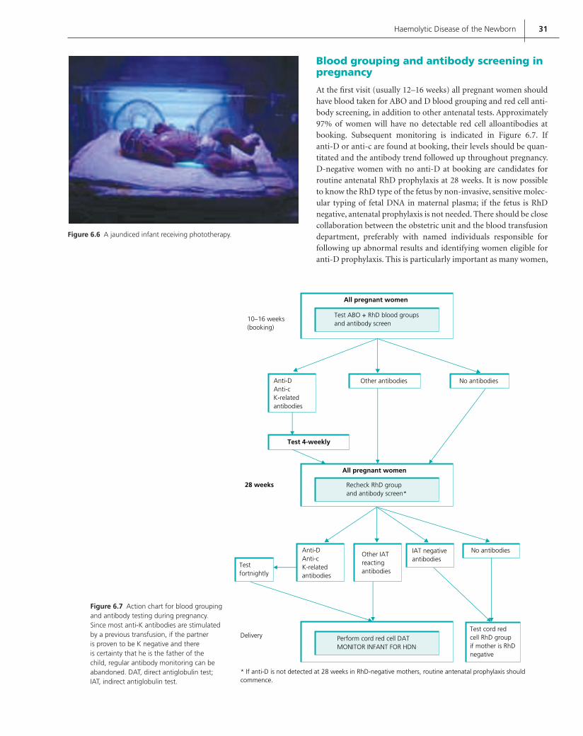
Haemolytic Disease of the Newborn 31
Figure 6.6 A jaundiced infant receiving phototherapy.
10–16 weeks(booking)
All pregnant women
Test ABO + RhD blood groupsand antibody screen
Anti-DAnti-cK-relatedantibodies
Other antibodies No antibodies
Test 4-weekly
All pregnant women
28 weeks
Testfortnightly
Anti-DAnti-cK-relatedantibodies
Other IATreactingantibodies
IAT negativeantibodies
No antibodies
Test cord redcell RhD groupif mother is RhDnegative
Perform cord red cell DATMONITOR INFANT FOR HDN
Delivery
* If anti-D is not detected at 28 weeks in RhD-negative mothers, routine antenatal prophylaxis shouldcommence.
Recheck RhD groupand antibody screen*
Figure 6.7 Action chart for blood grouping and antibody testing during pregnancy. Since most anti-K antibodies are stimulated by a previous transfusion, if the partner is proven to be K negative and there is certainty that he is the father of the child, regular antibody monitoring can be abandoned. DAT, direct antiglobulin test; IAT, indirect antiglobulin test.
Blood grouping and antibody screening in pregnancy
At the fi rst visit (usually 12–16 weeks) all pregnant women should have blood taken for ABO and D blood grouping and red cell anti-body screening, in addition to other antenatal tests. Approximately 97% of women will have no detectable red cell alloantibodies at booking. Subsequent monitoring is indicated in Figure 6.7. If anti-D or anti-c are found at booking, their levels should be quan-titated and the antibody trend followed up throughout pregnancy. D-negative women with no anti-D at booking are candidates for routine antenatal RhD prophylaxis at 28 weeks. It is now possible to know the RhD type of the fetus by non-invasive, sensitive molec-ular typing of fetal DNA in maternal plasma; if the fetus is RhD negative, antenatal prophylaxis is not needed. There should be close collaboration between the obstetric unit and the blood transfusion department, preferably with named individuals responsible for following up abnormal results and identifying women eligible for anti-D prophylaxis. This is particularly important as many women,
Contreras_C006.indd 31Contreras_C006.indd 31 12/18/2008 1:34:52 Shobha12/18/2008 1:34:52 Shobha

32 ABC of Transfusion
particularly those from immigrant or travelling communities, may either book late or have very fragmented antenatal care, and may fall through the system unless special safeguards are in place. Every Trust with an obstetric department should have a named obstetri-cian/fetal medicine specialist as well as a haematologist or transfu-sion medicine consultant from whom advice can be obtained about HDN. Previous obstetric history (severe fetal or neonatal anaemia, severe jaundice, etc.) is also particularly important and the relevant patients should be identifi ed early in the pregnancy so that appro-priate serological and fetal monitoring can be instituted. When a pregnant woman is found to have atypical antibodies, the labora-tory report should state the specifi city and clinical signifi cance of these antibodies.
Post-delivery tests
Infants born to women who have clinically signifi cant antibodies should have a cord blood sample taken for a DAT (Box 6.2). If the DAT is negative, HDN is unlikely. A positive DAT is not diagnostic of HDN; it only signifi es the coating of the infant’s red cells with maternally derived antibody. When the DAT is positive, the infant’s haemoglobin and bilirubin levels should be tested to diagnose HDN. Examining a blood fi lm is useful in diagnosing ABO HDN. The treatment of mild to moderate HDN is phototherapy. Exchange transfusion is indicated for severe HDN and top-up transfusions are indicated if anaemia persists in the infant (see Chapter 7).
Box 6.2 Post-delivery cord blood tests to be undertaken if the mother has clinically signifi cant antibody/ies
Direct antiglobulin test or DAT (positive if baby has corresponding • antigen/s).If the DAT is positive, test:•
haemoglobulin
bilirubin
blood fi lm (spherocytes are present if there is signifi cant
haemolysis)
The future
The very signifi cant reduction in the incidence and deaths due to HDN over the past 30–40 years is a result of both anti-D immunoprophylaxis and improved antenatal care and man-agement of sick fetuses and infants. In the near future sensi-tive molecular techniques on maternal plasma will allow routine determination of the D type of fetuses carried by D-negative mothers, thus avoiding antenatal prophylaxis when the fetus is D negative. Unfortunately, monoclonal anti-D for prophylaxis does not seem to be in the horizon. In recent years the labelling of all blood bags in England and Wales with the full Rh and K type has meant that K-negative women of childbearing potential can be given K-negative blood, which should further reduce the burden of HDN.
Further reading
British Committee for Standards in Haematology, Blood Transfusion Task
Force. Guidelines for blood grouping and red cell antibody testing during
pregnancy. Transfusion Medicine 1996; 6: 71–4.
Clarke CA. Preventing rhesus babies: the Liverpool research and follow up.
Archives of Disease in Childhood 1989; 64: 1734–40.
Joint working group of the British Blood Transfusion Society and the Royal
College of Obstetricians and Gynaecologists. Recommendations for the
use of anti-D immunoglobulin for Rh prophylaxis. Transfusion Medicine
1999: 9; 93–7.
Mollison PL, Engelfriet CP, Contreras M. Blood Transfusion in Clinical
Medicine,10th edn. Blackwell Science, Oxford, 1997.
National Institute of Clinical Excellence (NICE). Pregnancy: Routine anti-D
prophylaxis for rhesus negative women. NICE Guidelines No. 41. NICE
Publications, London, 2002.
Royal College of Obstetricians and Gynaecologists (RCOG) Anti-D
Immunoglobulin for Rh Prophylaxis. RCOG Green Top Guidelines. RCOG
Publications, London, 2002.
Vaughan JI, Warwich R, Letsky EA, Nicolini U, Rodeck CH, Fiasco NM.
Erythropoiesis suppression in foetal anaemia because of Kell alloimmuni-
sation. Journal of Obstetrics and Gynaecology 1994; 171: 247–52.
Contreras_C006.indd 32Contreras_C006.indd 32 12/18/2008 1:34:54 Shobha12/18/2008 1:34:54 Shobha

33
CHAPTER 7
Fetal and Neonatal Transfusion
Helen V. New and Sailesh Kumar
ABC of Transfusion, 4th edition, 2009. Edited by Marcela Contreras. © 2009
Blackwell Publishing, ISBN: 978-1-4051-5646-2.
Fetal transfusions are highly specialized procedures, performed in only a few fetal medicine centres in the UK. On the other hand, infants in neonatal units are frequently transfused, so optimiz-ing neonatal transfusions is an issue for many hospitals. Fetuses and neonates are among the most vulnerable recipients of the infectious, metabolic or immunological complications of blood transfusion. Moreover, problems with long-term side effects, including some infections, are likely to be greatest for this population as most will live for several decades after transfusion.
Special components are required for fetuses and neonates in order to minimize the risks of transfusion (see Table 7.1 for discussion), and these are described in detail in the 2004 British Committee for Standards in Haematology (BCSH) guidelines on transfusion for neonates and older children.
Fetal transfusion
Intrauterine transfusions may use either red cells to correct fetal anaemia such as caused by maternal red cell alloantibodies caus-ing haemolytic disease or, more rarely, platelets when there is fetal alloimmune thrombocytopenia. Routine antenatal and post-natal anti-D immunoglobulin (Ig) prophylaxis for all Rh-negative women (see Chapter 6) has decreased the incidence of haemolytic disease of the fetus and newborn, so the number of red cell intra-uterine transfusions has decreased markedly over the last 20 years.
Red cell transfusionThe commonest cause of fetal anaemia is haemolytic disease caused by red cell alloantibodies, although it may also occur following fetal infection with parvovirus B19, or be due to congenital red cell apla-sia. Survival rates of fetuses with anaemia have improved consider-ably since the introduction of intrauterine transfusion. Pregnancies complicated by red cell alloantibodies (particularly anti-D, anti-c and anti-K) may result in fetal anaemia secondary to transplacental passage of maternal IgG antibodies that bind to red cells carrying paternal antigens, leading to progressive fetal haemolysis. In severe cases the anaemic fetus develops ascites, subcutaneous oedema, and pleural and pericardial effusions (hydrops fetalis), and may die in utero (see Chapter 6).
The main advance in the management of pregnancies at risk of anaemia or where anaemia is suspected is the use of weekly fetal middle cerebral artery monitoring (Figure 7.1). Middle cerebral artery peak systolic velocities (Doppler) are raised in anaemic fetuses, and values greater than 1.5 MoM (multiples of the median) for the specifi c gestation are predictive of moderate or severe fetal anaemia with 100% sensitivity and a false positive rate of 12%. Middle cerebral artery Doppler results show very good correlation with amniotic fl uid bilirubin levels. This non-invasive method has supplanted the traditional technique of serial amniocentesis for bilirubin measurement.
Fetal blood sampling and intrauterine transfusionIf middle cerebral artery monitoring suggests anaemia, fetal blood sampling and intrauterine transfusion are indicated. The risks of the procedure depend on the age of gestation, the site of sampling and underlying pathology. The risk of fetal loss from uncompli-cated fetal blood sampling is between 1% and 3%; however if the
OVERVIEW
Fetuses and neonates are amongst the most vulnerable • transfusion recipients.
Special blood components are available in order to minimize the • risks of transfusion. It is important to increase awareness of these in order to ensure selection of the appropriate component.
Fetal transfusions are highly specialized procedures, with a risk • of fetal loss of at least 1% from each procedure.
Fetuses may be transfused with either red cells or platelets. Red • cell intrauterine transfusions have decreased following the introduction of routine anti-D for all RhD-negative women. Monitoring for fetal anaemia is now by non-invasive Doppler ultrasound of blood fl ow in the middle cerebral artery.
Sick babies in neonatal units are frequently transfused but • guidelines on transfusion indications are still largely based on consensus and expert opinion.
Strategies to reduce neonatal transfusion include local guidelines • to minimize the frequency and volume of blood taken for testing.
Contreras_C007.indd 33Contreras_C007.indd 33 12/10/2008 1:42:09 Shobha12/10/2008 1:42:09 Shobha

34 ABC of Transfusion
fetus is hydropic the risk may be as high as 20%. These are diffi cult procedures and should be performed by fetal medicine specialists in selected tertiary centres. Transfusions are started as late in preg-nancy as possible (ideally after at least 18 weeks’ gestation), and the intervals between transfusions should be maximized by giving as many red cells as possible in the allowed volume, i.e. using packed cells with a high haematocrit (0.70–0.85%).
Sampling can be performed from either the placental cord inser-tion site (Figure 7.2) or the intrahepatic vein. The intrahepatic approach is less likely to cause fetal complications but is technically more challenging. Complications of fetal blood sampling include fetal bradycardia, haemorrhage, cord haematoma, cardiac tampon-ade and fetal death. There may also be increased maternal alloim-munization. The procedure is done under continuous ultrasound guidance and facilities for immediate analysis of the fetal blood should be available.
Irradiated, group O blood, negative for cytomegalovirus (CMV), which should be less than 5 days old and which has been cross-matched with a maternal plasma sample, is used for fetal trans-fusion. A fi nal post-transfusion fetal haematocrit of around 0.45% is desirable. Transfusions may be given intravascularly (for immediate correction) or may be combined with concomitant intraperitoneal administration (when blood is slowly absorbed through the lym-phatics). This combination increases the interval between trans-fusions by maintaining adequate haemoglobin concentrations for longer. Although the timing of subsequent transfusions is depen-dent on the rate of decline of the fetal haematocrit, the presence of hydrops, and gestational age, in general a 3–5-weekly interval is the norm. Close monitoring with middle cerebral artery Doppler is essential and allows precise timing of further intervention.
Table 7.1 Some of the requirements to consider for fetal and neonatal blood components.
Issue to consider Component specifi cation
Blood group compatibility Use red cells compatible with any maternal antibodies, e.g. anti-A, anti-B, anti-D or anti-c (usually give group O RhD negative in the absence of signifi cant maternal antibodies)
Reduction of risk of morbidity from antibodies in donor plasma
Screen donors for high titre anti-A, anti-B and atypical antibodies. For neonates who are not group O, do not use group O FFP and avoid group O platelets where possible
Reduction of infection risk Leucodepleted; CMV negative; ‘accredited’ donors who have donated at least twice in the previous 2 years; methylene blue-treated FFP imported from the USA
Age of red cells Use fresh blood (5 days old) for fetal and large volume neonatal transfusions. Extracellular potassium levels rise during red cell storage. Red cells for top-up transfusion can be of any age, since it is better to dedicate paedipaks from the same donor for one recipient (Figure 7.3)
Suspension medium for red cells Use citrate-phosphate-dextrose (CPD) for IUT and neonatal exchange transfusions; saline-adenine-glucose-mannitol (SAG-M) for neonatal top-up transfusions (see text)
Haematocrit of packed red cells High (0.70–0.85) for IUT, to reduce volume overloadIntermediate (0.5–0.6) for neonatal exchange transfusionBroader range (0.5–0.7) for neonatal top-up transfusion
Prevention of transfusion-associated graft-versus-host disease
Irradiate red cells and platelets for IUT, any neonatal transfusion post IUT, exchange transfusions and some immunodefi ciency states
CMV, cytomegalovirus; FFP, fresh frozen plasma; IUT, intrauterine transfusion.
Figure 7.1 Circle of Willis in the fetus with the cursor placed on the middle cerebral artery, and the Doppler waveform.
Figure 7.2 Fetal blood sampling from the placental cord insertion site.
Contreras_C007.indd 34Contreras_C007.indd 34 12/10/2008 1:42:09 Shobha12/10/2008 1:42:09 Shobha
Fetal and Neonatal Transfusion 35
Overall, fetal hydrops usually reverses after one or two trans-fusions, and survival is about 85%, with a survival rate of greater than 90% when there has been severe disease but no hydrops.
Platelet transfusionFetal platelet transfusions are used for fetomaternal alloimmune thrombocytopenia. This is a rare condition due to maternal alloim-munization to fetal platelet antigens inherited from the father, and occurs in approximately one in 1000 to one in 1500 live births. The most common human platelet antigens involved are HPA-1a (85% of cases) and HPA-5b (10% of cases). Affected fetuses have a 10–30% risk of intracranial haemorrhage either antenatally or peripartum. There is uncertainty over the optimal antenatal management, which may include maternal treatment with intrave-nous Ig, with or without steroids, and intrauterine platelet trans-fusions. Many centres start with intravenous Ig, and if there is a poor response of the fetal platelet count will then add in steroids or move to weekly platelet transfusions until delivery.
The risks of intrauterine platelet transfusions in this case are similar to those of red cell transfusion, with a risk of fetal loss of at least 1% from each procedure. The platelets used are group-appropriate, negative for the platelet antigen to which the mother has made antibody, CMV negative, irradiated and concentrated.
Neonatal transfusion
Sick neonates are frequently transfused, but guidelines on trans-fusion indications (such as from the BCSH, 2004) are still largely based on consensus and expert opinion. There is evidence from reports to the UK Serious Hazards of Transfusion (SHOT) hae-movigilance scheme that there are a disproportionately high number of transfusion errors in this age group, mostly due to trans-fusion of the wrong blood component. It is therefore important to increase awareness of the components available and to ensure that such transfusions are only given in situations that are accepted as appropriate.
Red cell transfusionsBecause of concerns in the UK regarding possible transfusion transmission of variant Creutzfeldt–Jakob disease (vCJD) all blood is leucodepleted. Further measures are being taken to reduce the exposure of children to UK plasma as apparently most residual infectivity is present in plasma. The use of packed red cells in addi-tive solution (saline adenine glucose mannitol or SAG-M), which contains very little residual plasma, is being encouraged where possible, rather than the use of red cells in anticoagulant (citrate phosphate dextrose or CPD), which may have signifi cantly more plasma. Although there have been concerns over possible toxicity to neonates of the SAG-M constituents, they have been shown to be safe for small volume neonatal top-up transfusions. There is also evidence that they are safe for large volume transfusions in neonatal cardiac surgery (Mou et al. 2004), and the use of red cells in SAG-M is now recommended by the BCSH for this indication. However, for fetal transfusions and neonatal exchange transfusions red cells in CPD are still recommended, as the effect of additives in these situations is more uncertain.
Small volume top-upsMost red cell transfusions to neonates are small volume top-ups (10–20 ml/kg) for preterm babies. These transfusions are fre-quently given to treat anaemia from repeated blood sampling, but may also be required for other causes of anaemia such as pre-maturity, haemolysis, congenital infections or genetic disorders. The triggers for transfusion in these babies vary, depending on fac-tors such as the age of the baby and the cardiorespiratory status (Table 7.2). They are mostly based on observational clinical stud-ies and vary between units, with a general trend towards a more restrictive use of red cells over recent years.
Strict adherence to transfusion guidelines has been shown to reduce the number of transfusions given to neonates, and this should be coupled with local strategies to minimize the frequency and volume of blood taken for testing. The use of paedipaks (Figure 7.3) reduces donor exposure as multiple transfusions from one donor can be given to the same neonate over a period of several weeks.
Large volume transfusionsNeonates are given large volume transfusions (may be as much as two blood volumes) in the settings of exchange transfusion, car-diac surgery and sometimes during resuscitation or other surgery (Figure 7.4). They should have blood of neonatal specifi cation, which also needs to be fresh (less than 5 days old) in order not to have problems due to high potassium (see Table 7.1). When phototherapy fails, exchange transfusions are undertaken to treat hyperbilirubinaemia, with or without anaemia, for example in
Table 7.2 Suggested transfusion thresholds for neonates.
Transfusion threshold
Transfusion of red blood cellsAnaemia in the fi rst 24 hours Hb 12 g/dlNeonate receiving mechanical ventilation Hb 12 g/dlAcute blood loss 10% blood volume lostOxygen dependency (not ventilated) Hb 8–11 g/dl (depending on
clinical situation)Late anaemia, stable patient (off oxygen) Hb 7 g/dl
Transfusion of plateletsConsider in all neonates 30 platelets 109/LConsider if increased bleeding risk, for example:
• 1000 g and 1 week of age
clinically unstable (e.g. labile blood •
pressure)
previous major bleeding (e.g. grade •
3–4 intraventricular haemorrhage)
current minor bleeding (e.g. petechiae)•
coagulopathy•
planned surgery or exchange •
transfusion
50 platelets 109/L
Major bleeding <100 platelets 109/L
Adapted from BCSH (2004) and Murray and Roberts (2004).There is little clear evidence for much of this guidance, particularly regarding long-term outcomes, but they may serve as a starting point for developing local guidelines.
Contreras_C007.indd 35Contreras_C007.indd 35 12/10/2008 1:42:15 Shobha12/10/2008 1:42:15 Shobha

36 ABC of Transfusion
haemolytic disease of the newborn (HDN). The indication for exchange transfusion for hyperbilirubinaemia will depend on the bilirubin level and the gestation and age of the baby; there are charts and guidelines for neonatologists to follow in this situa-tion (see, for example, Figure 7.5). General experience in under-taking this procedure is decreasing and it is important to establish local practice guidelines.
The optimum haematocrit of the red cells used for exchange will vary depending on the level of anaemia of the neonate. The exchange procedure can be modifi ed as appropriate by including saline if the haematocrit is too high for a given situation. Therefore it is important for the blood provided to have a tightly controlled haematocrit in order to provide accurate information for neona-tologists. The BCSH guidelines currently recommend 0.5–0.6, and the National Blood Service (NBS) has to narrow this range still fur-ther, to 0.50–0.55.
PlateletsPlatelets are given to a signifi cant number of neonates on neona-tal units, largely for prophylaxis in the absence of bleeding, with transfusion rates of up to 9% reported in neonatal intensive care units. The common indications vary depending on whether the baby is term or preterm, but include thrombocytopenia due to perinatal asphyxia and sepsis. It is particularly important to remember fetomaternal alloimmune thrombocytopenia (known as neonatal alloimmune thrombocytopenia, or NAITP), which is usually detected in otherwise well term babies, as transfusions with platelets negative for the common human platelet antigens involved (HPA-1a and HPA-5b) are indicated and are specially provided by the NBS for this situation.
The platelet count at which it is appropriate to transfuse neo-nates is unclear. The few data from randomized studies suggest that there is no benefi t in transfusing preterm babies with platelet counts greater than 50 109/L in order to reduce intracranial hae-morrhage. Below this level, guidelines such as those from the BCSH recommend a threshold of 20–30 109/L, depending on the clinical situation (see Table 7.2). An observational study has recently been undertaken in the UK to look at the indications and outcome for neonatal platelet transfusions; it may be possible to use this as the basis for future randomized studies in this area.
Platelets for neonates follow the neonatal criteria as in Table 7.1, and are quarter splits of a single apheresis donation. Babies who are deemed likely to have several transfusions over a short time should be given more than one transfusion from the same donor, where possible.
Fresh frozen plasmaAccepted indications for the use of fresh frozen plasma (FFP) on neonatal units have become more restricted over recent years, such that it is no longer considered appropriate to use it as a vol-ume expander or during partial exchange transfusions for poly-cythaemia. Current guidelines recommend FFP primarily for bleeding as a result of vitamin K defi ciency and for bleeding or
Figure 7.5 Example guideline for exchange transfusion, taking into account the level of bilirubin and the age of the infant. This graph is for infants of 35 weeks’ gestation or more. The dashed lines for the fi rst 24 hours indicate uncertainty due to a wide range of clinical circumstances and a range of responses to phototherapy. (Adapted from American Academy of Pediatrics 2004.)
Birth
Tota
l ser
um
bili
rub
in (
µmo
l/L)
513
428
342
257
17124 h 48 h 72 h
Age
96 h 5 days 6 days 7 days
Infants at lower riskInfants at medium riskInfants at higher risk
Figure 7.3 A set of eight paedipaks split from a single unit of red cells. (Courtesy of Jan Green, National Blood Service.)
Figure 7.4 Transfusion of a neonate post cardiac surgery. (Courtesy of Ruth Allis.)
Contreras_C007.indd 36Contreras_C007.indd 36 12/10/2008 1:42:15 Shobha12/10/2008 1:42:15 Shobha

Fetal and Neonatal Transfusion 37
signifi cant bleeding risk where there are coagulopathies such as disseminated intravascular coagulation. However, it is important to remember that the laboratory coagulation screening times, in particular the activated partial thromboplastin time (APTT), may be signifi cantly longer than in adults, depending on the ges-tational and postnatal age of the baby (Table 7.3). It is therefore important to interpret coagulation results on these babies with caution, and the use of an APTT ratio based on adult ranges is likely to be particularly misleading when trying to diagnose a coagulopathy in a preterm baby.
There is little evidence from randomized trials regarding the use of FFP for neonates apart from the large prospective study undertaken by the Northern Neonatal Nursing Initiative (NNNI) Trial Group (1996), which showed no benefi t from the use of prophylactic FFP given to preterm babies to prevent intraven-tricular haemorrhage. This study did not include coagulation testing, so a subset of babies with a coagulopathy might have benefi ted. Overall, the use of FFP for neonates is of uncertain benefi t in most situations and its use should be minimized where possible, taking into account neonatal coagulation ranges and the clinical status of the baby.
The component provided by the NBS is methylene blue-treated FFP imported from the USA in order to minimize the risk of trans-fusion-transmitted infection including vCJD (Figure 7.6). The ABO blood group should be compatible with the baby for FFP and cryoprecipitate.
Cryoprecipitate may be given to neonates with low fi brino-gen levels (1 g/L) and a risk of bleeding. As it is usually given in
situations where babies are also receiving FFP, it is desirable to give cryoprecipitate only if the neonatal fi brinogen level remains low despite FFP transfusion.
Prescribing and administrationAs discussed above, the indications for neonatal transfusion are not always clear-cut. It is therefore particularly important to record clearly the clinical reasons for transfusion and the clinical and laboratory outcomes. Local guidelines on transfusion indica-tions should be followed, and ways of reducing transfusions such as by reducing blood sampling should be maximized. A common error is the selection of an incorrect blood component for trans-fusion, so it is important to ensure good training of junior doctors, nurses and laboratory staff. In addition, neonatal components must be prescribed correctly, and both the volume and time taken for the transfusion should be recorded in order to prevent incidents such as adult ‘units’ being given inappropriately quickly to a small baby (Table 7.4). Communication between neonatal units and blood banks needs to be close in order to make the best use of the allocation and use of red cell paedipaks.
All neonatal blood components should be transfused via a 170–200 m fi lter. Ideally this is part of a specifi c paediatric giving set with small priming volumes (Figure 7.7), but if such sets are not available the blood component can be drawn from the bag through a separate fi lter into a syringe and then delivered through a syringe driver. If a paediatric giving set is used, blood can be delivered either through a syringe driver or through a suitable volumetric pump (refer to local guidelines).
(a) Figures for adults and healthy full-term infants during the fi rst month of life.
Postnatal age
Test Day 1 Day 5 Day 30 Adult
PT (s) 13.0 (10.1–15.9) 12.4 (10.0–15.3) 11.8 (10.0–14.3) 12.4 (10.8–13.9)
APTT (s) 42.9 (31.3–54.5) 42.6 (25.4–59.8) 40.4 (32.0–55.2) 33.5 (26.6–40.3)
Fibrinogen (g/L) 2.83 (1.67–3.99) 3.12 (1.62–4.62) 2.70 (1.62–3.78) 2.78 (1.56–4.00)
(b) Figures for healthy preterm infants (30–36 weeks gestation) during the fi rst month of life.
Postnatal age
Test Day 1 Day 5 Day 30
PT (s) 13.0 (10.6–16.2) 12.5 (10.0–15.3) 11.8 (10.0–13.6)
APTT (s) 53.6 (27.5–79.4) 50.5 (26.9–74.1) 44.7 (26.9–62.5)
Fibrinogen (g/L) 2.43 (1.50–3.73) 2.80 (1.60–4.18) 2.54 (1.50–4.14)
APTT, activated partial thromboplastin time; PT, prothrombin time.Data from Andrew et al. (1988, 1990), expressed as means followed by ranges which include 95% of the population. Each mean value is based on tests from a minimum of 40 infants, all of whom received 1 mg vitamin K intramuscularly at birth. The few data available for earlier gestational ages show even more prolonged laboratory coagulation times (Andrew et al. 1990). Coagulation results are technique dependent and will vary between laboratories. However, it is diffi cult to create local neonatal coagulation ranges, so these ranges are often used to give local comparative guidance.
Table 7.3 Comparison of neonatal and adult laboratory coagulation ranges.
Contreras_C007.indd 37Contreras_C007.indd 37 12/10/2008 1:42:19 Shobha12/10/2008 1:42:19 Shobha

38 ABC of Transfusion
Figure 7.6 Neonatal fresh frozen plasma. (Courtesy of the National Blood Service.)
Table 7.4 Suggested volumes and rates of neonatal transfusions. These give general guidance, and the actual values used will depend on the clinical status of the baby at the time.
Component Volume usually administered
Rate
Red cell concentrates(1) Exchange
transfusion
(2) Top-up transfusion(3) Emergency large
volume transfusion
80–100 ml/kg (for anaemia)160–200 ml/kg (for hyperbilirubinaemia)10–20 ml/kg*
10–20 ml/kg
Depends on stability of baby: discuss with NICU consultant
5 ml/kg/h†
Rapid infusion only for resuscitation
Platelet concentrates 10–20 ml/kg 10–20 ml/kg/h
Fresh frozen plasma 10–20 ml/kg 10–20 ml/kg/h
Cryoprecipitate 5–10 ml/kg 10–20 ml/kg/h (i.e. over approx 30–60 min)
NICU, neonatal intensive care unit.* May also use the formula:Volume of packed cells (ml) weight (kg) desired rise in haemoglobin 4.† Furosemide is sometimes used, e.g. ventilation with acute respiratory distress.
Figure 7.7 Example of an administration set for neonatal transfusion. The red cell paedipak is attached to a paediatric giving set with an integral 200 m fi lter and three-way tap. In this case the blood is then drawn into a syringe for administration to the baby using a syringe driver, but the giving set is also suitable for use with an appropriate volumetric pump. (Courtesy of Rachel Moss, St Mary’s Hospital.)
Figure 7.8 Cover of the parent information leafl et on neonatal transfusion provided by the National Blood Service. (Courtesy of the National Blood Service.)
Contreras_C007.indd 38Contreras_C007.indd 38 12/10/2008 1:42:20 Shobha12/10/2008 1:42:20 Shobha

Fetal and Neonatal Transfusion 39
Parents are not generally required to give written consent for transfusion in the UK, but it is very important that the transfusion should be discussed with them in advance. To help with this pro-cess, the NBS has produced a parent information sheet to be made available to clinicians in hospitals (Figure 7.8).
Conclusion
Fetal and neonatal transfusions are specialized interventions, with long-term benefi ts when used appropriately. Despite the fact that only a small proportion of transfusions are given to this patient population they are often relatively complex, and require close attention in order to perform them correctly.
Acknowledgments
We are grateful to Sunit Godambe and Rachel Moss for comments on the text.
Further reading
British Committee for Standards in Haematology (BCSH). Transfusion
guidelines for neonates and older children. British Journal of Haematology
2004; 124: 433–53.
Bullock R, Martin WL, Coomarasamy A, Kilby MD. Prediction of fetal ane-
mia in pregnancies with red-cell alloimmunization: comparison of middle
cerebral artery peak systolic velocity and amniotic fl uid OD450. Ultrasound
in Obstetrics and Gynecology 2005; 25: 331–4.
Mari G, Deter RL, Carpenter RL, et al. Noninvasive diagnosis by Doppler
ultrasonography of fetal anemia due to maternal red-cell alloimmuniza-
tion. Collaborative Group for Doppler Assessment of the Blood Velocity
in Anemic Fetuses. New England Journal of Medicine 2000; 342:
9–14.
Maxwell DJ, Johnson P, Hurley P, Neales K, Allan L, Knott P. Fetal blood
sampling and pregnancy loss in relation to indication. British Journal of
Obstetrics and Gynaecology 1991; 98: 892–7.
Murray NA, Roberts IA: Neonatal transfusion practice. Archives of Disease in
Childhood Fetal Neonatal Edition 2004; 89: F101–F107.
New HV. Paediatric transfusion. Vox Sanguinis 2006; 90: 1–9.
References
American Academy of Pediatrics. American Academy of Pediatrics guideline:
management of hyperbilirubinemia in the newborn infant 35 weeks or
more weeks of gestation. Pediatrics 2004; 114: 297–316.
Andrew M, Paes B, Johnston M. Development of the hemostatic system in the
neonate and young infant. American Journal of Pediatric Hematology/
Oncology 1990; 12: 95–104.
Andrew M, Paes B, Milner R, Johnston M, Mitchell L, Tollefsen DM,
Castle V, Powers P. Development of the human coagulation system in the
healthy premature infant. Blood 1988; 72: 1651–7.
Mou SS, Giroir BP, Molitor-Kirsch EA, et al. Fresh whole blood versus recon-
stituted blood for pump priming in heart surgery in infants. New England
Journal of Medicine 2004; 351: 1635–44.
Northern Neonatal Nursing Initiative (NNNI) Trial Group. A randomized
trial comparing the effect of prophylactic intravenous fresh frozen plasma,
gelatin or glucose on early mortality and morbidity in preterm babies.
European Journal of Pediatrics 1996; 155: 580–8.
Rayment R, Brunskill SJ, Stanworth S, Soothill PW, Roberts DJ,
Murphy MF. Antenatal interventions for fetomaternal alloimmune
thrombocytopenia. Cochrane Database of Systematic Reviews 2005, Issue 1.
Art. No. CD004226.
Serious Hazards of Transfusion (SHOT). Serious Hazards of Transfusion
Annual Report 2003. Manchester, 2004.
Contreras_C007.indd 39Contreras_C007.indd 39 12/10/2008 1:42:27 Shobha12/10/2008 1:42:27 Shobha

40
CHAPTER 8
Plasma Products and Indications for Their Use
Hannah Cohen and Trevor Baglin
ABC of Transfusion, 4th edition, 2009. Edited by Marcela Contreras. © 2009
Blackwell Publishing, ISBN: 978-1-4051-5646-2.
The concept that the optimal use of plasma is achieved by fraction-ation is long established and a wide range of products is available for therapeutic use (Figure 8.1). This chapter considers all these products except albumin.
Most therapeutic plasma products are manufactured from pools of plasma derived from many thousands of donations. All donors are individually screened for mandatory microbiological agents (see Chapters 1 and 13) and the plasma products are made by processes that inactivate or remove (or both) any contaminating viruses.
Although the risks of viral transmission are very small, there can never be absolute assurance of freedom from risk. This concern, as well as about that of variant Creutzfeld–Jakob disease (vCJD), has driven the development of recombinant plasma products, and their use has, in some cases, superseded that of plasma-derived products. In the UK, precautions in place to reduce the risk of prion trans-mission include leucodepletion of all blood components, impor-tation of plasma for fractionation, exclusion of donors who have received a blood transfusion in the UK after 1980, and the use of viral-inactivated fresh frozen plasma (FFP), obtained from non-UK areas free of bovine spongiform encephalopathy (BSE) and vCJD. Such blood products are used for children under 16 years and for patients with thrombotic thrombocytopenic purpura (TTP) who require repeated infusions of large volumes of FFP.
Unpredictable and occasional fatal adverse effects of blood components have been highlighted by the Serious Hazards of Transfusion (SHOT) confi dential enquiry (see Chapter 15). Of particular concern are serious allergic reactions and anaphylaxis, and transfusion-related acute lung injury (TRALI): the risk of these acute transfusion reactions is approximately six-fold higher with FFP than with red cell transfusions. Haemolysis from trans-fused antibodies to blood group antigens, especially A and B, may also occur, usually as a result of ‘wrong blood transfusion’. Serious adverse events and reactions related to blood components should
OVERVIEW
There are few absolute and evidence-based indications for • treatment with plasma and plasma products; appropriate use will increase safety.
Transfusion of cryoprecipitate exposes the patient to multiple • donors and therefore fresh frozen plasma (FFP) is recommended for the bleeding associated with the hypofi brinogenaemia of disseminated intravascular coagulation, massive blood transfusion and thrombolytic agents, with cryoprecipitate reserved for instances when fi brinogen levels remain critically low (<1 g/L).
Viral-inactivated FFP obtained from non-UK areas free of bovine • spongiform encephalopathy and variant Creutzfeld–Jakob disease should be used for children under 16 years, patients with thrombotic thrombocytopenic purpura who require repeated infusions of large volumes of FFP, and, when necessary, patients with congenital defi ciencies of factor V and XI.
Recombinant factor VIII and IX concentrates are the products of • choice for patients with haemophilia A and B, respectively, and intermediate purity factor VIII for the treatment of moderate to severe von Willebrand’s disease. For these disorders and mild factor XI defi ciency desmopressin is preferable to plasma-derived products when the effects are equal.
Recombinant factor VIIa is useful in the management of • bleeding in patients with haemophilia who have inhibitory antibodies and it can be used to treat patients with rare congenital platelet disorders.
Anti-D immunoglobulin prevents primary Rh immunization and • haemolytic disease of the newborn.
Figure 8.1 Flow diagram of plasma fractionation.
Whole blood Plasmapheresis
Fresh plasma
Fresh frozen plasma
Cryoprecipitate
Factor VIIIconcentrate
Cryosupernatant
Albumin
Other concentrates
Immunoglobulins
Contreras_C008.indd 40Contreras_C008.indd 40 12/5/2008 6:02:09 PM12/5/2008 6:02:09 PM

Plasma Products and Indications for Their Use 41
be reported to SHOT via the Medicines and Healthcare products Regulatory Agency (MHRA) Serious Adverse Blood Reactions and Events (SABRE) electronic reporting system to meet the require-ments of the Blood Safety and Quality Regulations 2005 (see Chapter 20). SHOT also encourages reports on adverse events related to solvent detergent (SD) FFP, although a plasma prod-uct, for purposes of comparison with single donor FFP, and on anti-D immunoglobulin. Adverse reactions related to plasma-derived coagulation factors are monitored by the UK Haemophilia Centre Doctors’ Organisation (UKHCDO), and those to other plasma fractionated and recombinant products by the MHRA.
It should be appreciated that there are few absolute and evidence-based indications for treatment with plasma and plasma products and that some widely used products are unlicensed and prescribed for a ‘named patient’ by a ‘named doctor’. Especially in acquired disorders, in which sound evidence of effi cacy is usually weak-est, clinicians should take careful account of the risks of plasma and plasma products before treating patients. Table 8.1 lists the indications for use of different plasma products.
Fresh frozen plasma, cryoprecipitate and cryosupernatant
The indications for the transfusion of FFP, cryoprecipitate and cryosupernatant are very limited. However, although considerable progress has been made in limiting unnecessary transfusion of red cells the use of FFP continues to rise (from 307 804 units in 1999 to 347 882 units in 2005 in the UK). Several audits have shown inap-propriate use of FFP, and SHOT data show that acute transfusion reactions have been caused by FFP given inappropriately or for doubtful clinical indications. Current national British Committee for Standards in Haematology (BCSH) guidelines on the appro-priate use of FFP, cryoprecipitate and cryosupernatant should be incorporated into local protocols that are readily available in relevant clinical and laboratory areas, included in induction and update training, and subject to clinical audit. Appropriate use of these blood components will increase safety and should be driven in all hospital trusts by the hospital transfusion teams in accordance with the Department of Health circular Better Blood Transfusion: safe and appropriate use of blood. The risks of transmitting infection by FFP, cryoprecipitate and cryosupernatant are similar to those of other blood components unless pathogen-reduced plasma is used.
Fresh frozen plasmaThe two types of viral-inactivated FFP currently available in the UK are methylene blue and light-treated FFP (MBFFP) and SDFFP (available commercially as Octaplas®) (Figures 8.2–8.4). These products should be used for the treatment of congenital defi cien-cies of coagulation factors (e.g. factor V) when there is no specifi c factor concentrate available. Factor XI concentrate administration may be associated with thrombosis and in patients with factor XI defi ciency with a history of cardiovascular disease, in whom treat-ment with desmopressin (see below) is relatively contraindicated, SDFFP should be administered. Used aggressively, with plasma exchange, viral-inactivated FFP which should be viral-inactivated, is essential in the treatment of patients with TTP (see below). BCSH
guidelines on oral anticoagulation recommend prothrombin com-plex concentrates (two products, Octaplex® and Beriplex® P/N are licensed) rather than FFP for the reversal of the effects of oral anti-coagulation associated with serious bleeding.
Despite being widely advocated for patients with multiple coagu-lation defects, as in disseminated intravascular coagulation (DIC), massive transfusion and liver disease, the effect of FFP is poorly defi ned. In DIC, FFP is indicated if the patient is bleeding but should not be used if there is no bleeding regardless of the results of labo-ratory tests. In massive blood transfusion, whilst ‘formula replace-ment’ is not recommended, when rapid turnaround of coagulation tests cannot be guaranteed, FFP infusion should be considered after one blood volume is lost. A locally agreed algorithm is essen-tial. What is clear is that, if any benefi t is to be obtained, FFP must be given in adequate quantities and rapidly: 12–15 ml/kg (about 1 L for an adult), repeated if necessary, aiming to maintain the PT/APTT (prothrombin time/activated partial thromboplastin time) ratios at 1.5 and fi brinogen at 1.0 g/L. FFP should be used to correct the depletion of coagulation factors in bleeding associated with thrombolysis.
There is no justifi cation for the use of FFP as a volume expander because crystalloids are effective, safer, cheaper and more read-ily available. Patients likely to receive large or repeated doses of FFP should be offered vaccination against hepatitis A and B and post-transfusion surveillance of hepatitis A, B and C, human immunodefi ciency virus (HIV) and parvovirus status.
CryoprecipitateCryoprecipitate is prepared from FFP by slow thawing at 4 2°C (Figure 8.5). The resulting precipitate is then separated from the supernatant and refrozen for storage. Cryoprecipitate contains factor VIII, fi brinogen, von Willebrand factor (vWF), factor XIII and fi bronectin in higher concentrations than they are found in plasma, and its preparation is normally the fi rst step in plasma fractionation.
Fibrinogen concentrate (even though it is unlicensed) and fac-tor XIII concentrate (licensed as Fibrogammin®) are preferable to cryoprecipitate in the treatment of congenital hypodys- or hypo-fi brinogenaemia and factor XIII defi ciency, respectively. Transfusion of cryoprecipitate exposes the patient to multiple donors and therefore FFP has been recommended for bleeding associated with the hypofi brinogenaemia of DIC, massive blood transfusion and thrombolytic agents, with cryoprecipitate reserved for instances when fi brinogen levels remain critically low (1 g/L). One litre of FFP provides on average 2.69 g fi brinogen (range 1.54–5.00), while an adult therapeutic dose (two pools) of cryoprecipitate provides, on average, 4 g fi brinogen (range 0.54–3.79), in a volume of 320–340 ml (R. Cardigan, personal communication). As a result, a rise in fi brinogen can be achieved more rapidly with cryopre-cipitate than with FFP infusion; therefore – particularly in massive obstetric haemorrhage where profound hypofi brinogenaemia may occur – cryoprecipitate should be given ‘sooner rather than later’.
CryosupernatantThis is the supernatant plasma removed during the preparation of cryoprecipitate (see below).
Contreras_C008.indd 41Contreras_C008.indd 41 12/5/2008 6:02:09 PM12/5/2008 6:02:09 PM

42 ABC of Transfusion
Plasma exchange
To date, the only condition where FFP is recommended as replace-ment fl uid for PEX is acute TTP, where intensive PEX remains the mainstay of therapy, reducing mortality rates from 90% to 10–20%. Patients with TTP have defi ciency or inhibition of
ADAMTS 13 (vWF-cleaving protease), a metalloprotease that nor-mally cleaves ultralarge vWF multimers released from the vascu-lar endothelium. The resultant accumulation of ultralarge vWF multimers in TTP (Figure 8.6) leads to platelet activation and aggregation, which in turn leads to microvascular occlusion by
Table 8.1 Plasma product concentrates: indications for use.
Plasma product/recombinant concentrate Indications for use
Recombinant factor VIII Congenital defi ciency (haemophilia A)
Recombinant factor IX Congenital defi ciency (haemophilia B)
Recombinant factor VIIa Inhibitors in patients with haemophilia A and BCongenital defi ciency of factor VIICongenital platelet disorders?Uncontrolled bleeding states (trauma or surgery)
Prothrombin complex concentrates (PCC) Congenital defi ciencies of factor II and XReversal of oral anticoagulationInhibitors, especially to factor VIII?Severe liver disease
Activated prothrombin complex concentrates (PCC) Inhibitors, especially to factor VIII
Porcine factor VIII (recombinant product undergoing clinical trials)
Factor VIII inhibitors
Intermediate purity factor VIII concentrate Congenital defi ciency (moderate/severe von Willebrand’s disease)
8Y (intermediate purity factor VIII concentrate) Congenital defi ciency (ADAMTS 13 in thrombotic thrombocytopenic purpura)
Factor VII Congenital defi ciencyReversal of oral anticoagulation associated with severe bleeding (NB Octaplex® and Beriplex® P/N (licensed PCCs) contains factor VII)?Severe liver disease
Factor XI Congenital defi ciency (desmopressin is an alternative but viral-inactivated FFP is preferable in patients with a history of cardiovascular disease)
Fibrinogen Congenital hypo- and dysfi brinogenaemia
Factor XIII Congenital defi ciency
Fibrin sealant/glue Congenital and acquired haemostatic disorders to promote local haemostasis
Antithrombin (a recombinant product, ATryn®, is licensed (pan-European) for prophylaxis of venous thromboembolism in patients with congenital antithrombin defi ciency undergoing surgery)
Congenital defi ciency?Disseminated intravascular coagulation?Liver transplantation?Other acquired defi ciency states
Protein C Congenital defi ciency?Meningococcal purpura fulminans
Recombinant activated protein C Severe sepsis
Alpha-1 antitrypsin Hereditary defi ciency (emphysema, cirrhosis)
CI esterase inhibitor Hereditary angioedema
Fibronectin ?Acquired defi ciency states
Intravenous immunoglobulin (IVIg) Congenital ahypo- and hypogammaglobulinaemiaChronic lymphocytic leukaemiaImmune thrombocytopenic purpura?Other acquired immune disorders
Anti-D (usually intramuscular) Prevention of primary RhD immunization and haemolytic disease of the newborn
Anti-D (intravenous) Refractory idiopathic thrombocytopenic purpura
Contreras_C008.indd 42Contreras_C008.indd 42 12/5/2008 6:02:09 PM12/5/2008 6:02:09 PM

Plasma Products and Indications for Their Use 43
platelet-rich thrombi and ischaemic organ damage. Despite the documented low incidence of TTP in the UK these patients use 20–25% of national plasma stocks. The Department of Health has recommended that replacement in PEX for TTP should be with SDFFP in adults and with MBFFP in children under 16 years.Preliminary studies suggest that SDFFP, which, unlike standard FFP, lacks high molecular weight vWF multimers, may be more effective
than standard FFP or MBFFP in TTP. The use of cryosupernatant has now been superseded as it does not appear to be more effec-tive than standard FFP. Of note, SDFFP has been associated with venous thromboembolism, possibly due to acquired protein S defi -ciency, when used as replacement for PEX in TTP. Low molecular weight heparin thromboprophylaxis should therefore be instituted when the platelet count rises to 50 109/L. In addition, patients should be fi tted with graduated elastic compression stockings and care should be taken with indwelling lines, particularly to avoid
Figure 8.4 Pack of solvent detergent fresh frozen plasma (Octaplas®).
Figure 8.5 Pack of pooled cryoprecipitate.
Figure 8.2 Pack of methylene blue fresh frozen plasma for neonates.
Figure 8.3 Pack of methylene blue fresh frozen plasma for paediatric use.
Contreras_C008.indd 43Contreras_C008.indd 43 12/5/2008 6:02:10 PM12/5/2008 6:02:10 PM

44 ABC of Transfusion
infection. Concerns have been raised about the effi cacy of MBFFP as replacement in PEX for TTP as more PEX procedures may be required. Currently, methylene blue cryosupernatant is unavailable in the UK.
The optimal regimen for PEX is undefi ned, but the current recommendation is for at least 1.0 plasma volume exchange daily until at least 2 days after remission is achieved (defi ned as normal neurology, platelets 150 109/L, normal lactate dehy-drogenase levels, and rising haemoglobin concentration). More intensive PEX (twice daily or daily 1.5 volume exchanges) may be useful in patients with severe acute neurological or cardiological manifestations.
Recently, the monoclonal CD20 antibody rituximab, used ini-tially as second line therapy in patients with refractory TTP, has been associated with prompt remission and a reduction in the number of PEX required.
Factor VIII and IX concentrates
Factor VIII and IX concentrates are the products of choice for patients with haemophilia A (factor VIII defi ciency) who do not respond adequately to desmopressin (1-deamino-8-D-arginine vasopressin, DDAVP, see below) and for patients with haemo-philia B (factor IX defi ciency). Patients with factor VIII levels of less than 0.10 IU/ml are unlikely to respond adequately to DDAVP and require factor replacement. Patients with factor IX defi ciency
do not respond to DDAVP therapy. In the UK recombinant factors VIII and IX are now recommended, as there is no associ-ated risk of transmission of human-borne infectious disease. Factor concentrates are supplied as freeze-dried powders that are reconstituted with a small volume of sterile water before intravenous injection.
Factor VIII has a short half-life (about 8–12 hours in vivo) (Table 8.2), so twice-daily repeated injections are necessary for a sustained effect. Injection of 1 IU/kg of factor VIII concen-trate will raise the plasma factor VIII level by approximately 0.02 IU/ml. Factor IX has a longer half-life (about 12–18 hours) and once-daily injections may be adequate for a sustained effect. Injection of 1 IU/kg of factor IX concentrate will raise the plasma factor IX level by approximately 0.01 IU/ml. For example, to raise the factor level by 0.30 IU/ml (a normal level is 0.50 IU/ml) in a 50 kg male would require 750 units of factor VIII or 1500 units of factor IX concentrate.
The minimum level of factor VIII required for treatment of bleeding has not been determined from randomized clinical tri-als. Early mild haemarthrosis is often treated with a single infu-sion of factor concentrate to raise the plasma level to 0.15–0.20 IU/ml. More severe haemarthrosis requires repeated infusions and higher plasma levels (0.30–0.50 IU/ml). Intracranial bleed-ing requires normalization of factor levels with treatment for at least 2–4 weeks to achieve peak plasma levels of 1.0–1.50 IU/ml and troughs not less than 0.50 IU/ml. Treatment to cover surgery requires normalization of factor levels for 7–14 days depending on the type of surgery. Primary prophylaxis with factor concentrate two or three times weekly at doses to keep the trough factor level above 0.01 IU/ml reduces bleeding and hence chronic haemophilic arthropathy. Pharmacokinetic studies are used to optimize dos-ing schedules for individual patients. With the increased safety of
Figure 8.6 Ultralarge von Willebrand factor (vWF) multimers in patients with thrombotic thrombocytopenic purpura can be seen on sodium dodecyl sulfate polyacrylamide gel electrophoresis (SDS-PAGE) in lanes 2–6 above the dotted line (with normal vWF multimers in lanes 1, 7 and 8).
Table 8.2 Half-lives of infused coagulation factors contained in fresh frozen plasma.
Factor Half life of infused factor (hours)*
I (fi brinogen) 72–120II (prothrombin) 72V (proaccelerin) 12VII (proconvertin) 2–5VIII (antihaemophiliac factor) 8–12IX (Christmas factor) 12–18X (Stuart–Prower factor) 24–40XI (plasma thromboplastin antecedent) 60–80XII (Hageman’s factor) 40–50XIII (fi brin stabilizing factor) 216–240Antithrombin 24–60Protein C 6–8Protein S 12–60Fibronectin 24–72
* Half-lives of coagulation factors may be shortened when there is increased consumption, for example in disseminated intravascular coagulation, after surgery or during thrombotic episodes.
Contreras_C008.indd 44Contreras_C008.indd 44 12/5/2008 6:02:17 PM12/5/2008 6:02:17 PM

Plasma Products and Indications for Their Use 45
coagulation factor replacement products, prophylaxis has become the preferred treatment option in the UK for males with severe hae-mophilia. Programmes are typically initiated in the second or third year of life, so an indwelling venous access device (portacath) is often necessary.
Recombinant factor VIII preparations do not contain vWF and are not effective for the treatment of moderate to severe von Willebrand’s disease. Intermediate purity products are used to treat moderate to severe von Willebrand’s disease. Both high and intermediate purity plasma-derived factor VIII concentrates are available but are no longer used in the UK for treatment of haemophilia A. However, many patients with haemophilia were treated with these products before the availability of recombi-nant products. Despite the excellent safety record of virally inacti-vated plasma-derived concentrates it is acknowledged that current viral-inactivation techniques may fail to eliminate all virus trans-mission by non-lipid-enveloped viruses such as parvovirus. There is no evidence that vCJD can be transmitted by non-cellular blood products but there has been recent anxiety about the theoretical possibility that plasma-derived products could have been contami-nated. So far, there have been no documented cases of transmission of vCJD by plasma-derived factor concentrates.
Recombinant factor VIIa
Approximately one-quarter of patients with severe haemophilia A develop an inhibitory immunoglobulin G (IgG) antibody to fac-tor VIII. On average this is detectable after about 10 treatment exposures, and while the antibody is transitory in 50% of patients in the remainder it is permanent unless eradicated by immune-tolerance therapy employing daily high dose factor VIII until the antibody is eliminated. While the antibody is present infused fac-tor VIII is immediately neutralized, and this therapy is ineffective. For such patients, treatment with a ‘bypassing agent’ is required. High dose recombinant factor VIIa (90 g/kg/dose) generates thrombin in the absence of either factor VIII or IX and is used to treat bleeding in patients with inhibitors. Typically, up to three treatments at 2-hourly intervals are used. Recombinant factor VIIa can also be used to treat bleeding in patients with haemo-philia B with inhibitor antibodies to factor IX, but these occur in less than 1% of such patients. Recombinant factor VIIa can also be used to treat patients with factor VII defi ciency and the rare congenital platelet disorders Glanzmann’s thrombasthenia and Bernard Soulier syndrome. Recombinant VIIa is not licensed for the treatment of uncontrollable bleeding states following trauma or during major surgery.
Prothrombin complex concentrates
Prothrombin complex concentrates (PCCs) are plasma-derived intermediate purity products containing coagulation factors IX, X and II, and sometimes factor VII. They are used for the reversal of severe overanticoagulation and/or bleeding due to treatment with vitamin K antagonists (mainly warfarin in the UK). They are
also used for the treatment of the severe rare bleeding disorders of factor X and factor II defi ciency.
Prothrombin complex concentrates are now being used in patients with liver failure although this was previously considered a contraindication to their use. In liver disease PCCs can correct only part of the overall haemostatic abnormality, and carry a risk of provoking DIC. DIC can also occur in patients without liver disease who are treated with repeated doses of PCCs.
Activated PCCs are used as ‘bypassing agents’, as an alternative to recombinant factor VIIa, for the treatment of patients with severe haemophilia with an inhibitor.
Other coagulation factor concentrates
Other coagulation factor concentrates (factor XIII, fi brinogen, factor XI) have been produced by fractionation but are not always available. Currently a factor XIII concentrate (Fibrogammin®) is available in the UK for the treatment of patients with severe factor XIII defi ciency. Some concentrates of fi brinogen or factor XI that were previously available were associated with a signifi cant risk of thrombosis. At present viral-inactivated FFP is typically used for the treatment of patients with factor XI defi ciency. Concentrates of natural anticoagulants, antithrombin and protein C are occa-sionally used in patients with congenital, and sometimes acquired, defi ciencies of specifi c anticoagulants, for example protein C con-centrate for purpura fulminans. A recombinant activated protein C concentrate is licensed for the treatment of severe sepsis. A recom-binant antithrombin product (ATryn®) is licensed (pan-European) for prophylaxis of venous thromboembolism in patients with congenital antithrombin defi ciency undergoing surgery.
Desmopressin (DDAVP), a synthetic analogue of the non-apep-tide arginine vasopressin, can lead to a marked increase in blood levels of factor VIII and vWF. In patients with mild haemophilia A and von Willebrand’s disease DDAVP is effective in the treatment of bleeding episodes and to cover surgery. DDAVP may also be use-ful in patients with mild factor XI defi ciency. DDAVP has a similar effect to cryoprecipitate in the treatment of the bleeding tendency in chronic renal failure, where a functional defect in vWF–plate-let interaction appears to play a role in the abnormal haemostasis observed.
For all indications DDAVP is clearly preferable to plasma-derived products for reasons of both cost and safety when the effect is equal. DDAVP should be used with caution in elderly individuals and pregnant women, and avoided in those with evidence of arterio-vascular disease. In young children, precautions should be taken to prevent fl uid overloading and hyponatraemia, and DDAVP should probably be avoided in children under 2 years old.
Immunoglobulins
Specifi c immunoglobulinsSpecifi c immunoglobulins are obtained from donors whose plasma contains selected high titre IgG antibodies, as a result either of previous infection or of active immunization. Usually given by intramuscular injection, preparations are available for use in the
Contreras_C008.indd 45Contreras_C008.indd 45 12/5/2008 6:02:18 PM12/5/2008 6:02:18 PM

46 ABC of Transfusion
Figure 8.7 Fetal cells (darkly stained) in the maternal circulation of a RhD-negative woman as shown by the Kleihauer (acid elution) technique. A 500 IU intramuscular dose of anti-D immunoglobulin is suffi cient for 4 ml of red cells. If a fetomaternal haemorrhage of 4 ml is indicated by the Kleihauer test, the volume of fetomaternal haemorrhage must be confi rmed by fl ow cytometry and an adequate dose of anti-D administered (larger doses may be given intravenously) to prevent primary Rh immunization and haemolytic disease of the newborn.
00
20
50
75
100
200
5 10
Intravenous Ig
15 20Time (days)
Plat
elet
co
un
t x
109 /
L
25 30 35 40
Figure 8.8 Time course of a typical response of platelets to high doses of intravenous immunoglobulin (Ig) in adult (chronic) idiopathic thrombocytopenic purpura.
passive prophylaxis of varicella-zoster, tetanus, hepatitis B and other infections. Immunity lasts a few weeks. Anti-D immuno-globulin, usually given intramuscularly, is used in the prevention of primary Rh immunization and haemolytic disease of the newborn (see Chapter 6) (Figure 8.7), and intravenous anti-D in refractory idiopathic thrombocytopenic purpura (ITP).
Intravenous anti-D in ITP is suitable as second line therapy for RhD-positive patients who are not splenectomized (it is rarely effec-tive in splenectomized patients and therefore not recommended). The mechanism of action is believed to be mediated through the destruction of RhD-positive red cells, which are preferentially removed by the reticuloendothelial system, particularly the spleen, thus sparing autoantibody-coated platelets through Fc receptor blockade. The effect on the platelet count tends to last for only a few weeks. Common toxicities of anti-D include fever/chill reactions, which can be ameliorated by pretreatment with corticosteroids, and extravascular haemolysis; the risk of this is lessened by avoid-ing treatment in those with a positive direct antiglobulin test unless attributable to prior intravenous Ig or anti-D Ig therapy.
Non-specifi c immunoglobulinNon-specifi c (‘normal’) immunoglobulin is derived from the pooled plasma of non-selected donors and contains antibodies to all the viruses prevalent in the donor population. One of the main indications for its intramuscular use is in the passive prophylaxis of hepatitis A. Preparations made for intravenous use have the advan-tage that much larger doses may be given with minimal discomfort to the patient.
Intravenous Ig was introduced primarily for replacement ther-apy in patients with congenital hypogammaglobulinaemia, but its range of application has now broadened considerably to include replacement in acquired hypogammaglobulinaemia as seen in
chronic lymphocytic leukaemia, and in immunomodulation, espe-cially in some autoimmune disorders. Intravenous Ig is useful in patients with ITP when the platelet count has to be raised rapidly because of bleeding, or prior to intervention likely to induce bleed-ing. The usual dosage, a total of 2 g/kg over 2–5 days, elevates the platelet count in 75% of patients with ITP, 50% of whom achieve normal counts, but the responses are transient, lasting up to 3–4 weeks (Figure 8.8). Its mode of action is not certain but prob-ably includes blockade of Fc receptors on macrophages or other effectors of antibody-dependent cytotoxicity. Intravenous Ig has an excellent safety record although renal impairment or failure have been reported. These products are often used for unlicensed indications, including a variety of autoimmune neurological con-ditions, in the absence of any prospective, evidence-based clinical trial data.
Other plasma products
Several other plasma products are available, and there is little doubt that these concentrates can be effective in raising subnormal con-centrations of circulating plasma factors. Except in rare congenital defi ciency states, however, convincing evidence of clinical benefi t is generally weak and indications remain ill defi ned.
Acknowledgments
The authors are grateful to Professor Sam J. Machin for critical review of this chapter.
Further reading
Allford SL, Hunt BJ, Rose P, Machin SJ. Guidelines on the diagnosis and man-
agement of the thrombotic microangiopathic haemolytic anaemias. British
Journal of Haematology 2003; 120: 556–73.
Baglin TP, Keeling DM, Watson HG. Guidelines on oral anticoagulation
(warfarin): third edition – 2005 update. British Journal of Haematology
2006; 132: 277–85.
Contreras_C008.indd 46Contreras_C008.indd 46 12/5/2008 6:02:18 PM12/5/2008 6:02:18 PM

Plasma Products and Indications for Their Use 47
British Committee for Standards in Haematology (BCSH) Blood
Transfusion Task Force. Guidelines for the use of fresh frozen plasma,
cryoprecipitate and cryosupernatant. British Journal of Haematology
2004; 126: 11–28.
Health Service circular. Better Blood Transfusion: safe and appropriate use of
blood. Series No. 2007/001, Gateway reference 9058. Department of Health,
London, 2007.
Stainsby D, MacLennan S, Thomas D, Isaac J, Hamilton PJ. Guidelines on the
management of massive blood loss. British Journal of Haematology 2006;
135; 635–41.
UK Haemophilia Centre Doctors’ Organisation (UKHCDO). UKHCDO
guidelines on the selection and use of therapeutic products to treat
haemophilia and other hereditary bleeding disorders. Haemophilia 2003;
9: 1–23.
Contreras_C008.indd 47Contreras_C008.indd 47 12/5/2008 6:02:20 PM12/5/2008 6:02:20 PM

48
CHAPTER 9
Human Albumin Solutions and the Controversy of Crystalloids Versus Colloids
Neil Soni
ABC of Transfusion, 4th edition, 2009. Edited by Marcela Contreras. © 2009
Blackwell Publishing, ISBN: 978-1-4051-5646-2.
Albumin as a solution is available in two basic formats. The fi rst is 4.5% albumin solution, which is essentially isotonic with an ion distribution not dissimilar to plasma, and the second is 20% albumin, which is a hypertonic solution. It is often described as ‘salt-poor albumin’ as the ratio of sodium to albumin is approximately 25% of that in the 4.5% solution (Table 9.1).
The preparation of human albumin is by two pasteurization pro-cesses, effective against enveloped viruses such as human immuno-defi ciency virus (HIV), hepatitis B virus (HBV), hepatitis C virus (HCV) and non-enveloped viruses such as hepatitis A. Both the ethanol fractionation and heat treatment processes may have effects on the molecular structure, in particular the charge, which may then infl uence properties such as binding. At present UK supplies are derived from US plasma because of anxiety about prions (variant Creutzfeldt–Jakob disease or vCJD).
Physiology of albumin
Human albumin is a polypeptide with a molecular weight of 66 248. It is a small molecule (compared with fi brinogen, with a molecular weight of 340 000, or immunoglobulin G, with a molecular weight of 150 000) and is synthesiszed in hepatocytes at a rate of 9–12 g
per day in an adult. It is highly soluble and has a strong net negative charge. There are many variants of albumin. It is predominantly an extravascular protein with more albumin in the inter stitium than intravascularly, although at a lower concentration.
There is a normal circulation of albumin from the intravascular to interstitial spaces, with return via the lymphatic vessels.
Clinical properties of albumin
Albumin is strongly charged and has at least four discrete binding sites, which can bind reversibly to cations and anions and a wide range of other molecules including drugs (Table 9.2). Alterations in the quantity or structure of albumin will affect competitive binding and hence drug levels.
Albumin represents 75–80% of the plasma colloid osmotic pres-sure (COP). It is thus the major determinant of the plasma and interstitial COP component in Starling’s equation for fl uid fl ux across the capillary wall. The relative role of albumin alters in severe illness as the profi le of plasma proteins changes. Acid–base balance is infl uenced by the concentration of albumin. Free radical scaveng-ing is possible because of its thiol groups. Administered albumin will act as a ‘donor’ of thiol groups, and rejuvenate other intravas-cular proteins by donating electrons. It also has an anticoagulant and antithrombotic function. Albumin reputedly has a protective effect on membrane permeability but this is controversial.
OVERVIEW
Albumin is available as 4.5% isotonic and 20% hypertonic • solutions.
Serum albumin falls rapidly in many disease states. In acute • states the rapid fall is often due to redistribution.
Low serum albumin is a prognostic indicator in many chronic • disease states.
There are many indications, although few have an adequate • evidence base.
Suggestions that albumin is unsafe were clearly refuted in a • large clinical trial, SAFE, but specifi c areas where albumin may be of particular benefi t or be detrimental have not yet been delineated.
Table 9.1 Composition of albumin solutions.
4.5% albumin (500 ml bottle)
20% albumin (100 ml bottle)
Albumin (human), approx. 45 g/L (22.5 g) 200 g/L (20 g)
Colloid oncotic pressure 26–30 mmHg 100–120 mmHg
Sodium 120–160 mmol/L 70–160 mmol/L
Potassium 2 mmol/L 10 mmol/L
Stabilizers varySodium-n-octanote, mmol/LSodium acetyl tryptanoate
Bottle size 250 or 500ml 50 or 100 ml
Contreras_C009.indd 48Contreras_C009.indd 48 12/5/2008 6:02:56 PM12/5/2008 6:02:56 PM

Human Albumin Solutions 49
Clinical associations of serum albumin
The serum albumin concentration often falls in disease states and it is important to differentiate the meaning of the serum albu-min value in relatively stable, albeit chronic, states and the rapidly changing values found in acute severe illness. In chronic states, serum concentration refl ects the balance of synthesis and catabo-lism; in acute states, concentration may be signifi cantly infl uenced by both these. The predominant cause of the concentration change is redistribution. Low serum albumin is seen in many disease states (Table 9.3; Figures 9.1 and 9.2).
Preoperative hypoalbuminaemia may be a marker of malnutri-tion and is a predictor of poor outcome.
Table 9.2 Some ions, cations and molecules that bind to albumin.
Calcium, copper, silver, mercury
Amino acids
Fatty acids
Thyroxine
Glucose, galactose
Warfarin, phenytoin, diazepam, non-steroidal anti-infl ammatories, digoxin and many others
Table 9.3 Illness and low serum albumin.
Cause of low albumin Mechanisms/implications
Analbuminaemia No synthesis. No major clinical effects
Starvation Rapid reduction in albumin synthesis. Poor specifi c marker of nutrition, but rapid turnover proteins such as prealbumin are better nutritional markers. Associated with poor outcome
Liver disease Largely caused by redistribution but also catabolism and synthesis. It is not a strong specifi c synthesis marker
Renal disease Leakage in albuminuria and nephrosis. Also falls with dialysis
Pre-eclampsia Due to redistribution, not synthesis
Malignancy Reduced synthesis, and increased catabolism and redistribution. Cytokine activity also implicated (commonly tumour necrosis factor). Associated with poor prognosis
Stress response Synthesis of acute phase proteins increases. Albumin may decrease (not confi rmed)
Burns Catabolism, massive leak from site and redistribution. Reduction in synthesis
Trauma Stress response. Redistribution and catabolism
Surgery Stress response. Redistribution
Sepsis Redistribution, with later catabolism and reduced synthesis
Figure 9.1 This patient sustained severe burns to the head and neck, back, arms, chest and abdomen in a road traffi c accident. With an excess of 70% burns, he had a massive fl uid requirement on admission, but also recurrent requirements during debridement and grafting. Although he demonstrated hypoalbuminaemia as a consequence of both albumin loss and distribution, many units do not electively replace albumin but rather use nutritional support.
Figure 9.2 This patient shows several of the clinical features of cirrhotic liver failure with portal hypertension. He has obvious ascites, collateral veins in the abdominal wall, and possibly some muscle wasting. With this degree of liver failure it is likely that he is hypoalbuminaemic, but he is clearly active and mobile so albumin replacement is not indicated.
Contreras_C009.indd 49Contreras_C009.indd 49 12/5/2008 6:02:56 PM12/5/2008 6:02:56 PM

50 ABC of Transfusion
Prognostic value of serum albumin
In the relatively ‘steady state’ of severe chronic illness (i.e. not acute and fulminant), a reduced serum albumin is a relatively reli-able marker of the severity of illness and is associated with a poor outcome in many disease states (Table 9.4).
Indications
There are many indications for the use of albumin, including hypovolaemia and hypoalbuminaemia (see Table 9.5 for the more common indications). The former needs no explanation, but the latter is a consequence of being able to measure the serum concen-tration and to have the material available with which to correct the value.
As an unfortunate consequence of the association between albu-min value and disease outcome it has been impossible to separate the indications for albumin from the infl uence of those associations. A low serum albumin, which may be a marker of illness, nutritional state or of redistribution, has often been treated with albumin infu-sions to attempt to change the measured value in the hope that a corrected value would infl uence the disease state underlying it. This is refl ected in its usage and in its variable effi cacy. In Table 9.5 there is a column called effi cacy that indicates if the specifi c indica-tion is considered effective or not. In many of the indications there is widespread use but little evidence for specifi c effi cacy (Aboulghar et al. 2002; Barron et al. 2004; Maitland et al. 2005).
It is quite clear from Table 9.5 that there are a large number of indications where there is a lack of evidence of benefi t from the treatment. These need exploration, as do those with some evidence of benefi t. The use of albumin in primary peritonitis and in chil-dren with falciparum malaria are both indications with at least one study conferring benefi t, but in both this needs confi rmation. The Saline versus Albumin Fluid Evaluation (SAFE) study (see below),
Table 9.4 Low serum albumin has been shown to be associated with poor outcome in the following specifi c diseases and conditions.
AIDS (acquired immune defi ciency syndrome)/Pneumocystis carinii pneumoniaBone marrow transplantBreast cancerBurnsClostridium diffi cile colitisColorectal cancerGastric cancerJuvenile rheumatoid arthritisLeukaemiaLiver conditions: alcoholic cirrhosis cholangiocarcinoma hepatitis B cirrhosis hepatocellular cancer post shunt primary biliary cirrhosis pyogenic abscess
Lung cancerLymphomaMalignant melanomaMyelomaOvarian cancerPancreatic cancerPancreatitisPneumoniaPre-eclampsiaRenal conditions: continuous ambulatory glomerulonephritis peritoneal dialysis renal failureUlcerative colitis
Table 9.5 Indications for albumin use.
Solution General indication Specifi c indication Effi cacy
Colloid 4.5% Intravascular volume replacement Hypovolaemia Yes
Paracentesis in liver failure Probable
Primary peritonitis in liver failure Probable
Support colloid oncotic pressure No
Support serum albumin Non-specifi c: transport membrane integrity, coagulation, to prevent ileus
?
Protein-losing entero/nephropathy ? Some advocates
Nutrition No
Therapeutic plasma exchange It is used
Ovarian hyperstimulation syndrome At time of oocyte collectionMechanism unknown
Yes/maybe
To treat metabolic acidosis Use as a buffer ? Used in neonates
To treat falciparum malaria with acidosis in children
Used as volume replacement Yes, reduced mortality
Hypertonic solution/colloid 20%
Intravascular volume replacement Yes
Fluid redistribution Renal dialysis and to initiate diuresis Some advocates
Acute lung injury: fl uid redistribution, to initiate diuresis
Some advocates.
Ovarian hyperstimulation syndrome Intravascular volume Yes/maybe
Contreras_C009.indd 50Contreras_C009.indd 50 12/5/2008 6:03:01 PM12/5/2008 6:03:01 PM

Human Albumin Solutions 51
which was designed to evaluate any mortality outcome difference between albumin and saline when used for fl uid resuscitation in the critically ill, showed no difference in overall mortality although there were some interesting trends in the subgroup analysis (Finfer et al. 2004). These pose some new questions about potential indi-cations and contraindications (Table 9.6). These all need further targeted study and cannot at present be seen as either indications or contraindications.
The crystalloid/colloid controversy and the suggestion that the use of albumin is associated with a higher mortality
The use of intravenous fl uids to support the intravascular com-partment in the management of hypovolaemia is well established. The two main classes of fl uids are crystalloids and colloids. The principal difference is that the latter contain larger molecules and are therefore expected to stay in the intravascular compartment longer. Crystalloids are relatively free to move between compart-ments and will leave the intravascular space and distribute into the interstitium. Therefore it seems likely that colloid is more effi cient than crystalloid, requiring a smaller volume to achieve a sustained effect, and in normal physiological circumstances this is true. In clinical circumstances normal physiology becomes pathophysi-ology and the situation changes. Fluid tends to redistribute far more readily and so do some larger molecules. Colloid effi cacy is reduced, but for volume expansion it is still more effi cient than crystalloid and it is this concept that underpins the argument about which is better.
Crystalloids are cheap, easy to use and effective. Even though large volumes may be required, the fl uid will come back out as eas-ily as it went in. Apart from fl uid overload, which applies equally to colloids, specifi c complications are few. Excess saline can cause acidosis.
Colloids are more expensive and more effective, and smaller volumes are required to achieve the same effect, although once the larger molecules move into the interstitium there are those
that worry about how they get back out later. Specifi c complica-tions include a low incidence of anaphylactoid and anaphylactic reactions, potential alterations in coagulation, and the potential, although minor, risk of transmission of infectious agents in the human-derived products. There also appears to be a difference in propensity to acute renal failure in severe sepsis between the colloids: the gelatins have a possible benefi t over starches or albumin (Schortgen et al. 2001). There are some general points to consider when deciding on colloid use (Table 9.7).
Both crystalloids and colloids have proponents, and the longev-ity of the debate is a clear indication of the lack of hard evidence that either is advantageous.
Albumin is a colloid and was considered the gold standard. It is present in plasma and has multiple physiological functions; it can be measured and in illness is almost always at a lower level than nor-mal, implying a defi cit. There is a well known association between low serum albumin values and outcome, so the scene is set for this colloid not only to be effective as a volume expanding agent but to have a myriad of other benefi ts.
The controversy has been on two fronts. The fi rst was whether colloids are better than crystalloids, and the second was which col-loid is best, given that the alternative colloids to albumin are all syn-thetic and devoid of potential physiological benefi ts beyond plasma expansion. The information currently available refers to those col-loid solutions that are readily available and have been tested (Table 9.8). Balanced salt solutions, as carriers for colloids, are now avail-able and it has been suggested that they may be benefi cial, add-ing a new dimension to the debate. It seems logical to put colloid into a more balanced solution and, although there is some evidence to support this idea, more information is needed (Vercueil et al. 2005).
In summary there is a large amount of opinion but no hard evidence that there is a real difference in outcome either between crystalloids and colloids or between crystalloids and albumin. A new study or studies that could clearly show a difference was needed and it was into this scenario that two systematic reviews from the Cochrane Collaboration appeared, both in the British Medical Journal, within a relatively short time period (Cochrane Injuries Group Albumin Reviewers 1998; Schierhout & Roberts
Table 9.6 SAFE subgroup analyses: area of focus and relative risk (RR) of mortality and confi dence intervals (CI) when using albumin versus saline as fl uid resuscitation therapy.
Focus area RR and CI of using albumin versus saline
Benefi t/disadvantage
Trauma with head injury
RR: 1.6295% CI: 1.12–2.34P 0.009
Possible detriment from albumin
Trauma without head injury
RR: 1.0095% CI: 0.56–1.79P 1.00
No difference
Severe sepsis RR: 0.8795% CI: 0.74–1.02P 0.09
Possible advantage to albumin
Table 9.7 Considerations when using colloids.
Colloids may require less total volume to have the same volume expansion • effect. Benefi t?Crystalloids are cheap• Coagulation: colloids may affect coagulation. Antithrombotic effects of • dextran and hydroxyethyl starchClearance: starch may stay in the reticuloendothelial system• Renal failure: hydroxyethyl starch and albumin may increase the risk of • acute renal failure. There is evidence this may be a problem with higher molecular weight starchHead injury: colloids, specifi cally albumin, may have adverse effects (not • confi rmed)Disease states where albumin may be of benefi t: paediatric falciparum • malaria, primary peritonitis and severe sepsis
Contreras_C009.indd 51Contreras_C009.indd 51 12/5/2008 6:03:01 PM12/5/2008 6:03:01 PM

52 ABC of Transfusion
1998). In essence, they showed that both colloids and albumin were associated with a higher mortality than crystalloids. In the new era of evidence-based medicine – with systematic reviews becoming a gold standard akin to a randomized controlled trial – these studies had a profound effect in terms of evidence credibility. They were not the fi rst meta-analyses, but a previous analysis by Velanovich (1989), showing an advantage in the use of colloids in trauma, had not been generally noticed. There was a call to review practice and the net effect was a signifi cant deepening of the entire controversy.
Subsequent meta-analyses have failed to fi nd the same outcome. One analysis pointed out that a randomized controlled trial almost three times the size of the meta-analysis would be needed to pro-duce adequate power for a conclusion. There was widespread criti-cism of the studies. These criticisms were based on the numbers, population heterogeneity, treatment heterogeneity, and the time period studied during which fl uid management had altered con-siderably, as well as other aspects of these reviews. The real benefi t from these publications was that they triggered a massive random-ized controlled trial involving 6997 patients in Australia and New Zealand. This was the SAFE study mentioned above. It tested the use of saline against albumin for fl uid resuscitation in the criti-cally ill (Finfer et al. 2004); there was no difference in outcome. The lack of concordance between this well executed trial and the earlier meta-analyses casts further doubt on the validity of the latter. Since the initial report subgroup analysis has become available (see Table 9.6), and this has highlighted questions about specifi c clinical areas of use.
More recent Cochrane Collaboration updates have stated that ‘there is no evidence that albumin reduces mortality in critically ill patients with burns and hypoalbuminaemia’. Interestingly, the areas not yet tested by large trials and therefore dependent on meta-analysis, such as burns and hypoalbuminaemia, are still reported as indicating an increased risk, based on the meta-analysis of a few small trials; however, there is no comment on the validity of the statement (Alderson et al. 2004). Paradoxically, the use of albumin fell during this time period as obvious benefi ts over other colloids were hard to fi nd, while the cost issues in most countries mitigated in favour of synthetic colloids.
The current position is therefore that the colloid/crystalloid debate will continue. The only irrefutable fact is that crystalloids are cheaper, and while there is no clear advantage in the use of albumin over other colloids the obvious disadvantage of albumin is cost.
The controversy has been benefi cial as it has established that there is limited information about an ‘everyday’ management issue. Further work is needed to identify real benefi ts or real prob-lems, if they exist, from the use of colloids rather than crystalloids. Albumin has never been tested adequately in specifi c disease states to ascertain if the potential physiological benefi ts can be realized. In some disease states such as paediatric falciparum malaria there is signifi cant evidence to suggest possible benefi t. In others, such as primary peritonitis, the trial that demonstrated benefi t from albumin compared albumin with nothing. This trial needs to be repeated to determine if the effect was from albumin or from volume replacement. Likewise, in areas such as head injury the possibility of detriment needs to be clarifi ed.
There is a dearth of information in hypoalbuminaemia about the effects of reduced binding capability, the reduction in thiol groups for scavenging, or even the role in acid–base balance. The gross het-erogeneity of the critically ill as a population makes this group a diffi cult one to study. Investigation should target specifi c areas of use if benefi ts are to be identifi ed. The information available hints that benefi t may be found in disease states where the serum albu-min is low from depletion as well as distribution. This needs to be explored. In other situations, such as head injury, there is a sug-gestion that albumin, and therefore by default other members of the colloid group, might have detrimental effects. This information desperately needs clarifying.
Conclusion
There are no overwhelming benefi ts from any one volume expander. Serum albumin is a non-specifi c marker of disease associated with poor outcome. It is a simple matter of cause and effect. Low albu-min is the effect and treating it does not reverse the cause. Research into any specifi c benefi ts of crystalloids and each type of colloid is long overdue.
Acknowledgments
The author would like to thank Simon Finfer for his advice and help in preparing this chapter. Dr Cristina Martinez is thanked for providing fi gure 9.2.
Table 9.8 Available colloids.
Colloid Characteristics Problems
Albumin 4.5% Isotonic CostHypotension: rare
Albumin 20% HyperoncoticRelatively salt poor
Expensive
Gelatins MW 30 000 to 5000Shorter intravascular effectExcreted through kidney
Allergy: rare
Dextrans (complex polysaccharide)
Has anticoagulant effectMW ranges from 30 000 to 150 000Complex polysaccharides
Allergy: rareAnticoagulant effectNephrotoxicity rareMechanism unknown
Starches Wide range of characteristicsMW 200 000 to 450 000Long half-life in plasma
Larger MW starches affect coagulationRemains in reticuloendothelial systemItching
Balanced colloid
Starch in a more physiological electrolyte solutionPotentially better in terms of less acidosis
MW, molecular weight.
Contreras_C009.indd 52Contreras_C009.indd 52 12/5/2008 6:03:02 PM12/5/2008 6:03:02 PM

Human Albumin Solutions 53
Further reading
Aboulghar M, Evers JH, Al-Inany H. Intravenous albumin for preventing
severe ovarian hyperstimulation syndrome: a Cochrane review. Human
Reproduction 2002; 17(12): 3027–32.
Alderson P, Bunn F, Lefebvre C, Li WP, Li L, Roberts I, Schierhout G.
Human albumin solution for resuscitation and volume expansion in
critically ill patients. Cochrane Database of Systematic Reviews 2004; 4:
CD001208.
Barron ME, Wilkes MM, Navickis RJ. A systematic review of the comparative
safety of colloids. Archives of Surgery 2004; 139(5): 552–63.
Cochrane Injuries Group Albumin Reviewers. Human albumin administra-
tion in critically ill patients: systematic review of randomised controlled
trials. British Medical Journal 1998; 317(7153): 235–40.
Finfer S, Bellomo R, Boyce N, French J, Myburgh J, Norton R. A comparison
of albumin and saline for fl uid resuscitation in the intensive care unit.
New England Journal of Medicine 2004; 350(22): 2247–56.
Maitland K, Pamba A, English M, Peshu N, Marsh K, Newton C, Levin M.
Randomized trial of volume expansion with albumin or saline in children
with severe malaria: preliminary evidence of albumin benefi t. Clinical
Infectious Diseases 2005; 40(4): 538–45.
Schierhout G, Roberts I. Fluid resuscitation with colloid or crystalloid solu-
tions in critically ill patients: a systematic review of randomised trials.
British Medical Journal 1998; 316(7136): 961–4.
Schortgen F, Lacherade JC, Bruneel F, Cattaneo I, Hemery F, Lemaire F,
Brochard L. Effects of hydroxyethylstarch and gelatin on renal function in
severe sepsis: a multicentre randomised study. Lancet 2001; 357(9260):
911–16.
Velanovich V. Crystalloid versus colloid fl uid resuscitation: a meta-analysis of
mortality. Surgery 1989; 105(1): 65–71.
Vercueil A, Grocott MPW, Mythen MG. Physiology, pharmacology, and
rationale for colloid administration for the maintenance of effective
hemodynamic stability in critically ill patients. Transfusion Medicine
Reviews 2005; 19(2): 93–109.
Contreras_C009.indd 53Contreras_C009.indd 53 12/5/2008 6:03:02 PM12/5/2008 6:03:02 PM

54
CHAPTER 10
Treatment of Massive Haemorrhage in Surgery and Trauma
Tim Walsh
ABC of Transfusion, 4th edition, 2009. Edited by Marcela Contreras. © 2009
Blackwell Publishing, ISBN: 978-1-4051-5646-2.
The normal circulating blood volume is 70–80 ml/kg body weight or approximately 5 L for a 70 kg adult. There is no universally agreed defi nition of massive haemorrhage, but the replacement of the patient’s entire blood volume in 24 hours is commonly used. Signifi cant acute blood loss rapidly results in hypovolaemic shock unless managed appropriately.
Untreated haemorrhagic shock can result in rapid death due to ischaemia of the organs. After treatment is initiated, further com-plications can arise as a result of defi ciencies in blood clotting, hypothermia or electrolyte and acid–base imbalance. It is useful to broadly consider aspects of treatment in two phases: (i) the initial resuscitation phase; and (ii) later complications of resuscitation.
Initial resuscitation phase
Physiological responses to haemorrhageAcute haemorrhage results in the loss of circulating blood volume, which initiates refl ex compensatory mechanisms to maintain organ perfusion. These principally involve activation of the sympathetic nervous system to constrict venous beds in the gut (splanchnic cir-culation) and skin. This vasoconstriction reduces the capacity of the circulation to increase venous return to the heart, which helps maintain the cardiac output. Cardiac output is also enhanced by a direct effect of sympathetic nerves on the atrial node, and increased circulating adrenaline, which act to increase the heart rate and con-tractility. Sympathetic system activation also constricts the arteri-oles via the sympathetic nerves, increasing the overall resistance of the circulation (increased cardiac afterload). The net result is the maintenance of a blood pressure suffi cient to perfuse vital organs, principally the heart, brain and kidneys. The heart is particularly at risk during massive haemorrhage because its blood supply via the coronary arteries occurs only during diastole, when it normally extracts most of the available oxygen from haemoglobin (the oxy-gen extraction ratio is 60–70% compared to 20–40% in most other organs). If the oxygen supply to the heart is inadequate, death will rapidly follow.
Assessing the extent of haemorrhage
Clinical signsKey early signs of signifi cant haemorrhage are tachycardia and pallor (due to vasoconstriction in the skin). Hypotension, car-diac ischaemia, altered consciousness level and oliguria indicate that compensatory physiological mechanisms are not maintaining normal vital organ perfusion and are signs of severe haemorrhage. A commonly used classifi cation relating the percentage blood loss to typical associated clinical signs is shown in Table 10.1.
Measured blood lossIn the emergency setting, particularly outwith the operating theatre, accurate measurements of blood loss are diffi cult. Visible blood loss is often overestimated and non-visible blood loss, for example retroperitoneal or around major bones, is diffi cult to quantify. In the operating theatre, ongoing blood loss should be monitored by weighing swabs, and recording the losses in suction or drain
OVERVIEW
Immediate management of massive haemorrhage should focus • on the ABC resuscitation principles. All patients should receive supplemental oxygen, two large-bore cannulae, samples taken for haematology, coagulation, biochemistry and blood grouping, and administration of intravenous fl uid.
Controlling the source of bleeding is a priority. Until there is • source control, moderate hypotension (mean arterial pressure of 60–70 mmHg) is safe and preferable to hypervolaemia and/or normotension.
Blood transfusion is life saving in massive haemorrhage, but it is • important to remember that the haemoglobin concentration is infl uenced by plasma volume.
All hospitals should have a major haemorrhage protocol to • coordinate the clinical and laboratory response.
After initial stabilization, the aim is to prevent or reverse • complications that worsen outcomes such as hypothermia, electrolyte disturbances, metabolic acidosis and coagulopathy. This can be achieved through close monitoring, including near-patient techniques.
Contreras_C010.indd 54Contreras_C010.indd 54 12/10/2008 1:46:29 Shobha12/10/2008 1:46:29 Shobha

Treatment of Massive Haemorrhage 55
bottles. These measures are often inaccurate and should be used in conjunction with clinical signs.
Factors modifying normal physiological responses to haemorrhageConcurrent medication is the commonest reason for non-typical physiological responses to haemorrhage. Tachycardia is unreliable in patients taking -blocker drugs or other drugs that can slow the heart rate. This can result in an underestimation of blood loss severity. Patients on vasodilator drugs may have an exaggerated hypotensive response, for example angiotensin-converting enzyme (ACE) inhibitors or calcium channel blockers.
Initial investigations and proceduresIn the emergency setting, the patient should be managed using the ABC resuscitation principles. Two large-bore cannulae should be established and samples taken for haematology, coagulation, bio-chemistry and blood grouping. Requests for immediate laboratory processing should be made.
All hospitals should have an agreed protocol that can be trig-gered in the event of major haemorrhage. An example is shown in Figure 10.1. Key elements include:
a reliable mechanism for transporting samples and blood com-• ponents to the laboratories and to the patienta predetermined ‘pack’ of blood components to avoid confusion • or delaysa mechanism to alert relevant individuals such as blood bank • staff, haematologists and porters.
Principles of treatment
Oxygen therapyHigh fl ow oxygen therapy, preferably using a rebreathing mask that enables the delivery of inspired oxygen 60%, should always be administered. This ensures maximum saturation of available
haemo globin, increases dissolved plasma oxygen, and induces some constriction of blood vessels (hyperoxic vasoconstriction) that may help maintain blood pressure.
Fluid therapyInitial fl uid resuscitation aims to restore an adequate circulating volume. This is usually judged best by improvement in blood pres-sure, falling heart rate, better capillary return and peripheral tem-perature. In severe cases, signs of better organ perfusion such as improving conscious level are useful guides.
The choice of fl uid therapy is much debated. The most important principle is to relate the estimated expansion in circulating volume required to the predicted volume expected to remain in the circu-lation after redistribution. Initial resuscitation with approximately 20 ml/kg body weight of a crystalloid solution (1–2 L for a 70 kg adult) is appropriate for most clinical situations. During this period the degree of haemorrhage can be assessed from the history, clini-cal signs and response to fl uid therapy. The options for subsequent fl uids include a combination of:
further crystalloids• colloid solutions• red cells• coagulation factors.•
The relevant properties of each fl uid type, which are useful to con-sider in decision-making, are shown in Table 10.2. In practice, the combination administered will depend on the clinical condition of the patient, the results of blood tests and clinical judgment.
Source of bleeding controlFollowing initial resuscitation, the priority is to identify the source of bleeding and control it. This may require urgent transfer to the operating theatre or other specialist department depending on the site of haemorrhage. Delays to achieving source control can adversely affect patient outcome, especially when the haemorrhage is ongoing.
Table 10.1 A commonly used classifi cation of hypovolaemic shock (Baskett’s classifi cation).
Class I Class II Class III Class IV
Blood lossPercentage of .bloodVolume of blood (in 70 kg male) (L)
15750
15–30800–1500
30–401500–2000
402000
Blood pressureSystolicDiastolic
UnchangedUnchanged
NormalRaised (narrowed pulse pressure)
ReducedReduced
Very lowVery low
Pulse rate Mild tachycardia 100–120 120 (reduced volume) 120 (very weak)
Capillary refi ll Normal Slow (2 s) Slow (2 s) Undetectable
Respiratory rate Normal Normal Tachypnoea (20/min) Tachypnoea (20/min)
Urine fl ow rate (ml/h) 30 20–30 10–20 0–10
Extremities Normal colour Pale Pale Pale and cold
Complexion Normal Pale Pale Ashen
Mental state Alert Anxious and/or aggressive Anxious and/or aggressive and/or drowsy
Drowsy and/or confused or unconscious
Contreras_C010.indd 55Contreras_C010.indd 55 12/10/2008 1:46:29 Shobha12/10/2008 1:46:29 Shobha

56 ABC of Transfusion
Target blood pressure prior to source controlConsiderable research as been undertaken to determine the opti-mum blood pressure during resuscitation from massive haemor-rhage, especially following major trauma. Current evidence sup-ports partial correction of blood pressure to mean arterial pressure values of 60–70 mmHg, using incremental fl uid therapy, until the source of bleeding is controlled. The lower blood pressure reduces blood loss from damaged vessels and increases the chance of stable clot formation. After source control, more aggressive restoration of normal blood pressure with fl uids is appropriate to preserve organ function.
Optimum haemoglobin during massive haemorrhageDuring haemorrhage, the haemoglobin concentration is strongly infl uenced by the plasma volume. Resuscitation with crystalloid and colloid fl uids increases plasma volume. As a result, haemo-globin values can be misleadingly high prior to resuscitation and can decrease rapidly during fl uid therapy. Measured haemoglobin concentrations need to be interpreted in the context of the time when sampling occurred, because they may correlate poorly with the circulating red cell mass (Figure 10.2). When the patient is unstable, the estimated blood loss together with clinical signs are the best guide to when red cells should be transfused. The choice of
CALL 2 – Blood bank ……………………….
Tell them:How urgent is the need for blood?Patient information• Name • Hospital number • Major incident number • Sex and date of birth • ABO & Rh group (if known) What blood component and how much blood isrequestedWhere the blood is to be sentName and contact telephone number fordoctor in charge
Send samples tolaboratory…………………Pretransfusion testingsample
FBC and coagulationscreen
CALL 1 – Hospital switchboard
…………………………
Ask them to take detailsThere is a major haemorrhageName and location of patientContact name and telephone numberfor doctor in charge
Ask them to inform:
Blood bank …………………………………… Haematology lab
Red cellsneeded
Immediately
Red cellsneeded
In 15 minutes
Red cellsneeded
In 45 minutes
Areplatelets,
fresh frozenplasma and
cryoprecipitateneeded?
Allow time for preparation and delivery
Group O negative red cells are usuallyin very short supply. For male patients,group O positive cells may be used.
A sample should be sent to theblood bank as soon as possible toallow conversion to group-specificblood
If the patient has a historic recordand a group and screen on acurrent clinical sample, blood canbe made available immediatelyby electronic release
An antibody screen andcrossmatch will be carried outon the released units within 30minutes
Allow time for preparation anddelivery
If bleeding continues uncontrolledconsider antifibrinolytic orrecombinant factor VIIa
Send patient’s sample andrequest form urgently toblood bank
ABO and Rh group, antibodyscreen and crossmatch
Blood available 45 minutes aftersample received in the laboratory
Send patient’s sample andrequest form urgently to bloodbank
ABO and RhD group-specific redcells available for collection atblood bank
Blood available 15 minutes aftersample received in the laboratory
Use emergency group Onegative red cells fromblood bank
Send patient sample andrequest form urgently toblood bank
Blood available immediately
Use emergency group Onegative (or positive, if males)red cells in designated fridge
Send patient sample and requestform urgently to blood bank
Blood available immediately
Make two phone calls
Figure 10.1 An example of a major haemorrhage protocol.
Contreras_C010.indd 56Contreras_C010.indd 56 12/10/2008 1:46:29 Shobha12/10/2008 1:46:29 Shobha

Treatment of Massive Haemorrhage 57
red cell product should be determined by the perceived urgency of transfusion using clinical judgment (see Figure 10.1).
Once circulating volume has been restored, the haemoglobin concentration is a more reliable transfusion trigger. Although humans can tolerate haemoglobin concentrations of 4–5 g/dl under controlled conditions, these are not considered safe in clinical practice. A haemoglobin value of 7–9 g/dl is supported by available randomized trials in non-bleeding, critically ill patients. A value of 8–10 g/dl offers a ‘safety margin’ for further bleeding. Patients with heart disease should be kept at the upper end of this range.
Patient monitoringAs soon as available, patients should have continuous pulse oxim-etry, electrocardiogram and frequent non-invasive blood pressure
Table 10.2 Important properties of intravenous fl uids.
Fluid type Volume of 1000 ml infusion remaining in circulation after distribution
Tonicity Na+ and Cl– concentrations (mmol/L)
pH Comments
5% dextrose 60–80 ml Hypotonic Na+: 0Cl–: 0
4.0 No value as a resuscitation fl uid
Normal (0.9%) saline 200–250 ml Isotonic Na+: 154Cl–: 154
5.0 Excessive administration can contribute to acidosis (hyperchloraemic acidosis)
Ringer’s lactate 200–250 ml Isotonic Na+: 131Cl–: 112
6.5 Less likely to contribute to acidosis after initial resuscitation, but contains potassium and lactate, so caution in early phase
Haemaccel (succinylated gelatin) 1000 ml Isotonic Na+: 145Cl–: 145
7.4 Half-life in circulation depends on rate of metabolism of colloid particles; it is typically 4 h
Gelofusine (polygeline gelatin) 1000 ml Isotonic Na+: 154Cl–: 125
7.4 Half-life in circulation depends on rate of metabolism of colloid particles; it is typically 5 h
Hetastarch (example of starch solution-several available)
1000 ml Isotonic Na+: 154Cl–: 154
5.5 Various starch solutions are available. Their exact properties depend on the specifi c product. Some expand intravascular volume more than infusion volume through high oncotic pressure
Plasma protein fraction (human albumin solution 4.5%)
1000 ml Isotonic Na+: 150Cl–: 120
7.4 Evidence indicates no benefi t over cheaper, non-human derived fl uids
RCV 2 LHct 0.4
PV 3 L
RCV 1.6 LHct 0.28
RCV 1.6 LHct 0.4
2
4
6
0
8
Healthyeuvolaemic
Haemorrhagicshock with 1L
blood loss
Resuscitated withfluids to normal
circulating volume
Hb 9 g/dlHb 13 g/dlHb 13 g/dlBloodvolume (L)
PV 2.4 L PV 3.4 L
Figure 10.2 Relation between haemoglobin (Hb) concentration and plasma volume before and after resuscitation with fl uids. Hct, haematocrit; RCV, red cell volume; PV, plasma volume.
monitoring, in addition to the measurement of body temperature and respiratory rate. Invasive cardiovascular monitoring using arte-rial and central venous catheters are valuable, particularly during ongoing treatment. Placement of these catheters is often techni-cally diffi cult in shocked patients, and should not be undertaken by inexperienced staff. Attempts should not distract clinicians from resuscitation.
Later complications of massive haemorrhage
CoagulopathyCoagulopathy is an important component of the vicious circle that characterizes loss of control of the clinical condition. It is often multifactorial. Management relies on detection and correction of the contributing factors:
Dilution of coagulation factors1 in plasma occurs following massive transfusion. Protocolized prescriptions of blood components according to predicted blood loss or red cell transfusions are not recommended. Coagulation factor defi ciency is likely after the loss of 1–1.5 blood volumes. Blood component administra-tion should be guided by clinical evaluation (oozing or failure to generate clot) together with coagulation tests (see below).Thrombocytopenia2 can occur due to dilution and consumption. Platelets usually remain at a level 50 109/L until 1.5 blood volumes have been replaced.Hypothermia3 is a common and serious complication because it has multiple adverse effects (Table 10.3). Hypothermia can be prevented by using fl uid warming devices (Figure 10.3) capable of rapidly warming infused fl uids to body temperature. Modern patient warming devices are also highly effi cient.
Contreras_C010.indd 57Contreras_C010.indd 57 12/10/2008 1:46:30 Shobha12/10/2008 1:46:30 Shobha
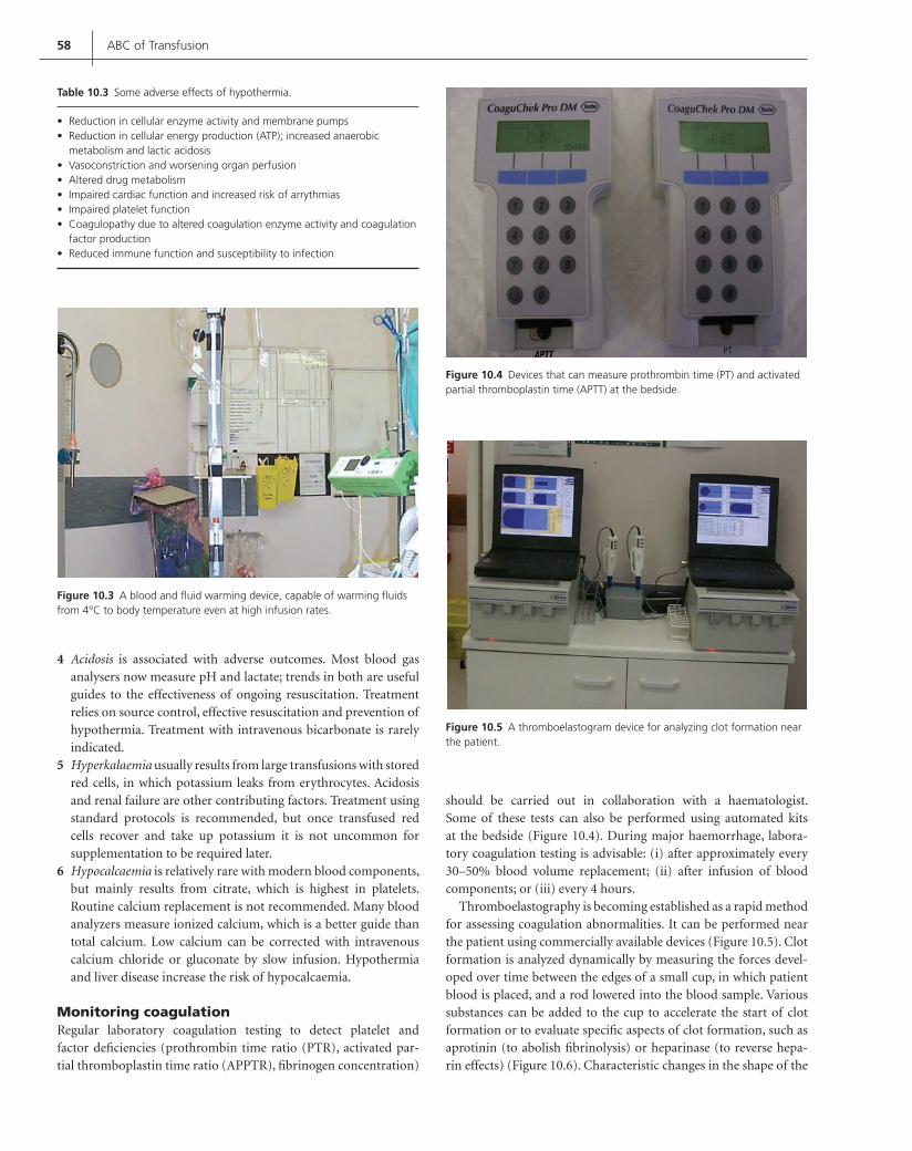
58 ABC of Transfusion
Acidosis4 is associated with adverse outcomes. Most blood gas analysers now measure pH and lactate; trends in both are useful guides to the effectiveness of ongoing resuscitation. Treatment relies on source control, effective resuscitation and prevention of hypothermia. Treatment with intravenous bicarbonate is rarely indicated.Hyperkalaemia5 usually results from large transfusions with stored red cells, in which potassium leaks from erythrocytes. Acidosis and renal failure are other contributing factors. Treatment using standard protocols is recommended, but once transfused red cells recover and take up potassium it is not uncommon for supplementation to be required later.Hypocalcaemia6 is relatively rare with modern blood components, but mainly results from citrate, which is highest in platelets. Routine calcium replacement is not recommended. Many blood analyzers measure ionized calcium, which is a better guide than total calcium. Low calcium can be corrected with intravenous calcium chloride or gluconate by slow infusion. Hypothermia and liver disease increase the risk of hypocalcaemia.
Monitoring coagulationRegular laboratory coagulation testing to detect platelet and factor defi ciencies (prothrombin time ratio (PTR), activated par-tial thromboplastin time ratio (APPTR), fi brinogen concentration)
should be carried out in collaboration with a haematologist. Some of these tests can also be performed using automated kits at the bedside (Figure 10.4). During major haemorrhage, labora-tory coagulation testing is advisable: (i) after approximately every 30–50% blood volume replacement; (ii) after infusion of blood components; or (iii) every 4 hours.
Thromboelastography is becoming established as a rapid method for assessing coagulation abnormalities. It can be performed near the patient using commercially available devices (Figure 10.5). Clot formation is analyzed dynamically by measuring the forces devel-oped over time between the edges of a small cup, in which patient blood is placed, and a rod lowered into the blood sample. Various substances can be added to the cup to accelerate the start of clot formation or to evaluate specifi c aspects of clot formation, such as aprotinin (to abolish fi brinolysis) or heparinase (to reverse hepa-rin effects) (Figure 10.6). Characteristic changes in the shape of the
Table 10.3 Some adverse effects of hypothermia.
Reduction in cellular enzyme activity and membrane pumps• Reduction in cellular energy production (ATP); increased anaerobic • metabolism and lactic acidosisVasoconstriction and worsening organ perfusion• Altered drug metabolism• Impaired cardiac function and increased risk of arrythmias• Impaired platelet function• Coagulopathy due to altered coagulation enzyme activity and coagulation • factor productionReduced immune function and susceptibility to infection•
Figure 10.3 A blood and fl uid warming device, capable of warming fl uids from 4°C to body temperature even at high infusion rates.
Figure 10.4 Devices that can measure prothrombin time (PT) and activated partial thromboplastin time (APTT) at the bedside.
Figure 10.5 A thromboelastogram device for analyzing clot formation near the patient.
Contreras_C010.indd 58Contreras_C010.indd 58 12/10/2008 1:46:30 Shobha12/10/2008 1:46:30 Shobha

Treatment of Massive Haemorrhage 59
trace can guide the use of blood components and drugs to correct coagulopathy (Figure 10.7).
Treatment of coagulopathy
Coagulation factorsThe PTR and APPTR should be kept at 1.5 by using fresh frozen plasma. Recommended dosing is 15 ml/kg (3–4 units for adults) per dose, although higher doses may be required for complete cor-rection. Fibrinogen concentration should be kept 1.0 g/L using fresh frozen plasma and/or cryoprecipitate. Platelet counts should be kept 50 109/L during ongoing haemorrhage. Using a trans-fusion trigger of 75 109/L is advisable if delays in delivery are anticipated.
FibrinolysisFibrinolysis results from the activation of plasminogen to plas-min. It is more common with larger blood loss, when treatment is delayed, with extensive tissue damage and in the presence of acidosis
and hypothermia. It can be detected by measuring D-dimers (or an equivalent test) and from thromboelastography. Fibrinolysis can be treated with agents such as tranexamic acid in consultation with a haematologist. These drugs are sometimes used prophylactically for surgical procedures with a high risk of massive haemorrhage, such as complex cardiac surgery and liver transplantation. Recent evidence suggests that aprotinin may worsen long term outcomes in cardiac surgery patients and tranexamic acid appears a safer alternative.
Recombinant activated factor VIIThis drug is not licensed for treatment of massive haemorrhage, but is increasingly used in this setting. Case reports and registries, and ongoing clinical trials, suggest a potential role in some patients. The drug promotes a ‘thrombin burst’, which may promote haemo-stasis when conventional measures have failed. It should only be used in consultation with a haematologist.
Recycling of autologous bloodCell salvage and reinfusion of the patient’s own blood is preferable to massive transfusion with allogeneic blood, which has undergone changes during storage that decrease the oxygen transport effec-tiveness of the red cells. Modern cell salvage devices can often be used during emergency situations, such as aortic aneurysm rupture and hepatic trauma (Figure 10.8; see Chapter 16).
Survival from major haemorrhage
Survival is increased by early intervention to control the source of bleeding and to prevent tissue ischaemia and organ failure. It is particularly important to avoid the combination of hypothermia, acidosis and coagulopathy (especially disseminated intravascular coagulation), which are associated with a high mortality.
Acknowledgments
The author thanks Dr Alastair Nimmo for providing the clinical pictures for this article.
Figure 10.6 A thromboelastogram trace from a patient prior to major vascular surgery. Each channel is a different assessment of clot formation created by adding different substances to the sample to inhibit aspects of coagulation.
Figure 10.7 An abnormal thromboelastogram trace from a patient with severe coagulopathy from massive haemorrhage. The characteristic ‘onion’ shape of the trace indicates fi brinolysis. The top right panel is relatively normal because aprotinin was added to the sample, showing that this drug would be an effective treatment for the patient.
Figure 10.8 Effective use of cell salvage during surgery associated with major bleeding. The machine can recycle blood continuously during overwhelming blood loss, as in this case.
Contreras_C010.indd 59Contreras_C010.indd 59 12/10/2008 1:46:35 Shobha12/10/2008 1:46:35 Shobha

60 ABC of Transfusion
Further reading
British Committee for Standards in Haematology. Guidelines on the manage-
ment of massive blood loss. British Journal of Haematology 2006; 135:
634–41.
Kwan I, Bunn F, Roberts I. Timing and volume of fl uid administration for
patients with bleeding. Cochrane Database of Systematic Reviews 2003,
Issue 1. Art. No. CD002245.
Levi M, Peters M, Buller HR. Effi cacy and safety of recombinant factor VIIa
for treatment of severe bleeding: a systematic review. Critical Care Medicine
2005; 33(4): 883–90.
McClelland DBL. Handbook of Transfusion Medicine, 4th edn. HM Stationery
Offi ce, London, 2007.
Spahn DR, Cerny V, Coats TJ. et al. Task Force for Advanced Bleeding Care
in Trauma. Management of bleeding following major trauma: a European
guideline. Critical Care 2007; 11(1): R17.
Contreras_C010.indd 60Contreras_C010.indd 60 12/10/2008 1:46:40 Shobha12/10/2008 1:46:40 Shobha

61
CHAPTER 11
Immunological Complications of Blood Transfusion
Marcela Contreras and Cristina Navarrete
ABC of Transfusion, 4th edition, 2009. Edited by Marcela Contreras. © 2009
Blackwell Publishing, ISBN: 978-1-4051-5646-2.
The transfusion of blood or its components can correct many defi ciencies, even if only temporarily, and in most cases this effect can be achieved without any untoward effects. Nevertheless, incom-patibilities of several kinds do occasionally cause trouble. Each blood cell has many different antigens on its surface, so the cells of a donor are virtually certain to contain antigens different from those of the recipient. In addition, the donor’s plasma may con-tain proteins that are foreign to the recipient. The donor’s plasma may also contain antibodies incompatible with antigens carried by cells in the recipient (for example, transfusion of group O plasma to a group A patient). Fortunately the transfusion of blood cells or plasma containing foreign antigens causes an immediate reac-tion only when the recipient’s serum contains a corresponding antibody, and then only rarely. In practice, severe reactions (caused, for example, by transfusion of incompatible red cells) are rare (Table 11.1), whereas mild reactions (caused, for example, by
reactions to foreign protein or hapten ingested by the donor and characterized by urticaria) are common.
Reactions to incompatible red cells
For a number of reasons, antibodies to red cell antigens are the most important in blood transfusion. Firstly, the volume of red cells transfused is usually greater than that of white cells or platelets, so that if incompatible red cells are transfused, the consequences are comparatively severe. Secondly, most individuals have ‘naturally occurring’ antibodies to either blood group A and/or B present on red cells. Thirdly, antibodies evoked by transfusions or pregnancy in women of childbearing age may subsequently cause haemolytic disease in their offspring.
Naturally occurring antibodies and immune antibodies to red blood cellsSome antibodies are found in subjects who have never been exposed to foreign blood cells, and the most important of these so-called ‘naturally occurring’ antibodies are anti-A, anti-B and
OVERVIEW
Immunological complications of transfusion can be due either • to: (i) antigens present in the transfused donor’s cells or plasma reacting with the recipient’s antibodies; or (ii) antibodies or lymphocytes present in the transfused donor’s plasma reacting with antigens or cells in the recipient.
The most frequent serious complications of transfusion are acute • haemolytic transfusion reactions due to ABO blood group incompatibility and transfusion-related acute lung injury (TRALI). ABO incompatibility is caused by giving the wrong blood to a patient. TRALI is caused by white cell antibodies present in the donor’s plasma reacting with incompatible cells in the recipient.
Overall, the most frequent but not serious complications of • transfusion are non-haemolytic febrile transfusion reactions and urticaria. The former have considerably decreased in the UK, since the introduction of universal leucodepletion.
Table 11.1 Immunological complications of blood transfusion.
Percentage frequency
Red cells Haemolytic reactions: Immediate 0.02 Delayed 0.2
White cells Febrile reactions* 5.10Acute lung injury 0.01
Platelets Post-transfusion purpura 0.01
Plasma proteins: Native Anaphylaxis 0.01 Ingested Urticaria 1–3
* The incidence of febrile transfusion reactions has decreased signifi cantly since the introduction of leucodepletion of red cells and platelets. It is now estimated to have a frequency of 0.1–0.2% or less.
Contreras_C011.indd 61Contreras_C011.indd 61 12/19/2008 1:53:16 PM12/19/2008 1:53:16 PM

62 ABC of Transfusion
anti-AB (Box 11.1). Most others are ‘immune’ antibodies and arise only after transfusion, or after pregnancy, when small numbers of fetal red cells enter the maternal circulation and may immunise the mother to antigens that the fetus has inherited from the father (see Chapter 6). Anti-RhD is the most common (and clinically signifi cant) immune antibody against red blood cells.
Most incompatible transfusions are caused not by serological errors but by giving blood to the ‘wrong’ patient, that is, to a dif-ferent patient from the one whose serum was tested before trans-fusion. The frequency with which incompatible transfusions are given is unknown, but their incidence is underreported; published estimates range from about 1/1000 to 1/20 000 transfusions (see Chapter 15).
Immediate haemolytic transfusion reactionsAn immediate haemolytic transfusion reaction means the occur-rence of signs of increased destruction of red cells soon after a transfusion. Mild haemolytic reactions may be caused in various ways – for example by transfusing blood that has been stored for too long – but severe haemolytic reactions are almost always caused by transfusing incompatible red cells (in most cases ABO incom-patible) (Boxes 11.2 and 11.3).
Potent ABO antibodies present in group O plasma (usually in the form of fresh frozen plasma or platelets, but also in whole blood and, in the past, some fractionated blood products) may lead to severe haemolysis in group A, B or AB recipients. The following are the main reasons why ABO incompatibility plays such a large part:
Anti-A, anti-B and anti-AB are the most common blood group 1 antibodies. They are present in the plasma of all subjects whose red cells lack the corresponding antigens. For example, a person of group O always has anti-A, anti-B and anti-AB in the plasma.If a mistake is made in identifying a patient and the wrong blood 2 is transfused (that is, blood that has been selected for a differ-ent patient), the chance (in a white subject) that it will be ABO incompatible is as high as one in three.
Anti-A and anti-B are usually potent immunoglobulin M 3 (IgM) antibodies (especially in group O subjects) and by activating the full complement pathway produce rapid intra-vascular lysis of incompatible red cells so that, if reactions to ABO incompatible blood occur, they are usually severe. Fortunately, the resulting morbidity is of the order of 20% and mortality is less than 10%.
The following features of haemolytic transfusion reactions are the result of transfusion of ABO incompatible red cells.
CircumstancesAs the cause of most incompatible transfusions is failure to identify the recipient correctly, many of such transfusions are given in emer-gencies and in surroundings where patients are not well known to those who are attending to them (Box 11.4). Thus most are given in operating theatres or intensive care units. Some patients who receive
Box 11.1 Antibodies
Naturally occurring Examples
Anti-Aproduced
No exposure to foreignred cells but exposureto bacteria containingfor example, A-like antigens
Immune
Anti-Rhproduced
Exposure to foreign redcells (for example Rh+)
by transfusion
or pregnancy(Rh+ fetus)
Box 11.2 Haemolytic reactions
Incompatible redcells (Group A)
Ag
Ab
Complex
Antibody inrecipient (anti- AB) Small fragments of
complement liberated(C3a, C5a)
Complementactivation
Haemolysis
Haemoglobinuria
Release ofthromboplasticsubstances
Disseminatedintravascularcoagulation
Cytokines
Symptoms and signsincluding hypotensionand renal failure
Box 11.3 Immunological complications
Incompatibility to:Red cells – Haemolysis:• – intravascular (ABO)– extravascular (Rh)White cells• Platelets• Plasma proteins•
Immune modulation:Benefi cial• Detrimental•
Box 11.4 Circumstances in which most incompatible transfusions are given
Patient wrongly identifi ed because:Unconscious (operation)• Wristband removed during operation • Human error• Sample for pretransfusion tests taken from the wrong patient•
Contreras_C011.indd 62Contreras_C011.indd 62 12/19/2008 1:53:17 PM12/19/2008 1:53:17 PM

Immunological Complications 63
incompatible blood are unconscious at the time, so that there are no symptoms that might call attention to what is happening.
Mechanisms of red cell destructionWhen ABO incompatible red cells are transfused – for example donor group A and recipient group O – the incompatible IgM anti-body (anti-A and anti-AB in this case) immediately binds to the donor’s red cells (Figure 11.1). The bound antibody activates com-plement leading to immediate lysis of donor red cells in the plasma. When the antibody is potent, virtually all the transfused red cells may be lysed within the bloodstream. Many other blood group allo-antibodies, leading to immediate haemolytic reactions, are IgG and activate complement, but as a rule they do so only partially, up to C3b, and the coated cells, after binding to monocytes/macrophages, are either ingested or destroyed by cytotoxicity as they pass through the liver and spleen. Some anti bodies – for example anti-Rh and a few anti-human leucocyte antigen (anti-HLA) – do not activate
complement (Figure 11.2), and these cause macrophages to remove coated red cells or platelets in the spleen only.
Symptoms and signsThe vast majority of patients experiencing an ABO incompatible transfusion reaction have no symptoms. However, about 20–25% of patients, particularly if group O, will have symptoms of vary-ing severity (Table 11.2). In the conscious patient, the transfusion of even a few millilitres of ABO incompatible blood may cause symptoms within 1 or 2 minutes. The patient becomes restless and complains of a feeling of oppression that is often accompanied by substernal pain, chills and fever. The patient may also complain of a burning face and may have abdominal pain and may vomit. In the unconscious patient the most important signs are hypotension and uncontrollable bleeding.
The main cause of the symptoms and of the hypotension is the release of the products of complement (C3a and C5a) into the plasma. These are polypeptides with a molecular weight of about 20 000, which cause the contraction of smooth muscle and the production of nitric oxide, oxygen radicals and cytokines such as tumour necrosis factor (TNF) and interleukin-1 (IL-1); they also cause degranulation of mast cells leading to the release of vasoac-tive substances (bradykinin and serotonin). Bleeding is caused by disseminated intravascular coagulation, which is thought to be triggered by procoagulant substances released from red cell stroma after lysis and also, possibly, by the direct activation of the coagula-tion system by complement, by antigen–antibody complexes and by cytokines.
Oliguria is common after the transfusion of ABO incompat-ible blood and is thought to be the result of changes in renal blood fl ow precipitated by hypotension, rather than haemoglobinuria (Box 11.5).
As soon as it is suspected that incompatible blood has been transfused, the transfusion should be stopped, and an intra-venous infusion of crystalloid solution should be started to maintain urinary output. Frusemide 80–200 mg should be given
Figure 11.1 IgM anti-A binding to two adjacent A sites activates the whole of complement pathway (C1 to C9); C8 and C9 make holes in the red cell membrane.
Haemoglobinliberatedinto plasma
IgM anti-A
Red cell ruptures
A sites
Figure 11.2 IgG anti-Rh does not activate complement. Red cells coated with antibody adhere to a macrophage or killer lymphocyte and are either ingested or lysed at the cell surface.
IgG anti-Rh
Rh sites
Antibody coated red cells
Red cells adheresto, and is lysedoutside, killerlymphocyte
Red cells adheresto, and is lysedoutside,macrophage
lysozymes Perforins
Haemoglobinliberated
Haemoglobinliberated
Bilirubinliberated
Red cells lysedwithin macrophage;haemoglobinmetabolised
Table 11.2 Immune red cell destruction.
Site of destruction
Predominantly intravascular
Predominantly extravascular
Characteristics of antibodies
Potent, lytic Antibodies that do not activate or only partially activate complement
(Anti-A and -B) (Anti-Rh, anti-K, anti-Jk, and others)
Symptoms Substernal pain,lumbar pain, restlessness
Nausea, shivering
Signs:
Immediate Hypotension, fever, uncontrollable bleeding
Fever
Later Haemoglobinuria Jaundice
Contreras_C011.indd 63Contreras_C011.indd 63 12/19/2008 1:53:17 PM12/19/2008 1:53:17 PM

64 ABC of Transfusion
intravenously over 4 hours. The labels on the transfused unit and on all relevant blood samples should be checked and the laboratory notifi ed; this is particularly important, because if the unit has been given to the wrong patient the right unit may have been – or may be about to be – given to another ‘wrong’ patient. If renal failure becomes established this can lead to death and the advice of a renal physician should be sought as soon as possible.
Although severe symptoms may follow the transfusion of only a few millilitres of incompatible red cells in group O patients with potent anti-A and anti-B, in patients with weak antibodies the transfusion of several units of ABO incompatible blood may cause only trivial effects. Very old and very young patients have weak ABO antibodies. As seen in Chapter 4, most recipients of blood in England belong to these age ranges.
Immediate haemolytic reactions as a result of transfusing incom-patible red cells may also be caused by the presence of any other red cell antibodies active at 37°C in the recipient’s blood. The next most common red cell antibody after anti-A and anti-B is anti-RhD. Rh antibodies are IgG, and extravascular haemolytic transfusion reactions caused by Rh incompatibility are usually much milder than those caused by ABO incompatibility. Other IgG antibodies, such as Kell, Duffy and Kidd, may also lead to extravascular hae-molytic transfusion reactions. In Rh incompatibility fever is often the only sign of an adverse reaction, although jaundice, develop-ing later, is quite common and, if IgG antibodies are very strong, haemo globinuria caused by extravascular red cell destruction (not associated with renal failure) sometimes develops.
Delayed haemolytic transfusion reactionsThese reactions occur in subjects who have been immunized to a foreign red cell antigen by an earlier transfusion or a previous pregnancy but in whom the concentration of antibody to the for-eign antigen is so low that it cannot be detected by tests before the transfusion. Following the current transfusion of red cells containing the same foreign antigen there is a secondary immune response, and after a few days the concentration of IgG antibody has risen suffi ciently to cause rapid destruction of those trans-fused red cells that contain the foreign antigen. The signs of a delayed haemolytic transfusion reaction are usually not noticed, but fever, a falling haemoglobin concentration and jaundice or, very rarely, haemoglobinuria, usually about 5–10 days after trans-fusion may occur (Figure 11.3). The specifi cities of the antibodies responsible for delayed haemolytic transfusion reactions are most often within the Rh system – that is, anti-c, anti-E and so on – or
in the Kidd (Jk) system, but they may have other specifi cities such as anti-K.
Delayed haemolytic transfusion reactions occur after about one in 500 transfusions; alone they are seldom fatal, but they may have a serious adverse effect on a patient who is already gravely ill.
Reactions to incompatible white cells
White cells present in transfused products also express antigens which, if not identical to those present in the recipient, can be recognized as foreign (non-self) resulting in the development of antibodies responsible for some of the serious complications of blood transfusion. On the other hand, antibodies (and sometimes immune cells) present in the transfused product may react directly with the relevant antigens in the recipient and in this way provoke a transfusion reaction. The most important white cell antibodies are those reacting with HLA antigens present on all white cells and with the human neutrophil antigens (HNAs) found primarily on neutrophils. (See Table 11.3 for the expression of antigens on blood cells and plasma.)
In contrast to red cell antibodies, the majority of HLA (and up to a certain extent HNA) antibodies are of immune origin, i.e. they
Box 11.5 Oliguria
Incompatible red cells + antibody:• complement activatedProducts of complement • activation hypotension oliguriaTreatment: Maintain urinary fl ow (infuse crystalloid solution, inject •
frusemideCall renal physician
Figure 11.3 Delayed haemolytic transfusion reaction.
Days after transfusion
Transfusion ofJk (a+) red cells
Jaundice orhaemoglobinuria
anti – Jka
Surv
ivin
g J
k (a
+ ) re
d c
ells
0 2 4 6 8 10 12
Table 11.3 Antigens expressed on white blood cells and platelets.
Class I (A,B,C)
HLA Class II (DR, DQ, DP)
HNA HPA ABO
Lymphocytes T + * Lymphocytes B + + Monocytes + + Granulocytes + * + Dendritic cells + ++ Platelets + + +Rbcs +a +Plasma (soluble) + + + + +
* On activated cells.a Immature rbc.
Contreras_C011.indd 64Contreras_C011.indd 64 12/19/2008 1:53:17 PM12/19/2008 1:53:17 PM

Immunological Complications 65
develop after exposure to foreign HLA or HNA molecules through pregnancy, transfusions or transplantation.
During pregnancy, the mother develops antibodies against the paternal HLA (as well as HNA and human platelet antigens (HPA)) specifi c antigens present in the fetus. These antibodies are normally IgG and of high affi nity. Antibodies that develop following transfu-sions are a mixture of IgG and IgM, are often transient and can be detected in ~10% of previously transfused patients and in about 30–70% of multitransfused patients. Antibodies that develop fol-lowing transplantation are predominantly IgG, but IgM can also be found transiently.
White cell antibodies present in blood transfusion recipients are implicated in the development of non-haemolytic febrile transfu-sion reactions (NHFTRs) and antibodies present in the transfused blood or blood component are associated with the development of transfusion-related acute lung injury (TRALI). On the other hand, viable lymphocytes present in blood or blood components are responsible for the development of transfusion-associated graft-versus-host disease (TA-GvHD).
Non-haemolytic febrile transfusion reactionsIn patients whose plasma contains potent leucocyte antibodies (mostly HLA but also HNA and sometimes platelet-specifi c anti-bodies), the transfusion of whole blood or blood components con-taining incompatible leucocytes or platelets may provoke severe febrile reactions. The most common feature is fever, often pre-ceded by shivering and beginning 30–60 minutes after the start of the transfusion. In some cases fl ushing of the face develops within 5 minutes of the start of the transfusion, presumably as a result of the release of cytokines and other pyrogens from the recipient’s granulocytes and monocytes, stimulated by the products of com-plement activation or by activation of these cells when engulfi ng or lysing antibody-coated cells (Box 11.6).
Febrile reactions are quite common in patients who have previ-ously been pregnant or who have had transfusions that have stim-ulated the formation of leucocyte antibodies. Most reactions are mild and can be dealt with by slowing the transfusion and giving
Figure 11.4 Chest radiograph of a patient who experienced a severe transfusion reaction as a result of potent leucoagglutinins in transfused plasma. There are numerous nodules, predominantly perihilar, and infi ltration of the lower lung fi elds.
Box 11.6 Febrile reaction to incompatible white cells
Destroyed byfull complementactivation
Engulfed by hostmonocytes
Release of pyrogen
Foreign leucocytes + leucocyte antibody
FEVER
antipyretics. In patients who have had at least two severe febrile reactions, blood or platelets with a reduced white cell load or that are leucodepleted should be transfused – that is, blood that has either had the buffy coat removed or has been passed through a special leucodepletion fi lter. In most cases, the removal of buffy coats, which reduces the white cell content 10-fold, will be suffi cient to avoid NHFTRs.
With the introduction of universal leucodepletion in the UK, NHFTRs have decreased signifi cantly. However some mild febrile reactions remain, presumably caused by cytokines and other biological response modifi ers such as TNF, IL-1a, IL-6 or IL-8 released by the remaining leucocytes present in stored blood or platelet concentrates.
Transfusion-related acute lung injuryIf the donor’s plasma contains potent leucoagglutinins that are incompatible with the recipient’s granulocytes, transfusion may cause a severe reaction characterized by chills, fever, a non-produc-tive cough, dyspnoea and severe hypoxaemia occurring during or in the 6 hours after transfusion. A chest radiograph shows numer-ous bilateral pulmonary nodules – predominantly perihilar – with infi ltration of the lower lung fi elds (Figure 11.4). Most cases require prompt mechanical ventilation. The donors are almost always multiparous women who have developed leucoagglutinins during pregnancy. This reaction is clinically indistinguishable from acute respiratory distress from other causes and there is no specifi c treat-ment, although symptoms usually improve within 48–96 hours with the pulmonary infi ltrates receding within 1–4 days. These reac-tions occur in approximately one in 3000–5000 transfusions and 5–10% of cases are fatal. The main blood components involved are those containing large volumes of plasma such as fresh frozen plasma and platelets, but red blood cell concentrates have also been implicated. HLA class I and class II and HNA antibodies in donor plasma have been implicated in 80% of clinically convincing cases.
Contreras_C011.indd 65Contreras_C011.indd 65 12/19/2008 1:53:18 PM12/19/2008 1:53:18 PM

66 ABC of Transfusion
These antibodies react with the leucocytes of the patient, trigger-ing the activation of the neutrophils and the complement cascade, the release of chemotactic factors, sequestration of neutrophils in the small vessels of the lung, and ultimately damage to the vascu-lar endothelium. There is an accumulation of leucocytes within the small vessels of the lung and lung oedema. Throughout this pro-cess there is secretion of infl ammatory cytokines such as IL-1 and TNF, which may induce and amplify the reaction (Figure 11.5).
Biologically active lipids present in stored, but not fresh, cellular blood components have been implicated by some, but other pre-disposing clinical conditions (e.g. recent surgery, cytokine therapy, active infection) also seem to be required. The laboratory investi-gations of these cases involve the screening of implicated donors for HLA and HNA antibodies. Female donors and male donors with a history of transfusion should be the fi rst ones to be investi-gated and, if antibody positive, the patient should be typed for the relevant antigen. The gold standard test is a leucocyte crossmatch between the donor’s plasma and the patient’s white cells. A positive crossmatch confi rms the diagnosis of TRALI.
Transfusion-associated graft-versus-host diseaseThis is a rare but usually fatal immunological complication of blood transfusion occurring approximately within 2 weeks of transfusion, which results from the engraftment and clonal expan-sion of viable, mostly HLA compatible, immunocompetent donor lymphocytes in an immunocompromised (though occasionally immunocompetent) host.
The clinical features classically include a skin rash, diarrhoea, fever, hepatitis and pancytopenia. Defi nitive diagnosis of TA-GvHD includes the detection of donor-derived cells or DNA in the blood or affected tissues of the recipient.
The incidence of TA-GvHD is not known, although it has been suggested that the disorder is underreported. The fi rst 2 years of haemovigilance in the UK indicated that TA-GvHD was the most common recorded cause of transfusion-related death. The blood
components implicated are those containing lymphoctyes and it can be prevented by the use of -irradiated blood components.
Reactions to incompatible platelets
Although it is doubtful whether platelets themselves can cause febrile reactions, transfusions of platelet concentrates quite often used to, because they contained contaminating leucocytes. In addition, because platelet concentrates contain plasma, all types of reactions due to plasma can be experienced by patients receiv-ing platelet transfusions. (Table 11.4 details the usual ranges of leucocyte and platelet concentrations found in blood.)
Platelets can cause a serious kind of delayed reaction known as post-transfusion purpura, characterized by severe thrombocytopenia approximately 1 week post-transfusion (Figure 11.6). The patient has become sensitized to a foreign platelet anti-gen (most commonly HPA-1a, previously termed PI-Al), usually as a result of previous pregnancies but rarely by transfusion. After a later transfusion of blood with platelets carrying this antigen, a secondary response develops, and this leads to destruction of the patient’s own HPA-1a negative platelets and severe thrombo-cytopenia. It is likely that the patient’s own HPA-1a negative platelets are destroyed by autoantibodies produced as a result of
Adherence of neutrophils topulmonary endothelium orepithelium
Endothelial/epithelial cellmembrane permeabilization
Lung oedema
Secretion of IL–1β, TNFα andIL-8 may amplify the reaction
•
•
•
•
C
C5chemotacticfactorC’ activation
HLA/HNA abs
HNA
HLA
inactive active
12
Endothelial cell
LFA-1ICAM-1
2
5
a
Figure 11.5 Diagram showing how TRALI occurs. HLA, human leucocyte antigen; HNA, human neutrophil antigen; ICAM-1, intercellular adhesion molecule; IL, interleukin; LFA-1, leucocyte function antigen; TNF, tumour necrosis factor.
Table 11.4 Leucocyte and platelet levels in blood.
Cell type Blood count
Total leucocytes 4.0–11.0 109/LNeutrophils 2.5–7.5 109/LEosinophils 0.04–0.4 109/LBasophils 0.01–0.1 109/LMonocytes 0.2–0.8 109/LLymphocytes 1.5–3.5 109/LPlatelets 140–400 109/L
Contreras_C011.indd 66Contreras_C011.indd 66 12/19/2008 1:53:18 PM12/19/2008 1:53:18 PM

Immunological Complications 67
the original alloimmunization, which become stimulated with the second antigenic challenge.
Although the incidence of HPA-1a-negative subjects among Europeans is two in 100, post-transfusion purpura is rare and occurs in approximately one in 700 000 transfusions, usually in female patients. Onset is severe and sometimes fatal. The best treat-ment is large doses of immunoglobulin given intravenously or plasma exchange.
Immunological refractoriness to platelet transfusionsPlatelet transfusion therapy plays a major role in the management of patients with haematological and oncological disorders with intermittent or long-lasting thrombocytopenia. However, some of these patients become refractory to the transfusion of platelets from random donors and this is characterized by the failure to gain adequate platelet count increments (<10 109/l) 1 hour post trans-fusion. Platelet refractoriness may be the result of immunological and/or non-immunological causes. Immunological refractoriness, caused by antibody-mediated destruction of transfused platelets, is normally due to HLA antibodies, although antibodies against HPAs and high titre ABO antibodies have also been implicated occasionally.
Different approaches are currently being used for the manage-ment of immunological platelet refractoriness including: (i) the provision of crossmatch-compatible platelets; (ii) the use of plate-lets negative for the HLA antigen(s) corresponding to the patient’s HLA antibody; and (iii) the use of complete or partially HLA-A and -B antigen-matched platelets from unrelated donors. All these different approaches have been shown to be effective. Although platelets express HLA-A, -B and -Cw antigens, matching at the HLA-A and -B locus antigens only is required.
The laboratory investigations to identify these cases and provide HLA-matched platelets include:
screening for and detection of HLA-specifi c antibodies• defi nition of antibody specifi city• HLA typing of patients• selection and issue of HLA-compatible platelets from a large • panel of HLA-typed platelet donorsdocumentation of post-transfusion increments.•
Reactions to plasma proteins
The commonest adverse reaction to plasma (or any blood com-ponent containing plasma) is urticaria (hives) (Figure 11.7). In fact, 1–3% of patients transfused with plasma experience urti-caria. The cause is presumably a reaction between plasma pro-teins or some foreign allergen (for example from pollen or milk) present in the donor’s plasma and a corresponding IgE antibody in the recipient. Urticaria is usually mild, but if it is severe it can be treated by slowing the transfusion and giving an antihistamine (e.g. chlorpheniramine 10 mg i.v. or i.m.). Patients who have developed urticaria after previous transfusions may be given antihistamine orally before subsequent transfusions.
Rarely, severe anaphylactic reactions occur, and they are char-acterized by hypotension and even shock, a feeling of a ‘lump’ in the throat, stridor, substernal pain, chest tightness, wheez-ing, dyspnoea and gastrointestinal symptoms including abdomi-nal cramps. These signs and symptoms are usually the result of a reaction between normal IgA in the donor’s plasma and so-called class-specifi c anti-IgA in the recipient’s plasma. This antibody is found only in ‘algA’ subjects, who are almost completely defi cient in IgA (<1 mg/L plasma). The incidence of algA subjects in the normal population is about one in 1000. However, severe ana-phylactic reactions are extremely rare and can lead to death. They should be treated promptly by stopping the transfusion and giv-ing adrenaline 0.5–1 mg subcutaneously or intramuscularly and hydrocortisone 100–200 mg intravenously. Anaphylactic reac-tions in patients with anti-IgA in their plasma can be prevented by using IgA-defi cient donors or, if unavailable, well washed red cells.
Immunomodulatory effects of transfusion
Some, though not all, studies in humans and experimental animals argue in favour of an immunosuppressive effect of transfusion with a detrimental effect as regards to an increased rate of postoperative bacterial infection and an increased rate of recurrence of colorectal carcinoma. There is also a possible benefi cial effect of some blood trans fusions on the survival of renal allografts. It is not known which
Figure 11.6 Post-transfusion purpura.
Later transfusionof Pl (A1+) blood
Purpura
105Days after transfusion
Plat
elet
co
un
t (×
109 /l
)
0
100
200
300
Figure 11.7 Urticaria is one of the commonest immunological complications of blood transfusion.
Contreras_C011.indd 67Contreras_C011.indd 67 12/19/2008 1:53:19 PM12/19/2008 1:53:19 PM

68 ABC of Transfusion
of the components of blood has a greater impact on the immune system, but leucocytes and especially lymphocytes are the favoured candidates.
Further reading
Bidwell J, Navarrete C, eds. Histocompatibility Testing. Imperial College Press,
London, 2000.
Bax J. Transfusion-related acute lung injury (TRALI): a serious adverse event
of transfusion. Vox Sanguinis 2005; 89: 1–10.
Kleinman S. A perspective on transfusion-related acute lung injury two years
after the Canadian Consensus Conference. Transfusion 2006; 46: 1465–8.
Mollison PL, Engelfriet CP, Contreras M. Blood Transfusion in Clinical
Medicine, 10th edn. Blackwell Scientifi c Publications, Oxford, 1997.
Navarrete C. The HLA system in blood transfusion. Baillière’s Clinical
Haematology 2000; 13(4): 511–32.
Contreras_C011.indd 68Contreras_C011.indd 68 12/19/2008 1:53:20 PM12/19/2008 1:53:20 PM

69
CHAPTER 12
Infectious Complications of Blood Transfusion: Bacteria and Parasites
John Barbara and Marcela Contreras
ABC of Transfusion, 4th edition, 2009. Edited by Marcela Contreras. © 2009
Blackwell Publishing, ISBN: 978-1-4051-5646-2.
Long before the transmission of human immunodefi ciency virus (HIV) became a prominent potential hazard of blood transfusion, considerable expertise in preventing the transmission of infection by transfusion had already been developed in the blood services. The fi rst and most important step in maintaining a safe blood supply will always be a rigorous process of selection of prospec-tive voluntary, unpaid blood donors (Box 12.1; see Chapter 1). The second is the use of specifi c microbiological screening tests (Box 12.2). Agents transmissible by transfusion can be either cell associated – for example cytomegalovirus (CMV) and human T-cell leukaemia virus type 1 (HTLV-1) – or plasma associated – for example hepatitis B virus – or both (e.g. HIV). If they are plasma associated, pooling large numbers of units of plasma (e.g. 15 000–20 000 U as in the production of factor VIII) greatly increases the chances of disseminating such contaminants. Due to the separation of each unit of blood into its components, even
without pooling, transfusion of blood components may result in up to four or fi ve patients being infected by a single contaminated donation. Fortunately, current inactivation procedures render fractionated plasma products virtually free of microbial infectivity. This offers a third approach to ensuring the transfusion of safe blood products and the concept is gradually being extended to labile components as well.
OVERVIEW
Careful selection of voluntary, unpaid donors remains a fi rst key • step to maximize blood safety.
Although ‘persistence’ is the hallmark of transfusion infections, • acute infections at high incidence may pose a threat.
Bacterial infections are a major component of the residual • microbial risks from transfusion in the developed world.
Rigorous history taking is essential for reducing the risk of • transmitting parasitic infections.
Visual checks of the blood bag prior to transfusion should • always be undertaken.
Most countries screen blood donors for human • immunodefi ciency virus, hepatitis B and C virus and syphilis by appropriate donor selection and laboratory testing.
Some countries also test for human T-cell leukaemia virus, • cytomegalovirus, West Nile virus and Chagas’ disease.
Inactivation procedures have increased the safety of fractionated • blood products and also of some labile blood components.
Box 12.1 Ensuring a safe blood supply
Donor selection• Microbiological screening tests• Microbial inactivation•
Box 12.2 Screening tests for blood donations (see Chapter 1)
Mandatory in the UKHepatitis B surface antigen• Antibody to human immunodefi ciency virus 1 (HIV-1) and HIV-2 in • combination with HIV antigenAntibody to • Treponema pallidum (syphilis)Antibody to hepatitis C virus (HCV)• Antibody to human T-cell leukaemia virus (HTLV) (on pooled • samples)HCV genome detection or nucleic acid technology for HCV (on • pooled samples)
Optional (for selected recipients)Antibody to cytomegalovirus•
Optional (on selected donors with relevant history):Antibody to• Plasmodium falciparum (malaria)Antibody to • Trypanosoma cruzi (Chagas’ disease)
Tests performed in some countriesHIV genome detection• Hepatitis B virus genome detection• Antibody to hepatitis B core• Alanine aminotransferase levels• BI9 genome detection• West Nile virus genome detection•
Contreras_C012.indd 69Contreras_C012.indd 69 12/5/2008 6:11:04 PM12/5/2008 6:11:04 PM

70 ABC of Transfusion
Properties of infections transmissible by transfusion
Agents transmitted by blood transfusion often possess a combination of some or all of the following properties:
they are present in the blood for long periods, sometimes in high • titresthey can cause subclinical infections or only mild symptoms• they have long incubation periods (sometimes years) before • clinical signs appearthey may exist in a latent or carrier state, or both; they are stable • in blood stored at 4°C.certain agents causing acute infection with only transient virae-• mia may be a threat if the rate of new (incident) infections is very high. An example is West Nile virus in North America.
Screening tests for blood donations
Screening tests are paradoxically usually directed at antibody to the agent rather than antigens from the agent, except in the case of hepatitis B virus. Antibody screening tests are markers for certain persistent or chronic infections and therefore indicate a potential for infectivity, especially when the inoculum is as large as a unit of blood or a blood component.
Various agents may be transmitted by transfusion, but in the UK there are only fi ve serological screening tests for blood dona-tions that are currently mandatory (Box 12.2). They are tests for: (i) hepatitis B surface antigen (HBsAg) for hepatitis B virus; (ii) antibody to HIV-1 and -2, in combination with HIV antigen; (iii) antibody to hepatitis C virus (HCV); (iv) antibody to Treponema pallidum (syphilis); and (v) antibody to HTLV performed on the pools of 48 samples used for detection of HCV genome (nucleic acid technology or NAT). Tests for several other agents are available, but it has not yet been considered necessary to extend the present range. The range of techniques for screening, to which process con-trol can be applied, includes enzyme-linked immunosorbent assay (ELISA), haemagglutination and gelatin particle agglutination. For a test to be suitable for screening blood for transfusion, several con-fl icting demands have to be met.
Quality control of microbiological screening of blood donations for transfusion in the UK is essential because the occurrence of donations positive for hepatitis B virus, HIV, HCV or syphilis is rare. In contrast to blood grouping, in which every sample produces a ‘positive’ result of some sort, in microbiological screening tests most donor serum samples are negative. Great vigilance is therefore required in carrying out the routine screening tests (Figures 12.1 and 12.2). In low prevalence populations even an apparently low rate of false positive results from a screening test implies that a posi-tive reaction has little predictive value. If, for example, an agent has an incidence of one in 100 000 donations, then a test with a speci-fi city of 99.9% will produce a false positive reaction once in every 1000 donations, or 100 false positive reactions for every true posi-tive (Table 12.1). It is therefore imperative that any donor samples that give a positive reaction in any of the mandatory screening tests should be sent to a reference laboratory for confi rmation before the donor is informed of the results. Blood centres in the UK and
in other developed countries, use assays that have low false positive rates, and they all have access to centralized reference laboratories that carry out a battery of confi rmatory tests, which virtually elimi-nates the possibility of mislabelling uninfected donors. In the UK these reference laboratories regularly coordinate their confi rmation protocols.
Specifi city of microbiological assays is vital if the confi dence of donors is to be maintained, and it must not be forgotten in the search for increased sensitivity. Fortunately recombinant and
Figure 12.1 Reagents for ELISA can be automatically dispensed, with full process control.
Figure 12.2 All assay steps can be performed automatically. Although microplate-based assays are illustrated here, fully enclosed systems such as the Abbot Laboratories PRISMTM machine are also in common use. This system has been fully assessed by the UK blood services to ensure that full process control of testing is achieved.
Table 12.1 Table of predictive values.
Prevalence: 1 in 100 000• Specifi city: 99.9% (minimum UK requirement)• False positive rate: 1 in 1000• 100 false positives for every true positive•
Contreras_C012.indd 70Contreras_C012.indd 70 12/5/2008 6:11:04 PM12/5/2008 6:11:04 PM

Bacterial and Parasitic Complications 71
synthetic antigens and modern molecular biological methods have produced remarkable improvements in the sensitivity and specifi city of assays, such as those for detecting HIV infection.
Bacterial complications of transfusion
Bacteria comprising normal skin fl ora, such as staphylococci, can contaminate blood donations at the time of collection; the blood’s own bactericidal powers, citrate and cold storage will, however, destroy many such contaminants. Due to their storage require-ments at room temperature, platelet concentrates carry the highest risk of bacterial contamination.
Bacterial complications of transfusion are rare in the UK because of the use of sterile, disposable collection sets and clean phle-botomy techniques. When they do occur, however, they can rap-idly be fatal, as a result of septicaemia, endotoxic shock, or both. Exogenous contaminants can be introduced into the blood mainly during collection or (rarely) during processing or the preparation and storage of platelets. At present, in the National Blood Service, all blood components are prepared in closed systems; blood is col-lected in multiple packs and the possibility of microbes entering the packs is negligible. Even washed red cells and frozen-thawed red cells are prepared in closed systems with the aid of sterile connect-ing devices. On the other hand, if any blood service prepares blood components in an open system, they should be processed in clean rooms and given a limited (24 hours) shelf life.
Serious and rarely fatal complications of blood transfusion may be caused by the accidental introduction of bacteria normally resi-dent on the skin, such as staphylococci, diphtheroids and strepto-cocci (Box 12.3). In addition, blood services across the world have also reported the rare accidental introduction of Gram-negative organisms, some of which were proven to also be present on the skin. To minimize the risk, in addition to rigorous arm cleansing (Figure 12.3), the fi rst 20 ml of the donation which may contain the highest bacterial load is ‘diverted’ to a pouch and used to provide samples for laboratory testing. Other environmental contaminants
have also been reported as causing serious (and often fatal) bacte-rial infections; these include pseudomonads, Achromobacter spp. and unusual coliforms, some of which grow preferentially at 4–8°C or at room temperature, but not at 37°C. Such bacteria use cit-rate as a source of energy, and this leads to the clotting of stored blood.
Bacteria that may cause low grade or asymptomatic infections in the donor (such as Salmonella or Yersinia species) are sometimes an endogenous source of contamination. Y. enterocolitica can be a particular problem as it grows in red cell concentrates stored at 4°C without causing haemolysis and produces a powerful endotoxin.
Conversely, bacteria that do not grow in blood stored at 4°C may grow rapidly in platelet concentrates that are routinely stored for 5 days at 20–22°C. Contaminated platelet concentrates have been implicated in fatal episodes of Salmonella spp., Escherichia coli and staphylococcal septicaemia. All blood components should be visu-ally inspected for evidence of bacterial contamination (e.g. as in Figure 12.4) prior to transfusion.
Reactions to the transfusion of contaminated blood result from the septicaemia itself, endotoxins, or both. They usually develop within minutes, with some or all of the following signs and symptoms: chills, rigors, fever, nausea, vomiting, bloody diar-rhoea, abdominal and muscle pains, hypotension (often leading to shock with fl ushing and dry skin) or hypertension (rarely), with subsequent renal failure, haemoglobinuria and disseminated intra vascular coagulation. It is very diffi cult to distinguish these symptoms from those caused by an ABO incompatible haemolytic transfusion reaction.
As soon as it is suspected that a contaminated unit is being – or has been – transfused, the transfusion should be stopped and blood samples as well as the packs of any units transfused should be sent to the blood bank and microbiology laboratory for investigation. The patient should be treated for shock before the results of labora-tory investigations are available. Appropriate broad-spectrum anti-biotics should be given intravenously, together with adequate fl uid replacement and vasopressive drugs, as necessary.
Recently, the UK haemovigilance system, Serious Hazards of Transfusion (SHOT), had recorded some reduction in the num-ber of bacterial transmissions. This may well be due to the imple-mentation of diversion of the fi rst 20 ml of blood collected and to enhanced arm cleansing. Continued monitoring is required to
Box 12.3 Bacteria and parasites transmissible by blood transfusion
BacteriaOccasional exogenous or endogenous bacterial contaminants • (e.g. Pseudomonas, Salmonella)Treponema pallidum • (syphilis)Brucellosis (donors giving a history are not accepted in the UK)•
ParasitesPlasmodium • species (malaria)Trypanosoma cruzi • (Chagas’ disease): endemic in Latin America. The parasite is present in 75% of seropositive subjects. Between I% and 22% of donors in Latin America are seropositiveToxoplasma gondii• : only a risk in immunosuppressed patients transfused with granulocytesBabesia microti • (Nantucket fever): potential risk in areas of North AmericaLeishmania donovanii• : occasional reports suggesting transmission
Figure 12.3 Taking a blood donation after thorough cleansing of the arm.
Contreras_C012.indd 71Contreras_C012.indd 71 12/5/2008 6:11:07 PM12/5/2008 6:11:07 PM

72 ABC of Transfusion
inform policy on the possible introduction of routine bacterial test-ing of platelet preparations as cases still occur.
Other complications of transfusion
Treponema pallidum (syphilis)Treponema pallidum (Figure 12.5) can only be transmitted by fresh blood and platelet concentrates because it is readily inactivated by refrigeration for 72 hours. The incubation period varies from 4 weeks to 4.5 months, the average being 9–10 weeks. It is only rarely transmitted by transfusion, but when it is, it presents as a secondary eruption. It responds to treatment with antibiotics, usually a course of benzylpenicillin (2 megaunits).
Screening for the antibody is mandatory, in the UK, by specifi c T. pallidum haemagglutination or particle agglutination assays or ELISA. In early primary syphilis, at the height of infectivity, screening tests may be negative. The detection rate of infectious donors is low because most positive donors have had the infec-tion and been treated. Donors with acute or latent infection, whilst
still relatively rare are now being seen more frequently. Screening for syphilis may also identify donors who have contracted other sexually transmitted diseases.
MalariaPlasmodium falciparum is the most dangerous of the human malar-ial parasites; the others are Plasmodium vivax, Plasmodium ovale and Plasmodium malariae. They are transmitted between people by an insect vector (e.g. Figure 12.6). The organisms are absolutely restricted to red blood cells (Figure 12.7), which may also contami-nate other blood components such as platelets. Freezing plasma will lyse any contaminated red cells and is therefore safe, but malaria parasites can survive storage of blood at 4°C for more than a week. The incubation period is from 1 week to 1 month, but for P. malar-iae it may be several months. Special note should be taken of unex-plained fevers after transfusion.
Occasional transmissions still occur in the UK despite the careful taking of histories prior to donation. Of 18 374 cases of malaria in Britain reported to the Malaria Reference Laboratory between 1977
Figure 12.5 Treponema pallidum examined by dark ground microscopy (approximate length 7 m). (Courtesy of Professor H.P. Lambert and Gower Medical Publishing.)
Figure 12.6 Central American malaria vector Anopheles albimanus feeding on a person. (Courtesy of Liverpool School of Tropical Medicine.)
Figure 12.4 If units of blood are left to settle, bacterial haemolysis can be detected. Uninfected unit shows clear demarcation between the plasma and red cells (right), in contrast to a haemolysed unit (left).
Figure 12.7 Ring forms of Plasmodium falciparum within red blood cells. (Courtesy of Dr P. Hewitt and Gower Medical Publishing.)
Contreras_C012.indd 72Contreras_C012.indd 72 12/5/2008 6:11:08 PM12/5/2008 6:11:08 PM

Bacterial and Parasitic Complications 73
and 1986, only four were caused by blood transfusion. However, a case of fatal transfusion-transmitted malaria has subsequently been reported. Most countries exclude donors who may have been exposed to malarial infections on the basis of their clinical and travel history. In the UK a recently approved ELISA to detect antibody to P. falciparum has allowed the acceptance of donors with a history of possible malarial exposure provided that this exposure was more than 6 months previously and that they are seronegative and free of symptoms. If a diagnosis of malaria after transfusion is made, conventional treatment should be started. Primaquine should not be used, however, as in cases of transfusion-transmitted malaria the parasite will be restricted to the red cells.
Conclusion
Meticulous cleansing of the skin and a clean venepuncture tech-nique are essential to reduce the risk of skin contaminants entering
Box 12.4 Requirements for a successful screening test
Sensitivity• Specifi city• Full process control• Ease of handling (bulk testing, training and automation)• Speed• Low cost•
blood donations. Diversion of the fi rst 20 ml of each blood donation appears to have reduced the number of bacterial transmissions. The following procedure should be carried out before all transfusions to minimize the chance of bacterial contamination:
Check that the pack is intact (no tears or pin holes).1 Ensure that blood is stored at the correct temperature with mini-2 mal time spent at room temperature (except platelets).Do not warm units of blood before transfusion.3 Look at packs that have been standing undisturbed to see if there 4 is evidence of haemolysis (for example a purple mass of red cells or brown red plasma) or clotting, which may be indicative of bacterial contamination The interface between cells and plasma should be clearly defi ned.
In order to minimize the risk of malarial transmission, history tak-ing of donors should be rigorous.
Further reading
Barbara JAJ, Regan FAM, Contreras MC, eds. Transfusion Microbiology.
Cambridge University Press, Cambridge, 2008.
Mollison PL, Engelfi et CP, Contreras M. Infectious agents transmitted by
transfusion. In: Blood Transfusion in Clinical Medicine, 10th edn. Blackwell
Science, Oxford, 1997, pp. 509–557.
Wendel S, Barbara JAJ. In: Lozano M, Contreras M, Blajchman M, eds. Global
Perspectives in Transfusion Medicine. AABB Press, Bethesda, MD, 2006,
pp. 55–101.
Contreras_C012.indd 73Contreras_C012.indd 73 12/5/2008 6:11:11 PM12/5/2008 6:11:11 PM

74
CHAPTER 13
Infectious Complications of Blood Transfusion: Viruses
John Barbara and Marcela Contreras
ABC of Transfusion, 4th edition, 2009. Edited by Marcela Contreras. © 2009
Blackwell Publishing, ISBN: 978-1-4051-5646-2.
Many transfusion-transmitted infections are caused by viruses and continue to arouse considerable public and medical interest (Table 13.1). Effective antiviral agents are still not available to treat all viral infections, so the safety of blood and blood com-ponents relies on ‘self-exclusion’ by potential donors who are at risk of contracting viruses that are transmissible by transfusion (often transmitted sexually or by intravenous drug misuse) and on laboratory screening for evidence of microbial infection. So far, inactivation methods are only routinely used for fractionated prod-ucts made from pooled plasma and for fresh frozen plasma (FFP). Photochemical methods to inactivate infectious agents and living cells in labile blood components have been developed. For example, psoralens together with ultraviolet light can irreversibly cross-link nucleic acids. Although a licensed commercial product is available for treating platelet preparations, it is not widely used. This chapter gives a brief review of the range of viruses that are transmitted by transfusion and of their properties.
Hepatitis B virus
The hepatitis B virus (HBV) is 42 nm in diameter and contains DNA (Figure 13.1). Reports of the isolation of cross-reacting
variants of HBV have been published. The virus is plasma borne and easily transmitted by all blood components and most blood products (for example factor VIII). It is not transmitted by pasteurized albumin. The chance of transmission is enhanced when plasma is pooled for the manufacture of blood products. However, the risk is removed with current viral inactivation procedures of fractionated blood components. The incubation period ranges from 2 to 6 months but is usually about 4 months (Figure 13.2). Although it is extremely infectious parenterally and is resistant to both chemical and heat inactivation, the number of transfusion-transmitted cases has been drastically reduced by screening blood donations. The few cases that do occur are due to seronegative donors
OVERVIEW
In the absence of donor selection, self-exclusion and donation • screening, many transfusion-transmitted infections are caused by viruses, mainly Hepatitis B and C viruses and HIV-1 and HIV-2.Nucleic acid detection (NAT) by genome amplifi cation techniques • is increasing in popularity.Incremental yields from NAT compared with antigen–antibody • ‘combi’ assays relate to the incidence of infection and the length of the ‘window period’.Pathogen inactivation/reduction of labile blood components has • not yet achieved its full potential.National surveillance of complications of blood transfusion • (haemovigilance) is a vital component of quality.See also overview for Chapter 12.•
Hepatitis B surface antigenHepatitis B core antigen
27 nm
42 nm
(b)
18–22 nm
The larger particle is the infective Dane particle(a)
Figure 13.1 Hepatitis B virus particles: (a) electron micrograph, and (b) diagram. (Courtesy of Butterworth Scientifi c Ltd.)
Table 13.1 Viruses transmissible by blood transfusion.
Plasma-borne viruses Cell-associated viruses
Hepatitis A (rarely) Cytomegalovirus
Hepatitis B and delta agent and Hepatitis B variants
Epstein–Barr virus (more than 95% of adults are immune)
Hepatitis C Human herpes virus 8
Hepatitis E (rarely) HTLV-1 (causes human T-cell leukaemia and tropical spastic paraparesis)
Other (non-hepatotropic) viruses (GBV-C/HGV; TTV; SEN-V)
HTLV-II (clinical relevance not clear; may be more common than HTLV-I in some countries)
HIV-1 and HIV-2 (also cellular) HIV-1 and HIV-2 (also plasma borne)
Serum parvovirus B19
West Nile virus
Dengue virus
Contreras_C013.indd 74Contreras_C013.indd 74 12/8/2008 10:10:28 AM12/8/2008 10:10:28 AM

Viral Complications 75
undergoing acute infection or to carriers with subliminal levels of hepatitis B surface antigen (HBsAg) in blood donations.
Screening for HBsAg is mandatory. Assays for antibody to hepatitis B core, HBc (total antibody and immunoglobulin M, IgM) are available for diagnosing acute hepatitis B infection. Assay for hepatitis B core (HBc) antibody should not replace that for HBsAg screening of donors; however in some countries both tests are done routinely on all blood units. In the UK only donors with a history of hepatitis or body piercing are tested for anti-HBc. Screening for the delta agent is unnecessary as delta depends on HBV to provide its sur-face antigen. Screening for antibody to HBsAg can be used to identify donors whose plasma is suitable for the preparation of hepatitis B immunoglobulin. In the UK, HBV is detected in approximately 1 in 25 000 donations overall, a lower rate than in the general population because individuals at high risk of having human immunodefi ciency virus (HIV) and, concomitantly, HBV are now excluding themselves from donation. Vaccine is available for protecting HBV-negative recipients of the products of pooled plasma (e.g. immunodefi cient patients on permanent IgG therapy) and for patients who need regular transfusions (e.g. those with thalassaemia). Vaccine escape mutants of HBV have been reported. This necessitates careful validation of HBsAg assays.
Hepatitis C virus
Hepatitis C virus (HCV) (Figure 13.3) is a fl avivirus causing most (in some countries, all) non-A, non-B hepatitis. Antibody to this agent can be detected by an enzyme-linked immunosorbent assay (ELISA). This uses antigen cloned from plasma known to transmit non-A, non-B hepatitis together with synthetic HCV antigens in some systems.
There may be at least two different viruses that transmit non-A, non-B hepatitis. Assays have been developed in which cloned anti-gens or synthetic peptides can react with antibody to HCV. The virus is plasma borne and has some routes of transmission in com-mon with HBV. The incubation period for hepatitis C is commonly from 6 to 12 weeks.
Some countries require the screening of blood donors for anti-body to hepatitis B core (anti-HBc) and the measurement of alanine aminotransferase (ALT) activity as surrogate markers for non-A,
non-B hepatitis. Most donations exhibiting only one of these abnor-mal markers, however, do not transmit hepatitis C, so ‘surrogate’ screening leads to unnecessary wastage of blood donations. The main causes of increased ALT activity in British blood donors are obesity and alcohol consumption. Assays for hepatitis C antibody are used routinely to screen blood donations. Improved screening assays based on recombinant or synthetic antigens including viral core protein have been developed. Very occasionally HCV may be transmitted by blood from donors in the early stages of infection. Direct testing for HCV nucleic acid using the nucleic acid ampli-fi cation technique (NAT) can detect infection during most of the ‘window period’ before seroconversion. In the UK, this is usually performed on pools of samples. HCV antigen detection is almost as sensitive as pooled NAT and is cheaper and more convenient. In England and Wales, where ELISA for anti-HCV as well as NAT for HCV genome are used, the current residual risk per donation of HCV infection is one in 30 million.
In the USA, before screening for HCV antibody was introduced, about 10% of transfusions caused signifi cant increases in transami-nase activity in recipients, and there were occasional cases of symp-tomatic hepatitis; this fi gure is now less than one in a million.
Acute infection is usually mild, but a proportion of patients do develop chronic liver disease. Confi rmed rates of positiv-ity for anti-HCV (and thus carrier rates) in UK new donors are approximately one in 2000. Modern methods of viral inactivation of plasma-derived factors VIII and IX will prevent transmission. Haemophiliacs who have received effectively inactivated factor VIII have proved negative for antibody to HCV, in contrast to those who received uninactivated concentrate, with a worldwide anti-HCV prevalence greater than 70%, a prevalence similar to that in intra-venous drug users. In the UK, haemophiliacs are no longer treated with plasma-derived products; they all receive recombinant coagu-lation factors.
Another fl avivirus distantly related to HCV has been cloned and named hepatitis G virus (Box 13.1). However, it is not hepa-totropic and the alternative name ‘GB virus C’ or GBV-C is more appropriate. Viraemia is present in 2% of blood donors and it has been shown to be transmissible by transfusion. It is considered to be non-pathogenic and is not causatively or predictively associated with elevated ALT levels in infected individuals. TTV and SEN-V are other cloned viruses initially thought to cause post-transfusion hepatitis. These circoviruses are considered non-pathogenic.
Hepatitis A and hepatitis E viruses only cause post-transfusion hepatitis very rarely because a carrier state does not occur.
Months0
Co
nce
ntr
atio
n o
f vi
rus
mar
kers
3 4 5 6 7 8 9
Anti-HBs
Anti-HBe
HBsAg
Infection
Prodrome
TransaminasesIncubation
DNAPolymeraseHBeAgHBV
Anti-HBc
Figure 13.2 Typical course of an acute infection with hepatitis B virus (HBV). Ag, antigen; HBe, hepatitis B e; HBc, hepatitis B core; HBs, hepatitis B surface. (Courtesy of British Journal of Hospital Medicine.)
Nucleocapsid protein
Envelope
Single-strandedRNA
Approximately10 000 nucleotides
Figure 13.3 A hepatitis C virus (diameter 50–60 nm). (Courtesy of Ortho Diagnostic Systems.)
Contreras_C013.indd 75Contreras_C013.indd 75 12/8/2008 10:10:30 AM12/8/2008 10:10:30 AM

76 ABC of Transfusion
Human immunodefi ciency virus
HIV-1 (Figure 13.5) was transmitted by transfusion before screen-ing for anti-HIV was introduced and before donors at high risk started excluding themselves from giving blood. HIV-2 occurs mainly in West Africa. Both are retroviruses, 100 nm in diameter, that carry their own RNA-dependent DNA polymerase (reverse transcriptase). Before screening was introduced, HIV had been transmitted by whole blood, red cell components, platelet concen-trates and FFP. It can contaminate factor VIII and factor IX concen-trates, but it can readily be inactivated chemically or by heat and modern concentrates do not transmit it. It has not been transmitted by albumin, immuno globulins or antithrombin III. With current anti-HIV assays, the seroconversion period is rarely longer than 1 month, and a primary illness similar to glandular fever may occur during this time. The incubation period for the acquired immune defi ciency syndrome (AIDS) is variable, with a likely median time of at least 7 years in adults (although the period is shorter for infants) (Figure 13.4).
just a few days. Although some countries such as the USA and the UK employ NAT for HIV detection in addition to serologi-cal assays, the residual risk per donation for HIV in England and Wales, before NAT for HIV was introduced, was only one in 5 million because of the sensitivity of serological testing and the low incidence of HIV infection in donors. This low incidence of ‘window period’ infections is not the case in countries with high rates of HIV infection, such as Thailand and South Africa, where donation screening for South Africa, HIV antigen has detected sev-eral positive donors who had not yet developed anti-HIV.
Transmission of HIV by transfusion has been extremely rare since the introduction of screening. HIV antibody is found in one in 100 000 donations overall in the UK. The rate is signifi cantly higher in new donors (one in 13 000) than in known donors (one in 176 000). ‘Seroconverting’ donations (those negative for HIV antibody but infectious because of recent infection) are therefore extremely rare. On only three occasions has a donation from a sero-negative donor been known to have transmitted HIV infection to 5 recipients in the UK since screening started in 1985. The virus can be inactivated in fractionated blood products by treatment with heat or chemicals, but components such as red cells or plate-lets cannot be heat treated. Methods for inactivating such cellular components are, however, being assessed, although so far they are logistically and fi nancially demanding.
Adult T-cell leukaemia and human T-cell leukaemia virus
Human T-cell leukaemia virus type I (HTLV-I) is another patho-genic retrovirus (Figure 13.6). The clinical importance of HTLV-II is not clear; in the West it is associated with intravenous drug use and worldwide it has been found in a few cases of hairy cell leukaemia. Much of what has been reported as antibody to HTLV in the USA is likely to be antibody to HTLV-II. Both agents are associated with white cells and are not transmitted in plasma. The incubation period for adult T-cell leukaemia is about 20 years, but even then only about 1% of patients who are seropositive develop the disease. HTLV-I can also (rarely) cause tropical spastic paraparesis (also known as HTLV-I associated myelopathy or HAM), which has a much shorter
Box 13.1 The alphabet of viruses causing viral hepatitis
Hepatitis A virus: ‘infectious hepatitis’• Hepatitis B virus: ‘serum hepatitis’ (and variants)• Hepatitis C virus: principal agent of non-A, non-B hepatitis• Hepatitis D virus: delta agent• Hepatitis E virus: enteric or epidemic• ‘Hepatitis F’ virus: the initial report of ‘fulminant’ hepatitis caused • by transfusion has never been subsequently corroborated(‘Hepatitis G’ virus: a non-hepatotrophic and non-pathogenic • virus more appropriately referred to as GBV-C)
0 1 2 3 4
p24Ag/DNA*
anti-p24
anti-gp41
RNA
HIV INFECTION
Months
*DNA positivity is maintained throughout the course of infection
Years
Figure 13.4 Sequence of events following human immunodefi ciency virus (HIV) infection. (Courtesy of Abbott Diagnostics Ltd.)
Figure 13.5 HIV-1 replicating in a lymphocytic leukaemia cell line culture. (Courtesy of Dr D. Robertson and Professor R.A. Weiss, Chester Beatty Laboratories, Institute of Cancer Research.)
Screening for HIV antibody is by an ‘antiglobulin’ or ‘sand-wich’ ELISA, capture ELISA or, in some countries, gelatin particle or ‘rapid’ (e.g. dipstick) assays. ‘Competitive’ assays specifi cally for anti-HIV-1 have been superseded by assays that can detect both anti-HIV-1 and -2. In recent years ‘combi’ ELISAs that detect anti-HIV-1 and -2 together with HIV antigen in a sin-gle assay have reduced the window period of HIV infection to
Contreras_C013.indd 76Contreras_C013.indd 76 12/8/2008 10:10:30 AM12/8/2008 10:10:30 AM

Viral Complications 77
incubation period than adult T-cell leukaemia. HTLV-I infection is endemic in the Caribbean, parts of Africa and Japan where 34% of the population are seropositive and where, before mandatory screening, transmission by transfusion was quite common.
In the UK the prevalence of anti-HTLV in blood donors var-ies from one in 20 000 to one in 80 000 or less, depending on the region. Blood donations are screened for HTLV antibodies using a very sensitive anti-HTLV assay on the pools of 48 samples prepared for HCV NAT testing.
Cytomegalovirus
Cytomegalovirus (CMV) (Figure 13.7) is a member of the herpes group of viruses, and latent infection of white cells in seropositive subjects may allow recrudescence of the virus. Viraemia in healthy donors is rare. The incubation period is up to 12 weeks, and blood transfusion can cause primary infection, reactivation of an endog-enous latent infection, or reinfection with a different strain of the virus.
ELISAs are currently used for screening in the UK. Because severe (and sometimes fatal) CMV disease may occur only after transmission to immunosuppressed patients, selective screen-ing of donors is suffi cient to fulfi l the demands of, in particular, CMV-negative recipients of bone marrow transplants and low birth weight premature infants. Granulocyte transfusions that are sero-positive for CMV are especially likely to transmit the virus. About half of all donors in the UK are seropositive, and the rate increases
Figure 13.7 Cytomegalovirus particles showing characteristic herpes virus morphology (100 000). (Courtesy of J.E. Richmond, Public Health Laboratory Service Virus Reference Laboratory.)
Figure 13.6 HTLV-I particles between cell membranes in a lymphocyte culture. (Courtesy of Dr D. Robertson and Professor R.A. Weiss, Chester Beatty Laboratories, Institute of Cancer Research.)
with age. Seropositivity also depends on the socioeconomic back-ground of the subject and on the geographical location. However, only between 3% and 12% of donor units have the potential for transmitting the virus (especially, but not exclusively, if IgM CMV antibody is detectable), but there is no screening test to identify specifi cally those seropositive donors who are likely to be infec-tious. Components from which the white cells have been removed (i.e. leucodepleted units and frozen-thawed red cells) have been shown not to transmit CMV. Despite this, the current consensus in the UK is that, if anti-CMV screening is in place for blood for immunosuppressed recipients, it should continue, even if blood is leucodepleted. IgG given intravenously with antiviral agents helps to ameliorate the effects of CMV disease in immunosuppressed patients.
The recently described HHV 8 (human herpes virus 8), the causative agent of Kaposi’s sarcoma, is white cell associated. It may therefore pose a potential risk for blood transfusion as transfusion has been implanted as a means of transmission.
Parvovirus B19
Although serum parvovirus is not usually pathogenic when trans-mitted by transfusion, it can lead to an aplastic crisis in a patient with chronic haemolytic anaemia (such as sickle cell anaemia) because of its inhibitory effect on red cell precursors. The risk of transmission by transfusion of non-pooled components is small because, as for hepatitis A, there is no carrier state and the period of viraemia is short in immunocompetent individuals. The titre of virus during the period of viraemia, however, is high and infectious units of plasma can contaminate batches of factor VIII; over 90% of recipients of untreated factor VIII are likely to be seropositive. Heat treatment of freeze-dried factor VIII at 80°C for 72 hours seems to inactivate most, if not all, of the virus. Some manufacturers of fractionated products therefore test pooled plasma with a low sen-sitivity B19 nucleic acid test, to exclude material containing high levels of viraemia.
Prion diseases
Despite continuing efforts to detect a nucleic acid involvement in prion disease (e.g. to help explain ‘strain’ differences), the infec-tious protein hypothesis remains the generally accepted model of pathogenesis. Prion transmission by transfusion has been clearly demonstrated in sheep and been strongly implicated in four human recipients. This important topic is covered in Chapter 14.
Conclusion
The number of viral infections that are potentially transmissible by blood transfusion seems daunting (see Table 13.1). Generally, persistent infections pose the major risk for transmission by trans-fusion. Acute infections only become a signifi cant risk when their incidence is very high (e.g. West Nile virus in the USA). In the UK, however, the incidence of most of these infections in the general population is low. Most potential donors who are at high risk of transmitting infectious agents have voluntarily stopped giving
Contreras_C013.indd 77Contreras_C013.indd 77 12/8/2008 10:10:31 AM12/8/2008 10:10:31 AM

78 ABC of Transfusion
blood, and donated blood is carefully screened, so the absolute numbers of infectious complications of blood transfusion are minute as demonstrated by the UK haemovigilance surveillance, SHOT (Serious Hazards of Transfusion, see Chapter 18). The pos-sible risk from high incidence pandemics of acute infections such as SARS (severe acute respiratory syndrome) – so far averted – or ‘bird fl u’ (the H5N1 strain of avian infl uenza) remain theoretical. However, transient viraemia has been demonstrated for both of these infections, so the situation is monitored closely. If outbreaks of either agent were to occur the risk of infection from transfusion should be considered in the context of the very high rates of com-munity-acquired infections predicted. A similar situation pertains for Chikungunya fever and Dengue viruses.
Patients are at much greater risk if they do not have transfusions when they genuinely need them than they are from the possible
infectious complications of transfusion, particularly as physicians are now more aware of the risks and far more discerning in their prescription of blood or its components.
Further reading
Barbara JAJ, Regan FAM, Contreras MC, eds. Transfusion Microbiology.
Cambridge University Press, Cambridge, 2008.
Mollison PL, Engelfi et CP, Contreras M. Infectious agents transmitted by
transfusion. In: Blood Transfusion in Clinical Medicine, 10th edn. Blackwell
Science, Oxford, 1997, pp. 509–557.
Wendel S, Barbara JAJ. In: Lozano M, Contreras M, Blajchman M (eds)
Global Perspectives in Transfusion Medicine. AABB Press, Bethesda, MD,
2006, pp. 55–101.
Contreras_C013.indd 78Contreras_C013.indd 78 12/8/2008 10:10:32 AM12/8/2008 10:10:32 AM

79
CHAPTER 14
Variant Creutzfeldt–Jakob Disease and its Impact on the UK Blood Supply
Patricia Hewitt, James Ironside and Marcela Contreras
ABC of Transfusion, 4th edition, 2009. Edited by Marcela Contreras. © 2009
Blackwell Publishing, ISBN: 978-1-4051-5646-2.
Prion diseases
Prion diseases (also known as transmissible spongiform encephalopathies or TSEs) are a group of fatal neurodegenera-tive diseases occurring in humans and other mammals (reviewed by Prusiner 2004). In humans, prion diseases occur in three major groups (Table 14.1), which represent sporadic, acquired or genetic forms of the diseases. Prion diseases are transmissible with prolonged incubation periods that in humans can reach several decades. Although the precise nature of the transmissible agents responsible for these diseases is uncertain, there is increasing evidence to support the prion hypothesis, which states that the transmissible agent or prion is devoid of nucleic acid and is composed mainly of an abnormal isoform of the prion protein (PrPSc). The normal form of the prion protein (PrPc) is expressed in many tissues in the body of the general population and occurs at highest levels in neurons within the central nervous system (CNS).
A number of factors have been identifi ed that infl uence the effi ciency of transmission of prions, including:
the route of transmission (intracerebral inoculation is most • effi cient)the amount of infected material inoculated• host genetic susceptibility (see below).•
Asymptomatic infection (or a “carrier state”) has been recog-nized experimentally for many years and has been identifi ed more recently in humans.
The commonest form of human prion disease is the sporadic form of Creutzfeldt–Jakob disease (sCJD), which occurs most com-monly in the seventh decade of life. The clinical features are those of a rapidly progressive dementia with a range of other neurological abnormalities including visual abnormalities and movement disor-ders (particularly ataxia and myoclonus), resulting in death around 4 months after the disease onset. sCJD occurs as a worldwide disorder affecting around one patient per million of the population each year.
Although rare, prion diseases have caught the public atten-tion in recent years due to the emergence of new forms of prion disease, bovine spongiform encephalopathy (BSE) and variant
OVERVIEW
The prevalence of variant Creutzfeldt–Jakob disease (vCJD) • infection in the population is unknown, but the number of cases of clinical disease has been falling since 1990.There is evidence that vCJD has been transmitted through blood • transfusion from asymptomatic donors prior to development of clinical disease.Precautionary measures to reduce the risk of transmission • through blood transfusion include donor selection measures (e.g. exclusion of transfused individuals as blood donors) and measures to reduce the prion load in blood (e.g. leucodepletion).Further measures designed to reduce even further the risk of • vCJD transmission through blood transfusion (blood screening tests and prion-reduction fi lters) are under development.Measures to reduce the risk of transmission, other than blood • processing methods, inevitably lead to a loss of donors or donations. Blood processing methods may lead to a reduction in red cell content of donations, also leading to a need for increased supply of blood donations.
Table 14.1 Classifi cation of human prion diseases.
Sporadic disorders of unknown aetiologySporadic Creutzfeldt–Jakob disease (CJD)• Sporadic fatal insomnia•
Familial disorders (associated with PRNP mutations or insertions)Familial CJD• Gerstmann–Straussler–Scheinker disease• Fatal familial insomnia•
Acquired disorders
Transmission from human to humanKuru (transmitted by ritualistic endocannibalism)• Iatrogenic CJD (transmitted by contaminated neurosurgical instruments • and electrodes, corneal and dura mater grafts and pituitary hormone extracts)Iatrogenic variant CJD (transmitted by blood transfusion)•
Transmission from bovine to humanVariant CJD (most likely by the consumption of BSE-contaminated meat • products)
BSE, bovine spongiform encephalopathy.
Contreras_C014.indd 79Contreras_C014.indd 79 12/10/2008 1:52:38 Shobha12/10/2008 1:52:38 Shobha

80 ABC of Transfusion
Creutzfeldt–Jakob disease (vCJD). BSE (or ‘mad cow disease’) occurred as an epidemic in UK cattle in the 1980s and 1990s, and during that time a very large number of BSE-infected cat-tle carcasses would have been likely to enter the human food chain, giving rise to possible human infection with BSE through contaminated meat products.
In order to address the question of transmission of BSE to humans, surveillance of all forms of Creutzfeldt–Jakob disease was reinstigated in the UK in 1990, with a National CJD Surveillance Unit established in Edinburgh. In 1996 the unit reported a series of 10 patients in the UK who were suffering from a novel form of prion disease, now known as vCJD (Will et al. 1996). In contrast to sCJD, the age of disease onset is much younger (mean 28 years), with a longer duration of illness (mean 14 months) and a char-acteristic clinical course beginning with psychiatric abnormalities (anxiety, depression, personality change), sensory abnormalities, ataxia, myoclonus and progressive dementia. Since then, experi-mental transmission studies to mice have shown that the prion responsible for vCJD has identical properties to the BSE prion, indicating that vCJD is the ‘human form of BSE’ (Bruce et al. 1997). A comparison of sCJD and vCJD is summarized in Table 14.2.
vCJD is also unique amongst other prion diseases in that PrPSc accumulation and infectivity can be readily detected outside the CNS, particularly in lymphoid tissues, where there is an accumula-tion of PrPSc within germinal centres that co-localizes to follicular dendritic cells. Experimental studies in animal models have indi-cated that the replication of the prion agents can occur at these sites, prior to neuroinvasion and well before the onset of neurologi-cal symptoms. The accumulation of PrPSc in lymphoid tissues prior to the onset of clinical disease in vCJD has also allowed the use of tonsil biopsy as an aid to premortem diagnosis in some patients.
Transmission of vCJD by blood
The widespread tissue distribution of PrPSc and infectivity in vCJD has given rise to concerns that infectivity might also be present in
blood. Studies in sheep infected with BSE have demonstrated trans-mission of infection by blood transfusion from donor sheep dur-ing the incubation stage of the disease. In 1997, a joint study was set up between the UK blood services and the UK National CJD Surveillance Unit, to examine whether there is any link between blood transfusion and Creutzfeldt–Jakob disease (Hewitt et al. 2006). By early 2007, this study had identifi ed four incidents of apparent transmission of vCJD infectivity by transfusion of non-leucodepleted red blood cells from three different donors who all subsequently died from vCJD. The fi rst case occurred in a patient who developed the clinical features of vCJD 6.5 years after trans-fusion, and the second occurred in a patient who died around 5 years after transfusion but without clinical features of vCJD. However, PrPSc accumulation was detected biochemically and by immunohistochemistry in lymphoid tissues (but not in the CNS) in this recipient, indicating an asymptomatic or preclinical infection. The third and fourth cases occurred around 8 years following transfusion, resulting in a clinical illness with the diagnostic features of vCJD.
All patients with the clinical features of vCJD to date (includ-ing the fi rst, third and fourth transfusion-associated cases) have been methionine homozygotes at codon 129 in the prion protein gene (PRNP) (Table 14.3). The incidence of vCJD has declined in the UK from a peak in 2000 (Figure 14.1), but it remains to be established whether BSE infection in other human PRNP genotypes will result in a similar clinical and neuropatho-logical phenotype to vCJD, hence continuing surveillance is required.
Impact of vCJD on the UK blood supply
Although fi rst described only 10 years ago, vCJD has already had a signifi cant impact on the UK blood supply. The direct impact of vCJD in relation to the UK blood supply is in two main areas–blood donors and blood donations. A third area which has also felt the impact is blood usage.
Table 14.2 Comparison of sporadic Creutzfeldt–Jakob disease (sCJD) and variant Creutzfeldt–Jakob disease (vCJD).
sCJD vCJD
Distribution Worldwide UK, with smaller numbers in 10 other countries
Incidence One per million population per annum Since 1996, 165 cases in UK (incidence in decline since 1999)
Mean age at onset (years) 65 28
Mean duration of illness (months) 4 14
Aetiology Unknown Likely dietary exposure to BSE
PRNP codon 129 genotypes affected MM, MV, VV MM (all clinical cases so far)
Infectivity present outside CNS Infrequent and at low levels Present in lymphoid tissues and peripheral nervous system
Associated with transmission by blood transfusion
No Yes (non-leucodepleted red blood cells)
BSE, bovine spongiform encephalopathy; CNS, central nervous system; M, methionine; V, valine.
Contreras_C014.indd 80Contreras_C014.indd 80 12/10/2008 1:52:38 Shobha12/10/2008 1:52:38 Shobha

Variant Creutzfeldt–Jakob Disease 81
Market research and donor surveys have been used in an effort to predict some of the impacts of vCJD on the blood supply. These initiatives, together with modelling of likely future supply and demand, help to inform planning and communications with donors.
Impact of vCJD on blood donorsNew donor selection criteria have been introduced with the aim of reducing the risk of collecting blood from a donor incubat-ing vCJD. These new criteria have reduced the number of avail-able blood donors and donations. The most important of the new criteria is the exclusion of individuals who have received a blood transfusion within the UK since 1980. Preliminary work, prior to implementation in April 2004, suggested that the impact would be to reduce the number of available blood donations by between 3% and 6%, depending on how the rule was applied (only to those with a certain history of transfusion, or also to those with a possible his-tory). Without compensatory recruitment efforts, such a loss would have created severe diffi culties in maintaining a blood supply.
There is a risk that some decisions in relation to blood safety/risk reduction could lead to disaffected donors, who stop donating even though not affected. An example is the donor notifi cation exercise carried out over the summer of 2005, when just over 100 donors were notifi ed that they were now considered at risk of vCJD ‘for public health purposes’ because their donations were transfused to a recipient who later developed vCJD. A small number of unaf-fected donors expressed concerns over this decision and indicated that they would consider whether to continue as a donor in future.
Table 14.3 Prion protein gene (PRNP) polymorphisms and human prion diseases.
PRNP codon 129 polymorphism frequency (%)
MM MV VV
Normal population 38 51 11sCJD 71 15 14vCJD 100 – –
M, methionine; V, valine.
There is no current evidence of a signifi cant problem, but other decisions might, if unpopular with donors, lead to a loss in willing donors.
Future initiatives, such as a possible blood screening test for vCJD, will inevitably lead to a loss of blood donors. Any screening test will produce a number of reactive results. A blood donation producing repeatedly reactive results cannot be used. There will therefore be less available donations. Confi rmation of screening results is likely to present challenges, especially in the early days of use of a screening test. It may be the case that there is no practical confi rmatory test available, in which case donors whose blood pro-duces repeat reactive tests may be unable to continue donating at all since it is unacceptable to keep taking donations that cannot be used. Furthermore, donors may not want to be tested, and may stop donating when, and if, a test is introduced. Although there is little evidence to suggest that this will happen, it remains an unknown.
Impact of vCJD on blood donationsUniversal leucodepletion of all blood components was introduced in the UK in the late 1990s and fully implemented by 1999 as a measure for reducing risk of vCJD transmission. This intervention inevitably added to the cost of blood components. It is too early to know whether it has been effective for the risk of vCJD transmis-sion, but there may be other, non-vCJD, benefi ts such as reduction in alloimmunization to human leucocyte antigens and fewer febrile non-haemolytic transfusion reactions.
There is currently no blood screening test available for evidence of vCJD infection, but several commercial companies are working to develop a test that could be applied to blood donations. A test will inevitably lead to a loss of donations through repeat reactive results (whether or not confi rmed). It is far too early to know the rates of repeat reactivity, false positivity and true positivity which might be expected. At present there is no immediate prospect of confi rmatory tests or alternative assays that could allow reinstate-ment of donors with falsely reactive screening tests, such as hap-pens currently for hepatitis B, C and human immunodefi ciency virus (HIV).
Prion removal fi lters are being developed for red cells as an approach to reducing the risk of transmission of prions by blood transfusion. Filters would be expected to lead to an increase in red cell losses, possibly increasing the number of units of red cells needed for a therapeutic dose. This in turn would require increased collection targets, and result in increased donor exposure for recipients.
Where alternative supplies exist, blood components may be imported from areas of the world with little or no evidence of vCJD. This is already the case in the UK for fresh frozen plasma for fractionation and for transfusion to children. Knock-on effects include added cost, wastage of large volumes of UK plasma expo-sure to other (different) risks presented by a non-UK donor base, and more complicated inventory and prescription procedures.
Impact of vCJD on blood usageWhen HIV fi rst became a transfusion issue, blood usage decreased in some areas through increased recipient awareness, and demands for alternative strategies. In the UK, there has been a big push for
Figure 14.1 Quadratic exponential model for vCJD diagnoses incidence trend.
19940
5
10
15
Dia
gn
ose
s 20
Quadratic modelObserved data25
30
1995 1996 1997 1998 1999 2000Year
2001 2002 2003 2004 2005 2006
Contreras_C014.indd 81Contreras_C014.indd 81 12/10/2008 1:52:39 Shobha12/10/2008 1:52:39 Shobha

82 ABC of Transfusion
‘better use of blood’ over the last 4–5 years which is now begin-ning to bear fruit (see Chapter 18). It is not totally clear whether the reduction in use is a vCJD effect, or is primarily due to cost pressures or other issues (e.g. changes in surgical and anaesthetic techniques).
Every new intervention has a cost. The introduction of a fur-ther test for blood donations will inevitably increase the cost of blood – through a direct effect (cost of the test kit and any addi-tional staff) and an indirect effect (cost of wasted donations, cost of additional recruitment efforts to replace lost donations/donors).
Further reading
Bruce E, Will RG, Ironside JW, et al. Transmissions to mice indicate that ‘new
variant’ CJD is caused by the BSE agent. Nature 1997; 389: 498–501.
Hewitt PE, Llewelyn CA, Mackenzie J, Will RG. Creutzfeldt–Jakob disease and
blood transfusion: results of the UK Transfusion Medicine Epidemiological
Review study. Vox Sanguinis 2006; 91: 221–30.
Prusiner SB. An introduction to prion biology and diseases. In: Crotty D &
Schaefer S, eds. Prion Biology and Diseases, 2nd edn. Cold Spring Harbor
Laboratory Press, New York, 2004: 1–87.
Will RG, Ironside JW, Zeidler M, et al. A new variant Creutzfeldt–Jakob
disease in the UK. Lancet 1996; 347: 921–5.
Contreras_C014.indd 82Contreras_C014.indd 82 12/10/2008 1:52:39 Shobha12/10/2008 1:52:39 Shobha

83
CHAPTER 15
Risks of Transfusion in the Context of Haemovigilance: SHOT – the UK Haemovigilance System
Dorothy Stainsby, Hannah Cohen and Brian McClelland
ABC of Transfusion, 4th edition, 2009. Edited by Marcela Contreras. © 2009
Blackwell Publishing, ISBN: 978-1-4051-5646-2.
Haemovigilance is generally defi ned as the systematic surveillance of adverse reactions and events relating to blood transfusion. Its aim is to accumulate evidence on the relative risks of blood transfu-sion that can be used to:
drive improvements in hospital transfusion practice• support development of national guidelines and local protocols• inform blood safety policy decisions at the national level• educate clinicians who use blood, and patients who receive it.•
The Serious Hazards of Transfusion (SHOT) scheme was estab-lished in 1996. It is UK-wide, professionally led and affi liated to the Royal College of Pathologists. Participation in the SHOT scheme is a requirement of the Chief Medical Offi cers’ Better Blood Transfusion initiatives and is a standard for compliance with the Clinical Negligence Scheme for Trusts (CNST) in England. The scheme is confi dential and complies with UK legislation on data protection. The most recent SHOT data can be found on their website, www.shotuk.org.
How SHOT works
When an adverse transfusion reaction or an error in the trans-fusion process occurs, it should be reported without delay to a
member of the hospital transfusion team (HTT). This team will usually include a consultant haematologist with responsibility for transfusion, a transfusion practitioner, and the blood transfusion laboratory manager (or senior biomedical scientist). The HTT is a source of expertise and advice on investigation and management of transfusion problems, and will ensure that untoward incidents are reported appropriately. Those reportable to SHOT are summarized in Table 15.1.
Reporting to SHOT is electronic, via the Serious Blood Reactions and Events (SABRE) on-line haemovigilance report-ing system developed by the Medicines and Healthcare Products Regulatory Agency (MHRA) to meet the requirements of the Blood Safety and Quality Regulations 2005 (see Chapter 20). Reporters are asked to complete a comprehensive questionnaire; this is then scrutinized by a member of the SHOT Working Group. This group is responsible for extracting the data and using it to compile the annual report and to formulate recommendations (Figure 15.1).
What have we learned from SHOT?
The total number of incidents reported to SHOT between 1996 and 2005 is shown in Figure 15.2. It is very likely that this is an underestimate, as, although 99% of NHS Trusts claim to par-ticipate in SHOT, only 69% of eligible hospitals reported incidents and/or near misses in 2005 and the rate of reporting varies widely. Case reporting depends on clinical vigilance and awareness, an open culture in which reporting is encouraged, and the resources to investigate and report cases.
During the same time period, 30 million blood components were issued by the UK blood services. Data from the National Blood Service (NBS)/NHS Blood Stocks Management Scheme indicates that the number of red cells transfused is about 5% less than those issued. There are no published data on numbers of patients trans-fused or the number of transfusion episodes, and risk estimates can only be based on the number of units transfused, rather than the number of patients exposed to the risk. Whilst acknowledging these limitations of both numerator and denominator data, it is possible to estimate the observed frequency of serious transfusion hazards, as shown in Table 15.2.
Such estimates can be useful when discussing the relative risk and benefi ts of transfusion for an individual patient, or when
OVERVIEW
Haemovigilance is the systematic surveillance of adverse • reactions and events relating to blood transfusion.
Haemovigilance data are used to drive improvements in safety • throughout the transfusion chain.
The Serious Hazards of Transfusion (SHOT) scheme is the UK’s • haemovigilance system.
SHOT data since 1996 show that ‘incorrect blood component • transfused’ (IBCT) is consistently the most frequently reported hazard, and can have fatal consequences.
The most common error resulting in IBCT is failure to carry out • fi nal identifi cation checks next to the patient immediately before the blood is given.
Contreras_C015.indd 83Contreras_C015.indd 83 12/5/2008 6:11:41 PM12/5/2008 6:11:41 PM

84 ABC of Transfusion
Making transfusion safer
Transfusion-associated mortality and morbidity between 1996 and 2005 is shown in Table 15.3. SHOT defi nes major morbidity as one or more of the following:
a requirement for intensive care admission and/or ventilation• acute renal failure• major haemorrhage due to transfusion-related coagulopathy• intravascular haemolysis• potential D sensitization of a female of childbearing potential•
Table 15.1 Categories of adverse reactions and events reportable to Serious Hazards of Transfusion (SHOT).
Category SHOT defi nition
Incorrect blood component (‘wrong blood’) transfused (IBCT)
Patient transfused with a blood component or product which did not meet the appropriate requirement or was intended for another patient
Acute transfusion reaction Adverse reactions occurring up to 24 hours following transfusion, excluding those due to IBCT
Delayed transfusion reaction Clinical adverse reactions (not simple serological reactions) occurring more than 24 hours following transfusion of blood components
Transfusion-related acute lung injury Acute dyspnoea with hypoxia and pulmonary infi ltrates within 6 hours of transfusion, with no other apparent cause
Transfusion-associated graft-versus-host disease
Development of the classical symptoms of fever, rash, liver dysfunction and pancytopenia occurring 1–6 weeks post-transfusion, without other apparent cause. Diagnosis supported by skin/marrow biopsy appearances and/or presence of circulating donor lymphocytes
Post-transfusion purpura Thrombocytopenia 5–12 days post-transfusion associated with antibodies in the patient directed against the HPA (human platelet antigen) system
Transfusion-transmitted infection Post-transfusion infection in which: the recipient had no evidence of infection pre-transfusion•
and eitherat least one component was donated by a donor with evidence of the same transmissible infection•
orat least one component was shown to have been contaminated with the infective agent•
Near miss event Any error which, if undetected, could result in the determination of a wrong blood group, or issue, collection or administration of an incorrect, inappropriate or unsuitable component but which was recognized before transfusion took place
Adverse event or reaction associated with autologous transfusion
Includes preoperative donation and blood salvage
Figure 15.1 The covers of eight SHOT reports from 1996 to 2004.
decisions are to be made about allocation of resources for blood safety. It is salutary to compare them with the very small residual risks of transfusion-transmitted viral infections for which blood is routinely tested (see Chapter 13).
Figure 15.2 Total incidents reported to SHOT, 1996–2005 (n = 3239). ATR, acute transfusion reaction; DTR, delayed transfusion reaction; IBCT, incorrect blood component transfused; PTP, post-transfusion purpura; TA-GvHD, transfusion-associated graft-versus-host disease; TRALI, transfusion-related acute lung injury; TTI, transfusion-transmitted infection.
IBCT71.5%(2317)
Unclassified0.2%
(7)
TA-GvHD0.4%(13)
PTP1.4%(46)
TTI1.6 %(52)
TRALI5.7%(185)DTR
8.8%(284)
ATR10.3%(335)
Contreras_C015.indd 84Contreras_C015.indd 84 12/5/2008 6:11:41 PM12/5/2008 6:11:41 PM

SHOT – the UK Haemovigilance System 85
Table 15.2 Frequency of reported serious hazards of blood transfusion in the UK estimated using data to 2004 (not all incident types are included).
Event Number of incidents reported
Frequency of reported events per 100 000 components issued
Incorrect blood component transfused (IBCT) 1832 7ABO incompatible transfusions (all components – included in IBCT) 249 1Death as a result of IBCT 20 0.07Transfusion-related acute lung injury (TRALI) 162 0.6Fatal TRALI 36 0.1Acute transfusion reaction (ATR) 267 1Transfusion-transmitted infection (including bacterial) 49 0.2Total adverse reactions/events 2630 10Total transfusion-related deaths 100 0.4
Total IBCT ATR DTR PTP TA-GvHD TRALI TTI
Death defi nitely attributed to transfusion (imputability 3)
46 7 2 6 1 13 8 9
Death probably attributed to transfusion (imputability 2)
13 3 4 1 0 0 5 0
Death possibly attributed to transfusion (imputability 1)
46 12 7 1 1 0 25 0
Subtotal 1 105 22 13 8 2 13 38 9
Major morbidity* probably or defi nitely attributed to transfusion reaction (imputability 2/3)
295 93 13 29 13 0 110 37
Minor or no morbidity as a result of transfusion reaction
2240 2191 306 246 31 0 37 6
Subtotal 2 3112 2284 319 275 44 0 147 43
Outcome unknown 15 11 3 1 0 0 0 0
Total† 3232 2317 335 284 46 13 185 52
ATR, acute transfusion reaction; DTR, delayed transfusion reaction; IBCT, incorrect blood component trans-fused; TA-GvHD, transfusion-associated graft-versus-host disease; TRALI, transfusion-related acute lung injury; TTI, transfusion-transmitted infection.* Excludes seven unclassifi able cases.† Excludes three cases reported prior to inception of SHOT.
Table 15.3 Transfusion-associated mortality and morbidity, 1996–2005.
persistent viral infection• acute symptomatic infection (viral, bacterial or protozoal).•
Other sections of this book highlight the immunological and infec-tious risks of transfusion and describe the measures taken in the UK to reduce these risks. Continuing SHOT data will be essential to monitor the impact of initiatives such as those aimed at reduc-ing bacterial contamination of platelets (see Chapter 12) and the implementation of male-only FFP to reduce the risk of transfusion-related acute lung injury (TRALI), currently the leading cause of transfusion-related mortality and morbidity (see Chapter 11).
The next section will therefore focus on incorrect blood compo-nent transfused – the most frequently reported transfusion hazard, comprising 71.5% of reported events and accounting in whole or
in part for reports of 22 deaths and 94 cases of major morbidity in the UK over a period of 9 years.
Incorrect blood component transfused: what goes wrong and how it can be prevented
The most serious consequence of an error in blood administration is a fatal ABO incompatible transfusion (Figure 15.3). This may be the end result of adverse events such as patient misidentifi cation, sample mislabelling, laboratory errors, collection of the wrong component from the blood bank, and/or administration to the wrong patient. Analysis of these events (Figure 15.4) reveals that in
Contreras_C015.indd 85Contreras_C015.indd 85 12/5/2008 6:11:44 PM12/5/2008 6:11:44 PM

86 ABC of Transfusion
approximately half of cases there is more than one error, that 70% of errors take place in clinical areas and 30% in hospital trans-fusion laboratories, and that failure of the fi nal patient identifi ca-tion check contributes to 30% of cases.
A blood sample for pre-transfusion testing that has been taken from the wrong patient is usually detectable by the laboratory if the patient has been previously tested and the results are accessed. These errors result in relatively few reports to SHOT. But clinical audit reveals that in one in 2000 samples sent to laboratories the blood in the tube does not match the patient details, and reports to SHOT of near misses reveal the true extent of sample misidentifi ca-tion (Figure 15.5).
Contributory factors to errors include: labelling the sample away from the bedside, failure to positively check patient identity, use of preprinted ‘addressograph’ labels (proscribed by the British Committee for Standards in Haematology, BCSH), phlebotomist distracted or multitasking, and sharing of tasks (e.g. handing over an unlabelled sample to be labelled by another person).
Poor techniques in blood sampling for diagnostic investigations may give rise to inappropriate transfusions, sometimes with disas-trous consequences. Successive SHOT reports include cases where
blood has been taken from a vein into which an infusion is running, or has been allowed to settle in a syringe, resulting in an errone-ous haemoglobin result and an inappropriate decision to transfuse, sometimes with fatal results. All staff undertaking phlebotomy must receive training and competence assessment. Hospital policies should state that blood samples must be taken from a free fl owing venepuncture site, the tube fi lled to capacity and adequately mixed. The phlebotomist must complete the tube label before leaving the patient, checking verbally with the patient that the identifi cation details are correct and against the ID wristband or equivalent.
The most critical stage in the transfusion chain, accounting for 40% of errors, is the collection of the blood from the blood bank or satellite refrigerator and its administration to the patient.
Consistently the commonest error in reports of ‘wrong blood’ to SHOT is a failure to carry out a fi nal patient identifi cation check. ‘Checking’ blood away from the bedside against paperwork such as a compatibility label, prescription chart or patient notes, whilst omitting to check against the patient wristband is a recipe for catas-trophe! It cannot be emphasized too strongly that all transfused patients must be identifi ed by a correct wristband, or an alterna-tive form of identifi cation that conforms to national standards, and this must be used to identify the patient before administering blood components (Figure 15.6). Adherence to standards for patient iden-tifi cation will also reduce errors in other areas of health care such as drug administration.
Transfusion errors can cause harm to patients in other ways than ABO incompatibility. Certain groups of patients (notably neonates and children, immunosuppressed patients and patients with red cell alloantibodies) have special and sometimes com-plex transfusion requirements. Failure to comply with transfusion guidelines (e.g. BCSH guidelines on indications for irradiation of components, and those on the provision of blood for neonates and children) can result in the transfusion of unsuitable blood com-ponents and expose patients to risk of harm. National guidelines should be translated into accessible local protocols supported by training and education, and expert advice from a haematologist should always be available.
Clinicians prescribing blood should ensure that all relevant information about the patient’s transfusion history, reason for transfusion and diagnosis are communicated to the laboratory
Figure 15.3 The most serious consequence of a ‘wrong blood’ error.
Figure 15.4 Where errors occur in the transfusion chain: distribution of 588 errors identifi ed in 348 IBCT (incorrect blood component transfused) reports analysed in 2003.
Collection from theblood bank, bedside
administration40% (232)
Hospitaltransfusionlaboratory31% (183)
Prescription,sampling, request
27% (161)
Blood centre2% (10)
Other <1% (2)
Figure 15.5 Distribution of errors in near miss reports, 1997–2005 (n = 4861).
57%
Miscellaneous errors (1)
13%
10%
11%
<1%9%
Sample errors (2782)
Laboratory componentselection handling, storage andissue errors (558)
Laboratory sample handlingand/or testing errors (468)
Component collection,transportation, ward handlingand administration errors (617)
Request errors (435)
Contreras_C015.indd 86Contreras_C015.indd 86 12/5/2008 6:11:45 PM12/5/2008 6:11:45 PM
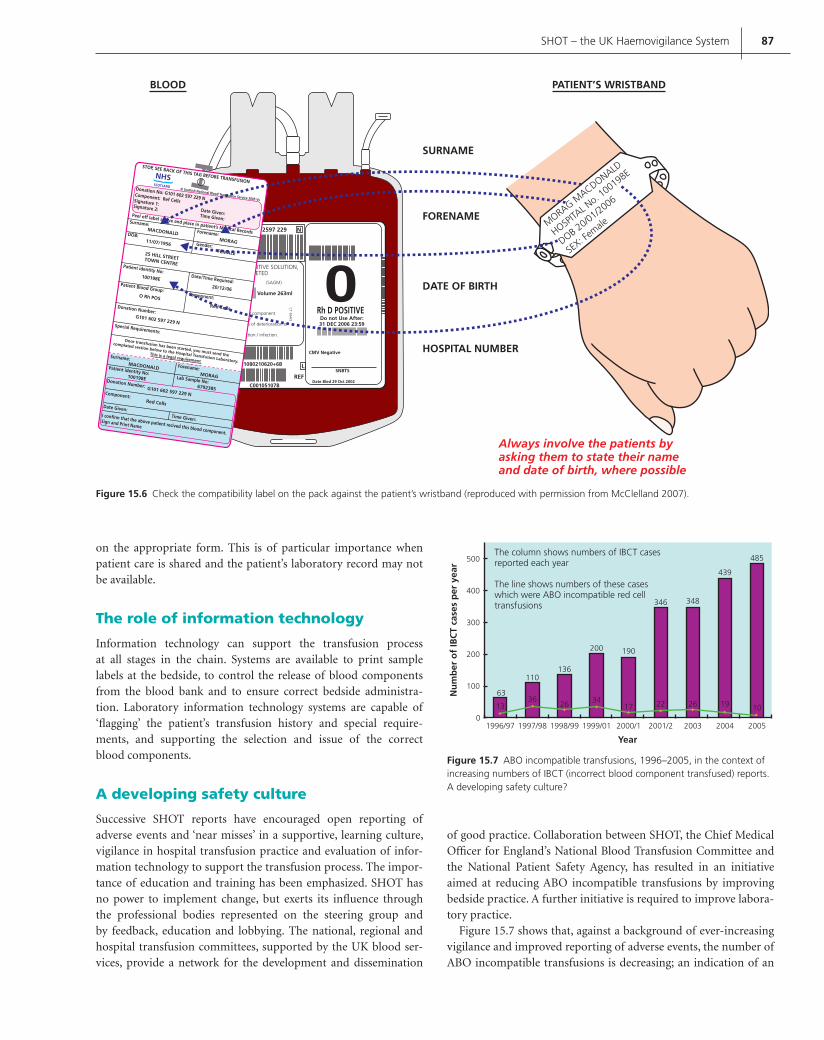
SHOT – the UK Haemovigilance System 87
Figure 15.7 ABO incompatible transfusions, 1996–2005, in the context of increasing numbers of IBCT (incorrect blood component transfused) reports. A developing safety culture?
63
1336
26 3417 22 26 19 10
2005200420032001/22000/11999/01
Year
Nu
mb
er o
f IB
CT
case
s p
er y
ear
1998/991997/981996/97
The column shows numbers of IBCT casesreported each year
The line shows numbers of these caseswhich were ABO incompatible red celltransfusions
0
100
200
300
400
500
110136
200 190
346 348
439
485
MORAG MACDONALD
HOSPITA
L No. 1
00198E
DOB 20/01/2006
SEX: F
emale
Always involve the patients byasking them to state their nameand date of birth, where possible
SURNAME
FORENAME
DATE OF BIRTH
HOSPITAL NUMBER
Rh D POSITIVEDo not Use After:31 DEC 2006 23:59
2597 229
POSITIVE SOLUTION,ETED
(SAGM)
/ componentys of deterioration or
ction / infection.
CT 664/2
Volume 263ml
C00105107B
31080210620+6B
N
CMV Negative
SNBTS
Date Bled 29 Oct 2002
L
REF
STOP, SEE BACK OF THIS TAG BEFORE TRANSFUSION
Surname:
DOB:
Patient identity No:
Patient Blood Group:
Donation Number:
Special Requirements:
Component:
Date/Time Required:
Gender:
MACDONALD
11/07/1956
25 HILL STREETTOWN CENTRE
100198E
Patient identity No:100198E Lab Sample No:
6792385
O Rh POS
G101 602 597 229 N
Donation Number: G101 602 597 229 N
Once transfusion has been started, you must send the
complated section below to the Hospital Transfusion Laboratory.
This is a legal requirement
Red Cells
Component:
Date Given:I confirm that the above patient recived this blood component.
Sign and Print Name
Time Given:
Red Cells
20/12/06
FEMALE
MORAG
Forename:
Surname:MACDONALD
MORAG
Forename:
Peel off label above and place in patient’s Medical Records
Donation No: G101 602 597 229 NComponent: Ref CellsSignature 1:Signature 2: Date Given:Time Given:
NHS
BLOOD PATIENT’S WRISTBAND
© Scottish National Blood Translusion Service 2005 Vs
SCOTLAND
Figure 15.6 Check the compatibility label on the pack against the patient’s wristband (reproduced with permission from McClelland 2007).
on the appropriate form. This is of particular importance when patient care is shared and the patient’s laboratory record may not be available.
The role of information technology
Information technology can support the transfusion process at all stages in the chain. Systems are available to print sample labels at the bedside, to control the release of blood components from the blood bank and to ensure correct bedside administra-tion. Laboratory information technology systems are capable of ‘fl agging’ the patient’s transfusion history and special require-ments, and supporting the selection and issue of the correct blood components.
A developing safety culture
Successive SHOT reports have encouraged open reporting of adverse events and ‘near misses’ in a supportive, learning culture, vigilance in hospital transfusion practice and evaluation of infor-mation technology to support the transfusion process. The impor-tance of education and training has been emphasized. SHOT has no power to implement change, but exerts its infl uence through the professional bodies represented on the steering group and by feedback, education and lobbying. The national, regional and hospital transfusion committees, supported by the UK blood ser-vices, provide a network for the development and dissemination
of good practice. Collaboration between SHOT, the Chief Medical Offi cer for England’s National Blood Transfusion Committee and the National Patient Safety Agency, has resulted in an initiative aimed at reducing ABO incompatible transfusions by improving bedside practice. A further initiative is required to improve labora-tory practice.
Figure 15.7 shows that, against a background of ever-increasing vigilance and improved reporting of adverse events, the number of ABO incompatible transfusions is decreasing; an indication of an
Contreras_C015.indd 87Contreras_C015.indd 87 12/5/2008 6:11:45 PM12/5/2008 6:11:45 PM

88 ABC of Transfusion
emerging safety culture that bodes well for patient safety. This must be nurtured by ensuring that hospitals can guarantee professional accountability, implement education programmes and assess and document competency.
The need for an overarching view of transfusion risks
From the inception of SHOT, a key recommendation has been a call for an overarching body with the expertise and remit to assess and prioritize transfusion risks. Transfusion safety is emotive, contro-versial and politically sensitive, and there is a need for objectivity and scientifi c rigour when allocating resources to safety initiatives.
Users of blood can contribute to transfusion safety by ensuring that robust protocols are in place, that all staff prescribing, han-dling and administering blood are competent to do so, and, above all, that blood freely and generously donated is used appropriately according to clinical need.
Further reading
British Committee for Standards in Haematology guidelines, www.bcsh-
guidelines.com.
Kleinman S, Chan P, Robillard P. Risks associated with transfusion of cellular
blood components in Canada. Transfusion Medicine Reviews 2003; 17(2):
120–62.
McClelland DBL, ed. Handbook of Transfusion Medicine, 4th edn. UK Blood
Services, London, 2007.
SHOT reports, www.shotuk.org.
Stainsby D, Jones H, Asher D et al. Serious hazards of transfusion.
A decade of Haemovigilance in the UK. Transfusion Medicine Reviews
2006; 20(4): 273–82.
Stainsby D, Russell J, Cohen H, Lilleyman J. Reducing adverse events in blood
transfusion. British Journal of Haematology 2005; 131: 1–8.
Taylor C, Jones H, Davies T et al. (2008) SHOT Annual Report 2007. ISBN
978-09558648-0-3.
Contreras_C015.indd 88Contreras_C015.indd 88 12/5/2008 6:11:46 PM12/5/2008 6:11:46 PM

89
CHAPTER 16
Alternatives to Allogeneic Blood Transfusion
Dafydd Thomas and Beverley Hunt
ABC of Transfusion, 4th edition, 2009. Edited by Marcela Contreras. © 2009
Blackwell Publishing, ISBN: 978-1-4051-5646-2.
There are a number of options available as transfusion alternatives. The successful avoidance of allogeneic transfusion will depend on whether the anaemia requiring treatment is chronic or acute and whether it is due to an underlying disease, to chemotherapeutic intervention or to trauma or surgery (Table 16.1). Current clinical practice in the UK is struggling to provide optimal preoperative preparation for patients. A suggested algorithm is highlighted in Figure 16.1.
The one area where the use of allogeneic blood transfusion has decreased signifi cantly in recent years, in the UK, is in the treatment of acute perioperative anaemia. This has been achieved by a num-ber of different strategies, all of which contribute to the overall aim of blood conservation, both of the patient’s own blood and of allo-geneic blood supplies. Measures that can be employed to minimize or eliminate the need for allogeneic transfusion during the peri-operative period are addressed here. The most signifi cant change in practice has been the acceptance by clinicians that withholding of allogeneic blood transfusion during this period, until there are signs or symptoms of anaemia, appears to be safe and results in a signifi cant decrease in allogeneic transfusion. Controlled anae-mia during the perioperative period does not increase mortality and patients soon recover, replenishing their own blood in the weeks following surgery. Published guidelines vary, but almost all state that transfusion to patients with a haemoglobin over 70 g/L is
unnecessary if asymptomatic, with a slightly higher haemoglobin threshold for the elderly or those with cardiorespiratory disease.
The more widespread use of perioperative red cell salvage has helped reduce peri-surgical use of allogeneic red cells.
Preoperative preparation
A signifi cant number of patients presenting for surgery are suffer-ing from iron-defi ciency anaemia. This may be as a result of their
OVERVIEW
Autologous transfusion can complement a blood conservation • programme.
Better preoperative assessment helps minimize allogeneic • transfusion.
Red cell salvage is now the most commonly used form of • autologous transfusion in the UK.
Local audit of surgical activity and the need for allogeneic blood • transfusion can help modulate blood conservation planning.
An integrated care pathway can help coordinate the • multidisciplinary perioperative approach.
Figure 16.1 Preoperative algorithm to help aid perioperative blood conservation.
Patient referred by generalpractitioner for operativeprocedure which is coded as aprocedure where bloodtransfusion is likely
• Full blood count• Urea + electrolytes• Group and save
Outpatient assessment • Review results• Prescribe iron oral/intravenous, if needed• Other haematinics, if needed• Discuss transfusion options• Plan stopping antithrombotic drugs if underlying condition allows
Hospital preoperative assessment Check response to changes inmedicationObtain informed consent fortransfusion planSecond group and save
Operative conservation Improved surgical haemostasisDiathermy, harmonic scalpels, gluesPharmacological agentsIntraoperative blood salvage
Table 16.1 Causes of anaemia that might require transfusion.
Acute Chronic
Trauma Dietary iron defi ciencySurgical loss MalignancyChemotherapy Infectious diseasesGastrointestinal bleeding Parasitic diseases (e.g. malaria)Pregnancy Thalassaemia, sickle cell disease
Contreras_C016.indd 89Contreras_C016.indd 89 12/19/2008 2:14:13 PM12/19/2008 2:14:13 PM

90 ABC of Transfusion
underlying illness or, as in a large number of elderly patients, as a result of poor diet. If noticed early in the preoperative period, oral iron is a very economic way of correcting this defi ciency. Alternatively if time is short or there is an intolerance or non-com-pliance with oral iron, the use of intravenous iron sucrose appears very safe, effi cacious and economic. If preoperative haemoglobin is increased, then there is a greater tolerance to blood loss.
Rational changes to preoperative medicationThe cessation of anticoagulants prior to surgery is a long accepted alteration to a patient’s usual medication. Stopping vitamin K antagonists and transfer to a low molecular weight heparin (LMWH) preoperatively is common practice. The heparin can then be stopped prior to surgery, so leaving only a narrow win-dow when haemostasis is normal to allow surgical intervention, and restarting LMWH postoperatively. Regional anaesthesia can be undertaken providing thromboprophylactic LMWH is stopped 12 hours before. Other agents that may increase bleeding during the operative period can be safely stopped in some instances, providing it is not considered to increase the patient’s risk of thromboembo-lism (e.g. clopidogrel, aspirin and other antiplatelet drugs). Aspirin needs to be stopped 7–10 days prior to surgery. Some patients with metal or drug eluting stents need to continue their dual antiplatelet therapy due to their high risk of rethrombosis; they thus have a high bleeding risk.
Operative haemostasis
Various surgical methods can be used to minimize operative blood loss. Operative haemostasis is the foundation upon which all other blood conservation methods are based. All other strategies are of little use if this has not been achieved.
The use of novel surgical techniques and instruments such as harmonic scalpels, water jet dissection, laparoscopic surgery and off-pump cardiac surgery all decrease the trauma to tissues and so may result in lower operative blood loss.
Autologous blood transfusion
In an attempt to further reduce the need for allogeneic blood, it is important that all measures to conserve the patient’s own blood are taken. The use of perioperative blood salvage is currently the favoured method of autologous transfusion in the UK. Other meth-ods include preoperative predeposit autologous donation (PAD), where a patient donates autologous blood in the weeks preceding surgery, and acute normovolaemic haemodilution, where venesec-tion and collection of one in two units of blood is undertaken by the anaesthetist immediately prior to surgery. During acute normo-volaemic haemodilution the blood loss is replaced with crystalloid or colloid solution to maintain isovolaemia and then the autolo-gous blood is returned at the end of surgery.
These last two methods have recently been discouraged in the UK, due to the cost and wastage in PAD and lack of evidence of signifi cant reduction in allogeneic red cell use in the latter. In addi-tion, stored PAD presents a similar risk of an incorrect blood com-ponent being transfused as with allogeneic blood transfusion. The
demands on blood supplies may change in the future and both of these methods may be revisited. They both remain of value in specifi c situations, such as for patients with extremely rare blood groups requiring transfusions. For such patients, it is justifi able to keep their blood frozen in a cryoprotectant.
Red cell salvage techniques
These techniques may be performed during the surgical procedure or following surgery. The surgical wound drains (which collect blood that is usually discarded) may be used as a reservoir from which the patient’s blood can be recycled. The collection to trans-fusion ratio is high, as only blood that is being lost is replaced. This latter fact helps to improve the cost effectiveness of perioperative techniques. The availability of cell salvage methods allows surgi-cal blood ordering schedules (see Chapter 3) to be reduced, there-fore decreasing blood bank work. There are two main techniques involved in perioperative salvage: (i) collection and reinfusion of wound drainage; and (ii) automated cell salvage.
Collection and reinfusion of wound drainageThe simplest method involves the collection of wound drainage blood and simple reinfusion. All these techniques consist of a main collection chamber that is connected to the wound drain via one port and a separate port to which an intravenous administration set can be attached (Figure 16.2). Reinfused blood passes through a
Figure 16.2 Simple postoperative collection system providing unwashed blood.
Contreras_C016.indd 90Contreras_C016.indd 90 12/19/2008 2:14:14 PM12/19/2008 2:14:14 PM

Alternatives to Allogeneic Blood Transfusion 91
40 m fi lter. No anticoagulant is added as the blood is assumed to be defi brinated, with clot formation having already occurred in the wound. The technique is probably best reserved for collection and reinfusion of blood in total knee replacement surgery and other joint replacement surgery.
AdvantagesThe main advantages of the simple collection systems are their relative ease of use and the avoidance of expensive and more com-plicated machines. The system can be used on the postoperative ward, where the collection and reinfusion of blood is under the care of the ward nursing staff.
DisadvantagesThere are potentially more complications with these devices as unwashed blood is being reinfused. The subsequent administration of anticoagulant, cellular debris, free haemoglobin and activated leucocytes may lead to physiological problems if large amounts of this blood are transfused. Surprisingly, few adverse reports have been published. In practice the reinfusion of small volumes seems clinically acceptable.
The procedure is not recommended in cases where there is con-tamination of the salvaged product with bowel contents, malignant cells or amniotic fl uid.
Automated cell salvageThe second method is regarded as the gold standard in operative recycling, where the collected blood is fi rst fi ltered and then under-goes centrifugal separation, washing and resuspension in sterile saline. This produces a red cell product free of the contaminants mentioned above with a packed cell volume of 0.5. The recycling process may only take 8 minutes if the patient is bleeding large volumes.
Blood is collected from the surgical wound via a suction catheter (Figure 16.3). This catheter has two lumens, one to deliver hepa-rinized saline (or other anticoagulant) (Figure 16.4) to the site of
Figure 16.3 Atraumatic suction catheter showing a wide bore and less angulation to minimize turbulent fl ow of salvaged blood.
Figure 16.4 An example of pre-prepared heparinized saline wash solution.
Figure 16.5 Cell salvage machine prepared for use.
haemorrhage, and the other to aspirate the already anticoagulated blood. The yield of red cells is improved if turbulence and red cell trauma is minimized. Red cell salvage is most successful when blood is aspirated from pooled blood rather than by constant suc-tion from wound surfaces. The collected blood is then aspirated via a roller pump into the centrifuge bowl, further to passing through a 40 m fi lter (Figure 16.5). After centrifugation, the supernatant is discarded and the red cells are washed and then suspended in normal saline for reinfusion (Figure 16.6).
AdvantagesThis technique provides washed red cells at a packed cell volume of 0.5. Disposable costs are minimized when blood salvage volumes
Contreras_C016.indd 91Contreras_C016.indd 91 12/19/2008 2:14:17 PM12/19/2008 2:14:17 PM

92 ABC of Transfusion
are large but probably become relatively cost effective for volumes of between 250 and 500 ml (equivalent to approximately 1 unit of packed cells). Fresh red cells are salvaged that have active oxygen carrying capacity. The alkaline pH of saved red cells minimizes aci-dosis associated with massive transfusion. There is no risk of hyper-kalaemia as red cell ionic pumps remain active and K+ remains within the red cells. The free haemoglobin levels are very low when compared with stored allogeneic red cells or simple reinfusion tech-niques. There remains a necessity to fully label all saved blood to avoid incorrect blood being transfused, in spite of the close prox-imity of all salvaged blood to the patient and the lack of need for storage (Figures 16.6 and 16.7).
Figure 16.6 Compact machine for providing washed postoperative red cell salvage.
DisadvantagesThe technique requires trained operators and automated equip-ment is expensive to purchase. The technique is not recommended in cases where there is contamination of the salvaged product with bowel contents, malignant cells or amniotic fl uid.
However, the National Institute for Clinical Excellence has given guarded support to the use of red cell salvage in obstetrics when there is signifi cant haemorrhage. In the case of surgical fi elds which may contain malignant cells, the use of cell salvage is left to the discretion of the surgeon.
Antifi brinolytics
Activation of fi brinolysis occurs during all forms of surgery. Antifi brinolytic drugs, such as tranexamic acid and aprotinin, block the breakdown of fi brin (Figure 16.8).
Tranexamic acid is a competitive inhibitor of plasmin and plas-minogen, preventing plasmin binding to fi brin. The Horrow regime (10 mg/kg followed by 1 mg/kg/h) has been shown to reduce blood loss in cardiac surgery. Other studies have used boluses of up to 5 g per patient, in bleeding patients, with no ill effect. -aminocaproic acid is also a synthetic lysine analogue that has a potency 10-fold weaker than tranexamic acid.
Aprotinin is a broad-spectrum serine protease inhibitor iso-lated from bovine lung, and is a powerful antiplasmin agent. The ‘high dose’ regimen (2 M KIU to the patient and a cardiopulmo-nary bypass prime and an infusion of 500 000 KIU/h; K stands for Kallikrein) has been shown to reduce perioperative bleeding in open cardiac surgery. Lower doses do produce adequate antiplas-min effects, however. A dose of 2 M units is approved for the treat-ment of established hyperfi brinolysis.
The effi cacy of antifi brinolytic agents in preventing blood loss in elective surgery, and especially in cardiac surgery, has been shown in numerous clinical trials. A Cochrane review suggested that, despite the theoretically greater potency of aprotinin, there was little clinical difference in effi cacy between it and tranexamic acid. Given prophylactically perioperative in elective surgery, these drugs reduce the need for red cell transfusion by about 30%, saving about 1 unit of red cells in patients requiring transfusion. The effi cacy of tranexamic acid in trauma will be assessed by the ongoing Clinical Randomisation of Antifi brinolytic in Signifi cant Haemorrhage (CRASH) II study (www.CRASH2.LSHTM.ac.uk), in which 20 000 trauma patients worldwide are being randomized to 1 g tranexamic acid followed by a 6–8-hour infusion of a further 1 g.
Thrombosis has been the feared theoretic side effect of antifi -brinolytic drugs; the Cochrane review of antifi brinolytics, however, cites studies that included over 8000 patients and demonstrated no increased risk. All antifi brinolytics are renally excreted and therefore dosage should be reduced in patients with renal failure. Because aprotinin is a bovine protein with an associated risk of anaphylaxis, a test dose must be given. After high dose aprotinin as many as 50% of patients develop specifi c antibodies within 3 months of exposure. An open study by Mangano et al. (2006) sug-gested that aprotinin usage in cardiac surgery was associated with an increased risk of myocardial infarction, stroke and renal failure. A further publication cited an increased risk of renal problems in
Contreras_C016.indd 92Contreras_C016.indd 92 12/19/2008 2:14:23 PM12/19/2008 2:14:23 PM

Alternatives to Allogeneic Blood Transfusion 93
patients receiving aprotinin compared to tranexamic acid. Blinded comparative study of aprotinin versus tranexamic acid versus -aminocaproic acid recruiting 3000 patients to assess safety and effi cacy. Following recent publications and developments aprotinin is no longer available for use in cardiac surgery.
Desmopressin (DDAVP) is a synthetic analogue of arginine vasopressin: 1-deamino-8-D-arginine vasopressin. It releases von Willebrand factor from the endothelium and increases platelet activity. Desmopressin has been used to minimize perioperative allogeneic blood loss in cardiac surgery, but there is no convinc-ing evidence that it does so in patients who do not have congenital bleeding disorders or uraemia.
Topical haemostatic sealants
In recent years, there has been a dramatic increase in topical haemo-static agents. Fibrin sealants and fi brin glues have been used as an adjunct to conventional methods of achieving haemostasis. The concept is to locally apply concentrated plasma derivatives to pro-mote the conversion of fi brinogen into fi brin. The common appli-cation methods for fi brin glues include spray bottles, syringes and silastic catheters through fl exible fi breoptic endoscopes.
Aside from fi brin glues, the most commonly used products are cellulose, gelatin, collagen, thrombin or aldehyde glues. The new
generation topical agents are based on the concept of a fusion matrix whereby a bovine gelatin matrix is mixed with a topical thrombin solution immediately prior to use. Other new genera-tion topical haemostatic agents include polyethylene glycol-based synthetic sealants, which have important advantages over biological substances, due to the lack of risk for transfusion-transmitted dis-eases. Synthetic sealants (e.g. Focalseal™) are also composed of two components, a primer and a sealant. These components are applied in two steps and then polymerized under the infl uence of a blue-green light from a xenon light source. Other synthetic new genera-tion sealants consist of albumin and glutaraldehyde or hydrogels in sprayable form.
Recombinant factor VIIa
The use of recombinant factor VIIa (rFVIIa) in managing bleeding became prominent after it was used in an exsanguinating Israeli soldier with severe haemorrhage after trauma. The soldier even-tually recovered. Subsequently a number of case studies and case series have described treatment with rFVIIa being benefi cial in the treatment of massive coagulopathic bleeding following trauma. A recent multicentre, randomized, double-blind, placebo-con-trolled study examined the effi cacy of rFVIIa in patients with blunt or penetrating trauma after they had received 6 units of red blood
AUTOLOGOUS TRANSFUSION
Untested bloodylnoesuSUOGOLOTUAroF
...…...……………………Hospital / NHS No
…...……………………………Last Name
.………...…………………………First Name
………...…………………………………DOB
………...………….Operator Name (Print)
Collection………...………Time…………………Date
ExpiryDate ………...………Time…………………
doolbsuogolotuafoepyT
HNA Post-op (washed)po-artnI Post-op (unwashed)
mls……………………………Total Volume---------------------------------------------------------
AFFIX IN TRANSFUSION RECORD
(This section must be completed andaffixed in patient’s transfusion record)
noisufsnarTsuogolotuA
……...…………………………………………emaNlluF
………..…………………………….oNSHN/latipsoH
:epyTHNA po-artnI Post-up
(washed)Post-up(unwashed)
.……………….………………………ybderetsinimdA
....…………………etaDTransfusion Started at:
.......………………emiT
slm...….…………………………………emuloVlatoT
noitcestuoleeP
!POTSDO NOT use addressograph labels
Handwrite the label from theinformation on the patient’s
identification band
Keep with the patient at alltimes
DO NOT Refrigerate
Reinfuse in accordance withmanufacturer’s guidelinesand hospital transfusion
policy
Before transfusion, carryout the following checksTake extra care in confirming thepatient’s identification.
.1
Check the information on the labelmatches the information on thepatient’s identification band. Wherepossible, ask the patient to statetheir NAME and D.O.B.
.2
No identification bandNo transfusion
dnaetadkcehC.3 foyripxefoemit.doolb
hctamtonodsliatedynafI.4
Do not transfuse.5 sinoitcaernoisufsnartafI
noisufsnartehtPOTS,detcepsus.ecivdalacidemkeesdna
Printed front and back on thin card
Easy peel out section
Reinforced hole at top to attach toblood bags.
Figure 16.7 An example of an autologous blood pack label.
Contreras_C016.indd 93Contreras_C016.indd 93 12/19/2008 2:14:28 PM12/19/2008 2:14:28 PM

94 ABC of Transfusion
cells, and received the fi rst dose of their assigned medication fol-lowing transfusion of a further 2 units of red blood cells (8 units in total), followed by a second and third dose, 1 and 3 hours after the initial dose. Treatment with rFVIIa in blunt trauma produced a signifi cant reduction in transfusion requirements in patients with blunt trauma surviving for more than 48 hours, and also reduced the incidence of acute respiratory distress syndrome in all patients with blunt trauma.
The current data suggest that rFVIIa may be benefi cial in uncon-trolled traumatic bleeding, but it remains unclear whether the benefi t is better than best practice in using blood components. rFVIIa causes localized activation of coagulation at bleeding sites, although there is an increased thrombotic risk. Since rFVIIa acts on the patient’s own clotting system, adequate levels of platelets and fi brinogen are required. The required dose of rFVIIa is still under debate and as yet it is not licensed for use in reducing mas-sive bleeding.
Further reading
A manual of blood conservation, www.transfusionguidelines.org.uk
Fergusson DA, Hébert PC, Mazer CD et al. A Comparison of Aprotinin and
Lysine Analogues in High-Risk Cardiac Surgery. New England Journal of
Medicine 2008; 358(22): 2319–31.
Mangano DT, Tudor IC, Dietzel C et al. The risk associated with aprotonin
in cardiac surgery. New England Journal of Medicine 2006; 354(4):
353–65.
Munoz M, Garcia-Erce JA, Villar J, Thomas D. Blood conservation strategies
in major orthopaedic surgery: effi cacy, safety and European regulations.
Vox Sanguinis 2008. DOI:10,1111/j.1423-0410.2008.01108X.
Thomas D, Thomson J, Ridler B. A Manual for Blood Conservation. tfm
Publishing, Shrewsbury, 2005.
Figure 16.8 Simplifi ed principles of fi brinolysis showing action points where aprotinin and tranexamic acid work. FDP, fi brin degradation product; PAI-1, plasminogen activator inhibitor-1.
PLASMINOGEN
PLASMIN
Tissueplasminogenactivator
ENDOTHELIUM
PAI-1
Fibrin FDPs
Alpha-2 antiplasminAPROTININTRANEXAMIC ACID
Contreras_C016.indd 94Contreras_C016.indd 94 12/19/2008 2:14:28 PM12/19/2008 2:14:28 PM

95
CHAPTER 17
Blood Substitutes and Oxygen Therapeutics
Kenneth C. Lowe
ABC of Transfusion, 4th edition, 2009. Edited by Marcela Contreras. © 2009
Blackwell Publishing, ISBN: 978-1-4051-5646-2.
Why is there a need for blood substitutes?
The ability to transfuse blood and blood components was one of the major medical advances of the twentieth century. Modern blood transfusion in developed countries is a universally practised and remarkably safe medical procedure. Blood or concentrated red cells are routinely transfused following extensive blood loss or as a treatment for chronic anaemia for which there is no alternative therapy. In both situations, the aim is to increase the arterial oxygen content and enhance oxygen delivery to the tissues.
In parallel with the evolution of modern transfusion medicine, there has been increasing interest in so-called ‘blood or red cell substitutes’. The reason highlights some of the disadvantages of blood transfusion (Box 17.1). Blood has to be stored refrigerated (at 4°C) and transport may be a problem. The useful shelf-life of blood or red cells is only about 42 days (35 days in the UK), even with the use of modern nutrient additives, such as mannitol, glucose and adenine. Inevitably, some donated blood is wasted.
Another problem is the need to type, antibody screen and cross-match blood, due to the natural occurrence of ABO anti bodies, the
polymorphism of red cell membrane antigens and their immuno-genicity. Despite pre-transfusion testing, immune-mediated transfu-sion reactions still occur due to alloimmunization to antigens on red cells, white cells, platelets and plasma proteins.
Aside from practical and physiological problems are cultural and religious objections to blood transfusion. This is best exempli-fi ed by Jehovah’s Witnesses and the biblical injunction in Leviticus (17:11): ‘Ye shall not eat the blood of no manner of fl esh: for the life of all fl esh is the blood thereof ’. Jehovah’s Witnesses interpret this so strictly that they refuse blood transfusions, even when their (or their child’s) life is threatened.
The realization in the 1980s that human immunodefi ciency virus (HIV) infection could be transmitted through the transfusion of blood products has led to increasing fear of blood donation and transfusion. This has occurred despite signifi cant improvements in donor screening procedures and the refi nement of educational policies.
All the problems outlined in Box 17.1 could, in principle, be solved by a synthetic substitute fl uid with the same osmolarity as red cells that could pick up oxygen in the lungs and deliver it to the tissues. Such a blood substitute should be free of transmissible agents, stable for long periods at room temperature, non-antigenic with no need for crossmatching, and should not produce adverse immunological reactions in humans.
The search for an effective blood substitute
Arguably, the search for a blood substitute began in the mid to late nineteenth century when physicians who were frustrated by the frequent lethality caused by heterologous blood transfusion used
OVERVIEW
A blood substitute has not been found. Although in widespread • use, the term blood substitute, as such, should be replaced by `red cell substitute', or, preferably, by `oxygen therapeutic drug.'
An ideal red cell substitute should have the same osmolarity as • red cell, being able to pick up oxygen from the lungs and deliver it to the tissues. It should be free of transmissible agents, stable at room temperature for lon periods of time, non-antigenic, with no need for crossmatching and should not produce adverse immunological reactions in the recipient. It should be cheap to manufacture.
The two approaches to developing an oxygen therapeutic are: • (1) haemoglobin solutions; (2) emulsions of perfl uorocarbons.
A bovine haemoglobin solution has been approved for clinical • trial, albeit only in South Africa. A few other products are at the stage where clinical trials are underway or are being planned.
Box 17.1 Disadvantages of blood for transfusion
Needs human volunteer donors• Increasingly diffi cult to recruit and retain donors• Storage• Transport• Short shelf-life• Potential to transmit blood-borne infectious agents• Errors in administration may cause ABO incompatible transfusion• Cultural and religious objections•
Contreras_C017.indd 95Contreras_C017.indd 95 12/5/2008 6:14:17 PM12/5/2008 6:14:17 PM
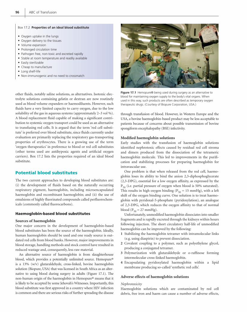
96 ABC of Transfusion
other fl uids, notably saline solutions, as alternatives. Isotonic elec-trolyte solutions containing gelatin or dextran are now routinely used as blood volume expanders or haemodiluents. However, such fl uids have a very limited capacity to carry oxygen, due to the low solubility of the gas in aqueous systems (approximately 2–3 vol %). A blood replacement fl uid capable of making a signifi cant contri-bution to systemic oxygen transport could be used as an alternative to transfusing red cells. It is argued that the term ‘red cell substi-tute’ is preferred over blood substitute, since fl uids currently under evaluation are primarily replacing the respiratory gas-transporting properties of erythrocytes. There is a growing use of the term ‘oxygen therapeutics’ in preference to blood or red cell substitutes (other terms used are antihypoxic agents and artifi cial oxygen carriers). Box 17.2 lists the properties required of an ideal blood substitute.
Potential blood substitutes
The two current approaches to developing blood substitutes are: (i) the development of fl uids based on the naturally occurring respiratory pigment, haemoglobin, including microencapsulated haemoglobin and recombinant haemoglobin; and (ii) the use of emulsions of highly fl uorinated compounds called perfl uorochem-icals (commonly called fl uorocarbons).
Haemoglobin-based blood substitutes
Sources of haemoglobinOne major concern in the development of haemoglobin-based blood substitutes has been the source of the haemoglobin. Ideally, human haemoglobin should be used and one ready source is out-dated red cells from blood banks. However, major improvements in blood storage, handling methods and stock control have resulted in reduced wastage and, consequently, less raw material.
An alternative source of haemoglobin is from slaughterhouse blood, which provides a potentially unlimited source. Hemopure® is a 13% (w/v) glutaraldehyde, cross-linked, bovine haemoglobin solution (Biopure, USA) that was licensed in South Africa as an alter-native to using blood during surgery in adults (Figure 17.1). The non-human origin of the haemoglobin in Hemopure® means that it is likely to be accepted by some Jehovah’s Witnesses. Importantly, this blood substitute was fi rst approved in a country where HIV infection is common and there are serious risks of further spreading the disease
through transfusion of blood. However, in Western Europe and the USA, a bovine haemoglobin-based product may be less acceptable to patients because of concerns about possible transmission of bovine spongiform encephalopathy (BSE) infectivity.
Modifi ed haemoglobin solutionsEarly studies with the transfusion of haemoglobin solutions identifi ed nephrotoxic effects caused by residual red cell stroma and dimers produced from the dissociation of the tetrameric haemoglobin molecule. This led to improvements in the purifi -cation and stabilizing processes for preparing haemoglobin for intravascular use.
One problem is that when released from the red cell, haemo-globin loses its ability to bind the anion 2,3-diphosphoglycerate (2,3-DPG), essential for a low oxygen affi nity, as expressed by the P50 (i.e. partial pressure of oxygen when blood is 50% saturated). This results in high oxygen binding (P50 = 15 mmHg), with a left shift of the oxygen binding curve. One solution is to treat haemo-globin with pyridoxal-5-phosphate (pyridoxylation), an analogue of 2,3-DPG, which reduces the oxygen affi nity to that of normal blood (P50 = 27 mmHg).
Unfortunately, unmodifi ed haemoglobin dissociates into smaller fragments and is rapidly excreted through the kidneys within hours following injection. The short circulatory half-life of unmodifi ed haemoglobin can be improved by the following:
Stabilizing the haemoglobin tetramer with intramolecular links 1 (e.g. using diaspirin) to prevent dissociation.Covalent coupling to a polymer, such as polyethylene glycol, 2 producing a conjugated tetramer.Polymerization with glutaraldehyde or o-raffi nose forming 3 intermolecular cross-linked haemoglobin.Encapsulating pyridoxylated haemoglobin within a lipid 4 membrane producing so-called ‘synthetic red cells’.
Adverse effects of haemoglobin solutions
NephrotoxicityHaemoglobin solutions which are contaminated by red cell debris, free iron and haem can cause a number of adverse effects,
Box 17.2 Properties of an ideal blood substitute
Oxygen uptake in the lungs• Oxygen delivery to the tissues• Volume expansion• Prolonged circulation time• Pathogen free, non-toxic and excreted rapidly• Stable at room temperature and readily available• Easily sterilizable• Cheap to manufacture• Long shelf-life• Non-immunogenic and no need to crossmatch•
Figure 17.1 Hemopure® being used during surgery as an alternative to blood for maintaining oxygen supply to the body’s vital organs. When used in this way, such products are often described as temporary oxygen therapeutic drugs. (Courtesy of Biopure Corporation, USA.)
Contreras_C017.indd 96Contreras_C017.indd 96 12/5/2008 6:14:17 PM12/5/2008 6:14:17 PM

Blood Substitutes and Oxygen Therapeutics 97
including renal failure. This is largely overcome by current manu-facturing procedures, which produce highly purifi ed haemoglobin solutions.
Cardiovascular effectsUnfortunately, even pure haemoglobin solutions can cause adverse effects, particularly on the cardiovascular system. Hence, modifi ed haemoglobin is desirable as a red cell substitute since the unmodi-fi ed molecule produces unwanted cardiovascular effects, particu-larly vasoconstriction, with a marked increase in blood pressure and a fall in cardiac output. This occurs because haemoglobin tetramers extravasated from the circulation bind to nitric oxide derived from the vascular endothelium. Nitric oxide is a potent vasodilator and its removal leads to unopposed vasoconstriction.
Clinical trials and status of haemoglobin solutionsClinical trials have been conducted with several commercial modi-fi ed haemoglobin-based red cell substitutes, including o-raffi nose intra- and intermolecularly cross-linked human haemoglobin (e.g. Hemolink™; Hemosol, Canada) and glutaraldehyde-polym-erized human haemoglobin (e.g. PolyHeme™; Northfi eld, USA). However, only Hemopure® has been approved for clinical use (Figure 17.2). One newer product, Hemospan™ (Sangart, USA), contains human haemoglobin conjugated to maleimide-treated polyethylene glycol, giving a molecular mass about twice that of Hemopure®. This prevents extravasation from the circulation,
thereby avoiding any hypertensive side effects. Hemospan™ also has a higher affi nity for oxygen (P50 = 5–6 mmHg) than blood and many earlier haemoglobin-based blood substitutes. The rationale is to avoid excessive oxygenation of tissues and prevent compen-satory arteriolar vasoconstriction causing hypertension. This new paradigm shift in the design of blood substitutes is controversial and not without its critics. The new bovine haemoglobin-based OxyVita™ (OxyVita, USA) has similar properties.
Encapsulated haemoglobinOne way of improving the circulation time of unmodifi ed haemo-globin is to enclose it within a lipid membrane. Haemoglobin has been encapsulated with 2,3-DPG (‘neohemocytes’) as the fi rst step towards creating synthetic red blood cells. If the haemo-globin is treated with pyridoxal-5-phosphate, a stable product is produced. Encapsulation prevents renal clearance of haemoglobin. Additionally, enzymes, such as superoxide dismutase and catalase, can be incorporated into the liposomes to scavenge potentially damaging oxygen radicals.
Human recombinant haemoglobinHuman haemoglobin has been synthesized by genetically modifi ed bacteria, yeast or plants (so-called ‘cell factories’). Such recombi-nant haemoglobins (rHbs) have oxygen binding properties simi-lar to normal human haemoglobin. This approach avoids using human or animal blood as raw material, thereby overcoming the problem of transmitting infectious agents. In a fi rst-generation rHb – rHb1.1, known as Optro™ (Baxter, USA) – some amino acid sequences were replaced to prevent molecular dissociation and giving acceptable oxygen binding characteristics. A new European project, Genomics and Blood Substitutes for 21st Century Europe (acronym EuroBloodSubstitutes), is developing a technological baseline for producing novel haem proteins and blood substitute components using microorganisms.
Recombinant haemoglobins are an exciting way forward for blood substitutes since ‘tailor-made’ molecules can, in principle, be produced. Recombinant technology is being used to produce haemoglobin mutants with altered affi nity for nitric oxide, thereby overcoming the transient, hypertensive side effects considered previously as a fl aw in the usefulness of haemoglobin red cell sub-stitutes. It is not yet clear whether this type of haemoglobin can be produced in a larger scale at lower cost. Box 17.3 details the advantages of rHb.
Perfl uorochemical emulsionsPerfl uorochemicals are organic compounds in which hydrogen atoms have been replaced with fl uorine. They are clear, odourless, inert fl uids, which are radiopaque. The compounds are extremely
Box 17.3 Advantages of recombinant haemoglobin
Virus free• No residual stromal contamination• Unconstrained supply• Can be engineered for specifi c requirements•
Figure 17.2 One unit of the haemoglobin-based blood substitute, Hemopure®, the fi rst such product to be licensed for clinical use. (Courtesy of Biopure Corporation, USA.)
Contreras_C017.indd 97Contreras_C017.indd 97 12/5/2008 6:14:19 PM12/5/2008 6:14:19 PM

98 ABC of Transfusion
unreactive chemically, due to the strength of the carbon–fl uorine bonds (Box 17.4).
Perfl uorochemical liquids dissolve large volumes of respiratory gases (up to 45 vol % oxygen per atmosphere at 37°C) and the rela-tionship between oxygen tension and partial pressure is linear. This contrasts with the characteristic sigmoid curve of whole blood (and haemoglobin). Because perfl uorochemical liquids are immiscible with blood and water, they must be emulsifi ed before intravascular use. The target droplet size is about 0.2 m. Following intravascular injection, emulsion droplets are removed from the circulation and temporarily stored in phagocytic cells of the monocyte–macrophage (reticuloendothelial) system. Eventually, the perfl uorochemicals are excreted from the body by exhalation.
Since the late 1970s, several emulsions, containing one or more perfl uorochemicals (Figure 17.3), have been developed as potential oxygen therapeutics. The fi rst emulsion produced commercially was Fluosol®, a 20% (w/v) formulation containing two fl uorocar-bons, perfl uorodecalin and perfl uorotripropylamine (Green Cross Corporation, Japan). A similar perfl uorocarbon-based oxygen carrier, Perftoran (Perftoran, Russia), was approved in Russia in 1996 for use in severe anaemia, haemorrhagic shock and cardiac surgery. Perftoran has been used in over 2000 patients, but little information on its use is available.
Fluosol® was the fi rst perfl uorochemical emulsion evaluated as a blood substitute/oxygen therapeutic in humans and extensive clini-cal trials were reported in the 1980s. It was approved (in 1990) by the American Food and Drug Administration Committee for use
Box 17.4 Properties of perfl uorochemical liquids
Chemically and biologically inert• High respiratory gas solubility• High density• Low viscosity• Low surface tension• Radiopaque• Easily sterilizable•
in restricted doses and in high risk patients, for oxygenating the distal coronary vascular bed during balloon angioplasty. However, in 1994, Green Cross ceased manufacturing Fluosol® due to the lack of interest from physicians and poor sales in their angioplasty indication. Improvements in angioplasty technology had made the need for Fluosol® redundant.
Fluosol® is now regarded as a fi rst generation fl uorocarbon emulsion and the experience gained from its clinical use informed the development of improved emulsions, involving:
the production of more concentrated emulsions, which, in • principle, can carry more oxygenmarked improvements in emulsion stability at room tempera-• ture, thus giving acceptable shelf-lives.
The current front runner amongst the second generation, con-centrated fl uorocarbon emulsions is Oxygent™, produced by the Alliance Pharmaceutical Corporation, San Diego. Oxygent™ is a 60% (w/v) emulsion of perfl ubron with 4% (w/v) egg yolk phos-pholipids as surfactant. Oxygent™ is stable for up to 1 year at room temperature and is rapidly eliminated from the body, with a half-time of about 4 days.
Oxygent™ has been tested in over 1300 patients. The infusion of perfl ubron emulsion into patients breathing oxygen was found to be more effective than autologous blood or colloid solution in maintaining cardiovascular function and tissue oxygen supply. In 500 general surgery patients, treatment with Oxygent™ reduced the frequency and volume of blood used. In 2001, Alliance suspended enrolment in a phase III cardiac surgery study with Oxygent™ to evaluate an increased incidence of stroke in patients. New licens-ing, development and marketing agreements with pharmaceuti-cal companies in China, Europe and Canada are being persued for the clinical and regulatory development of the drug for organ protection against hypoxia during surgery.
Conclusion
This chapter reviews potential blood substitutes or oxygen thera-peutic drugs. A bovine haemoglobin-based product has been approved for clinical use, albeit in a country where blood-borne disease is endemic. The development of other haemoglobin- and perfl uorochemical-based materials has progressed to the stage where advanced clinical trials are underway or planned.
Blood substitutes seem to have a defi nite role in surgery to avoid and minimize blood transfusions. The avoidance of allogeneic blood would be benefi cial to the patient and help the transfusion services conserve blood. This is a dynamic and forward-looking area of biotechnology. It is anticipated that products will emerge that will be useful, not only as intravascular gas carriers in trans-fusion medicine, but as biopharmaceuticals for other biomedical disciplines.
Further reading
Winslow RM, ed. Blood Substitutes. Academic Press, London, 2006.
Natanson C, Kern SJ, Lurie P et al. JAMA 2008; 299(17): 2304–12.
Figure 17.3 Chemical structures of two perfl uorochemicals. Both the cyclic (perfl uorodecalin) and linear (perfl ubron) compounds have been tested as red cell substitutes in humans.
F2
F2
F2
F
F2
F2
F2 F2
F2F3 F2 F2
F2 F2
F2
F2F
F2
Br
Perfluorodecalin
Perflubron
Contreras_C017.indd 98Contreras_C017.indd 98 12/5/2008 6:14:22 PM12/5/2008 6:14:22 PM

99
CHAPTER 18
Appropriate Use of Blood and Better Blood Transfusion
Mike Murphy
ABC of Transfusion, 4th edition, 2009. Edited by Marcela Contreras. © 2009
Blackwell Publishing, ISBN: 978-1-4051-5646-2.
Although the quality and safety of blood in the UK is amongst the best in the world, blood transfusion is not free from risk. The most logical approach to reducing the risk of transfusion is only to use blood when it is strictly clinically necessary and there is no alternative.
There has been a recent focus on better blood transfusion practice worldwide because of concerns about patient safety, documented variations in blood usage, and increasing costs of transfusion. In the UK, this work has been termed the Better Blood Transfusion ini-tiative. This chapter will review what activities have taken place to improve the use of blood and to promote alternatives to transfusion.
History of the Better Blood Transfusion initiative in the UK
The Health Service Circular (HSC) 1998/224 Better Blood Transfusion detailed the action required of hospitals and clinicians to improve transfusion practice. Its recommendations were based on presentations and workshops in a symposium on evidence-based blood transfusion held by the UK Chief Medical Offi cers (CMOs) in London in July 1998. A survey in 2001 of the implementation of these initial Better Blood Transfusion recommendations showed that most hospitals had established hospital transfusion committees (HTCs), were participating in the Serious Hazards of Transfusion (SHOT) scheme, and had protocols for the administration of blood (Table 18.1). However, there was evidence of poor training of clini-cal staff, few protocols for the appropriate use of blood and audits of transfusion practice, and limited use of autologous transfusion.
A second UK CMOs’ seminar on blood transfusion was held in October 2001, focusing on avoiding unnecessary transfusion. The HSC 2002/009 Better Blood Transfusion: appropriate use of blood was issued in July 2002, detailing the further actions required of hospi-tals, the National Blood Service (NBS) and clinicians to improve transfusion practice. Hospitals were required to develop hospital transfusion teams comprising a lead consultant for transfusion,
OVERVIEW
The promotion of safe and appropriate use of blood is part of • clinical governance in hospitals.
Education and training programmes for doctors should • emphasize the appropriate use of blood.
Clinical research is essential to provide an adequate evidence • base for transfusion and the use of alternatives to transfusion.
New approaches to the prevention of ‘wrong’ blood transfusion • should be considered including the use of automated bedside systems with barcode patient identifi cation.
Data on blood usage should be provided to clinical teams so • they can compare their practice with others.
Hospitals should improve information systems to facilitate • routine monitoring of blood usage.
Collaboration within clinical teams and between hospitals is • essential to promote good transfusion practice.
Better Blood Transfusion recommendations should have a high • priority in hospitals in terms of patient safety and ensuring best use of a scarce resource.
There is potential for reducing the use of fresh frozen plasma • and platelet transfusions, and for further reductions in the use of red cell transfusions.
Renewed efforts should be made to reduce blood usage in • medical patients.
The identifi cation and management of preoperative anaemia • avoids the need for perioperative red cell transfusion.
Better use should be made of alternatives to transfusion in • surgical patients.
Good communication is essential between clinicians and blood • bank staff about the timely and effective use of blood in patients with massive bleeding.
The decision to use recombinant activated factor VII in a patient • with uncontrolled bleeding must be made by individual clinicians guided by hospital policies and advice from haematologists with expertise in transfusion medicine.
Advances in information technology could be used to provide • ‘decision support’ for doctors considering transfusion in individual patients.
Patients should be better educated to exercise choice about • blood transfusion and the use of alternatives to transfusion.
Contreras_C018.indd 99Contreras_C018.indd 99 12/10/2008 1:53:15 Shobha12/10/2008 1:53:15 Shobha

100 ABC of Transfusion
a specialist practitioner of transfusion (usually, but not necessarily, a specialist nurse) and a senior biomedical scientist in transfusion. The role of the hospital transfusion team is to promote good trans-fusion practice by implementing actions approved by the HTC, which has the overall remit for improving hospital transfusion practice, and to be a resource for training all hospital staff involved in blood transfusion, primarily facilitated by specialist practitioners of transfusion.
A CMOs’ National Blood Transfusion Committee and 12 regional transfusion committees were established in England in 2001 with a primary remit to support the Better Blood Transfusion initiative.
The results of further surveys in 2003 and 2004 indicated some progress in the implementation of Better Blood Transfusion initia-tives, including training, and the number of hospitals with trans-fusion practitioners (see Table 18.1). However, the results also indicated the need for further progress in the training of some staff groups, particularly nurses and doctors, the development of hospital transfusion teams and protocols for the appropriate use of blood, the provision of information to patients, and the use of perioperative cell salvage.
Evidence base for the appropriate use of blood and alternatives to transfusion
Recent high quality evidence in the form of systematic reviews of the literature and well conducted clinical trials has disproved some widely accepted indications for transfusion, and has had a major impact on changing transfusion practice. Examples include:
A randomized clinical trial of a restrictive red cell transfusion 1 strategy (red cells transfused if the haemoglobin concentration
fell below 7 g/dl) was at least as effective and possibly superior to a liberal transfusion strategy (red cells transfused if the haemoglobin fell below 10 g/dl) in critically ill patients.A systematic review of 57 trials of fresh frozen plasma (FFP) 2 found that only three were of adequate design and power, and two showed no clinical benefi t of the use of FFP.A systematic review of alternatives to transfusion found that only 3 the use of antifi brinolytic drugs, such as aprotinin and tranexamic acid, and perioperative cell salvage consistently reduced the use of donor blood.
Audit as a tool to improve transfusion practice
Sequential national audits by the NBS and Royal College of Physicians have shown improvements in transfusion practice with fewer patients not wearing a wristband (6% in 2005 versus 10% in 2003) and fewer at risk from lack of observations (34% with no observations in the fi rst 30 minutes of transfusion in 2005, com-pared with 47% in 2003). However, there is still substantial evidence of transfused patients being put at risk of ‘wrong’ transfusions because of poor patient identifi cation (Figure 18.1) and inadequate monitoring during transfusion.
There is evidence of wide variation between hospitals in the use of blood for common surgical procedures. A recent audit in primary hip replacement surgery in the Oxford hospitals showed a range of 23–58% in the proportion of patients who were transfused. The majority of patients who were transfused only received 1 or 2 units of blood and most of these were discharged with a haemoglobin above 10 g/dl. If they had not been transfused, they would still have
Table 18.1 Progress on the implementation of Better Blood Transfusion strategies since 2001.
2001 2003 2004 2006
Hospitals completing the questionnaire survey
220/320 (69%) 122/259 (47%) 160/169 (95%) 156/173 (90%)
Participation in SHOT scheme 96% 100% 99% 95%
Hospitals with a HTC 91% 98% 99% 97%
% staff trained as: Phlebotomists Porters Nurses Medical staff
79%47%78%34%
97%75%52%53%
97%80%73%60%
74%69%83%87%
Transfusion practitioner in postLead consultant in post
14%–
50%74%
68%83%
92%90%
Protocol for the transfusion process 98% 97% 98% 96%
Protocols for the use of blood: Surgical blood ordering schedule Red cell transfusion*
67%34%
87%44% (critical care)34% (surgical)
92%46% (critical care)39% (surgical)
97%70% (critical care)58% (surgical)
HTC, hospital transfusion committee; SHOT, Serious Hazards of Transfusion.
* The question about red cell transfusion protocols was different between the 2001 and the later surveys. In 2001, the question asked if the hospital had a general red cell transfusion protocol. In 2003 and 2005, the question referred to specifi c protocols for critical care and surgical patients.
Contreras_C018.indd 100Contreras_C018.indd 100 12/10/2008 1:53:15 Shobha12/10/2008 1:53:15 Shobha

Appropriate Use of Blood 101
A summary of the indications for the use of blood is also pro-vided (see Chapter 4).The production of national patient information leafl ets for 2 adults, children and parents (Figure 18.4).The development of national and local contingency plans for 3 blood shortages, highlighting the need for each hospital to plan for acute and chronic blood shortages and to reduce blood usage as an important priority.More support from the NBS, specifi cally the development of its 4 hospital liaison function, comprising a transfusion liaison nurse, a hospital liaison manager and a joint medical consultant in each region to support specialist transfusion practitioners, blood transfusion laboratory scientifi c staff and clinicians in hospitals.
Review of progress since 2002Results of questionnaire surveys in 2003 and 2004 indicate progress since 2001 in the proportion of hospitals with HTCs, the training of some staff groups, the number of hospitals with transfusion prac-titioners, and the development of protocols for the use of blood (see Table 18.1). However, the results also indicated that further progress is needed.
Figure 18.4 Examples of patient information leafl ets.
Figure 18.1 Poor patient identifi cation should be avoided with the use of a handheld computer and barcode patient identifi cation for safe bedside transfusion. (Reproduced with permission from the front cover of the September 2003 edition of Transfusion)
10
5
10
15
20
30
35
40
45
25
2 3 4 5 6Hospital
% in
app
rop
riat
e tr
ansf
usi
on
7 8 9 10 11 12 13
Figure 18.2 The percentage of inappropriate transfusions in an audit of red cell transfusions in 13 hospitals in Northern Ireland. (With permission of the Northern Ireland Regional Transfusion Committee.)
Figure 18.3 The percentage of inappropriate transfusions with different grades of medical staff in an audit of red cell transfusions in Northern Ireland. JHO, junior house offi cer; SHO, senior house offi cer. (With permission of the Northern Ireland Regional Transfusion Committee.)
Consultant
% in
app
rop
riat
e tr
ansf
usi
on
0
5
10
15
20
25
30
35
Registrar
Grade
SHO JHO
had a haemoglobin above 8 g/dl, which would not be expected to impair postoperative recovery.
A recent audit in Northern Ireland estimated that 20% of red cell transfusions were inappropriate. This proportion of inap-propriate transfusions was similar in all 13 hospitals surveyed (Figure 18.2), and was not infl uenced by clinical specialty or grade of doctor requesting the transfusion (Figure 18.3). Repeated rounds of comparative audit, where data from individual hospitals or clini-cal specialties are readily compared with others, has been shown to be a powerful tool for improving practice in other areas of medicine such as myocardial infarction and stroke.
Better Blood Transfusion activities
Examples of Better Blood Transfusion initiativesSpecifi c initiatives in the UK have included the following:
Advice to hospitals on how to implement Better Blood 1 Transfusion strategies, including the resources required, in the form of a ‘toolkit’ (available on www.transfusionguidelines.org.uk).
Contreras_C018.indd 101Contreras_C018.indd 101 12/10/2008 1:53:15 Shobha12/10/2008 1:53:15 Shobha

102 ABC of Transfusion
Effect on blood usageAs a result of these efforts to use blood more appropriately and pos-sibly because of other factors, such as increases in the cost of blood, the use of red cell transfusions in England fell by 5.7% in 2004/5, and by a further 4.4% in 2005/6 (Figure 18.5). The use of FFP and platelet concentrates has changed very little.
Where does blood go?Red cell transfusions are most commonly used in medicine for patients with cancer or gastrointestinal bleeding, and in surgery for cardiac and orthopaedic procedures. Medical use now accounts for about 65% of red cell usage (for many years, medical and surgical use were both approximately 50%). Most of the recent reduction in red cell usage has occurred in surgery.
Strategies for reducing blood use in surgical patientsThere is a varied emphasis on blood management by clinical teams. Some teams carry out major procedures by attention to patient care throughout the perioperative period, resulting in avoidance of transfusion (Box 18.1; see also Chapter 16).
After the introduction of guidelines in one large hospital in Scotland, data on blood use in relation to haemoglobin before and after transfusion were fed back to clinical teams with suggestions about appropriate blood use. Audits were carried out before the initiative and after 1 year; red cell use decreased by 20% with no demonstrable effect on morbidity or mortality.
Strategies for reducing blood use in medical patientsGood blood management depends on many of the same principles as have been described for surgical patients, including the identifi -cation of the cause of anaemia and its appropriate investigation and treatment, the timely estimation of blood counts and haemostasis in massive blood loss, and the implementation of local policies for blood use with education and audit (Box 18.2).
Recombinant erythropoietin improves the quality of life of patients with chronic renal failure by the effective treatment of
anaemia. It is also effective in the avoidance of blood transfusion in some cancer patients.
Management of massive blood lossThe usual presentation is as an emergency in accident and emer-gency, obstetrics or surgery. The restoration of an adequate circu-lating volume is the most important immediate aim, followed by treatment of any surgical source of bleeding and the correction of haemostatic abnormalities with blood components.
Recombinant activated factor VII appears to be effective in the treatment of severe haemorrhage unresponsive to standard manage-ment, but as yet there are few data on its safety and effectiveness.
Future prospects for the Better Blood Transfusion initiative
There has been better recent recognition of the risks of transfusion and the safety of restrictive transfusion policies. Although blood usage is reducing, recent audit data continue to provide evidence of high levels of inappropriate use. Better education about good transfusion practice, wider adoption of algorithms for blood
Box 18.1 Strategies for reducing blood use in surgical patients
PreoperativeEstablishment of preoperative assessment clinics• Identifi cation and correction of anaemia• Avoidance of drugs interfering with haemostasis, e.g. • anticoagulants, or drugs with antiplatelet activity for a period of time prior to surgery
OperativeGood anaesthetic and surgical technique• Avoidance of hypothermia• Reduction of regional vascular pressure• Cell salvage• Point-of-care testing (for blood count and haemostasis) to guide • the use of blood componentsUse of antifi brinolytic drugs such as aprotinin and tranexamic acid•
PostoperativeImplementation of local guidelines for the use of blood, based on • national guidelines, with education, audit and feedback of data to clinical teamsMinimizing the volume of blood collected for laboratory samples• Cell salvage•
Figure 18.5 Change in the issues of red cell, platelet and fresh frozen plasma (FFP) units in England from 2003/4 to 2005/6.
Red cells Platelets
An
nu
al is
sues
(u
nit
s)
FFP0
500 000
1 000 000
1 500 000
2 000 000
2 500 000
2003/4
2004/5
2005/6
Box 18.2 Strategies for reducing blood use in medical patients
Identifi cation and correction of anaemia with appropriate • haematinicsUse of recombinant erythropoietin for patients with anaemia due • to chronic renal failure and some cancersTimely estimation of blood counts and haemostasis in massive • blood lossImplementation of local guidelines for the use of blood with • education, audit and feedback of data to clinical teams
Contreras_C018.indd 102Contreras_C018.indd 102 12/10/2008 1:53:19 Shobha12/10/2008 1:53:19 Shobha

Appropriate Use of Blood 103
management including alternatives to transfusion where relevant, and the adoption of conservative transfusion thresholds should result in further reductions in blood usage without compromis-ing patient safety. The Better Blood Transfusion initiative has been relaunched in 2007, following a further survey in 2006 of the implementation of the recommendations in the HSC 2002/009 (see Table 18.1), with a third UK CMOs’ seminar and the issue of HSC 2007/001 Better Blood Transfusion: safe and appropriate use of blood.
Further reading
Carless PA, Henry DA, Moxey AJ, O’Connell DL, Brown T, Fergusson DA. Cell
salvage for minimising perioperative allogeneic blood transfusion. Cochrane
Database of Systematic Reviews 2006, Issue 4. Art. No. CD001888.
Hebert PC, Wells G, Blajchman MA, et al. A multicenter, randomized,
controlled trial of transfusion requirements in critical care. New England
Journal of Medicine 2004; 340: 409–17.
Henry DA, Moxley AJ, Carless PA, et al. Anti-fi brinolytic use for minimising
perioperative allogeneic blood transfusion. Cochrane Database of Systematic
Reviews 2007, Issue 4. Art. No. CD001886.
Murphy MF, Howell C. Survey of the implementation of the recommenda-
tions in the Health Service Circular 2002/009 ‘Better Blood Transfusion’.
Transfusion Medicine 2005; 15: 453–60.
Stansworth SJ, Birchall J, Doree CJ, Hyde C. Recombinant factor VIIa for the
prevention and treatment of bleeding in patients without haemophilia.
Cochrane Database Systematic Reviews 2007; 18(2): CD005011.
Stanworth SJ, Brunskill SJ, Hyde CJ, McClelland DBL, Murphy MF. What is
the evidence base for the clinical use of FFP: a systematic review of ran-
domised controlled trials. British Journal of Haematology 2004; 126:
139–52.
References
Department of Health. Better Blood Transfusion: appropriate use of blood. HSC
2002/009. Department of Health, London, 2002.
Department of Health. Better Blood Transfusion: safe and appropriate use of
blood. HSC 2007/001. Department of Health, London, 2007.
Contreras_C018.indd 103Contreras_C018.indd 103 12/10/2008 1:53:19 Shobha12/10/2008 1:53:19 Shobha

104
CHAPTER 19
Stem Cell Transplantation and Cellular Therapies
Aleksandar Mijovic, Derwood Pamphilon and Suzanne Watt
ABC of Transfusion, 4th edition, 2009. Edited by Marcela Contreras. © 2009
Blackwell Publishing, ISBN: 978-1-4051-5646-2.
Haemopoietic stem cell transplantation
Haemopoietic stem cell (HSC) transplantation is currently the main therapeutic, and often the sole curative option for a variety of haematological disorders (Table 19.1). The original concept of allogeneic stem cell transplantation implied eradication of host myeloid and lymphoid cells by use of high dose cytotoxic drugs and/or radiation (‘conditioning’) prior to the infusion of the donor’s haemopoietic cells. Although toxic, complete ablation of the host haemopoietic and immune system was considered essen-tial for disease cure and to minimize the risk of graft rejection. The donor’s immune reaction against the host, graft-versus-host
disease (GvHD), complicates allogeneic HSC transplantation and is one of the main causes of morbidity and mortality. However, advances in transplantation immunology, coupled with clinical data, have highlighted the role of donor immune cells in estab-lishing engraftment and preventing disease recurrence via the graft-versus-tumour effect. The delicate balance between the benefi cial and harmful immunological effects of the graft became the fi eld of intense research, from which ensued the concept of non-myeloablative HSC transplantation (Table 19.2).
Autologous HSC transplantation, where the donor and the recipient are the same person, stemmed from the concept of chemotherapy intensifi cation (‘more is better’). Since more
OVERVIEW
Haemopoietic stem cell (HSC) transplantation is currently the • sole curative option for several haematological disorders. A paradigm shift from eradication of host cells to manipulation of immune mechanisms, in view of exploiting the antitumour potential of the graft, has occurred.
Peripheral blood has largely replaced bone marrow as the source • of HSCs; donor availability has been greatly improved by the establishment of volunteer donor panels, which currently have over 10 million donors worldwide.
Umbilical cord blood is a suitable source of allogeneic HSCs. It is • universally available, poses no risk to the donor and is associated with a lower incidence of graft-versus-host disease (GvHD), allowing the use of cells with up to two major human leucocyte antigen (HLA)–antigen mismatches.
Therapies using specifi c cell subsets allow selective antitumour • effects (cytotoxic T-lymphocytes, natural killer cells) or dampening of GvHD and autoimmununity (mesenchymal cells, T-regulatory cells).
Bone marrow, fetal or cord blood stem cells can undergo • differentiation into neural, cardiac muscle, liver, epithelial and skin cells. Stem cell-based therapies are likely to be used in the future for conditions such as Parkinson’s disease, diabetes, arthritis, multiple sclerosis and cardiovascular diseases.
Table 19.1 ‘Standard of care’ indications for haemopoietic stem cell transplantation (modifi ed from the European Group for Blood and Marrow Transplantation 2005).
AllogeneicAcute myeloid leukaemia: high risk/relapsedAcute lymphoblastic leukaemia: high risk/relapsedChronic myeloid leukaemiaMyelodysplastic syndromesAplastic anaemiaThalassemia majorPrimary immune defi ciencies
AutologousAcute myeloid leukaemia: high risk/relapsedNon-Hodgkin’s lymphoma, high grade/relapsedHodgkin’s disease: relapsedMultiple myeloma Germ cell cancer: relapsedEwing’s sarcoma: high risk/relapsed
Table 19.2 Principles of non-myeloablative (reduced intensity) conditioning transplantation.
Strong immunosuppressive effect (fl udarabine, campath 1H, • antilymphocyte globulin) to prevent rejectionReduced myelosuppression and organ toxicity• Minimise graft versus host disease by establishing mixed • chimerismConvert to full donor chimerism by tapering post-transplant • immunosuppresion and addition of donor lymphocyte infusions
Contreras_C019.indd 104Contreras_C019.indd 104 12/10/2008 1:57:41 Shobha12/10/2008 1:57:41 Shobha

Stem Cell Transplantation 105
chemotherapy causes unacceptable bone marrow toxicity, HSCs can be collected prior to, and infused after, intensive chemo/radio therapy in order to rescue the patient. In this setting, immu-nological disparity is not an issue, but reinfusion of malignant cells remains contentious. Gene marking experiments have shown that relapse may originate from the autologous graft, not just from the residual cells in the body that survive cytotoxic therapy. Various chemical and immunological methods of cleansing the graft of tumour cells (‘purging’) exist, but their clinical benefi ts are still unclear.
For years, the only source of HSC was the bone marrow, obtained by needle aspiration from the posterior iliac crest under general anaesthesia (Figure 19.1). Bone marrow harvest is a low risk proce-dure, though serious adverse events have occurred; the procedure requires admission to hospital for 1–2 days and causes discomfort for about a week.
By 1990 it was established that HSCs can be ‘mobilized’ from bone marrow into the blood by chemotherapy and haemopoietic growth factors (e.g. granulocyte and granulocyte-macrophage colony-stimulating factor (G-CSF and GM-CSF, respectively). HSC mobilization is a complex process involving several adhesion molecules that hold HSC in close contact with the bone marrow stroma (Figure 19.2). When the stem cells are released into the cir-culation, they can be collected by cytapheresis, using cell separators. Peripheral blood-derived HSC (PBSC) quickly became the pre-ferred source of HSC: in 2003, 97% of autologous transplants and 65% of allogeneic transplants in Europe were derived from periph-eral blood. This change was due to faster neutrophil and platelet regeneration, but also to the convenience of collection (Table 19.3). Problems of PBSC collection include poor venous access, which occasionally requires the placement of a venous catheter, and the need to administer a mobilizing agent. Patients often have a combi-nation of chemotherapy and a haemopoietic growth factor (nearly always G-CSF), but donors can only be given G-CSF. The drug has few serious side effects, although it commonly causes bone pain, headache and fatigue. Fears that G-CSF might stimulate dormant leukaemic clones have not materialized so far. The new mobilizing agent (AMD3100) targets specifi c mobilization mechanisms and holds much promise for the future (Figure 19.2). Finally, unless depleted of T lymphocytes, PBSC transplantation increases the risk of chronic GvHD.
Numerous studies have demonstrated the correlation between the speed of haematological regeneration after HSC transplantation and the cell dose infused. CD34, a sialomucin present on immature haemopoietic cells, is a surrogate marker for stem cells. At least 2 106/kg, and preferably 5 106/kg, CD34+ cells are required to achieve neutrophil and platelet regeneration within 10–14 days after autologous PBSC transplantation (Figure 19.3). In allogeneic transplantation, less than 2 106/kg CD34+ cells are associated with higher transplant-related mortality. Most transplant centres require 4 106/kg CD34+ cells for allogeneic transplants.
Limited availability of human leucocyte antigen (HLA) matched siblings prompted the search for alternative donors. Volunteer, unrelated donor panels rapidly grew, reaching over 10 million donors worldwide in 2006. As a result, donor availability increased from 25–40% to 70–80%. However, recruitment, running costs, bone marrow/PBSC harvesting and shipment have added to the overall costs of HSC transplantation.
In the mid-1990s umbilical cord blood emerged as a suit-able source of allogeneic HSCs (Figure 19.4). Cord blood can be
Figure 19.1 Harvesting bone marrow from the posterior iliac crest.
Figure 19.2 Mechanisms of haemopoietic stem cell mobilization. AMD3100, CXCR4 antagonist; CD26, serine-type proteinase; CD117, SCF receptor; CXCR4, chemokine receptor 4, ligand for SDF-1; IL-8, interleukin 8; MMP-9, metalloproteinase 9; SCF, stem cell factor; SDF-1, stroma-derived factor 1; VCAM-1, vascular cell adhesion molecule 1 (CD106); VLA-4, integrin, ligand of VCAM-1.
Ost
eocl
ast
Bone Bone Marrow Blood
MMP-9
IL-8
SCF
CD117
VLA-4
CXCR4SDF-1
Stromal cellPlerixafor
Granulocyte
CD26
Elastasecathepsine
Stem cell
Stem cell
Stem cells
VCAM-1
Table 19.3 Pros and cons of peripheral blood stem cell transplantation compared with bone marrow transplantation.
ProsHigher CD34+ cell countsFaster neutrophil and platelet recoveryConvenience of collectionLower costs compared to bone marrow harvestingNo admission to hospital; no general anaesthesia
ConsUse of mobilizing agentsVenous access may be a problemHigher risk of chronic graft versus host disease
QuestionableImprovement of survival
Contreras_C019.indd 105Contreras_C019.indd 105 12/10/2008 1:57:41 Shobha12/10/2008 1:57:41 Shobha

106 ABC of Transfusion
collected from siblings for specifi c patients, or from volunteer donors for unrelated patients. Cord blood is universally available in donor registries, poses no risk to the donor, and facilitates recruit-ment of ethnic minority donors. It is also virtually free of latent viruses (e.g. cytomegalovirus (CMV), Epstein–Barr virus (EBV)), and is associated with a lower incidence of GvHD, allowing the use of cord blood cells with up to two major HLA–antigen mis-matches. However, because of low absolute stem cell counts and consequently delayed haematological and immune regeneration, use of cord blood remained largely restricted to paediatric patients. In recent years, double cord blood transplantation and other trans-plant modalities circumvented the problem of the low stem cell dose in adults, and ex vivo cell expansion may widen the scope of cord blood transplantation further. Over 6000 cord blood trans-plants had been carried out worldwide by the end of 2006 and the results are encouraging.
HSCs may be given ‘fresh’, ideally up to 48 hours after collec-tion. Whereas this is the norm in the allogeneic setting, fresh cells are rarely used for autologous transplants because of limited conditioning options. Cryopreservation allows storage of HSCs, which remain viable and capable of marrow repopulation for an
undetermined time, but certainly for 5–10 years, if kept under adequate storage conditions. Although storage at –80°C for about 6 months does not affect function, most centres store HSCs at tem-peratures below –130°C in the vapour phase of liquid nitrogen (Figure 19.5). To minimize dehydration and ice formation dur-ing freezing, cells are frozen at a controlled rate, in the presence of cryoprotectants such as dimethyl-sulphoxide. When required for infusion into the patient, cells are rapidly thawed at 37°C and immediately infused.
The HSC product may need to be further manipulated to remove or select some of the cellular components. Prior to freezing, bone marrow or cord blood is processed to the mononuclear cell frac-tion, which contains the stem cells. In addition to volume reduc-tion, this removes the red cells and most of the plasma and can therefore be used in the setting of ABO incompatibility. In an attempt to prevent GvHD, peripheral blood or bone marrow can be T lymphocyte-depleted. Though the role of T cell depletion is con-troversial, several methods based on ‘negative selection’ exist: T cell rosetting, monoclonal antibodies, immunotoxins, etc. In addition, positive CD34+ cell selection by antibodies coupled to magnetic particles is used for T cell depletion, and to provide stem cells for ex vivo expansion.
Because of the increase in the demand and diversity of cellular products, the development of quality assurance systems became essential to guarantee the safety and quality of the product and service. Within the framework of the EU Directive on Tissues and Cells, and the Code of Practice for Tissue Banks and the Human Tissue Act in the UK, establishments that collect, process and store human stem cells have to be licensed or accredited for this function. Regulatory acts concern all parts of the process (Table 19.4).
Cellular therapies
ImmunotherapyDoctors and scientists have developed a number of ways to direct immune cells or antibodies to recognize and eliminate cancer or
Figure 19.3 Correlation of CD34+ cell dose in HSC harvest with the platelet recovery after autologous transplantation. CD34+ cell dose is given as millions per kg body weight.
0
10
20
30
40
50
60
70
80
90
100
Time after transplantation (days)
Plat
elet
rec
ove
ry (
%)
CD34 <1
CD34 = 1–5
CD34 >5
8070605045403530252015100
Figure 19.4 Cord blood collection with the placenta in utero. (Courtesy of Dr S. Querol.)
Figure 19.5 Storage of haemopoietic cells in tanks with liquid nitrogen.
Contreras_C019.indd 106Contreras_C019.indd 106 12/10/2008 1:57:42 Shobha12/10/2008 1:57:42 Shobha

Stem Cell Transplantation 107
virus-infected cells. Immunotherapy may be divided as follows (Figure 19.6):
Active immunotherapy1 : the patient’s own immune responses are stimulated, for example by vaccination with dendritic cells that present tumour antigens or DNA.Passive immunotherapy2 : immunity is provided by the transfer of lymphocytes (cellular immunotherapy) or antibodies (humoral immunotherapy) with antitumour reactivity. Lymphocytes may be activated in vitro by dendritic cells that stimulate antitumour responses.
In the treatment of leukaemia, doctors fi rst tried to stimulate the patients’ own anticancer immunity by immunizing them with leukaemic blast cells during remission. In the setting of alloge-neic stem cell transplantation, dramatic responses were described 16 years ago in three patients with chronic myeloid leukaemia (CML) who had relapsed after transplant but who achieved remis-sion when lymphocytes from the bone marrow donor were trans-fused to them. All three patients were alive 13 years later as the result of the graft-versus-leukaemia (GvL) effect. Subsequently it was shown that these donor leucocyte infusions were effective in 75% of CML patients. The response rate in patients with acute myeloid and lymphoblastic leukaemia (AML and ALL, respectively) is much lower (less than 25%). HSC transplant patients are also vulnerable to infections with CMV, EBV and adenovirus and impressive antivi-ral responses have been recorded after the infusion of virus-specifi c donor T lymphocytes in the post-transplant period.
In patients with solid tumours, the principal approach has been to immunize them with autologous tumour-loaded dendritic cells, which then present the tumour antigens to their T cells. This causes them to become cytotoxic to tumour cells; they are then called cytotoxic T lymphocytes (CTLs).
T lymphocytesOne of the reasons that leukaemia and other cancers develop is that they appear to be poorly recognized by the body’s immune system. Tumour cells must display antigens like CD80 (Figure 19.7) and CD54 – an important adhesion molecule – which are involved in immune recognition so that effector cells (e.g. T lymphocytes, nat-ural killer (NK) cells) are able to kill them. In some cancers, such as acute lymphoblastic leukaemia, these antigens are downregulated, helping the tumour cells to avoid detection.
Dendritic cells process tumour-derived peptides and present them bound to HLA antigens on their surface to activate the T cells via the T cell receptor (TcR). A second or co-stimulatory signal is given by the binding of CD80 and CD40 on dendritic cells with CD28 and CD40 ligand on T cells. This results in the production of lymphokines such as interleukin (IL) 12 and IL-2. T cells undergo expansion and may then recognize tumour cells as foreign. This is known as the major histocompatibility complex (MHC) restricted response. If one of the co-stimulatory signals is missing then a state of tolerance develops (Figure 19.7).
Natural killer cellsNatural killer cells are able to recognize tumour and virally infected cells. They lack the TcR but express other receptors, such as the inhibitory killer immunoglobulin-like receptors (KIRs) which bind to HLA class I A and B molecules. Where there is a mismatch for KIRs between the donor and patient, the NK cells can kill residual leukaemia in the patient – a GvL effect. Laboratory tests have dem-onstrated that all CML and AML cells could be killed by alloreac-tive NK cells, whereas in ALL cells only a minority were killed. In autologous HSC transplants, NK cell activity can be stimulated by IL-2 treatment.
Table 19.4 Components of quality assurance (modifi ed from the Department of Health Code of Practice for Tissue Banks).
Facilities (buildings, environment, materials, equipment)• Personnel, responsibilities and training• Donor selection• Control of tissues, materials and services (contracts/• agreements, storage, traceability)Process control (validation, release and discard of tissues, audits, • complaints, non-conformance)Finished product control (packaging, labelling, transport)• Documentation (standard operating procedures, • specifi cations, records)
Figure 19.6 Active and passive cellular immunotherapy. CTL, cytotoxic T lymphocyte; DC, dendritic cell.
Active immunotherapy: direct T cell stimulation
Passive immunotherapy: T cell transfer
PatientDC*
Tumour/viralantigens +
Tumour-loadedDC
Inject to patientto stimulate Tcell (CTL) growth
Tumour-loadedDC
+
T
T
T
Inject CTLto patient
CTL
CTL
CTL
Figure 19.7 Dendritic and T cells interact via cell surface ligands. Tumour or viral antigens are presented to the T cell receptor (TcR) in association with the major histocompatibility complex (MHC) II. Dendritic cells (DC) produce interleukin-12 (IL-12) and both the dendritic and T cells secrete interleukin-2 (IL-2), leading to T cell activation.
DC T cell
CD40
MHC II
CD80
CD40L
TcR
CD28
IL-12
IL-2
T
T
T
IL-2
IL-2
IL-2
Contreras_C019.indd 107Contreras_C019.indd 107 12/10/2008 1:57:45 Shobha12/10/2008 1:57:45 Shobha

108 ABC of Transfusion
Table 19.5 Generation of cytotoxic T lymphocytes with leukaemia specifi city.
Principle Method Specifi city
Incubate DCs with apoptotic blast cells – blast cell lysates
Culture with autologous/allogeneic lymphocytes
Unknown
Fuse normal DCs with blast cells
Culture with autologous/allogeneic lymphocytesInject directly to patient
Unknown
Culture blast cells with cytokine to produce ‘leukaemic DCs’
Culture with autologous/allogeneic lymphocytesInject directly to patient
Unknown
Utilize the overexpression of antigens by leukaemic cells, e.g. Wilms’ tumour 1 (WT-1)
Transfect TcR with WT-1 specifi city into autologous T cells and inject these to patient
WT-1
Allotransplant: patient positive for a minor histocompatibility antigen, e.g. HA-1; donor is negative
Stimulate donor T cells to expand those recognizing HA-1 and infuse to patient
HA-1
DC, dendritic cell; TcR, T cell receptor.
T regulatory cellsThese cells develop in the thymus and are essential for peripheral tolerance to self. They express CD4, CD25 (an activation marker) and a protein called Foxp3, and work via cell–cell contact. They may allow for activation of alloreactive T cells and therefore GvL responses, but prevent the massive expansion of T cells that causes GvHD. In keeping with this, patients with higher levels of T regula-tory cells after HSC transplant were less likely to have GvHD.
Clinical protocols using immunotherapy
LeukaemiaA number of methods are being developed to generate CTLs with antileukaemia activity (Table 19.5).
Haemopoietic stem cell transplantationAreas for possible immunotherapeutic intervention include:
Donor leucocyte infusions are used in the case of relapse in 1 patients with CML, acute leukaemia and multiple myeloma.Some transplant groups have shown that if CD8-positive T cells 2 are depleted from donor leucocytes prior to infusion, then the GvL effect is preserved but less GvHD occurs.Virus-specifi c CTLs can be infused to patients with post-3 transplant infections, e.g. CMV.T regulatory cells can be used to modulate GvHD.4
Solid tumoursPatients with renal, liver and prostate cancer and malignant mela-noma are currently being given dendritic cells pulsed with tumour antigens and the regression of their disease studied.
Stem cell plasticity and non-haemopoietic stem cells
The potential of adult HSCs to regenerate all blood cells has been known for many decades and has been successfully applied to cure
a variety of haematological disorders. Moreover, the plasticity of mammalian stem cells remains an area of controversy and intensive investigation.
There are two defi nitions of stem cell plasticity:Stem cells are plastic in their ability to balance their self-renewal 1 as undifferentiated cells with their capacity to differentiate into specifi c lineages.The second concept is their ability to switch to lineages that they 2 are not normally programmed to generate.
We can understand these concepts more easily if we apply them to two classes of stem cells: embryonic stem cells and ‘tissue-specifi c’ stem cells.
Embryonic stem cells can be derived from the inner cell mass of the blastocyst after fertilization and before implantation. These cells possess extensive self-renewal capacity and are able to give rise to identical offspring for many generations. They also have the potential to differentiate into mature cells. The most commonly used embryonic stem cell lines are those that have the potential to differentiate into the three germ layers – endoderm, mesoderm and ectoderm – and thus to generate most tissues in an organism. These cells may be regarded as truly ‘plastic’ as they not only have an enormous potential to regenerate undif-ferentiated stem cells, but are also able to switch into multiple somatic cell types found in the adult organism. Currently they are not used for clinical transplantation because of the need to further understand their differentiation potential in vitro and their functionality both in vitro and in pre-clinical trials, and to ensure their safety in vivo (i.e. that they do not form teratomas and that they mediate appropriate functional repair of tissues after transplantation).
Tissue-specifi c or ‘adult’ stem cells may be multipotent, but they have a much more restricted potential. Of those, HSCs still remain the best characterized adult mammalian stem cell type. Their major properties are:
rarity (frequency <1 in 10• 4 to 105 cells in tissues or blood)extensive proliferative capacity or ability to ‘self-renew’• ability to balance self-renewal with differentiation, ensuring a • sustained stem cell populationmultipotency, i.e. a single HSC can give rise to 10–11 functional • committed haemopoietic or blood cell lineages.
Newer concepts of stem cell plasticity imply the capability of stem cells to switch lineages and acquire the phenotype of a stem cell from a different tissue or organ from whence it is derived.
Bone marrow, fetal or cord blood derived HSCs have been reported to undergo a variety of conversions, where they differ-entiate into neural, cardiac muscle, liver, epithelial and skin cells, and even oocytes. An alternative explanation is that bone marrow, fetal blood or cord blood contains, in addition to HSCs, diverse stem cell types that mediate the effects described. These include: haemangioblasts or precursors for HSCs and endothelial stem cells, mesenchymal stem cells that are precursors for bone, cartilage, fat and haemopoietic and vascular supporting cells, and rare cells that resemble embryonic stem cells in their potentiality.
Besides haematological disorders, there is a group of human dis-eases for which stem cell-based therapies are likely to be benefi cial in the future, such as Parkinson’s disease, diabetes, bone and joint
Contreras_C019.indd 108Contreras_C019.indd 108 12/10/2008 1:57:46 Shobha12/10/2008 1:57:46 Shobha

Stem Cell Transplantation 109
disorders, multiple sclerosis, cardiovascular diseases, cancer, etc. Therapies for bone, joint or heart disorders using cells from hae-mopoietic tissues are either in existence or are under development. The key challenges that we face relate to a detailed understand-ing of the diversity of the stem cells themselves. To be therapeu-tically useful, we need to know their concentration, function and potential in a particular tissue or cell line, their molecular signature or identity, their potential to develop abnormalities, their ease of collection, isolation, manipulation, expansion, differentiation and reprogramming, and in vivo function.
Conclusion
Expansion of the donor base, a wide use of mobilized peripheral blood cells and cord blood, and non-myeloablative conditioning transplantation marked a decade of important progress in hae-mopoietic cell transplantation, rendering it safer and available to more patients. There has been a paradigm shift from myeloablation
of host cells to manipulation of immune mechanisms to support engraftment, exploit the antitumour potential of the graft, and provide cells capable of combating infectious agents. With more profound knowledge of stem cell biology, it might be possible to use stem cells derived from the bone marrow or blood for tissue repair in diverse conditions in the near future.
Further reading
Ballen KK. New trends in umbilical cord transplantation. Blood 2005; 105:
3786–92. www.embt.org
Department of Health Code of Practice for Tissue Banks, www.dh.gov.uk
European Group for Blood and Marrow Transplantation, www.ebmt.org
Ljungman P et al. Allogeneic and autologous transplantation for haemato-
logical diseases, solid tumours and immune disorders: defi nitions and cur-
rent practice in Europe. Bone Marrow Transplantation 2006; 37: 439–49.
Burt RK, Loy Y, Pearce W et al. Clinical applications of blood-derived and
marrow-derived stem cells for nonmalignant diseases. Journal of the
American Medical Association 2008; 299: 925–36.
Contreras_C019.indd 109Contreras_C019.indd 109 12/10/2008 1:57:46 Shobha12/10/2008 1:57:46 Shobha

110
CHAPTER 20
Blood Transfusion in a Regulatory Environment and the EU Directives
Shubha Allard, Clare Taylor and Angela Robinson
ABC of Transfusion, 4th edition, 2009. Edited by Marcela Contreras. © 2009
Blackwell Publishing, ISBN: 978-1-4051-5646-2.
Transfusion medicine in the UK and most developed countries is increasingly practised within a robust regulatory framework within a background of signifi cant interest in the safety of blood and blood components with particular reference to transfusion-transmitted infection. The care delivered by the UK blood services and hospital transfusion laboratories to patients and donors is subjected to rigorous control within licensing and accreditation systems and within the principles of clinical governance. There is considerable emphasis on a code of good clinical practice including guidelines, quality systems, standard operating procedures, audit, good manufacturing practice and haemovigilance.
Why is regulation needed?
Both the practice of transfusion medicine and its service infrastruc-ture have evolved rapidly over a few decades in line with scientifi c and clinical advances in the fi eld. In order to achieve acceptable quality and safety for both donors and recipients of blood and
blood components, minimum standards of practice are needed. This is done by both internal audit and quality control as well as by external quality assessment, accreditation and inspection. External agencies involved in this process need to be empowered to enforce change in blood establishments or hospital laboratories that are found to be substandard.
Microbiological safety, especially related to hepatitis C, human immunodefi ciency virus (HIV) (Figure 20.1) and new variant Creutzfeldt–Jakob disease (vCJD), has raised issues of national and international concern. More stringent donor selection criteria, new generations of serological tests with introduction of genomic test-ing, leucodepletion, viral inactivation processes, and testing for new pathogens have all been introduced. This in turn has fuelled debate in the transfusion world, the media and the law courts about the moral, medical and fi nancial desirability of these interventions. Despite high court rulings in France and the UK a supply of ‘risk free’ blood components cannot, of course, be achieved. However standardization and regulation of the entire process – collection, testing, processing, storage and distribution – reduce the likelihood of donors or patients coming to harm, and ultimately provide the framework for ‘standard current practice’ in any future debate or litigation relating to new pathogens or newly available tests.
OVERVIEW
Transfusion medicine is increasingly practised within a robust • regulatory framework.
The EU Directive on Blood Safety was written into UK Law as • the Blood Safety and Quality Regulations 2005 with the Medicines and Healthcare Products Regulatory Agency as the competent authority.
These regulations set standards of quality and safety for the • collection, testing, processing, storage and distribution of blood and blood components.
The key areas of impact for hospital transfusion laboratories • include the need for a quality system, education and training, traceability of all blood and components transfused, and adverse event reporting via the SABRE online system.
Only blood centres or hospital transfusion laboratories with • blood establishment status can carry out processing of blood or components including irradiation.
Figure 20.1 Automated microbiological testing. All donors are tested for hepatitis B and C, HIV, human T cell lymphotropic virus 1 and syphilis. Automation allows a rapid turnaround time for results. All techniques must be validated with clear documentation for compliance with the Blood Safety and Quality Regulations 2005.
Contreras_C020.indd 110Contreras_C020.indd 110 12/19/2008 2:24:32 PM12/19/2008 2:24:32 PM

Regulations and the EU Directives 111
Regulatory framework
The World Health Organization (WHO) and the Council of Europe (CoE) have recommended a code of ethical principles and guidelines for blood donation and transfusion practice and, while not legally binding, these formed the basis from which the standards were drafted for the subsequent European Union (EU) Directive on Blood Safety.
The various acts within the regulatory framework for blood transfusion are summarized in Table 20.1. The most recent of these, namely the EU Blood Directive, sets out far reaching and
Table 20.1 Legislation within the blood transfusion regulatory framework in the UK.
Act/Directive Date Summary of terms Regulatory/monitoring body
Medicines Act 1968 Manufacturer’s special licence needed for the collection, processing and issue of blood/blood products subject to inspection and demonstration of compliance with standards of good manufacturing practice. Failure to comply with regulations can result in closure of the organization with possible claims of negligence
Medicines and Healthcare Products Regulatory Agency (MHRA)
Consumer Protection Act 1987 Manufacturers (National Blood Service) and suppliers (hospital transfusion laboratories) are now considered liable if defective blood or blood product causes harm with no need to prove that any negligence took place. This has been the case since the successful litigation by patients infected with hepatitis C in 2001, where it was judged that blood should be regarded as a product and therefore subject to product liability under the Consumer Protection Act
NHS Act Healthcare 1999 Places a statutory duty of quality on all NHS organizations promoting minimum standards of healthcare throughout the UK
Commission for Audit and Inspection (CHAI), also known as Healthcare Commission
European Union Blood Directive
2005 Transposed into UK law as the Blood Quality and Safety Regulations. All blood establishments must be regularly inspected and licensed by the MHRA. The National Blood Service and hospital transfusion laboratories must be registered and have demonstrated compliance against standards for collection, processing, storage and distribution of blood and blood products by return of a completed questionnaire with an inspection visit of some sites
MHRA
Table 20.2 Areas of impact of the EU regulations in the UK.
Processing – activities only allowed to take place in a BE, so work shifted from BB, with logistical, fi nancial, staffi ng and training consequences
Washing cells, altering haematocrit, pooling of cryoprecipitate, granulocyte collection, preoperative autologous deposit, irradiation of components
Traceability – fully ‘vein to vein’ (donor to patient) in both directions, with records kept for 30 years (Figures 20.2–20.4)
System mostly in place for donors and BEs but documentation systems required in hospitals to document positively ‘fi nal fate’ of components
Quality management systems Requirement for full documentation at every stage, in line with the CPA and GMP standards ‘Orange Book’ and ‘Red Book’ (Figure 20.5). Records necessary in storage, cold chain, calibration and servicing of equipment in BBs
Adverse event reporting (Figure 20. 6) Requirement for AE reports to go to CA fi rst (not via SHOT) has resulted in a new SABRE system, which collates data required for EU commission as well as allowing full SHOT reporting
Training and education Donor attendants, laboratory staff, clinical staff – porters, phlebotomists, nurses, doctorsDocumentation of all training
AE, adverse event; BB, blood bank; BE, blood establishment; CA, competent authority; CPA, clinical pathology accreditation; EU, European Union; GMP, good manufacturing practice; SABRE, Serious Adverse Blood Reactions and Events; SHOT, Serious Hazards of Transfusion.
legally binding regulations for the UK blood services and hospital transfusion laboratories (Table 20.2).
EU Blood Directive and the Blood Safety and Quality RegulationsThe EU Directive on Blood Safety (2002/98/EC) set standards of quality and safety for the collection, testing, processing, storage and distribution of human blood and blood components. This was written into English law as the UK Blood Safety and Quality Regulations 2005, and was implemented in November 2005. In the UK, the ‘competent authority’ overseeing the implementation
Contreras_C020.indd 111Contreras_C020.indd 111 12/19/2008 2:24:33 PM12/19/2008 2:24:33 PM
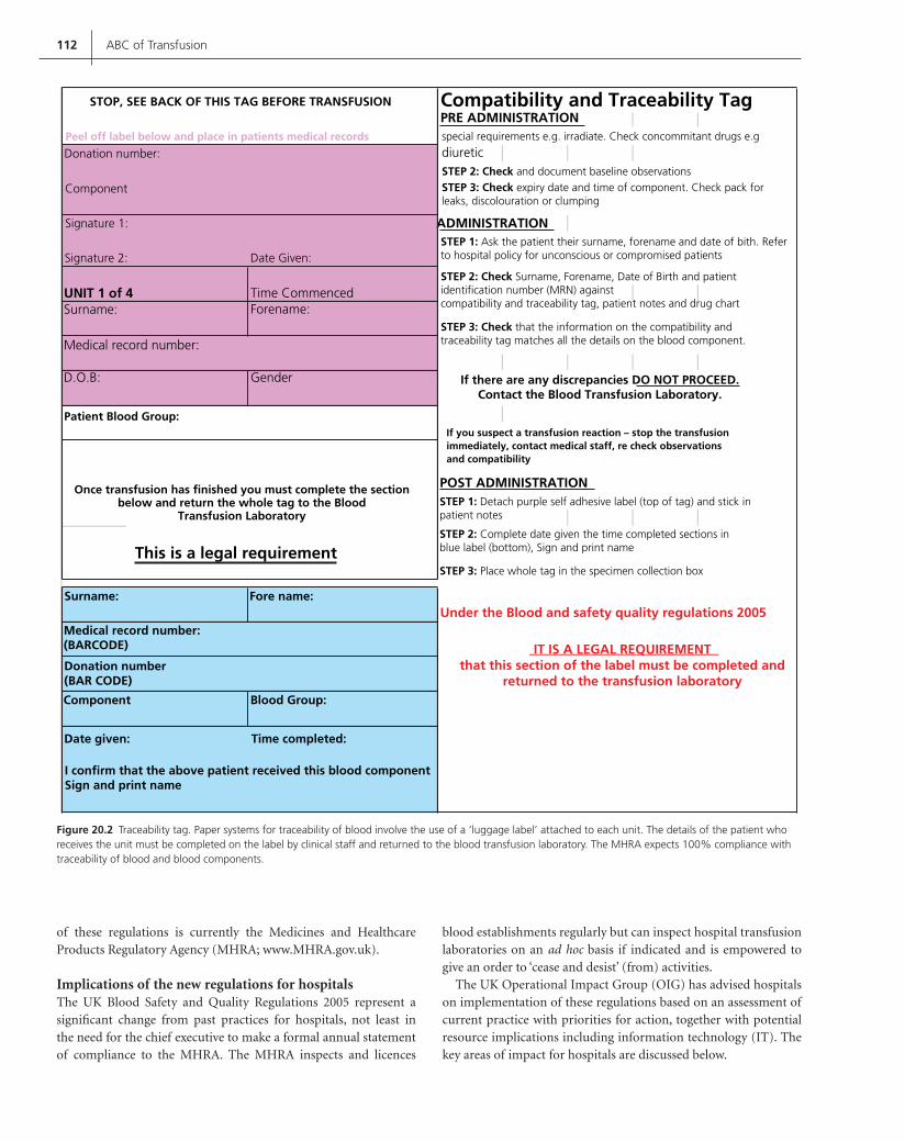
112 ABC of Transfusion
of these regulations is currently the Medicines and Healthcare Products Regulatory Agency (MHRA; www.MHRA.gov.uk).
Implications of the new regulations for hospitalsThe UK Blood Safety and Quality Regulations 2005 represent a signifi cant change from past practices for hospitals, not least in the need for the chief executive to make a formal annual statement of compliance to the MHRA. The MHRA inspects and licences
blood establishments regularly but can inspect hospital transfusion laboratories on an ad hoc basis if indicated and is empowered to give an order to ‘cease and desist’ (from) activities.
The UK Operational Impact Group (OIG) has advised hospitals on implementation of these regulations based on an assessment of current practice with priorities for action, together with potential resource implications including information technology (IT). The key areas of impact for hospitals are discussed below.
Figure 20.2 Traceability tag. Paper systems for traceability of blood involve the use of a ‘luggage label’ attached to each unit. The details of the patient who receives the unit must be completed on the label by clinical staff and returned to the blood transfusion laboratory. The MHRA expects 100% compliance with traceability of blood and blood components.
STOP, SEE BACK OF THIS TAG BEFORE TRANSFUSION Compatibility and Traceability TagPRE ADMINISTRATION
Peel off label below and place in patients medical records
ADMINISTRATION
Signature 2:
Signature 1:
Component
Donation number:
Date Given:
UNIT 1 of 4Surname: Forename:
Time Commenced
D.O.B:
Medical record number:
Gender If there are any discrepancies DO NOT PROCEED.Contact the Blood Transfusion Laboratory.
If you suspect a transfusion reaction – stop the transfusionimmediately, contact medical staff, re check observationsand compatibility
Once transfusion has finished you must complete the sectionbelow and return the whole tag to the Blood
Transfusion Laboratory
Patient Blood Group:
POST ADMINISTRATION
This is a legal requirementSTEP 3: Place whole tag in the specimen collection box
STEP 1: Detach purple self adhesive label (top of tag) and stick inpatient notes
STEP 1: Ask the patient their surname, forename and date of bith. Referto hospital policy for unconscious or compromised patients
STEP 2: Check Surname, Forename, Date of Birth and patientidentification number (MRN) againstcompatibility and traceability tag, patient notes and drug chart
STEP 2: Check and document baseline observations
special requirements e.g. irradiate. Check concommitant drugs e.g
diuretic
STEP 3: Check expiry date and time of component. Check pack forleaks, discolouration or clumping
STEP 3: Check that the information on the compatibility andtraceability tag matches all the details on the blood component.
STEP 2: Complete date given the time completed sections inblue label (bottom), Sign and print name
Fore name:Surname:Under the Blood and safety quality regulations 2005
IT IS A LEGAL REQUIREMENTthat this section of the label must be completed and
returned to the transfusion laboratoryDonation number(BAR CODE)
Medical record number:(BARCODE)
Time completed:
Blood Group:
Date given:
Component
I confirm that the above patient received this blood componentSign and print name
Contreras_C020.indd 112Contreras_C020.indd 112 12/19/2008 2:24:33 PM12/19/2008 2:24:33 PM

Regulations and the EU Directives 113
Quality systemsHospital transfusion laboratories must have a comprehensive qual-ity system in place based on the principles of ‘good practice’. This includes stringent requirements for storage and distribution of blood and blood components with particular emphasis on ‘cold chain’ management together with validation and calibration of processes and equipment and change control. A designated quality manager is pivotal to the effective operation of a quality manage-ment system.
TraceabilityThe regulations require that hospitals must have complete trace-ability of the fate of each unit of blood/blood components (red
Figure 20.3 Traceability instructions. All clinical staff involved in blood transfusion must have training in maintaining traceability of all blood and blood components and this can be reinforced by use of training posters available in clinical areas. (Reproduced courtesy of the Scottish National Blood Service.)
Figure 20.5 The ‘Red Book’ contains guidelines for all materials produced by the UK blood transfusion services for both therapeutic and diagnostic use. The guidelines refl ect the legally binding requirements of the Blood Safety and Quality and Regulations 2005 (see www.transfusionguidelines.org.uk).
Figure 20.4 Electronic blood-tracking systems assist with the traceability of blood and can also be used for the electronic issue of blood at sites remote from the blood bank.
Contreras_C020.indd 113Contreras_C020.indd 113 12/19/2008 2:24:34 PM12/19/2008 2:24:34 PM

114 ABC of Transfusion
cells, platelets, plasma and cryoprecipitate). These records must be kept for 30 years. The data set that is required for compliance includes: donation number, component type, identifi cation of sup-plying blood establishment, transfused recipient identifi cation, if not transfused, confi rmation of fi nal disposition, date of trans-fusion or fi nal fate of component if not transfused.
Positive affi rmation is required from all clinical areas regarding the fate of each unit of blood or blood component. The traceability information can be stored in a paper or electronic format but com-pliance must be 100%. Options for traceability systems include: (i) a paper-based system involving returning the label or signed paper document to the laboratory (Figures 20.2 and 20.3) and manual or electronic update of laboratory records; or (ii) a fully electronic sys-tem with barcode readers used by nursing and medical personnel when blood components are administered to patients (Figure 20.4). This would involve barcoded wrist identity bands for patients and additional hardware and software.
The OIG has stated that ultimately only effective electronic sys-tems can ensure total traceability and recommends the inclusion of electronic blood tracking systems in the national IT programmes (e.g. Connecting for Health).
Education and trainingAll staff involved in the blood transfusion process must receive training and all training records must be available for inspection by the MHRA.
HaemovigilanceAdverse incidents must be reported as soon as known via the SABRE (Serious Adverse Blood Reactions and Events) online system hosted by the MHRA (Figure 20.6). This also includes online reporting to SHOT (Serious Hazards of Transfusion). See Chapter 15 for further details on haemovigilance.
Processing activitiesThe only processing activity that may be performed legally under these regulations by a hospital transfusion laboratory, is thaw-ing of components (i.e. fresh frozen plasma and cryoprecipitate). Processing activities formerly undertaken in hospital trans fusion laboratories but which are now permitted only in licensed blood establishments include irradiation, preoperative autologous deposit, granulocyte collection, splitting or pooling of components, and adjusting of haematocrit. Stem cell and donor lymphocyte
Figure 20.6 The UK Blood and Quality Safety Regulations 2005 require that serious adverse events and reactions related to transfusion of blood and blood components are reported to the MHRA. There are two guidance documents, namely (a) Background and Guidance on reporting Serious Adverse Events and Serious Adverse Reactions and (b) SABRE – A User Guide (see www.mhra.gov.uk).
(a) (b)
Contreras_C020.indd 114Contreras_C020.indd 114 12/19/2008 2:24:38 PM12/19/2008 2:24:38 PM

Regulations and the EU Directives 115
collection, and intra- and postoperative cell salvage techniques, are excluded from the blood regulations, though stem cell and donor lymphocyte collection will be regulated under tissues legislation.
AmendmentsAmendments based on Commission Directives 2005/61/EC and 2005/62/EC, containing further technical requirements with regard to blood and blood components, have been transposed into English law coming into force in August 2006. These have introduced fur-ther defi nitions for community facilities where blood may be trans-fused. The amendments also impose the requirement for hospital transfusion laboratories to have a system of traceability and to comply with the quality system requirements of Commission Directive 2005/62/EC. Transfusion laboratories are required to retain records of any serious adverse events that may affect the qual-ity or safety of blood and blood components for at least 15 years.
Implications of the new regulations for blood establishmentsUnlike hospital transfusion laboratories, blood establishments were already regulated by the MHRA and undergoing regular inspec-tions. The secretary of state, via its ‘competent authority’, can grant a blood establishment licence to existing hospital transfusion labo-ratories to carry out processing activities (e.g. irradiation or granu-locyte collection) with the newly licensed facility being inspected every 2 years.
The regulations set out details of the quality system and trace-ability requirements for blood establishments as well as require-ments for testing and specifi cations for blood components and their labelling. An annual report must be sent to the EU via the competent authority with details of testing, including additional testing for epidemiological reasons, compliance with the quality system, including storage, transport and distribution conditions, and donor information and recruitment details.
Other standards and guidelinesThere are several other sources of regulations and guidelines encom-passing standards for the practice of clinical blood transfusion.
Better Blood TransfusionAlthough not part of the regulatory framework, the two Health Service circulars, published in 1998 and 2002, with further update in 2007, have helped to ensure that the Better Blood Transfusion initiative is an integral part of NHS practice with responsibil-ity fi rmly in the hands of NHS Trust management as part of the clinical governance structure. The key objectives include avoidance of unnecessary transfusion and making transfusion practice safer with better information for patients and the public. These initia-tives have been discussed in detail in Chapter 19.
NHS Litigation AuthorityThe NHS Litigation Authority (NHSLA) is a Special Health Authority, responsible for handling negligence claims made against NHS bodies in England. Although membership of the scheme is voluntary, the majority of NHS Trusts (including
Foundation Trusts) currently belong to the scheme. Discounts are available to those trusts that achieve the relevant NHSLA risk management standards and these include blood transfusion practice.
Clinical Pathology Accreditation schemeThe Clinical Pathology Accreditation (CPA) scheme, introduced to UK pathology laboratories in 1992, is an external audit of the abil-ity to provide a service of high quality and is confi rmed by peer review. There are objective standards for all aspects of hospital laboratory work including blood transfusion laboratories, such as staffi ng, procurement and maintenance of equipment, use of stan-dard operating procedures, reporting of results, internal audit, and external quality assessment with emphasis on continual quality improvement.
National Patient Safety Agency Safer Practice NoticeThe National Patient Safety Agency (NPSA) was established in 2001 as a Special Health Authority with the aim of making the NHS ‘an organisation with a memory’, which learns from its mistakes via reporting and analysis of adverse events and near miss incidents.
Although not a regulation, the NPSA Safer Practice Notice Right Patient – right blood (NPSA 2006) has important implications for hospitals. The emphasis is on the fi nal pre-transfusion bedside check, the need to consider information technology to improve transfusion safety, and the need for training and regular compe-tency testing for all relevant staff involved in the transfusion process (including phlebotomists, blood transfusion laboratory staff, por-ters, nurses and medical staff).
Where next with regulation?
The Treaty of Amsterdam in 2000 empowered the European Parliament to legislate on the quality and safety of organs and sub-stances of human origin, blood and blood derivatives. The outcome to date has been the EU Directive on Blood Safety and the Tissues and Cells Directive.
In the fi eld of blood safety there are two outstanding areas that the EU had originally planned future action in:
Educational programmes for health professionals in the optimal 1 use of blood.The promotion of public information/education on blood col-2 lection and transfusion.
There will be commission follow-up of the implementation of the EU Blood Directives in community states. Action is being taken to standardize the training of inspectors, with the potential aim of a European-wide inspection system for blood services.
It can be anticipated that sooner or later proposals will be drawn up for the regulation of organ donation. However, article 152 of the Treaty of Amsterdam places some restriction on what the EU can legislate. The relevant clause states: ‘community action in the fi eld of public health shall respect the responsibilities of Member States for the organisation and delivery of health services and medical care, in particular measures shall not affect national provisions on donations or medical use of organs and blood’. In
Contreras_C020.indd 115Contreras_C020.indd 115 12/19/2008 2:24:47 PM12/19/2008 2:24:47 PM

116 ABC of Transfusion
other words, at present the EU cannot legislate on the delivery of clinical services.
Whilst harmonization and regulation of quality and safety standards for these ‘substances of human origin’ is to be wel-comed, the UK concern is that where the technical details are overproscriptive they are likely to need regular and occasionally rapid updating to respond to advances in knowledge and technol-ogy. The EU regulatory committee procedures necessary to make amendments to any of the EU Blood Directives is yet to be tested. Should this regulatory committee review process take too long, there is the potential for an adverse impact on quality and safety of ‘substances of human origin’ with the law requiring compliance with outdated standards.
Further reading
Murphy M, Pamphilon DH. Practical Transfusion Medicine. Blackwell
Publishing, Oxford, 2005.
Operational Impact Group. OIG Report. www.transfusionguidelines.org.uk,
under new regulations.
References
Department of Health. Better Blood Transfusion. HSC 1998/224. Department
of Health, London, 1998.
Department of Health. Better Blood Transfusion: appropriate use of blood. HSC
2002/009. Depa rtment of Health, London, 2002.
Department of Health. Better Blood Transfusion: safe and appropriate use
of blood. HSC 2007/001. Department of Health, London, 2007.
European Parliament and Council. Implementing Directive 2002/98/EC as
regards standards of quality and safety for the collection, testing, process-
ing, storage and distribution of human blood and blood components.
Offi ce Journal of the European Union 2003; L33/30.
European Parliament and Council. Implementing Directive 2005/61/EC as
regards traceability requirements and notifi cation of serious adverse reac-
tions and events. Offi ce Journal of the European Union 2005; L256/32.
European Parliament and Council. Implementing Directive 2005/62/EC as
regards community standards and specifi cations relating to a quality
system for blood establishments. Offi ce Journal of the European Union
2005; L256/41.
National Patient Safety Agency (NPSA). Right Patient – right blood.
NPSA/2006/14. National Patient Safety Agency 2006, www.npsa.nhs.uk.
UK Blood Safety and Quality Regulations 2005. Statutory Instrument 2005,
No. 50. © Crown Copyright 2005. www.opsi.gov.uk/si/si2005/20050050.htm.
Contreras_C020.indd 116Contreras_C020.indd 116 12/19/2008 2:24:48 PM12/19/2008 2:24:48 PM

117
Index
ABO blood groups 131ABO haemolytic disease of newborn 30,
30, 31ABO incompatibility 19, 85–7, 86, 87ABO-related haemolytic disease of newborn 30,
30, 31acidosis 58ADAMTS 13 42adult stem cells 108adult T-cell leukaemia virus 76–7AIDS 50, 76
albumin 48, 76binding to 49clinical associations 49, 49clinical properties 48–9, 49crystalloid vs colloid 51–2, 51, 52effect of illness 49indications for use 50–51, 50, 51physiology 48prognostic value 50, 50
albumin solutions 48alloimmune thrombocytopenia 24arm cleansing 71anti-A antibodies 61, 62anti-AB antibodies 61, 62anti-B antibodies 61, 62antibody screening 11, 12, 13anti-D antibodies 29–30, 29, 62antifi brinolytics 92–3aprotinin 92audit 100–102autoimmune thrombocytopenic purpura 24autologous blood transfusion 90, 93
bacterial contamination 19–20, 71–2, 71, 72Baskett’s classifi cation of hypovolaemic
shock 55Bernard-Soulier syndrome 24, 45Better Blood Transfusion initiative 99–103,
115activities of 101–2, 101, 102audit 100–101, 101effect on blood use 102, 102evidence base 100future prospects 102–3history 99–100, 100management of massive blood loss 102reduction of blood use 102
bloodgroups 9, 11, 12, 13ordering for surgery 13–14, 14supply and demand 6–9
blood cell separator 22blood components
demand for 8, 8ordering 10, 12supply and demand 6–9
blood donations, impact of vCJD on 81blood donors 1–5
adverse effects of donation 2age profi le 1demographics 1, 1impact of vCJD on 81safety of 2screening 69, 70–71selection 1–2, 2, 3
blood grouping 5, 10–11in pregnancy 31–2, 31reverse 10
blood loss 54blood prescription chart 11Blood Safety and Quality Regulations 2005 2blood stocks management 8, 8blood substitutes 95–8
haemoglobin-based 96–7, 97need for 95perfl uorochemical emulsions 97–8, 98properties of 95search for 95–6
blood supply 6–8, 6, 7blood usage, impact of vCJD on 81–2bone marrow failure 23–24, 25bovine spongiform encephalopathy 79–80
cardiopulmonary bypass surgery 24cell salvage 59, 59cellular therapies 106–9, 107, 108Chagas’ disease 4Clinical Pathology Accreditation scheme 115CMV: see cytomegaloviruscoagulation factors
in coagulopathy 59half-lives 44see also individual factors
coagulopathy 57–8, 58treatment 59
colloids 51–2, 52colloid osmotic pressure 48compatibility testing see crossmatchingConsumer Protection Act 1987 111cord blood 106Creutzfeldt–Jakob disease, variant 1, 6, 40, 79–82
impact on UK blood supply 80–82blood donation 81blood donors 81blood usage 81–2
prion diseases 79–80, 79, 80transmission by blood 80, 81, 81
Creutzfeldt-Jakob disease, sporadic 80crossmatching 9, 10–14
electronic 13see also traceability
cryoprecipitate 41, 43crystalloids 51–2, 51cytomegalovirus 4, 25, 69, 77
DDAVP, see desmopressindesmopressin 45, 93dilutional thrombocytopenia 242,3-diphosphoglycerate 18Duffy antibodies 11
EC Blood Directive (2004/33/EC) 2EDTA 10education 114electronic blood-tracking systems 113electronic crossmatching 13ELISA 70, 70, 72, 73, 75, 77embryonic stem cells 108ethylene diaminetetra-acetic acid see EDTAEuropean Union Blood Directive 2005 111–14,
111, 112–14areas of impact 111
factor VII, recombinant activated 59factor VIIa, recombinant 45, 93–4factor VIII concentrate 44–5factor IX concentrate 44–5factor XIII concentrate 45fetal blood sampling 33–5fetal transfusion 33–5, 34
platelet transfusion 35red cell transfusion 33–5, 34
fetomaternal haemorrhage 28–9
Note: Page numbers in bold represent tables, those in italics represent fi gures.
Contreras_Index.indd 117Contreras_Index.indd 117 12/5/2008 6:16:25 PM12/5/2008 6:16:25 PM

118 Index
fi brinogen concentrate 41fi brinolysis 59, 94fi brin sealants 93fi ltration 15fl uid therapy 55, 57Fluosol 98fresh frozen plasma 7–8, 7, 41, 43
for neonates 36–7, 38
gel column agglutination 13Glanzmann’s thrombasthenia 24, 25, 45good practice 112–13graft-versus-host disease 66granulocyte colony-stimulating factor 22granulocyte concentrates
preparation of 22–3properties of 23
granulocyte transfusions 25–6administration 25–6, 26adverse effects 26contraindications 26indications 26
GVHD see graft versus host disease
haemoglobin-based blood substitutes 9, 96–7adverse effects 96–7clinical trials 97, 97encapsulated haemoglobin 97human recombinant haemoglobin 97modifi ed haemoglobin solutions 96sources of haemoglobin 96, 96
haemoglobin-oxygen dissociation curve 18haemolytic disease of newborn 27–31
ABO-related 30, 30, 31aetiology and pathogenesis 27–8, 27, 28anti-D Ig 29–30, 29blood grouping/antibody screening in
pregnancy 31–2, 31immunoprophylaxis 28–9, 28post-delivery tests 32Rh 27, 32
haemolytic transfusion reactions 62, 62acute 61delayed 64, 64
haemopoietic stem cells 108–9haemorrhage, massive see massive haemorrhagehaemostasis
massive haemorrhage see massive haemorrhage
operative 90haemovigilance see Serious Hazards of
Transfusion (SHOT)HBV see hepatitis B virusHCV see hepatitis C virusHemolink 97Hemopure 96, 96, 97Hemospan 97heparin sodium 91hepatitis A virus 76hepatitis B virus 2–3, 74–5, 74, 74, 75hepatitis C virus 2–3, 75–6, 75hepatitis G virus 75histocompatibility antigens 5HIV 76, 76, 77HLA see human leucocyte antigensHNA see human neutrophil antigensHPA see human platelet antigens
human albumin see albuminhuman leucocyte antigens 5human neutrophil antigens 64human platelet antigens 7human T-cell leukaemia virus 76–7
type 1 69human T cell lymphotropic virus 3hydrops fetalis 27, 30hyperkalaemia 58hypoalbuminaemia 49, 49hypocalcaemia 58hypothermia 22, 57, 58hypovolaemic shock 55
idiopathic thrombocytopenic purpura 46, 46immunoglobulins 45–6immunological complications of
transfusion 61–8, 61transfusion-associated graft versus host
disease 65, 66immunomodulation 67–8plasma protein reactions 67platelet incompatibility 66–7red cell incompatibility 61–4white cell incompatibility 64–6
immunological refractoriness to platelet transfusions 25
immunomodulation 20immunoprophylaxis 28–9, 28immunotherapy 106–9, 107, 108
active 107natural killer cells 107passive 107T lymphocytes 107, 108T regulatory cells 108
infectious complications 69–73bacterial 71–2, 71, 72malaria 72–3, 72parasitic 69properties of infections 70screening tests for blood donations 69, 70–71, 70Treponema pallidum (syphilis) 72, 72vCJD see Creutzfeldt–Jakob diseaseviruses 74–8
information technology 87intrauterine transfusion 33–5inventory management 8–9iron overload 20
Jehovah’s witnesses 17, 19, 95
Kell antibodies 11, 30kernicterus 27Kidd antibodies 11Kleihauer test 29–30, 29, 46
labellingpatient identifi cation 101red cell salvage 94traceability tag 112
leucocyte depletion 6leukaemia, immunotherapy 108, 108
malaria 72–3, 72markers of infection 3–5, 4massive haemorrhage 54–60
assessment of extent 54, 55
complicationscoagulation problems 58–9, 58, 59coagulopathy 57–8, 58
factors modifying normal physiological response 55
initial investigations/procedures 55physiological responses to 54recycling of autologous blood 59, 59survival 59treatment 56
bleeding control 55coagulopathy 59fl uid therapy 55intravenous fl uids 57optimum haemoglobin 56–7, 57oxygen therapy 55patient monitoring 57
target blood pressure 56Medicines Act 1968 111Medicines and Healthcare Products Regulatory
Agency (MHRA) 83
National External Quality Assurance Scheme 30National Patient Safety Agency, Safer Practice
Notice 115natural killer cells 107neonatal transfusion 34, 35–9
fresh frozen plasma 36–7, 38large volume transfusions 35–6, 36platelet transfusion 36prescribing and administration 37–9, 38, 38red cell transfusion 35–6, 35, 36small volume top-ups 35, 36thresholds for 35volumes and rates 38
neonatesactivated partial thromboplastin time 37fi brinogen 37prothrombin time 37
NHS Act Healthcare 1999 111NHS Litigation Authority 115non-haemolytic febrile transfusion
reactions 25, 65non-haemopoietic stem cells 108—9nucleic acid testing (NAT) 74, 75, 77
oliguria 63, 64operative haemostasis 90Optro 97ordering 10, 12oxygen carriers 96Oxygent 98oxygen therapy 55OxyVita 97
parvovirus B19 77pathogen inactivation 74patient identifi cation 101perfl uorochemical emulsions 97–8, 98Perftoran 98phototherapy 31plasma exchange 42–4plasma products 40–47, 40, 42
coagulation factors 44–5, 44cryoprecipitate 41, 43cryosupernatant 41fresh frozen plasma 7–8, 7, 36–7, 38, 41, 43
Contreras_Index.indd 118Contreras_Index.indd 118 12/5/2008 6:16:26 PM12/5/2008 6:16:26 PM

Index 119
immunoglobulins 45–6indications for use 42prothrombin complex concentrates 45
plasma proteins, reactions to 67, 67platelets 7, 7platelet concentrates
preparation of 22–3properties of 23
platelet function abnormalities 24platelet incompatibility 66–7platelet recovery 24–5platelet transfusions 22–5
administration 24–5adverse effects 25antigen matched 25contraindications 25crossmatched 25fetal 35indications 23–4, 24neonate 36
PolyHeme 97post-transfusion purpura 25pregnancy, blood grouping/antibody
screening 31–2, 31preoperative anaemia 89–90, 89prion diseases 77, 79–80, 79, 80
see also Creutzfeldt–Jakob disease, new variantprothrombin complex concentrates 45PTP see post-transfusion purpurapurpura, post-transfusion 67
quality systems 113
Red Book 113red cells 6–7, 7, 16
alloantibodies 20preparation 15storage 15–16, 16, 17uses 16
red cell antigens, population frequency 13red cell incompatibility 61–4
circumstances 62–3haemolytic transfusion reactions 62, 62
acute 61delayed 64, 64
mechanisms of red cell destruction 63, 63naturally occurring and immune
antibodies 61–2, 62symptoms and signs 63–4, 63
red cell salvage 90–92automated 91–2, 91, 92collection and reinfusion of wound
drainage 90–91, 90labelling 94
red cell transfusions 15–21administration 17–18adverse effects 19–20, 20, 21emergency 20–21expected benefi ts 18–19fetal 33–5, 34indications for 17neonate 35–6, 35, 36rationale for use 16–17recipients 15transfusion thresholds 3
regulatory issues 110–16future action 115–16need for regulation 110, 110regulatory framework 111–15, 111see also individual regulations
reverse blood grouping 10RhD antigen 11RhD haemolytic disease of the newborn 27RhD prophylaxis 28RhD sensitization 27, 28
saline adenine glucose mannitol (SAG-M) 15, 17, 35
Saline versus Albumin Fluid Evaluation (SAFE) study 50, 51, 52
Serious Adverse Blood Reactions and Events (SABRE) 41, 83, 114
Serious Hazards of Transfusion (SHOT) 19, 20, 40, 71, 83–8, 114
blood safety issues 84–5, 85categories of adverse reactions 84development of safety culture 87–8frequency of adverse reactions 85lessons from 83–4, 84operation of 83, 84prevention of incompatibity reactions 85–7,
86, 87role of information technology 87
serological testing 5SHOT see Serious Hazards of Transfusionspherocytosis 30stem cells, non-haemopoietic 108–9stem cell plasticity 108–9stem cell transplantation 104–6, 104, 105,
105, 106supply and demand 6–9surgery
operative haemostasis 90ordering blood for 13–14, 14
thrombocytopenia 22, 57alloimmune 24dilutional 24
transfusion-associated graft-versus-host disease (TA-GVHD) 20, 24, 34, 65, 84, 85
transfusion reactions 10, 13, 19, 20, 25, 40, 61, 62, 65, 81
thromboelastography 58–9, 58, 59T lymphocytes 107, 108topical haemostatic sealants 93traceability 113–14
instructions 113tag 112
training 114TRALI see transfusion-related acute lung injurytranexamic acid 92transfusion reaction 19–21, 20, 21transfusion recipients
age of 16safety of 2
transfusion-related acute lung injury (TRALI) 20, 40, 65–6, 65, 66
transfusion-related mortality/morbidity 85transmissible spongiform encephalopathies see
prion diseasesT regulatory cells 108Treponema pallidum (syphilis) 72, 72Trypanosoma cruzi 69, 71
UK Blood and Quality Safety Regulations 2005 114
UK Haemophilia Centre Doctors’ Organisation (UKHCDO) 41
UK Operational Impact Group 112urticaria 19, 67
vCJD see Creutzfeldt–Jakob disease, variant CJDviral contamination by transfusion 74–8
adult T-cell leukaemia virus 76–7cytomegalovirus 77hepatitis B virus 74–5, 74, 74, 75hepatitis C virus 75–6, 75HIV 76, 76, 77human T-cell leukaemia virus 76–7parvovirus B19 77viral inactivation 74, 75
volume overload 20von Willebrand factor 44von Willebrand’s disease 45
West Nile virus 1white cell incompatibility 4–6
graft-versus-host disease 66non-haemolytic febrile transfusion
reactions 65transfusion-related acute lung injury 65–6,
65, 66
Contreras_Index.indd 119Contreras_Index.indd 119 12/5/2008 6:16:26 PM12/5/2008 6:16:26 PM

Contreras_Index.indd 120Contreras_Index.indd 120 12/5/2008 6:16:26 PM12/5/2008 6:16:26 PM


