37
Medical Imaging and the Abdomen Pratheep Suntharamoorthy
| Date post: | 13-Apr-2017 |
| Category: |
Documents |
| Upload: | meducationdotnet |
| View: | 591 times |
| Download: | 1 times |

Medical Imaging and the Abdomen
Pratheep Suntharamoorthy

Learning Objectives
• Describe different diagnostic imaging techniques
• Explain the different modalities of GI imaging• Identify core anatomical features on GI
radiographs• State systematic approach to presenting an
abdominal film • Use systematic approach to present an
abdominal film

X-rays• Electrons released from a hot
cathode.
• When electrons collide with anode, they produce X-rays.
• X-rays are absorbed by bone, but pass through soft tissue.
• When X-rays used to be taken with photographic film. Now computer detection used more frequently.
• Contrast also used in X-rays.
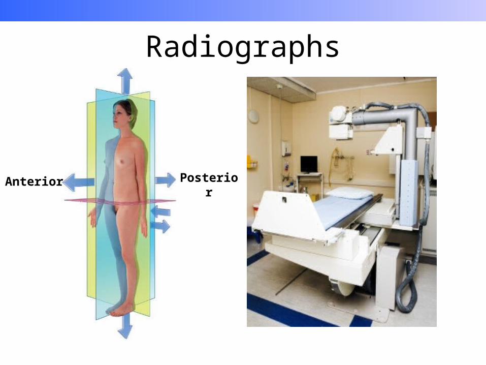
Radiographs
Anterior Posterior
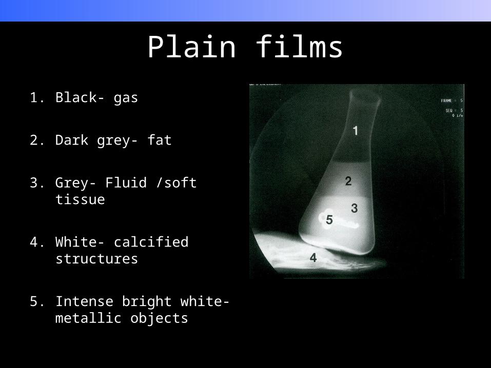
Plain films1. Black- gas
2. Dark grey- fat
3. Grey- Fluid /soft tissue
4. White- calcified structures
5. Intense bright white- metallic objects
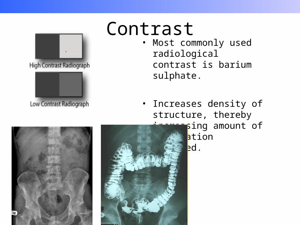
Contrast• Most commonly used
radiological contrast is barium sulphate.
• Increases density of structure, thereby increasing amount of X-radiation absorbed.

Computed tomography• X-radiation with greater
exposure.
• Essentially breaking up a 3D structure and putting it back together as a 2D structure.
• CT can detect and differentiate a wide range of x-ray intensities by electronic detection.

CT abdomen

CT Abdomen

CT Head
A- Orbit D- External Auditory canal
B- Sphenoid sinus E- Mastoid Air cellsC- Temporal lobe F- Cerebral
Hemisphere
Anatomy
Normal Anatomy
University of Virginia Medical Education
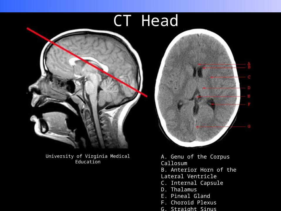
CT Head
University of Virginia Medical Education A. Genu of the Corpus CallosumB. Anterior Horn of the Lateral VentricleC. Internal CapsuleD. ThalamusE. Pineal GlandF. Choroid PlexusG. Straight Sinus

CT Stroke

Advances in Imaging

Magnetic resonance imaging• Developed in Nottingham
• Uses magnetic properties of hydrogen
• Interaction between radio waves and hydrogen nuclei produce image.
• T1 ad T2 weighted scansNational High Magnetic Field Laboratory

MRI• T1 weighted scans: water and
fluid containing tissue are dark and fat containing tissue are bright.

MRI• T2 weighted scans: water and
fluid containing tissue are bright and fat containing tissue are dark
• Indications for MRI in abdominal pathology are malignancy, liver lesions, and MRCP.

Nuclear Imaging• Positron emission
tomography• Patient injected with a small
amount of radioactive drug.• The drug localises to a part
of the body according to metabolic properties.
• Once there it decays, due to a short half life, emitting gamma radiation.
• This gamma radiation is picked up by a gamma camera.

Abdominal Imaging

Plain films1. Black- gas
2. Dark grey- fat
3. Grey- Fluid /soft tissue
4. White- calcified structures
5. Intense bright white- metallic objects
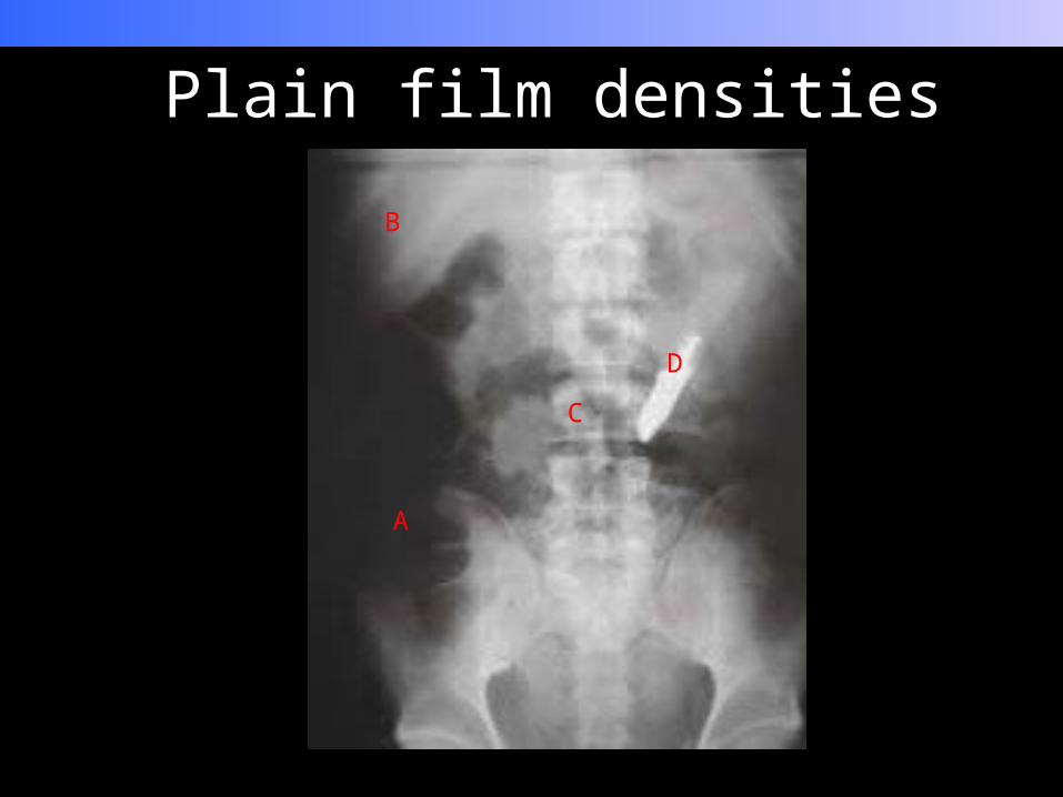
A
B
C
D
Plain film densities

Abdominal Radiographs• Commonly taken in supine
position
• Anterio- posterior direction
• Abdominal plain films have radiation dose equivalent to 35 CXR (or 4 months background radiation)
Anterior
Posterior

Anatomy

Barium Swallow

Barium Meal

Barium follow through
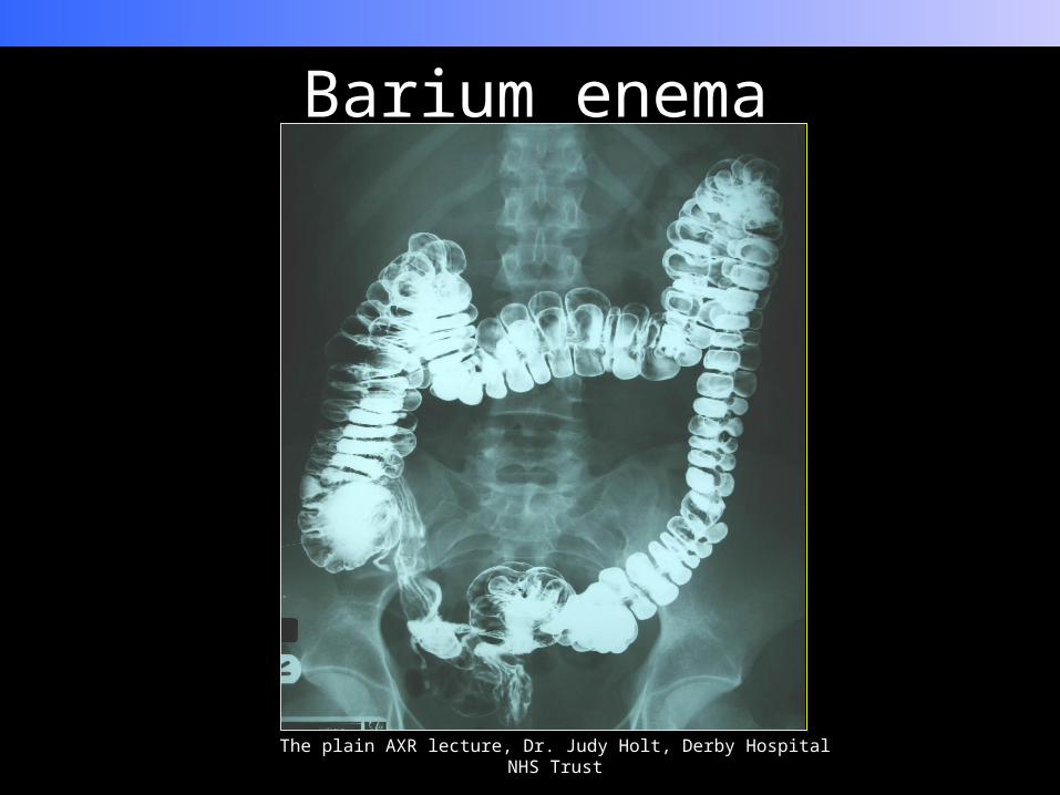
Barium enema
The plain AXR lecture, Dr. Judy Holt, Derby Hospital NHS Trust

Comparison of large and small bowel
Small bowel•Valvulae conniventes
•Many loops
•Centrally placed
•3-5cm
•No solid faeces
Elsevier. Drake et al. Gray’s Anatomy for students

Large bowel•Haustra
•Fewer loops
•Peripheral distribution
•>5cm•present
Elsevier. Drake et al. Gray’s Anatomy for students


Systematic approach
• Demographics• What modality• Bowel gas pattern• Soft tissue outline• Areas of calcification• Skeletal abnormalities

Case presentation 1• 57 year old male presents with
abdominal distension and vomiting.
Remember:• Demographics• What modality• Bowel gas pattern• Soft tissue outline• Areas of calcification• Skeletal abnormalities
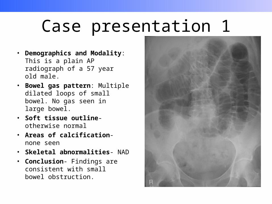
Case presentation 1• Demographics and Modality:
This is a plain AP radiograph of a 57 year old male.
• Bowel gas pattern: Multiple dilated loops of small bowel. No gas seen in large bowel.
• Soft tissue outline- otherwise normal
• Areas of calcification- none seen• Skeletal abnormalities- NAD• Conclusion- Findings are
consistent with small bowel obstruction.

Case presentation 2• 45 year old female patient
admitted with poor renal function.
Remember:• Demographics• What modality• Bowel gas pattern• Soft tissue outline• Areas of calcification• Skeletal abnormalities

Case presentation 2• Demographics and Modality:
This is a plain AP radiograph of a 45 year old female.
• Bowel gas pattern: No apparent distension of small or large bowel
• Soft tissue outline- otherwise normal
• Areas of calcification- Calcification seen over renal areas bilaterally
• Skeletal abnormalities- NAD• Conclusion: Findings are
consistent with nephrocalcinosis

Case presentation 3• 50 year old male with known
Crohn’s presents with an acute abdominal pain, following an ERCP.
Remember:• Demographics• What modality• Bowel gas pattern• Soft tissue outline• Areas of calcification• Skeletal abnormalities

Case presentation 3• Demographics and Modality:
This is a plain PA radiograph of a 50 year old male
• Bowel gas pattern: crescenteric appearance on the underside of diaphragm
• Soft tissue outline- otherwise normal
• Areas of calcification-NAD• Skeletal abnormalities- NAD• Conclusion: Findings are
consistent with extraluminal gas (pneumoperitoneum)

Thank you


