33
Abdominal wall reconstruction Ari Leppäniemi Abdominal Center Meilahti hospital University of Helsinki Finland

Abdominal wall reconstruction
Ari Leppäniemi
Abdominal Center
Meilahti hospital
University of Helsinki
Finland

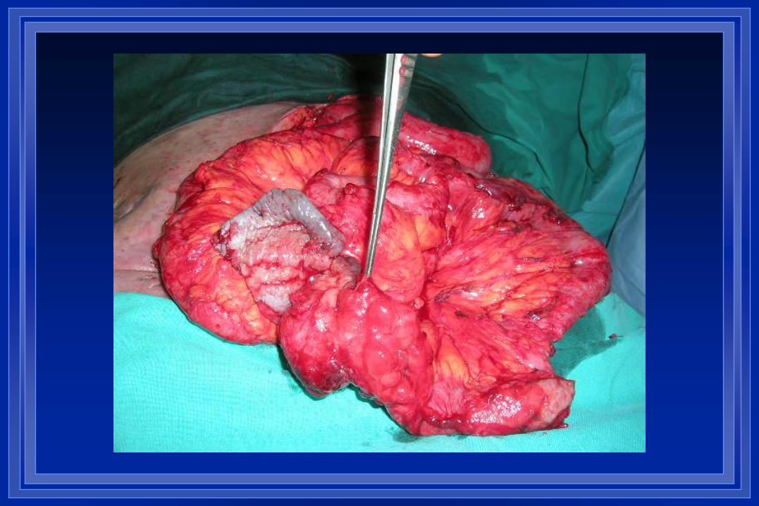
Planned hernia with early skin-grafting

Massive ventral hernia

Planned hernia
- fascial defect with original skin cover
- fascial replacement (mesh)
- fascial approximation (component separation)
- combination
- fascial and skin defect

Components separation (Ramirez et al. 1990),
originally described by Albanesi 1951

Mini-invasive components separation

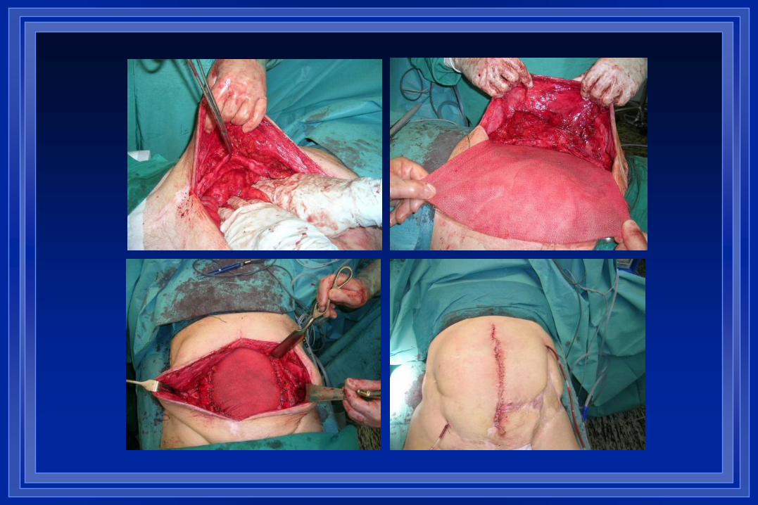
Mesh repair

Biological meshes
- partially remodeling prostheses
- porcine dermal collagen, human dermal
collagen, bovine pericardium collagen
- completely remodeling prostheses
- porcine intestinal mucosa
- different remodeling times
- resistance to mechanical stress (partially remodeling
meshes)
- low adhesiogenic power ?
- resistance to infection (contamination) ?

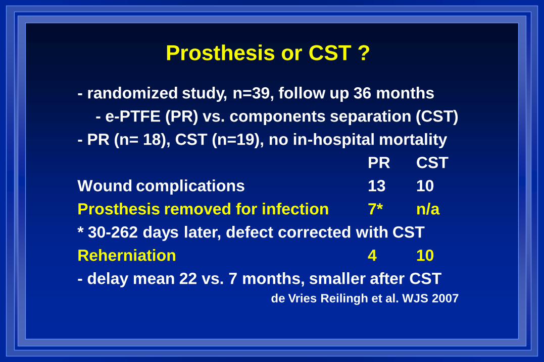
Prosthesis or CST ?
- randomized study, n=39, follow up 36 months
- e-PTFE (PR) vs. components separation (CST)
- PR (n= 18), CST (n=19), no in-hospital mortality
PR CST
Wound complications 13 10
Prosthesis removed for infection 7* n/a
* 30-262 days later, defect corrected with CST
Reherniation 4 10
- delay mean 22 vs. 7 months, smaller after CST de Vries Reilingh et al. WJS 2007

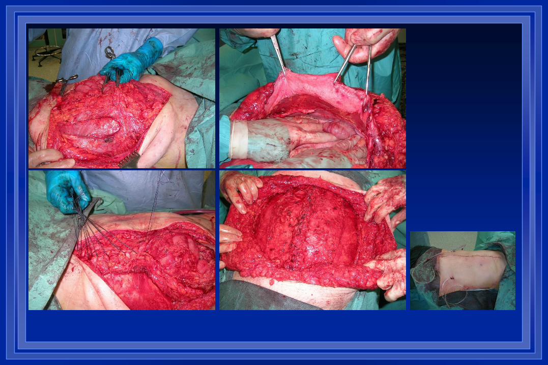
Component separation + mesh

Planned hernia
- fascial defect with original skin
cover
- fascial and skin defect
- split-thickness skin graft
late abdominal wall
reconstruction

Planned hernia with early skin-grafting

Maturation of the skin graft
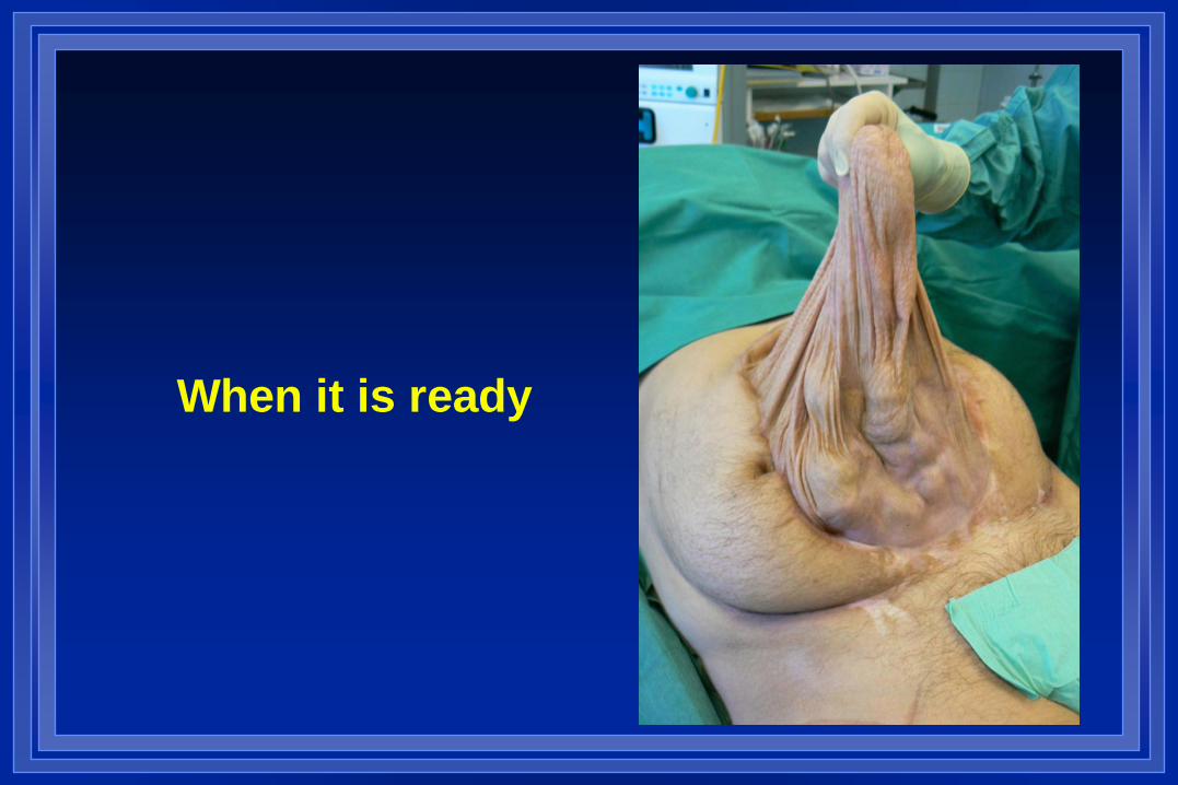
When it is ready

Abdominal wall reconstruction with
Tensor fascia lata (TFL) -flap
- myofascial or myofascial
cutaneous flaps
- pedicled TFL (Wangensteen
1934)
- free vascularized TFL (Hill et
al.1979)

Pedicled TFL

TFL microvascular flap


Creating the vascular loop

Microvascular anastomosis

Microvascular TFL-flap
- n = 20, mean age 52 (range 43-78) years
- mean follow up 5 (range 0.5-12) years
- perioperative mortality 0
- total flap necrosis 1
- distal tip necrosis 2
- postoperative bleeding 1
- intra- abdominal infection 0
- deep surgical site infection 0
- hernia recurrence (after 3 months) 1 Tukiainen and Leppäniemi 2011

Management options (Leppäniemi & Tukiainen WJS 2011)
Defect Primary Addit/alternat.
Small hernia, intact skin
No contamination CS Mesh (M)
Contamination CS Biological mesh (Mb)
Small hernia, grafted skin
No contamination CS +M or flap
Contamination CS +Mb or flap
Large hernia, intact skin
No contamination CS + flap or M
Contamination CS + flap or Mb
Large hernia, grafted skin
No contamination Flap + CS + M
Contamination Flap + CS + Mb

Case
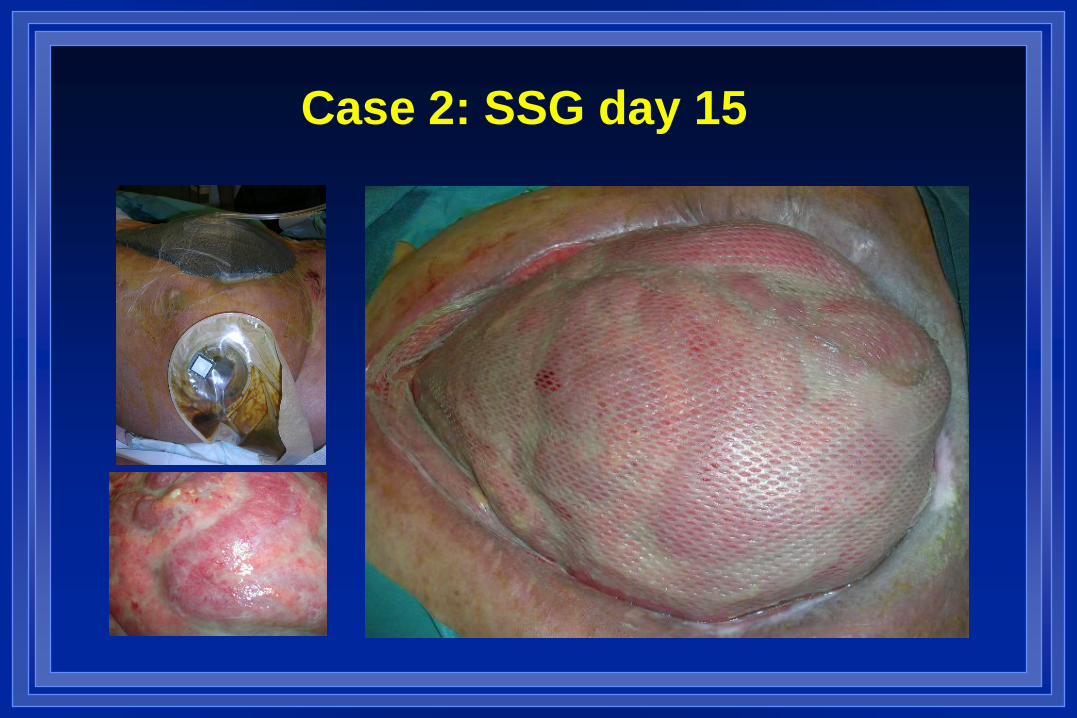
Case 2: SSG day 15

Case 2: 1 year later (not ready yet!)

Case 2: 2 years later: Laparotomy
(CS + biological mesh)

Conclusions
- aim for early fascial closure after open
abdomen
- when unable to close, think planned
hernia at 3 weeks
- start with component separation
- be ready to use other options (mesh,
flap) or a combination of techniques
- involve plastic surgeons early !

Thank you !


