42
Abnormalities of Amniotic fluid L. Sekhavat M.D

Abnormalities of Amniotic fluid
L. Sekhavat M.D

Meconium Staining
The ranged 7- 22% of pregnanciesIt is uncommon prior to 38 weeks
and increases after 40 weeks
Incidence

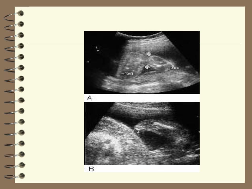
Staining of the amnionic membrans is obvius within 1-3h after meconium passege

MeconiumWhat is it? Is the earliest stools of an infant Thick dark material made up of GI secretions:
intestinal epitelial cells , lanugo, mucus, amniotic fluid, bile and water that is sometimes secreted in amniotic fluid.
Meconium is a yellow or greenish-black color and very sticky, tarry in its texture
May be classified as light or heavy.
Meconium passage is associated with:Increased fetal acidemiaIncreased perinatal mortality
and morbidityIncreased C/SIncreased neonatal mortality

MeconiumComplicates 12% of birthOf these 5% aspirate meconium (1/200
preg)Very toxic to lungsEvidence that it inactivates surfactant
ManagementNo difference in the blood pH of neonates
with and without meconium if the heart rate is normal
If the heart pattern abnormal then the incidence of acidosis is increased and the fetus should be delivered

Grading of meconium and managementGrade 1: Enough AF + light meconium
Discharg of oxytocin + follow upGrade 2: Enough AF + thick meconium
Fetal blood sampling and AB evaluationGrade 3: Oligohydramnios + thick
meconium
Urge delivery or C/S

Meconium - newborn 35% who aspirate meconium develop
complications: 10% pneumothorax 66% persistant pulmonary hypertension
related to meconium 4% die

Treatment is standard suctioningof baby mouth and nose.
Head and neck are delivered
Shoulder is delivered
when
before

Disorders of Amniotic fluid
volume
Normally:Amnionic fluid volume increases
to about 1 lit or more by 36 wks
In postterm there may by only 100-200ml
.
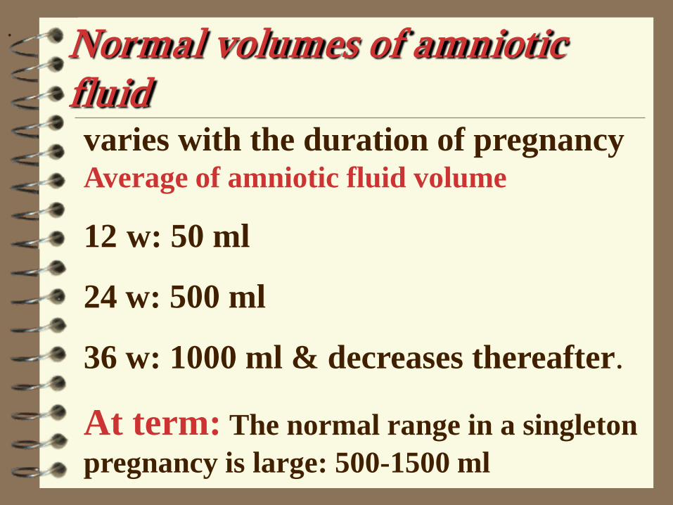
varies with the duration of pregnancyAverage of amniotic fluid volume
12 w: 50 ml
24 w: 500 ml
36 w: 1000 ml & decreases thereafter.
At term: The normal range in a singleton pregnancy is large: 500-1500 ml
Normal volumes of amniotic fluid

polyhydramnios
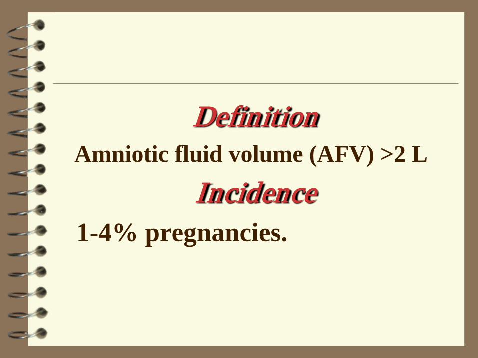
DefinitionAmniotic fluid volume (AFV) >2 L
Incidence1-4% pregnancies.
Types
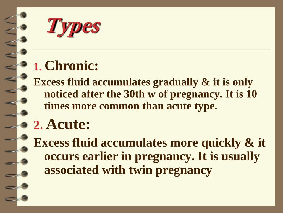
1. Chronic:Excess fluid accumulates gradually & it is only
noticed after the 30th w of pregnancy. It is 10 times more common than acute type.
2. Acute:Excess fluid accumulates more quickly & it
occurs earlier in pregnancy. It is usually associated with twin pregnancy
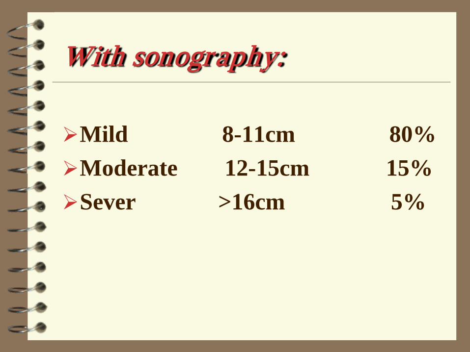
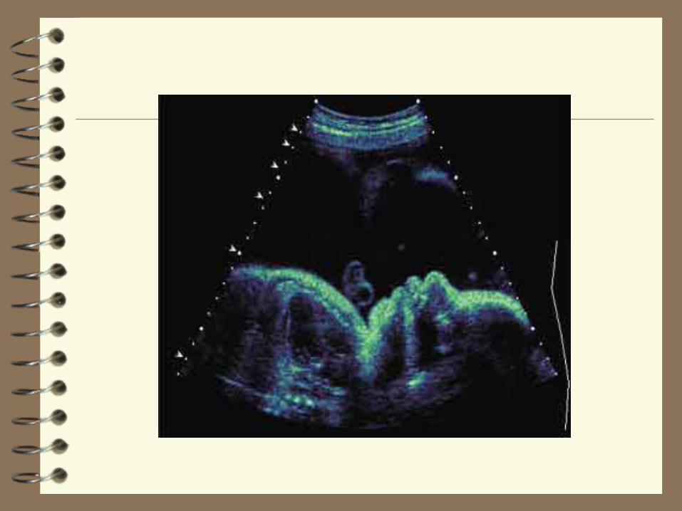
With sonography:
Mild 8-11cm 80%Moderate 12-15cm 15%Sever >16cm 5%

pathogenesisEarly in pregnancy the amnionic cavity is
filled with fluid similar to extracellular fluid
In the first half of pregnancy transfer of water across the amnion and fetal skin
During the second trimester the fetus begins to urinate, swallow and inspire
.
Causes*Fetal:1- Multiple pregnancy
2- Hydrops fetalis
3- Fetal anomalies

.
Fetal anomaliesNeural tube defect (Anencephaly , Spina bifida )
1- Increased transudation of CSF
2- Excessive urination* stimulation of cerebrospinal centers
Duodenal atresia
Thoraco-oesophageal fistula
. * Maternal:Diabetes mellitusMaternal hyperglycemia Fetal hyperglycemia
Osmotic diuresis
Pre-eclampsia
Heart or renal failure
*Idiopathic

SymptomsDyspeneaEdemaOliguriaDyspepsia

DiagnosisUterine enlargment ( larger than
the period of pregnancy)Difficulty in palpating fetal partDifficulty in hearing fetal heart Sonography

.With sonography
A. Confirm diagnosis:*Vertical pocket >8cm
*AFI >24 cm (AFI > 97.5 percentile for gestational age)
B. Detect the degree:* Mild
* Moderate
* Sever
C. Detect the cause

.
1. Twins
2. Ovarian cyst
3. Full bladder
4. Hydatiform mole
5. Ascite
All are resolved by U/S
Differential Diagnosis
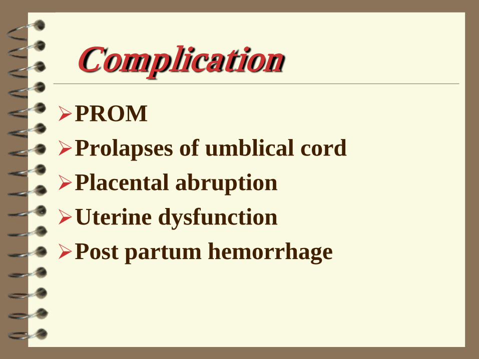
ComplicationPROMProlapses of umblical cordPlacental abruptionUterine dysfunctionPost partum hemorrhage
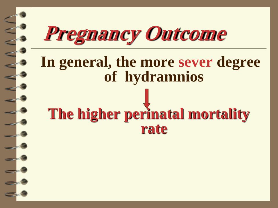
Pregnancy OutcomeIn general, the more sever degree
of hydramnios
The higher perinatal mortality rate

ManagmentMinor degrees of hydramnios rarely
require treatmentModerate degrees can usually managed
until labor ensuesSever degrees ( dyspnea or abdominal
pain or other complication), hospitalization become necessary
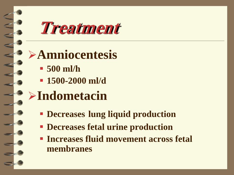
TreatmentAmniocentesis 500 ml/h 1500-2000 ml/d
Indometacin Decreases lung liquid production Decreases fetal urine production Increases fluid movement across fetal
membranes

Oligohydramnios
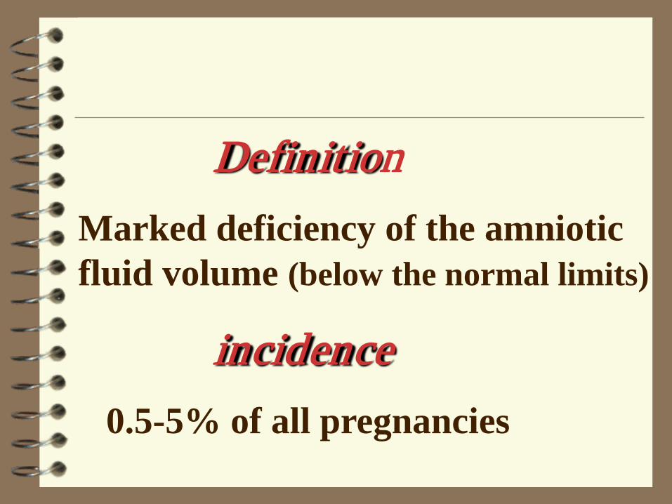
DefinitionMarked deficiency of the amniotic fluid volume (below the normal limits)
incidence0.5-5% of all pregnancies
In general:Oligohydramnios developing early
in pregnancy is less commonand
Has a bad prognosis
PlacentaAbruption
DrugProstaglandin synthetase
inhibitors, Angiotensin converting
enzyme inhibitors
idiopatic
FetalChromosomal abnormalitiesCongenital anomaliesFetal deathIUGR Postterm PROMTwin-twin transfusion
Maternaluteroplacental insufficiencyHypertensionDiabetes
Causes

Uterus is small for date
Fetus: easily felt & immobile
FHS easily heard
U/S: Vertical pocket <1cm or <2cm;
AFI <5 cm
Clinical picture
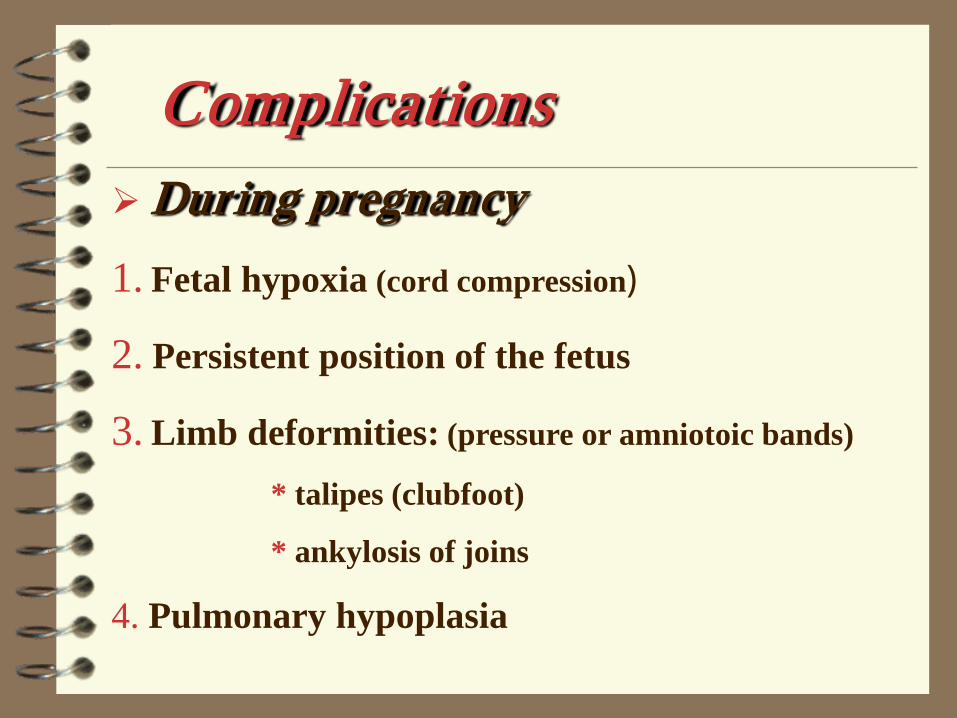
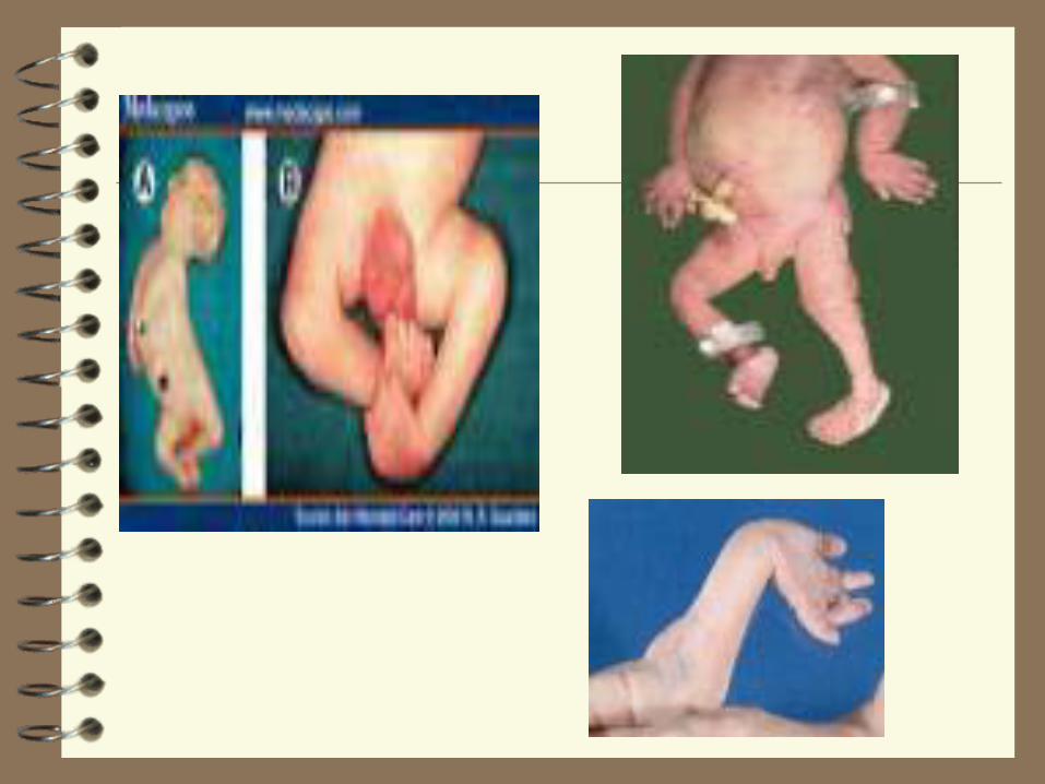
ComplicationsDuring pregnancy1. Fetal hypoxia (cord compression)
2. Persistent position of the fetus
3. Limb deformities: (pressure or amniotoic bands)
* talipes (clubfoot)
* ankylosis of joins
4. Pulmonary hypoplasia

Increased variable deceleration Increased c/s rate
During labor

Amnioinfusion: infusion of saline into the uterine cavity through the abdominal wall by a spinal needle
To increase the AFV
To dilute meconium
Treatment


