Hirosaki Med.J. 64 (Suppl.):S28―S44,2013 ABO-INCOMPATIBLE KIDNEY TRANSPLANTATION -THE SURPRISING ABSENCE OF HYPERACUTE REJECTION- Professor Kota Takahashi MD, PhD. Abstract Owing to the shortage of deceased donors in Japan, since 1989, we have performed ABO-incompatible kidney transplantation(ABO-IKTx)to expand the indication for living donor kidney transplantation. During the past 2 decades, about 2,000 ABO-IKTxs were performed. Since 2001 the success rate for these kidney transplants has reached 96% for 1-year graft survival and 91% for 5-year graft survival, similar to outcomes of ABO-compatible kidney transplantation(ABO-CKTx) . This dramatic improvement in results means that ABO-IKTx has become accepted as a therapeutic alternative for end-stage renal failure. Today ABO-IKTx accounts for 30% of all living donor kidney transplantations performed in Japan. In 1901 Karl Landsteiner discovered the presence of human ABO blood groups, and for many years ABO-IKTx was considered contraindicated, as the medical community assumed that this procedure would result in immediate/ hyperacute ABO-related rejection followed by graft loss. When we actually attempted the procedure, however, we found that acute antibody-mediated rejection (AMR) developed, but not a single instance of ABO-related hyperacute rejection. This phenomenon can be demonstrated epidemiologically. Yet, that does not answer the fundamental question of "Why is hyperacute rejection absent?" This article addresses potential mechanisms of acute AMR across ABO mismatched barriers as well as summarizing graft/patient survival of ABO-IKTxs in Japan. Hirosaki Med.J. 64, Supplement:S28―S44,2013 Key words: ABO-incompatiblity; kidney transplantation; hyperacute rejection; accommodation; antibody-mediated rejection Division of Urology, Dept. of Transplant and Regenerative Medicine, Graduate School of Medical and Dental Sciences, Niigata University. Niigata, Japan. Chief Assistant Director Niigata University Medical and Dental Hospital Address : Asahaimachi 1-757, Chuo-ku, Niigata, 951- 8510, Japan Tel:+81-25-227-2284 Fax +81-25-227-0784 E-mail: [email protected]Introduction Time certainly flies. It has been more than 20 years now since we performed the first ABO- incompatible kidney transplantation in Japan, on January 19, 1989 1-7) . The very first ABO- IKTx was conceptualized and performed by Guy P.J. Alexandre in June of 1982 8-10) . However, transplant surgeons in Japan have done a great deal to popularize this surgery and to improve its outcome. As a result, Japan can boast of expertise second to none in the area of ABO- IKTx, a procedure which is relatively rare in the international medical community. In 1901 Karl Landsteiner discovered the existence of human ABO blood groups 11) , and for many years ABO-IKTx was considered to be contraindicated, as the medical community assumed that this procedure would result in immediate hyperacute rejection (HAR)followed by graft loss. However, when we actually attempted the procedure we found that acute AMR developed, but not a single instance of ABO-related HAR. This phenomenon can be demonstrated epidemiologically 13-15) . Yet, that does not answer the fundamental question of "Why is hyperacute rejection absent?" I was sure that my readers would not be satisfied until this mechanism was clearly elucidated, and neither would I. That is
Transcript
Hirosaki Med.J. 64(Suppl.):S28―S44,2013
ABO-INCOMPATIBLE KIDNEY TRANSPLANTATION -THE SURPRISING ABSENCE OF HYPERACUTE REJECTION-
Professor Kota Takahashi MD, PhD.
Abstract Owing to the shortage of deceased donors in Japan, since 1989, we have performed ABO-incompatible kidney transplantation(ABO-IKTx) to expand the indication for living donor kidney transplantation. During the past 2 decades, about 2,000 ABO-IKTxs were performed. Since 2001 the success rate for these kidney transplants has reached 96% for 1-year graft survival and 91% for 5-year graft survival, similar to outcomes of ABO-compatible kidney transplantation (ABO-CKTx). This dramatic improvement in results means that ABO-IKTx has become accepted as a therapeutic alternative for end-stage renal failure. Today ABO-IKTx accounts for 30% of all living donor kidney transplantations performed in Japan. In 1901 Karl Landsteiner discovered the presence of human ABO blood groups, and for many years ABO-IKTx was considered contraindicated, as the medical community assumed that this procedure would result in immediate/hyperacute ABO-related rejection followed by graft loss. When we actually attempted the procedure, however, we found that acute antibody-mediated rejection (AMR) developed, but not a single instance of ABO-related hyperacute rejection. This phenomenon can be demonstrated epidemiologically. Yet, that does not answer the fundamental question of "Why is hyperacute rejection absent?" This article addresses potential mechanisms of acute AMR across ABO mismatched barriers as well as summarizing graft/patient survival of ABO-IKTxs in Japan. Hirosaki Med.J. 64, Supplement:S28―S44,2013
Division of Urology, Dept. of Transplant and Regenerative Medicine, Graduate School of Medical and Dental Sciences, Niigata University. Niigata, Japan.Chief Assistant Director Niigata University Medical
Time certainly flies. It has been more than 20 years now since we performed the first ABO-incompatible kidney transplantation in Japan, on January 19, 19891-7). The very first ABO-IKTx was conceptualized and performed by Guy P.J. Alexandre in June of 19828-10). However, transplant surgeons in Japan have done a great deal to popularize this surgery and to improve its outcome. As a result, Japan can boast of expertise second to none in the area of ABO-IKTx, a procedure which is relatively rare in the international medical community. In 1901 Karl Landsteiner discovered the
existence of human ABO blood groups11), and for many years ABO-IKTx was considered to be contraindicated, as the medical community assumed that this procedure would result in immediate hyperacute rejection (HAR) followed by graft loss. However, when we actually attempted the procedure we found that acute AMR developed, but not a single instance of ABO-related HAR. This phenomenon can be demonstrated epidemiologically13-15). Yet, that does not answer the fundamental question of "Why is hyperacute rejection absent?" I was sure that my readers would not be satisfied until this mechanism was clearly elucidated, and neither would I. That is
S 29
why this manuscript was written. We have reported elsewhere two new ground-breaking findings in 2009 and 2010 regarding ABO- IKT16, 17). We are confident that these findings will overturn the conventional wisdom about ABO-incompatible transplantation, and will mark a fundamental change in therapeutic strategies.
I. Definitions and explanation of terms To avoid misunderstanding by readers, I would like to define and explain the following important terms in advance.
1. Hyperacute rejection, and acute antibody-mediated rejection HAR, as used in this manuscript refers to an acute AMR occurring within 24 hours of transplantation. Acute AMR is defined according to the Banff classification.
2. ABO histo-blood group related antigens ABO histo-blood group related antigens consist of the following 2 types of antigens.
2.1. ABO histo-blood group antigens These were formerly described as pure antigens. They are found on the surface of human erythrocytes, tissue cells, and vascular endothelial cells, and are antigens in the original sense of the word (Fig.1). ABO histo-blood group antigens are further classified into two subtypes: ABO blood group ant igens and ABO histo group ant igens , according to the different proteins that are bound to the carbohydrate chain of each antigen.2.2. ABO histo-blood group associated antigens The process of evolutionary development has led to the presence of numerous ABO histo-blood group associated antigens within the plant and animal kingdoms. These carbohydrate antigens sometimes provoke antigen stimulation in humans, and can cause urticaria and tissue damage.Bacterial surfaces also harbor ABO histo-blood group associated antigens. From the perspective of the host defense mechanism, the healing process for complications such as sepsis in ordinary patients involves the production of neutralizing antibodies against the antigens of the causative bacteria. However, in ABO-
Fig.1. ABO histo-blood group related antigens ABO histo-blood group related antigens consist of the 2 types of antigens.
1. ABO histo-blood group antigens
ABO blood group antigens ABO histo group antigens
Erythrocytes
2. ABO histo-blood group associated antigens
Bacteria etc.in plant and animal kingdoms
Endothelial cells
S 30 K. Takahashi
incompatible transplant patients the development of sepsis during the critical period can result in those neutralizing antibodies responding to ABO histo group antigens on the surface of the vascular endothelial cells in the transplant organ, causing acute AMR that damages the transplant organ7, 15, 18).
II. Clinical outcome of ABO-incompatible kidney transplantation in Japan
1. Excellent long-term outcomes of ABO-incompatible living donor transplantation in Japan
In Japan, 1,878 ABO-IKTxs were performed between January 1, 1989 and December 31, 2010. Fig. 2 shows the patient and the graft survival rates for those procedures (Fig.2). The overall patient survival rate was 97% for the first year, 95% for the first three years, 93% for the first 5 years, and 90% for the first 9 years. The 1-year graft survival rate was 93%, 3-year 89%, 5-year 84%, and 9-year72%.
We subsequently analyzed survival outcomes for each of the earlier era (1989 – 2000) and recent era (2001 – 2010). The patient and graft survival rates for the 451 transplants performed during the earlier era were 92, 82% for the first year, 89, 76% for the first three years, 86,70% for the first 5 years, and 84, 58%, respectively. For the 1,427 procedures performed during the recent era, the patient and graft survival rates were 98, 96% for the first year, 97, 93% for the first three years, 96, 91% for the first 5 years and 91,83%, respectively, showing significant improvement (P < 0.01) in the recent era regarding both patient and graft survival. Today ABO-IKTx accounts for 30% of all living kidney transplantations performed in Japan. This has made Japan the world-leading country in original research and clinical results for this procedure, which is not yet widely studied in most nations around the world19). Four main factors contributed to such rapid and sharp improvement as follows.
Fig.2. Results for ABO-incompatible kidney transplantation in Japan show a sharp improvement in findings for the 1,427 patients treated since 2001.Results are comparable to those obtained in ABO-compatible transplantation (ABO-incompatible Kidney Transplantation Committee 2010).
0
10
20
30
40
50
60
70
80
90
100
0 5 10 15 20
%
10
20
30
40
50
60
70
80
90
100
0 5 10 15 20
Patient Survival Rate Graft Survival Rate
Overall ③(1,878 cases)
‘~2000’ ②(451 cases)
‘2001~’ ①(1,427 cases)
93% 89% 84%
82% 76% 70%
96% 93% 91%
( 1yr ) ( 3 yr ) ( 5 yr )
72%
58%
83%
( 9 yr )
③
②
①
90%97% 95% 93%
92% 89% 86%
98% 97% 96%
( 1yr ) ( 3 yr ) ( 5 yr ) ( 9 yr )
84%
91%
③
②
①
(Kaplan-Meier)
n=1,878
years
The Japan ABO-incompatible Kidney Transplantation Committee
S 31
2. Changes over 20 years that potentially improve graft/patient survival rates.Factor 1: The disproving of hyperacute rejection The common wisdom has been that organ transplantation between incompatible blood groups would result immediately in HAR and graft loss. However, this concept turned out to be a completely unsupported assumption. We provided epidemiological proof that HAR was not caused by the ABO histo-blood group antigens12-15). The anti-A/anti-B antibodies that elicit acute AMR immediately after transplantation are clearly not natural antibodies. Instead, our results show them to be de novo antibodies that are produced after transplantation, as a result of stimulation and sensitization by the ABO histo group antigens present on the surface of the vascular endothelial cells in the graft and by ABO histo-blood group associated antigens
(bacteria, etc.) 7, 15, 18). The de novo antibodies are synthesized after the graft is introduced into the body of the recipient17). This fact has extremely important implications for therapeutic strategy in ABO-incompatible organ transplantation. If posttransplant antibody production can be suppressed, then acute AMR can be avoided; i.e., the most important treatment step for ensuring a successful graft outcome is pretransplant suppression of B cell function (desensitization therapy) 7, 15, 17, 18-30). Factor 2: The mechanism of onset of acute antibody-mediated rejection, and types of rejection reaction Factor 2, as I have described previously, involved clarification of the mechanism of onset of acute AMR as elicited by ABO histo-blood group related antigens. We found that the acute AMR induced by ABO histo-blood group related antigens (ABO histo-blood group antigens and ABO histo-blood group associated antigens) could be categorized into two different mechanisms: those resulting from antigen stimulation and
those attributable to immunological response to such stimuli 7, 15, 18). The acute AMR ceases to be elicited one to two weeks after transplantation. Thus, the condition termed "accommodation" will be established if the graft can survive for the first one to two weeks7, 12-15, 18). Factor 3: Multicenter cooperative research and statistical analysis The third factor is associated with the establishment of the Japan ABO-incompatible K idney Transp lant at ion Commit tee t o systematize the conducting of this type of kidney transplantation in multiple institutions across Japan in 1997. That organizat ion conducts an annual statistical analysis of kidney transplantation procedures and outcomes in Japan, and sponsors two academic conferences each year to publicize and utilize those findings. At the beginning of the earliest period, perioperative care was not yet fully developed. Our surgical experience was limited, the findings described above were still in the future, and we were struggling to establish effective therapeutic strategies. In the early period we classified all ABO-incompatible kidney transplant patients as immunologically high-risk cases, and we thought that splenectomy concurrently at transplantation was essential for inhibiting antibody production. However, the relevant morbidities including blood loss and acute pancreatitis led to fatal complications in a number of patients. There were also some cases in which overzealous antibody removal resulted in a loss of clotting factor. This produced bleeding tendencies and serum protein loss associated with conditions such as accelerated hypovolemia and ultimately resulting in acute renal failure. As phys ic ians had no knowledge of posttransplant induction and the establishing of accommodation, but instead assumed that acute AMR could occur at any time during the life of the graft, all ABO-incompatible kidney
S 32 K. Takahashi
transplant patients were considered high-risk, and immunosuppressive therapy was aggressively pursued. This increased the chance of opportunistic infection with organisms such as cytomegalovirus, and prolonged the pathological status of the patient. Based on the statistical analysis of these data, we were able to make use of previous clinical experience and to increase both patient survival rate and graft survival rate3-6). Factor 4: Clinical applications of novel immunosuppressants The fourth factor was the use of novel immunosuppressants, which we initiated from around the year 2000. That was when drugs that suppressed B cells involved in antibody production, specifically mycophenolate mofetil and rituximab, first became available. A monoclonal antibody against CD25 that suppresses activated T cells also became available for clinical use at about that time. From new findings based on the first and second factors described above, we realized that the most important point in our therapeutic strategy was the inhibition of antibody formation through pretransplant desensitization therapy. The clinical application of these new immunosuppressants contributed greatly to our improved therapeutic results7, 15, 17,
18-30).
III. Mechanism of acceptance of ABO-incompatible kidney transplantation
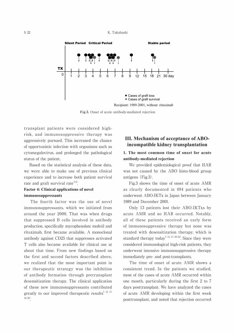
1. The most common time of onset for acute antibody-mediated rejection We provided epidemiological proof that HAR was not caused by the ABO histo-blood group antigens (Fig.3). Fig.3 shows the time of onset of acute AMR as clearly documented in 494 patients who underwent ABO-IKTx in Japan between January 1989 and December 2001. Only 13 patients lost their ABO-IKTxs by acute AMR and no HAR occurred. Notably, all of these patients received an early form of immunosuppressive therapy but none was treated with desensitization therapy, which is standard therapy today7, 15, 17, 20-22). Since they were considered immunological high-risk patients, they underwent intensive immunosuppressive therapy immediately pre- and post-transplants. The time of onset of acute AMR shows a consistent trend. In the patients we studied, most of the cases of acute AMR occurred within one month, particularly during the first 2 to 7 days posttransplant. We have analyzed the cases of acute AMR developing within the first week posttransplant, and noted that rejection occurred
Fig.3. Onset of acute antibody-mediated rejection
1 2 3 4 5 6 7 8 9 12 15 18 21 30 day 0
TX
Cases of graft lossCases of graft survival
Silent Period Critical Period Stable period
Recipient: 1989-2001, without rituximab
S 33
despite highly aggressive immunosuppressive therapy implemented in the immediate pre and postoperative period, and that graft loss was also encountered in almost every case. Subsequently the incidence of graft loss decreased. Beginning at approximately day 20 posttransplant , occurrences of AMR can be attributed primarily to factors such as inadequate immunosuppression and bacterial infection7, 15).The above data can be summarized followings. 1. HAR due to ABO histo-blood group related
antigens does not arise within the first 2 days posttransplant. We refer to this period as the “silent period”.
2. Acute AMR tends to occur within 2 to 7 days posttransplant. The incidence decreases after this period, and we have found no instances of acute AMR occurring more than 1 month posttransplant. We call this dangerous period the “critical period”.
3. To look as the reverse side of the phenomenon described in item 2, accommodation was established 1 to 2 weeks posttransplant in many cases. Once accommodation has been established, there are no further instances of acute AMR, and this continues for the life of the graft. This period is termed the “stable period”.
2. Why is ABO-related hyperacute rejection absent in ABO-incompatible kidney transplantation? -- Mechanism of onset for ABO-related acute antibody-mediated rejection The statistical analysis performed in 1967 by Gleason, et al. on kidney transplant cases showed 1 year graft survival rate as low as 25% for ABO-IKTx. That report virtually shut the door to this type of transplantation31). I raised a question about this long-standing misconception and finally disproved the occurrence of HAR due to ABO incompatibility based on the above –mentioned epidemiological studies. I defined the phenomenon of accommodation
as the situation in which, although the vascular endothelial cells in the graft carry ABO histo-blood antigens on their surface, and the blood of the recipients contains antibodies to those antigens, no antigen-antibody reaction occurs, and there is no occurrence of acute AMR5, 7,
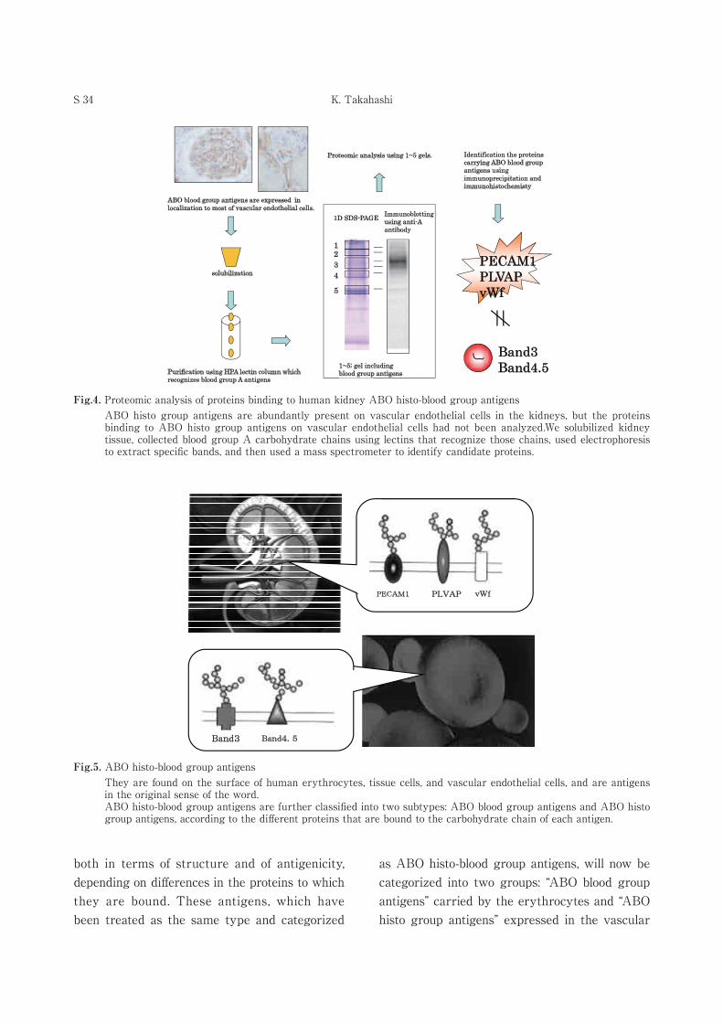
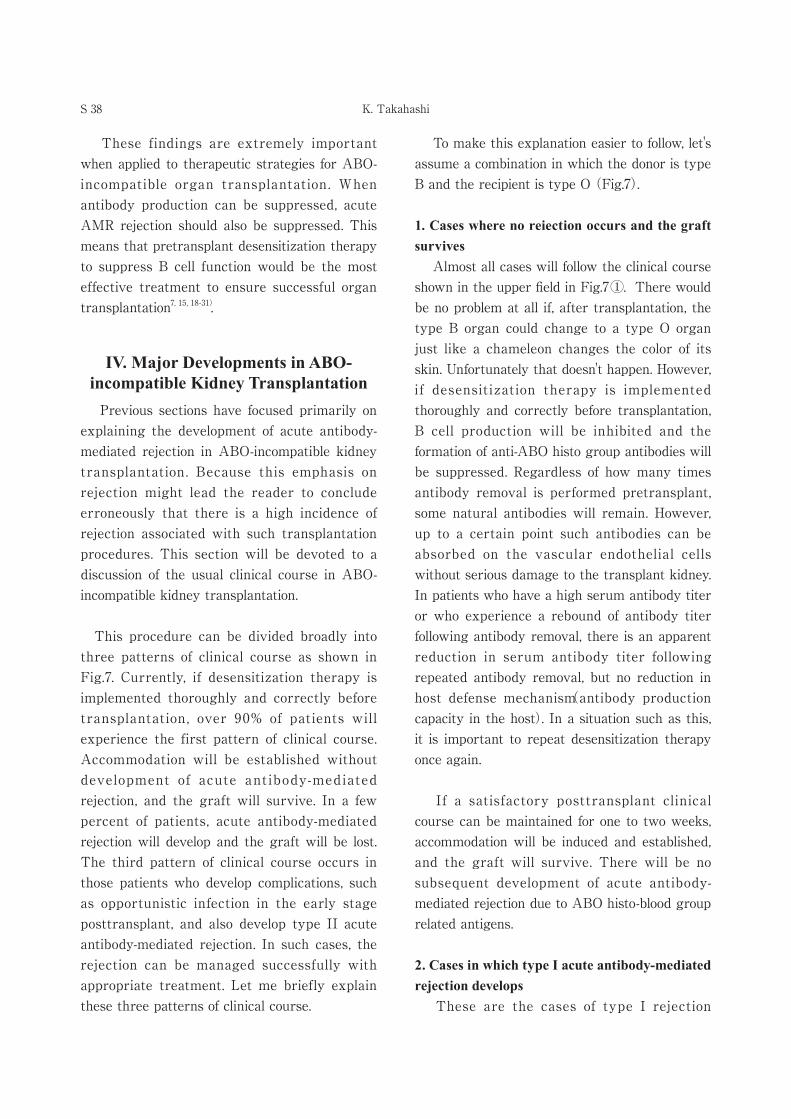
12-15). If I reflect on this definition, however, it contradicts itself. To explain this contradiction, I came up with the idea of key (antibody) and keyhole (antigen) mismatch. We have reported elsewhere two new ground-breaking findings in 2009 and 2010 regarding ABO- IKTx16, 17). We are confident that these findings will overturn the conventional wisdom about ABO-incompatible transplantation, and will mark a fundamental change in therapeutic strategies. The first finding involves ABO histo-blood group antigens, which are classed as carbohydrate antigens. Conventionally, emphasis has been placed on the saccharide chains only, and related phenomena have been interpreted accordingly. However, the saccharide chains in ABO histo-blood group antigens are present in the form of glycoproteins, and the binding proteins are termed "carrier proteins" or "anchor proteins." It has been reported that the carbohydrate chains of ABO histo-blood group antigens on erythrocyte membranes form glycoproteins by bonding to proteins such as band 3 or band 4.532, 33). We performed proteomic analyses in order to determine which type of proteins would be bound to ABO histo-blood group antigens on the vascular endothelial cells (Fig.4). The results showed that those antigens, unlike the ones on erythrocyte membrane, primarily bind to proteins such as PECAM1 (platelet endothelial cell adhesion molecule 1), PLVAP (plasmalemmal vesicle associated protein), and vWf (von Wilebrand factor) (Fig. 5) 16). The above data indicate that ABO histo-blood group antigens on vascular endothelial cells differ from those on the erythrocyte membrane,
S 34 K. Takahashi
both in terms of structure and of antigenicity, depending on differences in the proteins to which they are bound. These antigens, which have been treated as the same type and categorized
as ABO histo-blood group antigens, will now be categorized into two groups: “ABO blood group antigens” carried by the erythrocytes and “ABO histo group antigens” expressed in the vascular
Fig.4. Proteomic analysis of proteins binding to human kidney ABO histo-blood group antigens ABO histo group antigens are abundantly present on vascular endothelial cells in the kidneys, but the proteins binding to ABO histo group antigens on vascular endothelial cells had not been analyzed.We solubilized kidney tissue, collected blood group A carbohydrate chains using lectins that recognize those chains, used electrophoresis to extract specific bands, and then used a mass spectrometer to identify candidate proteins.
Immunoblotting using anti-A antibody
1
1D SDS-PAGE
234
5
solubilization
Purification using HPA lectin column which recognizes blood group A antigens
Proteomic analysis using 1~5 gels.
ABO blood group antigens are expressed in localization to most of vascular endothelial cells.
Identification the proteins carrying ABO blood group antigens using immunoprecipitation and immunohistochemisty
PECAM1PLVAPvWf
Band3Band4.51~5; gel including
blood group antigens
Fig.5. ABO histo-blood group antigens They are found on the surface of human erythrocytes, tissue cells, and vascular endothelial cells, and are antigens in the original sense of the word.ABO histo-blood group antigens are further classified into two subtypes: ABO blood group antigens and ABO histo group antigens, according to the different proteins that are bound to the carbohydrate chain of each antigen.
PECAM1 PLVAP vWf
Band3 Band4.5
S 35
endothelial cells. The host immune system probably do not identify the saccharides and binding proteins separately, but instead recognize them as part of a whole, and then proceed to form antibodies that have a high affinity for that whole. It can also recognize the structural differences between ABO blood group antigens and ABO histo group antigens, and can initiate immunological responses accordingly. Such immunological responses will lead to anti-ABO blood group antibodies in the case of ABO blood group antigens, and anti-ABO histo group antibodies in the case of ABO histo group antigens. Thus, the serum natural anti-A/anti-B antibodies react well with erythrocyte membrane ABO blood group antigens but do not necessarily react with ABO histo group antigens on vascular endothelial cells having a different structure. This is the major factor contributing to the absence of HAR in ABO-IKTx . If the "key" doesn't fit the "keyhole," the "door" won't open, and HAR cannot develop. To further expand this theory, the antibody that is responsible for posttransplant ABO-related acute AMR must be an antibody that was newly synthesized after transplantation
(de novo antibody, i.e. anti-ABO histo group antibody) after that has been sensitized by ABO histo group antigens on the vascular endothelial cells of the graft. If true, this theory explains the existence of approximately 2 days of “silent period” with no sign of HAR. This is the time required for sensitization, i.e. the time required for host immune system to recognize ABO histo group antigens and then to produce anti-ABO histo group antibodies. In cases where pretransplant desensitization therapy has not been adequately implemented, memory B cells are sensitized by ABO blood group antigens and are class -switched to produce IgG antibodies. After being stimulated by ABO histo group antigens, the memory B
cells produce anti-ABO histo group antibodies having a different affinity to these antigens. The anti-ABO histo group antibodies react in turn with ABO histo group antigens on the vascular endothelial cells to trigger a type I acute AMR15). Transplant procedures are followed by a two-day “silent period” free of acute AMR because it takes time for host immune system to recognize ABO histo group antigens on the vascular endothelial cells of the graft and to respond immunologically. That immunological response produces anti-ABO histo group antibodies to complete the antigen-antibody reaction, i.e., acute AMR18).
3. Mechanism of acute antibody-mediated rejection in ABO-incompatible kidney transplantation: Which anti-A/anti-B antibodies are responsible, natural or de novo? The second finding relates to the known fact that ABO-incompatibility associated acute AMR is caused by anti-A/anti-B antibodies. Conventionally it had been believed that this rejection response was caused by natural antibodies present in the recipient. If we consider the first theory, however, it becomes clear that the antibodies eliciting acute AMR could not be produced without transplantation of the donor organ. That is to say, the recipient host immune system must be sensitized to the ABO histo group antigens on the vascular endothelial cells, resulting in the new production of anti-A/anti-B de novo antibodies. Anti-ABO blood group antibodies and anti-ABO histo group antibodies are presumably very similar in structure. As of this writing, there are no assay systems available for demonstrating the differences between those two antibodies. I once considered the use of commercially-available umbilical cord veins (HUVEC), but realized that no ABO histo group antigens are present on the surface of the HUVEC. Then I investigated an indirect method to prove my theory.
S 36 K. Takahashi
One day I was studying the development of acute AMR in four patients, and I happened to note the presence of Complement 4d (C4d) deposits in the peritubular capillaries (PTCs). In three of these four patients, the acute AMR was so severe that the graft was lost. However, perhaps coincidentally, after transplantation the postreperfusion one-hour biopsy findings were negative for C4d deposition in the PTCs in all four of these patients (Table 1). Since then, we have focused special attention on C4d deposition in PTCs in cases of ABO-CKTx and ABO-IKTx performed at Niigata University Medical and Dental Hospital between January 2000 and December 200817, 18). C4d deposition in the PTCs is a comparatively specific and sensitive response associated with the immunological reaction to a kidney allograft. These deposits indicate that the antigens on the surface of the vascular endothelial cells are exposed to antibodies in recipient serum, and that an antigen-antibody reaction occurred34-40). Particularly in ABO-IKTx, if we see C4d deposits in the PTCs upon renal graft biopsy we can then infer that the ABO histo-blood group antigens on the vascular endothelial cells have been exposed to ABO histo-blood group antibodies. Therefore, by tracking the course of C4d deposition over time, we can infer whether these deposits are caused by natural antibodies or by de novo antibodies that were formed after transplantation. We thus began to monitor the
time course of C4d deposits in the PTCs. We performed consecutive 3 renal graft biopsies (0 hour, 1 hour, and protocol or episode biopsies) in 31 patients who underwent ABO-I TKx to examine the complement 4d (C4d) deposition in peritubular capillaries (PTCs) and to estimate when ABO blood type antigens on endothelial cells in PTCs reacted with these antibodies. As a control group, we also studied 37 ABO-CKTx patients during the same period. In - depth repor t s on t hese forms of immunosuppressive therapy are available in the literature7, 29, 30). In ABO-I KTx group, C4d deposition was observed in 0%, 16.1%, and 70.9% of the patients at the first (0-hour), 1-hour, and the protocol/episode biopsy, respectively. In all 4 patients with acute AMR, both 0- and 1-h biopsy specimens were negative for C4d deposition, whereas those with the protocol/episode biopsy were positive. In ABO-C KTx group, all of 3 biopsy specimens were negative for C4d (Fig.6). The first biopsy was the 0 hour biopsy, conducted prior to reperfusion of the kidney graft, so findings were negative, just as in the ABO-C KTx group. As the second renal biopsy was performed 1 hour after reperfusion, the C4d deposition that was detected at that time was attributable to natural antibodies. The C4d deposition detected at the third biopsy, after a certain posttransplant period, can be explained by three different mechanisms. Thus we have
Table 1. ABO-incompatible kidney transplant patients with severe acute AMR
(1) C4d deposition that is a continuation from the second period, (2) C4d deposition caused by de novo serum anti-A/anti-B antibodies, and
(3) C4d deposition due to de novo serum anti-A/anti-B antibodies in addition to the deposition caused by natural antibodies during the second period. Even with repeated antibody removal pretransplant, it is impossible to remove all serum anti-A/ anti-B antibodies. There will naturally be some antibodies remaining in the blood. We assumed that those antibodies would be adsorbed on the transplant kidney, and that the positive rate would thus be nearly 100% at the second biopsy. However, the actual value of 16.1% was much lower than our expectations. In these 16.5% positive C4d recipients at the time of 1-hr biopsy (the second biopsy), clinical acute rejection not occurred. In considering possible causes of this outcome, we realized that we had been measuring serum anti-A/anti-B antibody titers, and that this actually measured the antibodies responding to ABO blood group antigens on the erythrocytes. Those antibodies were believed to respond in the same way as the ABO histo group antigens
on the surface of the vascular endothelial cells31). Our clinical outcomes to date indicate that high anti -A/anti -B antibody t iter in recipients pretransplant is not necessarily associated with the development of ABO-related acute AMR, and that the graft can survive in such patients. We noted this dissociation between pretransplant antibody titer and clinical results in many of our patients, but were unable to provide any specific explanation7, 18, 41). While the natural antibodies are highly reactive because of their strong affinity for the ABO blood group antigens on the erythrocyte surface, they may not respond so strongly to the ABO histo group antigens on the vascular endothelial cells, which have a different structure. These factors may contribute to the low incidence
(16.1%) of positive C4d deposition at the second biopsy. Acute AMR after ABO-IKTx is induced primarily by serum anti-A and anti-B antibodies produced de novo after transplantation, rather than by such antibodies remaining after pre-transplant ant ibody remova l . Successfu l outcomes of ABO-I KTx require the prevention of acute AMR.
0
10
20
30
40
50
60
70
80
0
0
016.1
0
70.9
Compatible (n=37)
Incompatible (n=31)
ProtocolEpisode <2months
1hr
0hr
(%)
(n=5)
(n=22)
Fig.6. PTCs C4d Deposition in ABO-compatible and ABO-incompatible living donor kidney ransplantation
S 38 K. Takahashi
These findings are extremely important when applied to therapeutic strategies for ABO-incompatible organ transplantation. When antibody production can be suppressed, acute AMR rejection should also be suppressed. This means that pretransplant desensitization therapy to suppress B cell function would be the most effective treatment to ensure successful organ transplantation7, 15, 18-31).
IV. Major Developments in ABO-incompatible Kidney Transplantation
Previous sections have focused primarily on explaining the development of acute antibody-mediated rejection in ABO-incompatible kidney transplantation. Because this emphasis on rejection might lead the reader to conclude erroneously that there is a high incidence of rejection associated with such transplantation procedures. This section will be devoted to a discussion of the usual clinical course in ABO-incompatible kidney transplantation.
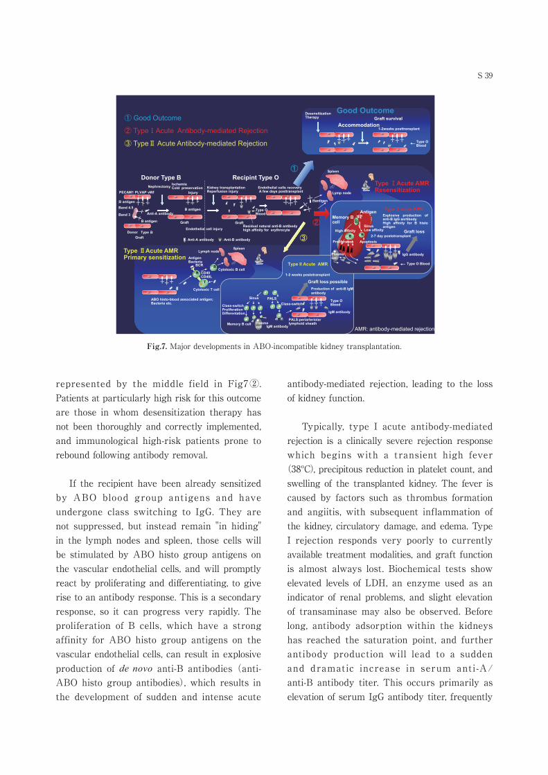
This procedure can be divided broadly into three patterns of clinical course as shown in Fig.7. Currently, if desensitization therapy is implemented thoroughly and correctly before transplantation, over 90% of patients will experience the first pattern of clinical course. Accommodation will be established without development of acute ant ibody-mediated rejection, and the graft will survive. In a few percent of patients, acute antibody-mediated rejection will develop and the graft will be lost. The third pattern of clinical course occurs in those patients who develop complications, such as opportunistic infection in the early stage posttransplant, and also develop type II acute antibody-mediated rejection. In such cases, the rejection can be managed successfully with appropriate treatment. Let me briefly explain these three patterns of clinical course.
To make this explanation easier to follow, let's assume a combination in which the donor is type B and the recipient is type O (Fig.7). 1. Cases where no reiection occurs and the graft survives Almost all cases will follow the clinical course shown in the upper field in Fig.7①. There would be no problem at all if, after transplantation, the type B organ could change to a type O organ just like a chameleon changes the color of its skin. Unfortunately that doesn't happen. However, i f desensit ization therapy is implemented thoroughly and correctly before transplantation, B cell production will be inhibited and the formation of anti-ABO histo group antibodies will be suppressed. Regardless of how many times antibody removal is performed pretransplant, some natural antibodies will remain. However, up to a certain point such antibodies can be absorbed on the vascular endothelial cells without serious damage to the transplant kidney. In patients who have a high serum antibody titer or who experience a rebound of antibody titer following antibody removal, there is an apparent reduction in serum antibody titer following repeated antibody removal, but no reduction in host defense mechanism(antibody production capacity in the host). In a situation such as this, it is important to repeat desensitization therapy once again.
If a satisfactory posttransplant clinical course can be maintained for one to two weeks, accommodation will be induced and established, and the graft will survive. There will be no subsequent development of acute antibody-mediated rejection due to ABO histo-blood group related antigens.
2. Cases in which type I acute antibody-mediated rejection develops These are the cases of type I rejection
S 39
represented by the middle field in Fig7②. Patients at particularly high risk for this outcome are those in whom desensitization therapy has not been thoroughly and correctly implemented, and immunological high-risk patients prone to rebound following antibody removal.
If the recipient have been already sensitized by ABO blood group ant igens and have undergone class switching to IgG. They are not suppressed, but instead remain "in hiding" in the lymph nodes and spleen, those cells will be stimulated by ABO histo group antigens on the vascular endothelial cells, and will promptly react by proliferating and differentiating, to give rise to an antibody response. This is a secondary response, so it can progress very rapidly. The proliferation of B cells, which have a strong affinity for ABO histo group antigens on the vascular endothelial cells, can result in explosive production of de novo anti-B antibodies (anti-ABO histo group antibodies), which results in the development of sudden and intense acute
antibody-mediated rejection, leading to the loss of kidney function.
Typically, type I acute antibody-mediated rejection is a clinically severe rejection response which begins with a transient high fever
(38ºC), precipitous reduction in platelet count, and swelling of the transplanted kidney. The fever is caused by factors such as thrombus formation and angiitis, with subsequent inflammation of the kidney, circulatory damage, and edema. Type I rejection responds very poorly to currently available treatment modalities, and graft function is almost always lost. Biochemical tests show elevated levels of LDH, an enzyme used as an indicator of renal problems, and slight elevation of transaminase may also be observed. Before long, antibody adsorption within the kidneys has reached the saturation point, and further antibody production will lead to a sudden and dramat ic increase in serum ant i -A/anti-B antibody titer. This occurs primarily as elevation of serum IgG antibody titer, frequently
Fig.7. Major developments in ABO-incompatible kidney transplantation.
Cytotoxic T cell
Spleen
CD40CD40L
Type ⅡAcute AMRPrimary sensitization
ABO histo-blood associated antigen; Bacteria etc.
AntigenBacteria
BCR
Lymph node
BB
T
Cytotoxic B cell
Sinus
B
Antigen
B antigen
Class-switchProliferationDifferentation
B B
Donor Type B
PECAM1 PLVAP vWf
Donor Type BGraft
Anti-A antibody
B antigen
Band 4.5
Band 3
Nephrectomy
Graft
B antigen
IschemiaCold preservation
Injury
Recipint Type OKidney transplantationReperfusion injury
Endothelial cell injury
GraftResidual natural anti-B antibodyhigh affinity for erythrocyte
:Anti-A antibody :Anti-B antibody
Endothelial cells recovery A few days posttransplant
Type OBlood
②
③
Spleen
Lymp node
Bantigen
Type ⅠAcute AMRResensitization
①
DesensitizationTherapy Graft survival
Accommodation1-2weeks posttransplant
Type OBlood
Graft loss
Type Ⅰacute AMRExplosive production ofanti-B IgG anrtibodyHigh affinity for B histoantigen
Type O Blood
IgG antibody
2-7 day postotransplant
Type II Acute AMR
1-2 weeks postotransplant
Graft loss possible
PALS:periarteriolarlymphoid sheathMemory B cell IgM antibody
PALSClass-switch
SinusLow affinityHigh affinity
Proliferation Apoptosis
Memory B cell
Type OBlood
Plasmacell
Plasmacell
Production of anti-B IgM antibody
IgM antibody
Good Outcome① Good Outcome
② TypeⅠAcute Antibody-mediated Rejection
③ TypeⅡ Acute Antibody-mediated Rejection
AMR: antibody-mediated rejection
S 40 K. Takahashi
accompanied by parallel elevation of IgM antibody titer.
3. Cases in which type II acute antibody-mediated rejection develops The clinical course shown in the bottom field in Fig7③. represents cases in which type II acute antibody-mediated rejection has developed. After kidney transplantat ion but before accommodation is induced and established, if the recipient incurs initial sensitization by ABO histo-blood group associated antigens that are cross-reactive to donor ABO histo group antigens, then antibody production will be elicited as an immunological response, resulting in acute antibody-mediated rejection. As noted previously, the initial sensitization is a primary response, so its pathological progression is slower than type I rejection, and the clinical course is prolonged. This response generally develops late in the critical period. In situations such as this, serum IgM antibody titer is elevated, and IgG antibody titer generally remains unchanged.
This rejection is prone to develop in the early stage posttransplant as a result of opportunistic infection, particularly bacterial sepsis. If the recipient is exposed to a bacterial infection, naturally antibodies will be produced as part of the host defense mechanisms. However, this is truly a "two-edged sword" in the case of a posttransplant patient. The antibodies will act to neutralize the bacterial cells, but they will also produce a cross-reaction with the ABO histo group antigens of the vascular endothelial cells, giving rise to an acute antibody-mediated rejection.
At the microscopic level , the bacteria/microorganisms act as antigens. When antigens react with B cell receptors, those antigens are engulfed, digested into peptides , and presented on the cell surface together with the
histocompatible antigens. T cells activated by reacting with these same antigens then react with the B cell surface antigen peptides, so that there is contact between the T cells and the B cells. This kind of antigen-specific T cell-B cell contact is followed by a reaction between CD40L
(ligand) on the activated T cells and CD40 on the B cells, inducing a signal mediated by CD40 in the B cells. This results in B cell activation in response to bacterial antigens. The activated B cells proliferate and differentiate to plasma cells which are thought to produce antibodies.
Because the condition progresses slowly in the type II rejection, the graft can be rescued with early diagnosis and appropriate treatment.
These are the three main patterns of clinical course that can be expected with ABO-incompatible kidney transplantation.
V. New strategies for immunosuppression in ABO-incompatible kidney
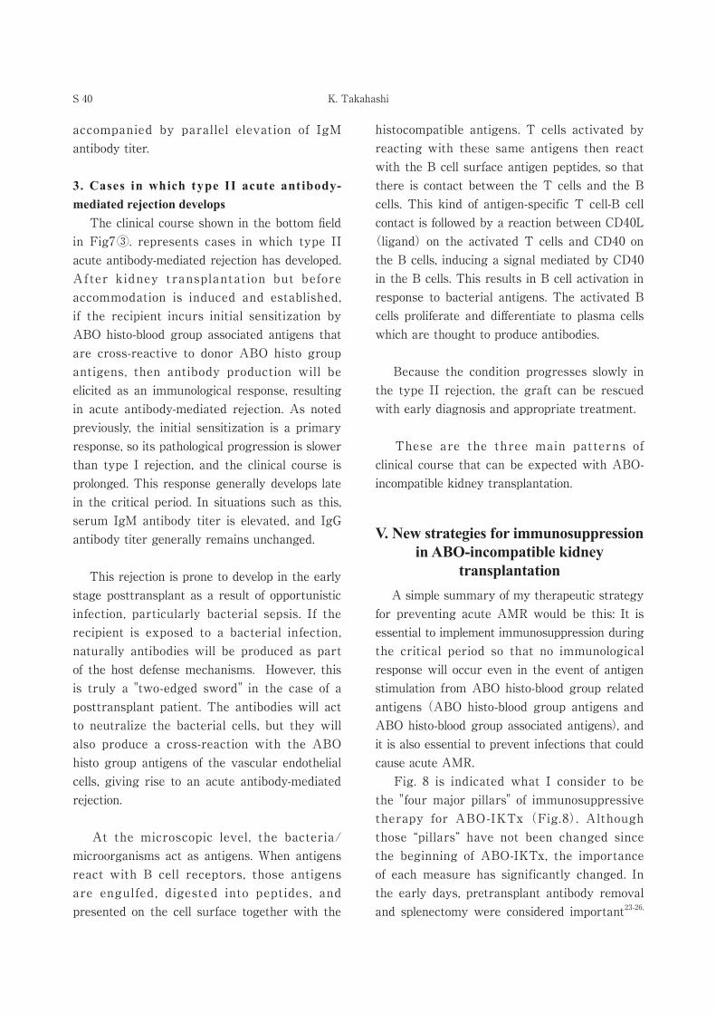
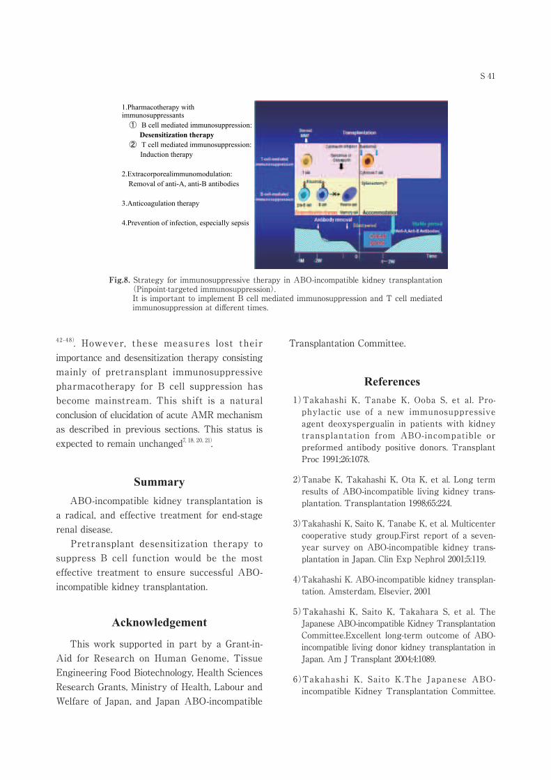
transplantation A simple summary of my therapeutic strategy for preventing acute AMR would be this: It is essential to implement immunosuppression during the critical period so that no immunological response will occur even in the event of antigen stimulation from ABO histo-blood group related antigens (ABO histo-blood group antigens and ABO histo-blood group associated antigens), and it is also essential to prevent infections that could cause acute AMR. Fig. 8 is indicated what I consider to be the "four major pillars" of immunosuppressive therapy for ABO-IKTx (Fig.8). Although those “pillars” have not been changed since the beginning of ABO-IKTx, the importance of each measure has significantly changed. In the early days, pretransplant antibody removal and splenectomy were considered important23-26,
S 41
42 - 4 8). However, these measures lost their importance and desensitization therapy consisting mainly of pretransplant immunosuppressive pharmacotherapy for B cell suppression has become mainstream. This shift is a natural conclusion of elucidation of acute AMR mechanism as described in previous sections. This status is expected to remain unchanged7, 18, 20, 21).
Summary ABO-incompatible kidney transplantation is a radical, and effective treatment for end-stage renal disease. Pretransplant desensitization therapy to suppress B cell function would be the most effective treatment to ensure successful ABO-incompatible kidney transplantation.
Acknowledgement
This work supported in part by a Grant-in-Aid for Research on Human Genome, Tissue Engineering Food Biotechnology, Health Sciences Research Grants, Ministry of Health, Labour and Welfare of Japan, and Japan ABO-incompatible
Transplantation Committee.
References1)Takahashi K, Tanabe K, Ooba S, et al. Pro-
phylactic use of a new immunosuppressive agent deoxyspergualin in patients with kidney transplantation from ABO-incompatible or preformed antibody positive donors. Transplant Proc 1991;26:1078.
2)Tanabe K, Takahashi K, Ota K, et al. Long term results of ABO-incompatible living kidney trans-plantation. Transplantation 1998;65:224.
3)Takahashi K, Saito K, Tanabe K, et al. Multicenter cooperative study group.First report of a seven-year survey on ABO-incompatible kidney trans-plantation in Japan. Clin Exp Nephrol 2001;5:119.
4)Takahashi K. ABO-incompatible kidney transplan-tation. Amsterdam, Elsevier, 2001
5)Takahashi K, Saito K, Takahara S, et al. The Japanese ABO-incompatible Kidney Transplantation Committee.Excellent long-term outcome of ABO-incompatible living donor kidney transplantation in Japan. Am J Transplant 2004;4:1089.
6)Takahashi K, Saito K.The Japanese ABO-incompatible Kidney Transplantation Committee.
Fig.8. Strategy for immunosuppressive therapy in ABO-incompatible kidney transplantation (Pinpoint-targeted immunosuppression).It is important to implement B cell mediated immunosuppression and T cell mediated immunosuppression at different times.
1.Pharmacotherapy with immunosuppressants① B cell mediated immunosuppression:
Desensitization therapy② T cell mediated immunosuppression:
Induction therapy
2.Extracorporealimmunomodulation: Removal of anti-A, anti-B antibodies
3.Anticoagulation therapy
4.Prevention of infection, especially sepsis
S 42 K. Takahashi
Present Status of ABO-incompatible kidney transplantation in Japan. Xenotransplan t 2006;13:118.
7)Takahash i K . ABO- incompat ib le k idney transplantation – Establishing a scientific frame-work. Amsterdam, Elsevier, 2001.
8)Alexandre GPJ, Bruyere MDE, Squifflet JP, et al. Human ABO incompatible living donor renal homografts. Neth J Med 1985;28:231.
9)Alexandre GPJ, Squifflet JP, Bruyere MDE, et al. ABO incompatible related and unrelated living donor renal allografts. Transplant Proc 1986;18:452.
10)Alexandre GPJ, Latinne D, Gianello P, et al. Pre-formed cytotoxic antibodies and ABO-incompatible grafts. Clin Transplant 1991;5:583.
11)Tagareli A, Karl Landsteiner. A hundred year later. Transplantation 2001;72:3.
12)Takahashi K. Accommodation in ABO-incompatible kidney transplantation: Why do kidney grafts survive? Transplant Proc 2004 (Suppl.2S):193.
13)Takahashi K. Accommodation in ABO-incompatible kidney transplantation. Amsterdam, Elsevier, 2004
14)Takahashi K. A new concept of accommodation in ABO-incompatible kidney transplantation. Clin Transplant 2005;19 (Suppl.14):76.
15)Takahashi K. Recent findings in ABO-incompatible kidney transplantation: classification and therapeutic strategy for acute antibody-mediated rejection due to ABO-blood-group-related antigens during the critical period preceding the establishment of accommodation. Clin Exp Nephrol 2007;11:1281.
16)Tasaki M, Yoshida Y, Takahashi K, et al. Iden-tification and characterization of major proteins carrying ABO blood group antigens in the human kidney transplantation. Transplantation 2009;87:1125.
17)Takahashi K, Imai N, Saito K, et al. Mechanism of acute antibody-mediated rejection in ABO-incompatible kidney transplantation: Which anti-A/anti-B antibodies are responsible, natural or de novo? Transplantation 2010; Letter to the editors 89:635.
18)Takahash i K . ABO- incompat ib le k idney transplantation- Why is hyperacute rejection absent? Amsterdam Elsevier, 2011.
19)Saito K, Takahashi K, The Japan ABO-incompatible transplantation Committee. In:Takahashi K, Tanaka K, editors. New strategies for ABO-incompatible kidney transplantation-2012-. Ⅰ.1st ed. Tokyo:Nihon Igakukan;2012. pp.3-15.
20)Saito K, Nakagawa Y, Takahashi K, et al. Pinpoint targeted immunosuppression; Anti-CD20/MMF desensitization with anti-CD25 in successful ABO-incompatible kidney transplantation without splenectomy. Xenotransplant 2006;13:111.
21)Takahashi K. Pinpoint targeted immunosuppres-sion in ABO-incompatible kidney transplantation – Desensitization therapy without splenectomy.in Takahashi K (ed), ABO-incompatible Organ Transplantation from Japan. Amsterdam, Elsevier, 2006; 73-87.
22)Sawada T, Fuchinoue S, Teraoka S.Successful A1-to-O ABO-incompatible kidney transplantation after a preconditioning regimen consisting of anti-CD20 monoclonal antibody infusion, splenectomy, and double-filtration plasmapheresis. Transplantation 2002;74:1207.
23)Tyden G, Kumlien G, Fehrman I. Successful ABO incompatible kidney transplantations without splenectomy, using antigen-specific immunoadsorption and rituximab. Transplantation 2003;27:1207.
24)Warren DS, Zachary AA, Montgomery RA, et al. Successful renal transplantation across simultaneous ABO incompatible and positive crossmatch barriers. Am J Transplant 2004;4:561.
25)Tyden G , Kuml ien G , Genberg H , e t a l . ABO incompatible kidney transplantations without splenectomy, using antigen-specific immunoadsorpt ion and r i tux imab . Am J Transplant 2005;5:145.
26)Norden G, Briggs D, Breimer ME, et al. ABO-incompatible live donor renal transplantation using blood group A/B carbohydrate antigen immunoadsorption and anti-Cd20 antibody treatment. Xenotransplant 2006;13(2):143
27)Stegall MD. Overcoming antibody barrier to
S 43
transplantation: ABO-incompatible and positive cross-match kidney transplantation at Mayo Clinic – Rochester. in:Takahashi K (ed), International Congress Series 1292 ABO-incompatible organ transplantation from Japan. Elsevier, Amsterdam, 2006;113-9.
28)Montgomery RA, Locke JE, King KE, et al. ABO incompatible renal transplantation: A paradigm ready for broad implementation. Transplantation 2009;87:1246.
29)Zhou W, Ohdan H, Tanaka Y, et al. NOD/SCID mice engrafted with human peripheral blood lymphocytes can be a model for investigating B cells responding to blood group A carbohydrate determinant. Transpl Immunol 2003;12:9.
30)Ire T, Ohdan H, Zhou W, et al. The persistent elimination of B cells responding to blood group A carbohydrates by synthetic group A carbohydrates and B-1 cell differentiation blockade: novel concept in preventing antibody-mediated reject ion in ABO- incompat ib le transplantation. Blood 2007;110:4567.
31)Gleason RE, Murray JE. Report from kidney transplant registry: Analysis of variables in the function of human kidney transplants. Transplantation 1967;52:343.
32)Finne J, Krusius T, Rauvala H, Jarnefelt J. Molecular nature of the blood group ABH antigens of the human erythrocyte membrane.Review Fr. Transfus Immunohematology 1980;23: 545.
33)Finne J. Identification of the blood-group ABH-active glycoprotein components of human erythrocyte membrane. Eur. J Biochem 1980;104:181.
34)Regele H, Bohmig GA, Exner M, et al.Capillary deposition of complement split product C4d in renal allografts is associated with basement membrane injury in peritubular and glomerular capillaries: A contribution of humoral immunity to chronic allograft rejection. J Am Soc Nephrol 2002;13:2371.
35)Kanetsuna Y, Yamaguchi Y, Horita S, et al.C4d and/or immunoglobulins deposition in peritubular capillaries in perioperative graft biopsies in ABO-incompatible renal transplantation. Clin Transplant 2004;18 (Suppl.11):13.
36)Hass M, Rahman MH, Racusen LC, et al. C4d and C3d staining in biopsies of ABO- and HLA-incompatible renal allografts: correlation with histologic findings. Am J Transplant 2006;6:1829.
37)Imai N, Nishi S, Takahashi K, et al. Immunohisto-chemical evidence of activated lectin pathway in kidney allografts with peritubular capillary C4d deposition. Nephrol Dial Transplant 2006;21:2589.
38)Haga H, Egawa H, Monabe T, et al. Acute humoral rejection and C4d immuno staining in ABO blood type-incompatible liver transplantation. Liver Transplant 2006;12:457.
39)Sakashita H, Haga H, Manabe T, et al. Significance of C4d staining in ABO-incompatible /compatible liver transplantation. Modern Pathology 2007;20: 576.
40)Takahashi K. ABO-incompatible organ transplan-tation. Curr Opin Organ Transplant 2007;12:409.
41)Kobayashi T, Saito K. A series of surveys on assay for anti-A/B antibody by Japanese ABO-incompatible Transplantation Committee .Xenotransplant 2006;136.
43)Bannett AD, Bensinger WI, Raja R, et al. Immu-noadsorption and renal transplant in two patients with a major ABO incompatibility. Transplantation 1987;43:909.
44)Takahashi K, Agishi T, Oba S, et al. Extracorporeal plasma treatment for extending indication of kidney transplantation ABO-incompatible and preformed antibody-positive kidney transplantation Therapeutic Plasmapheresis IX. Cleveland, ESAO Press, 1990;61-3.
45)Agishi K, Takahashi K, Ota K, et al. Japanese Biosynsorbe Reserch. Immunoadosorption od anti-A or anti-B antibody for successful kidney transplantation between ABO incompatible pairs and its limitation. ASAIO Trans. 1991;37:496.
46)Ota K, Takahashi K, Agishi T, et al. Japanese Biosynsorb ABO-incompatible kidney transplant group: Multicenter trial of ABO-incompatible kidney transplantation. Transplant Int 1992;5
(suppl.): 40.
S 44 K. Takahashi
47)Nishi S, Hasegawa S, Takahashi K, et al. The safety measures for double filtration plasma apheresis (DFPP) before ABO incompatible kidney transplantation.in Takahashi K (ed), International Congress Series 1292, ABO-incompatible Organ Transplantation from Japan.Amsterdam, Elsevier, 2006;91-5.
48)Alexamdre GPJ, Squifflet Jp, Bruyere MDE, et al. Splectomy as prerequisite for successful human ABO-incompatible renal transplantation. Transplant Proc. 1985;17:139.