34
Dr Ashish Jain, Associate Professor, Department of Transfusion Medicine, PGIMER, Chandigarh ABO isoagglutinin titration: application, method and controversies

Dr Ashish Jain,
Associate Professor,
Department of Transfusion Medicine, PGIMER, Chandigarh
ABO isoagglutinin titration: application, method
and controversies

ABO isoagglutinins Isohemagglutinins are naturally present antibodies to non-self A and B blood group
antigens.
Also k/a ABO hemagglutinins or ABO isohemagglutinins or ABO antibodies.
Occur as ‘naturally occurring’ antibodies; but their origin is still unclear.
Whether these antibodies are produced through some inherited, “natural” innate
mechanism, not requiring antigenic stimulation or, instead, follow classical adaptive
immune-mediated mechanisms.
Landsteiner’s Law
Whichever ABO antigens are lacking ona given person’s RBCs, that person willalways have the corresponding antibodyor isohemagglutinin
Dr Karl Landsteiner(14th November,1901)

Development of ABO isoagglutinins
In most infants, anti-A and anti-B agglutinins (presumably IgM) produced by the infant
can first be demonstrated at 3–6 months.
The titre of anti-A and anti-B agglutinins reaches its maximum at the age of 5–10
years.
It may be wholly IgM or partly IgM and partly IgG, partly IgM and partly IgA or may
be made of all three immunoglobulins

Characteristics of ABO isoagglutinins
IgG anti-A and anti-B are found far more commonly in group O than in B or A subjects.
IgG subclasses: IgG2 ≥ IgG1 ≥ IgG3
Both IgM and IgG may be hemolytic, bind complement; IgA is not hemolytic.
Naturally occurring anti-A and anti-B react more strongly at 4°C than at 37°C.
Cross reacting anti-A,B: Anti-A,B in group O serum is an antibody directed against a an
epitope shared by both A and B (cannot be distinguished by differential adsorption).

Applications of ABO Ab titration
Hemolytic disease of the fetus and newborn (HDFN): IgG anti-A, anti-B, and anti-A,B are all
capable of causing HDFN: almost only occurs in A1, B, or A1B babies of group O mothers.
ABO incompatible solid organ transplantation: ABO antibodies can cause hyperacute
rejection of incompatible kidney, liver, and heart transplants.
ABO incompatible Hematopoietic stem cell transplant (HSCT): HSC do not express ABO
antigens, so ABO is often disregarded when selecting a stem cell donor. However, major
ABO incompatibility may lead to hemolysis of infused red cells with a bone marrow
transplant.
Transfusion of platelets containing ABO incompatible plasma: screening for donor anti-A and
anti-B hemolysins, and high titers of IgM and IgG is suggested when using ABO non-identical
platelets.

Titration: methods
Titration
Tube techniqueColumn
agglutination technology (Gel)
Solid phase red cell adherence
(SPRCA)
Flow cytometry
Titration is semi-quantitative technique of measuring the concentration of an antibody in a
serum.
Performed using Double dilution technique (Serial dilution).
Dilution is expressed as: 1 in 16 which means that the dilution factor is 16.
Titer is simply the inverse of dilution at which the end point agglutination (1+) is achieved.

RBC phenotype: A1 or A2Concentration of RBC in final
mixture
Time and temperature of incubation
Technique of reading the endpoint
Factors affecting ABO Ab titers
Wider range for anti-A (8-2048) than anti-B (8-256)
Redman et al (UK, 1990) showed that there is no significant difference between Black, White and Asian people

Tube technique (TT)
It is the most commonly performed method in laboratories.
The room temperature (RT) incubation technique and the indirect antiglobulin test (IAT) have
been interpreted as the methods detecting IgM and IgG, respectively.
Both IgM and IgG of ABO Ab can agglutinate RBCs at RT (20-24°C) or below and
efficiently activate the complement at 37°C.
Therefore titers using RT techniques or IAT on dithiothreitol (DTT) untreated samples may be
more reflective of the mixed concentration of IgM and IgG of ABO Ab.

14 nm 35 nm
24 nm
IgG IgM
Intercellular distance
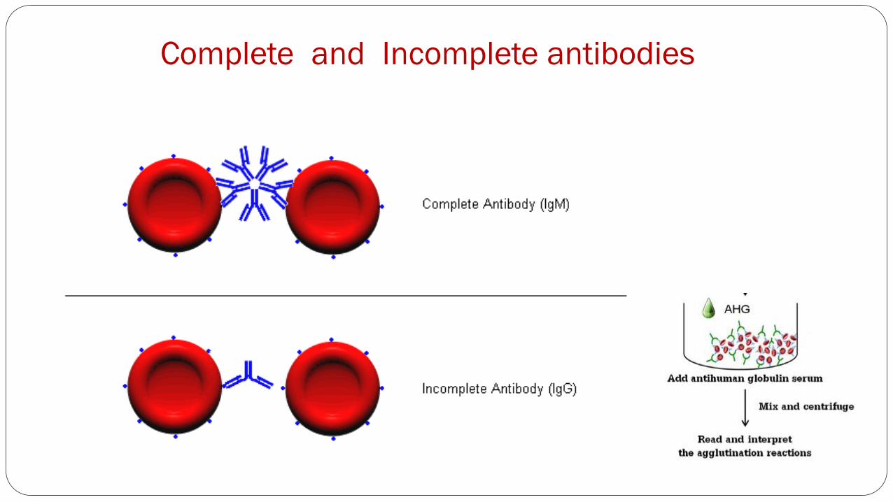
Complete and Incomplete antibodies

Titer: doubling dilutionLabel 10 test tubes (1:1, 1:2, 1:4, 1:8, 1:16, 1:32............)
Add one volume (100µl) of saline to all test tubes except the first tube
Add an equal amount of serum to each of the first two tube
Using a clean pipette mix the contents of the 1 in 2 dilution several times and transfer one volume (100µl) into the next tube
Continue the same process for all the dilutions, using a clean pipette to mix and transfer each dilution and save the last transfer volume
Add 1 drop of the corresponding red cell suspension (5%) into each test tube. Mix well, keep these test tubes at room temperature for at least 15min, centrifuge at 1000 rpm for 1min
Observe the highest dilution that produces macroscopic agglutination (1+)

Titer: doubling dilution (contd…)
Normal saline
1 2 3 4 5 6 7 8 9
Serum
100μl
NEAT

Tube Agglutination Grading

Disadvantages
Variation in cell suspension
Cell loss during washing
Alteration in cell:serum ratio
Inter-observer variation (1+ and wk+ are subjective: End-point ??)
Stability of test results
Variation in repeat testing

Column agglutination technology (gel)
Conceptualized by Lapierre (1985)
Principle: Controlled centrifugation of red cell with/without serum through a porous dextran or
polyacrylamide gel column of defined pore size under defined sets of incubation.
Gel acts as a sieve so that unagglutinated cells settle at bottom and cells forming lattice get trapped at
various zones across the column.
LISS/COOMBS gel card contains Anti-IgG(Poly) + Anti C3d (mono)
Preparation of 0.8%-1% LISS suspension of red cells (No
need to wash the cells)
Dispense 50µL in the reaction chamber at acute angle
Dispense 25µL of recipient serum on its top gently
Incubate for 15 minutes at 37*C
Spin at 1000 rpm for 10 minutes
Interpret

Advantages
more qualitative in grading the strength of
agglutination reaction
the inter-observer variation is minimal
less time-consuming
uses smaller volumes of serum and RBCs
Limitation: COST ??
0

Microplate technique
MICROPLATES
Small tray with 96 small wells
Holds 200-300microlitres of reagent
Three types: V-type, flat-bottom, U-type
Advantage
More sensitive –very weak cell suspension can be used
Very small amount of reagents are needed
Titrations are easier with multi channel pipettes
Grades of reaction can be compared
Disadvantage: high viscosity in serum/plasma causes red cells to adhere to side of wells

Solid phase red cell adherence assay (SPRCA)
Components of antigen-antibody reaction is immobilized onto a solid medium.
On centrifugation antigen positive cells spread out while antigen negative cells form a button at the
bottom of the well.
Excess plasma is blotted out and anti IgG bound indicator red cells are added to give visible reaction.
SPRCA: Available in automated platforms

SPRCA (Contd.)
Antigen coat
+ Test serum or plasma, incubation at 37oC
Antibody attached to RBC’s antigen
Wash to remove unbound antibody
+Indicator RBC
PositiveNegative
Indirect test
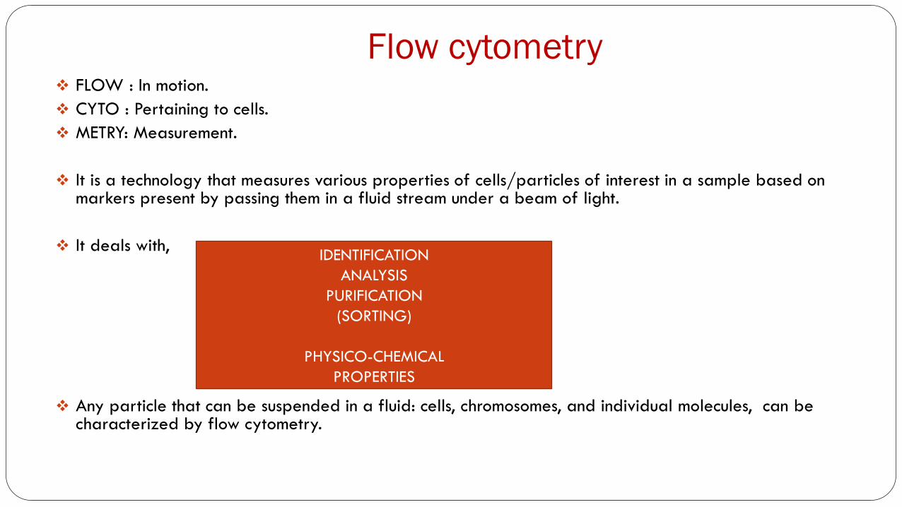
Flow cytometry FLOW : In motion.
CYTO : Pertaining to cells.
METRY: Measurement.
It is a technology that measures various properties of cells/particles of interest in a sample based on markers present by passing them in a fluid stream under a beam of light.
It deals with,
Any particle that can be suspended in a fluid: cells, chromosomes, and individual molecules, can be characterized by flow cytometry.
IDENTIFICATION
ANALYSIS
PURIFICATION
(SORTING)
PHYSICO-CHEMICAL
PROPERTIES

Detection of ABO antibodies by flow cytometry
ABO fluorescence-activated cell sorting (ABO-FACS) to quantify binding of anti-A/B IgM,
IgG and IgG subclasses to human A or B red blood cells.
The sensitivity and specificity of anti-A/B IgM to predict the blood group was 93% and
96% respectively.
IgG2 was the predominant IgG subclass.
The correlation of anti-A/B IgM and IgG in the ABO-FACS with haemagglutination titres
was 0.870 and 0.783, respectively (n= 240; P < 0.001).
It opens the possibility of isotype-specific monitoring of anti-A/B antibodies levels after
ABO-incompatible solid organ and stem cell transplantation.
Stussi et al. Isotype-specific detection of ABO blood group antibodies using a novel flow cytometric method. Br J Haematol 2005:130;954-63.

Controversies in ABO titration
The preferred method ?
Standardization ?
End points ?
Critical titre levels (specially for ABOi-KT) ?


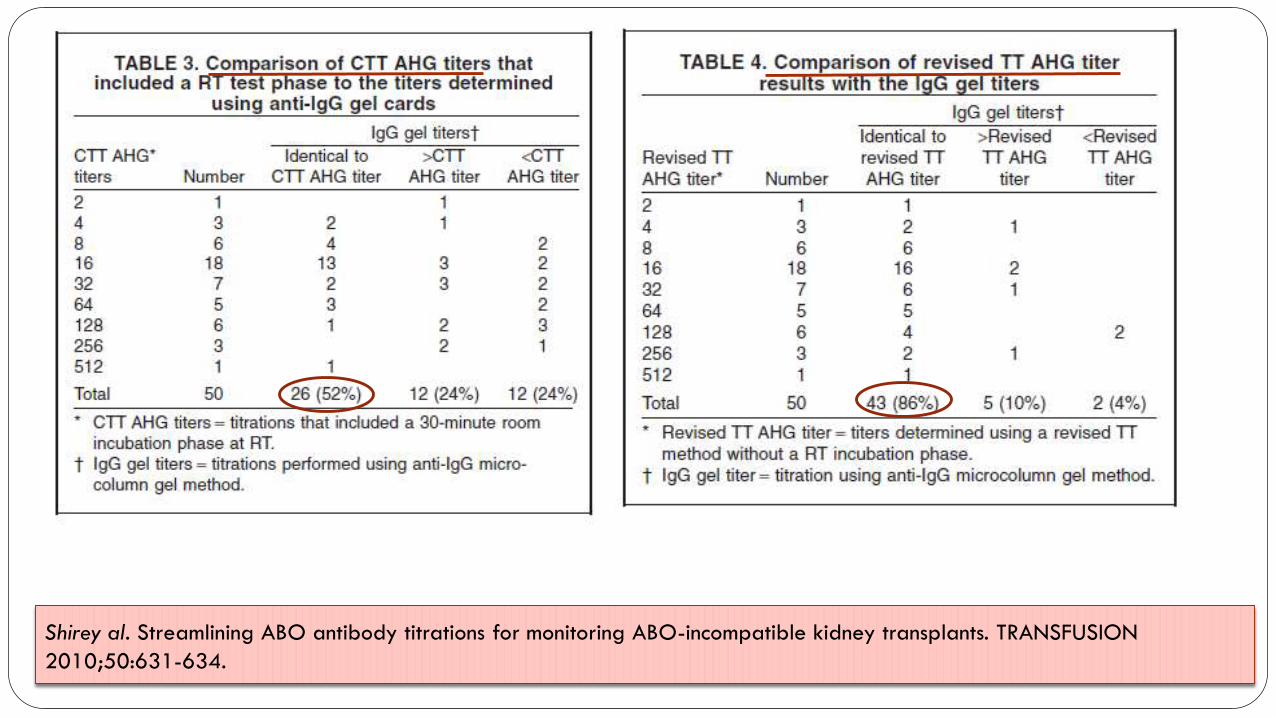
The AHG titer results using the Tube Technique (TT) method werecompared to IgG gel titers. Forty-three (86%) of the titer results wereidentical and 7 (14%) varied by one standard dilution. Five (10%) wereone dilution greater than the TT-AHG titer, and two (4%) were onedilution lower than the TT AHG titer. No IgG gel titer varied more than onestandard dilution from the TT AHG titers.
CONCLUSIONS: The Tube and IgG gel titers are comparable . The IgG gel method offers the best titer turnaround time, eliminating 45 minutes of incubation time alone. Implementation of this technique would benefit ABO INKT patients by providing titer results in a more timely manner.

Tube v/s gel
CTT titration method: Titrations were incubated at ambient (22-25°C) RT
followed by incubation for 30 minutes at 37°C with subsequent conversion to the
AHG test phase using monospecific anti-IgG.
Revised TT titration method: Titrations were performed according to the CTT
titration method except the 30-minute RT incubation test phase was omitted.
Anti-IgG gel titers: Titrations were performed using anti-IgG gel cards (Micro
Typing Systems, MTS, Ortho Clinical Diagnostics, Raritan, NJ)
Shirey al. Streamlining ABO antibody titrations for monitoring ABO-incompatible kidney transplants. TRANSFUSION
2010;50:631-634.
Shirey al. Streamlining ABO antibody titrations for monitoring ABO-incompatible kidney transplants. TRANSFUSION
2010;50:631-634.

Streamlining ABO titration
The IgG gel method offers the best turnaround time requiring only 15 minutes
of incubation at 37°C and eliminates the tedious reading of TT agglutination
reactions.
The gel reactions are stable, batch titrations can be easily accommodated by
IgG gel.
AuBuchon and co-workers have reported that using a weak-positive titer end-
point may reduce titration variability.

End-points
The variance between laboratories was not significantly reduced with the uniform method
using a 1+ end-point.
A statistically significant reduction in the variance of anti-D and anti-A titres by the TT
(including the IAT phase) was seen when 19 laboratories re-analysed their results using a w+
end-point.
Titration against red cells of the specified phenotype provided by the participating
laboratory did not appear to introduce additional variance.
Results reported based on the gel card technique at the AHG phase (1+ end-point) showed
reduced variance compared to tube-based techniques.
Blackwell Publishing Ltd
AuBuchon et al. Reducing the variation in performance of antibody titrations. Vox Sanguinis (2008);95:57–65

SPRCA v/s Gel
ABO titration assays on the Fully automated SPRCA platform (Galileo-NEO, Immucor).
318 IgG and 105 IgM titrations were performed.
The results were compared to the manual gel card method (Bio-Rad) without
pretreatment with DTT.
The typically one dilution difference vs the gel card method can be possibly
explained by the presence of IgM affecting the gel card and that different antigen
concentration are employed to that in the automated assay.
Preuss E, et al. Vox Sanguinis (2015) 109 (Suppl. 1), 1–379

Total v/s IgG ABO Ab titers
Median titers of anti-B and anti-A in all
blood groups were higher in CAT without
DTT than in CAT with DTT, especially for
group O individuals.
Park ES et al. Comparison of total and IgG ABO antibody titers in healthy individuals by using tube and column agglutination techniques. Ann Lab Med 2014;34:223-229

Case (PGIMER, Chandigarh)
Patient NK; 31yr/ Male; CKD ESRD
Posted for ABO incompatible renal transplant.
Blood group of the patient: O RhD Positive
Donor: A2RhD Positive
Antibody screen and DAT: Negative (Gel)
Titers:
Anti-A Titer TUBE TECHNIQUE GEL TECHNIQUE
Ig M Ig G Ig M Ig G
Pre-TPE-1 64 64 64 128
Post-TPE-1 4 4 4 4
Desensitization: Glycosorb

64
4
8 8 8
16
8
16 16 16 16 16 16 16 16
0
10
20
30
40
50
60
70
Pre TPE PostTPE
PostRenal
TxDay-2
PostRenal
TxDay-6
PostRenal
Tx Day-7
PostRenal
TxDay-8
PostRenal
TxDay-9
PostRenal
TxDay-10
PostRenal
TxDay-11
PostRenal
TxDay-12
PostRenal
TxDay-13
PostRenal
TxDay-14
PostRenal
TxDay-15
PostRenal
TxDay-16
PostRenal
TxDay-17
Ig M
Ig G
Serial titers (IgG Gel)
Pre-TPE Post-TPE
GLYCOSORB

Conclusion
Interlaboratory variations in the technical procedures and results do occur in
measurement of the ABO Ab titer.
CAT significantly decreases variation as compared to the tube test.
Individual centres should develop their own protocols based on:
• Available resources
• Validation of applied methods
• Goals of isoagglutinin titer
A periodically conducted assessment could help in continued improvement of the
results of ABO Ab titer measurement.


