40
About Hydrocephalus A Book for Families

About Hydrocephalus
A Book for Families


This booklet is written for parents of children with hydrocephalusand people with hydrocephalus in the hope that the information willgive you a better understanding of the condition and how it can bemanaged. Although this book was originally written for parents, it con-tains basic information about hydrocephalus that is valuable to every-one—parents of children with hydrocephalus, families and individuals.In recent years, there have been remarkable advances in the treatmentof hydrocephalus. With early detection and effective treatment, the out-look for children with hydrocephalus is promising. Many people withhydrocephalus lead normal lives with few limitations. Research andexperience show that children with hydrocephalus have excellentopportunities to attain their full potential through programs that stimu-late their development.
Hydrocephalus affects about one in every 500 to 1,000 childrenborn. It is caused by a wide variety of medical problems, and the cir-cumstances of each child’s condition are unique. You will probably havemany questions concerning your child’s particular problems that arebeyond the scope of this booklet, but you will find that your knowledgeabout the condition will increase steadily as time passes.
A number of experienced medical professionals and families of chil-dren with hydrocephalus participated in making this booklet. They havedealt with many of the issues facing you today. We hope that theirexperiences, knowledge and perceptions will help you discover yourown path to understanding and coping with hydrocephalus.
We wish to give special thanksto the people with hydrocephalus and their families
who participated in the makingof this booklet.
Foreword

Introduction 3
Anatomy and Physiology 4
Hydrocephalus 10
Causes 12
Diagnostic Tests 16
Treatment 18
Complications 24
Caring for Your Child 30
As Your Child Grows Up 32
Looking to the Future 34
Resources 35
Health Record 36
Contents

“When we were first given the shocking news aboutour child, it was hard for us to look beyond andrealize that there was a lot of support out there, andthat we would cope somehow.”
Hydrocephalus is an abnormal accumulation of fluid—cerebro-spinal fluid, or CSF—within cavities called ventricles inside the brain.Hydrocephalus is commonly treated by a surgical procedure, performedby a neurosurgeon, in which a tube called a shunt is placed into thechild’s body. The shunt channels the flow of fluid away from the brainor spinal cord into another part of the body, where the fluid can beabsorbed and transported to the bloodstream. This is a relatively com-mon operation—in fact, an average of 75,000 shunt operations are per-formed each year in this country. In most cases, the procedure success-fully controls hydrocephalus, but, unlike many surgical procedures thatcan cure a condition, the placement of a shunt does not cure hydro-cephalus. Except in rare cases, hydrocephalus is a lifelong condition.And as with any longterm medical condition, complications can occur towhich parents must be alert. The changes that signal a possible compli-cation require your understanding, because a complication left undiag-nosed and untreated could cause severe brain damage, or threaten thelife of your child.
In the following pages, we explain the nature and causes of hydro-cephalus, its diagnosis, treatment protocols and follow-up care. We alsoprovide important information about shunt malfunctions and infections,including a quick-reference table on page 27.
Introduction

4
Brain, Spinal Cord and Their Protective Coverings
The brain and spinal cord form the central nervous system. Thesevital structures are surrounded and protected by the bones of the skulland the vertebral column, as shown in the drawing on the oppositepage. The bones of the skull are often referred to as the cranium. Ininfants, the skull is actually composed of separate bones, and aninfant’s soft spot (anterior fontanel) is an area where four skull bonesnearly come together. The places where the bones meet and grow arecalled sutures. The vertebral column, which encases the entire spinalcord, is composed of bones called vertebrae. The cerebral columnbegins at the base of the skull and extends all the way down to thetailbone.
The brain’s major components are the cerebrum, the cerebellumand the brain stem. The cerebrum is the central processing area for thebody’s incoming and outgoing messages. It is also the area responsiblefor speech, thought and memory. The cerebellum primarily helps coor-dinate our body movements. The brain stem controls basic functionslike heart rate, breathing and blood pressure. The spinal cord extendsfrom the brain stem, through a very large opening (the foramen mag-num) in the base of the skull, and down the spine. At the level ofeach vertebra in the spine, nerve fibers arise from the spinal cord andemerge through openings between the vertebrae. These are the spinalnerves, which carry messages to and from various regions of our bod-ies.
Anatomy and Physiology
Madison was diagnosed with an arachnoidcyst and was shunted at 10 weeks of age.Her parents felt very frightened when theyfirst received Madison’s diagnosis. “She hasproven to us that she is capable of anything.She has always been a magical child and hasa spark for life that touches everyonearound her.”

5
Skull
Cerebrum
Cerebellum
Brain stem
Spinal cord
Vertebralcolumn
Spinalnerves
The brain and spinal cordand the protective bones cov-
ering them.
Lying between the brain and skull are three other pro-tective coverings. These are the membranes (meninges),which completely surround the brain and spinal cord.An important fluid—the cerebrospinal fluid (CSF)—flowsin a space between these membranes that is called thesubarachnoid space. CSF is in constant circulation andserves several important functions. Because it surroundsthe brain and spinal cord, the CSF acts as a protectivecushion against forceful blows to the head and spine.Though it is clear and colorless, CSF contains manynutrients and proteins that are needed for the nourish-ment and normal function of the brain It also carrieswaste products away from surrounding tissues.

Ventricles
CSF is produced within the cavities of the brain that are calledventricles. Below is a drawing of the ventricles. As you look at thedrawing, imagine the ventricles as chambers filled with fluid. There arefour in all: the two lateral ventricles, the third ventricle and the fourthventricle. As you can see, the ventricles are interconnected by narrowpassageways. Your neurosurgeon can learn valuable information aboutyour child’s condition by closely monitoring the size and shape ofthese ventricles.
6
The ventricles are interconnectedby narrow passageways.
Third ventricle
Lateral ventriclesAqueduct of Sylvius
Foramen of Monro
Fourth ventricle
Foramens (openings) ofLuschke and Magendie

7
Lateralventricles
Choroidplexus Subarachnoid
space
Arachnoid villi
Sagittal sinus
Spinalnerves
Fourthventricle
Aqueductof Sylvius
Thirdventricle
7
Cerebrospinal fluid (CSF) circulatorypathway. The drawing shows a view of the cen-
ter of the brain. The solid arrows show the majorpathway of CSF flow. The broken arrows show
additional pathways.

Travis, who has congenital hydro-cephalus and cerebral palsy, has hadmany complications including meningi-tis and numerous shunt revisions. Healso underwent a cranial expansion. Hisfather says, “Travis has a great attitudeabout life. From the moment he wakesup until the moment he goes to bed, heis enthusiastic about everything.”
Cerebrospinal Fluid Circulation and Absorption
CSF is formed within the ventricles by small, delicate tufts of spe-cialized tissue called the choroid plexus. The solid arrows in the draw-ing on the previous page show the major pathway of CSF flow.Beginning in the lateral ventricles, CSF flows through two passagewaysinto the third ventricle. From the third ventricle it flows down a long,narrow passageway (the aqueduct of Sylvius) into the fourth ventricle.From the fourth ventricle it passes through three small openings(foramina) into the subarachnoid space surrounding the brain andspinal cord. Most of the CSF is absorbed through tiny, specialized cellclusters (arachnoid villi) near the top and midline of the brain. CSFpasses through the arachnoid villi into a large vein (the sagittal sinus)and is absorbed into the bloodstream. Once in the bloodstream, it iscarried away and filtered by our kidneys and liver in the same way asare our other body fluids.
The ventricular system is the major pathway for the flow of CSF.CSF also flows directly from the ventricles into the brain tissue sur-rounding them. This is shown by the broken arrows. Here the CSFpasses through the spaces between the cells to where it eventuallyenters the subarachnoid space. It is believed that the brain tissue doesnot absorb any CSF, but simply provides another pathway for the fluidmoving to the subarachnoid space. Some small amounts of CSF arealso absorbed into lymphatic channels along the membranes coveringthe nerves (nerve sheaths) as they leave the brain stem and spinalcord.
8

9
Born two months prematurely withan intraventricular hemorrhage, Ianwas shunted at seven months. Hisshunt malfunctioned when he wasthree years old and his doctorsdecided he was a good candidate forendoscopic third ventriculostomy.After five years, Ian remains shunt-free. “We consider ourselves verylucky,” says his father, John.“Endoscopic third ventriculostomyis not for everyone, but it workedfor us.”
Endoscopic Third Ventriculostomy
Endoscopic third ventriculostomy (ETV) is a relatively new procedurefor the treatment of hydrocephalus. The surgery involves making a hole inthe floor of the third ventricle to allow free flow of spinal fluid into thebasal cisterns for absorption. This concept is an old one, and other proce-dures utilizing this type of approach have been tried for many years. Theimprovement in endoscopic equipment combined with the ability of MRIto visualize actual brain anatomy prior to the procedure have led to a newenthusiasm for ETV.
ETV is clearly appropriate for treating obstructive (noncommunicating)hydrocephalus. It is controversial as to whether it is effective in treatingnon-obstructive (communicating) hydrocephalus, although some neurosur-geons have used it successfully in these cases. In order to perform the pro-cedure, the ventricles must be large enough to see the appropriate brainstructures. In cases of so-called slit ventricle syndrome, or when the childis already shunted, it may be necessary to disable the shunt temporarily inorder to increase the size of ventricles for the surgery.
Many neurosurgeons do not perform ETV on children below the ageof two years because the failure rate is higher than for older children. Five-year patency rates for ETV are in the 50–80 percent range, dependingupon the anatomy of the child and the cause of the hydrocephalus. Theinitial complication rate of ETV is higher than that for shunt placement,but, if successful, the procedure eliminates the need for a shunt as well asthe associated risks of shunt malfunction. Even when ETV is initially suc-cessful, it is still important for the child to have periodic neurosurgicalevaluations.
ETV is an important alternative to shunting for obstructive hydro-cephalus in older children, and it may be useful in other cases as well. Thedecision whether to perform an ETV or to place a shunt is best made onan individual basis for each child.

“Since Tess was born with hydrocephalus, we havelearned to live each day to its fullest and to putour faith in modern medicine and technology.”
Our bodies produce approximately a pint (500 ml) of CSF daily,continuously replacing CSF as it is absorbed. Under normal conditionsthere is a delicate balance between the amount of CSF that is pro-duced and the rate at which it is absorbed. Hydrocephalus occurswhen this balance is disrupted. Although there are many factors thatcan disrupt this balance, the most common is a blockage, or obstruc-tion, somewhere along the circulatory pathway of CSF. The obstructionmay develop from a variety of causes, such as brain tumors, cysts,scarring and infection. Specific causes will be discussed more fully in alater section.
Because CSF is produced continuously, when it is blocked it willbegin to accumulate upstream from the site of the obstruction, muchlike a river swells behind a dam. Eventually, as the amount of fluidaccumulates, it causes the ventricles to enlarge and pressure toincrease inside the head. This condition is known as hydrocephalus.
Obstruction of the CSF pathway often occurs within the ventricles.Although it can occur anywhere in the ventricular system, the site ofblockage usually lies either within the narrow passageways connectingthe ventricles or where the CSF exits the fourth ventricle into the sub-arachnoid space. For example, because of its long, narrow structure,the aqueduct of Sylvius is especially vulnerable to becoming narrowedor obstructed, so that it blocks the flow of CSF. Likewise, when thesmall openings of the fourth ventricle fail to develop, or developimproperly, they also may obstruct the flow of CSF. Hydrocephalus ofthis kind is called noncommunicating hydrocephalus because the
10
Hydrocephalus
Tess was born two months early with anintraventricular hemorrhage, and she wasshunted at four months of age. Through aHigh Risk Infant Development Program, shebegan receiving physical and occupationaltherapy when she was two months old. Hermom and dad report that Tess is just aboutready to take her first steps.

11
ventricles no longer provide free passage of CSF through them into thesubarachnoid space.
In some cases of hydrocephalus, CSF flows unrestricted throughthe ventricles, but once it reaches the subarachnoid space its flow isimpeded as it passes over the surfaces of the brain. In other cases, theabsorptive sites (arachnoid villi) are blocked. Because the ventriclesremain open and communicate with each other, this type of hydro-cephalus is called communicating hydrocephalus.
Signs and Symptoms of Hydrocephalus
In an infant, the most obvious sign of hydrocephalus is an abnor-mal enlargement of the baby’s head. The soft spot (fontanel) may betense and bulging. The scalp may appear thin and glistening, and thescalp veins may appear to have unnatural fullness (prominence) aswell. When you feel your baby’s head along the suture lines, you mayfind that the bones are separated. Symptoms to watch for are vomiting,sleepiness, irritability and downward deviation of the baby’s eyes (thesunsetting sign).
Toddlers whose sutures have not yet closed also show the signs ofhead enlargement. Older toddlers and children, once their sutures haveclosed, will show other symptoms of raised intracranial pressure (ICP)caused by their enlarged ventricles. Often these symptoms includeheadache, nausea, vomiting and sometimes blurred or double vision.The child might have problems with balance, delayed development insuch areas as walking or talking, or poor coordination. As with infants,a child may be more irritable or tired than normal. The child mayshow a change in personality or be unable to concentrate or remem-ber things, and their school performance may decline. Older childrenmay have difficulty waking up and staying awake. While at times thesymptoms are very noticeable, other times they can be very subtle andprogress so slowly that only in retrospect are they appreciated.
Eight-year-old Will was diagnosed withaqueductal stenosis at one month of ageand shunted at six weeks. Says his mother,“We had a lot of fear when Will was firstdiagnosed, but he has surprised us. Willhas developed into a warm, highly social,athletic boy who is meeting all expecta-tions at school.”

A variety of medical problems can cause hydrocephalus. In manychildren the problem is there at birth—this kind of hydrocephalus isreferred to as congenital. Most cases of congenital hydrocephalus arethought to be caused by a complex interaction of genetic and environ-mental factors. Hydrocephalus that develops later in life in some chil-dren, and even in adults, but is caused by a condition that existed atbirth, is still considered a form of congenital hydrocephalus. Whenhydrocephalus develops after birth and is caused by a factor such ashead injury, meningitis or a brain tumor, it is termed acquired hydro-cephalus. Parents must not blame themselves for their child’s hydro-cephalus. In almost all cases the circumstances contributing to a child’scondition are beyond the parent’s control.
Aqueductal Obstruction (Stenosis)
The most common cause of congenital hydrocephalus is obstruc-tion of the cerebral aqueduct—the long, narrow passageway betweenthe third and fourth ventricle. Aqueductal obstruction may result fromnarrowing or blockage of the aqueduct, or may be caused by infec-tion, hemorrhage or a tumor. Fluid accumulates upstream from theobstruction, producing hydrocephalus.
Neural Tube Defects, or Myelomeningocele
Spina bifida, meaning “open spine,” actually refers to the conditionin which the structures (vertebrae, muscles, ligaments, etc.) supportingand protecting the spinal cord are impaired, not the spinal cord itself.Although commonly used, the term spina bifida is better replaced bythe term neural tube defect, or NTD. A myelomeningocele is anopen NTD wherein the spinal cord is exposed at birth and is oftenlacking CSF. This form of NTD is associated with widespread abnor-malities of the central nervous system, including the Chiari II malfor-mation and hydrocephalus that occur in 90 percent of NTDs. In theChiari II malformation, part of the cerebellum and the fourth ventricle
12
Causes
Sam was born with myelomeningocele andhydrocephalus. He’s a high school fresh-man who enjoys nature, boating, comput-ers, playing the trombone in his schoolband and competing in wheelchair basket-ball and track.
13
extend downward through the opening at the base of the skull, block-ing the flow out of the fourth ventricle and therefore producing hydro-cephalus.
Intraventricular Hemorrhage
Intraventricular hemorrhage is an acquired form of hydrocephalusand most frequently affects premature newborns. It occurs when smallblood vessels lying alongside the ventricular lining rupture. Blood mayblock or scar the ventricles or may plug the arachnoid villi, the sites ofCSF absorption along the sagittal sinus. Less frequently, intraventricularhemorrhage may result from a malformation of blood vessels withinthe brain, from a tumor lying near the ventricles or from injury to thehead.
Meningitis
Meningitis is an inflammation of the membranes (meninges) of thebrain and spinal cord. It may be caused by bacterial infections or, lessfrequently, viral infections, which can scar the delicate membranes thatline the CSF pathway. Hydrocephalus may develop following meningi-tis if this scarring restricts or obstructs the flow of CSF as it passesthrough the narrow passageways of the ventricles or as it passes overthe surfaces of the brain in the subarachnoid space.
Head Trauma
A head injury can damage the brain’s tissues, nerves or blood ves-sels. Blood from these ruptured vessels may enter the CSF pathways.Because this blood causes inflammation, there may be scarring of themeninges, or blood cells may block the CSF absorptive sites. Whenthis occurs, the CSF flow becomes restricted and hydrocephalus devel-ops.
“Having a child with a life-threatening health problemcan be a very lonely experi-ence. Finding an informedcommunity has turned loneli-ness and fear into an opportu-nity for sharing and personalgrowth.”

Tumors
In children, brain tumors most commonly occur in the back of thebrain (posterior fossa). As a tumor grows it may fill or compress thefourth ventricle, blocking the flow of spinal fluid. In other areas of thebrain a tumor may similarly block or compress the ventricular system,causing hydrocephalus.
Arachnoid Cysts
Arachnoid cysts are congenital in origin and may occur anywherein the brain. In children, they are often located in the back of thebrain and in the region of the third ventricle. They are CSF-filled cyststhat are lined with the arachnoid membrane (one of the threemeningeal coverings). Some arachnoid cysts are self-contained, whileothers may be connected by a passageway with the ventricles or sub-arachnoid space. The entrapped fluid may block the CSF pathways,producing hydrocephalus.
Dandy-Walker Syndrome
In the Dandy-Walker syndrome, the fourth ventricle is enlargedbecause of partial or complete closure of its outlets. In addition, a por-tion of the cerebellum fails to develop. The Dandy-Walker syndromecan be associated with abnormal, or a lack of, development of otherparts of the brain as well. Obstruction at the aqueduct may also occur.In some instances, two shunts are placed in the child’s ventricles—onein the lateral ventricle and another in the fourth ventricle—to managethe hydrocephalus.
14
“Networking is a vital thing.A lot of this
is up to the parents.Parents have to start
reaching out.”

15
“It was a real crisis for me.It was a grieving process.
I was grieving for my perfect child,and had to let that vision go.”
Creating a strong partnership between professional and fam-ily is an essential component of quality health care. Pediatricneurosurgeon Dr. Phil Cogen explains follow-up scans toKevin, who has communicating hydrocephalus subsequentto an intraventricular hemorrhage, and his mother,Antoinette.

Ultrasonography
Ultrasonography is a medical technique that uses high-frequencysound waves to outline structures within the head. It takes little time toperform and is a simple, painless procedure. By the passing of soundwaves through the open fontanel of infants, good pictures (images) ofthe ventricles can be obtained to diagnose and follow the course ofhydrocephalus. Because the skull blocks sound waves, ultrasonogra-phy cannot be used in an older child, once the fontanel is closed,unless there is a skull defect (a hole in the skull) through which thesound waves may pass.
Computed Tomography (CT Scans)
CT scanning is a safe, reliable and painless procedure for diagnos-ing and assisting in the management of hydrocephalus. It is a sophisti-cated technique in which an x-ray beam is passed through a patient’sbody and pictures of the internal structures, in this case the brain, aremade by the computer.
Magnetic Resonance Imaging (MRI)
Like the CT scan, MRI is a diagnostic technique that producesimages of the brain—but unlike CT scanning, MRI does not use x-rays.Instead, MRI uses radio signals and a very powerful magnet to scanthe patient’s body, and the signals are then formed into pictures by acomputer. MRI is a painless procedure and has no known side effects.There are two types of MRI scans: The Single Shot Fast Spin Echo,which takes about three minutes and rarely requires sedation, is usedto assess ventricular size. The full MRI, which takes 30 to 60 minutesand may require sedation, shows more minute details. Before thelonger scans are performed, small children are given a sedative to min-imize movement that would cause blurring of the images.
16
Diagnostic Tests

17
Above left: CT scans showing the ventricles asviewed from the top of the head.
Above right: Ultrasounds showing the ventriclesas viewed from the top of the head.
Bottom left: MRI scans showing the ventriclesfrom the side view.
Enlarged ventricles Ventricles after shunt placement
Enlarged ventricles Normal ventricles
Enlarged ventricles Normal ventricles

Today, one of the best and most effective treatments forhydrocephalus is a surgical procedure in which a flexible tube called ashunt is placed into the child’s CSF system. Recent medical andtechnological advances have led to a new and growing interest inanother treatment, endoscopic third ventriculostomy (ETV), which isdiscussed on page 9.
ShuntingShunt Systems
The shunt diverts the flow of CSF from the ventricles into anotherregion of the body, most often the abdominal cavity or a chamber ofthe heart called the atrium. The shunt tube is about 1/8 inch in diame-ter and is made of a soft and pliable plastic (usually Silastic) that iswell tolerated by our body tissues. Shunt systems come in a variety ofmodels but have similar functional components. Catheters (tubing) anda flow-control mechanism (one-way valve) are components commonto all shunts.
The parts of a shunt are named according to where they areplaced in the body. The portion of the tube that is inserted into theventricles is called the ventricular catheter. The peritoneal catheter isthe portion of the tube that passes the CSF into the abdomen (peri-toneal cavity). If the tube is placed into the right atrium of the heart, itis called the atrial catheter. A valve regulates the pressure of the CSFflow and prevents the backward flow of spinal fluid toward the ventri-cles. There are a number of different shunt systems currently available,examples of which are shown on the next page.
Most shunt systems have an access area—usually referred to as areservoir—that allows easy entrance into the system with a fine-gaugeneedle in order to obtain CSF. This procedure, called tapping theshunt, allows the neurosurgeon to measure CSF pressure at that partic-ular point in time. In some cases—but not all—this can give the neuro-
18
Treatment
Krystal was born prematurely and had avery stormy course in the neonatal nursery.An intraventricular hemorrhage causedhydrocephalus, and an externalized shuntwas needed to clear an infection. Her pedi-atric neurosurgeon, Dr. Hal Rekate, says,“Considering her birthweight and her strug-gles to survive, Krystal is a testimony to thestrength of babies.”

19

20
Redmond’s hydrocephalus was not diag-nosed until he was 12 years old; however,doctors believe it is congenital. After seriouscomplications, many revisions and a cran-iotomy, Redmond is now embracing lifewith enthusiasm and energetic determina-tion.
surgeon a sense of whether or not the shunt is functioning properly.The CSF that is removed from the reservoir can be sent for analysis,such as a study of white blood cell count or a culture to look for evi-dence of infection.
Reservoirs, which are recommended for all shunts, are most com-monly associated with the ventricular catheter or incorporated into thevalve system. Some reservoirs are compressible and can act as flushingdevices. Depending upon the design of the system, the reservoir canpush fluid either (most commonly) toward the head or away from thehead. Some systems have a double reservoir, so that when the valve orreservoir is pumped, fluid can be pushed in either direction.
Unfortunately, whether or not the shunt pumps normally has littlecorrelation with the functioning of the shunt. Many shunts may beworking normally even though they don’t pump well, while others thatpump normally may be malfunctioning. It is not advisable to rely onthe pumping characteristics as the sole measure of the shunt’s func-tioning. Unless specifically advised by your neurosurgeon, pumping ofthe shunt is not recommended, as it can produce overdrainage orplugging of the system.
Placement
Like the parts of the shunt, the procedures used to place the shuntalso are given their names according to where the shunt is placed inthe body. The illustration at the right shows the placement of theshunt in the two most commonly performed procedures.
A ventriculoperitoneal (VP) shunt diverts CSF from the ventri-cles into the peritoneal cavity, the space in the abdomen where ourdigestive organs lie. The tip of the peritoneal catheter rests in this cavi-ty near the loops of the intestine and bowel, but not inside them. TheCSF shunted to this area is reabsorbed into the bloodstream.

VP Shunt VA Shunt
A ventriculoatrial (VA) shunt diverts CSF from the ventricles intothe right atrium of the heart. The atrial catheter is placed into a vein inthe neck and then gently advanced through the vein into the atrium ofthe heart. Here the CSF passes directly into the bloodstream.
Placement of the VP shunt is generally the preferred procedure.On the whole, it has fewer risks and is easier to perform than the VAshunt procedure. Although other sites in the body may be used todivert the flow of CSF from the ventricles (such as the lungs, in a ven-triculopleural shunt), one would be chosen only if the usual sites forshunt placement cannot not be used or if the neurosurgeon determinesit to be the most favorable for a particular child’s circumstances.
Surgery
Your neurosurgeon should explain the type of shunt and place-ment procedure he or she plans to use. The insertion of a shunt is arelatively short and uncomplicated procedure. The child is brought tothe operating room and is placed under general anesthesia. To ensurecleanliness, a small region of the scalp may be clipped or shaved, and,for a ventriculoperitoneal shunt, the entire area from the scalp to theabdomen is scrubbed with an antiseptic solution. Sterile drapes areplaced over the child. Incisions are made in the head and abdominalareas. The shunt tube is passed beneath the skin, in the fatty tissuethat lies just below the skin. A small hole is made in the skull, and themembranes between the skull and brain are opened. The ventricularend of the shunt is gently passed through the brain into the lateralventricle. The abdominal (peritoneal) end is passed into the abdominalcavity through a small opening in the lining (peritoneum) of theabdomen. This is where the CSF will ultimately be absorbed. The inci-sions are then closed. When the procedure is completed, sterile band-ages may be applied to the incisions and the child is taken to therecovery room, where the anesthesia is allowed to wear off.
22
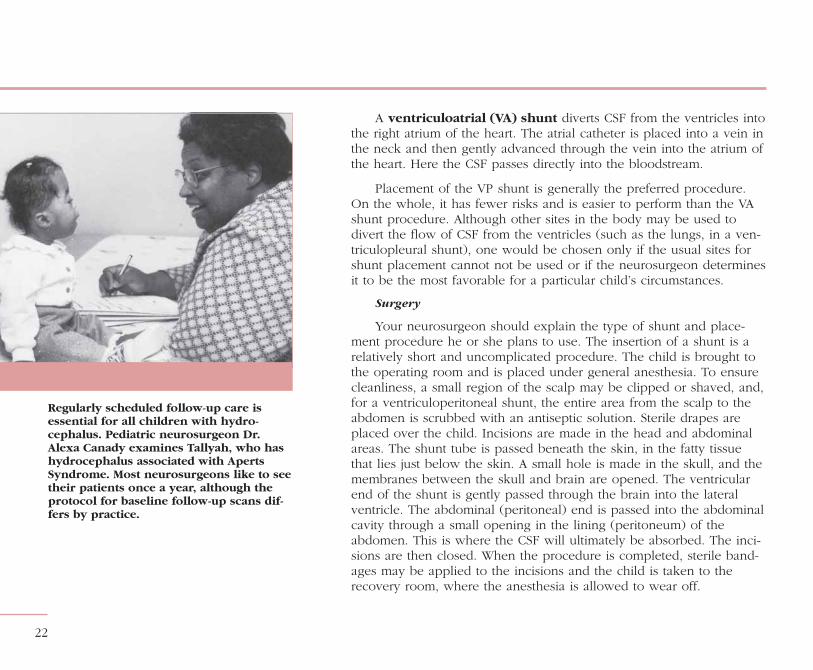
Regularly scheduled follow-up care isessential for all children with hydro-cephalus. Pediatric neurosurgeon Dr.Alexa Canady examines Tallyah, who hashydrocephalus associated with ApertsSyndrome. Most neurosurgeons like to seetheir patients once a year, although theprotocol for baseline follow-up scans dif-fers by practice.

After the operation, the child will be watched closely as he or sherecovers from surgery and the anesthesia. The neurosurgeon and nurs-es will check the child’s vital signs and neurological status for signs ofincreased intracranial pressure (ICP) that would warn of a shunt mal-function. If the child is an infant, they will check the anterior fontanelfor fullness and measure the head circumference at regular intervals.They will also watch the incisions for signs of infection. Some redness,swelling and tenderness are normal for the first week after surgery.The child may run a mild temperature for two or three days after theoperation—this, too, is quite normal. If the child has a high fever or afever that lasts for more than a few days, a surgeon or a pediatricianwill do evaluations to determine what is causing it and how it shouldbe treated. The neurosurgeon may specify that the child should stay ina certain body position for a period of time after the operation. Forexample, if greater drainage of CSF is necessary, the neurosurgeonmay recommend that the child’s head be elevated.
Shunt surgery usually involves minimal pain for the child. Somechildren experience neck and/or abdominal tenderness. Generally,mild analgesics such as acetaminophen are given. However, othermedications are available to make the child comfortable, especially forthe first few days after the operation. If all goes well and no complica-tions arise, the child will be released from the hospital within one tothree days.
After placement of a shunt, the size of the child’s ventricles usuallydecreases. In infants, the fontanel becomes soft and may appearsunken, and the skull sutures will narrow or possibly even overlap.Except in infants, the shunt usually is not visible under the skin. Achild whose hydrocephalus was diagnosed and treated early in infancywill have the same head size as his or her peers.
23
Ben and his twin brother were born pre-maturely, and Ben was shunted at sixmonths due to intraventricular hemor-rhage. Despite mild cerebral palsy andsome learning disabilities, Ben is a social-ly engaging young man who is developinghis talents in speaking and writing as amass communications major in college.Ben enjoys the Internet and his latestinterest is on-line investing.

Although hydrocephalus is almost always treated successfully withsurgical placement of a shunt, shunt malfunction and, less frequently,infection occur in many cases. Shunt malfunction, which is caused byobstruction, simply means that the shunt is not able to divert enoughCSF away from the ventricles in the brain. Shunt infection is caused bythe child’s own bacterial infection. These are serious problems andmust be treated appropriately.
Obstruction
When shunt malfunction occurs, it is usually a problem with a par-tial or complete blockage of the shunt. The fluid backs up from thesite of the obstruction and, if the blockage is not corrected, almostalways results in recurrent symptoms of hydrocephalus. Shunt obstruc-tion can occur in any of the components of the shunt. Most common-ly, the ventricular catheter becomes obstructed by tissue from thechoroid plexus or ventricles. The catheters or the valve may becomeblocked with blood cells or bacteria.
Infection
Shunt infection usually is caused by a child’s own bacterial organ-isms; it is not acquired from exposure to other children or adults whoare ill. The most common organism to produce infection isStaphylococcus epidermidis, which is normally found on the sur-face of the child’s skin and in the sweat glands and hair follicles deepwithin the skin. Infections of this type are most likely to occur one tothree months after surgery but may occur up to six months after theplacement of a shunt. Children with VP shunts are at risk of develop-ing a shunt infection secondary to abdominal infection, whereas chil-dren with VA shunts may develop generalized infection, which canquickly become serious. In either case, the shunt infection must betreated immediately to avoid life-threatening illness or possible braindamage.
24
Complications
“It’s important that every child have
a good self-image.We feel our job as
parents is to prepare all our children to lead
a normal life when they leave the nest.”
25
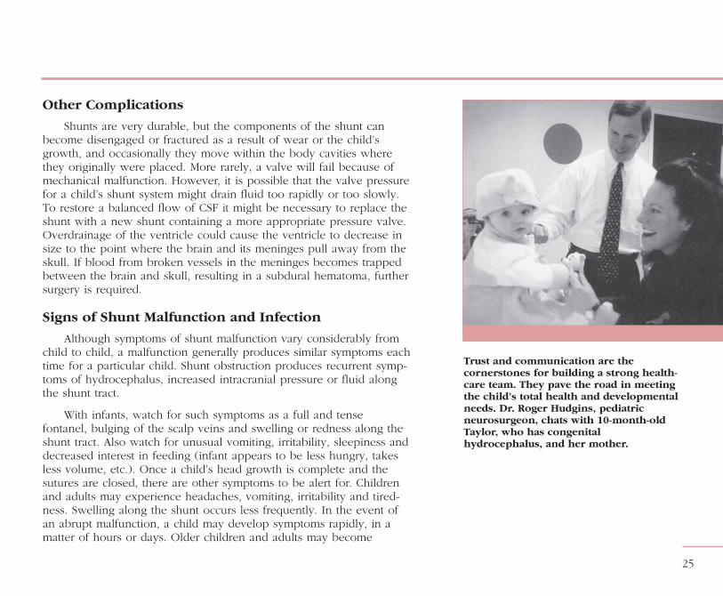
Trust and communication are thecornerstones for building a strong health-care team. They pave the road in meetingthe child’s total health and developmentalneeds. Dr. Roger Hudgins, pediatricneurosurgeon, chats with 10-month-oldTaylor, who has congenitalhydrocephalus, and her mother.
Other Complications
Shunts are very durable, but the components of the shunt canbecome disengaged or fractured as a result of wear or the child’sgrowth, and occasionally they move within the body cavities wherethey originally were placed. More rarely, a valve will fail because ofmechanical malfunction. However, it is possible that the valve pressurefor a child’s shunt system might drain fluid too rapidly or too slowly.To restore a balanced flow of CSF it might be necessary to replace theshunt with a new shunt containing a more appropriate pressure valve.Overdrainage of the ventricle could cause the ventricle to decrease insize to the point where the brain and its meninges pull away from theskull. If blood from broken vessels in the meninges becomes trappedbetween the brain and skull, resulting in a subdural hematoma, furthersurgery is required.
Signs of Shunt Malfunction and Infection
Although symptoms of shunt malfunction vary considerably fromchild to child, a malfunction generally produces similar symptoms eachtime for a particular child. Shunt obstruction produces recurrent symp-toms of hydrocephalus, increased intracranial pressure or fluid alongthe shunt tract.
With infants, watch for such symptoms as a full and tensefontanel, bulging of the scalp veins and swelling or redness along theshunt tract. Also watch for unusual vomiting, irritability, sleepiness anddecreased interest in feeding (infant appears to be less hungry, takesless volume, etc.). Once a child’s head growth is complete and thesutures are closed, there are other symptoms to be alert for. Childrenand adults may experience headaches, vomiting, irritability and tired-ness. Swelling along the shunt occurs less frequently. In the event ofan abrupt malfunction, a child may develop symptoms rapidly, in amatter of hours or days. Older children and adults may become

“If you deal with a life-threatening condition, ithelps you appreciate each day, and what you have.”
26
increasingly tired, may have difficulty waking up and staying awakeand, unless treated promptly, may go into a coma.
Shunt infection frequently results in fever and may occur alone orin conjunction with shunt obstruction. Occasionally, shunt infectionmay produce reddening or swelling along the shunt tract.
Knowing what symptoms to watch for will help you become moreat ease. Although the early symptoms of shunt malfunction or infec-tion—fever, vomiting and irritability—are the same as for many child-hood illnesses, you will learn to determine the symptoms associatedwith your child’s shunt. Should you have any doubt about your child’ssymptoms, don’t hesitate to call or visit your pediatrician for an evalua-tion. Remember, although shunt complications can be serious, they canalmost always be treated successfully when they are discovered early.A review of symptoms to watch for is given on the next page.
Michael sustained a brain injury at birthand was shunted 35 years ago. He and hiswife, Eva, are the proud parents ofMichelle. “Shunt technology was very newwhen I was born. I feel lucky to be alive.”

Symptoms of Shunt Malfunction or Infection
Infants Toddlers Children and Adults
Enlargement of the baby’s head
Fontanel is full and tense whenthe infant is upright and quiet
Prominent scalp veins
Swelling along the shunt tract
Vomiting
Irritability
Sleepiness
Downward deviation of the eyes
Less interest in feeding
Fever*
Redness along the shunt tract*
Head enlargement
Vomiting
Headache
Irritability and/or sleepiness
Swelling along the shunt tract
Loss of previous abilities(sensory or motor function)
Fever*
Redness along the shunt tract*
Vomiting
Headache
Vision problems
Irritability and/or tiredness
Personality change
Loss of coordination or balance
Swelling along the shunt tract
Difficulty in waking up or stayingawake
Decline in academic performance
Fever*
Redness along the shunt tract*
27
This list of symptoms is for your reference only and is not a diagnostic aid.If you are in doubt about your child’s medical condition, consult your physician immediately.
*Fever and redness along the shunt tract both indicate infection.

Shunt Revisions
A shunt complication usually requires another operation to make asurgical revision of the shunt. Depending on the cause of the compli-cation, some or all of the components of the shunt will be replaced. Inthe event of infection, the child is given a course of antibiotic therapyand usually the entire shunt is replaced.
Although there are exceptional cases in which children receive ashunt and have no further need for revisions or replacements, thesecases are rare. Experience shows us that some children undergo sever-al revisions throughout their lives. Whether or not other complicationswill arise depends on your child’s particular medical problems andbodily reactions to the surgical procedure and the shunt.
Hydrocephalus, left untreated, may cause severe brain damagewith physical and mental retardation. We wish there were simpleanswers about when permanent damage can result—but muchdepends upon the timeliness and effectiveness of the treatment or theoccurrence and severity of complications. The best way to prevent thepossibility of your child’s having brain damage is early detection ofproblems, should they occur. This is why it is so important that youlearn the signs and symptoms of shunt malfunction and infection andhave your child evaluated regularly by the pediatrician, neurosurgeonand neurologist.
It is important to develop a strong relationship with the health-care team and to share information as well. Your pediatrician or family practice physician will provide your child’s primary health care and will consult your neurosurgeon if a problem with the shunt
28
“To me, information always helped to soothe
and make me feel like I could contribute
something, rather than being out of control.”

29
Richard has three shunts as a result ofcongenital hydrocephalus with no knowncause. An energetic high school student,Richard specializes in computers—hedesigned and maintains his own website.His mother says that at first they weredevastated by the news and didn’t knowwhat to do, but now hydrocephalus is justa normal part of their lives.
is suspected. Your neurosurgeon will monitor your child’s ventriclesand will take care of problems associated with the shunt. A neurologistmay follow your child’s neurological status, as well as growth anddevelopment. You are an integral part of your child’s health-care team,with your own knowledge of your child’s health and history. Togetherwith the medical professionals, you have the combined skills to pro-vide excellent care for your child.
Some families find peace of mind—and a sense of control—inhaving their child carry or wear a medical identification device such asa surgical shunt I.D. card or a medical I.D. bracelet or necklace. Bothmethods of identification provide valuable medical information such asthe names, addresses and telephone numbers of doctors to be contact-ed in an emergency; shunt type, manufacturer and pressure setting (ifappropriate); and any additional information regarding medical condi-tions or allergies. Whether or not an identification device is importantto you, it is crucial to keep your child’s medical reports up-to-date andeasily accessible.
All children have a need to be their own person as they matureand explore their world. A child with hydrocephalus is no different. Itis essential that you treat your child as you would any other child, andthat you afford him or her every opportunity to live as normal a life aspossible. The shunt is a very durable device and should pose no spe-cial problems to normal handling or to childhood bumps and falls.Your child should be able to participate in most activities, with thepossible exception of rough contact sports.
You will find that your knowledge and understanding of yourchild’s condition will increase together with your confidence and com-fort in caring for him or her. When you have questions about hydro-cephalus, write them down as they occur to you—and bring the listwith you when you visit your doctor. You may find it helpful to talk toanother family whose child has a similar problem. Also, realize thatthere are many resources available to families with children who havespecial needs. Begin by asking your nurse or doctor about some ofthese possibilities.
Friends and relatives can also offer valuable emotional support.And don’t forget—all parents need to take time out for themselves.Allow a relative or a responsible sitter to care for your child from timeto time. Leave important information and telephone numbers whereyou can be reached. When your family travels, get the names of med-ical resource personnel in the area to which you are going, and besure to bring along important medical information as a safety measure.Although the likelihood of an emergency is remote, such preparednesswill allow you greater peace of mind and will avoid unnecessaryinconvenience should a problem arise. Some families choose to live inan area where access to a large medical center is convenient. If youlive far from a major care center, you must plan to travel at times toensure the best possible care for your child.
30
Caring for Your Child
“The family is the central influence in the lives of
children.”

Depending on your child’s medical problems, observation andconsultation by other specialists may be needed. Your child should bereferred to a neuro-ophthalmologist to have his or her vision exam-ined. Sometimes parents who wish to have more children are referredto a specialist for a genetic consultation. The geneticist assesses par-ents and their child to find possible genetic causes for the child’s med-ical problem and to determine the likelihood of another baby’s beingborn with the same defect. Occupational therapists and educationalpsychologists can provide valuable assistance in your child’s develop-ment. As advocates for your child, you and your physician mustencourage strong communication and a team effort toward meetingyour child’s total health and developmental needs. Also, let your physi-cian know your needs and concerns as a parent. And finally, make theeffort to gather perspectives from the various health-care providersworking with your child, in order to develop a framework of under-standing about your child and this condition. This understanding, com-bined with your own experience as a parent, will enable you to offerthe encouragement, support and resources your child needs to findacceptance and success in the world.
31
Lexi, age four, her little sister, Chiara, andher mom and dad savor special momentstogether. Diagnosed while in utero at 20weeks, Lexi underwent cranial reconstruc-tion surgery while still a baby.
“It was hard not to be scared and worried all thetime.We felt so much better after we got accurateinformation on hydrocephalus.”

As with all children, the age at which a child with hydrocephalusdevelops physical and intellectual skills varies. Many children withhydrocephalus have normal intelligence, physical development andcoordination, but they may be slower in acquiring such skills as eye-hand coordination and in learning to walk. Each child is different, andeach child’s level of attainment in skills depends upon many factors.Your child’s developmental progress will be influenced by the natureof the problem causing the hydrocephalus, by the degree of braindamage, if any, that occurred before treatment and by infections orother complications. But a child’s overall development and adaptationto the world also depends upon the individual child and the attitudeand opportunities afforded him or her by parents and environment.
Seeking a specialized pediatric psychologist can help to maximizeyour child’s physical, intellectual, emotional and social development.Very early on, your child should be evaluated by a pediatric psycholo-gist who has special skills in neuropsychological and emotional assess-ment. By giving your child a variety of diagnostic tests, the psycholo-gist will be able to identify the strengths and weaknesses in his or herabilities. And because there are thought to be critical developmentstages at which optimum learning takes place, we urge that your childhave regular evaluations.
As an infant, your child will be evaluated for such things as alert-ness, movement and tracking (responsiveness to sound and movingobjects). As your child gets older, she or he will be evaluated for ver-bal, intellectual and reasoning skills, as well as for social and emotion-al growth. All are crucial to sound and full development. Early identifi-cation and intervention can help to compensate for known deficienciesand can stimulate your child’s developing abilities, offering your childevery opportunity to achieve his or her fullest potential. If your pedia-trician and neurosurgeon are unfamiliar with a pediatric psychologist
32
As Your Child Grows Up

33
who does such diagnostic testing, ask them to check with localresources or the nearest medical center and refer you to one.
As your child reaches school age, developmental testing can pro-vide valuable information to help teachers meet your child’s education-al needs. Federal law requires all public schools to address and pro-vide for the educational needs of all children, including those withspecial needs. Share relevant information with teachers and others if itwill benefit your child. If you notice a decline in your child’s academicperformance, realize that many factors can contribute to these changes,including shunt malfunction. Consult your health-care team for an eval-uation. They may request an additional evaluation by a psychologistand, through their assessments, they will determine if your child has aproblem with the shunt and may identify other contributing factors andoffer helpful recommendations.
Challenge your child to seek out his or her potential. Your accept-ance and love will have great impact on how he or she perceives him-or herself and, ultimately, succeeds in the world. Your positive attitudeand encouragement will afford your child the greatest opportunity tolive a full and happy life.
“When life gets overwhelming for me, I remindmyself to let go and trust—and then to trust, and
let go.”
We must look to the future with vision and hope. Medical sci-ence is an advancing and dynamic field. Today we have solutions tomedical problems that were not even dreamed of in the past. Andthrough science and technology people will continue to expand thelimits of what is possible. As we go forward, we must have faith inourselves and our children. When faced with life’s challenges, we dis-cover not only personal strengths but also a greater capacity for com-passion and love. It is from endeavors like these that we find truevalue and meaning in life.
34
Looking to the Future
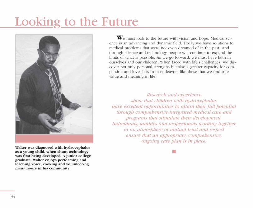
Walter was diagnosed with hydrocephalusas a young child, when shunt technologywas first being developed. A junior collegegraduate, Walter enjoys performing andteaching voice, cooking and volunteeringmany hours in his community.
Research and experienceshow that children with hydrocephalus
have excellent opportunities to attain their full potentialthrough comprehensive integrated medical care and
programs that stimulate their development.Individuals, families and professionals working together
in an atmosphere of mutual trust and respectensure that an appropriate, comprehensive,
ongoing care plan is in place.
■

35
The Hydrocephalus Association is a national, 501 (c) (3) nonprofit organization founded in 1983 to provide support,education and advocacy to individuals, families and professionals. Our goal is to provide comprehensive services thatempower individuals and families to seek out the best medical care, programs and resources that meet their needs nowand in the future.
As the nation’s largest and most widely respected organization dedicated solely to hydrocephalus, the Association hasbeen instrumental in creating a community of individuals, families and health-care professionals addressing the complexi-ties of hydrocephalus in all age groups—infants, children, young adults and adults. We continually update and expand ourresources to keep pace with new technologies in the diagnosis and treatment of hydrocephalus and stay current with theneeds of the individuals we serve.
Hydrocephalus is a chronic condition. With early detection, effective treatment and appropriate interventional services,the future for individuals with hydrocephalus is promising. We invite your inquiries.
Resources
About Hydrocephalus—A Book for Families (English or Spanish)Prenatal Hydrocephalus—A Book for Parents
About Normal Pressure Hydrocephalus—A Book for Adults and Their FamiliesDirectory of Pediatric Neurosurgeons
Directory of Neurosurgeons for Adult Onset HydrocephalusLINK Directory
Quarterly NewsletterThe Resource Guide
A Teacher’s Guide to HydrocephalusFact and Information Sheets on a wide range of topics related to hydrocephalus
Annual Educational ScholarshipsAnnual Neurosurgical Resident’s Prize
Biennial National Conference for Families and Professionals
Hydrocephalus Association � 870 Market Street � Suite 705 � San Francisco, CA 94102 � Tel:(415)732-7040Toll-free:(888)598-3789 � Fax:(415)732-7044 � E-mail:[email protected] � Website:www.hydroassoc.org
Resources

36
� Name � � Birthdate
� Parent/Guardian � Home � Work
� Neurosurgeon � � Address
� Pediatrician � � Address
� Other Doctors:Name � � Address
Name � � Address
� Type of Hydrocephalus/Other Conditions
� Shunt Name/Type � Pressure Setting � Allergies
� Brief Surgical/Medical History
� Medications
� Developmental/Neuropsychological Testing � Date � Follow-up
� Notes/CT, MRI Scans or Head Ultrasounds
Health Record

About Hydrocephalus—A Book for Parents was originally publishedby the University of California, San Francisco, in 1986, under the guidance of Michael S. B. Edwards, M.D., and Margie Derechin, M.S.N., R.N. Text, illustration and design were by Lynne Larson and the editor was Susan Eastwood.
2000 Edition
Editor: Rachel A. Fudge
Graphic Design: Tina Keker Design
Photography: Philip Harvey
Medical Consultants:
Marvin Bergsneider, M.D. J. Gordon McComb, M.D.Alexa Canady, M.D. David G. McLone, M.D., Ph.D.Philip H. Cogen, M.D., Ph.D. Harold L. Rekate, M.D.Samuel F. Ciricillo, M.D. Mary Smellie-Decker, R.N., M.S.N.James M. Drake, M.D. Marion L. Walker, M.D.Michael S. B. Edwards, M.D. Donna Wallace, R.N., M.S., C.P.N.P.Roger J. Hudgins, M.D. Michael A. Williams, M.D.
Revision of this booklet was made possible through funds contributed by:
Aesculap, Inc.Codman, a Johnson & Johnson CompanyIntegraMedtronic NeurosurgeryVygon NeuroHydrocephalus Association
© Copyright 2002 Hydrocephalus Association, San Francisco, California

Hydrocephalus Association � 870 Market Street � Suite 705 � San Francisco, CA 94102 � Tel:(415)732-7040Toll-free:(888)598-3789 � Fax:(415)732-7044 � E-mail:[email protected] � Website:www.hydroassoc.org


