22
ABOVE KNEE AMPUTATION
| Date post: | 31-Dec-2015 |
| Category: |
Documents |
| Upload: | jade-gamble |
| View: | 359 times |
| Download: | 13 times |

ABOVE KNEE AMPUTATION

ARTHROSCOPY

KNEE JOINT
Block 14 GNK 483

Anatomy
• Classification• Bones• Capsule• Ligaments• Synovial membrane• Cruciate ligaments• Menisci• Nerve supply
• Blood supply• Relationships• Bursae• Movements
Classification
• Synovial hinge joint• Medially & laterally: femur & tibia (hinge and
rotation)• Intermediate: Femur & patella (gliding joint

Capsule
• Absent anteriorly• Synovial membrane protrudes to form the
bursa beneath the tendon of quadriceps femoris
• It is anteriorly reinforced by the tendon of quadriceps femoris, the patella and the patella retinacula

Capsule
• Inferiorly, the capsule is fixed to the margins of the tibia, except where the tendon of popliteus passes
• The capsule is reinforced on both sides by the expansions of vastus lateralis and medialis, as well as behind by the oblique popliteal ligament from the insertion of semimembranosus
Ligaments5 extracapsular
• Patellar ligaments – quadriceps reflex L2,3,4
• Lateral collateral ligament• Medial collateral ligament• Oblique popliteal ligament
– is an expansion from the insertion of semimembranosus
• Arcuate popliteal ligament– Y-shaped ligament posterior
that bridges the tendon of popliteus

Ligaments4 intracapsular
• Synovial membrane• Cruciate ligaments• Menisci• Tendon of popliteus
Synovial Membrane
• Infrapatellar fold is a reflexion from the posterior surface of the patella-forms a fold to the intercondylar fossa
• The synovial membrane from the posterior part of the capsule is reflected around the front of the cruciate ligaments-they are therefore intracapsular but extrasynovial

Cruciate LigamentsExtrasynovial
• Anterior– Taut in extension– Prevents post
displacement of femur on tibia
– Prevents hyperextension of the knee

Cruciate LigamentsExtrasynovial
• Posterior– Stronger– Taut during flexion– Prevents ant
displacement of femur on tibia
– Prevents hyperflexion of the knee
Menisci - General
• Medial & lateral• C-shaped• Fibrous cartilage• Upper surface concave• Outer margins thicker,
convex,attached to capsule• Inner margins thinner,
concave,free border• Transverse ligament extends
between the 2 ant parts of the menisci

Menisci
• Medial meniscus– Semicircular (oval)– Wider at back than in
front– Attached to medial
collateral ligament– Most commonly
injured

Menisci
• Lateral meniscus– Almost circular– Even width front and
back– Separated from
lateral collateral ligament via popliteal tendon

Nerve Supply
• Br of femoral n to vastus medialis
• Obturator n (the br passes with the femoral a thro the add hiatus to popliteus fossa
• Sciatic n

Blood Supply
• Genicular anatomosis formed by brrs of popliteal a
• Middle genicular a pierces the capsule & supplies cruciate lig’s, synovial mem, outer margins of menisci
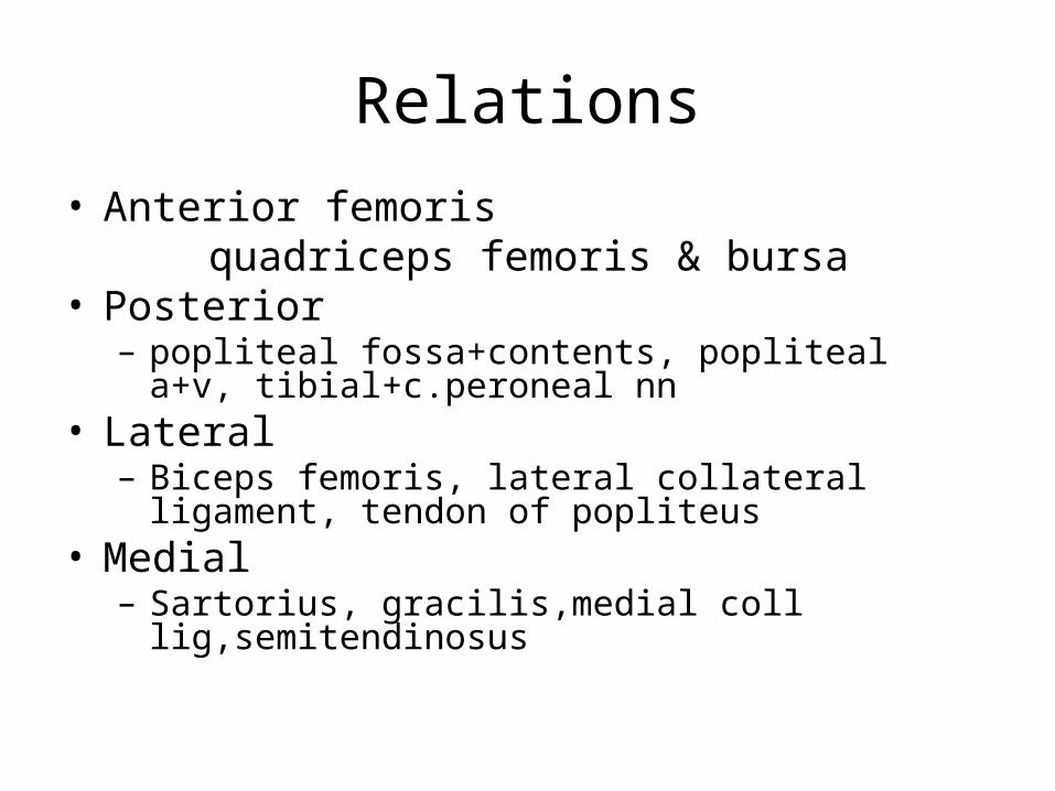
Relations• Anterior femoris quadriceps femoris & bursa• Posterior – popliteal fossa+contents, popliteal a+v, tibial+c.peroneal
nn• Lateral– Biceps femoris, lateral collateral ligament, tendon of
popliteus• Medial– Sartorius, gracilis,medial coll lig,semitendinosus
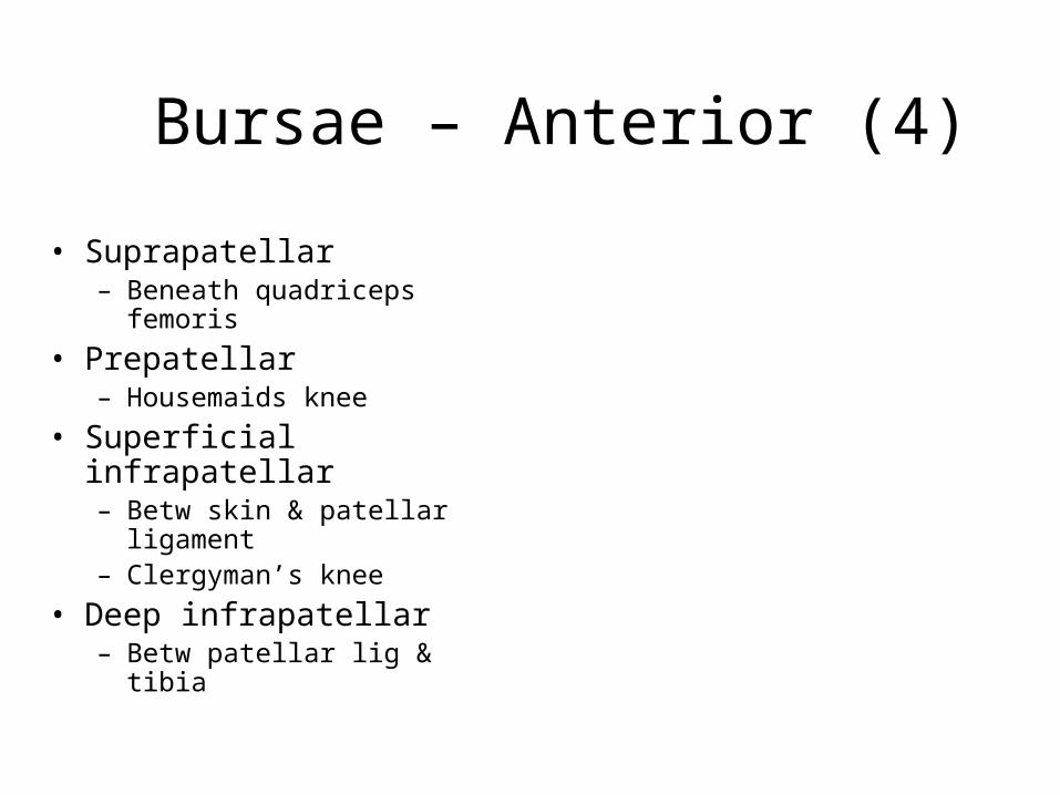
Bursae – Anterior (4)
• Suprapatellar – Beneath quadriceps
femoris• Prepatellar
– Housemaids knee• Superficial infrapatellar
– Betw skin & patellar ligament
– Clergyman’s knee• Deep infrapatellar
– Betw patellar lig & tibia

Bursae – Posterior (2)
• Beneath popliteus• Beneath
semimembranosus

Bursae – Posterior (2)
• Others:Biceps femorisLateral head of gastrocnemiusMedial head of gastrocnemiusAnserine bursa below insertion of:– Sartorius– Gracilis– semitendinosus

Movements
• Flexion– hamstrings+sartorius,gracilis,gastroc
• Extension– quadriceps femoris+medial rotation
• Rotation– Passive: screw home mechanism– Active: medially in a flexed knee (SarGrTen)
laterally via Biceps femoris


