2
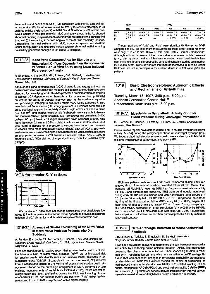
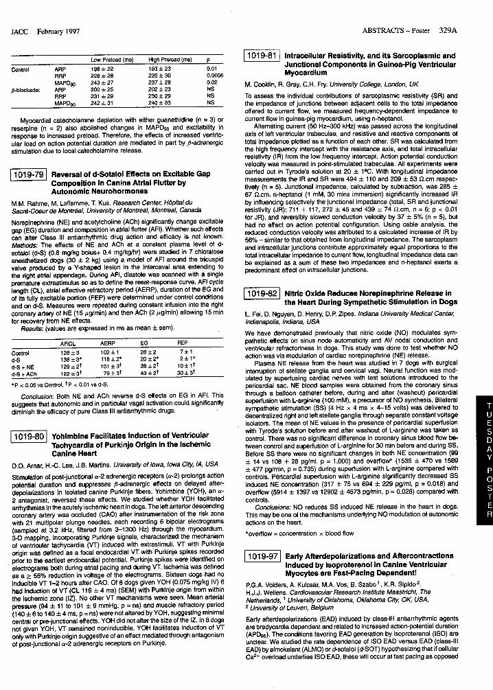
328A ABSTRACTS – Poster JACC February 1997 the annulus and papillav muscle (PM), consistent with chordal tension limit- ing excursion. We therefore examined the MV by 2D eohocardiogrephy in 58 patients with LVdyefunotion (36with IMLC and 22 without) vs 21 normal con- trols. Results: In most patients with IMLC vsthose without, 1) the AL showed abnomnalbending in systole, 2) AL opening was restricted to the annulus-PM line, and 3) the opening excursion angle u of the AL was markedly reduced. Conclusions: In most patients with IMLC, abnormal systolic and diastolic leaflet mnfiguration and reatrioted motion auggest abnormal leaflet tethering created by geometric changes in the mitral-LV complex. m 101836 Isthe Vena Contracta Area forStenoticand Regurgitant OrificeaDependent on Hemodynamic Varieblea?An In Vitro StudyusingLaserInduced Fluorescence Imaging R. Shandas, N. Trujillo, E.A. Gill, J. Kwon, C.G. DeGrolf, L. Valdes-Cruz. The Children’s Hospital, University of Colorado Health Sciences Centec Denver, CO, USA Although the vena contracta area (VCA) of atenotic and regurgitant jets has been shown toreprasentthe true measureof diaeaseseverity, there isnogold atanderd for quantifying VCA. This has presented problems when attempting to aaseas VCA dependence on hemodynamics (pressure, flow, pulsatility) aa well as the ability of Doppler methods such as the mntinuity equation and proximal jet imaging to accurately reflect VCA. Using a precise in vitro Iaaer induced fluorescence (LIF) imaging system to illuminate perpendicular cross-sectional regions immediately distal to rigid orifices of various sizes (0.2-4.9 cmz) and shapes (circular, slit, Y-shaped), we were able to isolate and measure VCA (Figure) for steady (25-150 cckec) and pulsatile (20-100 Cc/beat;60 bpm) flows. VCA ragion (minimum cross-sectionaljet area) was found between 0.1 cm and 0.5 cm distal to all orifices at all flow rates. VCA was found to depend on a pressure to viscous force ratio. Low pressure to viscous force ratios (increased viscous effects) caused VCA to approach anatomic areas while increasing the ratio (decreasingviscous effects)caused an asymptotic decrease in VCA towards a constant value (79% + 3.3% of anatomic area). VCA did not change significantly over the pulsatile cycle (Graph). VCA for circular&Y orifices w—-uw- a ::: *! ... ..... ..... : 4 ........... ..... . ...... t ....i..-~q--.””” .,-””” i * .... <..... +.... }.. ?..;..... 9: : U We conclude: 1) VCA does not change significantly over physiologic flow rates; 2) A ratio of pressure to viscous forces appears to provide an accurate reflector of VCA dynamics and its relationship to actual anatomic area. m 101837 AbeenceofSevereThickeningoftheMitralVelve inMitrelValveProlapaePatientswhoDie Suddenly A. Pandey, E.K. Louie, T.L. McKiernan, S. Bharati. The Hear?Institute for Children, Christ Hospital, Oak Lawn, IL, USA, Loyola Uni~ Medical Centec Maywood, IL, USA Prior eohocsrdiographic atudies report that a mitral leaflet width >5 mm identifies a subaet of mitral valve prolapse (MWJ pts at increased risk for audden death. We directly measured intrinsic leaflet thicknaas in 24 autopsiad hearts (12 MVP, 12 controls with normal mitral valves, NL) selected from a consecutive series of 376 victims of unexplained sudden death. An obaewer, blinded to the pathologic assignment of MVP, performed in situ triplicate measurements of leaflet body thickness (THb), leaflet coaptation margin thickness (THc), and leaflet closure line thickness including chordal attachments (THch) for anterior (AMV) and posterior (PMV) mitral leaflets (measured in mm to 0.01 mm precision with a digital caliper): AMV PMV THb THc THch THb THc THch MVP 0.8 + 0.2 0.9 * 0.3 2.0 + 0.6 0.s ● 0.2 0.9 ● 0.4 1.7 * 0.8 NL 0,6 k 0.1 0.6 + 0.2 1.6 + 0.7 0.5 i 0.1 0.5 * 0.1 0.9 * 0.2 P <0,01 ns ns <0,005 <0.01 <0005 Though portions of AMV and PMV were significantly thicker for MVP compared to NL, the maximum measurements from either leaflet for MVP were only: THb = 1.2 mm, THc = 1.9 mm, and THch = 3.2 mm. Conclusions: Although intrinsic thickness of the mitral valve is increased in MVP relative to NL, the maximum leaflet thicknesses observed were considerably lees than the 5 mm threshold proposed by echocardiographic studies aa a marker for sudden death. Our study shows that marked ’increases in intrinsic leaflet thickness are not a prerequisite for sudden death in mitral valve prolapse patients. mEl Basic Electrophysiology: Autonomic Effects and Mechanisms of Arrhythmias Tuesday,March18, 1997, 3:00 p.m.–5:OOp.m. AnaheimConventionCenter,Hall E PresentationHour:4:00 p.m.–5:OOp.m. -[ MuacleNerveSymPathetic ActivitYC0ntr0k3 BloodPressureduringVasovagal Preayncope D.L. Jardine, S.1.Bennett, R. Frethey, H. Ikram, LG. Crozier. Christchurch Hospital, NewZaa/and Previous case reports have demonstrated a fall in muscle sympathetic newe activity [MNSA] during the presyncopal phase of vasovaaal svnco!Je WS1. We hypothesized that blood pressure would correlate dir&tly with MNSA at this time irrespective of parasympathetic activity [PA]. Eighteen patients with recurrent VS were compared during eatiy 8@ head-up tilt to 17 controls all of whom tolerated tilt for 45 min. Mean blood pressure [MBP], MNSA, heart rate [HR], high frequency heart rate variability [HFHRV], and baroreceptor sensitivity [BS] were continuously monitored. During early tilt, BP was maintained and MNSA increased [both groups p < 0.01] while PA activity fell [HFHRV, BS p < 0.01]. Presyncope, defined as the time of the first sustained fall in MBP during tilt [p < 0.05], began at a mean time of 15.2 + 3 min and lasted 170 + 10 sec. During presyncope, MBP and MNSA decreased in direct correlation [p < 0.001] while HFHRV and BS remained low. HR also correlated with MNSA [p < 0.001] suggesting that sympathetic withdrawal rather than parasympathetic activity mediatea vasovagal syncope. \ 1019-78 I Beta-Adrenergic MediaticJnof Mechenoelectrical Feedback B.B. Lerman, K. Todaka, E. Engelstein, D. Burkhoff. New York Hospital-Cornell Medical Centec New York, NY USA It has been previously shown that augmented preload increases myooardial excitability by shortening action potential duration (APD). The mechaniam governing this phenomenon is unknown. Since shortening of APD is madi- ated by K+ depolarizationcurrents which are sensitive to cAMP, we hypoth- esized that load-dependent changes in myooerdial excitability are mediated by stimulation of cAMP. We therefore studied the effects of propranolol on load-induced changes in electrical excitability in 7 isolated ejecting canine hearts. Monophaaic APD (MAPDso)and electrical excitability (relative [RRP] and abaolute [ARP] refractory periods derived from strength-intetval curves) were determined at low and high loads before and after p-blockade.