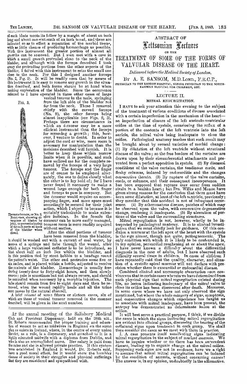
183 of each blade meets its fellow by a margin of about an inch long and about one-sixteenth of an inch bioad, and these are indented so as to effect a separation of the morbid tissue, with as little change of producing haemorrhage as possible. With this instrument the greater portion of almost all growths can be removed. But I soon met with a case in which a small growth protruded close to the neck of the bladder, and although with the forceps described I took away the protruding portions from the other a"pects of the bladder, I failed with this instrument to seize the small one close to the neck. For this I designed another forceps (No. 2, Fig. 2). It will be readily seen that by means of this instrument it is easy to remove any growth in the situa- tion described, and both forms should be at hand when making exploration of the bladder. Since the occurrence referred to I have operated in three other cases of large vestal tumour m tne maie, springing from the left side of the bladder not far from the neck. These I removed chiefly with the curved forceps (No. 2), the other forceps being almost inapplicable (see Figs. 2, 3). Perhaps there are cirumstances in which an eeraseur may be a more efficient instrument than the forceps for removing a growth; this, how- ever, I venture to doubt. In order to adjust the cord or wire, more room is necessary for manipulation than the incisions described will furnish. It is better to keep these within narrow limits when it is possible, and such have sufficed me for the complete re- moval by the forceps of a very large tumour. The forceps and the finger are of course to be employed alter- nately, the one to define clearly what the other is to lay hold of; for I have never found it necessary to make a wound large enough for both finger and forceps to pass in company. But the écraseur would require an accom- panying finger, and more space must accordingly be secured for their joint action, which I object to because it is The.amef.rcep., or No. 2. certainly undesirable to make exten- Front view, showing di. sive incisions. In the female the vergence of bladts by ecraseur can be more easily managed, joint, topreventBipping because room is more readily acquired . of the soft parts at neck .th t t. of the bladder when without section. closed. After the chief portions of tumour have been removed from the bladder, it should be washed out with a current of cool water, by means of a syringe and tube through the wound, after which a large vulcanised catheter is to be introduced, so that about half an inch lies within the bladder, and in this position tied by stout bobbin to a bandage round the patient’s waist. The other end protrudes some five or six inches, and is placed within a bottle, adjusted to receive the contents of the bladder.’ Haemorrhage is usually free during twenty-four to forty-eight hours, and then slowly ceases; pain is sometimes but not always severe, and should be controlled when necessary by a morphia injection. The tube should remain from five to eight days and then be re- moved, when the wound rapidly heals and all the urine soon passes by the natural channel. A brief résumé of some fifteen or sixteen caes, six of which are those of vesical tumour removed in the manner described, will be given in the next number. AT the annual meeting of the Salisbury Medical Club and Provident Dispensary, held on the 26th ult., Dr. Roberts recommended the proper trainiog and educa- tion of women to act as midwives in England on the same plan as exists in Ireland, where, in the centre of every union district, as a rule, is a dispensary, and attached to it is a highly educated midwife with a diploma from Dublin, and who is also an accomplished nurse. Her salary is paid from the rates and she is allowed private practice. If this system were established in England it would, Dr. Roberts said, have a good moral effect, for it would show the humbler classes of society in their struggles and physical sufferings that they are considered and sympathised with. ABSTRACT OF Lettsomian Lectures ON THE TREATMENT OF SOME OF THE FORMS OF VALVULAR DISEASE OF THE HEART. Delivered before the Medical Society of London, BY A. E. SANSOM, M.D.LOND., F.R.C.P., PHYSICIAN TO THE LONDON HOSPITAL, SENIOR PHYSICIAN TO THE NORTH- EASTERN HOSPITAL FOR CHILDREN, ETC. LECTURE II. MITRAL REGURGITATION. I HAVE to ask your attention this evening to the subject of the treatment of various conditions of disease associated with a certain imperfection in the mechanism of the heart- an imperfection of closure of the left auriculo-ventricular orifice at the time of systole, occasioning the reflux of a portion of the contents of the left ventricle into the left auricle, the mitral valve being inadequate to close the orifice. Pathological anatomy teaches that such result may be brought about by several varieties of morbid change: (1) By dilatation of the left ventricle without structural disease of the valve; so the free borders of the curtains are drawn upon by their circumferential attachments and pre- vented from a perfect apposition in systole. (2) By diseased conditions of the valve curtains, the tendinous cords, and fleshy columns, induced by endocarditis and the changes consecutive thereto. (3) By rupture of the valve curtains, cords, or columns, and their consequent incompetence. It has been supposed that rupture may occur from sudden strain in a healthy heart; but Drs. Wilks and Moxon have given strong reasons for the conclusion that there must have been some dilatation, at least of the left ventricle, previously; they consider that this accident is not of infrequent occur- rence. (4) By atheromatous disease, patches of which may be observed upon the valve, with consecutive degenerative change, rendering it inadequate. (5) By ulceration of por- tions of the valve and the surrounding structures. Mitral regurgitation is not, however, wholly to be in- terpreted by pathological anatomy. It is to clinical investi- gation that we must chiefly look for guidance. Of this con- dition a murmur at the left apex of the heart with the systole is the s’gn almost, though not quite, pathognomonic. The only condition with which it is likely to be confounded is, in my opinion, pericardial roughening at or about the apex. I have never known a difficulry ahout the differential diagnosis in the case of adults, but I have observed such difficulty several times in children. In cases of children I have repeatedly said that the quality, character, and situa- tion of a systolic apical murmur will not declare with pre- cision whether there is exocardial or endocardial disease. Combined clinical and necroscopic observation foon con- vincesus that in certain cases wherein we have determinedfrom such physical sign that mitral regurgitation existed during life, no lesion indicating inadequacy of the mitral valve to close its orifice has been discovered after death. Moreover, in some cases where we have not only observed the sign mentioned, but where the whole category of sign", symptoms, and consecutive changes which experience has taught us to associate with mitral inadequacy, have been present, the autopsy has demonstrated no determinate lesion at the orifice. It will best serve a practical purpose, I think, if we divide the cases in which the signs indicating mitral regurgitation are evident into clinical groups, discussing the bearing of the collateral signs upon treatment in each group. We shall thus consider the cases as we meet with them in practice. I. A case presents itself manifesting signs indicating mitral regurgitation in the subject of marked ancenaia. We have to inquire whether or no there has been antecedent disease, leading up to organic change at the mitral orifice. Supposing such signs are not in evidence, have we a right to assume that actual mitral regurgitation can be induced by the condition of anaemia, without concurring causes? The answer is, in my opinion, undoubtedly in the affirmative.
Transcript
183
of each blade meets its fellow by a margin of about an inchlong and about one-sixteenth of an inch bioad, and these areindented so as to effect a separation of the morbid tissue,with as little change of producing haemorrhage as possible.With this instrument the greater portion of almost all
growths can be removed. But I soon met with a case inwhich a small growth protruded close to the neck of thebladder, and although with the forceps described I tookaway the protruding portions from the other a"pects of thebladder, I failed with this instrument to seize the small oneclose to the neck. For this I designed another forceps(No. 2, Fig. 2). It will be readily seen that by means ofthis instrument it is easy to remove any growth in the situa-tion described, and both forms should be at hand whenmaking exploration of the bladder. Since the occurrencereferred to I have operated in three other cases of large
vestal tumour m tne maie, springingfrom the left side of the bladder notfar from the neck. These I removedchiefly with the curved forceps(No. 2), the other forceps beingalmost inapplicable (see Figs. 2, 3).Perhaps there are cirumstances inwhich an eeraseur may be a moreefficient instrument than the forcepsfor removing a growth; this, how-ever, I venture to doubt. In order toadjust the cord or wire, more room isnecessary for manipulation than theincisions described will furnish. It isbetter to keep these within narrowlimits when it is possible, and suchhave sufficed me for the complete re-moval by the forceps of a very largetumour. The forceps and the fingerare of course to be employed alter-nately, the one to define clearly whatthe other is to lay hold of; for I havenever found it necessary to make awound large enough for both fingerand forceps to pass in company. Butthe écraseur would require an accom-panying finger, and more space mustaccordingly be secured for their jointaction, which I object to because it is
The.amef.rcep., or No. 2. certainly undesirable to make exten-Front view, showing di. sive incisions. In the female thevergence of bladts by ecraseur can be more easily managed,joint, topreventBipping because room is more readily acquired
. of the soft parts at neck .th t t.of the bladder when without section.closed. After the chief portions of tumour
have been removed from the bladder,it should be washed out with a current of cool water, bymeans of a syringe and tube through the wound, afterwhich a large vulcanised catheter is to be introduced, so
that about half an inch lies within the bladder, andin this position tied by stout bobbin to a bandage roundthe patient’s waist. The other end protrudes some five orsix inches, and is placed within a bottle, adjusted to receivethe contents of the bladder.’ Haemorrhage is usually freeduring twenty-four to forty-eight hours, and then slowlyceases; pain is sometimes but not always severe, and shouldbe controlled when necessary by a morphia injection. Thetube should remain from five to eight days and then be re-moved, when the wound rapidly heals and all the urinesoon passes by the natural channel.A brief résumé of some fifteen or sixteen caes, six of
which are those of vesical tumour removed in the mannerdescribed, will be given in the next number.
AT the annual meeting of the Salisbury MedicalClub and Provident Dispensary, held on the 26th ult.,Dr. Roberts recommended the proper trainiog and educa-tion of women to act as midwives in England on the sameplan as exists in Ireland, where, in the centre of every uniondistrict, as a rule, is a dispensary, and attached to it is ahighly educated midwife with a diploma from Dublin, andwho is also an accomplished nurse. Her salary is paid fromthe rates and she is allowed private practice. If this systemwere established in England it would, Dr. Roberts said,have a good moral effect, for it would show the humblerclasses of society in their struggles and physical sufferingsthat they are considered and sympathised with.
ABSTRACT OF
Lettsomian LecturesON THE
TREATMENT OF SOME OF THE FORMS OFVALVULAR DISEASE OF THE HEART.
Delivered before the Medical Society of London,BY A. E. SANSOM, M.D.LOND., F.R.C.P.,
PHYSICIAN TO THE LONDON HOSPITAL, SENIOR PHYSICIAN TO THE NORTH-EASTERN HOSPITAL FOR CHILDREN, ETC.
LECTURE II.MITRAL REGURGITATION.
I HAVE to ask your attention this evening to the subjectof the treatment of various conditions of disease associatedwith a certain imperfection in the mechanism of the heart-an imperfection of closure of the left auriculo-ventricularorifice at the time of systole, occasioning the reflux of aportion of the contents of the left ventricle into the leftauricle, the mitral valve being inadequate to close theorifice. Pathological anatomy teaches that such result maybe brought about by several varieties of morbid change:(1) By dilatation of the left ventricle without structuraldisease of the valve; so the free borders of the curtains aredrawn upon by their circumferential attachments and pre-vented from a perfect apposition in systole. (2) By diseasedconditions of the valve curtains, the tendinous cords, andfleshy columns, induced by endocarditis and the changesconsecutive thereto. (3) By rupture of the valve curtains,cords, or columns, and their consequent incompetence. Ithas been supposed that rupture may occur from suddenstrain in a healthy heart; but Drs. Wilks and Moxon havegiven strong reasons for the conclusion that there must havebeen some dilatation, at least of the left ventricle, previously;they consider that this accident is not of infrequent occur-rence. (4) By atheromatous disease, patches of which maybe observed upon the valve, with consecutive degenerativechange, rendering it inadequate. (5) By ulceration of por-tions of the valve and the surrounding structures.
Mitral regurgitation is not, however, wholly to be in-terpreted by pathological anatomy. It is to clinical investi-gation that we must chiefly look for guidance. Of this con-dition a murmur at the left apex of the heart with the systoleis the s’gn almost, though not quite, pathognomonic. Theonly condition with which it is likely to be confounded is,in my opinion, pericardial roughening at or about the apex.I have never known a difficulry ahout the differential
diagnosis in the case of adults, but I have observed suchdifficulty several times in children. In cases of children Ihave repeatedly said that the quality, character, and situa-tion of a systolic apical murmur will not declare with pre-cision whether there is exocardial or endocardial disease.Combined clinical and necroscopic observation foon con-
vincesus that in certain cases wherein we have determinedfromsuch physical sign that mitral regurgitation existed duringlife, no lesion indicating inadequacy of the mitral valve toclose its orifice has been discovered after death. Moreover,in some cases where we have not only observed the signmentioned, but where the whole category of sign", symptoms,and consecutive changes which experience has taught usto associate with mitral inadequacy, have been present, theautopsy has demonstrated no determinate lesion at theorifice.
It will best serve a practical purpose, I think, if we dividethe cases in which the signs indicating mitral regurgitationare evident into clinical groups, discussing the bearing of thecollateral signs upon treatment in each group. We shallthus consider the cases as we meet with them in practice.
I. A case presents itself manifesting signs indicatingmitral regurgitation in the subject of marked ancenaia. Wehave to inquire whether or no there has been antecedentdisease, leading up to organic change at the mitral orifice.Supposing such signs are not in evidence, have we a rightto assume that actual mitral regurgitation can be inducedby the condition of anaemia, without concurring causes?The answer is, in my opinion, undoubtedly in the affirmative.
184
In cases of anaemia and chlorosis a murmur is sometimesheard exactly in the site of that indicating mitral regurgita-tion. Assuming that in these cases there is a veritable re-gurgitation, how is such brought about? The explanationis, I think, given by the careful experiments conducted byLudwig and Hesse at Leipzig, which have been admirablysummarised by Dr. Donald Macalister.l The mechanismfor the closure of the left auriculo-ventricular orifice doesnot reside in the valve curtains alone; the surroundingmuscles of the ventricle have an active share, not merely infloating up the valve curtains, but in reducing the size oithe aperture which these valve curtains have to close. It inot that the orifice is dilated, but that it is insufflcie2?,tlgcontracted, the aid of the muscles of the wall of the ventriclewhich normally produce such contraction being lost.
It is important in regard to treatment to differentiatemitral regurgitation due to disease of the valves from thatdue to adynamia of ventricle, supposing a systolic apicalmurmur to be manifest in a markedly anaemic subject. Thetwo signs I would most rely on as pointing to an anaemiccausation of the murmur are: (1) an absence of notable car-diac dilatation; (2) a heightened tension in the systemicarteries. I have never known in these cases any markedimprovement follow the administration of the usual cardiactonics, such as digitalis and iron. In the cases attendedwith haemorrhage it is, of course, of the first importance toarrest this at its source. Rest and the administration ofassimilable food are no less important indications. In thisconnexion I may call attention to the great value I haveobserved to attach to supplementary alimentation by therectum in such cases. I have long tried the plan of usingdefibrinated ox blood for a nutrient enema, as advocated bymy friend Dr. A. H. Smith of New York. In comparingresults, however, with those in which artificially digestedfood has been employed, I felt that the balance of evidenceis in favour of the latter plan. I have had prepared mixedpeptone enemata-beef, milk, and farinaceous food-whichhave been proved to preserve a perfectly good condition forlong periods. These have the advantage of being availableat a moment’s notice, it being only needful to render themdiffluent with warm water. From two to four ounces areinjected slowly into the rectum, and repeated every three orfour hours. In many cases I have caused to be added thedry ox blood (sanguis bovinus exsiccatus), in the proportionof-a drachm to the ounce. I have lately, however, adopteda simpler plan with good results, using, instead of pep-toned food, equal parts of warm milk and cod-liver oil as anutritive enema.In the treatment of cases of idiopathic ansemia I have
found no drug treatment so efficient as the administration ofarsenic (Fowler’s solution in small doses gradually increased).I have observed, as has been recorded by others, completerecovery, with the disappearance of the cardiac murmur,under such treatment, combined with rest and carefulnutrition.
II. We will now assume that a systolic apex murmur ispresent in a patient showing signs of a aaeecrosis of the cervicalsympathetic. It has been frequently noted that a murmurat the apex has existed in the subjects of exophthalmicgoitre (Graves’ or Basedow’s disease) ; yet, on post-mortemexamination, no disease at the mitral orifice has been dis-covered. In these cases anaemia may be present, but not ofnecessity. It is not causally related with the phenomena.Organic heart disease may co-exist, but such coincidence israre. It is important to recognise, especially with regard totreatment, that in the subjects of Graves’ disease mitralregurgitation occurs without valvular lesion. The record offatal cases in which disease of the cervical sympatheticganglia has been actually demonstrated in Graves’ disease isnow tolerably extensive. Trousseau, Cruise, and ]B,[cDonnell,Keith, and Shingleton Smith have recorded cases in whichsome of the ganglia (usually the inferior cervical) have beenenlarged, atrophied, or degenerated.As regards treatment, in the cases which I have seen,
ordinary tonics and digitalis have been of very little benefit,but great improvement has followed galvanisation of thecervical sympathetic. I have employed the continuouscurrent from twenty to forty elements (Lecla.ncbe). Onepole may be placed behind the lower jaw in front of thesterno-mastoid, and the other either at sb corresponding pointof the oDDosite side. or at the nade of the neck right or left
1 Remarks on the Form and Mechanism of the Heart, &c., BritishMedical Journal, Oct. 28th, 1882, p. 825.
of the vertebra prominens, or above the sternum at the inneredge of the insertion of the sterno-mastoid.
III. I now turn to a third group of cases, and assume thatthe indications of mitral regurgitation are manifest duringthe evolution of certain fevers In the course of typhoidfever, for example, a systolic murmur may be discovered atthe apex. There is no history of its existence before theattack, but it has arisen during the course of the disease.The murmur is an evanescent one. To what is it due? Thechanges are, according to M. Hayem’s observations, not inthe endocardium nor pericardium, but in the muscle of theheart. In fatal cases the muscular fibres present a granularand fatty degeneration or a special form of vitreous degene-ration, the areas of morbid change being disseminated in anirregular manner here and there throughout the cardiacmuscle. There are, besides, a multiplication of the mus.cular nuclei and aggregation of cellular elements. In fact,the disease is a form of myocarditis. It is, I think, suffi.ciently proven that the murmur occasionally heard at theapex in cases of typhoid is due to regurgitation on accountof imperfect apposition of the valves of left or right sidesfrom enfeeblement by disease of the muscular fibres in certainareas of the heart wall. It does not appear that the occur.rence of such murmur renders the prognosis more grave;but sudden death, in all probability from myocarditis, mayoccur in typhoid without any special evidence of directcardiac impairment previously. Its occurrence, however,should make us watchful, and cases presenting any ofthe phenomena indicating myocarditis in typhoid shouldbe observed and treated with a view of preventingsubsequent dilatation. Analogous myocarditis has beendescribed in variola by MM. Desnos and Huchard, andin severe forms of intermittent fever, as observed in Africaby M. Vallin.
It is obvious that a recognition of the nature of the altera-tion which produces a mitral regurgitant murmur in thecases we have been considering must have an importantbearing on treatment. We need not fear that endo-carditis has arisen as a complication, nor have we to debateas to an anti-rheumatic plan of treatment. The indicationis to keep the disturbed muscle of the heart as tranquil aspossible, and, of course, to promote as good a nutrition asthe circumstances will permit.
IV. I now come to the fourth group, and assume that amurmur indicating mitral regurgitation is observed in the sub-jects of acute o7subacute rheumatism. Attention has been fre-quently drawn to the fact that murmurs may arise in the courseof evolution of the disease and yet disappear, and patients onconvalescence may be supposed to be free from cardiac com-plication. I have in my former lecture deprecated this as atoo hasty conclusion. It may be well to inquire, in the firstplace, what is the probable nature of these transitory orevanescent murmurs, which are by no means uncommon, forthey occur, as the statistics of the London Hospital for 1880and 1881 show, in about ten per cent. of the cases. Rheu-matism is a disease notably attended with anaemia. Is it
probable that these bruits are of the nature of those whichwe have considered to be causally related with anaemia!The evidence collated for me by Dr. Gabbett as to the siteof such transient murmurs is, I think, against this view.It is well known that the murmurs noted in connexionwith ansemia, though sometimes heard at the apex and in.dicating mitral regurgitation, are far more frequentlyaudible at the base over the site of the pulmonaryartery or aorta. Even when heard at the apex theyare usually accompanied by other murmurs at the base.In rheumatism, however, the usual site of the evanescentmurmur is the apex. The totals for 1881 show as fol-lows : Transient murmurs in mitral area, 15; at baseand apex, 7 ; in aortic area, 5; in pulmonic area, 31. Itwould appear that a murmur which might suggest an
anaemic causation is almost confined to a first attack ofrheumatism ; after two or more attacks no basic transitorymurmurs are recorded. Then, as regards the transient sys-tolic murmur in the mitral area, we may ask whether itmay be due to myocarditis. If so, it does not resemblein associated phenomena the murmur observed in typhoid,&c. The peculiar perturbations of rhythm are not recorded,and it would appear probable that if there be myocarditis, itdoes not occur in disseminated areas, as in typhoid. May itnot be that the temporary regurgitation is due to a localisedmyocarditis developed in the neighbourhood of a swollenvalve or inflamed endocardium ? Thus, though the swollenvalve might not be in itself incompetent, a temporary in-
185
competence would be produced by the impairment of theforce of the muscle. As the myocarditis subsided the valvewould again become competent, but probably in many in-stances to present a renewed imperfection when the swellingin the course of time has given rise to fibrous change andconsequent shrinking. I draw attention to this as a cautionas to the expression of any opinion that a valve is soundafter a murmur is developed during the early stages anddisappearing during the later period of rheumatic fever.Supposing that mitral regurgitation is left after rheumatic
endocarditis, it is well known that compensation may beeffected and the health of the patient be preserved for verylong periods with no subjective symptom of cardiac unsound-ness. The chief factor in inducing such compensation is aconservative hypertrophy of the right ventricle, and the sign ofsuch compensation (supposing the amount of blood regurgitat-ing to be very small) is au accentuated second sound over thepulmonary semilunar valves. Supposing that we are satisfiedthat there is due compensation, medicinal treatment may beentirely unnecessary. I have no doubt that a vast amountof injury has been done to patients by a shaking of the headof the auscultator over the subject of a mitral murmur, who,perhaps, was no worse at the time of examination than hewas ten, twenty, or thirty years before, and who might con-tinue uninfluenced for harm by his cardiac complication allhis days. He should be cautioned against strain, againstexposure, and against irregularities of diet, &c. He may bebetter occasionally for treatment by iron tonics, cod-liveroil, or strychnine; but any special cardiac treatment is outof place. Not so, however, if there be evidence that com-pensation is beginning to fail. I will pass in brief reviewthe chief agents which are of service in such cases :-
1. Digitalis is facile p of drugs in the treatment ofimperfect compensation. Alittle overa suitabledose, however,may induce nausea, vomiting, anuria, irregularity of pulse,and, instead of slowing, an enhanced rapidity of heart’s action.Whilst a dose which produces favourable result is constantand discoverable in regard to a large majority of patients,in a minority such dose is inconstant, and even unattainable.As regards the preparation used we may have differences ofresult, and we know that, as in the case of so many vegetableproducts, the energy of different samples may vary. Thepharmaceutical equivalents of the officinal drugs (P.B.) area little awkward : one grain of the dried and powderedleaves = one-third of an ounce of the infusion = eightminims of the tincture. Practically, I consider the tincturemost reliable, and that usually in small doses five minims toten minims increased only in exceptional cases, and thenoccasionally reduced. Next in value I consider the powderedleaves (half a grain to two grains), the combination of whichwith alkalies I shall hereafter consider. In some cases,even by increasing the dose, no apparent influence appearsto be exerted by the drug ; then digitaline, especiallywhen hypodermically injected, has been observed to
give in many cases good results. The digitaline hithertoprepared has probably scarcely ever been the purealkaloid; but it appears likely that by a new process it canbe obtained in a state of purity. The usual dose for hypo-dermic administration is y grain. For a child of ten yearsof age with dropsy and great distress from mitral regurgita-tion, I found, after injection of j- grain of digitaline hypo-dermically injected at intervals of four hours, the pulse-ratereduced by eight per minute after each injection. In thiscase recovery took place from the urgent symptoms, and thechild was sent to a convalescent home. She relapsed, how-ever, and died three months afterwards, when away fromour observation. When the right ventricle has dilated sofar that there is marked tricuspid regurgitation, the bene-ficial action of digitalis is by no means SD decided. Never-theless (especially when purgatives are also administered),the signs of tricuspid regurgitation may pass away. Inother cases no such favourable result attends. In fact, asa priori considerations might suggest, any increased power ofsystole which the digitalis may bring about serves the moreto force back the blood through the imperfect tricuspidorifice into the venous channels. Yet I have seen goodresults when the administration of digitalis has been com-bined with abstraction of blood by leeches or cupping.
2. Belladonna is, I think, only useful in the treatment offailure of compensation in cases of mitral regurgitation whencombined with, or occasionally substituted for, digitalis.Belladonna, like digitalis, increases the power of systole andraises the arterial tension. As Dr. Lauder Brunton hasshown, it paralyses the cardiac terminals of the vagus and
reduces irritability by an anaesthetic effect on the sensorynerves of the heart. Very useful occasionally, it by nomeans compares with digitalis for prolonged employment.The hypodermic injection of grain of digitaline with grain of atropine I have found very satisfactory.
3. Casca.-A tincture made from the bark of the ordealbark of West Africa has been employed as a substitute fordigitalis. Dr. Lauder Brunton in his Gulstonian Lecturesfor 1877 published the results of elaborate experiments as toits physiological action. In kind this action appears muchto resemble that of digitalis. Dr. Brunton has said:"Digitalis has hitherto been our great resort in mitraldisease, but I think it probable that in casca we possess adrug more powerful still; at least its effect upon the arte-rioles appears to be greater than that of digitalis, and it is
quite possible that it may succeed in those cases of advancedmitral disease where digitalis fails." I have employed thetincture of casca, substitutively, for digitalis, in a number ofcases, but I have not been able to convince myself that ithas any more beneficial action in mitral disease.
4. Caffeine.-Gnbler, Shapter, Leech, Milliken, Braken-ridge, Huchard, and others have recorded observations show-ing the beneficial action of caffeine (or its citrate) in cases ofcardiac disease, especially where dropsy is a marked sym-ptom. Some of the cases show very forcibly that a beneficialinfluence has been exerted by the drug. There are many appa-rentlycontradictory data as tothe physiological action, but thecardinal points are: that it at first quickens, but soon afterslows, the heart’s action ; that it increases the generalarterial tension, and acts in a very pronounced manner as adiuretic in cardiac dropsy. Dr. Brakenridge advises thatdigitalis be administered previously to, or in conjunctionwith, the citrate of caffeine, and that small doses (threegrains) should be employed. M. Huchard, however, recom-mends that caffeine, and not its citrate, should be used, andthat in larger doses (four grains to six grains); it producesdiuresis more rapidly than digitalis, and has none of its
nauseating effect. I have employed citrate of caffeinein substitution for digitalis without any marked benefitbeing manifest; indeed, I have found that in some cases ithas induced insomnia. Nevertheless, I consider that theevidence is such that I should certainly employ it in anycase where, in cardiac dropsy, a rapid diuretic effect isdesirable.
5. Convallaria majalis.-This is the well-known lily ofthe valley, long employed by the Russian peasantry as aremedy for dropsy. Prof. See has shown that it has an actionmuch resembling that of digitalis. An extract of the wholeplant is employed, in doses of from five to eight grains threetimes a day. In cases of mitral regurgitation with severesymptoms it entirely relieved the cardiac distress and, mani-festing a decided diuretic action, removed the dropsy.Professor See considers that it may be used in all forms ofheart failure, for it has none of the nauseating effects ofdigitalis ; nor does it exhaust the contractility of the heartand arteries. I have employed it as a substitute for digitalis,and am convinced of its action in promoting a stronger ventri-cular contraction; but I am not yet convinced of its supe-riority to digitalis.
6. lIforphia.-The hypodermic injection of’ morphia, asadvocated by Dr. Clifford Allbutt, is a most valuable adjunctto the treatment of failure of compensation in cases of mitralregurgitation. I have found preparations of opium by themouth generally disagree, but not so when the alkaloid ishypodermically injected. It is often very advantageous tocombine the morphia with atropia or digitaline.V. By no means all the cases which come before us
showing mitral regurgitation are to be explainedby the modesof causation we have hitherto discussed. In a considerableminority such regurgitation is secondary to continued hightension in the aorta and arteries. It is important forprognosis and treatment to discriminate the cases ofmiti’Ctl regurgitation duc to laeihte7aed arterial tension.In such the apical murmur is usually post-systolic, thesigns of hypertrophy preponderate, the patient is usually,though not always, of middle or advanced age, the adventof signs has been gradual. Often the arteries may beobserved to be tortuous and hard. The most importantsigns, however, are the discovery either of aortic disease orof accentuation of the aortic second sound with pulse ofhigh tension. Chronic renal disease may be also manifested.When not so complicated great improvement often follows aprolonged treatment by alkalies with iodide of potassium. Acarefully regulated diet is most important, and those patients
186
do best, I am convinced, who entirely abstain from alcohol.It is by no means infrequent to find a murmur of regnrgita-tion brought about by mch cause wholly to disappear.Their epi-phenomena. are often to be successfully treated bythe administration of nitro-glycerine or the inhalation ofnitrite of amyl.
NOTES ON SURGERY.
BY WILLIAM S. SAVORY,SURGEON TO ST. BARTHOLOMEW’S HOSPITAL
Un ::Jyme’s Amputation,EVERYONE knows that Mr. Syme attached very great
importance to certain details of the admirable operation ofamputation at the ankle-joint that goes by his name. Heinsisted especially upon the position of the incision acrossthe sole. "The foot betng placed at a right angle to theleg, a line drawn from the centre of one malleolus to that ofthe other, directly across the sole of the foot, will show theproper extent of the posterior flap. The knife should beentered close up to the fibular malleolus, and carried to apoint on the same level of the opposite side, which will be alittle below the tibial malleolus." " Thus he laid it down,and he dissected the flap off the os calcis from below upward.These directions were for a long while rigidly observed, butof late years surgeons have been less particular in the direc-tion of the incisions. That across the sole is often madeobliquely backward, at the expense of the flap. The in-cision across the front of the joint is also varied, sometimesbeing quite transverse, at others curved towards the toes.With regard to the heel, of course the more obliquely back-ward the incision of the sole is made the less difficulty willthere be in the reflection of the flap, if done from belowupward ; but it seems to me of importance to preserve atleast the whole of the heel, so that it is best to make theincision a vertical one. The thick integument of this regionforms so capital a pad on the extremity of the stump thatcare should be taken to secure the whole of thi, and tobring it well forward in the first instance, for during repairand afterward there is a tendency in this to be drawn back-ward. Of course it will not be forgotten that after thisoperation the person stands and walks directly upon theextremity of the stump. With regard to the particularpoints where the extremities of the vertical incision shouldbe, some surgeons keep them both on a level with theexternal malleolus, but I prefer to have them rather moreforward-that is to say, on a line with the extremity of theinternal malleolus, but not extending higher than the levelof the external one, for the base of the flap is thereby somuch broader. This is, I think, an advantage, and, so faras I can see, there is no objection to it. But of all changesin the operation I should attach most importance to the wayin which the dissection is done. I greatly prefer, aftermaking both incisions, to open the joint from the front, andthen to work from above downward. This mode of dis-secting out the os calcis is far easier than the original planof dissecting from below upward, and there is less danger ofinadvertently cutting into the substance of the flap. I have
adopted this plan now for several years in many cases, and Icannot doubt that it is a much better one of performing theoperation. By dissecting out the os calcis from above down-ward, and so escaping the only difficulty in the operation-that of turning off the heel-there is no temptation, as inthe other way, by carrying the first incision obliquely back-ward, to sacrifice some portion of the flap.
On Ligature of Arteries.Of late years a practice, which appears to me to be fraught
with mischief, has come into fashion in the operation forligature of important arteries in their continuity. I meanthis : that after the neighbourhood of the vessel has beenreached by a few preliminary incisions, it has become thecustom to lay aside the knife and to seek for and expose theartery by tearing through the investing tissues with the endof a director or some similar instrument. The motive forthis practice, which, I think, has been derived from the Con-tinent, is obvious enough, and similar to that which formerlysuggested the use of a silver blade. It is the fear of in-juring the artery itself, or of some branch near to its origin,
by the knife, when dissecting closely upon Jt, and so ofhaving to deal at a critical stage of the operation withtroublesome hfjeruorrhage. But if the knife be used withfair knowledge and ordinary skill, the artery itself, or evena branch of Jt, ought not to be in danger; and assuredly therisk of yet more serious injury in another way fiom forciblytearing through the textures with a blunt instrument isimminent. Indeed, it is almost impossible to avoid bruisingthe artery or vein, or to make a clean and satkfdctoryisolation of the artery by this plan. It gets exposed, indeed,and a ligature can be passed around it; but the surface ofthe vessel, when thus separated from its sheath, does notappear smooth as it ought to do, and already before theoperation is finished there is often evidence of some ex-travasation of bloud upon the surface. The less completethe isolation of the artery at the particular spot where theligature is to be applied, so much the greater is the risk ofinjuring the vein by the needle. Herein, I think, lies thechief cause of the dangerous accident of transfixing the vein.If all the work of exposure and isolation of the artery is tobe done with a director or some such instrument, if no
cutting is to be allowed and only tearing practised, what isto become of the rule, so universally insisted on, of openingthe sheath with the least possible amount of disturbance,and of separating it from the artery only to an extentsufficient to allow the needle to pass? How is the sheath tobe opened and treated, according to the recognised principlesof surgery, with a director? vVhile it has happened to me,in more than a single instance, to see the artery injured bothon the dead and Jiving by the abuse of a director, I havenever seen the artery itself cut by the knife, even when ithas been used too freely by awkward hands. I believe asurgeon who is fairly up to his work will tie an artery moresafely by exposing it with the knife than with such an in-strument as a director, and there is probably no operation insurgery in which the result so depends on the manner ofperforming it. It is not enough to tie the vessel and nothingbut the vessel. It is of supreme importance to tie the vesselwith the least possible amount of disturbance or injury.And, while writing on this subject, I may add that, withthis object in view, perhaps it is more convenient to carrythe needle round the artery, without the ligature, and topass the thread through it atterward.
FIBRINOUS COAGULA IN THE HEART ANDPULMONARY ARTERY.
BY SIR JOSEPH FAYRER, M.D., F.R.S.(Continued from p. 52.)
IN a recent communication I referred to thrombosis of the
pulmonary artery and clots in the heart as a cause of deathin certain diseases, and especially after injuries and surgicaloperations, even where there has been no indication of
pyaemia or septicaemia. Though unable to explain the patho-genesis of this dyscrasia, I remarked that whilst it seemedto be most liable to occur in malarial climates where the
subject of it had become more or less cachectic, yet it is notconfined to malarially affected individuals, but may, andprobably does, take place under other circumstances in allclimates, and that it is perhaps a more frequent direct causeof death than is generally supposed. Of its importance asa cause of disease of a secondary character disseminatedthroughout the body in the form of local death, of parenchymain the viscera, which, if life be prolonged sufficiently, becomedisseminated abscesses from suppuration taking place roundthe dead patches; or, again, of the influence of thlOmbosis ofthis character when it occurs in the arterial system, pro-ducing gangrene or other dangerous results, much might besaid, and I have described it in detail elsewhere; but tor thepresent I refer only to that form which affects the right side ofthe heart and pulmonary artery, whether in the form ofclots moulded into the auricle and ventricle, extending intothe ramifications of the pulmonary artery like the branchesof a tree, or in the pulmonary artery itself ; or it may be im-pacted in the form of a small nodule or flake in the ostiamof the pulmonary artery, equally dangerous in its effects bypreventing access of blood to the lungs.The symptoms of this dangerous morbid state are described (
in the following brief abstract of cases, so it is needless to