No. 3469. FEBRUARY 22, 1890. ABSTRACT OF THE Erasmus Wilson Lectures ON THE RELATION OF MORPHOLOGY AND PATHOLOGY. Delivered at the Royal College of Surgeons, BY J. BLAND SUTTON, F.R.C.S. ENG., ASSISTANT SURGEON TO THE MIDDLESEX HOSPITAL. LECTURE III. THE MORPHOLOGY OF CANCER. MR. PRESIDENT AND GENTLEMEN,-Many surgeons have attempted to elucidate the nature of cancer by studying it ’in the human species from statistics, from critical and ex- fuaustive surveys of the histological details of various kinds .of malignant tumours, in a more thorough and patient manner than I could think of attempting. My plan has ’been to study it from a morphological standpoint. Until recently the term has been used by a few pathologists ’I in an indefinite sense. Even now in clinical surgery and medicine it is employed of ben to signify a malignant tumour that, if left to itself, destroys life by involving surrounding atructures, and by dissemination through bloodvessels and lymphatics. The adjective "malignant" in its relation to tumours is applied to infective granulomata, sarcomata, .and most epithelial tumours, three groups which are structurally quite distinct from each other. It is to the I last group that cancers belong. Throughout this lecture the term "cancer" will be used as synonymous with mali!!- ,4any adenoma. An adenoma is a neoplasm conforming in histological details to the type of a secreting gland. The 1ess perfectly an adenoma mimics a gland, the more likely i ds it to exhibit malignant properties, and come under the i denomination "cancer." The intimate relationship between glands and cancer is shown by the fact that adenomata, simple and malignant, .arise in situations frequented by glands, and histologists - equally competent will often apply the terms" adenoma" , i .and "cancer" to the same tumour when submitted to them I - separately. Adenomata and cancers have a common feature i in the possession of epithelium disposed in a definite manner. This will lead us to study epithelial structures in general. Epidermic appendages, whether hair or wool, bristles or leathers, glands or teeth, arise from surface epithelium ; and though these structures are so unlike when fully formed, they develop from identical germs, and, what is equally remarkable, glands, hair, and teeth may be formed erra- tically under similar conditions. Gland-germs arise from surface cells, and dip into the underlying connective tissue. The germ, at first solid, gradually acquires a central - cavity-the acinus or alveolus. A gland thus formed may extend itself indefinitely by offshoots, and become com- pound. Although most glands develop from germs during cetal life, some-e.g., the uterine glands-arise after birth. The close relationship existing between hair, glands, and teeth is worthy of closer study in connexion with cancer I ’than at first sight appears. In December I described before the Obstetrical Society, London, an ovarian dermoid. The tumour consists of two parts—a small or cystic half the size of an orange, con- taining a few hairs and much sebaceous matter, and a some- what larger half nearly solid, presenting an appearance to the naked-eye like an ordinary ovarian adenoma. On ’examining the solid portion microscopically the following structures were found in a piece of tissue one inch square : I. Teeth in all stages of development, with enamel-organ, gubernaculum, and dentine papilla. 2. Epithelial in- growths giving rise to hairs and sebaceous glands. 3. Epi- thelial pearls : these are collections of large epithelial cells perfectly circumscribed and looking not unlike the globular lens of a fish’s eye. 4. Groups of sweat glands were present in many parts of the sections. 5. Clusters of epithelial- lined cavities which, so far as their histological characters determined, might be called adenomata, adenoid cancer, or epithelial odontomes. Doubtless it sounds strange to say that teeth and cancer are related it is nevertheless true. In the hut lecture I explained that certain aberrations of the enamel-organ appear as tumours, which in structure have been interpreted as adenoma and as cancer until more exact researches showed their true nature. Some of these are truly malignant. The relation of hair and cancer has long been indicated clinicallv in the tendency of hairy moles to become cancerous. This relationship has been further’ illustrated in an interesting observation by M. Panas.l He points out that cancer of the conjunctiva almost invariably occurs at the corneo-scleral margin on the outer side, and this is the commonest situation in which to find dermoid patches. M. Panas regards i t as probable that this relation is more than incidental, and draws attention to the frequency of pinguecula in the same situation. Pingueculae are com- posed of epithelial structures and fat. It is usual to regard sebaceous glands as appendages of hairs; how far this view is correct is well open to question. The fine downy hairs on the skin of the nose have very large glands, and the largest sebaceous glands in the human body are actually found in a situation where hair is normally absent--viz., the nymphs. In a general sense we may say that hairless regions are glandular and glandular tracts hairless. For instance, the skin of frogs is hairless, but it is beset with large mucous glands. The correlation of glands and hair is by no means casual ; indeed, it is one of intimate character. The more elaborate the dermal covering the fewer and less important the glands. Thus, in birds, epidermic appendages, in the form of feathers, reach a high standard of development; sebaceous gland, in these specialised forms, are almost limited to the cluster at the root of the tail, known as the uropygial gland ; even this is wanting in some birds. I was interested to find that the " velvet " covering the growing antlers of deer is beset with very fine downy hair. On microscopical examination I find that the velvet contains sebaceous glands in great number and of large size, reminding us of the relation of the hairs and glands on the tips of human noses. Speaking generally, it may be said that exposure and irritation favour the develop- i ment of hair, whilst seclusion promotes the formation of glands, and is unfavourable to the development of hair. The nymphae illustrate this point; these folds in the adult female are overlapped and obscured by the greater labia. They contain sebaceous glands of large size, but no hairs. Not infrequently the smaller labia elongate and emerge from the protected position they usually occupy; the pro- truded portions become pigmented, the sebaceous glands on the exposed segments are small and inconspicuous, and not infrequently hairs will be found growing from them. Warmth and moisture seem to be favouring conditions for the development of glands. For instance, in the inguinal region of the sheep close beside the dugs a shallow recess exists, usually containing a quantity of unctuous material or smegma; this substance is very abundant in pregnant ewes. The inner wall of this recess is almost devoid of wool, but is beset with a number of sebaceous glands very large in size, thus furnishing another example of glands growing luxuriantly in hairless, but protected spaces-a con- dition of things further exemplified in the case of the opossum, which contains, in some species, sixteen nipples growing under the protection furnished bv the cutaneous abdominal fold known as the marsupium. These facts assist us to comprehend the wealth of glands which characterised the interior of many dermoids, both at the angle of the orbit and in the ovary. As instances of seclusion favouring the development of glands, reference may be made to the Meibomian glands in the palpebral conjunctiva, to Tyson’s glands in the recess between the glans and the reflexion of the prepuce: The glands in the axilla and folds of the nates are large, abundant, conspicuous, and thoroughly active. Mention may also be made of the glands in the external ear passage. These parts, when compared with cutaneous regions in general, may be fairly described as richly glandular. The influence of seclusion on the formation of glands, especially when combined with equable temperature and moisture, is conspicuous with mucous membranes. The mouth has its salivary glands, the alimentary canal from the stomach downwards is rich in glands of various kinds, and the same applies to the Fallopian tube and uterus. 1 Gaz. Méd. de Paris, June 26th, 1886.
Transcript
No. 3469.
FEBRUARY 22, 1890.
ABSTRACT OF THE
Erasmus Wilson LecturesON THE
RELATION OF MORPHOLOGY ANDPATHOLOGY.
Delivered at the Royal College of Surgeons,BY J. BLAND SUTTON, F.R.C.S. ENG.,
ASSISTANT SURGEON TO THE MIDDLESEX HOSPITAL.
LECTURE III.
THE MORPHOLOGY OF CANCER.
MR. PRESIDENT AND GENTLEMEN,-Many surgeons haveattempted to elucidate the nature of cancer by studying it’in the human species from statistics, from critical and ex-fuaustive surveys of the histological details of various kinds.of malignant tumours, in a more thorough and patientmanner than I could think of attempting. My plan has’been to study it from a morphological standpoint. Until
recently the term has been used by a few pathologists ’Iin an indefinite sense. Even now in clinical surgery andmedicine it is employed of ben to signify a malignant tumourthat, if left to itself, destroys life by involving surroundingatructures, and by dissemination through bloodvessels andlymphatics. The adjective "malignant" in its relation totumours is applied to infective granulomata, sarcomata,.and most epithelial tumours, three groups which are
structurally quite distinct from each other. It is to the Ilast group that cancers belong. Throughout this lecturethe term "cancer" will be used as synonymous with mali!!-,4any adenoma. An adenoma is a neoplasm conforming inhistological details to the type of a secreting gland. The1ess perfectly an adenoma mimics a gland, the more likely ids it to exhibit malignant properties, and come under the idenomination "cancer."The intimate relationship between glands and cancer is
shown by the fact that adenomata, simple and malignant,.arise in situations frequented by glands, and histologists- equally competent will often apply the terms" adenoma" , i.and "cancer" to the same tumour when submitted to them I- separately. Adenomata and cancers have a common feature iin the possession of epithelium disposed in a definite manner.This will lead us to study epithelial structures in general.Epidermic appendages, whether hair or wool, bristles or
leathers, glands or teeth, arise from surface epithelium ; andthough these structures are so unlike when fully formed,they develop from identical germs, and, what is equallyremarkable, glands, hair, and teeth may be formed erra-tically under similar conditions. Gland-germs arise fromsurface cells, and dip into the underlying connective tissue.The germ, at first solid, gradually acquires a central- cavity-the acinus or alveolus. A gland thus formed mayextend itself indefinitely by offshoots, and become com-pound. Although most glands develop from germs duringcetal life, some-e.g., the uterine glands-arise after birth.The close relationship existing between hair, glands, andteeth is worthy of closer study in connexion with cancer I’than at first sight appears.
In December I described before the Obstetrical Society,London, an ovarian dermoid. The tumour consists of twoparts—a small or cystic half the size of an orange, con-taining a few hairs and much sebaceous matter, and a some-what larger half nearly solid, presenting an appearance tothe naked-eye like an ordinary ovarian adenoma. On’examining the solid portion microscopically the followingstructures were found in a piece of tissue one inch square :I. Teeth in all stages of development, with enamel-organ,gubernaculum, and dentine papilla. 2. Epithelial in-growths giving rise to hairs and sebaceous glands. 3. Epi-thelial pearls : these are collections of large epithelial cellsperfectly circumscribed and looking not unlike the globularlens of a fish’s eye. 4. Groups of sweat glands were presentin many parts of the sections. 5. Clusters of epithelial-lined cavities which, so far as their histological charactersdetermined, might be called adenomata, adenoid cancer, or
epithelial odontomes. Doubtless it sounds strange to saythat teeth and cancer are related it is nevertheless true.In the hut lecture I explained that certain aberrations ofthe enamel-organ appear as tumours, which in structurehave been interpreted as adenoma and as cancer until moreexact researches showed their true nature. Some of these aretruly malignant. The relation of hair and cancer has longbeen indicated clinicallv in the tendency of hairy molesto become cancerous. This relationship has been further’illustrated in an interesting observation by M. Panas.l He
points out that cancer of the conjunctiva almost invariablyoccurs at the corneo-scleral margin on the outer side, andthis is the commonest situation in which to find dermoidpatches. M. Panas regards i t as probable that this relation ismore than incidental, and draws attention to the frequencyof pinguecula in the same situation. Pingueculae are com-posed of epithelial structures and fat.
It is usual to regard sebaceous glands as appendages ofhairs; how far this view is correct is well open to question.The fine downy hairs on the skin of the nose have verylarge glands, and the largest sebaceous glands in the humanbody are actually found in a situation where hair is normallyabsent--viz., the nymphs.In a general sense we may say that hairless regions are
glandular and glandular tracts hairless. For instance, theskin of frogs is hairless, but it is beset with large mucousglands. The correlation of glands and hair is by no meanscasual ; indeed, it is one of intimate character. The moreelaborate the dermal covering the fewer and less importantthe glands. Thus, in birds, epidermic appendages, in theform of feathers, reach a high standard of development;sebaceous gland, in these specialised forms, are almostlimited to the cluster at the root of the tail, known as theuropygial gland ; even this is wanting in some birds. Iwas interested to find that the " velvet " covering thegrowing antlers of deer is beset with very fine downyhair. On microscopical examination I find that the velvetcontains sebaceous glands in great number and of largesize, reminding us of the relation of the hairs and glandson the tips of human noses. Speaking generally, it maybe said that exposure and irritation favour the develop-i ment of hair, whilst seclusion promotes the formation of
glands, and is unfavourable to the development of hair.
The nymphae illustrate this point; these folds in the adultfemale are overlapped and obscured by the greater labia.They contain sebaceous glands of large size, but no hairs.Not infrequently the smaller labia elongate and emergefrom the protected position they usually occupy; the pro-truded portions become pigmented, the sebaceous glands onthe exposed segments are small and inconspicuous, andnot infrequently hairs will be found growing from them.Warmth and moisture seem to be favouring conditions forthe development of glands. For instance, in the inguinalregion of the sheep close beside the dugs a shallow recessexists, usually containing a quantity of unctuous materialor smegma; this substance is very abundant in pregnantewes. The inner wall of this recess is almost devoid ofwool, but is beset with a number of sebaceous glands verylarge in size, thus furnishing another example of glandsgrowing luxuriantly in hairless, but protected spaces-a con-dition of things further exemplified in the case of theopossum, which contains, in some species, sixteen nipplesgrowing under the protection furnished bv the cutaneousabdominal fold known as the marsupium. These facts assist
us to comprehend the wealth of glands which characterisedthe interior of many dermoids, both at the angle of the orbitand in the ovary.As instances of seclusion favouring the development of
glands, reference may be made to the Meibomian glands inthe palpebral conjunctiva, to Tyson’s glands in the recessbetween the glans and the reflexion of the prepuce: Theglands in the axilla and folds of the nates are large,abundant, conspicuous, and thoroughly active. Mentionmay also be made of the glands in the external ear passage.These parts, when compared with cutaneous regions ingeneral, may be fairly described as richly glandular. Theinfluence of seclusion on the formation of glands, especiallywhen combined with equable temperature and moisture, isconspicuous with mucous membranes. The mouth has itssalivary glands, the alimentary canal from the stomachdownwards is rich in glands of various kinds, and the sameapplies to the Fallopian tube and uterus.
1 Gaz. Méd. de Paris, June 26th, 1886.
388
As I propose to show the relationship of glands to cancer,especially in connexion with the uterus, it will be necessaryto consider the three sections-Fallopian tube, uterus, andcervix, in detail.
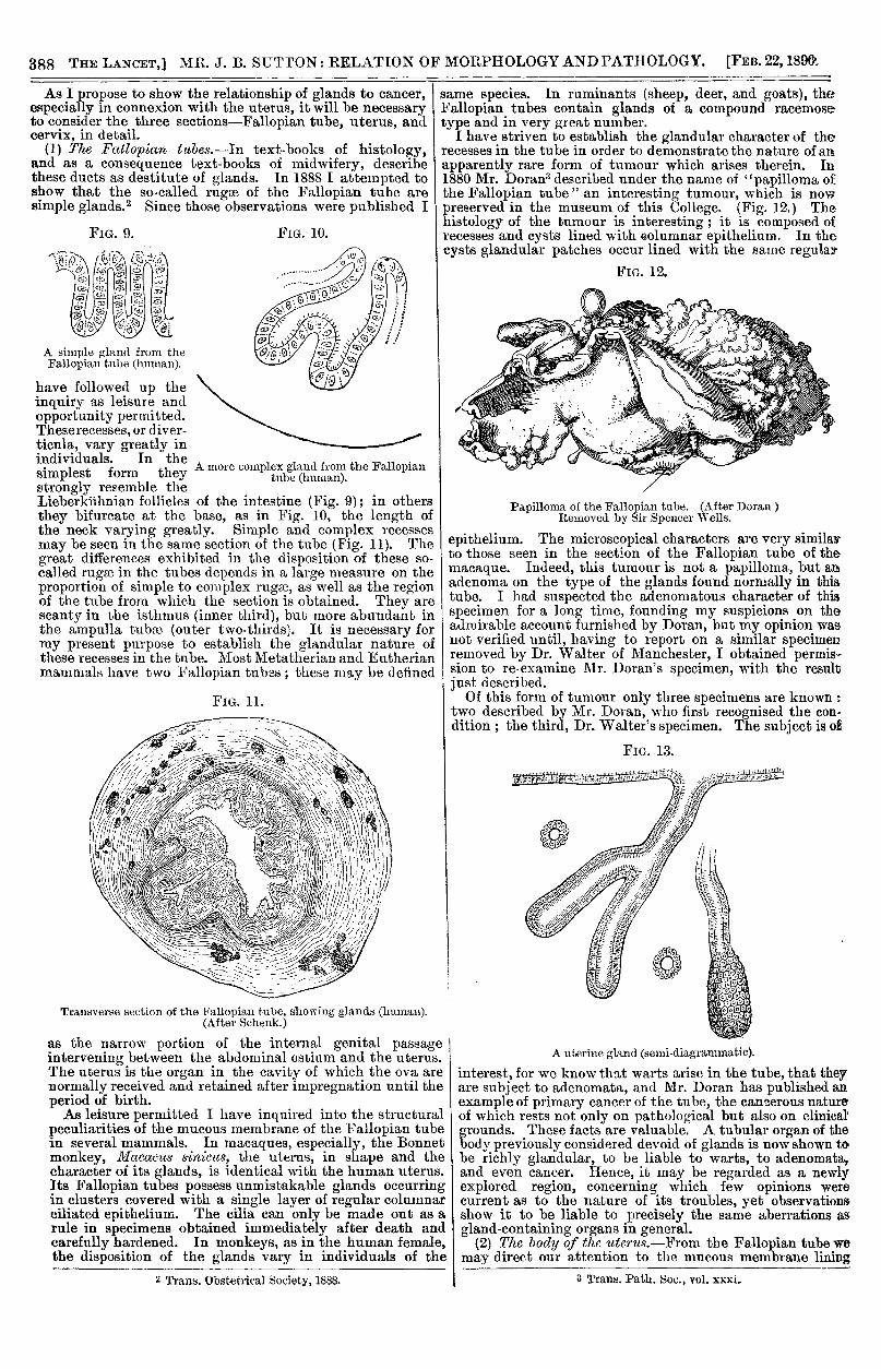
(1) The Fallopian tubes.-In text-books of histology,and as a consequence text-books of midwifery, describethese ducts as destitute of glands. In 1888 I attempted toshow that the so-called rugse of the Fallopian tube aresimple glands. 2 Since those observations were published I
A simple glRn<l from theFallopian tube (hnumn).
have followed up theinquiry as leisure andopportunity permitted.These recesses, or diver-ticula, vary greatly inindividuals. In the
Asimplest form they 1strongly resemble theLieberkuhnian follicles of the intestine (Fig. 9); in othersthey bifurcate at the base, as in Fig. 10, the length ofthe neck varying greatly. Simple and complex recessesmay be seen in the same section of the tube (Fig. 11). Thegreat differences exhibited in the disposition of these so-called rugae in the tubes depends in a large measure on theproportion of simple to complex rugse, as well as the regionof the tube from which the section is obtained. They arescanty in the isthmus (inner third), but more abundant inthe ampulla tube (outer two-thirds). It is necessary formy present purpose to establish the glandular nature ofthese recesses in the tube. Most Metatherian and Eutherianmammals have two Fallopian tubes ; these may be defined
Transverse section of the Fallopian tube, showing glands (human).(After Schenk.)
as the narrow portion of the internal genital passageintervening between the abdominal ostium and the uterus.The uterus is the organ in the cavity of which the ova arenormally received and retained after impregnation until theperiod of birth.As leisure permitted I have inquired into the structural
peculiarities of the mucous membrane of the Fallopian tubein several mammals. In macaques, especially, the Bonnetmonkey, Macacus si2zicit3, the uterus, in shape and thecharacter of its glands, is identical with the human uterus.Its Fallopian tubes possess unmistakable glands occurringin clusters covered with a single layer of regular columnarciliated epithelium. The cilia can only be made out as arule in specimens obtained immediately after death andcarefully hardened. In monkeys, as in the human female,the disposition of the glands vary in individuals of the
2 Trans. Obstetrical Society, 1888.
same species. In ruminants (sheep, deer, and goats), theFallopian tubes contain glands of a compound racemosetype and in very great number.
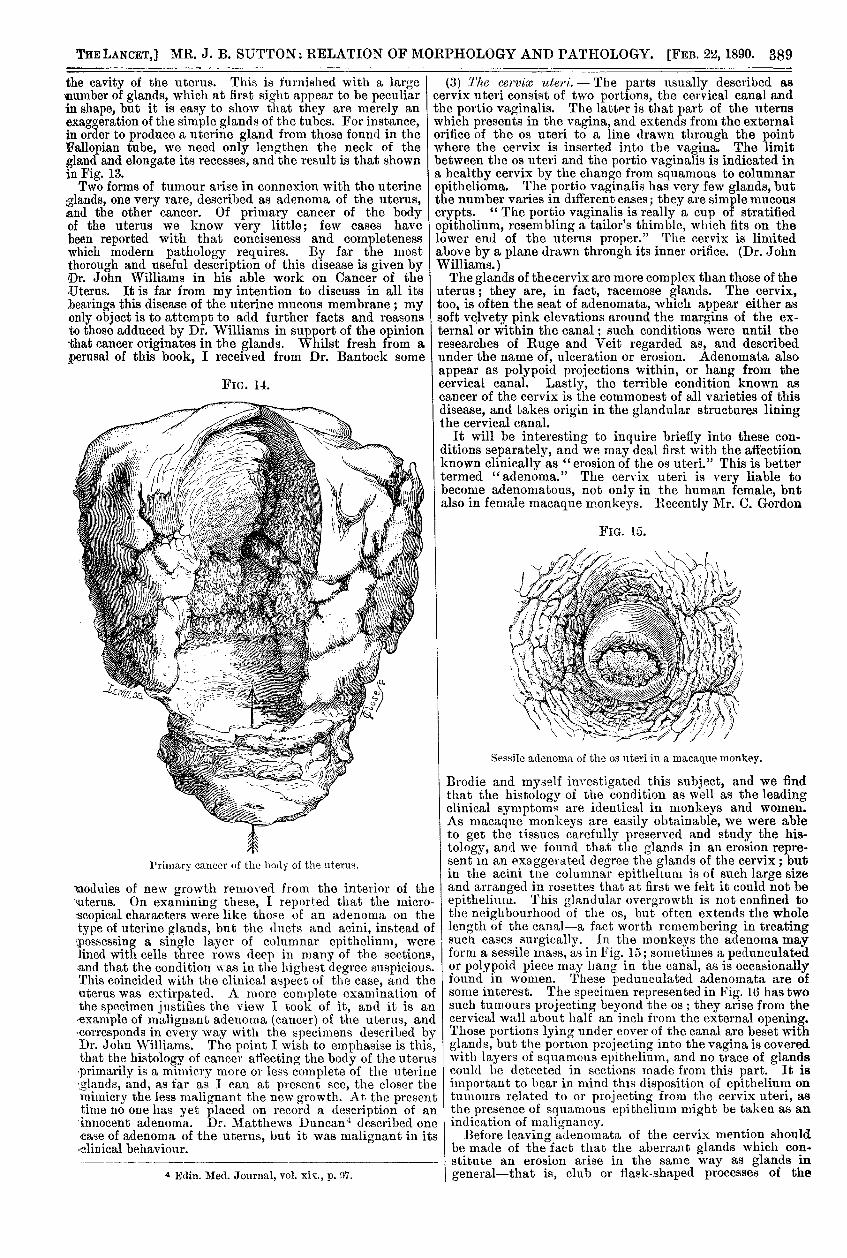
I have striven to establish the glandular character of therecesses in the tube in order to demonstrate the nature of anapparently rare form of tumour which arises therein. In1880 Mr. Doran3 described under the name of "papilloma of £the Fallopian tube " an interesting tumour, which is nowpreserved in the museum of this College. (Fig. 12.) Thehistology of the tumour is interesting ; it is composed ofrecesses and cysts lined with eolumnar epithelium. In thecysts glandular patches occur lined with the same regular
,r
Papilloma of the Fallopian tube. (After Doran )Removed by Sir Spencer Wells.
epithelium. The microscopical characters are very similarto those seen in the section of the Fallopian tube of themacaque. Indeed, this tumour is not a papilloma, but anadenoma on the type of the glands found normally in thistube. I had suspected the adenomatous character of thisspecimen for a long time, founding my suspicions on theadmirable account furnished by Doran, but my opinion wasnot verified until, having to report on a similar specimenremoved by Dr. Walter of Manchester, I obtained permis-sion to re-examine Mr. Doran’s specimen, with the resultjust described.Of this form of tumour only three specimens are known o
two described by Mr. Doran, who first recognised the con.dition ; the third, Dr. Walter’s specimen. The subject is of
A uterine gland (semi-diagrammatic).
interest, for we know that warts arise in the tube, that theyare subject to adenomata, and Mr. Doran has published anexample of primary cancer of the tube, the cancerous natureof which rests not only on pathological but also on clinical’grounds. These facts are valuable. A tubular organ of thebody previously considered devoid of glands is now shown tobe richly glandular, to be liable to warts, to adenomata,and even cancer. Hence, it may be regarded as a newlyexplored region, concerning which few opinions werecurrent as to the nature of its troubles, yet observationsshow it to be liable to precisely the same aberrations asgland-containing organs in general.
(2) The body of the ’M6rMs.—From the Fallopian tube wemay direct our attention to the mucous membrane lining
3 Trans. Path. Soc., vol. xxxi.
389
the cavity of the uterus. This is furnished with a large’number of glands, which at first sight appear to be peculiarin shape, but it is easy to show that they are merely anexaggeration of the simple glands of the tubes. For instance,in order to produce a uterine gland from those found in theFallopian tube, we need only lengthen the neck of thegland and elongate its recesses, and the result is that shownin Fig. 13.Two forms of tumour arise in connexion with the uterine
glands, one very rare, described as adenoma of the uterus,.and the other cancer. Of primary cancer of the bodyof the uterus we know very little; few cases havebeen reported with that conciseness and completenesswhich modern pathology requires. By far the most
thorough and useful description of this disease is given byDr. John Williams in his able work on Cancer of theUterus. It is far from my intention to discuss in all itsbearings this disease of the uterine mucous membrane ; myonly object is to attempt to add further facts and reasonsto those adduced by Dr. Williams in support of the opinionthat cancer originates in the glands. Whilst fresh from aperusal of this book, I received from Dr. Bantock some
Primary cancer of the body of the uterus.
modules of new growth removed from the interior of the’uterus. On examining these, I reported that the micro-scopical characters were like thoe of an adenoma on thetype of uterine glands, but the ducts and acini, instead of’.possessing a single layer of columnar epithelium, werelined with cells three rows deep in many of the sections,and that the condition was in the highest degree suspicious.This coincided with the clinical aspect of the case, and theuterus was extirpated. A more complete examination ofthe specimen justifies the view I took of it, and it is anexample of malignant adenoma (cancer) of the uterus, and’corresponds in every way with the specimens described byDr. John Williams. The point I wish to emphasise is this,that the histology of cancer affecting the body of the uterusprimarily is a mimicry more or less complete of the uterine.glands, and, as far as I can at present see, the closer themimicry the less malignant the new growth. At the presenttime no one has yet placed on record a description of aninnocent adenoma. Dr. Matthews Duncan4 described onecase of adenoma of the uterus, but it was malignant in its- clinical behaviour.
4 Edin. Med. Journal, vol. xix., p. 97.
(3) The cervix ’uteri. - The parts usually described ascervix uteri consist of two portions, the cervical canal andthe portio vaginalis. The latter is that part of the uteruswhich presents in the vagina, and extends from the externalorifice of the os uteri to a line drawn through the pointwhere the cervix is inserted into the vagina. The limitbetween the os uteri and the portio vaginalis is indicated ina healthy cervix by the change from squamous to columnarepithelioma. The portio vaginalis has very few glands, butthe number varies in different cases; they are simple mucouscrypts. " The portio vaginalis is really a cup of stratifiedepithelium, resembling a tailor’s thimble, which fits on thelower end of the uterus proper." The cervix is limitedabove by a plane drawn through its inner orifice. (Dr. JohnWilliams.)The glands of thecervix are more complex than those of the
uterus; they are, in fact, racemose glands. The cervix,too, is often the seat of adenomata, which appear either assoft velvety pink elevations around the margins of the ex-ternal or within the canal; such conditions were until theresearches of Ruge and Veit regarded as, and describedunder the name of, ulceration or erosion. Adenomata alsoappear as polypoid projections within, or hang from thecervical canal. Lastly, the terrible condition known ascancer of the cervix is the commonest of all varieties of thisdisease, and takes origin in the glandular structures liningthe cervical canal.
It will be interesting to inquire briefly into these con-ditions separately, and we may deal first with the affectiionknown clinically as " erosion of the os uteri." This is bettertermed "adenoma." The cervix uteri is very liable tobecome adenomatous, not only in the human female, butalso in female macaque monkeys. Recently Mr. C. Gordon
Sessile adenoma of the os nteri in a macaque monkey.
Brodie and myself investigated this subject, and we findthat the histology of the condition as well as the leadingclinical symptoms are identical in monkeys and women.As macaque monkeys are easily obtainable, we were ableto get the tissues carefully preserved and study the his-tology, and we found that the glands in an erosion repre-sent in an exaggerated degree the glands of the cervix; butin the acini tne columnar epithehum is of such large sizeand arranged in rosettes that at first we felt it could not beepithelium. This glandular overgrowth is not confined tothe neighbourhood of the os, but often extends the wholelength of the canal-a fact worth remembering in treatingsuch cases surgically. In the monkeys the adenoma mayform a sessile mass, as in Fig. 15; sometimes a pedunculatedor polypoid piece may hang in the canal, as is occasionallyfound in women. These pedunculated adenomata are ofsome interest. The specimen represented in Fig. 16 has twosuch tumours projecting beyond the os ; they arise from thecervical wall about half an inch from the external opening.Those portions lying under cover of the canal are beset withglands, but the portion projecting into the vagina is coveredwith layers of squamous epithelium, and no trace of glandscould be detected in sections made from this part. It isimportant to bear in mind this disposition of epithelium ontumours related to or projecting from the cervix uteri, asthe presence of squamous epithelium might be taken as anindication of malignancy.
Before leaving adenomata of the cervix mention shouldbe made of the fact that the aberrant glands which con-stitute an erosion arise in the same way as glands ingeneral-that is, club or flask-shaped processes of the
390
epithelium dip into the underlying tissues, and subsequentlyacquire a lumen. In one of our specimens we were able tosatisfy ourselves that before the ingrowths could acquire alumen many of them were shed, leaving sockets in thetissue which they had invaded. In the next stage we areconfronted with malignant adenomata, and it is a rematk-able fact that to the naked eye typical cases of adenomaand cancer are very unlike, yet microscopically the condi-tion is often so very similar that it would be hazardous fora pathologist to pronounce a positive opinion one way oranother. As far as I have examined the adenomatous andcancerous growths of the ceivix uteri, I can only say thatin the clinically malignant cases the microscope showedthat the glands were less perfectly formed-that is to say,the acini, instead of being lined with a regular peripherallayer of cells, were filled with ill-formed cells, and in manyinstances the gland-germs had not become hollow beforetheir constituent cells underwent degenerate change. To
put the matter briefly, cancer of the uterine cervix (ex-cluding the portio vaginalis) is, in histological details, bestdescribed as consisting of imperfectly developed glands onthe type of those normally found in the lining membrane ofthe canal.To my mind, it is very suggestive that in the three parts
of the uterus-Fallopian tube, uterus proper, and cervicalcanal-adenoma and cancer occur, and in each instancethe histological type mimicked is that of the glands peculiarto each sectior. Careful inquiries conducted in other
Pedunculated adenomata of the cervix uteri (llol11u). !
regions of the body return the same answer; such facts asthese indicate more clearly than anything else that canceror malignant adenoma has a local origin. These facts makeus ask ourselves, If cancer is of local origin and its point ofdeparture is in glands, what is the irritant that initiatessuch disastrous changes? This part of the question I amnot prepared to consider, not even to suggest a theory; myaim has been simply to show that cancer belongs to a groupof structures comprising hair and its modifications, teth,and glands. We pass by insensible gradations from glandsto innocent adenomata, and from these to malignant adeno-mata or cancers. Speaking generally, the environmentwhich promotes gland development is favourable to thedevelopment of cancer-viz., seclusion, an equable tempera-ture, and moisture.
It is also important to bear well in mind that the term"cancer" must be used in a dennite sense, if we are expectedto find a causc, much less a cure, for this disease. Shoulda parasite be demonstrated as the cause of cancer, and thisis extremely probable, it will in no sense invalidate itsright to rank with adenomata., for its structural charactersare so entirely different from other forms of tumours attri-butable to micro-organisms that they ought never to beconfounded. Lastly, I feel sure if the word " cancer" werebanished from our terminology, pathology would be thegainer, and mankind at large far happier for the expurgation.
SOUTH DEVON AND EAST CORNWALL HOSPITAL.-During the year ]880 1267 patients had been treated. Inthe out-patients’ department ]8üG cases of minor accidentsor ailments received surgical treatment, and 653 operationswere performed. The nUl ses’ eaminn3 during the yearamounted to JS897 8s., and, after payment of disbursementsincidental to the staff, a credit halance remained in hand of:E318 16.
Clinical LectureON
SOME OF THE MORE PROMINENT SYMPTOMSIN THE PRESENT EPIDEMIC OF
INFLUENZA.Delivered at St. George’s Hospital,
BY THOMAS WHIPHAM, M.D., F.R.C.P.,PHYSICIAN TO THE HOSPITAL.
GENTLEMEN,—In the following remarks I shall limit,
myself to the clinical aspects of the prevailing disease’known as influenza. Even if time allowed me to enter into>the history of previous epidemics which have occurredsince the days of Hippocrates, I should merely recapitulate-what is already contained in the text-books on medicine.I propose therefore to call your attention to-day to some of-the more prominent characteristics of the complaint as wehave observed them here during the past few weeks. At.the same time, I must ask your indulgence for many short-comings ; for, as you know, we have had a g?eat outbreakamong our nursing staff, and this has rendered observations.of details more or less imperfect. Our chief experience-here has been among the nurses, comparatively few-ordinary patients having been admitted in consequence ofthe compulsory closure of three wards for the reason that.we had not a sufficient number of nurses unaffected by theinfluenza to carry on the ordinary work of the hospital. The,onset of thedisease in the majority of the cases was, as has been,described in the few accurate records of previous epidemics,almost sudden. Aperson in his ordinary health, with but short,or perhaps no warning, may be attacked with a feeling ofgreat chilliness, rigors, vertigo, of extreme depression, or all,of them-a depression so great that some of ihe patients.have described it as closely resembling that of sea sickness,-even to the carelessness as to whether the end should be lifeor death. Coincidently with the rigor, or perhaps in somecases following it, in the course of an hour or so occurredvery severe pain in the frontal and occipital regions of thehead, in the back of the neck, in the eyes, and in the backand legs. The pain in the head and back was of unusual)intensity, and some patients who are liable to headaches ofgreat severity have remarked of the pain in influenza that.in all their past experience they never felt any to equal it."I think it will drive me mad," one or two have said.There has been something peculiar also in the pain in,the eyes; the greater number have described it has."pain at the back of the eyes," and, aceoiding as.
they were more or less fanciful, have likened the feelingto that of something pulling at the back of the eyeball.One woman told us that "it felt as if the back ofher eyes were being rubbed with hot saad."
" Others havedescribed intense pain in the chest, the patients’ descriptions-of which much resembled those of angina pectoris. In oneor two of the patients who have come under observation,there has been, before the more acute symptoms set in, a-.most uncontrollable drow,,iness-accoriipanied, perhaps, with,some frontal headache. This drowsiness was so great in.one case that the lady was, as a matter of fact, onlyawake--during the first twenty-four liours-when she was.aroused to take nourishment ; in another case, the patient,on the afternoon of Jan. 3rd, had slight frontal headache,and was unable to keep awake. She went to bed at,11 o’clock and slept well till 3 A. Ill. , when she awoke with,intense pain all over, especially in the head and back..With the onset of severe pain the drowsiness ceased, and’the patients had no sleep for about thirty-six hours. Thissleeplessness occurred in some instances in the absence ofany very great pain. By the time that the pain anddepression had become iairly established the face hadassumed a characteristic appearance--i.e., it had becomepale, or perhaps somewhat dusky, and the eyes wered heavy, and half closed, as at the commencement ofsmall-pox or typhus fever ; but, in addi&ioR to this, theconjunctive, in these cases of influenza, were red and turgid,and with a slight tendency to laerymation. The aspectwas somewhat that of measles, but without the coryza..