Accumulation of 86Rb, 43K and Heavy Metal Ions in the Anulus of the Moss Mnium cuspidatum (1.) Leysser: A Parallel to Guard Cell Ion Uptake UTA MAIER-MAERCKER Physik Department der Technischen Universitat Munchen, D-8046 Garching bei Munchen, James-Franck-Strafle, F.R.G. Received May 25, 1982 . Accepted July 15, 1982 Summary 86Rb and 43K were mainly found within the fully developed anulus. Parallel feeding experi-. ments using ThS04 and AgN03 led to accumulation at the same sites. The uptake of the cat- ions from the transpiration stream the anulus has in common with the guard cells of vascular plants. There is also a certain structural analogy of both. Among the common features the abundance of starch within the anulus led to the belief that ion accumulation depends on primary processes which may also be involved in the initiation of ion flux changes in stomatal movements. Key words: Mnium cuspidatum, anulus, stomata movement, ion accumulation, microautoradio· graphy. Introduction In the mature moss capsule there is a local change in the character of the cells where the lid meets the urn. Following uptake of water the mature capsule is split by the detaching anulus, a 2-3 layered ring of specialized cells (Maier, 1973 a, b) which are able to change shape and increase volume prior to and during this event (Maier, in preparation). This is an interesting parallel to guard cells of vascular plants; also these were said to increase their volume while changing shape (Raschke, 1979). While the capsule wall may loosen gradually along the line of dehiscence during maturation, there is a critical point where dehiscence occurs instantly (Maier, 1973 b). Because this point varies within one and the same moss population, it is diffi- cult to arrive at a quantitative evaluation of the environmental factors which initiate the process of dehiscence; but little experience is needed to notice that this process depends on high humidity and is favoured by light. Also in this respect, the swelling cells of the anulus and the guard cells of opening stomata are alike and because guard cells of open stomata accumulate potassium and rubidium ions, Mnium cuspidatum was subjected to the micro-autoradiographic method applied to stomata (Maier- Maerckcr, 1981; Maier-Maercker and Jahnke, 1980) in order to demonstrate ion uptake in the anulus cells if taking place. Z. Pjlanzenphysiol. Bd. 108. S. 107-111. 1982.
Transcript
Accumulation of 86Rb, 43K and Heavy Metal Ions in the Anulus of the Moss Mnium cuspidatum (1.) Leysser: A Parallel to Guard Cell Ion Uptake
UTA MAIER-MAERCKER
Physik Department der Technischen Universitat Munchen, D-8046 Garching bei Munchen, James-Franck-Strafle, F.R.G.
Received May 25, 1982 . Accepted July 15, 1982
Summary
86Rb and 43K were mainly found within the fully developed anulus. Parallel feeding experi-. ments using ThS04 and AgN03 led to accumulation at the same sites. The uptake of the cations from the transpiration stream the anulus has in common with the guard cells of vascular plants. There is also a certain structural analogy of both. Among the common features the abundance of starch within the anulus led to the belief that ion accumulation depends on primary processes which may also be involved in the initiation of ion flux changes in stomatal movements.
In the mature moss capsule there is a local change in the character of the cells where the lid meets the urn. Following uptake of water the mature capsule is split by the detaching anulus, a 2-3 layered ring of specialized cells (Maier, 1973 a, b) which are able to change shape and increase volume prior to and during this event (Maier, in preparation). This is an interesting parallel to guard cells of vascular plants; also these were said to increase their volume while changing shape (Raschke, 1979).
While the capsule wall may loosen gradually along the line of dehiscence during maturation, there is a critical point where dehiscence occurs instantly (Maier, 1973 b). Because this point varies within one and the same moss population, it is difficult to arrive at a quantitative evaluation of the environmental factors which initiate the process of dehiscence; but little experience is needed to notice that this process depends on high humidity and is favoured by light. Also in this respect, the swelling cells of the anulus and the guard cells of opening stomata are alike and because guard cells of open stomata accumulate potassium and rubidium ions, Mnium cuspidatum was subjected to the micro-autoradiographic method applied to stomata (MaierMaerckcr, 1981; Maier-Maercker and Jahnke, 1980) in order to demonstrate ion uptake in the anulus cells if taking place.
Z. Pjlanzenphysiol. Bd. 108. S. 107-111. 1982.
108 UTA MAIER-MAERCKER
Material and Methods
Sporophytes of Mnium cuspidatum (L.) Leysser were gathered in the state of approaching dehiscence. The capsules were cut off at the small apophysis, care being taken to avoid perforation of the capsule proper. The detached capsules were then inserted into small holes punched into parafilm with a hot needle. This film had been stretched over a small petri dish containing cotton wool soaked with 20 mM solutions of either 86RbCI or 43KCI, the solution touching only the cut end at the base of the capsule. 86RbCI was purchased from Amersham and Buchler, 43KCI was produced at the cyclotron of the Technische Universitat Munchen, using a 7 MeV triton beam (Wegmann et al., 1981). Label activity was 296· 104 Bq' ml- 1 for 86Rb and 444· 104 Bq' ml- I for 4lK.
Together with another small petri dish containing water for moistening the surrounding air the capsules were kept in a chamber under dim light. Feeding occurred for 48 h with 86RbCI and for 22 h with 43KCI; 22 h is the half time period of the short lived 43K. After removing the contaminated capsule base, strips were cut longitudinally and attached with the internal surface to a microscope slide. The specimens were then covered with dry stripping film (Kodak, AR 10) spread on a second slide. By wrapping both slides with scotch tape, film and specimen were firmly held in place for the 5 days of exposure at -16°C. Chemography was excluded by control experiments.
In parallel experiments capsules were fed with either TlzS04 (1 %) or AgN03 (100 mM). The cations were precipitated with a methanolic solution of NaCI (Maercker, 1965; MaierMaercker, 1979 b).
For electron microscopy the tissue was fixed in 3 % glutaraldehyde followed by a treatment with 2 % OS04 and embedded in Durcupan ACM. The sections were afterwards stained with lead citrate and examined in a Philips 200. Phosphorylase activity was localised following the technique of Pearse (1972).
Results
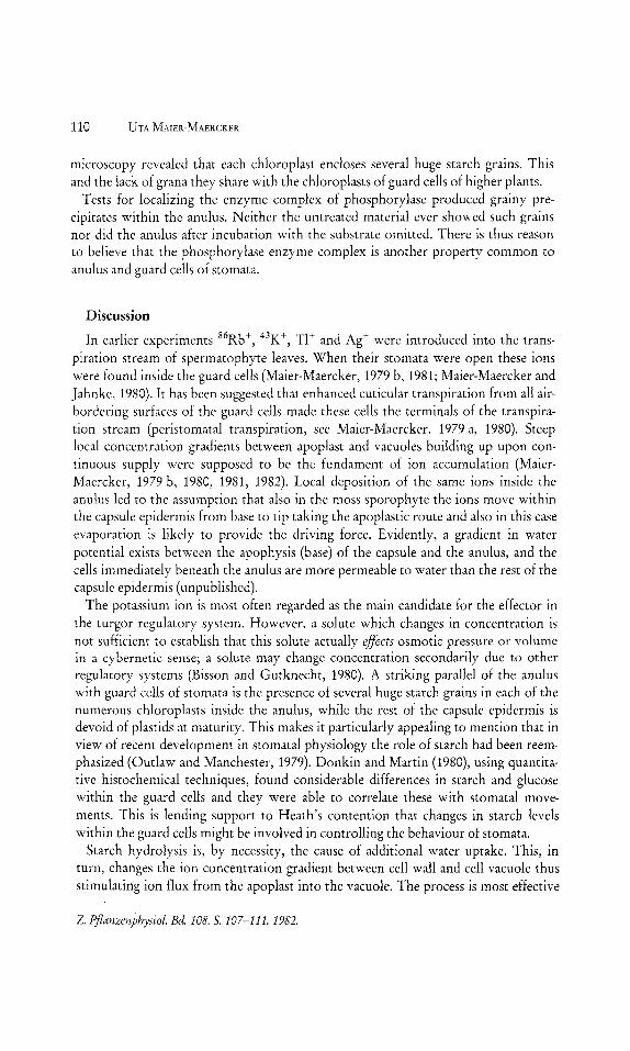
The outcome of the experiments strongly depended on the state of the material. If the conditions led to instant dehiscence there were no detectable amounts of ions inside the cells of the detached anulus. These had to be built up during a period of feeding preceeding dehiscence. In the course of several hours 86Rb and 43K were accumulated by cells close to the line of dehiscence. The radiation pattern obtained was somewhat serrate in appearance (Fig. 2) and there is a striking resemblance between it and the anulus in Fig. l.
In the following example (Fig. 3) the anulus can actually be seen in detail. Using 43K, resolution allowed the identification of individual cells and of the «band» holding them all together (Fig. 3). In lid and urn accumulation of 86Rb was moderate, slightly higher only in the cells adjacent to the anulus.
After feeding mature capsules with TlzS04 or AgN03 the cations of either sub
stance were also found inside the anulus and accumulation was exceptionally heavy (Figs. 4, 5). As with 86Rb, accumulation occurred also in the walls of the adjacent cells of the operculum and urn, while everywhere else in the capsule wall epidermis accu
mulation had been moderate (Fig. 6). Within the capsule epidermis only the highly specialized cells of the anulus have
dense cytoplasm and only they are packed full with chloroplasts (Fig. 7). Electron
Z. Pjlanzenphysiol. Bd. 108. 5.107-111.1982.
3
rr rt~ ~ . ~ . 1" ! . . , .
,. . , ~ ---'. -.. ~.
2
4
Fig. 1: Mnium cuspidatum: Early stage of dehiscence. The capsule wall is seen to burst under the pressure of the swelling anulus.
Fig. 2: Autoradiograph showing 86Rb accumulation in the anulus.
Fig. 3: 4JK accumulation in an isolated anulus. Resolution allows to distinguish individual cells and the "band».
Fig. 4: TI+ inside the cells of the anulus after feeding the detached capsule with ThS04.
Fig. 5: Deposits of Ag + in the anulus after feeding the capsule with AgN03•
6,--_~' __ 7
Fig. 6: Deposition of TI+ in the zone of weakness.
Fig. 7: Epidermis of a capsule ready for dehiscence. The cells of the anulus are crammed with chloroplasts in contrast to all other cells of the lid (top) and urn (bottom).
Z. P/lanzenphysiol. Ed. 108. 5.107-111. 1982.
110 UTA MAIER-MAERCKER
microscopy revealed that each chloroplast encloses several huge starch grains. This and the lack of grana they share with the chloroplasts of guard cells of higher plants.
Tests for localizing the enzyme complex of phosphorylase produced grainy precipitates within the anulus. Neither the untreated material ever showed such grains nor did the anulus after incubation with the substrate omitted. There is thus reason to believe that the phosphorylase enzyme complex is another property common to anulus and guard cells of stomata.
Discussion
In earlier experiments 86Rb +, 43K +, TI + and Ag + were introduced into the transpiration stream of spermatophyte leaves. When their stomata were open these ions were found inside the guard cells (Maier-Maercker, 1979 b, 1981; Maier-Maercker and Jahnke, 1980). It has been suggested that enhanced cuticular transpiration from all airbordering surfaces of the guard cells made these cells the terminals of the transpiration stream (peristomatal transpiration, see Maier-Maercker, 1979 a, 1980). Steep local concentration gradients between apoplast and vacuoles building up upon continuous supply were supposed to be the fundament of ion accumulation (MaierMaercker, 1979 b, 1980, 1981, 1982). Local deposition of the same ions inside the anulus led to the assumption that also in the moss sporophyte the ions move within the capsule epidermis from base to tip taking the apoplastic route and also in this case evaporation is likely to provide the driving force. Evidently, a gradient in water potential exists between the apophysis (base) of the capsule and the anulus, and the cells immediately beneath the anulus are more permeable to water than the rest of the capsule epidermis (unpublished).
The potassium ion is most often regarded as the main candidate for the effector in the turgor regulatory system. However, a solute which changes in concentration is not sufficient to establish that this solute actually effects osmotic pressure or volume in a cybernetic sense; a solute may change concentration secondarily due to other regulatory systems (Bisson and Gutknecht, 1980). A striking parallel of the anulus with guard cells of stomata is the presence of several huge starch grains in each of the numerous chloroplasts inside the anulus, while the rest of the capsule epidermis is devoid of plastids at maturity. This makes it particularly appealing to mention that in view of recent development in stomatal physiology the role of starch had been reemphasized (Outlaw and Manchester, 1979). Donkin and Martin (1980), using quantitative histochemical techniques, found considerable differences in starch and glucose within the guard cells and they were able to correlate these with stomatal movements. This is lending support to Heath's contention that changes in starch levels within the guard cells might be involved in controlling the behaviour of stomata.
Starch hydrolysis is, by necessity, the cause of additional water uptake. This, in turn, changes the ion concentration gradient between cell wall and cell vacuole thus stimulating ion flux from the apoplast into the vacuole. The process is most effective
Z. Pjlanzenphysiol. Bd. 108. S. 107-111. 1982.
Accumulation of ions in the moss anulus 111
upon continuous supply. By contributing to the osmotic gradient the ions assist in further inflow (Raven, 1977). In the anulus the presence of starch and the localization of phosphorylase activity suggest the application of the same principle of rapid and effective turgor increase. Besides, toxic metal ions (thallium and silver) accumulate in the guard cells and in the cells of the anulus. There is no reason to believe that K+ is taken up by an active K+ pump mechanism. The same conclusion had been reached concerning guard cells of stomata (see Maier-Maercker, 1979 b, 1980, 1981).
Acknowledgements
The author is greatly indepted to Prof. Dr. H. Morinaga and to H. Muthig. This work has been supported by the Deutsche Forschungsgemeinschaft.
References
BISSON, M. A. and J. GUTKNECHT: Osmotic regulation in Algae. In: Plant membrane transport: Current conceptual issues, R. M. SPANSWICK, W. J. LUCAS, and J. DAINTY (Eds.). Elsevier! North Holland Biomedical Press, 131-142 (1980).
DONKIN, M. E. and E. S. MARTIN: Changes in starch and glucose levels in the epidermis of Com· melina communis in relation to stomatal movements. Plant, Cell and Environment 3, 403-414 (1980).
MAERCKER, U.: Zur Kenntnis der Transpiration der Schlieihellen. Protoplasma 60, 61-78 (1965).
MAIER, K.: Dehiscence of the moss capsule. II. The anulus: Analysis of its functional apparatus. Osterr. Bot. Z. 122, 75-98 (1973 a).
- III. Anulus function and the lid stability: A study with the light- and scanning electron microscope. Osterr. Bot. Z. 122, 99-114 (1973 b).
MAIER-MAERCKER, U.: "Peri stomatal transpiration» and stomatal movement: A controversial view. I. Additional proof of peristomatal transpiration by hygrophotography and a comprehensive discussion in the light of recent results. Z. Pflanzenphysio!. 91, 25-43 (1979 a).
- IV. Ion accumulation by peristomatal transpiration. Z. Pflanzenphysio!. 91, 239-254 (1979 b).
- V. Rubidium-86 in the epidermal transpiration stream. Z. Pflanzenphysio!. 101, 447-460 (1981).
- VI. Lanthanum de~osits in the epidermal apoplast. Z. Pflanzenphysio!. 100, 121-130 (1980). Accumulation of 6Rb and 43K ions in the cells surrounding the air pores of Conocephalum conicum. Z. Pflanzenphysio!. 105, 97-102 (1982).
MAIER-MAERCKER, U. and A. JAHNKE: Microautoradiography with 43K: A method for the reliable tracing of ion transport in stomata. Z. Pflanzenphysio!. 100, 35-42 (1980).
OUTLAW, W. H. Jr. and J. MANCHESTER: Guard cell starch concentration quantitatively related to stomatal aperture. Plant Physio!' 64, 79-82 (1979).
PEARSE, A. G. E.: Histochemistry: theoretical and applied. Churchill, Edingburg, 1972. RAVEN, J. A.: Regulation of solute transport at the ceilleve!. Symp. Soc. Exp. Bio!. 31, 73-99
(1977). RASCHKE, K.: Movements of stomata. In: W. HAUPT and M. E. FEINLEIB (Eds.): Physiology of
Movements. Encyclopedia of Plant Physio!. New Series Vo!' 7, 383-441 (1979). WEGMANN, H., H. HUENGES, H. MUTHIG, and H. MORINAGA: Acceleration of tritons with a
compact cyclotron. Nuclear Instruments and Methods 179, 217-222 (1981).