Page 1
i
ACCURACY OF SCREENING FOR DIABETIC RETINOPATHY AND MACULA
EDEMA AT KENYATTA NATIONAL HOSPITAL
DR. LAZARUS WAMBUA MUTINDA
H58/81317/15
A Dissertation Submitted to the University of Nairobi
Ophthalmology Department in Partial Fulfillment for the Award of Degree of Master of
Medicine in Ophthalmology - M.Med(Ophth)
©2018
Page 2
ii
DECLARATION
I declare that this research is my original work and has never been published or presented for a
degree in any other University.
Signed: ___________________________ Date: _____________________
DR. LAZARUS WAMBUA MUTINDA
Page 3
iii
APPROVAL BY SUPERVISORS
1. DR. MUCHAI GACHAGO
MB. ChB, M. Med (Ophthal) (Nrb), ICO, FEACO, FVRS (LMU,LVPEI)
Lecturer
Department of Ophthalmology, University of Nairobi
Signed__________________________ Date________________________
2. PROF. JEFITHA KARIMURIO
MB. ChB, M. Med (Nrb), MSc-CEH (London),FEACO,PhD(Melbourne)
Associate Professor
Department of Ophthalmology, University of Nairobi
Signed______________________ Date_________________
3. DR. STANLEY NGARE
MB. ChB, M. Med (Nrb)
Consultant Physician and Diabetologist
Department of Medicine, Kenyatta National Hospital
Signed_____________________ Date_________________
Page 4
iv
ACKNOWLEDGEMENTS
I would like to thank the following: The almighty God for seeing me through my residency and
the period I carried out this thesis; My supervisors, Dr.Gachago , Professor Karimurio and Dr.
Ngare for their critical review, input and support during the study; Dr. Kibata and Upperhill Eye
and Laser Centre (UHEAL) staff for their support during data collection; KNH diabetic and eye
clinic staff for their support during data collection; The staff and my fellow students in the
department of ophthalmology and my family for the moral support and prayers accorded to me.
Page 5
v
TABLE OF CONTENTS
DECLARATION .......................................................................................................................................................... II
APPROVAL BY SUPERVISORS .................................................................................................................................. III
ACKNOWLEDGEMENTS ........................................................................................................................................... IV
LIST OF FIGURES ..................................................................................................................................................... VII
LIST OF TABLES ...................................................................................................................................................... VIII
LIST OF ABBREVIATIONS ....................................................................................................................................... VIII
ABSTRACT ............................................................................................................................................................... XI
1. INTRODUCTION ............................................................................................................................................ I
2. LITERATURE REVIEW ............................................................................................................................................ 2
2.1 PATHOGENESIS ................................................................................................................... 2
2.1.1 Biochemical processes .............................................................................................. 3
2.1.2 Hemodynamic alterations ........................................................................................ 3
2.1.3 Paracrine factors....................................................................................................... 4
2.2 MACULA EDEMA................................................................................................................. 4
2.3 SCREENING FOR DIABETIC RETINOPATHY ........................................................................... 4
2.3.1 Rationale for screening ............................................................................................ 5
2.3.2 Methods of screening ................................................................................................ 5
2.3.3. Screening modalities ................................................................................................ 5
2.3.4 Screening intervals for diabetic retinopathy .......................................................... 9
2.4 NATURAL HISTORY ............................................................................................................. 9
2.5 CLASSIFICATION OF DIABETIC RETINOPATHY AND MACULA EDEMA ................................. 10
2.6 PREVALENCE OF DIABETIC RETINOPATHY AND MACULA EDEMA ...................................... 10
3. JUSTIFICATION ................................................................................................................................................... 12
4. OBJECTIVE .......................................................................................................................................................... 13
4.1 BROAD OBJECTIVE ............................................................................................................ 13
4.2 SPECIFIC OBJECTIVES ........................................................................................................ 13
5. METHODS ........................................................................................................................................................... 14
Page 6
vi
5.1 STUDY DESIGN ................................................................................................................. 14
5.2 STUDY PERIOD ................................................................................................................. 14
5.3 STUDY AREA ..................................................................................................................... 14
5.4STUDY POPULATION .......................................................................................................... 14
5.5 SAMPLE SIZE ESTIMATION................................................................................................. 14
5.6 SAMPLE SELECTION METHODS.......................................................................................... 15
5.6.1 Inclusion Criteria .................................................................................................... 15
5.6.2 Exclusion criteria .................................................................................................... 15
5.7 DATA COLLECTION, MANAGEMENT AND ANALYSIS ........................................................ 15
5.7.1 Data Collection Procedure ..................................................................................... 15
5.7.2 Data Instruments .................................................................................................... 16
5.8 DATA MANAGEMENT AND ANALYSIS ............................................................................... 16
5.9 ETHICAL CONSIDERATIONS .............................................................................................. 18
5.9.1 Confidentiality ........................................................................................................ 18
5.9.2 Potential risks and benefits .................................................................................... 18
5.9.3 Approval by Ethics Committees ............................................................................ 18
6.0 RESULTS. .......................................................................................................................................................... 19
6.1 SOCIO-DEMOGRAPHIC CHARACTERISTICS ......................................................................... 20
6.1.1 Age............................................................................................................................ 20
6.1.2 Sex ............................................................................................................................ 21
6.1.3 Best corrected visual acuity ................................................................................... 21
6.1.4 Duration of Diabetes ............................................................................................... 24
6.2. Fundus Examination Findings using the fundus camera ..................................... 24
6.2.1 DR Grading using the fundus camera .................................................................. 24
6.2.2Maculopathy............................................................................................................. 25
6.3 Fundus Examination using indirect ophthalmoscope ............................................ 26
6.3.1 ETDR grading of diabetic retinopathy using indirect ophthalmoscope. ........... 26
6.3.2 Grading of clinically significant macula edema by indirect ophthalmoscope .. 27
6.4 SENSITIVITY, SPECIFICITY AND PREDICTIVE VALUES ........................................................ 28
6.4.1 Accuracy of Diagnosis for DR (All grades) .......................................................... 28
Page 7
vii
6.4.2 Accuracy of Diagnosis for No DR ......................................................................... 28
6.4.3 Accuracy of Diagnosis for mild DR....................................................................... 29
6.4.4 Accuracy of Diagnosis for Moderate and Severe NPDR..................................... 30
6.4.5 Accuracy of Diagnosis for PDR ............................................................................. 30
6.5 OPTICAL COHERENCE TOMOGRAPHY (OCT) FINDINGS .................................................... 31
6.5.1 Macula edema as per OCT .................................................................................... 31
6.5.2Accuracy of diagnosis for macula edema using the fundus photography .......... 32
7.0 DISCUSSION ..................................................................................................................................................... 33
8.0 STUDY LIMITATIONS ........................................................................................................................................ 36
9.0 CONCLUSION .................................................................................................................................................... 37
10.0 STUDY RECOMMENDATIONS ......................................................................................................................... 38
10.0REFERENCES .................................................................................................................................................... 39
11.0 APPENDICES ................................................................................................................................................... 45
APPENDIX I: THE BASIC PRINCIPLES FOR DISEASE SCREENING AS PER THE WHO. ............... 45
APPENDIX II: ETDRS GRADING OF DIABETIC RETINOPATHY .............................................. 46
APPENDIX III: CLINICALLY SIGNIFICANT MACULAR EDEMA, AS DEFINED BY ETDRS. ........ 47
APPENDIX IV: KNH-UON ERC APPROVAL ....................................................................... 48
APPENDIX V: DEPARTMENT OF MEDICINE KNH APPROVAL FORM ..................... 50
APPENDIX VI: CONSENT .................................................................................................. 51
APPENDIX VII: INFORMED CONSENT ........................................................................... 52
APPENDIX VIII: FOMU YA RIDHAA ..................................................................................... 55
APPENDIX IX: QUESTIONNAIRE .......................................................................................... 58
APPENDIX X: ENGLISH NATIONAL SCREENING PROGRAM FOR GRADING DIABETIC
RETINOPATHY ........................................................................................................................ 60
APPENDIX XI: WORK PLAN ............................................................................................. 60
APPENDIX XII: BUDGET ................................................................................................... 61
LIST OF FIGURES
Figure 1: Flow diagram ................................................................................................................. 19
Page 8
viii
Figure 2: Distribution of the studied patients by age in years (n=99) .......................................... 20
Figure 3: Distribution by sex (n=99) ............................................................................................ 21
Figure 4: Grades of diabetic retinopathy on fundus photography (n=198) ................................. 25
Figure 5: Eyes with maculopathy on fundus photography (n=198). ............................................ 25
Figure 6: Eyes which had retinal photocoagulation on fundus photography(n=198) ................... 26
Figure 7: Percentage of eyes which had DR on fundoscopy (n=198) .......................................... 27
Figure 8: Percentage of eyes with clinically significant macula edema(n=198) .......................... 28
Figure 9: Percentage of eyes with Macula edema after OCT scanning(n=118) .......................... 32
LIST OF TABLES
Table 1: Demographics ................................................................................................................. 17
Table 2: Sensitivity, specificity and predictive values .................................................................. 17
Table 3: Grading of visual acuity per eye according to WHO categorization of blindness and
visual impairment.......................................................................................................................... 22
Table 4: Grading of visual acuity in the best eye as per WHO categorization of blindness and
visual impairment.......................................................................................................................... 23
Table 5: Duration of diabetes in years .......................................................................................... 24
Table 6: Accuracy of Diagnosis for All grades of DR (n=94) ...................................................... 28
Table 7: Shows accuracy of Diagnosis for No DR (n=97) ........................................................... 29
Table 8: Shows accuracy for diagnosis for mild DR .................................................................... 29
Table 9: Shows accuracy of Diagnosis for moderate and Severe NPDR...................................... 30
Table10: Table Shows accuracy of Diagnosis for PDR................................................................ 30
Table11: Accuracy of diagnosis for macula edema using the fundus photography ..................... 33
LIST OF ABBREVIATIONS
Page 9
ix
DM Diabetes Mellitus
DR Diabetic Retinopathy
VTDR Visually Threatening Diabetic Retinopathy
PDR Proliferative Diabetic Retinopathy.
DME Diabetic Macula Edema
CSME Clinically Significant Macula Edema
ME Macula Edema
CI Confidence interval
VEGF Vascular Endothelial Growth Factor
ETDRS Early Treatment Diabetic Retinopathy Study
WHO World Health Organization
MOPC Medical outpatient Clinic
KNH Kenyatta National Hospital
UK United Kingdom
NVD Neovascularization at the disc
NVE Neovascularization elsewhere
DRS Diabetic Retinopathy Study
SLB Slit Lamp Examination
Page 10
x
ACE Angiotensin converting enzymes
bFGF basic Fibroblast Growth Factor
IGF-1 Insulin Growth Factor 1
PDGF Platelet Derived Growth Factor
EGF Epidermal Growth Factor
TGF-β2 Transforming Growth Factor beta 2
MESA Multi Ethnic Study of Atherosclerosis
DRSP Diabetic Retinopathy Screening Programs
OCT Optical Coherence Tomography
Page 11
xi
ABSTRACT
Background: Low income countries in Asia and Africa have both the highest prevalence of
diabetes mellitus (DM) and expected rise in disease burden. Many patients with diabetes are
unaware of their diagnosis and may not receive treatment in a timely fashion. These diabetic
retinopathy screening programs (DRSP) where regular eye examinations are done,can minimize
the risk of visual loss.
In Kenya, most of the patients with diabetes are screened for diabetic retinopathy (DR) with
indirect fundoscopy and only few facilities are using the fundus camera. While dilated
fundoscopy has been found to be an effective method of screening, it requires to be performed by
an experienced person. Fundus camera has been preferred because it does not need a highly
experienced person and more people can also be screened per day compared to dilated
fundoscopy. Current fundus cameras used for screening are non-mydriatic. However its accuracy
has not been validated in our setup.
Objective: The general objective was to assess the accuracy of screening for diabetic retinopathy
in diabetic patients attending the medical outpatient clinic at Kenyatta National Hospital (KNH).
The specific objectives were: to compare grading of DR using fundus photographs by the
technician (screener) and Early Treatment Diabetic Retinopathy Study (ETDRS) clinical grading
by the ophthalmologist and to compare diagnosis of macula edema using fundus photographs
and optical coherence tomography (OCT).
Materials and methods: This was a cross sectional hospital based study. Patients were recruited
in the MOPC.56 patients attending medical outpatient clinic (MOPC) were randomly selected
then screened. First consent was taken then a questionnaire administered then fundus
photographs were captured in all the patients. After retinal photography, patients were dilated
and fundoscopy was done on all patients regardless of their DR status. A questionnaire was
administered to all patients with DR who gave consent. The eyes of all patients screened for DR
were scanned using OCT. The primary outcome was presence or absence of DR or macula
edema.
Page 12
xii
Results: This study revealed that the mean duration of diabetes was 10.8 years. The sensitivity
of the fundus camera in the diagnosis of no DR is 94.8% and a specificity of 86.2%. The positive
and negative predictive value for identification of no DR was 87.6% and 94.1% respectively.
Sensitivity for identifying mild DR was 60.6%. The Sensitivity for identifying moderate and
severe DR was 74.0% and a specificity of 99.3%. Diagnosis of proliferative diabetic retinopathy
(PDR) had a sensitivity and specificity of 90.9% and 99.4% respectively. Sensitivity of all grades
of DR was 86.2% and specificity of 94.9%.Sensitivity of diagnosis for macula edema was 57.1%
and a specificity of 89.8%.
Conclusion: The fundus camera is accurate and can therefore be effectively used to screen for
diabetic retinopathy but not for diabetic macula edema. Its accuracy is higher for more advanced
stages of DR.
Recommendation: Since the fundus camera is an effective tool for screening of diabetic
retinopathy its use be increased nationally due its high accuracy and specificity. KNH needs an
OCT as it has been seen that fundus photography tends to over-estimate diabetic macula edema
(DME).
Page 13
i
1. INTRODUCTION
Diabetic retinopathy (DR) is defined as damage to the micro-vascular system of the retina
accompanied by structural change to the retina due to prolonged hyperglycaemia. It occurs in
both type 1 and type 2 diabetes mellitus (DM) 26
.
Diabetic patients are 25 times more likely to become blind than the general population. If DR is
detected and treated early enough, the risk of vision loss and blindness, as well as the complexity
and cost of treatment, can be reduced significantly, utilizing well-established and widely
available treatments35
.
Effective screening and treatment programs can greatly reduce the burden of blindness. The
current standard or screening for DR is either a dilated eye examination performed by an
ophthalmologist or dilated ETDRS (Early Treatment Diabetic Retinopathy Study) 7-standard
field stereoscopic 30° fundus photography35
.
Fundus camera was introduced in KNH three years ago. Before the introduction of the camera
diabetic retinopathy screening was done using dilated fundoscopy. Our study aimed to compare
the sensitivity and specificity of fundus camera in screening for diabetic retinopathy and
sensitivity of OCT machine in screening for macula edema.
Page 14
2
2. LITERATURE REVIEW
2.1 Pathogenesis
The exact mechanisms by which elevated glucose initiates the vascular disruption in retinopathy
remain unclear. The vascular disruptions of DR and diabetic macula edema (DME) are
characterized by abnormal vascular flow, changes in permeability, and non-perfusion of
capillaries.
A hallmark of early DR is the change in the structure and cellular composition of the
microvasculature. Endothelial cells are responsible for maintaining the blood-retinal barrier, and
damage to them results in increased vascular permeability. In early stages of DME, breakdown
of the inner blood-retinal barrier may occur, resulting in accumulation of extracellular fluid in
the macula 2.
Pericytes are essential cellular components in the regulation of retinal capillary perfusion, and
damage to these cells in diabetes leads to altered retinal hemodynamics, including abnormal
autoregulation of retinal blood flow. Because pericytes help regulate retinal capillary perfusion,
damage to these cells immediately disrupts retinal haemodynamics8. Loss of retinal pericytes
represents another early feature of DR and correlates with micro-aneurysm formation. Another
common feature of DR is the thickening of the capillary basement membrane and increased
deposition of extracellular matrix components26
. This feature may contribute to the development
of abnormal retinal hemodynamics, including abnormal autoregulation of retinal blood flow.
There is evidence that retinal leukostasis may also play an important role in the pathogenesis of
DR. Leukocytes possess large cell volume, high cytoplasm rigidity, a natural tendency to adhere
to the vascular endothelium, and a capacity to generate toxic superoxide radicals and proteolytic
enzymes. In diabetes, there is increased retinal leukostasis, which affects retinal endothelial
function, retinal perfusion, angiogenesis, and vascular permeability. In particular, leukocytes in
diabetes are less deformable, higher proportions are activated, and they may be involved in
capillary nonperfusion, endothelial cell damage, and vascular leakage in the retinal
microcirculation20
.
Page 15
3
As a result of occluded capillaries, retinal ischemia stimulates a pathologic neovascularization
mediated by angiogenic factors, such as vascular endothelial growth factor (VEGF), basic
fibroblast growth factor (bFGF) , insulin-like growth factor-1 (IGF-1) , angiopoietin-1 and -2 ,
stromal-derived factor-1, epidermal growth factor (EGF) , transforming growth factor-beta 2
(TGF-β2) , platelet-derived growth factors (PDGFs) , and erythropoietin 19
. VEGF promotes
angiogenesis; causes breakdown of the blood-retinal barrier, stimulation of endothelial cell
growth, and neovascularisation; and increases vascular permeability in the ischemic retina5.
2.1.1 Biochemical processes
In non-enzymatic glycation hyperglycemia leads to formation and accumulation of advanced
glycation end products that accumulate in the retina affecting the functioning of retinal vascular
endothelial cells. In the polyol pathway mechanism, the enzyme aldose reductase causes
increased conversion of glucose to sorbitol. In addition, it also reduces galactose conversion to
galactitol. Accumulation of sorbitol and galactitol results in inhibition of biosynthetic and
degenerative enzymes resulting in basement membrane thickening. In early stages of the disease,
protein kinase C induces various cytokines and angiogenic factors including vascular endothelial
growth factor (VEGF). In the later stages, the VEGF is dependent on protein kinase C for both
angiogenic and permeability effects. Increase in oxygen-free radicals may impair endothelium
dependent vasodilatation and increase the apoptosis of retinal capillary cells 27
.
2.1.2 Hemodynamic alterations
The variety of hematologic abnormalities seen in diabetes, such as increased erythrocyte
aggregation, decreased red blood cell deformability, increased platelet aggregation and adhesion,
predispose the patient to sluggish circulation, endothelial damage, and focal capillary occlusion.
This leads to retinal ischemia, which, in turn, contributes to the development of diabetic
retinopathy27
.
Page 16
4
2.1.3 Paracrine factors
A variety of growth factors have been implicated in the pathogenesis of diabetic retinopathy.
Endoplasmic reticulum stress response to nutritional deprivation in diabetes regulates the
expression of VEGF and platelet derived growth factor at the level of mRNA translation27
.
2.2 Macula edema
Diabetic macular edema (DME) is the leading cause of vision loss in patients living with
diabetes. The common pathway that leads to macular edema is breakdown of the blood–retinal
barrier (BRB). The BRB consists of the inner and the outer BRB. The inner BRB is formed by
tight junctions between retinal capillary endothelial cells, the surrounding basal lamina,
perycytes, astrocytes and microglia. The outer BRB is formed by the tight junctions between
retinal pigment epithelium (RPE) cells. Impaired integrity of the BRB leads to leakage of plasma
solutes into the interstitial spaces, causing edema through increased osmotic pressure. Fluid
subsequently accumulates in different spaces within and underneath the retina4.
VEGF is a glycoprotein secreted by retinal pigment epithelium cells, Muller cells, ganglion
cells and capillary endothelial cells. VEGF increases vascular permeability via multiple
mechanisms, including: Leukocyte mediated extracellular injury, formation of fenestration and
the dissolution of tight junctions resulting in transcellular bulk flow27
.
2.3 Screening for diabetic retinopathy
Screening is defined as, “The process of examining a group of people for the presence of a
disease”with its prerequisites being:
The disease must appear in a defined population
The population must be identifiable
The disease must present a health problem
There must be effective treatment for the disease
Screening must be cost effective and improve quality of life.
Page 17
5
Screening for DR helps to detect early sight-threatening retinopathy, allowing treatment it in a
timely fashion and in this way help to avoid expensive, advanced treatment or even prevent the
development of blindness. Principles for screening in medicine (Appendix I) were established
by Wilson and Jungner in 1968 and accepted by the World Health Organization (WHO)39
the
same year.
2.3.1 Rationale for screening
Screening for DR is important because patients are diagnosed early and those not affected are
advised on how to prevent DR. As a result, patients with DR are treated promptly and vision loss
from DR is avoided.
2.3.2 Methods of screening
There are several methods currently available for visualizing the retina, including direct and
indirect fundoscopy, fundus photography and slit lamp biomicroscopy. Fluorescein angiography
is held as the gold standard for detecting DR however, there are side effects to fluorescein
making it less desirable for screening.9
For the initial screening examination, it is preferred that evaluation be done by an
ophthalmologist or optometrist who is experienced in diagnosing and treating DR. Fundoscopy is
a reasonable screening method when performed by well-trained personnel on a dilated pupil. The
accuracy of fundoscopy is substantially lower when performed by primary care physicians 13
.
In one study of 1949 patients participating in the Wisconsin Epidemiology Study of Diabetic
Retinopathy (WESDR), there was 86% agreement between ophthalmoscopy and the results of
fundus photography, with no significant inter-observer differences22
.
2.3.3. Screening modalities
For any DR screening programme to function effectively it must fulfill certain basic criteria.
Firstly, the screening test must have sufficiently high sensitivity (true positive rate) to ensure that
substantial numbers of patients with sight-threatening retinopathy are not missed. Secondly, it
must have sufficiently high specificity (true negative rate) to ensure that ophthalmic departments
Page 18
6
are not overwhelmed with unnecessary referrals30
. The British Diabetic Association proposed
that any screening programme for diabetic retinopathy should have at least 80% sensitivity and
specificity, and it is against these figures that any screening modality for diabetic retinopathy is
judged33
.
2.3.3.1 Direct ophthalmoscope
Direct fundoscopy alone has no role in a screening programme since the method consistently
fails to meet the 80% sensitivity and specificity targets. Direct fundoscopy with mydriasis was
shown to have a sensitivity of 65% when used by ophthalmologists, 33-66% with general
practitioners and 48-83% with optometrists.11, 25, and 41
2.3.3.2 Retinal photography
Retinal photography, without mydriasis, utilizing 45° Polaroid colour prints was the first retinal
photographic technique to be applied to DR screening. Whilst Polaroid photography offered an
instant hard-copy image of the retina, concerns were soon raised about the adequacy of the
technique to detect sight-threatening retinopathy in the peripheral retina, particularly when the
pupils were small.33
In contrast, retinal photography through dilated pupils using 35 mm
transparencies has proved highly effective, achieving sensitivities and specificities of 89% and
86%, respectively.3
Non-mydriatic photography has been shown to be less sensitive mainly due to the pupillary
constriction of the second eye following flash photography in the first.39
Therefore, retinal
photography with non-mydriatic cameras following dilation was the recommended method for
screening in the UK by the National Screening committee 2000.33
In a study done in Brazil there was a high significant agreement (kappa = 0.97, P =.0001)
between the degree of retinopathy detected by a single non-mydriatic monochromatic digital
photograph and that seen in seven standard 35-mm color stereoscopic mydriatic fields.
Sensitivity of direct fundoscopy compared with color photography was 34%, with a specificity of
100%14
.
Fundus photography in a dilated eye has been shown to increase sensitivity upto 87%.This has
been made possible when done using high resolution cameras and seven field fundus
photographs15
.Thirty degree seven field-ETDRS photography has been used as the gold standard
Page 19
7
for screening of diabetic retinopathy. Three color 45-degree non-mydriatic fundus fields have
been found to be more superior when compared to one field non-mydriatic photography32
.
2.3.3.3 Slit Lamp Biomicroscopy
The results with the slit lamp biomicroscope have been much more impressive, yielding
sensitivities and specificities as high as 80% and 95%, respectively30
. The widespread
availability was an advantage for this method of detection. However, slit lamp biomicroscopy
requires considerable skill and the procedure could be time consuming.
2.3.3.4 Optical Coherence Tomography (OCT)
Optical Coherence Tomography is a new diagnostic tool that can perform tomograph/cross-
sectional imaging of biological tissues with less or equal to 10μm axial resolution using light
waves. It is a non-contact, non- invasive device whereby a broad bandwidth near infrared light
beam (820 nm) is projected onto the retina. The light gets reflected from the boundaries between
the microstructures and also gets scattered differently from the tissues with different optical
properties. It then compares the time delay of the light reflecting from various layers of the retina
with the time delay of the light reflected from the mirror at a known distance.
The device has an interferometer that combines the reflected pulses from the retina as well as
those from the reflecting mirror, resulting in a phenomenon known as interference. This
interference is then measured by a photodetector, which determines the distance traveled by
various beams of light varying the distance to the reflector mirror. This finally produces a range
of time delays for comparison. The interferometer integrates several data points over 2mm of
depth to construct a tomogram of retinal structures. It is a real time tomogram using a color
scale. Different colors represent the degree of light back scattering from different depths of the
retina.A systemic review of 6 journals from 1998 to 2006 showed a sensitivity of 79% and a
specificity of 88%of detecting macula edema10
.
Patients with diabetes, mild to moderate non-proliferative DR and evidence of diabetic
maculopathy on non-stereoscopic retinal photographs have a 42.1% chance of having no macular
edema on SDOCT imaging as defined by standard OCT definitions of DME when graded by a
retinal specialist28
.
Page 20
8
Data has shown that many eyes diagnosed as having DME or CSME on monocular fundus
photographs have no DME based on OCT CST, while many eyes diagnosed as not having DME
or CSME on monocular fundus photographs have DME on OCT. Compared with the DME
prevalence based on OCT CST, monocular fundus photographs overestimated the prevalence of
macular edema by 40.2% (95% CI, 32.8%-47.7%; P < .001) and 27.2% (95% CI, 19.2%-
35.3%; P < .001) when using Multi Ethnic Study of atherosclerosis(MESA) definitions of DME
and CSME, respectively42
.
2.3.3.5 Fluorescein angiography (FA).
FA is generally used for treatment planning. It is a method in which sodium fluorescein is
intravenously administered followed by a rapid sequence of photo of the retina to evaluate its
circulation through the retinal vasculature. A method using orally administered fluorescein has
also been developed. Normally, fluorescein cannot pass through the tight junctions of retinal
capillaries; however, in some disease states, such as DR and DME, dye leakage occurs. The
method is useful in detecting early alterations of the blood-retinal barrier, capillary closure, and
micro-aneurysm formation. The major advantage of FA over fundus photography is its ability to
detect macular ischemia denoted by non-perfusion of the retinal capillaries and to detect subtle
DME as evidenced by fluorescein leakage from the capillaries. An automated method of
quantifying micro-aneurysms from digitized fluorescein angiograms was shown to reliably detect
micro-aneurysms with a sensitivity of 82%.
FA and fundus photography are comparable for the detection of no DR or mild and moderate
DR. Similar results were reported for comparing digital color photography and oral FA
(sensitivity for DR, 87% for both methods), although FA was more sensitive for detecting DME
(sensitivity 48% for photography and 87% for FA; P< 0.01).Drawbacks to using FA as a
screening procedure are its invasiveness, time constraints, expensive equipment, and adverse
reactions. Allergic-type reactions to sodium fluorescein have been reported in patients
undergoing FA, although the incidences of serious complications are rare34
.
Page 21
9
2.3.3.6 Combined modalities
The remaining option for diabetic retinopathy screening is to combine screening modalities and
site camera systems within optometrist practices. Combination of screening modalities is not a
new idea. Previous studies have shown that sensitivities of around 90% can be achieved by
optometrists using fundoscopy and dilated fundus photography, and these figures are all the
more impressive when one considers that they were achieved with the direct ophthalmoscope24
.
One disadvantage of a combined modality program is the capital set-up costs.
2.3.4 Screening intervals for diabetic retinopathy
The goal of screening is to identify eyes with sight-threatening DR before symptoms occur, so
that photocoagulation or other treatments can be applied in a timely and appropriate manner. A
study done by Massino et al, suggested that screening can be repeated safely at 2-year intervals
in any patient with type 1 or 2 diabetes and no retinopathy, giving a 95% probability of
remaining free of referable lesions according to the same standard adopted by previous reports .
It also shows that DR progresses more rapidly to referable severity in patients with type 2
diabetes on insulin treatment and ≥10 years known disease duration. On the other hand, patients
with a shorter duration of diabetes can potentially be seen even less frequently (e.g. at 3-year
intervals), though prudence is always of the essence, considering that information on the duration
of type 2 diabetes is often imprecise16
.
2.4 Natural history
In general, the progression of retinopathy is orderly, advancing from mild non-proliferative
abnormalities, characterized by increased vascular permeability, to moderate and severe non-
proliferative diabetic retinopathy (NPDR), characterized by vascular closure, to proliferative
diabetic retinopathy (PDR), characterized by the growth of new blood vessels on the retina and
posterior surface of the vitreous. Pregnancy, puberty, and cataract surgery can accelerate these
changes29
.
Vision loss due to DR results from several mechanisms. First, central vision may be impaired by
macular edema or ischemia. Second, the new blood vessels of PDR and contraction of the
Page 22
10
accompanying fibrous tissue can distort the retina and lead to tractional retinal detachment,
producing severe and often irreversible vision loss. Third, the new blood vessels may bleed,
adding the further complication of subhyaloid or vitreous hemorrhage29
.
2.5 Classification of diabetic retinopathy and macula edema
DR is a potentially blinding disease in which the threat to sight is through two main mechanisms:
growth of new vessels leading to intraocular hemorrhage and possible retinal detachment with
profound sight loss, and localized damage to the macula with loss of central visual acuity.
It can be classified as non-proliferative diabetic retinopathy (NPDR) which refers to presence of
intra-retinal vascular changes prior to the development of extra-retinal fibrovascular tissue. It is
staged using ETDRS grading system as no DR, mild, moderate, and severe NPDR as shown in
appendix II. Proliferative diabetic retinopathy (PDR) is the presence of neovascularization due to
diabetes induced ischemia and its associated complications. It is staged as early, high risk or
advanced eye disease5 as shown in appendix II.
Clinically significant macula edema (CSME) is defined as macula edema that meets the minimal
criteria for size and location as shown in appendix III.
2.6 Prevalence of diabetic retinopathy and macula edema
There are approximately 93 million people with DR, 17 million with proliferative DR, 21
million with diabetic macular edema, and 28 million with visually threatening diabetic
retinopathy (VTDR) worldwide. VTDR was defined as proliferative diabetic retinopathy (PDR)
and /or diabetic macula edema (DME). Analyses of 35 studies done between 1980-2008 showed
that the overall age-standardized prevalence of any DRin diabetic patients was 34.6% (95% CI
34.5–34.8), PDR was 6.96% (6.87–7.04), DME was 6.81% (6.74–6.89), and VTDR was 10.2%
.Analyses confined to studies with similar methodologies and rigorous outcome definitions
showed that the age-standardized prevalence was 35.4% (35.2–35.6) for any DR, 7.24% (7.15–
7.33) for PDR, 7.48% (7.39–7.57) for DME, and 11.7% (11.6–11.8) for VTDR 40
(PDR and /or
DME).
Page 23
11
According to Wisconsin Epidemiological Study of Diabetic Retinopathy (WESDR) non
proliferative DR affects 99% of type 1 diabetic patients after 20 years and 60% of type 2 diabetic
patients over the same period. Proliferative DR occurs in 50% of type 1 diabetic patients in
20years and 25% of type 2 diabetic patients in 25years. Central vision loss may be due to macula
oedema or ischaemia. It may also be caused by tractional retinal detachment, vitreous
haemorrhage and neovascular glaucoma13
.
In a systematic review, 62 studies from 21 African countries were included: three population-
based surveys; two cohort studies; five case–control studies; 32 diabetes clinic-based studies,
nine eye clinic-based studies and 11 other hospital-based surveys. Included studies varied
considerably in terms of patient selection, method of assessing the eye and retinopathy
classification. In population-based studies, the reported prevalence range in patients with
diabetes for diabetic retinopathy was 30.2 to 31.6%, proliferative diabetic retinopathy 0.9 to
1.3%, and any maculopathy 1.2 to 4.5%. In diabetes clinic-based surveys, the reported
prevalence range for diabetic retinopathy was 7.0 to 62.4%, proliferative diabetic retinopathy 0
to 6.9%, and any maculopathy 1.2 to 31.1%.6
A recent study in Northern Tanzania showed a prevalence of 27.9% for DR, 6.1% for
maculopathy and 2.9% for PDR31
.
In a study done in Nakuru Kenya, to estimate the prevalence and factors associated with DR
among people aged ≥50 years a total of 277 patients were screened for DR by slit lamp
biomicroscopy (SLB) and 195 also underwent retinal photography. The prevalence of any DR
diagnosed by retinal images among diabetics was 35.9% (95%, CI-: 29.7–42.6%). The most
common grades of DR were mild and moderate non-proliferative DR (NPDR; 22.1%, 95% CI
16.1–29.4%), while severe NPDR and proliferative DR were less frequent (13.9%, 95% CI 10.0–
18.8%) 18
.A study done in 2007 in the diabetic clinic KNH showed a prevalence of 22.6%40
and
another one done in 2011 showed a prevalence of 31.9%39
.
Page 24
12
3. JUSTIFICATION
The specificity and sensitivity of the fundus camera in screening for DR has not been validated
in our setup. The England national committee guidelines on screening for sight threatening
disease describes a good screening tool as one with a sensitivity of above 80%, however there
are some studies which have shown the fundus photograph sensitivity to be less than 80%12
.The
study was to ensure that the proper method of screening in our set up is recommended so that not
to miss patients with diabetic retinopathy and also not to refer patients to posterior segment clinic
unnecessarily. Increasing prevalence of DR in Kenya calls for use of an accurate method of
screening to identify patients who require treatment.
Page 25
13
4. OBJECTIVE
4.1 Broad objective
The broad objective was to assess the accuracy of screening for diabetic retinopathy and macula
edema using the fundus camera and optical coherence tomography (OCT) in diabetic patients
attending the medical outpatient clinic at KNH.
4.2 Specific objectives
1. To compare grading of DR using fundus photographs by the technician (screener) and ETDRS
clinical grading criteria by the ophthalmologist.
2. To compare diagnosis of macula edema using fundus photographs and OCT.
Page 26
14
5. METHODS
5.1 Study Design
This was a hospital based, cross-sectional study.
5.2 Study Period
The study was conducted between September 2017 and July 2018.
5.3 Study area
The study was done in the diabetic medical outpatient clinic at KNH. The hospital is located in
Nairobi the capital city and it is the main national referral hospital. The diabetic clinic is located
in the old outpatient clinic. It run clinic five days a week but Wednesday is mainly an education
day for patients and on average 25 patients are seen daily and 60 patients on Fridays. The clinic
has a catchment population of about 2500.
5.4Study Population
All type 2 diabetic patients attending the diabetic medical outpatient clinic at KNH were eligible
for this study.
5.5 Sample size estimation
Formula for assessment of a diagnostic test used1;
TP+FN [ ( )]
N (sN) =
N=56 patients
Where:
TP=True positive
FN=False negative
Z=Confidence interval normal distribution value i.e. 95%, z=1.96
Sen. =sensitivity of the test, 80%
W =Accuracy, within 17.5%
Page 27
15
N (sN) =sample size powered for sensitivity.
P=Prevalence, 35.9%
5.6 Sample Selection methods
Simple random sampling method was used. Patients were allocated numbers on a daily basis.
Numbers were then selected at random from the pool.
5.6.1 Inclusion Criteria
Patients with type 2 diabetes attending diabetic medical outpatient clinic in KNH..
5.6.2 Exclusion criteria
Patients with other retinopathies.
Patients with type 1 diabetes.
Patients under 18 years of age (cannot give consent)
5.7 Data Collection, Management and Analysis
5.7.1 Data Collection Procedure
Ethical approval (Appendix IV) was obtained from the Kenyatta National Hospital/University of
Nairobi Ethics and Research Committee (KNH/UON ERC) and department of medicine KNH
(Appendix V). Informed consent was obtained from each patient in either English or Kiswahili
using the informed consent document in AppendixVI, VII and VIII.
Personal information such as age and sex was taken, best corrected visual acuity (BCVA) and a
questionnaire filled (appendix VIII) by the principal investigator. The Snellen chart for those
who can read was used. Illiterate, E, chart was used for those who could not read. Charts were
placed at 6metres.
Patient’s fundus photograph findings were recorded according to the ENSC grading (Appendix
IX) by the technician. The patient’s pupils were dilated using 1.0% tropicamide, 1 drop in each
Page 28
16
eye, repeated after every 5 minutes for 15-20minutes if required. A detailed fundus examination
was performed and graded using a non- contact fundus examination (+90D) Volk lens with a slit
lamp bio-microscope and indirect ophthalmoscope by the principal investigator. The fundoscopy
findings were confirmed by the vitreoretinal surgeon. A sketch of the fundus was drawn and
changes suggestive of diabetic retinopathy were noted. Eyes were graded according to EDTRS
grading system (Appendix II) OCT scanning was performed on the 60 patients to determine the
presence or absence of macula edema. Patients found to have clinically significant macula edema
were referred to the posterior segment clinic or any facility of their choice for further
management and follow up.
5.7.2 Data Instruments
A predesigned questionnaire as used to collect data (Appendix VIII). Snellen chart and E chart
were used to assess for vision for the literate and illiterate respectively. A fundus camera was
used to take fundus photographs. Tropicamide 1% was then used to dilate patients. Slit lamp and
a 78D were used for fundoscopy. Indirect ophthalmology was done with a 20D.Spectral domain
OCT was for scanning.
5.8 Data Management and Analysis
Data was collected using structured questionnaires (Appendix VIII) and entered into a password
protected Microsoft Access Database. The hard copy data forms were stored in a lockable
cabinet in the Principal Investigator’s office. Upon completion of data entry, hard copy forms
were compared with the entered data to identify errors and corrections made appropriately.
Descriptive statistics were carried out and were summarized with frequencies and percentages
while continuous variables were summarized using measures of central tendency such as mean,
median, mode and standard deviation.
The sensitivity and specificity of fundus test were estimated using simple proportions. SPSS
program was used to analyze data.
Page 29
17
Table 1: Demographics
Particular Response
Age in years
Sex
Male
Female
Table 2: Sensitivity, specificity and predictive values
Gold standard (Fundoscopy)
Retinopathy
present
Retinopathy
absent
Total
Fundus
Camera
Retinopathy present A B A+B
Retinopathy absent C D C+D
Total A+C B+D A+B+C+D
Page 30
18
Where;
Sensitivity: A/ (A+C) × 100
Specificity: D/ (D+B) × 100
Positive Predictive Value: A/ (A+B) × 100
Negative Predictive Value: D/ (D+C) × 100
Prevalence (A+C)/ (A+B+C+D)
5.9 Ethical Considerations
5.9.1 Confidentiality
The identity of the patients was kept anonymous during data collection. No record of the
identity of the patient or file number was made. No photocopies of medical records were made.
The information of the patient was only available to the statistician and investigator for analysis.
5.9.2 Potential risks and benefits
The study was not made to harm the patient in anyway. Fundoscopy involves shining a bright
light into the patient’s eyes but the examination has been found to be safe. No adverse events
noted with OCT which is safe and non-invasive. Tropicamide, the drug used for pupillary
dilation is safe and has no major side effects. Patients were advised that they may experience
blurring of vision which won’t disappear as the drug wears out in about six hours. Participation
in the study was voluntary and one could opt out at any stage of the study. Patients diagnosed of
any condition during screening were referred to the appropriate clinic for further management.
All the examinations done to the patient were safe.
5.9.3 Approval by Ethics Committees
Written ethical approval to conduct the study was sought from the Ethics and Research
Committee of University of Nairobi and Kenyatta National Hospital (Appendix IV).
Page 31
19
6.0 RESULTS.
The figure below shows that 99 patients were selected, 60 patients had OCT of both eyes done.
Figure 1: Flow diagram
198 Eyes (99 patients) screened using both the fundus camera and fundoscopy
80 Eyes no OCT done (2 Eyes had Tractional RD) 118 Eyes OCT done
6 OCTs not interpreted due to cataract
112 OCTs Interpreted
Page 32
20
6.1 Socio-demographic characteristics
6.1.1 Age
The mean age of the patients was 59.4 years with standard deviation of 13.4 years and within the
range of 25 and 92 years. The figure below shows population distribution by age.
Figure 2: Distribution of the studied patients by age in years (n=99)
4.0 4.0
17.7
34.3
24.2
13.1
2.0 1.0
0
5
10
15
20
25
30
35
40
21-30 31-40 41-50 51-60 61-70 71-80 81-90 91-100
Perc
en
tage
Age in years
Page 33
21
6.1.2 Sex
The male to female ratio was approximately 1:2.and this difference was statistically significant
(p value=0.01). The figure below shows the distribution of the sample population by sex.
Figure 3: Distribution by sex (n=99)
6.1.3 Best corrected visual acuity
It was noted that most patients had normal vision according to WHO classification. 10.1% of the
patient had moderate visual impairment. It was also noted that the patients who had severe visual
impairment had age related macular degeneration(ARMD).The table below shows the number of
eyes with the best corrected visual acuity in different categories.
64%
36%
Female Male
Page 34
22
Table 3: Grading of visual acuity per eye according to WHO categorization of blindness
and visual impairment
Category Visual Acuity Right Eye Left Eye
Worse
than
Equal to
or better
than
Number of
patients
% Number
of patients
%
Normal 6/18 88 88.9 85 85.9
Moderate Visual
Impairment
6/18 6/60 8 8.1 11 11.1
Severe Visual
Impairment
6/60 3/60 2 2.0 2 2.0
BLINDNESS) 3/60 1 1.0 1 1.0
TOTAL 99 100 99 100
Page 35
23
Table 4: Grading of visual acuity in the best eye as per WHO categorization of blindness
and visual impairment
Category < ≥ No. %
Normal 6/18 88 88.9
Moderate Visual Impairment 6/18 6/60 10 10.1
Severe Visual Impairment 6/60 3/60 1 1.0
BLINDNESS) 3/60 0 0
TOTAL 99 100
Page 36
24
6.1.4 Duration of Diabetes
The average duration was 10.8 years (SD 7.3) within the range of 2months to 36 years
distributed as shown below.
Table 5: Duration of diabetes in years
Age Clusters No of patients Percentage
<1yr 2 2.0
1-5 19 19.2
6-10 27 27.3
11-15 25 25.3
16-20 18 18.2
21-25 2 2.0
26-30 3 3.0
31-35 2 2.0
36-40 1 1.0
TOTAL 99 100
6.2. Fundus Examination Findings using the fundus camera
6.2.1 DR Grading using the fundus camera
Fifty three percent of the patients had no DR as per the diagnosis made by thefundus camera
photographs using the English National Screening program for grading diabetic retinopathy
(Appendix X) as shown below.
Page 37
25
Figure 4: Grades of diabetic retinopathy on fundus photography (n=198)
6.2.2Maculopathy
The pie chart below shows that 16% of the eyes had macula edema as per fundus photography.
Figure 5: Eyes with maculopathy on fundus photography (n=198).
53.3
18.8 19.2
5.6 3.0
0
10
20
30
40
50
60
R0 R1 R2 R3 Ungradable
Perc
en
tage
Grades of diabetic retinopathy
80%
16%
4%
M0 M1 Ungradable
Page 38
26
6.2.3 Photocoagulation
Only 4.1% 0f the patients had had retinal photocoagulation as in the figure shown below
Figure 6: Eyes which had retinal photocoagulation on fundus photography(n=198)
6.3 Fundus Examination using indirect ophthalmoscope
6.3.1 ETDR grading of diabetic retinopathy using indirect ophthalmoscope.
49.7% patients had no diabetic retinopathy. 5.6% had PDR as per the bar chart below.
93%
4% 3%
P0 P1 Ungradable
Page 39
27
Figure 7: Percentage of eyes which had DR on fundoscopy (n=198)
6.3.2 Grading of clinically significant macula edema by indirect ophthalmoscope
Nineteen percent of the eyes had CSME as shown below.
49.7
17.8
9.1
17.8
5.6
0
10
20
30
40
50
60
No DR Mild NPDR Moderate
NPDR
Severe NPDR PDR
Perc
en
tage
Grade of retinopathy
80%
19%
1%
No CSME CSME Ungradable
Page 40
28
Figure 8:Percentage of eyes with clinically significant macula edema(n=198)
6.4 Sensitivity, specificity and predictive values
6.4.1 Accuracy of Diagnosis for DR (All grades)
Diagnosis for DR had a sensitivity of 86.2% (95% CI: 77.5% - 92.4%) and a specificity of 94.9%
(95% CI: 88.4% - 98.3%) as shown below. Eight eyes were ungradable by the fundus camera
and so they could not be compared.
Table 6: Accuracy of Diagnosis for All grades of DR (n=94)
Fundoscopy (Gold standard)
Present Absent Total
Fundus
photography
Present 81 5 86
Absent 13 92 105
Total 94 97 191
Statistic Value
Sensitivity 86.2% (95% CI: 77.5% - 92.4%)
Specificity 94.9% (95% CI: 88.4% - 98.3%)
Positive Predictive Value 94.2% (95% CI: 87.3% - 97.5%)
Negative Predictive Value 87.6% (95% CI: 81.0% - 97.5%)
6.4.2 Accuracy of Diagnosis for No DR
Diagnosis for no NPDR had a sensitivity of 94.9% (95% CI: 88.3% - 99.3%) and a specificity of
86.2% (95% CI: 77.5% - 92.4%)
Page 41
29
Table 7: Shows accuracy of Diagnosis for No DR(n=97)
Fundoscopy (Gold standard)
Present Absent Total
Fundus
photography
Present 92 13 105
Absent 5 81 86
Total 97 94 191
Statistic Value
Sensitivity 94.9% (95% CI: 88.3% - 99.3%)
Specificity 86.2% (95% CI: 77.5% - 92.4%)
Positive Predictive Value 87.6% (95% CI: 81.0% - 92.2%)
Negative Predictive Value 94.1 % (95% CI: 87.3% - 97.5%)
Prevalence 50.8 %( 95% CI: 43.5% - 58.1%)
6.4.3 Accuracy of Diagnosis for mild DR
Diagnosis for mild NPDR had a sensitivity of 60.6% (95% CI: 42.1% - 77.1%) and a specificity
of89.2% (95% CI: 83.3% - 93.6%)as shown below
Table 8:Showsaccuracy for diagnosis for mild DR
Fundoscopy (Gold standard)
Present Absent Total
Fundus
photography
Present 20 17 37
Absent 13 141 154
Total 33 158 191
Statistic Value
Sensitivity 60.6% (95% CI: 42.1% - 77.1%)
Specificity 89.2% (95% CI: 83.3% - 93.6%)
Positive Predictive Value 54.1% (95% CI: 41.0% - 66.6%)
Negative Predictive Value 91.6% (95% CI: 87.6% - 94.3%)
Page 42
30
6.4.4 Accuracy of Diagnosis for Moderate and Severe NPDR
Diagnosis for moderate and severe NPDR had a sensitivity of 74.0 % (95% CI: 59.7% - 85.4%)
and a specificity of 99.3% (95% CI: 96.1% - 99.9%) as shown in the table below
Table 9:Shows accuracy of Diagnosis for moderate and Severe NPDR
Fundoscopy (Gold standard)
Present Absent Total
Fundus
photography
Present 37 1 38
Absent 13 140 153
Total 50 141 191
Statistic Value
Sensitivity 74.0 % (95% CI: 59.7% - 85.4%)
Specificity 99.3% (95% CI: 96.1% - 99.9%)
Positive Predictive Value 97.4% (95% CI: 84.0% - 99.6%)
Negative Predictive Value 91.6% (95% CI: 87.2 %- 94.5%)
6.4.5 Accuracy of Diagnosis for PDR
Diagnosis for PDR had a sensitivity of 90.9% (95% CI: 58.7% - 99.8%) and a specificity of
99.4% (95% CI: 96.9% - 99.9%) as shown in the table below.
Table10: Table Shows accuracy of Diagnosis for PDR
Fundoscopy (Gold standard)
Present Absent Total
Page 43
31
Fundus
photography
Present 10 1 11
Absent 1 179 180
Total 11 180 191
Statistic Value
Sensitivity 90.9% (95% CI: 58.7% - 99.8%)
Specificity 99.4% (95% CI: 96.9% - 99.9%)
Positive Predictive Value 90.9% (95% CI: 58.4% - 98.6%)
Negative Predictive Value 99.4 % (95% CI: 96.5%- 99.9%)
6.5 Optical Coherence Tomography (OCT) findings
6.5.1 Macula edema as per OCT
Eighty two percent of the patients did not have CSME as shown in the pie chart below.
Page 44
32
Figure 9: Percentage of eyes with Macula edema after OCT scanning(n=118)
6.5.2Accuracy of diagnosis for macula edema using the fundus photography
The sensitivity of fundus camera in diagnosis for macula edema was found to be 57.1% (95%CI:
28.9% - 82.3%) with a specificity of 89.8% (95%CI: 82.0% - 95.0%).
82%
12%
6%
Absence of macula edema Presence of macula edema Ungradable
Page 45
33
Table11: Accuracy of diagnosis for macula edema using the fundus photography
OCT (Gold standard)
Present Absent Total
Fundus photography Present 8 10 18
Absent 6 88 94
Total 14 98 112
Statistic Value
Sensitivity 57.1% (95%CI: 28.9% - 82.3%)
Specificity 89.8% (95%CI: 82.0% - 95.0%)
Positive Predictive Value 44.4% (95%CI: 27.6% - 62.7%)
Negative Predictive Value 93.6% (95%CI: 88.9 %- 96.4%)
Prevalence 12.5% (95%CI:41.5% t-o 56.0%)
7.0 DISCUSSION
The accuracy of diagnosis for DR had a sensitivity of 86.2% and a specificity of 94.9% and the
sensitivity of fundus camera in diagnosis for macula edema was found to be 57.1% and a
specificity of 89.8%.
Page 46
34
Majority of patients, 63(63.6%), screened were female. This is comparable to many studies done
in Africa have shown that majority of patients with diabetes are female. A study in Nakuru
showed that 52% were female38
.A study done in 2017 by international diabetic federation (IDF)
showed that the global diabetes prevalence in adults aged 20–79 years was estimated at 8.8%.
There were differences in the prevalence of diabetes by age group, World Bank income group,
and geographical region. Diabetes prevalence peaked at ages 65–69 years for men and ages 75–
79 years for women12
.
Diabetic retinopathy is one of the leading causes of irreversible blindness. Most patients (88.9%)
in this study had mild or no visual impairment in their best eye as per the WHO guidelines. A
small percentage (10.1%) had moderate visual impairment. Only one patient had severe visual
impairment. The patient had age related macular degeneration in both eyes. A similar study done
in Nakuru in 2015 showed that 79.3% of the patients had no visual impairment38
.
The mean duration of diabetes was found to be 10.8 years with a standard deviation of 7.3 years,
within the range of 0.2 to 36 years. The patient with the shortest duration of two months had
proliferative diabetic retinopathy and macula edema. This may be attributed to late diagnosis.
This is comparable to a study done in Nakuru which found that most patients had duration of
over 10 years. A study done in 2014 by Massino et al, to estimate the delay between onset and
diagnosis of type 2 diabetes found that type 2 diabetes may arise 4 to 6 years before clinical
diagnosis is reached17
.
Fundus photography examination revealed that most patients (53.3) had no diabetic retinopathy.
Majority of the patients had R1 (18.8%) and R2 (19.2%).5.6% of the patients had R3.A small
percentage (3%) of photographs was ungradable. The major reason for photographs being
ungradable was unclear ocular media and miotic pupil. Nonmydriatic fundus photographs have
been reported to be of decreased quality compared with mydriatic fundus photographs. The rate
of upgradable photographs in prior studies varies from 6% to 36% among those taken without
pupillary dilation compared with 2% to 7% of photography performed after pupillary dilatation7.
Fundus photography diagnosed 16% of the eye with diabetic maculopathy. It was found that 4%
of the patients had retinal photocoagulation.
Page 47
35
Fundoscopy revealed that majority of the patients (49.7%) had no diabetic retinopathy. Patients
with mild and moderate diabetic retinopathy were 26.9%, severe NPDR 17.8% and 5.6% had
PDR. This was comparable to the findings in a study done in Nakuru which found 22.1% of the
patients to have mild and moderate NPDR and 13.9% to have severe NPDR and PDR18
.it was
observed that most patients with severe diabetic retinopathy had been referred to the eye clinic
but had not been seen.
The sensitivity for identifying no DR was 94.8% while that of mild NPDR was 60.6%. Moderate
and Severe NPDR had a sensitivity of 74.0% and PDR had a sensitivity of 90.9%. The
specificity for identifying no DR was 86.2%, mild NPDR was 89.2%, moderate and severe
NPDR was 95.5%% and PDR was 99.4%. This study revealed that the ability of the fundus
camera to diagnose no DR had a sensitivity of 94.8%.This means that out of 100 people with no
diabetic retinopathy 95 were correctly diagnosed by the fundus camera. This showed that the
fundus camera had higher sensitivity for diagnosis of advanced disease than mild disease. This is
comparable to a study done in Nakuru in 2015 which showed that the fundus camera had a
sensitivity of 91%, specificity of 69.9% 21
.
The prevalence of DR was 50.7% which is high compared to previous studies. A study done in
Nakuru found a prevalence of 35.9%21
.Mwale et al in 2007 found a prevalence of 22.6%23
at
KNH diabetic clinic and Wambugu et al found a prevalence of 31.9%37
in 2007 at KNH diabetic
clinic. During data collection there was a doctor’s strike and the number of patients attending the
clinic was very low. Among the few patients were patients who had attended the clinic for
management of diabetic foot who were also sampled.
It was found that 11.7% of patients who had OCT scanning had macula edema. This is the lowest
percentage compared to 16% maculopathy by fundus photography and 19% by slit lamp
biomicroscopy though it was not clinically significant. The prevalence of macula edema was
12.5% which is also high. The same reasons for high prevalence of DR above could explain this.
A study done by Yu et al, involving 246 eyes found that 48.5% of patients we diagnosed with
CSME by the fundus camera compared to 27.2% diagnosed by OCT42
. Therefore it appears that
fundus photography used for screening tends to over-estimate the presence of DME.
Page 48
36
.
8.0 STUDY LIMITATIONS
This was a hospital based cross-sectional study and the following limitations were encountered:
1. Thirty nine patients did not go for OCT scanning and this resulted in a high dropout rate
Page 49
37
of 39.4%.
2. The high prevalence of DR may have affected the sensitivity and specificity.
.
9.0 CONCLUSION
1. Fundus photography is an accurate method of screening for diabetic retinopathy.
2. Accuracy of identification of moderate and severe DR was higher than mild DR
3. Fundus camera has a low accuracy in screening for diabetic macula edema compared to
OCT.
Page 50
38
10.0 STUDY RECOMMENDATIONS
1. Since the fundus photography is accurate for DR, it should be expanded for use as a
screening modality.
Page 51
39
2. KNH needs an OCT as it has been seen that fundus photography tends to over-
estimate DME.
10.0REFERENCES
Page 52
40
1. Adam J,Wesley O, Ian A. Sample size calculation for studies designed to evaluate diagnostic
test accuracy.Journal of agricultural,biological and environment statistics. 2007
2. Antcliff R, Marshall J. The pathogenesis of edema in diabetic maculopathy. Semin
Ophthalmol 1999; 14:223–232
3. Barrie T, MacCuish A. Assessment of non-mydriatic fundus photography in detection of
diabetic retinopathy. Br Med J 1986; 293: 1304-1305
4.Bobak B,Meidong Z,Thomas H,Andrew C. Diabetic macular oedema: pathophysiology,
management challenges and treatment resistance.Diabetologia 2016;59(8):1594-1608.
5. Brad Bowling.Kanski’s Clinical Ophthalmology.A systematic Approach-Sydney:Elsevier
Ltd,8th Edition :2016.pg 536.
6. Burgess P, Maccormick J, Harding S, Bastawrous A, Beare N, Garner P. Epidemiology of
diabetic retinopathy and maculopathy in Africa: a systematic review.Diabetic Med 2012; 30:
399-412.
7. Cedric L, Beau B, David W, Kevin P, Nancy J, Valerie B. Quality of nonmydriatic digital
fundus photography obtained by nurse practitioners in the emergency department: The FOTO-
ED Study. N Eng J Med 2011; 364 (4):387-389.
8. Ciulla T, Harris A, Latkany P, Piper H, Arend O, Garzozi H, et al. Ocular perfusion
abnormalities in diabetes. ActaOphthalmolScand 2002; 80:468–477.
9. Garvican L, Clowes J, Gillow T. Preservation of sight in diabetes: developing a national risk
reduction programme. Diabetic Med. 2000; 17: 627-634.
10. Gianni V, Francesca M, Andrea F,EmilioR, UgoM, Francesco B et al .Optical Coherence
Tomography versus Stereoscopic Fundus Photography or Biomicroscopy for Diagnosing
Diabetic Macular Edema. InvestOphthalmol Vis Sci November 2007; 48:4963-4973.
Page 53
41
11. Harding S, Broadbent D, Neoh C, White M, Vora J. Sensitivity and specificity of
photography and direct fundoscopy in screening for sight threatening eye disease. The Liverpool
Diabetic Eye Study. Br Med J 1995; 311: 1131-1135.
12. Joao R, Yadi H, Leanor G, David C, Jonathan S, Cho N et al. IDF Diabetic Atlas: Global
estimates for the prevalence of diabetes for 2015 to 2040. Diabetes Res ClinPract2017;128:40-
50.
13. Klein R, Klein B, Scotts E, Mathew D, Davis L .The Wisconsin Epidemiological Study of
Diabetic Retinopathy II. Prevalence and risk factors of diabetic retinopathy when age at is 30
years or more. Arch Ophthamol 1984; 102: 527-532.
14. Lin D, Blumenkranz M, Brothers R, Grosvenor D. The sensitivity and specificity of single –
field nonmydriatic monochromatic digital fundus photography with remote image interpretation
for diabetic retinopathy screening: a comparison with fondoscopy and standardized mydriatic
color photography. Am J Ophthalmol 2002; 134 (2): 204-213.
15. Mary G. The accuracy of digital-video retinal imaging to screen for diabetic retinopathy: an
analysis of two digital-video retinal imaging systems using standard stereoscopic seven-field
Photography and dilated clinical examination as reference standards. Trans Am
OphthalmolSoc2004; 102: 321-340.
16. Massimo P, Mauro M, Sara S, Elena L, Marina T, Elena S et al. Clinical characteristics
influencing screening intervals for diabetic retinopathy. Diabetologia 2013; 56 (10): 2147–
2152.
17. Massino P, Gialia C, Dario C, Roberta R, Marina T. Estimating the delay between onset and
diagnosis of type 2 diabetes from the time course of retinopathy prevalence. Diabetes Care 2014
37:1668-1674.
18. Mathenge W, Bastawrous A, Peto T, Leung I, Yorston D, Foster A, et al. Prevalence and
correlates of diabetic retinopathy in a population-based survey of older people in Nakuru, Kenya.
Ophthalmic epidemiology 2014; 21(3):169 –177.
Page 54
42
19. Miller J, Adamis A, Aiello L. Vascular endothelial growth factor in ocular
neovascularization and proliferative diabetic retinopathy. Diabetes Metab 1997; 13: 37– 50.
20. Miyamoto K, Ogura Y. Pathogenetic potential of leukocytes in diabetic
retinopathy. SeminOphthalmol 1999; 14: 233– 239.
21.Morten B, Michael D, James C ,Wanjiku M ,Andrew B ,Tunde P. Results of Automated
Retinal Image Analysis for Detection of Diabetic Retinopathy from the Nakuru Study,
Kenya.Plos One 2015;10:1371.
22. Moss S, Klein R, Kessler S, and Richie K. Comparison between ophthalmoscope and fundus
photography in determining severity of diabetic retinopathy. Ophthalmology 1985; 92:62.
23. Mwale C, Karimurio J,Njuguna M. Prevalence of refractive errors in type 2 diabetes. East
Afr Med Jl. 2007; 84 (6):259-63.
24. O'Hare J, Hopper A, Mad haven C,MChany, Purewal T, Harney B et al. Adding retinal
photography to screening for diabetic retinopathy: a prospective study in primary care. Br Med
J 1996; 312: 679-82.
25. Owens D, Gibbins R, Lewis P, Wall S, Allen J, Morton R. Screening for diabetic retinopathy
by general practitioners: ophthalmoscopy or retinal photography as 35 mm colour transparencies.
Diabetic Med 1998; 15: 170-175.
26. Retina and vitreous. American Academy of Ophthalmology. Section 12. Canada: European
Board of Ophthalmology Subcommittee ; 2016 p82-83.
27. Sandeep S. Diabetic Retinopathy. New Delhi: Jaypee Brothers Medical Publishers(P) ltd,first
edition ; 2012.p8-10.
28. Sarah M, Christian S, Amanda C, Dawn S, Vikas T, Martin D, et al. SDOCT Imaging to
identify macular pathology in patients diagnosed with diabetic maculopathy by a ddigital
photographic retinal sceening programme. PLoS One 2011; 6 (5): 14811.
29. Sharon D, Emily C, Elia J, Lucia S, Jennifer K, Brian L et al. Diabetic Retinopathy. A Position
Statement by the American Diabetes Association.Diabetes Care 2017; 40 (3): 412-418.
Page 55
43
30. Squirrel D, Talbot J. Screening for diabetic retinopathy. J R Soc Med 2003; 6: 273-276.
31. Stanifer J, Cleland C, Makuka G, Egger J, Maro V, Maro H, et al. Prevalence, Risk Factors
and Complications; of Diabetes in the Kilimanjaro Region: A Population-Based Study from
Tanzania. Plos ONE 2016 11 (10).
32. Stela V, Elisa B, Francesca M, Elizabella P, Monica V, Fabiano C. Screening of diabetic
retinopathy 1 and 3 non mydriatic 45 degree digital fundus photographs versus 7 standard early
treatment diabetic retinopathy study fields. Am J Ophthalmol 2009; 148 (1): 111-118.
33. The National Screening committee. National screening programme for sight-threatening
diabetic retinopathy: Fact sheet on Quality Assurance, 2000.
34. Thomas A, Armando G, Bernard Z. Diabetic retinopathy and diabetic macula edema:
pathophysiology, screening and novel therapies. Diabetic Care 2003; 26 (9): 2653-2664.
35. Tillman F, Naresh M, Ryan P, Cece C. Accuracy of primary care clinicians in screening for
diabetic retinopathy using single image retinal photograph. Ann Fam Med 2008; 6 (5): 428-434.
36. Ute E, Lala C, Christian K,Milko E, Manuel F, Simon P et al.Macular Thickness
Measurements in Healthy Eyes Using Six Different Optical Coherence Tomography Instruments.
Investigative Ophthalmology & Visual Science 2009; 50, 3432-3437.
37. WambuguN.The prevalence, patterns and associations of diabetic retinopathy in black
African diabetic patients attending medical diabetic clinic at Kenyatta National Hospital.MMed
thesis 2011.
38. Wanjiku M, Andrew B, Tunde P, Irene L,David Y, Allen F et al. Prevalence and Correlates
of Diabetic Retinopathy in a Population-based Survey of Older People in
Nakuru,Kenya.OphthalmicEpidemiol, 2014; 21(3): 169–177.
39. Wilson J, Jungner G. The principles and practice of screening for disease, Public Health
Papers, Geneva: World Health Organ, 1968.
Page 56
44
40. Yau J, Rogers S, Kawasaki R, Lamoureux E, Kowalski J, Bek T et al. Meta-Analysis for
Eye Disease (META-EYE) Study Group. Global prevalence and major risk factors of diabetic
retinopathy. Diabetes Care 2012; 35 (3): 556-564.
41. Younis N, Broadbent D, James M, Harding S, Vora J. Current status of screening for diabetic
retinopathy in the UK. Diabetic Med. 2002; 19 (4): 44-49.
42. Yu T, Yulia W, Susan B, Neil M. Comparison of prevalence of diabetic edema based on
monocular Yu T, Mangkol fundus photography and optical coherence tomography. JAMA
Ophthalmol 2016;134 (2): 222-228.
Page 57
45
11.0 APPENDICES
APPENDIX I: The basic principles for disease screening as per the WHO.
The condition sought should be an important health problem.
There should be an accepted treatment for patients with recognized disease.
Facilities for diagnosis and treatment should be available.
There should be a recognizable latent or early symptomatic stage.
There should be a suitable test or examination.
The test should be acceptable to the population.
The natural history of the condition, including development from latent to declared disease,
should be adequately understood.
There should be an agreed policy on whom to treat as patients.
The cost of case finding (including diagnosis and treatment of patients diagnosed) should be
economically balanced in relation to possible expenditure on medical care as a whole.
Case finding should be a continuing process and not a ‘once and for all’ project.
Page 58
46
APPENDIX II: ETDRS grading of diabetic retinopathy
1.Nonproliferative Diabetic Retinopathy (NPDR)
1.1.Mild NPDR
At least one retinal microaneurysm and one or more of the following:
Retinal hemorrhage, hard exudate, soft exudate, etc.
1.2.Moderate NPDR
Hemorrhages or microaneurysms or both in at least one quadrant and one or more of the following:
Soft exudates, venous beading, and IRMA.
1.3.Severe NPDR
Hemorrhages or microaneurysms or both in all four quadrants. Venous beading in two or more
quadrants.
IRMA is in at least one quadrant.
2.Proliferative Diabetic Retinopathy (PDR)
2.1. Early PDR (proliferative retinopathy without DRS high-risk characteristics). One or more of the
following:
– NVE
– NVD
– Vitreous or preretinal hemorrhage and NVE <1/2 disc area.
2.2. High-risk PDR (proliferative retinopathy with DRS high-risk characteristics). One or more of the
following:
– NVD >1/4-1/3 disc area
– NVD; vitreous or preretinal hemorrhage
– NVE >1/2 disc area; preretinal or vitreous hemorrhage.
2.3.Advanced PDR
High-risk PDR; traction retinal detachment involving macula or vitreous hemorrhage obscuring ability
to grade NVD/NVE.
Page 59
47
APPENDIX III: Clinically significant macular edema, as defined by ETDRS.
It includes any one of the following lesions:
1. Retinal thickening at or within 500 microns from the center of the macula
2. Hard exudates at or within 500 microns from the center of the macula, if there is thickening of
the adjacent retina
3. An area or areas of retinal thickening at least 1 disc area in size, at least part of which is within
1 disc diameter of the center of the macula
Page 60
48
APPENDIX IV: KNH-UoN ERC Approval
Page 62
50
APPENDIX V: DEPARTMENT OF MEDICINE KNH APPROVAL FORM
Page 63
51
APPENDIX VI: CONSENT
Consent information
I, Dr. Lazarus Mutinda of the Department of Ophthalmology, University of Nairobi, am
conducting a study to access the accuracy of screening for diabetic retinopathy (DR) using
fundus camera in patients with diabetes at KNH.DR is one of the potentially vision threatening
condition that requires early diagnosis and treatment to prevent blindness. My supervisors for the
study are, Dr. Muchai Gachago and Prof. Jefitha Karimurio of the University of Nairobi
Ophthalmology department and Dr. Stanley Ngare of KNH, department of Internal Medicine.
The study will be a cross sectional study, eye examination of patients after the fundus
photography then OCT scanning and using questionnaires to get data from the patients. Eligible
participants for the study are diabetic patients who attend medical outpatient clinic. Informed
consent will be sought from participants before any data is collected. Once consent has been
obtained, the participants will undergo medical history taking and an eye exam. Information
obtained will be captured in a formulated questionnaire. The study period will run from January
2017 to June 2018. Study results will be disseminated to all participants after the study. This
study will be beneficial since information obtained will be useful in the screening for DR.
Permission and authorization for the study will be sought from all the responsible authorities
before commencement of the study and therefore all the risks of data abstraction will be dealt
with. Throughout the study, the data obtained will be treated with strict confidentiality.
Any question and concern can be addressed to Lazarus Mutinda at [email protected]
/0721157630 or Dr. Muchai at [email protected] /0722873059 or Prof. Jefitha
Karimurio at [email protected] /0722760121 or Dr. Ngare at
[email protected] /0722881579. Questions can also be addressed to the University of
Nairobi/ Kenyatta National Hospital Review committee. By signing below, you indicate your
permission for the data abstraction.
Sign________________________________ Date_____________
Page 64
52
APPENDIX VII: INFORMED CONSENT
Title of Study: Accuracy of screening for diabetic retinopathy and macula edema in
patients attending the diabetic medical clinic at Kenyatta National Hospital
Sponsor: SELF
Principal Investigator
Dr. Lazarus Mutinda
University of Nairobi
Supervisor
Dr. Muchai Gachago
University of Nairobi
Supervisor
Prof. Jefitha Karimurio
University of Nairobi,
Supervisor
Dr. Stanley Ngare
Kenyatta National Hospital
Introduction
My name is Dr.Lazarus Mutinda. I am doing my postgraduate degree in Ophthalmology at the
University of Nairobi. My thesis is on the accuracy of screening for diabetic retinopathy and
macula edema using in patients with diabetes at KNH and it is a cross sectional study from
January 2017 to June 2018.
The purpose of this consent form is to give you information that might help you to decide
whether to participate in the study or not. You are allowed to ask questions related to the study
and implications on your part. The consenting process will take place in a private place that is
comfortable to you.
Page 65
53
Investigator’s statement
Diabetic retinopathy is one of the potentially vision threatening condition that requires early
diagnosis and treatment to prevent blindness. Blindness can be caused by vitreous hemorrhage,
retinal detachment or macula edema. The aim of this study is to evaluate the accuracy of
screening for DR and macula edema in patients attending medical outpatient clinic at KNH.
Dilated fundoscopy is being used as the gold standard for DR screening.
Purpose of study
The results of this study will enable us to know the accuracy of screening for DR using fundus
camera in patients with diabetes at KNH.
Study design and site
The study will be a cross sectional study done at KNH.
Procedures to be followed
The principal investigator together with the vitreoretinal surgeon will evaluate how accurate the
fundus camera is able to screen for DR
Study process
Patients will be recruited at the MOPC, taken fundus photograph then taken to the
ophthalmology department for visual acuity assessment, pupillary dilatation, slit-lamp
biomicroscopy and OCT scanning. The information will be captured in questionnaire.
Benefits
The results of the study will reveal the accuracy of screening for DR and macula edema. This
will potentially help in improving the efficiency of screening patients with diabetes for DR
Risks of accessing records
There is no risk if we access the medical records in this study. We will maintain privacy and
confidentiality of all information obtained.
Assurance of confidentiality
The information given and records obtained will remain confidential and will not appear when
we present this study or publish its results. You will receive a copy of the consent form.
Storage of data
The data will be stored in secure cabinets and computers with password/s and will only be
accessible to the investigators.
Page 66
54
Range of information desired
Patient demographic data, eye examination findings, OCT scanning and final diagnosis in
relation to DR.
Right to refuse or withdraw
It is important that you understand the following general principles that will apply to all
participants in the study:
1. Participation is entirely voluntary.
2. You may withdraw from this study at any time without penalty or loss of benefits.
Please feel free to ask any questions that you may have. Do you agree to participate?
I acknowledge that this consent form has been fully explained to me in a language that I
understand and had the opportunity to ask questions which have been answered to my
satisfaction. I agree voluntarily to participate in this study and understand that I have the right to
withdraw at any time without penalty.
Participant's name: _____________________________________________
Participant's signature or thumb print: ________________________
Date: ____________
Study No:
For participants who are illiterate giving oral consent.
Name of witness: _____________________________________________
Signature of witness: ____________________________ Date: ___________________
Investigator's signature: __________________________ Date: ___________________
Contact: If you have questions in future, please contact The Secretary, University of Nairobi,
College of Health Sciences Ethical Review Committee, P. O. Box 19676-00202, Nairobi, and
Telephone: 020-2726300-9 ext. 44355, email [email protected]
Page 67
55
APPENDIX VIII: Fomu ya Ridhaa
Kuanzishwa
Jina langu ni Daktari Lazarus Mutinda, mwanafunzi katika idara ya Oftalmologia katika Chuo
Kikuu cha Nairobi. Mimi ninafanya utafiti juu ya usahihi wa uchunguzi wa ugonjwa wa kisukari
katika macho ukitumia kamera ya fundus kwa wagonjwa wa kisukari katika hospitali kuu ya
Kenyatta.
Madhumuni ya utafiti
Tunata kakujua usahihi wa kamera ya fundus kuchunguza ugonjwa wa kisukari katika macho.
Pia tunataka kutambua usahihi uchunguzi wa wagonjwa walio na selimapafu kutumia machine
ya OCT.
Msingi wa kushiriki
Kushiriki katika utafiti huu ni kwa hiari yako.Unaweza kuwachakushiriki wakati wowote wa
kipindi cha utafiti huu. Kutoshiriki ama kutoka kwa utafiti huu, hakutadhuru matibabu yako
katika hospitali ya Kenyatta kwa njia yoyote.
Utaratibuwa utafiti
Baadaya kupeana idhini, yakushiriki katika utafiti huu, utaulizwa maswali kuhusu shidayako ya
jicho, kasha utaangaliwa macho kutumia kamera ya fundus. Baadaye utaangaliwa nyuma ya
macho na pia utapigwa picha ya sehemu ya nyuma ya macho na machine ya OCT.
Usiri
Chochote utakachochangi akatika utafiti huu kitawekwa siri. Sitatumia majina yako katika ripoti
zozote.
Page 68
56
Faida ya utafiti huu.
Matokeo ya utafiti huu yanaweza kuchapishwa katika vitabu vya matibabu au jarida kwa
madhumuni ya kufundisha. Pia mtokeo haya yatachangia katika kuelewa zaidi ugonjwa huu,
katika jami iyetu.
Utaonyeshwa picha ya maumbile ya sehemu ya nyuma ya jicho, utakayopigwa,ilikujuahali ya
macho yako
Hatari na usumbufu
Katika harakati za uchunguzi na picha ya jicho hakuna uvamizi, wala maumivu yoyote. Baadhi
ya maswali utakayo ulizwa yanaweza kuwa ya kibinafsi lakini faragha na uaminifu zitazingatiwa
wakati wote.
Ombi la taarifa
Unaweza kuuliza maswali zaidi kuhusu utafiti huu wakati wowote. Utafahamishwa kuhusu
matokeo ama jambo lolote muhimu kwa afya yako, litakalogunduliwa katika utafiti huu.
Mawasiliano
Unaweza kuwasilianana Daktari Lazarus W. Mutinda, nambari ya simu 0721157630 au Daktari
Muchai Gachago (UON idara ya Ofthalmologia) nambari ya simu 0722760121 au Prof Jefitha
Karimurio (UON idara ya ofthalmologia), nambari ya simu 0718057138 au Daktari Stanley
Ngare (KNH) nambari ya simu 0722881579 au KNH / UON Kamati ya maadili S.L.P. 20723-
00202 Nairobi, namba ya simu. +2542726300 Ext 44102 na barua pepe [email protected]
Page 69
57
Ridhaa
Baada ya kusoma na kuelewa fomu hii ya ridhaa, maswali yangu yote yamejibiwa, sahihi yangu
hapa chini inaonyesha nia yangu ya kushiriki katika utafiti huu na idhini yangu kutumia matokeo
na kushirikiana na wengine.
Mimi ...................................................................(Mgonjwa/mzazi) wa
................................................... minesoma na nikaelezwa lengo la utafiti huu n aDt Lazarus W.
Mutinda. Ninatoa ridhaa ya kushiri kikatika utafiti huu katika hospitalikuu ya Kenyatta.
Sahihi................................................... ..Tarehe ...............................
Gumba................................................ ..Tarehe........................... ..
Ninathibitisha ya kwamba nimemueleza mgonjwa nakujibu maswali yake kuhusu utafiti huu.
Sahihi ya mpelelezi...... ......................................
Dt Lazarus W.Mutinda
Simu 0721157630
Page 70
58
APPENDIX IX: Questionnaire
A. Demographics:
1. Hospital patient Number 2. Patient Code No:
3. Age: Years 4. Sex: Male Female
5. Best Corrected Visual Acuity (BCVA) /pinhole Right Eye________ Left
Eye_________
B. Fundus Examination Findings using the fundus camera
Right Eye Left Eye
1. Retinopathy 0 (R0) ---------------
2. Retinopathy 1 (RI) -----------------
3. Retinopathy 2 (R2) ---------------
4. Retinopathy 3 (R3) ----------------
5. Maculopathy 0 (MO) ---------------
6. Maculopathy 1 (R1) ---------------
7. Photocoagulation 0 (PO) ----------
8. Ungradable Yes No
9. Others
C) Grading of diabetic retinopathy using indirect ophthalmoscope
Right Eye Left
Eye
1. No Diabetic Retinopathy --------
Page 71
59
2. Mild Nonproliferative Diabetic Retinopathy --------
3. Moderate Nonproliferative Diabetic Retinopathy ---------
4. Severe Nonproliferative Diabetic Retinopathy ------------
5. Proliferative Diabetic Retinopathy -------------------------
6. Advanced Diabetic eye Disease ----------------------------
7. Diabetic macula Edema -----------------------------------
E.Optical Coherence Tomography (OCT) FINDINGS
Right Eye Left
Eye
1. Presence of Diabetic Macula Edema
2. Absence of Diabetic macula Edema
If present;
3. Centre involving
4. Non Centre involving
5. Central thickness in micrometers ----------------
Page 72
60
APPENDIX X: English National Screening program for grading diabetic Retinopathy
Retinopathy
Ro – No diabetic retinopathy
R1 – background microaneurysms, retinal hemorrhage and exudates
R2 – (pre proliferative) venous bleeding, venous loop or reduplication intraretinal micro vascular
abnormality (IRMA) cotton wool spots and blot hemorrhage
R3 – (proliferative) new vessels at disc and elsewhere, pre-retinal or vitreous haemorrhage, pre –
retinal fibrosis and tractional detachment.
Maculopathy
M0- No maculopathy
M1 -Exudates within one disc diameter of fovea or circinate or group of exudates within the
macula. Retinal thickening within 1 disc diameter of the centre of the fovea or any
microaneuryms or hemorrhage within 1 disc diameter of the centre of the fovea.
Photocoagulation-Evidence of focal/grid laser to macular or evidence of peripheral scatter laser
APPENDIX XI: WORK PLAN
YEAR 2017/2018 Jun Jul Aug Sept Oct Nov Dec Jan Feb Mar Apr May
Proposal presentation
Ethics approval
Data collection
Data analysis
Report writing
Dissemination of the
result
Page 73
61
APPENDIX XII: BUDGET
M. Med Thesis Budget
TITLE: ACCURACY OF SCREENING FOR DIABETIC RETINOPATHY AND MACULA
EDEMA IN PATIENTS WITH DIABETES AT KENYATTA NATIONAL HOSPITAL
Principal Investigator: Dr Lazarus Mutinda
Item Quantity Unit Cost Total
Cost
Proposal/Ethical approval and ministry of Education approval
Proposal writing & printing 6 copies Ksh 10 per page 5000
Binding Proposal 6 copies 100 600
Ethics 1 2000 2000
Airtime Ksh. 3 per minute 2000
Subtotal 9600
Data Collection
Typing and Printing of Questionnaires 60 per copy 300
Photocopy of questionnaires 18 per copy 10000
Stationary –pens, rubbers etc 2000
Flash Disc 16GB Hp 1 4500 4500
Tropicamide eye drops 5 200 1000
Box files for filing questionnaires 10 450 each 4500
Subtotal 22300
Page 74
62
Contracted services
Statistician 1 50000
Research assistant 1 25000
Subtotal 75000
Printing costs and binding of Final book
Finished book printing (120 pages
approximately)
8 copies- 100 pages Ksh 10 per page 8000
8 copies- coloured20
pages
Ksh 30 per page 4800
Binding Finished book 2 copies- marking 100 per book 200
8 final copy(black
cover)
300 2400
Subtotal 15400
TOTAL BUDGET 122300
Signature: -------------------------- Date: …………….


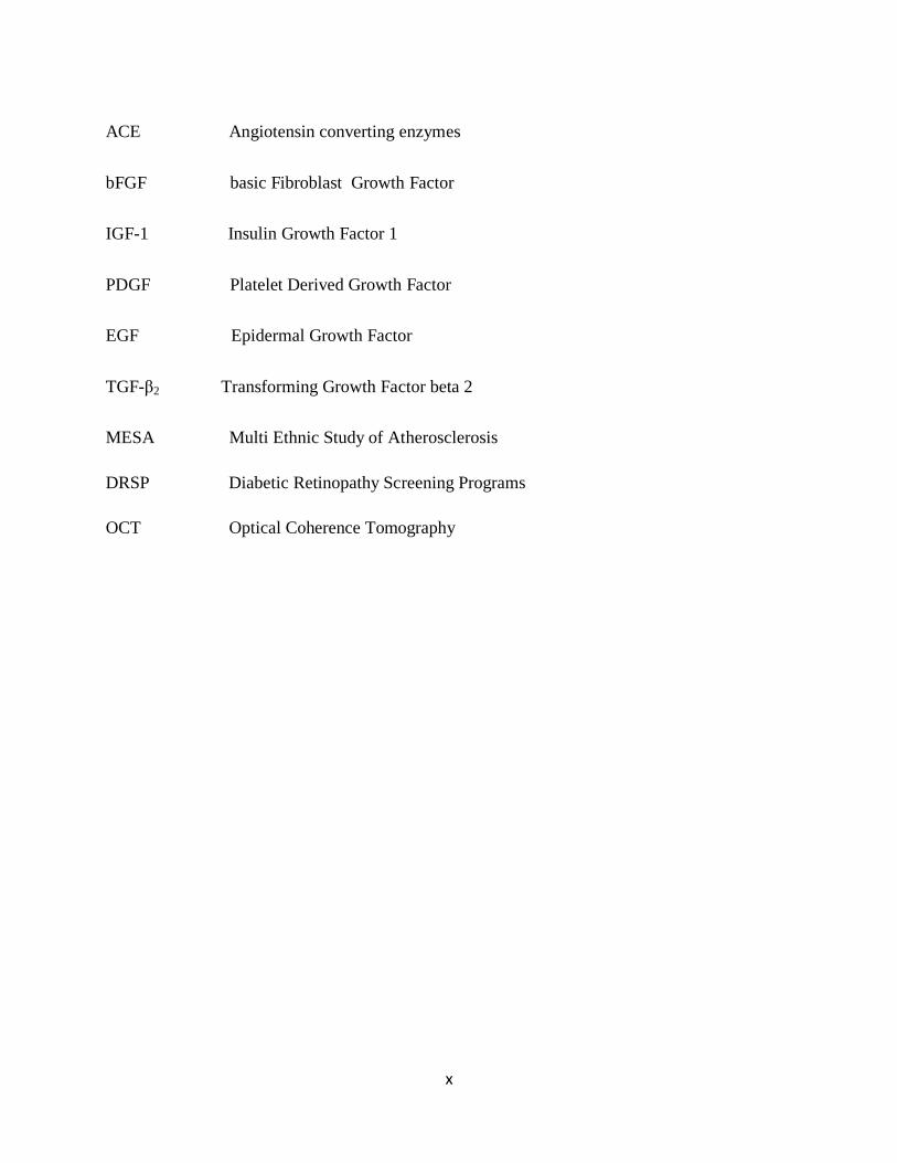
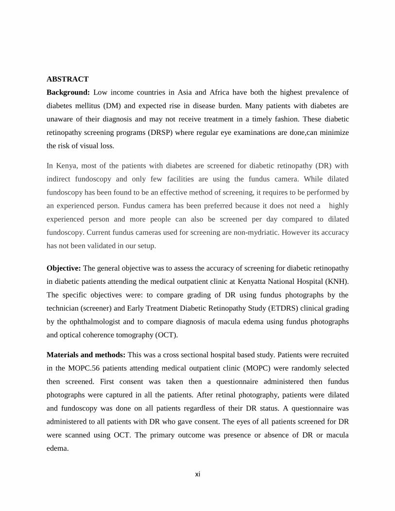


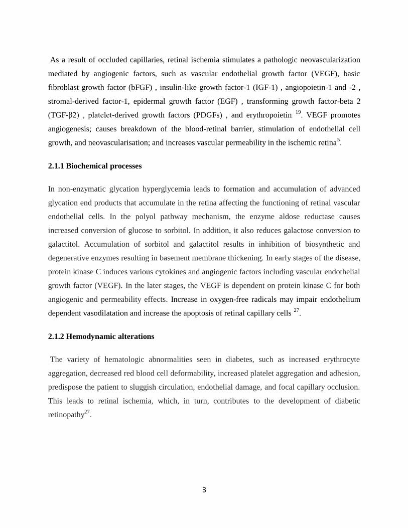


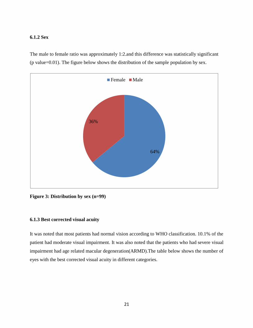


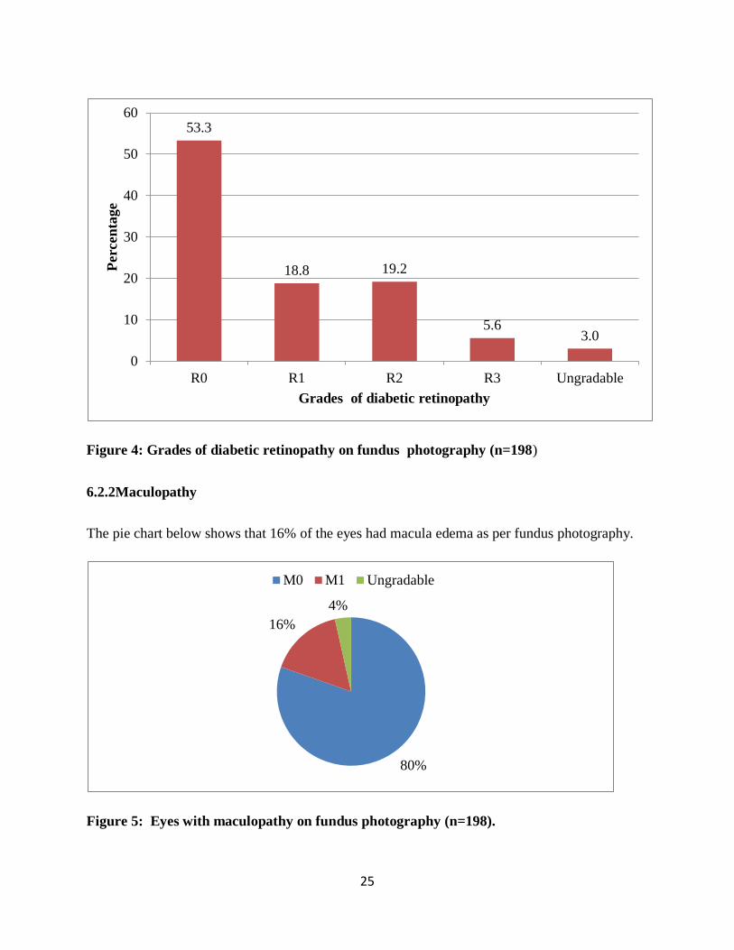
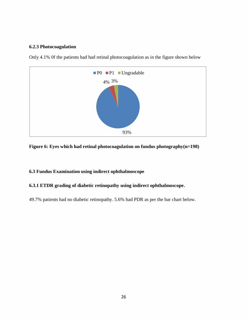
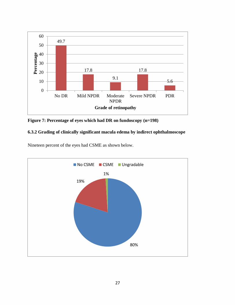

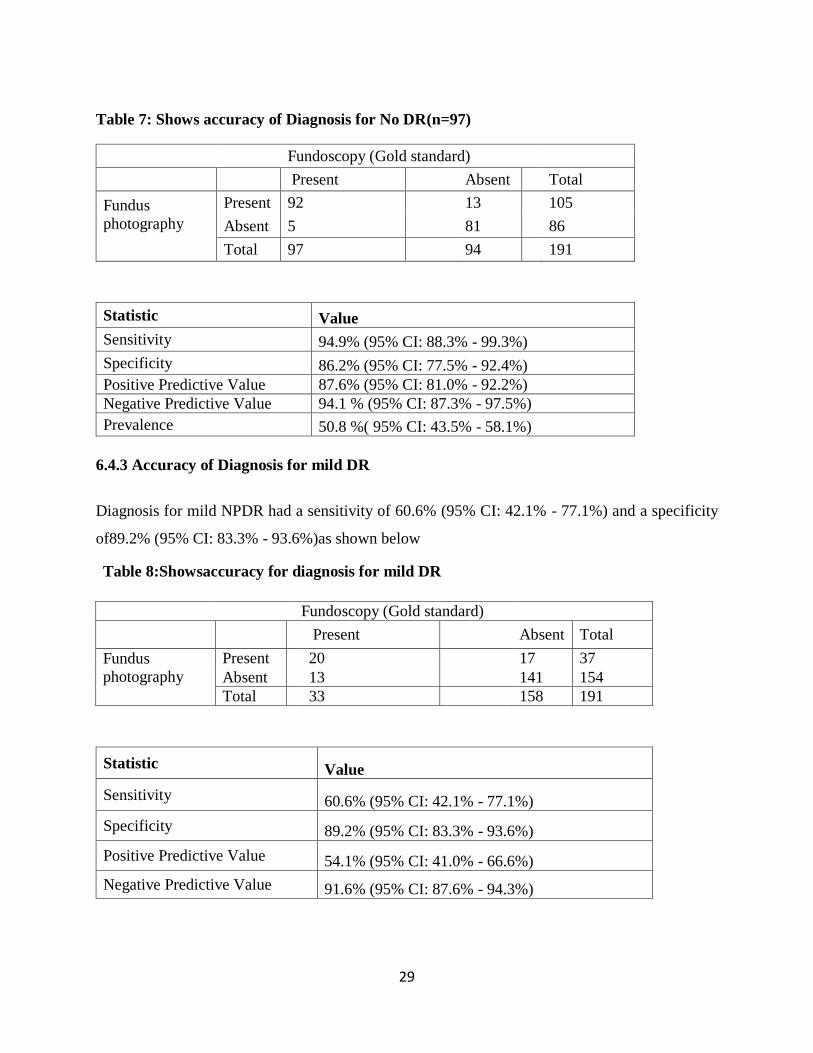
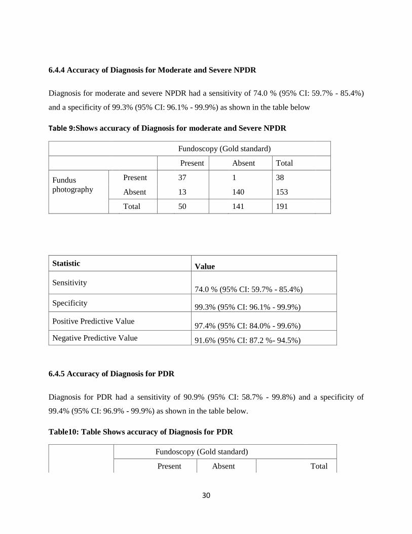

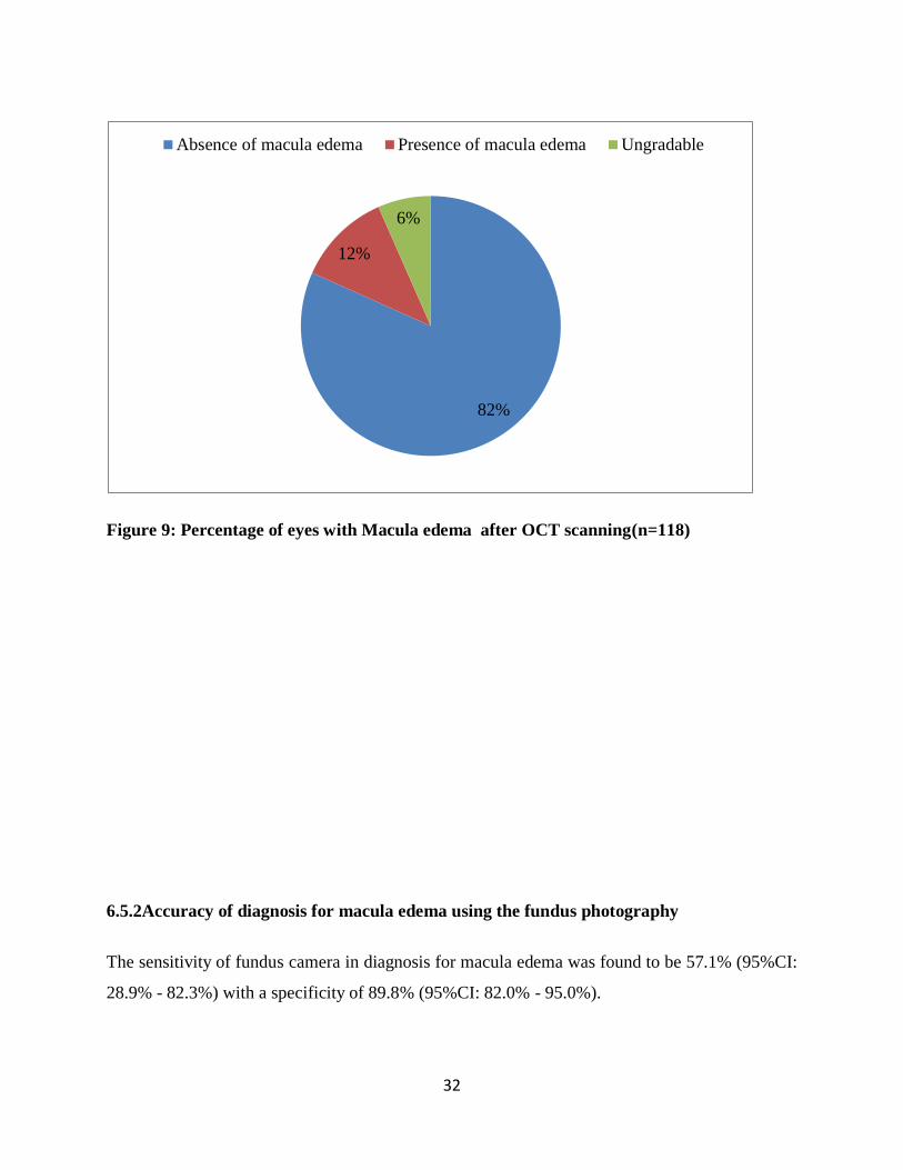


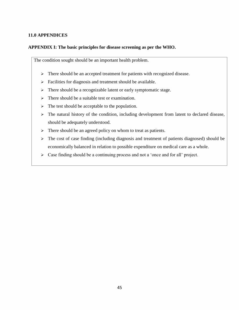

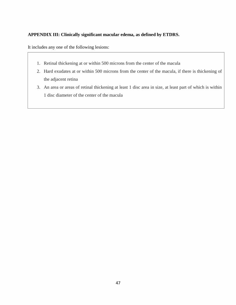


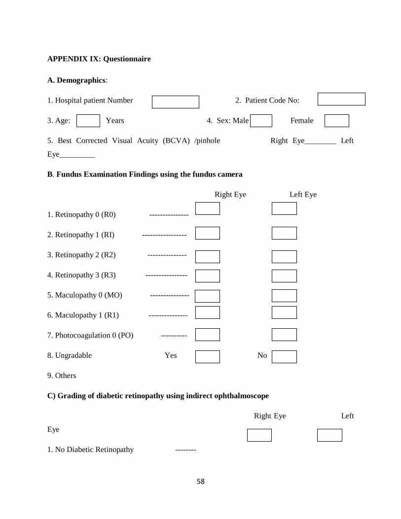
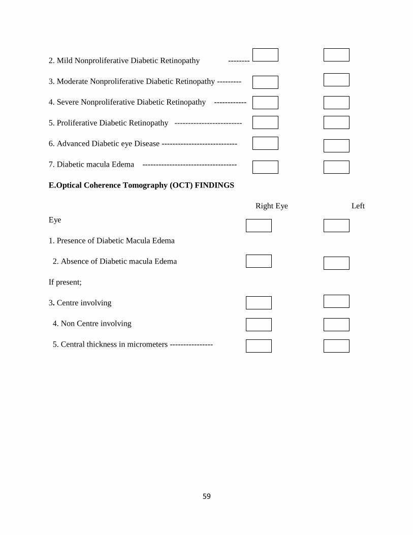
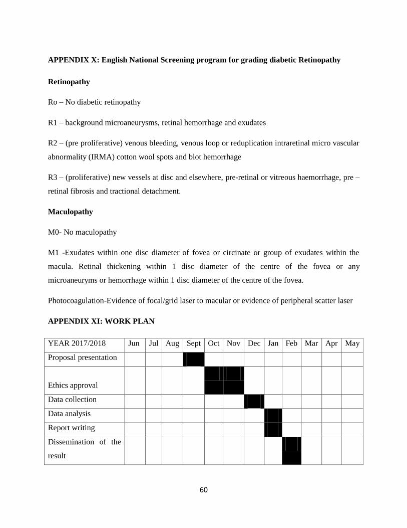
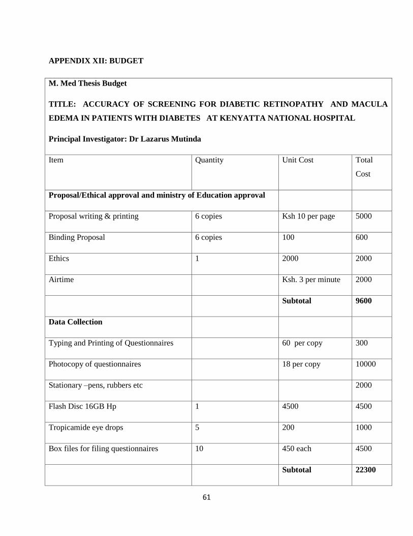


![The Guide - Diabetic Retinopathy - Vision Lossvisionloss.org.au/wp-content/uploads/2016/05/The... · the guide [diabetic retinopathy] What is Diabetic Retinopathy? Diabetic Retinopathy](https://static.documents.pub/doc/80x56/5e3ed00bf9c32e41ea6578a8/the-guide-diabetic-retinopathy-vision-the-guide-diabetic-retinopathy-what.jpg)


