+ All Categories
Home > Documents > Accuracy of the interferon-gamma release assay for the ... · PDF fileDepartment of...

Accuracy of the interferon-gamma release assay for the ... · PDF fileDepartment of...

Date post: 06-Mar-2018
Category:
Upload: vuongque
View: 216 times
Download: 1 times
Share this document with a friend
17
Submitted 2 March 2015 Accepted 21 April 2015 Published 21 May 2015 Corresponding author Fu-Qiang Wen, [email protected] Academic editor Li Zuo Additional Information and Declarations can be found on page 13 DOI 10.7717/peerj.951 Copyright 2015 Pang et al. Distributed under Creative Commons CC-BY 4.0 OPEN ACCESS Accuracy of the interferon-gamma release assay for the diagnosis of tuberculous pleurisy: an updated meta-analysis Cai-Shuang Pang 1 , Yong-Chun Shen 1 , Pan-Wen Tian 1 , Jing Zhu, Mei Feng, Chun Wan and Fu-Qiang Wen Department of Respiratory and Critical Care Medicine, West China Hospital of Sichuan University and Division of Pulmonary Diseases, State Key Laboratory of Biotherapy of China, China 1 These authors contributed equally to this work. ABSTRACT Background and Objectives. The best method for diagnosing tuberculous pleurisy (TP) remains controversial. Since a growing number of publications focus on the interferon-gamma release assay (IGRA), we meta-analyzed the available evidence on the overall diagnostic performance of IGRA applied to pleural fluid and peripheral blood. Materials and Methods. PubMed and Embase were searched for relevant English papers up to October 31, 2014. Statistical analyses were performed using Stata and Meta-DiSc. Pooled sensitivity, specificity, positive likelihood ratio (PLR), negative likelihood ratio (NLR), positive predictive value (PPV), negative predictive value (NPV) and diagnostic odds ratio (DOR) were count. Summary receiver operating characteristic curves and area under the curve (AUC) were used to summarize the overall diagnostic performance. Results. Fifteen publications met our inclusion criteria and were included in the meta analysis. The following pooled estimates for diagnostic parameters of pleural IGRA were obtained: sensitivity, 0.82 (95% CI [0.79–0.85]); specificity, 0.87 (95% CI [0.84–0.90]); PLR, 4.94 (95% CI [2.60–9.39]); NLR, 0.22 (95% CI [0.13–0.38]); PPV, 0.91 (95% CI [0.85–0.96]); NPV, 0.79 (95% CI [0.71–0.85]); DOR, 28.37 (95% CI [10.53–76.40]); and AUC, 0.91. The corresponding estimates for blood IGRA were as follows: sensitivity, 0.80 (95% CI [0.76–0.83]); specificity, 0.70 (95% CI [0.65–0.75]); PLR, 2.48 (95% CI [1.95–3.17]); NLR, 0.30 (95% CI [0.24–0.37]); PPV, 0.79 (95% CI [0.60–0.87]); NPV, 0.75 (95% CI [0.62–0.83]); DOR, 9.96 (95% CI [6.02–16.48]); and AUC, 0.89. Conclusions. This meta analysis suggested that pleural IGRA has potential for serv- ing as a complementary method for diagnosing TP; however, its cost, high turn around time, and sub-optimal performance make it unsuitable as a stand-alone diagnostic tool. Better tests for the diagnosis of TP are required. Subjects Epidemiology, Health Policy, Respiratory Medicine, Statistics Keywords Interferon-gamma release assay, Tuberculous pleurisy, Diagnosis, Meta-analysis How to cite this article Pang et al. (2015), Accuracy of the interferon-gamma release assay for the diagnosis of tuberculous pleurisy: an updated meta-analysis. PeerJ 3:e951; DOI 10.7717/peerj.951
Transcript

Submitted 2 March 2015Accepted 21 April 2015Published 21 May 2015

Corresponding authorFu-Qiang Wen,[email protected]

Academic editorLi Zuo

Additional Information andDeclarations can be found onpage 13

DOI 10.7717/peerj.951

Copyright2015 Pang et al.

Distributed underCreative Commons CC-BY 4.0

OPEN ACCESS

Accuracy of the interferon-gammarelease assay for the diagnosis oftuberculous pleurisy: an updatedmeta-analysisCai-Shuang Pang1, Yong-Chun Shen1, Pan-Wen Tian1, Jing Zhu,Mei Feng, Chun Wan and Fu-Qiang Wen

Department of Respiratory and Critical Care Medicine, West China Hospital of SichuanUniversity and Division of Pulmonary Diseases, State Key Laboratory of Biotherapy of China,China

1 These authors contributed equally to this work.

ABSTRACTBackground and Objectives. The best method for diagnosing tuberculous pleurisy(TP) remains controversial. Since a growing number of publications focus on theinterferon-gamma release assay (IGRA), we meta-analyzed the available evidence onthe overall diagnostic performance of IGRA applied to pleural fluid and peripheralblood.Materials and Methods. PubMed and Embase were searched for relevant Englishpapers up to October 31, 2014. Statistical analyses were performed using Stata andMeta-DiSc. Pooled sensitivity, specificity, positive likelihood ratio (PLR), negativelikelihood ratio (NLR), positive predictive value (PPV), negative predictive value(NPV) and diagnostic odds ratio (DOR) were count. Summary receiver operatingcharacteristic curves and area under the curve (AUC) were used to summarize theoverall diagnostic performance.Results. Fifteen publications met our inclusion criteria and were included in themeta analysis. The following pooled estimates for diagnostic parameters of pleuralIGRA were obtained: sensitivity, 0.82 (95% CI [0.79–0.85]); specificity, 0.87 (95% CI[0.84–0.90]); PLR, 4.94 (95% CI [2.60–9.39]); NLR, 0.22 (95% CI [0.13–0.38]); PPV,0.91 (95% CI [0.85–0.96]); NPV, 0.79 (95% CI [0.71–0.85]); DOR, 28.37 (95% CI[10.53–76.40]); and AUC, 0.91. The corresponding estimates for blood IGRA were asfollows: sensitivity, 0.80 (95% CI [0.76–0.83]); specificity, 0.70 (95% CI [0.65–0.75]);PLR, 2.48 (95% CI [1.95–3.17]); NLR, 0.30 (95% CI [0.24–0.37]); PPV, 0.79 (95%CI [0.60–0.87]); NPV, 0.75 (95% CI [0.62–0.83]); DOR, 9.96 (95% CI [6.02–16.48]);and AUC, 0.89.Conclusions. This meta analysis suggested that pleural IGRA has potential for serv-ing as a complementary method for diagnosing TP; however, its cost, high turnaround time, and sub-optimal performance make it unsuitable as a stand-alonediagnostic tool. Better tests for the diagnosis of TP are required.

Subjects Epidemiology, Health Policy, Respiratory Medicine, StatisticsKeywords Interferon-gamma release assay, Tuberculous pleurisy, Diagnosis, Meta-analysis

How to cite this article Pang et al. (2015), Accuracy of the interferon-gamma release assay for the diagnosis of tuberculous pleurisy: anupdated meta-analysis. PeerJ 3:e951; DOI 10.7717/peerj.951

INTRODUCTIONTuberculous pleurisy (TP) is the most common form of extrapulmonary tuberculosis,

accounting for 23% of all tuberculosis cases and 30% of cases of disease-causing pleural

effusion (PE) (Vidal et al., 1986; Corbett et al., 2003; Valdes et al., 2003), which involves

exudate containing primarily lymphocytes. Direct diagnosis of TP would be the best way

to avoid misdiagnosis and the resulting inappropriate treatment (Lin et al., 2009), but

this remains a challenge. Definitive diagnosis of TP depends on isolating Mycobacterium

tuberculosis from PE or pleural tissue. Conventional methods, such as PE culture, pleural

biopsy and Ziehl-Neelsen staining, show poor sensitivity for detecting the limited amounts

of bacteria in the PE of affected patients (Escudero et al., 1990; Valdes et al., 1998). Culturing

PE is also time-consuming. Pleural biopsy is invasive and technically difficult, so its

effectiveness depends on technical skill (Perez & Jimenez, 2000). It may not be suitable

for elderly and children, individuals with underlying co-morbidities, and those at high risk

of bleeding. The tuberculin skin test is cross-reactive for Bacille Calmette Guerin (BCG)

and many non-tuberculous mycobacteria, increasing the risk of misdiagnosis (Lawrence,

2000; Stead & To, 1987; Liebeschuetz et al., 2004). The limitations of these conventional

approaches to diagnosing TP highlight the need to identify new diagnostic tools.

The PE of patients with TP has been shown to contain significantly higher levels of

T lymphocytes and interferon (IFN)-γ than peripheral blood (North & Jung, 2004;

Sharma et al., 2002), and the PE of these patients contains higher IFN-γ levels than the

PE of uninfected individuals (Yamada et al., 2001). In fact, T lymphocytes that have

previously been exposed to MTB release more IFN-γ on repeat exposure. This inspired

the development of a T-cell IFN-γ release assay (IGRA), which is now licensed as a blood

test for diagnosis of latent tuberculosis (Lalvani, 2007; Pai, Zwerling & Menzies, 2008).

Whether IGRA can be used to diagnose TP is controversial. A previous meta-analysis

concluded that it showed poor sensitivity and specificity for this purpose (Zhou et al.,

2011). Nevertheless, a growing number of studies have focused on extending the use

of IGRA to the diagnosis of TP (Hooper, Lee & Maskell, 2009). Therefore, the present

meta-analysis was undertaken to comprehensively assess the overall accuracy of IGRA for

the diagnosis of TP.

MATERIAL & METHODSSearch strategy and study selectionPubMed and Embase were searched for articles published before October 31, 2014. The

following search terms were used: “pleural effusion/pleural fluid, pleurisy/pleuritis AND

elispot, OR quantiferon, OR interferon-gamma assays, OR interferon-gamma release

assays, OR t cell assays.” The related-articles function was also used, and reference lists in

relevant articles were searched manually.

Studies were included in our meta-analysis if they (1) used IGRA testing for the diagno-

sis of tuberculous pleurisy (2) reported sufficient data to calculate true positive, false pos-

itive, false negative , and true negative of IGRA for the diagnosis of TP, and (3) constituted

original research published in English. Studies available only as abstracts were excluded.

Pang et al. (2015), PeerJ, DOI 10.7717/peerj.951 2/17

Data extraction and quality assessmentTwo reviewers independently checked all potentially relevant studies, and disagreements

were resolved by consensus. Data were collected from each study, including first author,

year of publication, country, participant characteristics, IGRA method, samples, cut-off

values, sensitivity, specificity and methodological quality. For each study we constructed

2 × 2 contingency tables in which we calculated true positive, false positive, false negative,

and true negative rates.

The methodological quality of the studies was assessed using the 14-items Quality

Assessment for Studies of Diagnostic Accuracy (QUADAS) guidelines (Whiting et al.,

2003). When a criterion was fulfilled, a score of 1 was given, 0 if a criterion was unclear, and

−1 if a criterion was not achieved. This evaluation instrument rates studies on a quality

scale of up to 14 points.

Statistical analysesStandard methods recommended for meta-analyses of diagnostic test evaluations (Deville

et al., 2002) were used. Stata 12.0 and Meta-DiSc 1.4 were used for statistical analysis. The

following accuracy measures were calculated for each study: sensitivity, specificity, positive

likelihood ratio (PLR), negative likelihood ratio (NLR), positive predictive value (PPV),

negative predictive value (NPV) and diagnostic odds ratio (DOR). Summary receiver op-

erating characteristic (SROC) curves and area under the curve (AUC) were also calculated

(Moses, Shapiro & Littenberg, 1993; Irwig et al., 1995; Vamvakas, 1998). Heterogeneity

across studies was detected using chi-square and Fisher’s exact tests. We planned to use

a random-effects model to synthesize data if heterogeneity was present (P < 0.05 and

I2 > 50%) (Shen et al., 2012). Based on this rule, pooled average sensitivity, specificity and

other diagnostic parameters of pleural and blood IGRA were calculated using, respectively,

a random-effects model and a fixed-effects model (Irwig et al., 1995; Vamvakas, 1998).

Potential presence of publication bias was tested using funnel plots and the Egger’s test. All

statistical tests were two-sided, and the threshold of significance was set at P < 0.05.

RESULTSStudy inclusion and characteristicsStudy identification and selection were outlined in Fig. 1. In the end, 15 publications of

IGRA to diagnose patients with TP were eligible for inclusion (Wilkinson et al., 2005; Ariga

et al., 2007; Losi et al., 2007; Baba et al., 2008; Chegou et al., 2008; Dheda et al., 2009; Lee

et al., 2009; Keng et al., 2013; Ates et al., 2011; Eldin et al., 2012; Kang et al., 2012; Liu et al.,

2013; Liao et al., 2014; Chung et al., 2011; Gao et al., 2012). In the studies by Dheda et al.

(2009) and Kang et al. (2012), IGRA was performed in two formats: as an enzyme-linked

immunosorbent spot (ELISPOT) assay, and as an enzyme-linked immunosorbent assay

(ELISA). Thus, each of these publications was treated as two independent studies in our

meta-analysis, giving 17 studies in our meta-analysis altogether. ELISPOT was used in

seven studies (Wilkinson et al., 2005; Losi et al., 2007; Dheda et al., 2009; Lee et al., 2009;

Kang et al., 2012; Liu et al., 2013; Liao et al., 2014), while ELISA was used in the remaining

Pang et al. (2015), PeerJ, DOI 10.7717/peerj.951 3/17

Figure 1 Flow diagram of included and excluded studies.

10 studies (Ariga et al., 2007; Baba et al., 2008; Chegou et al., 2008; Dheda et al., 2009; Keng

et al., 2013; Ates et al., 2011; Eldin et al., 2012; Kang et al., 2012; Chung et al., 2011; Gao et al.,

2012). Across all studies, 17 analyses of PE (Wilkinson et al., 2005; Ariga et al., 2007; Losi et

al., 2007; Baba et al., 2008; Chegou et al., 2008; Dheda et al., 2009; Lee et al., 2009; Keng et al.,

2013; Ates et al., 2011; Eldin et al., 2012; Kang et al., 2012; Liu et al., 2013; Liao et al., 2014;

Gao et al., 2012). and 14 analyses of blood (Wilkinson et al., 2005; Ariga et al., 2007; Losi et

al., 2007; Baba et al., 2008; Chegou et al., 2008; Dheda et al., 2009; Lee et al., 2009; Ates et al.,

2011; Eldin et al., 2012; Kang et al., 2012; Liu et al., 2013; Liao et al., 2014; Chung et al., 2011)

were conducted. Ten studies were conducted in Asia, five in Africa, and two in Europe. Key

characteristics of included studies, along with QUADAS score, were shown in Table 1.

Nine studies included at least 60 patients (Ariga et al., 2007; Chegou et al., 2008; Dheda

et al., 2009; Keng et al., 2013; Ates et al., 2011; Liu et al., 2013; Liao et al., 2014; Chung et al.,

2011; Gao et al., 2012). Mean sample size in the 17 analyses of pleural IGRA was 76 (range

18–332), involving a total of 806 patients with TP and 482 without TP. Mean sample size

in the 14 analyses of blood IGRA was 80 (range 34–332), involving altogether 730 patients

with TP and 383 without TP.

Diagnostic accuracyIn the 17 analyses of pleural IGRA, diagnostic sensitivity ranged from 0.44 to 1.0 (Fig. 2);

pooled sensitivity was 0.82 (95% CI [0.79–0.85]; I2= 92%). Specificity ranged from 0.5 to

1.0, and pooled specificity was 0.87 (95% CI [0.84–0.90]; I2= 82.5%). Other pooled esti-

mates of diagnostic parameters were as follows: PLR, 4.94 (95% CI [2.60–9.39]); NLR, 0.22

(95% CI [0.13–0.38]); PPV, 0.91 (95% CI [0.85–0.96]); NPV, 0.79 (95% CI [0.71–0.85]);

and DOR, 28.37 (95% CI [10.53–76.4]). Chi-square values for these parameters suggested

considerable heterogeneity among studies (Table 2): sensitivity, 199.86; specificity, 91.18;

PLR, 129.36; NLR, 180.23; PPV, 132; NPV, 157; and DOR, 81.01 (all P < 0.001).

Pang et al. (2015), PeerJ, DOI 10.7717/peerj.951 4/17

Table 1 Key characteristics of the studies included in the meta-analysis.

First author Settings IGRA method Samples Test results QUADAS score

TP FP FN TN

Wilkinson UK ELISPOT PE 10 1 0 7 10

Blood 10 – 0 –

Ariga Japan ELISA PE 27 1 1 46 11

Blood 21 14 6 33

Losi Italy, Germany, Netherlands ELISPOT PE 19 5 1 16 10

Blood 18 7 2 14

Baba South Africa ELISA PE 12 2 15 4 10

Blood 17 0 7 6

Chegou South Africa ELISA PE 13 2 10 13 13

Blood 16 5 6 12

Dheda South Africa ELISPOT PE 38 8 6 9 11

Blood 30 7 6 9

ELISA PE 23 6 19 12

Blood 26 4 4 9

Lee Taiwan ELISPOT PE 18 3 1 18 9

Blood 14 2 4 19

Ates Turkey ELISA PE 21 6 22 23 11

Blood 30 14 13 15

Chuang South Korea ELISA Blood 42 17 12 26 8

Kang South Korea ELISPOT PE 15 8 0 3 9

Blood 18 6 2 8

ELISA PE 10 5 5 5

Blood 4 6 0 7

Eldin Egypt ELISA PE 16 3 4 15 7

Blood 14 7 6 11

Gao China ELISA PE 54 2 4 18 8

Keng Taiwan ELISA PE 24 2 31 57 8

PE 22 1 9 56

Liu China ELISPOT PE 53 2 2 41 9

Blood 51 10 4 33

Liao China ELISPOT PE 269 0 12 51 8

Blood 220 7 61 44

Notes.ELISPOT, enzyme-linked immunosorbent spot; ELISA, enzyme-linked immunosorbent assay; FN, false negative; FP, false positive; IGRA, T-cell interferon-γ releaseassay; PE, pleural effusion; QUADAS, quality assessment for studies of diagnostic accuracy; TN, true negative; TP, true positive.

For 14 analyses of blood IGRA, diagnostic sensitivity ranged from 0.71 to 0.93 (Fig. 2),

and specificity ranged from 0.56 to 1.0. Pooled estimates of the other diagnostic parameters

were as follows: sensitivity, 0.8 (95% CI [0.76–0.83]; I2= 31.9%); specificity, 0.7 (95% CI

[0.65–0.75]; I2= 54.4%); PLR, 2.48 (95% CI [1.95–3.17]); NLR, 0.3 (95% CI [0.24–0.37]);

PPV, 0.79 (95% CI [0.60–0.87]); NPV, 0.75 (95% CI [0.62–0.83]); and DOR, 9.96 (95%

CI [6.02–16.48]). Chi-square values for most of these parameters indicated no significant

heterogeneity among studies (Table 2): sensitivity, 19.09 (P = 0.12); NLR, 18.02 (P = 0.16);

Pang et al. (2015), PeerJ, DOI 10.7717/peerj.951 5/17

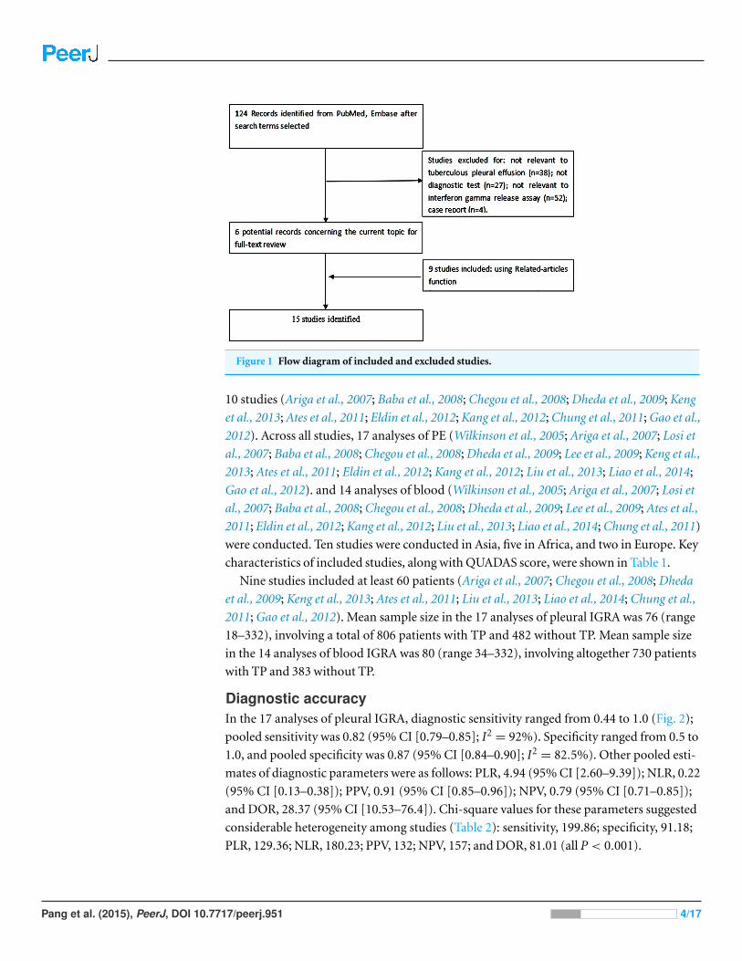
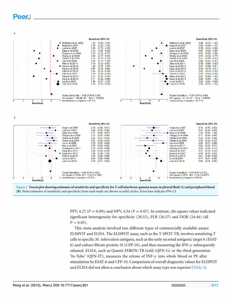
Figure 2 Forest plot showing estimates of sensitivity and specificity for T-cell interferon-gamma assays in pleural fluid (A) and peripheral blood(B). Point estimates of sensitivity and specificity from each study are shown as solid circles. Error bars indicate 95% CI.

PPV, 4.27 (P = 0.09); and NPV, 4.54 (P = 0.07). In contrast, chi-square values indicated

significant heterogeneity for specificity (28.52), PLR (26.57) and DOR (24.44) (all

P < 0.05).

This meta-analysis involved two different types of commercially available assays:

ELISPOT and ELISA. The ELISPOT assay, such as the T-SPOT-TB, involves sensitizing T

cells to specific M. tuberculosis antigens, such as the early secreted antigenic target 6 (ESAT-

6) and culture filtrate protein 10 (CFP-10), and then measuring the IFN-γ subsequently

released. ELISA, such as Quanti-FERON-TB Gold (QFN-G) or the third-generation

‘In-Tube’ (QFN-IT), measures the release of INF-γ into whole blood or PE after

stimulation by ESAT-6 and CFP-10. Comparison of overall diagnostic values for ELISPOT

and ELISA did not allow a conclusion about which assay type was superior (Table 2).

Pang et al. (2015), PeerJ, DOI 10.7717/peerj.951 6/17

Table 2 Pooled results for accuracy of interferon-gamma assays to diagnose tuberculous pleurisy.

Pleural effusion Blood

Total ELISPOT ELISA Total ELISPOT ELISA

Number of study 17 7 10 14 6 8

Sensitivity(95% CI) 0.82(0.79–0.85) 0.95(0.93–0.97) 0.65(0.60–0.70) 0.80(0.76–0.83) 0.82(0.78–0.85) 0.76(0.70–0.81)

Heterogeneity*(P) 199.86(<0.001) 7.93(0.24) 67.65(<0.001) 19.09(0.12) 9.99(0.075) 6.17(0.52)

Specificity(95% CI) 0.87(0.84–0.9) 0.84(0.78–0.89) 0.89(0.85–0.93) 0.70(0.65–0.75) 0.77(0.69–0.83) 0.64(0.57–0.71)

Heterogeneity(P) 91.18(<0.001) 50.63(<0.001) 38.25(<0.001) 28.52(0.008) 12.57(0.028) 9.34(0.23)

PLR(95% CI) 4.94(2.60–9.39) 5.62(1.65–19.14) 4.6(2.16–9.82) 2.48(1.95–3.17) 3.21(2.09–4.94) 2.00(1.63–2.45)

Heterogeneity(P) 129.36(<0.001) 94.35(<0.001) 42.71(<0.001) 26.57(0.014) 12.18(0.03) 6.13(0.525)

NLR(95% CI) 0.22(0.13–0.38) 0.08(0.04–0.16) 0.41(0.27–0.62) 0.30(0.24–0.37) 0.22(0.16–0.31) 0.38(0.30–0.50)

Heterogeneity(P) 180.23(<0.001) 12.33(0.06) 48.66(<0.001) 18.02(0.16) 6.13(0.294) 5.23(0.632)

PPV(95% CI) 0.91(0.85–0.96) 0.87(0.8–1.03) 0.96(0.68–1.32) 0.79(0.60–0.87) 0.74(0.64–0.84) 0.84(0.75–1.19)

Heterogeneity(P) 132(<0.001) 46.52(0.03) 61.25(<0.001) 4.27(0.09) 6.79(1.22) 5.76(0.46)

NPV(95% CI) 0.79(0.71–0.85) 0.75(0. 65–0.87) 0.84(0.7–1.41) 0.75(0.62–0.83) 0.72(0.58–0.81) 0.76(0.65–0.88)

Heterogeneity(P) 157(<0.001) 9.68(<0.001) 16.9(<0.001) 4.54(0.07) 7.01(1.13) 11.53(0.06)

DOR(95% CI) 28.37(10.53–76.4) 88.85(16.10–490.43) 14.10(4.56–43.54) 9.96(6.02–16.48) 19.82(11.67–33.66) 5.46(3.46–8.61)

Heterogeneity(P) 81.01(<0.001) 23.47(0.001) 43.65(<0.001) 24.44(0.03) 4.93(0.43) 6.38(0.496)

AUC(SEM) 0.91(0.03) 0.98(0.01) 0.84(0.08) 0.84(0.03) 0.89(0.02) 0.78(0.04)

Notes.* Q value.

AUC, area under the curve; DOR, diagnostic odds ratio; ELISPOT, enzyme-linked immunosorbent spot; ELISA, enzyme-linked immunosorbent assay; NLR, negativelikelihood ratio; PLR, positive likelihood ratio; PPV, positive predictive value; NPV, negative predictive value.

We assessed the overall diagnostic performance by calculating SROC curves and the

corresponding AUC. The SROC curve for pleural IGRA was not positioned near the

desirable upper left corner, and the point where sensitivity equals specificity (Q) was 0.84;

the optimum AUC was 0.91 (Fig. 3A). The corresponding SROC curve for blood IGRA

showed Q of 0.77 and AUC of 0.84 (Fig. 3B). Although neither the pleural or blood AUC

was entirely satisfactory, this summary analysis suggests that pleural IGRA shows much

better diagnostic performance than blood IGRA.

Multiple regression analysis and publication biasThe quality of the 17 studies in this meta-analysis varied considerably, with only five

studies earning high QUADAS scores (≥ 11; Table 1). These scores were used in a

meta-regression analysis to assess the effect of study quality on the relative DOR (RDOR)

of IGRA for the diagnosis of TP (Table 3). Higher- and lower-quality studies did not differ

significantly in RDOR for either pleural or blood IGRA (Table 3). Seven studies were

performed in areas with a low tuberculosis incidence (Wilkinson et al., 2005; Ariga et al.,

2007; Losi et al., 2007; Keng et al., 2013; Ates et al., 2011; Eldin et al., 2012; Kang et al.,

2012) and 10 studies (eight publications) were performed in areas with a high tuberculosis

incidence (Baba et al., 2008; Chegou et al., 2008; Dheda et al., 2009; Lee et al., 2009; Liu et al.,

2013; Liao et al., 2014; Chung et al., 2011; Gao et al., 2012). Diagnostic accuracy of pleural

IGRA depended significantly only on assay method (ELISPOT vs ELISA, P = 0.023),

Pang et al. (2015), PeerJ, DOI 10.7717/peerj.951 7/17

Figure 3 Summary receiver operating characteristic (SROC) curves for T-cell interferon-gamma as-says in pleural fluid (A) and peripheral blood (B). Solid circles represent each study included in themeta-analysis, with circle size representing the sample size in each study. The regression SROC curvessummarize the overall diagnostic accuracy.

Pang et al. (2015), PeerJ, DOI 10.7717/peerj.951 8/17

Table 3 Weighted meta-regression to assess the effects of study setting, IGRA method and studyquality on diagnostic accuracy of IGRA.

Covariate Number of studies Coefficient RDOR (95% CI) P-value

Pleural effusion

QUADAS score

≥11 5 −1.44 0.24(0.02–2.70) 0.225

<11 12

Setting

Area with low TB incidence 7 0.36 1.44(0.10–21.14) 0.777

Area with high TB incidence 10

Method

ELISPOT 7 −3.17 0.04(0.00–0.60) 0.023

ELISA 10

Peripheral blood

QUADAS score

≥11 5 −0.81 0.45(0.15–1.35) 0.137

<11 9

Setting

Area with low TB incidence 7 1.12 3.06(1.16–8.10) 0.028

Area with high TB incidence 7

Method

ELISPOT 6 −1.26 0.28(0.13–0.62) 0.0048

ELISA 8

Notes.RDOR, relative diagnostic odds ratio; QUADAS, quality assessment for studies of diagnostic accuracy; TB, tuberculosis;ELISA, enzyme-linked immunosorbent assay; ELISPOT, enzyme-linked immunosorbent spot.

but not on study quality or tuberculosis incidence. Diagnostic accuracy of blood IGRA

depended significantly on both assay method and tuberculosis incidence.

Results of the RDOR analysis were shown in Table 3.

Publication bias was analyzed by using funnel plots and the Egger’s test. Since the funnel

plots for publication bias showed asymmetry (Fig. 4), Egger’s tests were performed, which

confirmed significant risk of publication bias in the meta-analyses for both blood IGRA

and pleural IGRA (both P < 0.001).

DISCUSSIONIGRA has an advantage over conventional methods of diagnosing M. tuberculosis infection,

because it is based on specific antigens, such as ESAT-6 and CFP-10, that are absent

from BCG and most environmental mycobacteria. Whether this assay is suitable for

diagnosing TP is controversial. In fact, Zhou et al. (2011) conducted a meta-analysis to

analyze the diagnostic role of IGRA for TP. According to his inclusion criteria, only seven

publications were included. Several years have passed, and some new studies have been

added, so we conducted this updated meta-analysis. Our meta-analysis summarizes the

available evidence on this question in an effort to provide guidance for TP diagnosis.

Our results showed that the pooled sensitivities of pleural and blood IGRA were 0.82 and

Pang et al. (2015), PeerJ, DOI 10.7717/peerj.951 9/17

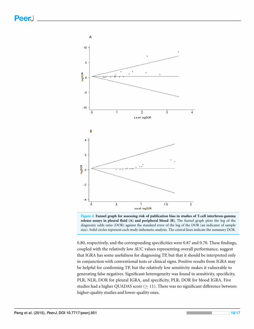
Figure 4 Funnel graph for assessing risk of publication bias in studies of T-cell interferon-gammarelease assays in pleural fluid (A) and peripheral blood (B). The funnel graph plots the log of thediagnostic odds ratio (DOR) against the standard error of the log of the DOR (an indicator of samplesize). Solid circles represent each study inthemeta-analysis. The central lines indicate the summary DOR.

0.80, respectively, and the corresponding specificities were 0.87 and 0.70. These findings,

coupled with the relatively low AUC values representing overall performance, suggest

that IGRA has some usefulness for diagnosing TP, but that it should be interpreted only

in conjunction with conventional tests or clinical signs. Positive results from IGRA may

be helpful for confirming TP, but the relatively low sensitivity makes it vulnerable to

generating false negatives. Significant heterogeneity was found in sensitivity, specificity,

PLR, NLR, DOR for pleural IGRA, and specificity, PLR, DOR for blood IGRA. Five

studies had a higher QUADAS score (≥ 11). There was no significant difference between

higher-quality studies and lower-quality ones.

Pang et al. (2015), PeerJ, DOI 10.7717/peerj.951 10/17

We assessed pleural and blood IGRAs using SROC curves and DOR tests, both of which

combine sensitivity and specificity. SROC curves, which are unlikely to be affected by a

diagnostic threshold effect (Jones & Athanasiou, 2005), showed an optimum cut-off of

0.84 for pleural IGRA and 0.77 for blood IGRA, while the corresponding AUCs were 0.91

and 0.84, suggesting less than fully satisfactory overall accuracy. The DOR of a test is the

ratio of the odds of obtaining a positive test result in the disease group to the odds of

obtaining a positive test result in the no-disease group (Zhou et al., 2011). When DOR

>1, higher values indicate better discriminatory test performance. We calculated a pooled

DOR of 28.37 for pleural IGRA and of 9.96 for blood IGRA, suggesting that IGRA and

particularly pleural IGRA may be helpful for diagnosing TP. We found higher pooled

sensitivity and specificity for pleural IGRA than a previous meta-analysis (Zhou et al.,

2011), which likely reflects our inclusion of more articles. Similarly we calculated a higher

pooled DOR for pleural IGRA (19.0, 95% CI [4.8–75.8]) than that meta-analysis did. We

conclude that pleural IGRA has better prospects than blood IGRA for widespread clinical

implementation. This was possibly due to compartmentalization of antigen-specific

effector T cells, which could be recruited and concentrated at the site of infection, such

as pleural cavity. ESAT-6-specific, IFN-γ secreting T-cells have a 15-fold concentration in

PE relative to peripheral blood in patients with TP (Wilkinson et al., 2005).

Potentially more clinically meaningful than DOR and SROC, PLR and NLR are often

used as measures of diagnostic accuracy. PLR indicates how much the odds of a condition

are increased by a positive test, while NLR indicates how much they are decreased by a

negative test. Larger PLR means greater diagnostic accuracy, whereas a smaller NLR is

better. The pooled PLR of 4.94 for pleural IGRA suggests that patients with TP have a

nearly five-fold greater chance of a positive test result than patients without TP. Even

though this PLR is larger than that reported in a previous meta-analysis (Zhou et al., 2011),

it is still too small for clinical purposes. At the same time, we calculated a pooled NLR of

0.22 for pleural IGRA, indicating that the probability that a patient with a negative result

has a 22% chance of having TP, which is not low enough to reliably rule out false negatives.

The corresponding PLR and NLR for blood IGRA were even less satisfactory.

The pooled PPV for pleural IGRA was 0.91, indicating that 9% of positive results may be

false positives. The NPV of pleural IGRA was 0.79, suggesting a negative rate of 21%. The

corresponding values for blood IGRA were less satisfactory. Although these PPV and NPV

values are higher than those reported in a recent meta-analysis (Zhou et al., 2011), they are

still not as high as necessary for reliable clinical performance.

Our results are consistent with the observation that pleural and blood IGRAs give a

relatively high rate of false positive test results because IGRA cannot distinguish active

from latent tuberculosis (Hooper, Lee & Maskell, 2009; Dheda et al., 2009). In the present

meta-analysis, we found pleural IGRA to show a lower rate of false positive results

than false negative results. Previous studies showed IGRA, especially T-SPOT-TB, to be

helpful in the diagnosis of latent tuberculosis (Lalvani, 2007; Pai, Zwerling & Menzies,

2008), while the overall accuracy of the technique for diagnosing TP was lower than for

diagnosing latent tuberculosis (Diel et al., 2011) but higher than for diagnosing active

Pang et al. (2015), PeerJ, DOI 10.7717/peerj.951 11/17

tuberculosis (Sester et al., 2011). This dependence of diagnostic accuracy on tuberculosis

form may reflect the fact that patients with latent M. tuberculosis infection live with

superior immunologic function, such that smaller pathogen load can elicit an effective

response to tuberculosis antigen. Another explanation is significant heterogeneity among

studies. A third possible explanation is transient exposure to non-replication persistent

M. tuberculosis in the pleural space of patients without PE.

Two types of IGRAs are commercially available: the ELISA-based QFT-G or QFT-IT,

and the ELISPOT-based T-SPOT-TB. Although both ELISPOT and ELISA measure IFN-γ

release after T cell stimulation by ESAT-6 and CFP-10, ELISPOT has been reported to be

more stable and sensitive (Liebeschuetz et al., 2004). Indeed, we found the sensitivity, PLR,

DOR and AUC to be higher for pleural ELISPOT than for pleural ELISA (Table 2). On

the other hand, the specificity and NLR were lower for pleural ELISPOT than for pleural

ELISA. In the blood-based assay, sensitivity, specificity, PLR, DOR and AUC were higher

for ELISPOT than for ELISA, but NLR was lower for ELISPOT than for ELISA. Therefore,

we cannot determine whether ELISPOT or ELISA shows greater overall accuracy for

diagnosing TP. This requires larger studies that compare the two types of IGRAs in parallel.

The reliability of meta-analysis in general is limited by the methodological quality and

heterogeneity of included studies (Petitti, 2001). Quality scoring was compiled for every

study on the basis of title, introduction, methods, results and discussion. When a criterion

was fulfilled, a score of 1 was given, 0 if a criterion was unclear, and −1 if a criterion was not

achieved. Quality of study can be interpreted into different scores by the use of QUADAS,

thus, easy to be carried out and compared. Overall the quality of study design and

reporting diagnostic accuracy of most studies were good to a certain extent and five studies

had a higher QUADAS score (≥ 11). IGRA performance was similar in higher-quality

studies (QUADAS ≥ 11) and lower-quality ones. Pleural IGRA studies showed significant

heterogeneity in meta-analyses of sensitivity, specificity, PLR, NLR and DOR. Whether the

study used ELISPOT or ELISA significantly affected the diagnostic accuracy of both pleural

and blood IGRAs. We also found that whether a study was performed in an area of low

or high tuberculosis incidence significantly affected the accuracy of blood IGRA, but not

of pleural IGRA. A previous study concluded that IGRA was more sensitive and specific

than conventional methods in areas of high tuberculosis prevalence (Gao et al., 2012). This

contrasts with studies in low-incidence areas showing that pleural fluid T-cells in pleural

fluid respond to stimulation with ESAT-6 and CFP-10 are significantly more than do to

T-cells in peripheral blood (Ariga et al., 2007; Losi et al., 2007), perhaps reflecting the fact

that most patients in such areas are immunocompetent. Our observation of a differential

effect of study area on the two types of IGRAs may reflect country biases in the studies

examining each type of IGRA. Future studies should address this question in detail.

Theoretically, tuberculosis antigen-specific responses like the one measured by IGRA

should allow clinicians to distinguish PE from alternative diagnosis and provide greater

discriminatory value than non-specific inflammatory biomarkers such as unstimulated

IFN-γ or adenosine deaminase (ADA). However, comparing our findings with those of

previous meta-analyses (Zhou et al., 2011; Liang et al., 2008) suggests that IGRA has lower

Pang et al. (2015), PeerJ, DOI 10.7717/peerj.951 12/17

overall accuracy than either IFN-γ or ADA for diagnosing TP. In fact, one study found that

combining ADA and IFN-γ to diagnose TP led to 100% specificity (Keng et al., 2013). The

authors of that study were unsure why IFN-γ and ADA perform better than IGRA. Future

studies should investigate this question.

Some limitations should be discussed in this meta-analysis. First, we included only

studies published in PubMed and Embase, and we excluded abstracts, letters to the editor

and articles written in languages other than English. This may have led to publication

bias, which is indeed suggested by our funnel plots and Egger’s test. Second, only five of

the 15 publications diagnosed TP based on bacteriological or histological assessment,

or on the gold standard combination of both (Wilkinson et al., 2005; Ariga et al., 2007;

Eldin et al., 2012; Liu et al., 2013; Gao et al., 2012). The remaining 10 publications used

a mixture of bacteriological, histological or clinical assessment (Losi et al., 2007; Baba et

al., 2008; Chegou et al., 2008; Dheda et al., 2009; Lee et al., 2009; Keng et al., 2013; Ates et

al., 2011; Kang et al., 2012; Liao et al., 2014; Chung et al., 2011). Third, the results of this

meta-analysis may be less applicable to severely immunocompromised subjects, since

IGRA depends on host immunity and many studies excluded indeterminate results from

analysis. This may have led to systematic error in some studies.

CONCLUSIONOur meta-analysis suggests that pleural IGRA shows much better diagnostic performance

than blood IGRA. Pleural IGRA has potential for serving as a complementary method

for diagnosing TP; but that its sub-optimal performance, cost and high turnaround time

make it unsuitable as a stand-alone diagnostic tool. Better tests for the diagnosis of TP are

required.

ACKNOWLEDGEMENTSWe are indebted to the authors of the primary studies included in this meta-analysis;

without their contributions, this work would not have been possible.

ADDITIONAL INFORMATION AND DECLARATIONS

FundingThis work was supported by grants 81230001 and 81300032 from the National Natural

Science Foundation of China. The funders had no role in study design, data collection and

analysis, decision to publish, or preparation of the manuscript.

Grant DisclosuresThe following grant information was disclosed by the authors:

National Natural Science Foundation of China: 81230001, 81300032.

Competing InterestsThe authors declare there are no competing interests.

Pang et al. (2015), PeerJ, DOI 10.7717/peerj.951 13/17

Author Contributions• Cai-Shuang Pang conceived and designed the experiments, performed the experiments,

analyzed the data, wrote the paper, prepared figures and/or tables, reviewed drafts of the

paper.

• Yong-Chun Shen conceived and designed the experiments, performed the experiments,

wrote the paper, prepared figures and/or tables, reviewed drafts of the paper.

• Pan-Wen Tian analyzed the data, prepared figures and/or tables.

• Jing Zhu, Mei Feng and Chun Wan contributed reagents/materials/analysis tools.

• Fu-Qiang Wen reviewed drafts of the paper.

Supplemental InformationSupplemental information for this article can be found online at http://dx.doi.org/

10.7717/peerj.951#supplemental-information.

REFERENCESAriga H, Kawabe Y, Nagai H, Kurashima A, Masuda K, Matsui H, Tamura A, Nagayama N,

Akagawa S, Machida K, Hebisawa A, Nakajima Y, Yotsumoto H, Mori T. 2007. Diagnosis ofactive tuberculous serositis by antigen-specific interferon-gamma response of cavity fluid cells.Clinical Infectious Diseases 45:1559–1567 DOI 10.1086/523591.

Ates G, Yildiz T, Ortakoylu MG, Ozekinci T, Erturk B, Akyildiz L, Caglar E. 2011. Adapted T cellinterferon-gamma release assay for the diagnosis of pleural tuberculosis. Respiration 82:351–357DOI 10.1159/000323184.

Baba K, Sørnes S, Hoosen AA, Lekabe JM, Mpe MJ, Langeland N, Dyrhol-Riise AM. 2008.Evaluation of immune responses in HIV infected patients with pleural tuberculosisby the QuantiFERONTB-Gold interferon-gamma assay. BMC Infectious Diseases8:35 DOI 10.1186/1471-2334-8-35.

Chegou NN, Walzl G, Bolliger CT, Diacon AH, van den Heuvel MM. 2008. Evaluation of adaptedwhole-blood interferon-gamma release assays for the diagnosis of pleural tuberculosis.Respiration 76:131–138 DOI 10.1159/000128575.

Chung JH, Han CH, Kim CJ, Lee SM. 2011. Clinical utility of QuantiFERON-TB GOLD In-Tubeand tuberculin skin test in patients with tuberculous pleural effusions. Diagnostic Microbiologyand Infectious Disease 71:263–266 DOI 10.1016/j.diagmicrobio.2011.06.015.

Corbett EL, Watt CJ, Walker N, Maher D, Williams BG, Raviglione MC, Dye C. 2003. Thegrowing burden of tuberculosis: global trends and interactions with the HIV epidemic. Archivesof Internal Medicine 163:1009–1021 DOI 10.1001/archinte.163.9.1009.

Deville WL, Buntinx F, Bouter LM, Montori VM, de Vet HC, van der Windt DA, Bezemer PD.2002. Conducting systematic reviews of diagnostic studies: didactic guidelines. BMC MedicalResearch Methodology 2:9 DOI 10.1186/1471-2288-2-9.

Dheda K, van Zyl-Smit RN, Sechi LA, Badri M, Meldau R, Meldau S, Symons G, Semple PL,Maredza A, Dawson R, Wainwright H, Whitelaw A, Vallie Y, Raubenheimer P, Bateman ED,Zumla A. 2009. Utility of quantitative T-cell responses versus unstimulated interferon-γfor the diagnosis of pleural tuberculosis. European Respiratory Journal 34:1118–1126DOI 10.1183/09031936.00005309.

Pang et al. (2015), PeerJ, DOI 10.7717/peerj.951 14/17

Diel R, Goletti D, Ferrara G, Bothamley G, Cirillo D, Kampmann B, Lange C, Losi M,Markova R, Migliori GB, Nienhaus A, Ruhwald M, Wagner D, Zellweger JP, Huitric E,Sandgren A, Manissero D. 2011. Interferon-gamma release assays for the diagnosis of latentMycobacterium tuberculosis infection: a systematic review and meta-analysis. EuropeanRespiratory Journal 37:88–99 DOI 10.1183/09031936.00115110.

Eldin EN, Omar A, Khairy M, Mekawy AH, Ghanem MK. 2012. Diagnostic value of exvivopleural fluid interferon-gamma versus adapted whole-blood quantiferon-TB gold intube assays in tuberculous pleural effusion. Annals of Thoracic Medicine 7:220–225DOI 10.4103/1817-1737.102181.

Escudero BC, Garcıa CM, Cuesta CB, Molinos ML, Rodrıguez RS, Gonzalez PA, Martınez GJ.1990. Cytologic and bacteriologic analysis of fluid and pleural biopsy specimenswith Cope’s needle: study of 414 patients. Archives of Internal Medicine 150:1190–1194DOI 10.1001/archinte.150.6.1190.

Gao Y, Ou Q, Huang F, Wang S, Shen L, Shen Y, Wu J, Zheng J, Weng X, Zhang W, Shao L. 2012.Improved diagnostic power by combined interferon-gamma release assay and nested-PCRin tuberculous pleurisy in high tuberculosis prevalence area. FEMS Immunology and MedicalMicrobiology 66:393–398 DOI 10.1111/1574-695X.12006.

Hooper CE, Lee YC, Maskell NA. 2009. Interferon-gamma release assays for the diagnosis ofTB pleural effusions: hype or real hope? Current Opinion in Pulomnary Medicine 15:358–365DOI 10.1097/MCP.0b013e32832bcc4e.

Irwig L, Macaskill P, Glasziou P, Fahey M. 1995. Meta-analytic methods for diagnostic testaccuracy. Journal of Clinical Epidemiology 48:119–130 DOI 10.1016/0895-4356(94)00099-C.

Jones CM, Athanasiou T. 2005. Summary receiver operating characteristic curve analysistechniques in the evaluation of diagnostic tests. Annals of Thoracic Surgery 79:16–20DOI 10.1016/j.athoracsur.2004.09.040.

Kang JY, Rhee CK, Kang NH, Kim JS, Yoon HK, Song JS. 2012. Clinical utility of twointerferon-gamma release assays on PF for the diagnosis of tuberculous pleurisy. Tuberculosisand Respiratory Diseases 73:143–150 DOI 10.4046/trd.2012.73.3.143.

Keng LT, Shu CC, Chen JY, Liang SK, Lin CK, Chang LY, Chang CH, Wang JY, Yu CJ, Lee LN.2013. Evaluating pleural ADA, ADA 2, IFN-γ and IGRA for diagnosing tuberculous pleurisy.Infect 67:294–302 DOI 10.1016/j.jinf.2013.05.009.

Lalvani A. 2007. Diagnosing tuberculosis infection in the 21st century: new tools to tackle an oldenemy. Chest 131:1898–1906 DOI 10.1378/chest.06-2471.

Lawrence JG. 2000. Targeted tuberculin testing and treatment of latent tuberculosisinfection. American Journal of Respiratory and Critical Care Medicine 161:221–247DOI 10.1164/ajrccm.161.supplement 3.ats600.

Lee LN, Chou CH, Wang JY, Hsu HL, Tsai TH, Jan IS, Hsueh PR, Yang PC. 2009. Enzyme-linkedimmunospot assay for interferon-g in the diagnosis of tuberculous pleurisy. ClinicalMicrobiology and Infection 15:173–179 DOI 10.1111/j.1469-0691.2008.02655.x.

Liang QL, Shi HZ, Wang K, Qin SM, Qin XJ. 2008. Diagnostic precision of adenosinedeaminase in tuberculous pleurisy: a meta-analysis. Respiratory Medicine 102:744–754DOI 10.1016/j.rmed.2007.12.007.

Liao MF, Yang Q, Zhang J, Zhang M, Deng Q, Liu H, Graner MW, Kornfeld H, Zhou B,Chen X. 2014. Interferon-gamma immunospot assay of pleural effusion mononuclearcells for diagnosis of tuberculous pleurisy. Clinical and Vaccine Immunology 21:347–353DOI 10.1128/CVI.00680-13.

Pang et al. (2015), PeerJ, DOI 10.7717/peerj.951 15/17

Liebeschuetz S, Bamber S, Ewer K, Deeks J, Pathan AA, Lalvani A. 2004. Diagnosis oftuberculosis in South African children with a T-cell-based assay: a prospective cohort study.Lancet 364:2196–2203 DOI 10.1016/S0140-6736(04)17592-2.

Lin MT, Wang JY, Yu CJ, Lee LN, Yang PC. 2009. Mycobacterium tuberculosis andpolymorphonuclear pleural effusion: incidence and clinical pointers. Respiratory Medicine103:820–826 DOI 10.1016/j.rmed.2008.12.023.

Liu F, Gao M, Zhang X, Du F, Jia H, Yang X, Wang Z, Zhang L, Ma L, Wu X, Xie L, Zhang Z.2013. Interferon-gamma release assay performance of pleural fluid and peripheral blood inpleural tuberculosis. PLoS ONE 8:e0083857 DOI 10.1371/journal.pone.0083857.

Losi M, Bossink A, Codecasa, Jafari C, Ernst M, Thijsen S, Cirillo D, Ferrarese M, Greinert U,Fabbri LM, Richeldi L, Lange C. 2007. Use of a T-cell Interferon gamma release assayfor the diagnosis of tuberculous pleurisy. European Respiratory Journal 30:1173–1179DOI 10.1183/09031936.00067307.

Moses LE, Shapiro D, Littenberg B. 1993. Combining independent studies of a diagnostic test intoa summary ROC curve: data analytic approaches and some additional considerations. Statisticsin Medicine 12:1293–1316 DOI 10.1002/sim.4780121403.

North RJ, Jung YJ. 2004. Immunity to tuberculosis. Annual Review of Immunology 22:599–623DOI 10.1146/annurev.immunol.22.012703.104635.

Pai M, Zwerling A, Menzies D. 2008. Systematic review: T-cell-based assays for the diagnosisof latent tuberculosis infection: an update. Annals of Internal Medicine 149:177–184DOI 10.7326/0003-4819-149-3-200808050-00241.

Perez RE, Jimenez CD. 2000. The use of adenosine deaminase and adenosine deaminaseisoenzymes in the diagnosis of tuberculous pleuritis. Current Opinion in Pulomnary Medicine6:259–266 DOI 10.1097/00063198-200007000-00002.

Petitti DB. 2001. Approaches to heterogeneity in meta-analysis. Statistics in Medicine20:3625–3633 DOI 10.1002/sim.1091.

Sester M, Sotgiu G, Lange C, Giehl C, Girardi E, Migliori GB, Bossink A, Dheda K, Diel R,Dominguez J, Lipman M, Nemeth J, Ravn P, Winkler S, Huitric E, Sandgren A, Manissero D.2011. Interferon-gamma release assays for the diagnosis of active tuberculosis:a systematic review and meta-analysis. European Respiratory Journal 37:100–111DOI 10.1183/09031936.00114810.

Sharma SK, Mitra DK, Balamurugan A, Pandey RM, Mehra NK. 2002. Cytokine polarizationin miliary and pleural tuberculosis. Journal of Clinical Immunology 22:345–352DOI 10.1023/A:1020604331886.

Shen YC, Liu MQ, Wan C, Chen L, Wang T, Wen FQ. 2012. Diagnostic accuracy of vascularendothelial growth factor for malignant pleural effusion: a meta-analysis. Experimental andTherapeutic Medicine 3:1072–1076.

Stead WW, To T. 1987. The significance of the tuberculin skin test in elderly persons. Annals ofInternal Medicine 107:837–842 DOI 10.7326/0003-4819-107-6-837.

Valdes L, Alvarez D, San Jose E, Penela P, Valle JM, Garcıa-Pazos JM, Suarez J, Pose A. 1998.Tuberculous pleurisy: a study of 254 patients. Archives of Internal Medicine 158:2017–2021DOI 10.1001/archinte.158.18.2017.

Valdes L, Pose A, San Jose E, Martinez Vazquez JM. 2003. Tuberculous pleural effusions.European Journal of Internal Medicine 14:77–88 DOI 10.1016/S0953-6205(03)00018-9.

Vamvakas EC. 1998. Meta-analyses of studies of the diagnostic accuracy of laboratory tests:a review of the concepts and methods. Archives of Pathology and Laboratory Medicine122:675–686.

Pang et al. (2015), PeerJ, DOI 10.7717/peerj.951 16/17

Vidal R, de Gracia J, Ruiz J, Fite E, Monso E, Martın N. 1986. Controlled study of 637 patientswith tuberculosis: diagnosis and therapeutic results with 9- and 6-month regimens. MedicinaClinica 87:368–370.

Whiting P, Rutjes AW, Reitsma JB, Bossuyt PM, Kleijnen J. 2003. The development of QUADAS:a tool for the quality assessment of studies of diagnostic accuracy included in systematic reviews.BMC Medical Research Methodology 3:25 DOI 10.1186/1471-2288-3-25.

Wilkinson KA, Wilkinson RJ, Pathan A, Ewer K, Prakash M, Klenerman P, Maskell N, Davies R,Pasvol G, Lalvani A. 2005. Ex vivo characterization of early secretory antigenic target 6-specificT cells at sites of active disease in pleural tuberculosis. Clinical Infectious Diseases 40:184–187DOI 10.1086/426139.

Yamada Y, Nakamura A, Hosoda M, Kato T, Asano T, Tonegawa K, Itoh M. 2001. Cytokinesin pleural liquid for diagnosis of tuberculous pleurisy. Respiratory Medicine 95:577–581DOI 10.1053/rmed.2001.1103.

Zhou Q, Chen YQ, Qin SM, Tao XN, Xin JB, Shi HZ. 2011. Diagnostic accuracy of T-cellinterferon-gamma release assays in tuberculous pleurisy: a meta-analysis. Respirology16:473–480 DOI 10.1111/j.1440-1843.2011.01941.x.

Pang et al. (2015), PeerJ, DOI 10.7717/peerj.951 17/17


Recommended