Accurate whole-brain segmentation for Alzheimer’s disease combining an adaptive statistical atlas and multi-atlas Zhennan Yan 1 , Shaoting Zhang 1 , Xiaofeng Liu 2 , Dimitris N. Metaxas 1 , Albert Montillo 2 ? , and the Australian Imaging Biomarkers and Lifestyle flagship study of ageing † 1 CBIM, Rutgers, The State University of New Jersey, Piscataway, NJ, USA 2 GE Global Research, Niskayuna, NY, USA Abstract. Accurate segmentation of whole brain MR images including the cor- tex, white matter and subcortical structures is challenging due to inter-subject variability and the complex geometry of brain anatomy. However a precise solu- tion would enable accurate, objective measurement of structure volumes for dis- ease quantification. Our contribution is three-fold. First we construct an adaptive statistical atlas that combines structure specific relaxation and spatially varying adaptivity. Second we integrate an isotropic pairwise class-specific MRF model of label connectivity. Together these permit precise control over adaptivity, al- lowing many structures to be segmented simultaneously with superior accuracy. Third, we develop a framework combining the improved adaptive statistical atlas with a multi-atlas method which achieves simultaneous accurate segmentation of the cortex, ventricles, and sub-cortical structures in severely diseased brains, a feat not attained in [18]. We test the proposed method on 46 brains including 28 diseased brain with Alzheimer’s and 18 healthy brains. Our proposed method yields higher accuracy than state-of-the-art approaches on both healthy and dis- eased brains. Keywords: brain segmentation, Alzheimer’s, adaptive atlas, multi-atlas, MRF 1 Introduction The neurologic study and clinical diagnosis of many brain diseases, such as Alzheimer’s Disease (AD) or hydrocephalus, often requires magnetic resonance (MR) imaging of the brain. Segmentation of distinct brain structures from MR images is a vital process for objective diagnosis, treatment planning, therapy monitoring and drug development. Manual labeling of brain structures in MR images by a human expert can require up to 1 ? Corresponding author: Tel:(518)-387-4791, Email: [email protected]† Data used in the prepa- ration of this article was obtained from the Australian Imaging Biomarkers and Lifestyle flagship study of ageing (AIBL) funded by the Commonwealth Scientific and Indus- trial Research Organisation (CSIRO) which was made available at the ADNI database (www.loni.ucla.edu/ADNI). The AIBL researchers contributed data but did not participate in analysis or writing of this report. AIBL researchers are listed at www.aibl.csiro.au.
Transcript
Accurate whole-brain segmentation for Alzheimer’sdisease combining an adaptive statistical atlas and
multi-atlas
Zhennan Yan1, Shaoting Zhang1, Xiaofeng Liu2, Dimitris N. Metaxas1, AlbertMontillo2 ?, and the Australian Imaging Biomarkers and Lifestyle flagship study of
ageing†
1 CBIM, Rutgers, The State University of New Jersey, Piscataway, NJ, USA2 GE Global Research, Niskayuna, NY, USA
Abstract. Accurate segmentation of whole brain MR images including the cor-tex, white matter and subcortical structures is challenging due to inter-subjectvariability and the complex geometry of brain anatomy. However a precise solu-tion would enable accurate, objective measurement of structure volumes for dis-ease quantification. Our contribution is three-fold. First we construct an adaptivestatistical atlas that combines structure specific relaxation and spatially varyingadaptivity. Second we integrate an isotropic pairwise class-specific MRF modelof label connectivity. Together these permit precise control over adaptivity, al-lowing many structures to be segmented simultaneously with superior accuracy.Third, we develop a framework combining the improved adaptive statistical atlaswith a multi-atlas method which achieves simultaneous accurate segmentationof the cortex, ventricles, and sub-cortical structures in severely diseased brains,a feat not attained in [18]. We test the proposed method on 46 brains including28 diseased brain with Alzheimer’s and 18 healthy brains. Our proposed methodyields higher accuracy than state-of-the-art approaches on both healthy and dis-eased brains.
The neurologic study and clinical diagnosis of many brain diseases, such as Alzheimer’sDisease (AD) or hydrocephalus, often requires magnetic resonance (MR) imaging ofthe brain. Segmentation of distinct brain structures from MR images is a vital processfor objective diagnosis, treatment planning, therapy monitoring and drug development.Manual labeling of brain structures in MR images by a human expert can require up to 1
? Corresponding author: Tel:(518)-387-4791, Email: [email protected] †Data used in the prepa-ration of this article was obtained from the Australian Imaging Biomarkers and Lifestyleflagship study of ageing (AIBL) funded by the Commonwealth Scientific and Indus-trial Research Organisation (CSIRO) which was made available at the ADNI database(www.loni.ucla.edu/ADNI). The AIBL researchers contributed data but did not participate inanalysis or writing of this report. AIBL researchers are listed at www.aibl.csiro.au.
2 Z. Yan et al.
week per subject and is operator dependent. For large data sets, manual segmentation ofindividual structures is not practical; however automating the segmentation is difficultdue to image artifacts, noise, complex textures, complex shapes and partial volume ef-fect. In recent decades, many approaches have been proposed to segment human organsor tissues in MR images or other modalities [11], for example classification based meth-ods [6], deformable model based methods [10], and atlas-guided approaches [1, 7, 9, 12,14, 16]. Among these approaches, atlas based methods are the most commonly used ap-proaches for brain image segmentation. In medical image segmentation (e.g. [15]), anatlas is defined as a pair of an MR intensity scan (e.g. T1) and its corresponding manualsegmentation. Given several atlases, there are two ways to segment a new target image.The first is to learn a single statistical atlas that models the spatial priors for individualstructures. A single probabilistic atlas is fit in a Bayesian framework for voxel classifi-cation [1, 14]. Single statistical atlas based methods are accurate when the target scanhas similar anatomical characteristics as the atlas populations. The second approach isto register the set of atlases (multi-atlas) to the target image and then compute the finalsegmentation via a label fusion approach [9, 7, 12, 16]. A multi-atlas method tends to becomputationally expensive due to the required multiple non-linear registrations [2, 17]and also has limited ability to handle diseased brains with anatomical characteristicsthat vary from the training atlases.
Recently, several adaptive statistical atlas-based expectation maximization (EM) al-gorithms were proposed to deal with the above limitation of atlas based methods. Oneby Shiee et al. [13] was applied to segment brains with ventriculomegaly. Another byCardoso et al. [3] was applied to measure cortical thickness. In their work the brainis segmented into only 4-6 coarse structures: white matter (WM), gray matter (GM),ventricles, cerebrospinal fluid (CSF). However, the subcortical GM structures (e.g. hip-pocampus), which are critical in clinical diagnoses are not handled. In [18], we proposedan extended adaptive statistical atlas (EASA) using spatially varying adaptivity prior tosegment many structures from whole brain MR scans, and use a strategy to combine thestatistical atlas with multi-atlas to enhance the accuracy of segmenting diseased brains.This method can fail for some structures such as the cerebral cortex which is criticalfor disease detection (e.g. cortical thinning in AD). One reason is the use of a simplisticMarkov Random Field (MRF) model that assumes every pair of voxel label arrange-ments has the same probability. This can lead to leaking, an unregulated, undesirableform of atlas adaptivity.
In this work, we segment the whole brain into 34 anatomical structures simultane-ously and accurately by improving the adaptive atlas methods [13, 18]. Our method-ological contributions include: (1) structure specific relaxation along with the spatialadaptivity prior, (2) an isotropic pairwise class-specific MRF model, (3) a complete hy-brid framework of our proposed method, denoted EASA++, with a multi-atlas approachwhich improves segmentation accuracy on diseased brains, especially for the cerebralcortex and ventricles. We evaluate our hybrid method on 46 brains. We qualitativelyevaluate our results on all 46 brains including 19 with moderately enlarged ventricles,and quantitatively evaluate on 27 brains including 18 normal brains, and 9 AD brainswith severely enlarged ventricles. Finally we compare our performance against state-of-the-art approaches such as FreeSurfer [5].
Accurate Whole-Brain Segmentation 3
2 Methods
Background for the Extended Adaptive Single Statistical Atlas (EASA) and Weight-ed Majority Voting (WMV) multi-atlas methods. In [13] an EM-based adaptive sin-gle statistical atlas (ASA) brain segmentation method was proposed to address the com-mon clinical situation in which the target brain to be segmented is poorly representedby the brains in the training set. This method assumes that the brain consists ofK struc-tures (k = 1, . . . ,K), the number of voxels in the T1 MR image is N (i = 1, . . . , N ),and that the intensity distribution of each structure follows a Gaussian distribution. Theobserved image is modeled by a K-component Gaussian Mixture Model (GMM) [14]with unknown parameters: the mixing coefficients πik and θk = (µk, σ
2k), where µk
and σ2k are the means and variances, respectively. The true label for voxel i is denoted
as zi (a K × 1 binary-valued vector), while the prior probability that voxel i belongs tostructure k is written as pi = (pi1, . . . , piK), and its posterior probability as wik. Sincebrain structure labels are piecewise constant, a MRF prior on zi’s is incorporated to thecomplete model:
f(Z,X|π,θ) = 1
Norm
N∏i=1
K∏k=1
(πikG(xi; θk))zik exp
−β ∑j∈Ni
K∑l=1,l 6=k
zikzjl
where xi is the intensity in target image at voxel i, G(xi; θk) is the Gaussian model ofstructure k, Norm is the MRF normalizer term and Ni is the 6-connected neighbor-hood of voxel i. The EM algorithm is used to solve the maximum a posteriori (MAP)estimation of parameters πik and θi. Assuming πi follows a Dirichlet distribution andapplying a Gaussian smooth filter on wtik, they used πt+1
ik ≈ (1 − κ)pik + κ(G ∗ wtik)to trade off spatial prior fidelity and the current EM estimate. The method in [13] islimited in that it can only parcellate 4 coarse anatomical classes (WM, GM, exteriorCSF and ventricular CSF).
In [18] we extended ASA (denoted EASA) to parcellate many more (30+) structuresthroughout the brain. This was achieved through two steps. First we took the globalinvariant relaxation parameter κ = 0.5 and made it spatially variant. The spatiallyvariant adaptivity map κ(x):R3 7→ R depends on the coordinates of voxel x ∈ R3.Second to segment diseased brains more accurately, we combined EASA with a multi-atlas label fusion approach called intensity weighted majority voting (WMV) [9]. WMVextends canonical majority voting label fusion by incorporating the intensity of thetarget subject to further guide the final label selection. This approach, which we denotesimply as [18], applies the multi-atlas WMV to the target. This (1) generates a roughinitial parcellation for subsequent EM-based EASA, and (2) enables the creation ofASA priors that are target subject specific since the multi-atlas WMV maps the trainingatlases to the target.
While [18] successfully extended ASA to segment 30+ structures the method’s limi-tations include an inaccurate segmentation of structures, such as the cerebral-cortex andneighboring WM. There was also limited quantitative evaluation in [18]. In this paperwe propose a new ASA approach, EASA++, and two new hybrid approaches, Hybrid2and Hybrid3, which address these methodological limitations and we perform a thor-ough quantitative evaluation of the impact of our new methods.
4 Z. Yan et al.
Our proposed EASA++ and hybrid approaches. Though the IBSR3 atlas is ex-tensively used in the brain segmentation literature, its manual labels contain errors in-cluding an over segmentation of cortical gray (exterior CSF voxels labeled as cortex).Consequently multi-atlas approaches, such as WMV, can fail to achieve good corticalGM parcellation because the propagated atlases consistently over segment the target’scortex. [18] does not improve cortical GM segmentation because all registered atlasesare incorrect in the same way, causing low label entropy (high confidence) which wouldhave otherwise triggered relaxation and subject-specific adaptivity. This motivates ourfirst improvement to improve segmentation accuracy for cortex, external CSF, and WM,by making the spatially varying relaxation map κ(x) dependent on the anatomical struc-ture. This yields greater adaptivity for structures whose manually segmentation is prob-lematic. Formally, our new relaxation map is a function of location x and structurek:
where the white matter structures, kWM ′s include left and right cerebral WM, α1, α2 ∈(0..1) are empirically determined structure specific relaxation coefficients. κ(x) is com-puted via voxel label entropy: H(x) =
∑Kk=1−rk(x)log (rk(x)) where rk(x) is the
rate that voxel x is labeled as k in training data. More relaxation is allowed at voxelswith larger entropy and less where entropy is lower.
Studying the evolution of labels of [18], we observe that if the initial segmentationhad miss-labeled a voxel as another similar-intensity class to the true label, then in-correct label can be propagated to neighboring voxels with similar intensity values. Tosuppress such error propagation, we replace the isotropic MRF of [18], with a pairwiseclass-specific MRF model:
f(Z,X|π,θ) = 1
Norm
N∏i=1
K∏k=1
(πikG(xi; θk))zik exp
−∑j∈Ni
K∑l=1,l 6=k
β(l, k)zikzjl
where β(l, k) is the the K×K MRF parameter matrix. These parameters are estimatedin 3 steps. First for neighboring voxels throughout the whole atlas, we compute thepairwise class probability. Specifically we compute the probability that a voxel withlabel c2 appears next to the voxel with label c1 as Ppair(c1, c2) = # of pairs 〈c1,c2〉
# of voxels with label c1and average Ppair(c1, c2) over all atlases. Finally we map Ppair(c1, c2) to β(c1, c2)values, where the larger the value of Ppair(c1, c2) the smaller β(c1, c2) should be tomake the model have a larger probability of connecting classes. We model the relationas follows:
β(c1, c2) =
{ γPpair(c1,c2)
if (Ppair(c1, c2) > γ)
1 otherwise
where γ ∈ (0..1).Having described EASA++, we combine it with multi-atlas method forming the
new hybrid algorithm Hybrid3, which is illustrated in Fig 1. After removing the skull3 http://www.cma.mgh.harvard.edu/ibsr/.
Accurate Whole-Brain Segmentation 5
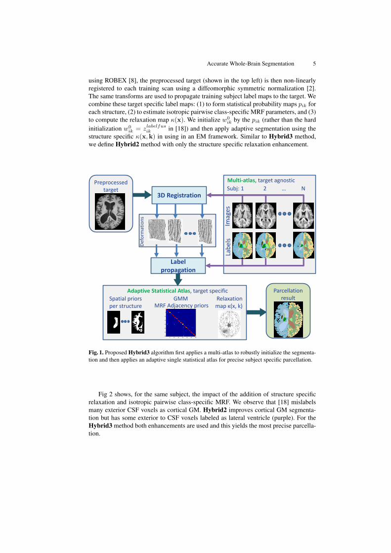
using ROBEX [8], the preprocessed target (shown in the top left) is then non-linearlyregistered to each training scan using a diffeomorphic symmetric normalization [2].The same transforms are used to propagate training subject label maps to the target. Wecombine these target specific label maps: (1) to form statistical probability maps pik foreach structure, (2) to estimate isotropic pairwise class-specific MRF parameters, and (3)to compute the relaxation map κ(x). We initialize w0
ik by the pik (rather than the hardinitialization w0
ik = zlabelfusik in [18]) and then apply adaptive segmentation using thestructure specific κ(x,k) in using in an EM framework. Similar to Hybrid3 method,we define Hybrid2 method with only the structure specific relaxation enhancement.
MA-EASA++ work flow
Def
orm
atio
ns
3D Registration
Label propagation
Multi-atlas, target agnostic
Imag
es
Lab
els
Subj: 1 2 … N
Parcellation result
Adaptive Statistical Atlas, target specific
Spatial priors per structure
GMM MRF Adjacency priors
Relaxation map κ(x, k)
5 10 15 20 25 30 35
5
10
15
20
25
30
35
Preprocessed target
Fig. 1. Proposed Hybrid3 algorithm first applies a multi-atlas to robustly initialize the segmenta-tion and then applies an adaptive single statistical atlas for precise subject specific parcellation.
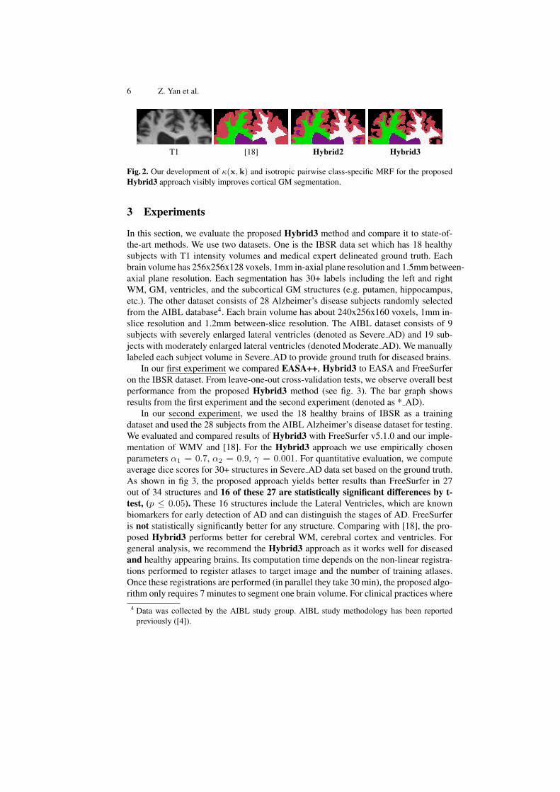
Fig 2 shows, for the same subject, the impact of the addition of structure specificrelaxation and isotropic pairwise class-specific MRF. We observe that [18] mislabelsmany exterior CSF voxels as cortical GM. Hybrid2 improves cortical GM segmenta-tion but has some exterior to CSF voxels labeled as lateral ventricle (purple). For theHybrid3 method both enhancements are used and this yields the most precise parcella-tion.
6 Z. Yan et al.
T1
Hybrid1
Hybrid2
Hybrid3
T1
T1
Hybrid1
Hybrid2
Hybrid3
[18]
T1
Hybrid1
Hybrid2
Hybrid3
Hybrid2
T1
Hybrid1
Hybrid2
Hybrid3
Hybrid3
Fig. 2. Our development of κ(x,k) and isotropic pairwise class-specific MRF for the proposedHybrid3 approach visibly improves cortical GM segmentation.
3 Experiments
In this section, we evaluate the proposed Hybrid3 method and compare it to state-of-the-art methods. We use two datasets. One is the IBSR data set which has 18 healthysubjects with T1 intensity volumes and medical expert delineated ground truth. Eachbrain volume has 256x256x128 voxels, 1mm in-axial plane resolution and 1.5mm between-axial plane resolution. Each segmentation has 30+ labels including the left and rightWM, GM, ventricles, and the subcortical GM structures (e.g. putamen, hippocampus,etc.). The other dataset consists of 28 Alzheimer’s disease subjects randomly selectedfrom the AIBL database4. Each brain volume has about 240x256x160 voxels, 1mm in-slice resolution and 1.2mm between-slice resolution. The AIBL dataset consists of 9subjects with severely enlarged lateral ventricles (denoted as Severe AD) and 19 sub-jects with moderately enlarged lateral ventricles (denoted Moderate AD). We manuallylabeled each subject volume in Severe AD to provide ground truth for diseased brains.
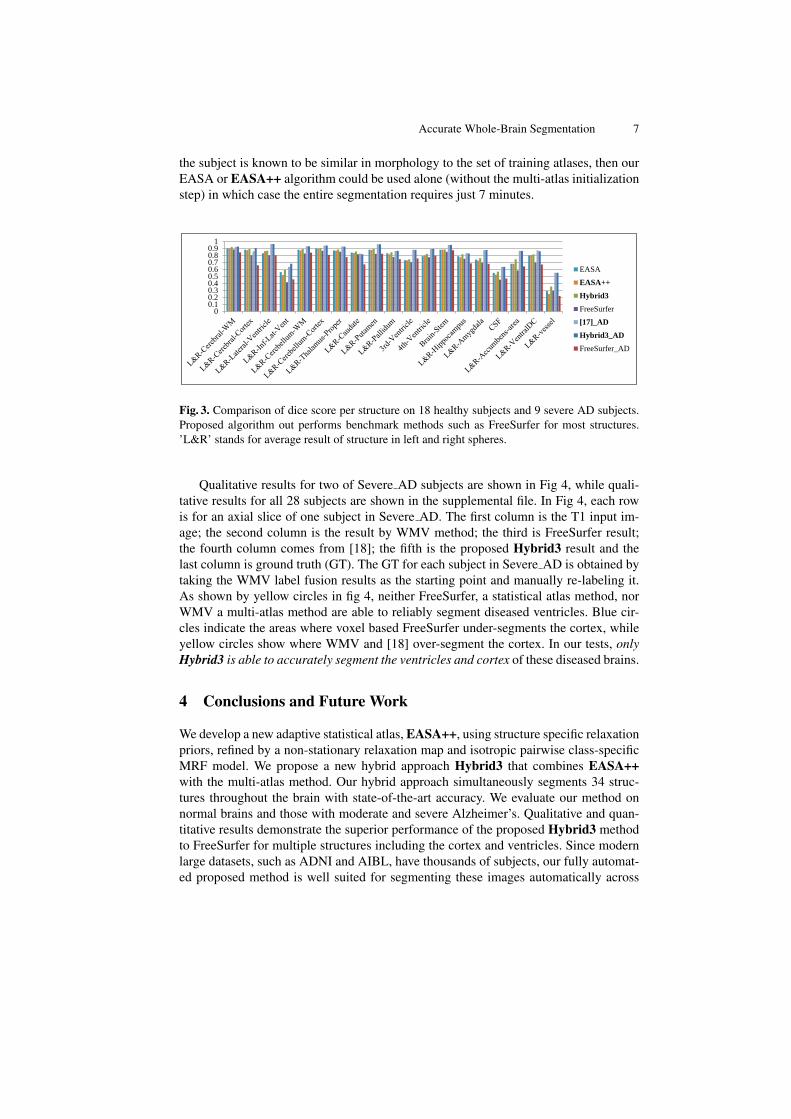
In our first experiment we compared EASA++, Hybrid3 to EASA and FreeSurferon the IBSR dataset. From leave-one-out cross-validation tests, we observe overall bestperformance from the proposed Hybrid3 method (see fig. 3). The bar graph showsresults from the first experiment and the second experiment (denoted as * AD).
In our second experiment, we used the 18 healthy brains of IBSR as a trainingdataset and used the 28 subjects from the AIBL Alzheimer’s disease dataset for testing.We evaluated and compared results of Hybrid3 with FreeSurfer v5.1.0 and our imple-mentation of WMV and [18]. For the Hybrid3 approach we use empirically chosenparameters α1 = 0.7, α2 = 0.9, γ = 0.001. For quantitative evaluation, we computeaverage dice scores for 30+ structures in Severe AD data set based on the ground truth.As shown in fig 3, the proposed approach yields better results than FreeSurfer in 27out of 34 structures and 16 of these 27 are statistically significant differences by t-test, (p ≤ 0.05). These 16 structures include the Lateral Ventricles, which are knownbiomarkers for early detection of AD and can distinguish the stages of AD. FreeSurferis not statistically significantly better for any structure. Comparing with [18], the pro-posed Hybrid3 performs better for cerebral WM, cerebral cortex and ventricles. Forgeneral analysis, we recommend the Hybrid3 approach as it works well for diseasedand healthy appearing brains. Its computation time depends on the non-linear registra-tions performed to register atlases to target image and the number of training atlases.Once these registrations are performed (in parallel they take 30 min), the proposed algo-rithm only requires 7 minutes to segment one brain volume. For clinical practices where
4 Data was collected by the AIBL study group. AIBL study methodology has been reportedpreviously ([4]).
Accurate Whole-Brain Segmentation 7
the subject is known to be similar in morphology to the set of training atlases, then ourEASA or EASA++ algorithm could be used alone (without the multi-atlas initializationstep) in which case the entire segmentation requires just 7 minutes.
00.10.20.30.40.50.60.70.80.9
1
EASA
EASA++
Hybrid3
FreeSurfer
[17]_AD
Hybrid3_AD
FreeSurfer_AD
Fig. 3. Comparison of dice score per structure on 18 healthy subjects and 9 severe AD subjects.Proposed algorithm out performs benchmark methods such as FreeSurfer for most structures.’L&R’ stands for average result of structure in left and right spheres.
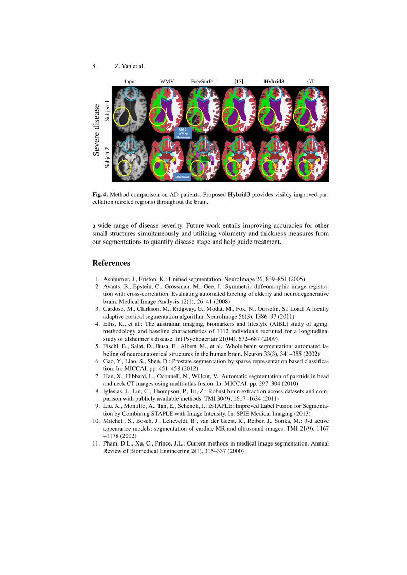
Qualitative results for two of Severe AD subjects are shown in Fig 4, while quali-tative results for all 28 subjects are shown in the supplemental file. In Fig 4, each rowis for an axial slice of one subject in Severe AD. The first column is the T1 input im-age; the second column is the result by WMV method; the third is FreeSurfer result;the fourth column comes from [18]; the fifth is the proposed Hybrid3 result and thelast column is ground truth (GT). The GT for each subject in Severe AD is obtained bytaking the WMV label fusion results as the starting point and manually re-labeling it.As shown by yellow circles in fig 4, neither FreeSurfer, a statistical atlas method, norWMV a multi-atlas method are able to reliably segment diseased ventricles. Blue cir-cles indicate the areas where voxel based FreeSurfer under-segments the cortex, whileyellow circles show where WMV and [18] over-segment the cortex. In our tests, onlyHybrid3 is able to accurately segment the ventricles and cortex of these diseased brains.
4 Conclusions and Future Work
We develop a new adaptive statistical atlas, EASA++, using structure specific relaxationpriors, refined by a non-stationary relaxation map and isotropic pairwise class-specificMRF model. We propose a new hybrid approach Hybrid3 that combines EASA++with the multi-atlas method. Our hybrid approach simultaneously segments 34 struc-tures throughout the brain with state-of-the-art accuracy. We evaluate our method onnormal brains and those with moderate and severe Alzheimer’s. Qualitative and quan-titative results demonstrate the superior performance of the proposed Hybrid3 methodto FreeSurfer for multiple structures including the cortex and ventricles. Since modernlarge datasets, such as ADNI and AIBL, have thousands of subjects, our fully automat-ed proposed method is well suited for segmenting these images automatically across
8 Z. Yan et al.
Input WMV GT Hybrid3 FreeSurfer
Subje
ct 1
S
ubje
ct 2
Sev
ere
dis
ease
Mo
derate d
isease
Input WMV Hybrid3 FreeSurfer
Subject 1
S
ubject 2
Unknown
GM or WM or
Unknown
[17]
Fig. 4. Method comparison on AD patients. Proposed Hybrid3 provides visibly improved par-cellation (circled regions) throughout the brain.
a wide range of disease severity. Future work entails improving accuracies for othersmall structures simultaneously and utilizing volumetry and thickness measures fromour segmentations to quantify disease stage and help guide treatment.
tion with cross-correlation: Evaluating automated labeling of elderly and neurodegenerativebrain. Medical Image Analysis 12(1), 26–41 (2008)
3. Cardoso, M., Clarkson, M., Ridgway, G., Modat, M., Fox, N., Ourselin, S.: Load: A locallyadaptive cortical segmentation algorithm. NeuroImage 56(3), 1386–97 (2011)
4. Ellis, K., et al.: The australian imaging, biomarkers and lifestyle (AIBL) study of aging:methodology and baseline characteristics of 1112 individuals recruited for a longitudinalstudy of alzheimer’s disease. Int Psychogeriatr 21(04), 672–687 (2009)
5. Fischl, B., Salat, D., Busa, E., Albert, M., et al.: Whole brain segmentation: automated la-beling of neuroanatomical structures in the human brain. Neuron 33(3), 341–355 (2002)
6. Gao, Y., Liao, S., Shen, D.: Prostate segmentation by sparse representation based classifica-tion. In: MICCAI. pp. 451–458 (2012)
7. Han, X., Hibbard, L., Oconnell, N., Willcut, V.: Automatic segmentation of parotids in headand neck CT images using multi-atlas fusion. In: MICCAI. pp. 297–304 (2010)
8. Iglesias, J., Liu, C., Thompson, P., Tu, Z.: Robust brain extraction across datasets and com-parison with publicly available methods. TMI 30(9), 1617–1634 (2011)
9. Liu, X., Montillo, A., Tan, E., Schenck, J.: iSTAPLE: Improved Label Fusion for Segmenta-tion by Combining STAPLE with Image Intensity. In: SPIE Medical Imaging (2013)
10. Mitchell, S., Bosch, J., Lelieveldt, B., van der Geest, R., Reiber, J., Sonka, M.: 3-d activeappearance models: segmentation of cardiac MR and ultrasound images. TMI 21(9), 1167–1178 (2002)
11. Pham, D.L., Xu, C., Prince, J.L.: Current methods in medical image segmentation. AnnualReview of Biomedical Engineering 2(1), 315–337 (2000)
Accurate Whole-Brain Segmentation 9
12. Rousseau, F., Habas, P., Studholme, C.: A supervised patch-based approach for human brainlabeling. TMI 30(10) (2011)
13. Shiee, N., Bazin, P., Cuzzocreo, J., Blitz, A., Pham, D.: Segmentation of brain images usingadaptive atlases with application to ventriculomegaly. In: IPMI. pp. 1–12 (2011)
14. Van Leemput, K., Maes, F., Vandermeulen, D., Suetens, P.: Automated model-based tissueclassification of MR images of the brain. TMI 18(10), 897–908 (1999)
15. Wang, H., Suh, J.W., Das, S.R., Pluta, J., Altinay, M., Yushkevich, P.A.: Regression-basedlabel fusion for multi-atlas segmentation. In: CVPR. pp. 1113–1120 (2011)
16. Warfield, S., Zou, K., Wells, W.: Simultaneous truth and performance level estimation (sta-ple): an algorithm for the validation of image segmentation. TMI 23(7), 903–921 (2004)
17. Wu, G., Kim, M., Wang, Q., Shen, D.: S-HAMMER: Hierarchical attribute-guided, symmet-ric diffeomorphic registration for MR brain images. Human brain mapping (2012)
18. Yan, Z., Zhang, S., Liu, X., Metaxas, D., Montillo, A., AIBL: Accurate segmentation ofbrain images into 34 structures combining a non-stationary adaptive statistical atlas and amulti-atlas with applications to alzheimer’s disease. In: ISBI (2013)