24
ACID BASE PHYSIOLOGY

ACID BASE PHYSIOLOGY
(a) To explain and describe acid-base chemistry using the Henderson-Hasselbach equation.
Definition of acid and base:
- “Bronsted-Lowry definition” is most commonly accepted in medicine: o Acid – Substance that donates a proton or hydrogen ion to another in solution o Base – Substance that accepts protons or hydrogen ions from another in solution
Overview of Hydrogen ion (H+):
- H+ is a hydrogen atom without its orbital electron → essentially a “proton” - In aqueous solution, H+ is hydrated to form a “hydronium ion” (H3O
+):
- “H+ activity” (AH+) is a measure of how many H+ “seem” to be present in solution → This is determined by:
o (i) Activity coefficient of H+ (g) o (ii) Concentration of H+ ([H+]) – Quantity of H+ “actually” present in solution
Overview of pH system:
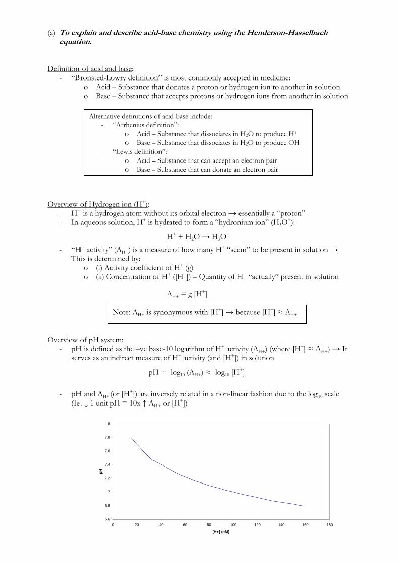
- pH is defined as the –ve base-10 logarithm of H+ activity (AH+) (where [H+] ≈ AH+) → It serves as an indirect measure of H+ activity (and [H+]) in solution
- pH and AH+ (or [H+]) are inversely related in a non-linear fashion due to the log10 scale (Ie. ↓ 1 unit pH = 10x ↑ AH+ or [H+])
Alternative definitions of acid-base include: - “Arrhenius definition”:
o Acid – Substance that dissociates in H2O to produce H+ o Base – Substance that dissociates in H2O to produce OH-
- “Lewis definition”: o Acid – Substance that can accept an electron pair o Base – Substance that can donate an electron pair
H+ + H2O → H3O+
AH+ = g [H+]
Note: AH+ is synonymous with [H+] → because [H+] ≈ AH+
pH = -log10 (AH+) ≈ -log10 [H+]
6.6
6.8
7
7.2
7.4
7.6
7.8
8
0 20 40 60 80 100 120 140 160 180
[H+] (nM)
pH
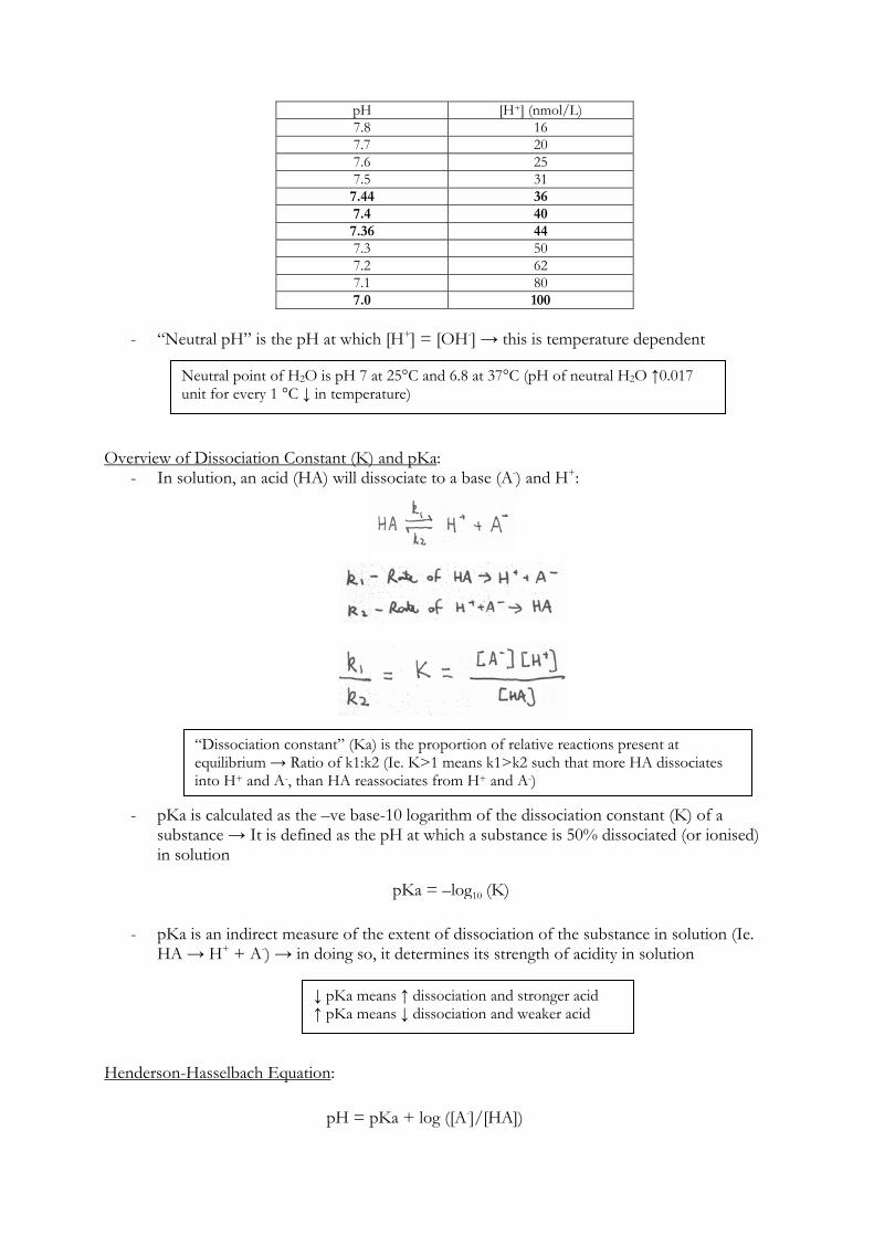
pH [H+] (nmol/L) 7.8 16 7.7 20 7.6 25 7.5 31
7.44 36 7.4 40 7.36 44 7.3 50 7.2 62 7.1 80 7.0 100
- “Neutral pH” is the pH at which [H+] = [OH-] → this is temperature dependent
Overview of Dissociation Constant (K) and pKa:
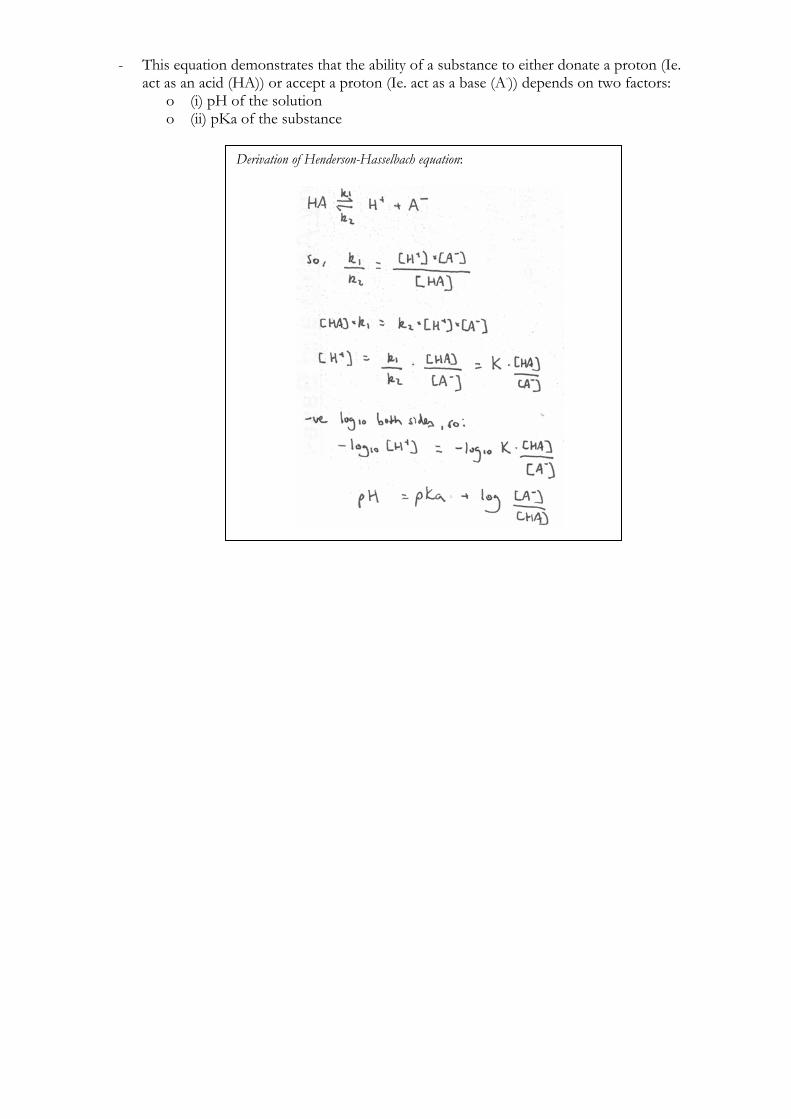
- In solution, an acid (HA) will dissociate to a base (A-) and H+:
- pKa is calculated as the –ve base-10 logarithm of the dissociation constant (K) of a substance → It is defined as the pH at which a substance is 50% dissociated (or ionised) in solution
- pKa is an indirect measure of the extent of dissociation of the substance in solution (Ie. HA → H+ + A-) → in doing so, it determines its strength of acidity in solution
Henderson-Hasselbach Equation:
Neutral point of H2O is pH 7 at 25°C and 6.8 at 37°C (pH of neutral H2O ↑0.017 unit for every 1 °C ↓ in temperature)
pKa = –log10 (K)
↓ pKa means ↑ dissociation and stronger acid ↑ pKa means ↓ dissociation and weaker acid
“Dissociation constant” (Ka) is the proportion of relative reactions present at equilibrium → Ratio of k1:k2 (Ie. K>1 means k1>k2 such that more HA dissociates into H+ and A-, than HA reassociates from H+ and A-)
pH = pKa + log ([A-]/[HA])
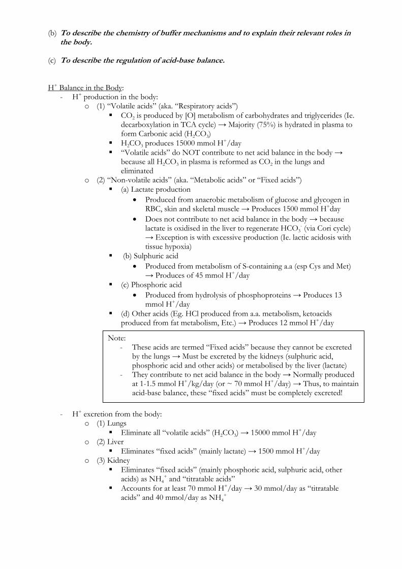
- This equation demonstrates that the ability of a substance to either donate a proton (Ie. act as an acid (HA)) or accept a proton (Ie. act as a base (A-)) depends on two factors:
o (i) pH of the solution o (ii) pKa of the substance
Derivation of Henderson-Hasselbach equation:
(b) To describe the chemistry of buffer mechanisms and to explain their relevant roles in the body.
(c) To describe the regulation of acid-base balance. H+ Balance in the Body:
- H+ production in the body: o (1) “Volatile acids” (aka. “Respiratory acids”)
CO2 is produced by [O] metabolism of carbohydrates and triglycerides (Ie. decarboxylation in TCA cycle) → Majority (75%) is hydrated in plasma to form Carbonic acid (H2CO3)
H2CO3 produces 15000 mmol H+/day “Volatile acids” do NOT contribute to net acid balance in the body →
because all H2CO3 in plasma is reformed as CO2 in the lungs and eliminated
o (2) “Non-volatile acids” (aka. “Metabolic acids” or “Fixed acids”) (a) Lactate production
Produced from anaerobic metabolism of glucose and glycogen in RBC, skin and skeletal muscle → Produces 1500 mmol H+day
Does not contribute to net acid balance in the body → because lactate is oxidised in the liver to regenerate HCO3
- (via Cori cycle) → Exception is with excessive production (Ie. lactic acidosis with tissue hypoxia)
(b) Sulphuric acid Produced from metabolism of S-containing a.a (esp Cys and Met)
→ Produces of 45 mmol H+/day (c) Phosphoric acid
Produced from hydrolysis of phosphoproteins → Produces 13 mmol H+/day
(d) Other acids (Eg. HCl produced from a.a. metabolism, ketoacids produced from fat metabolism, Etc.) → Produces 12 mmol H+/day
- H+ excretion from the body: o (1) Lungs
Eliminate all “volatile acids” (H2CO3) → 15000 mmol H+/day o (2) Liver
Eliminates “fixed acids” (mainly lactate) → 1500 mmol H+/day o (3) Kidney
Eliminates “fixed acids” (mainly phosphoric acid, sulphuric acid, other acids) as NH4
+ and “titratable acids” Accounts for at least 70 mmol H+/day → 30 mmol/day as “titratable
acids” and 40 mmol/day as NH4+
Note: - These acids are termed “Fixed acids” because they cannot be excreted
by the lungs → Must be excreted by the kidneys (sulphuric acid, phosphoric acid and other acids) or metabolised by the liver (lactate)
- They contribute to net acid balance in the body → Normally produced at 1-1.5 mmol H+/kg/day (or ~ 70 mmol H+/day) → Thus, to maintain acid-base balance, these “fixed acids” must be completely excreted!
- Acid-base balance in the body: o To maintain acid-base balance (and pH) in the body → Daily acid production
must EQUAL daily acid excretion → So “Net acid balance” must equal ZERO
o Net acid production (NAP): Normally ~ 70 mmol of H+ is produced daily from “fixed acids” (esp
phosphoric acid, sulphuric acid, other acids) o Net acid excretion (NAE):
Normally all “fixed acids” produced are excreted by the kidneys (~ 70 meq/day) → 30 mmol/day as “titratable acids” and 40 mmol/day as NH4
+ Almost all filtered HCO3
- is reabsorbed → HCO3- excretion ~ 0
mmol/day Overview of Acid-Base Homeostasis:
- [H+] in body fluid is precisely regulated → maintained at low plasma [ ] of 40 nmol/L (normal range 35-45 nmol/L) to keep extracellular pH ~ 7.4 (normal range pH 7.35-7.44) and intracellular pH ~ 6.8
- This tight regulation of [H+] and pH is achieved by the following processes: o (1) Buffering (1st line of defence) – Systems that immediately minimise changes in
pH in the event that an acid or base is added to the body o (2) Compensation (2nd line of defence) – Physiological processes that attempts to
normalise pH by restoring the HCO3-/PCO2 ratio to normal → Either respiratory
(rapid; mins-hrs) or renal (slow; hrs-days) → pH generally not completely restored to 7.4
NAE = NH4+ + “Titratable acids” – HCO3
-
Derangements of [H+] and pH can result in systemic effects (Eg. altered CNS reflexes, CVS depression, Etc.; See below) as a result of direct intracellular disturbances:
- (i) Altered protein function (Eg. enzyme activity, transporter activity, Etc.) - (ii) Altered membrane excitability - (iii) Disruption in metabolic pathways (esp energy production) - (iv) Ion trapping of biological molecules in compartments, Etc.
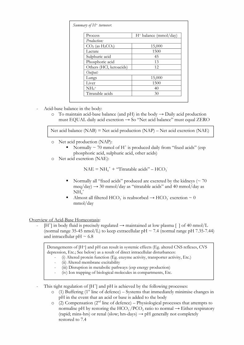
Summary of H+ turnover:
Process H+ balance (mmol/day) Production: CO2 (as H2CO3) 15,000 Lactate 1500 Sulphuric acid 45 Phosphoric acid 13 Others (HCl, ketoacids) 12 Output: Lungs 15,000 Liver 1500 NH4+ 40 Titratable acids 30
Net acid balance (NAB) = Net acid production (NAP) – Net acid excretion (NAE)
o (3) Correction (3rd line of defence) – Mechanism that corrects acid-base derangement through correction of primary disorder
Acid-Base Homeostasis: Buffering Overview of buffer systems:
- A “buffer” is a solution containing a weak acid and its conjugate base → Resists profound pH changes when exposed to a stronger acid or base by reversibly binding H+
Buffer + H+ ↔ H.Buffer
- Effectiveness of a buffer is dependent on:
o (i) Amount of buffer present → ↑ [buffer] ↑ the effectiveness of the buffer o (ii) Buffer’s pKa and pH of the carrier solution → Majority of buffering activity
(80%) occurs within +/- 1 pH of the buffer’s pKa, with the maximal effect at its pKa (thus buffer effectiveness ↑ if buffer pKa and solution pH within +/- 1 unit)
o (iii) “Open” (physiological) vs “closed” (chemical) buffer system → Buffer effectiveness ↑ with open systems
Buffering systems of the body:
- There are two types of buffer systems in body: o (1) Bicarbonate buffers (H2CO3-HCO3
- buffer system) → Can only buffer “fixed/metabolic” acids (because it cannot buffer itself!)
o (2) Non-bicarbonate buffers (Hb, Phosphate and Protein buffer systems) → Can buffer both “respiratory” and “fixed/metabolic” acids
- Extracellular buffering: o (1) Blood – Mainly HCO3
- and Hb (proteins and PO4- have minor roles)
(i) RBC – 1°ly Hb (35%; buffers 90% of H2CO3) and HCO3- (18%);
proteins and PO4- are negligible
(ii) Plasma – 1°ly HCO3- (35%; buffers 70% of “metabolic” acids), protein
(7%) and PO4- (2%)
o (2) ISF – 1°ly HCO3- (ISF has ↑ capacity to buffer “metabolic” acids than blood
because ISF volume (and HCO3- content) is 3x ↑ cf. blood)
- Intracellular buffering: o (i) 1°ly protein and PO4
3- → because they occur at ↑ [ ] intracellullarly and have pKa’s closer to intracellular pH (~ 6.8)
o (ii) Fixed acid extrusion → Extrusion of IC H+ in exchange for a strong electrolyte (Na+, Cl-, lactate) via an anti-port transporter
o (iii) Organellar buffering → Sequester or release H+ from IC organelle o (iv) Metabolic buffering → Alter production of acidic metabolites
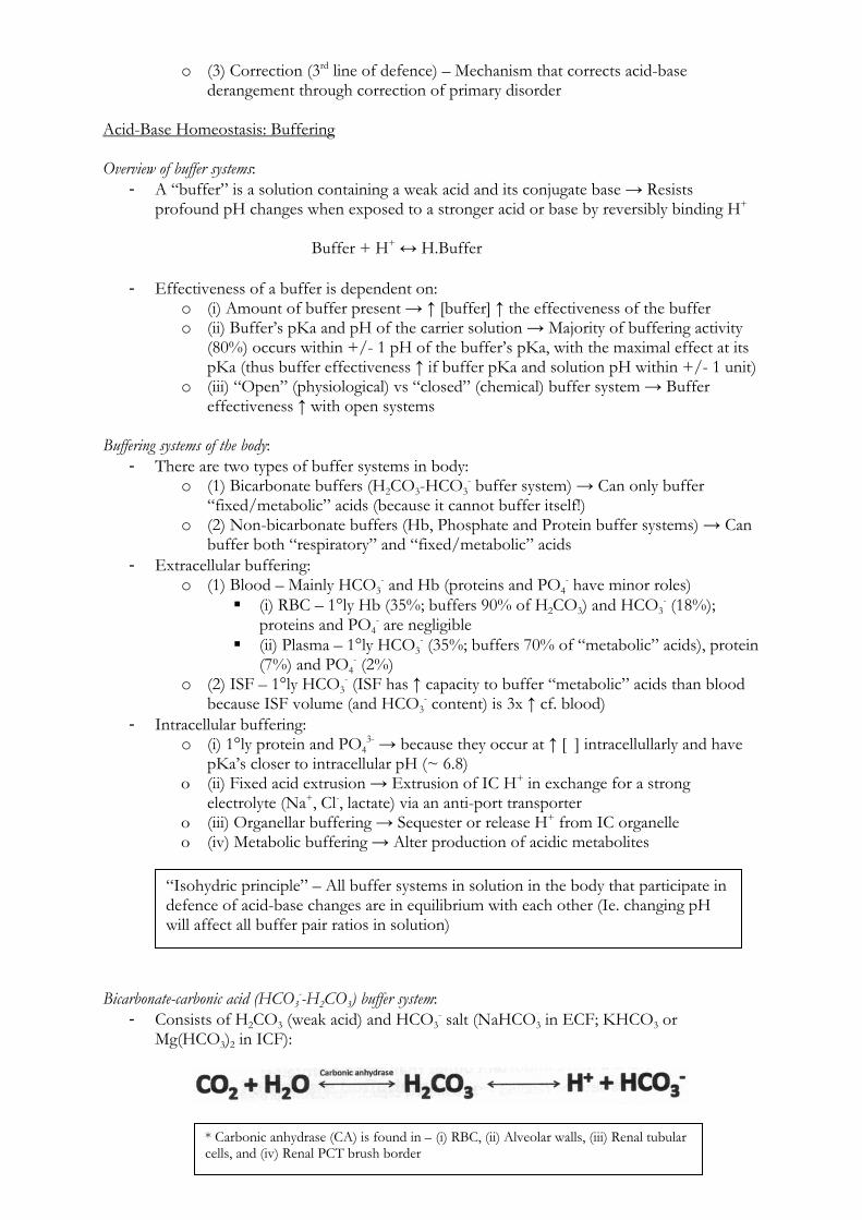
Bicarbonate-carbonic acid (HCO3
--H2CO3) buffer system: - Consists of H2CO3 (weak acid) and HCO3
- salt (NaHCO3 in ECF; KHCO3 or Mg(HCO3)2 in ICF):
* Carbonic anhydrase (CA) is found in – (i) RBC, (ii) Alveolar walls, (iii) Renal tubular
cells, and (iv) Renal PCT brush border
“Isohydric principle” – All buffer systems in solution in the body that participate in defence of acid-base changes are in equilibrium with each other (Ie. changing pH will affect all buffer pair ratios in solution)
o In presence of strong acid – H+ from acid buffered by HCO3
- → shifts to production of CO2 production (via H2CO3) → CO2 excreted from lungs
o In presence of strong base – Buffered by H2CO3 → Forms HCO3- which is
excreted from kidneys - Most important buffer in ECF (RBC, plasma and ISF) → 80% buffering capacity:
o It’s low pKa (6.1) relative to physiological pH and relatively small amounts in ECF ↓ its effectiveness as a buffer system in ECF
o BUT this is offset by the fact that the system is “open” system → Can be controlled independently by the: (i) Lungs – Excretion of CO2 (or H2CO3) regulated rapidly by changes in
minute ventilation (ii) Kidneys – Excretion of HCO3
- and H+ regulated more slowly Haemoglobin (Hb) buffer system:
- Hb is an intracellular protein in RBCs BUT acts as the major non-HCO3- buffer in ECF
- Mechanisms for buffering capacity of Hb: o (1) ↑ [Hb] within RBCs (150 g/L) → Large amount of buffer present o (2) Structure of Hb
(a) Multiple (38) Histidine residues with Imidazole side chain (pKa 6.8) on globin chains of Hb → Anionic sites on Imidazole binds H+ → Contributes to MAIN buffering capacity of Hb at physiological pH
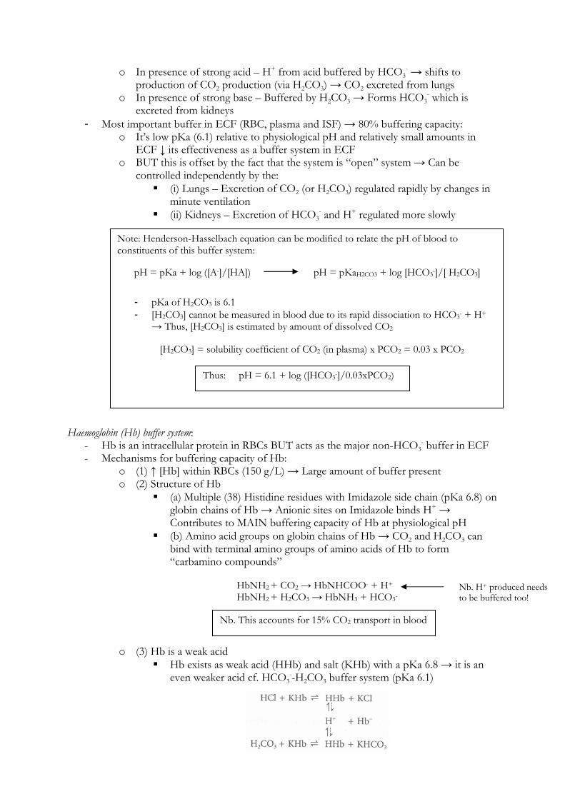
(b) Amino acid groups on globin chains of Hb → CO2 and H2CO3 can bind with terminal amino groups of amino acids of Hb to form “carbamino compounds”
o (3) Hb is a weak acid Hb exists as weak acid (HHb) and salt (KHb) with a pKa 6.8 → it is an
even weaker acid cf. HCO3--H2CO3 buffer system (pKa 6.1)
Note: Henderson-Hasselbach equation can be modified to relate the pH of blood to constituents of this buffer system:
- pKa of H2CO3 is 6.1 - [H2CO3] cannot be measured in blood due to its rapid dissociation to HCO3- + H+
→ Thus, [H2CO3] is estimated by amount of dissolved CO2
pH = pKa + log ([A-]/[HA]) pH = pKaH2CO3 + log [HCO3-]/[ H2CO3]
[H2CO3] = solubility coefficient of CO2 (in plasma) x PCO2 = 0.03 x PCO2
Thus: pH = 6.1 + log ([HCO3-]/0.03xPCO2)
HbNH2 + CO2 → HbNHCOO- + H+ HbNH2 + H2CO3 → HbNH3 + HCO3-
Nb. This accounts for 15% CO2 transport in blood
Nb. H+ produced needs to be buffered too!

Thus, in presence of ECF acid → Hb buffers extracellular H+ → leads to HCO3
- formation within RBC → HCO3- accumulates and diffuses down
its electrochemical gradient out of RBC → ↑ plasma HCO3-
o (4) RBC contains carbonic anhydrase and has ↑ solubility for CO2: Hb contributes to > 90% of blood capacity to buffer CO2 (as H2CO3) Tissue CO2 diffuses into RBC whereby it is hydrated to form H2CO3 by
CA → (i) H+ produced then preferentially binds DeoxyHb (to form Reduced Hb), thus allowing offloading of O2 to tissues, and (ii) HCO3
- produced diffuses down its electrochemical gradient out of RBC → ↑ plasma HCO3
- o (5) DeoxyHb is a better buffer than OxyHb (Haldane Effect)
DeoxyHb (pKa 8.2) is a better buffer than OxyHb (pKa 6.6) because it is a weaker acid and dissociates to a greater extent
In tissue capillaries → OxyHb unloads O2 to form DeoxyHb → This facilitates:
Uptake and buffering of extracellular H+ (as reduced Hb; HHb) produced from hydration of tissue CO2 and dissociation of H2CO3 → 30% Haldane effect
Uptake of tissue CO2 (as carbamino compounds) → 70% Haldane effect
Thus, pH venous blood is only slightly more acidic than arterial blood in spite of large tissue CO2 produced daily!
Protein buffer system:
- Amino acid side chains can buffer H+: o Most amino groups (pKa 9) and carboxyl groups (pKa 2) have pKa very far from
physiological pH → Contributes little to buffering o Imidazole group on Histidine (pKa 6.8) has the closest pKa to physiological pH
→ Most important amino acid in proteins for buffering (esp in Hb; See above) - Much more important buffer in ICF (cf. ECF) because → (i) [proteins] intracellularly are
much ↑, and (ii) intracellular pH is more acidic (Ie. closer to pKa of amino acid moieties) Phosphate buffer system:
- Phosphoric acid is a tribasic acid:
“Isohydric buffering” – For each mmol OxyHb that is reduced, 0.7mmol H+ can be taken up by Hb, and 0.7mmol CO2 can enter venous system without significantly changing pH
Note: Despite being a protein, Hb has 6x ↑ buffering capacity than protein proteins because - (i) [Hb] is 2x of plasma protein - (ii) Hb has 3x buffering capacity gram per gram that of plasma protein → due to 3x ↑
content of imidazole-containing histidine residues
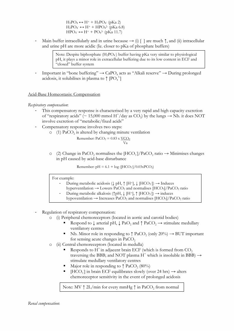
- Main buffer intracellularly and in urine because → (i) [ ] are much ↑, and (ii) intracellular and urine pH are more acidic (Ie. closer to pKa of phosphate buffers)
- Important in “bone buffering” → CaPO4 acts as “Alkali reserve” → During prolonged acidosis, it solubilises in plasma to ↑ [PO4
3-] Acid-Base Homeostasis: Compensation Respiratory compensation:
- This compensatory response is characterised by a very rapid and high capacity excretion of “respiratory acids” (~ 15,000 mmol H+/day as CO2) by the lungs → Nb. it does NOT involve excretion of “metabolic/fixed acids”
- Compensatory response involves two steps: o (1) PaCO2 is altered by changing minute ventilation
o (2) Change in PaCO2 normalises the [HCO3-]/PaCO2 ratio → Minimises changes
in pH caused by acid-base disturbance
- Regulation of respiratory compensation: o (i) Peripheral chemoreceptors (located in aortic and carotid bodies)
Respond to ↓ arterial pH, ↓ PaO2 and ↑ PaCO2 → stimulate medullary ventilatory centres
Nb. Minor role in responding to ↑ PaCO2 (only 20%) → BUT important for sensing acute changes in PaCO2
o (ii) Central chemoreceptors (located in medulla) Responds to H+
in adjacent brain ECF (which is formed from CO2 traversing the BBB; and NOT plasma H+ which is insoluble in BBB) → stimulate medullary ventilatory centres
Major role in responding to ↑ PaCO2 (80%) [HCO3
-] in brain ECF equilibrates slowly (over 24 hrs) → alters chemoreceptor sensitivity in the event of prolonged acidosis
Renal compensation:
H3PO4 ↔ H+ + H2PO4- (pKa 2) H2PO4- ↔ H+ + HPO42- (pKa 6.8) HPO4- ↔ H+ + PO43- (pKa 11.7)
Note: Despite biphosphate (H2PO4-) buffer having pKa very similar to physiological pH, it plays a minor role in extracellular buffering due to its low content in ECF and “closed” buffer system
Remember: PaCO2 = 0.83 x VCO2
Va
Remember: pH = 6.1 + log ([HCO3-]/0.03xPCO2)
For example: - During metabolic acidosis (↓ pH, ↑ [H+], ↓ [HCO3-]) → Induces
hyperventilation → Lowers PaCO2 and normalises [HCO3]/PaCO2 ratio - During metabolic alkalosis (↑pH, ↓ [H+], ↑ [HCO3-]) → induces
hypoventilation → Increases PaCO2 and normalises [HCO3]/PaCO2 ratio
Note: MV ↑ 2L/min for every mmHg ↑ in PaCO2 from normal

- This compensatory response is characterised by a very slow (7-10 days) and low capacity excretion of “fixed/metabolic acids” (~ 70 mmol H+/day) → normalises the [HCO3
-
]/PaCO2 ratio → Minimises changes in pH caused by acid-base disturbance
- Renal regulation of acid-base balance involves tubular H+ secretion: o At least 4390 mmol H+ is actively secreted by kidneys each day:
(i) Majority is used to reabsorb filtered HCO3- → 4320 mmol H+ is
actively secreted by the tubular system each day to facilitate reabsorption of all 4320 mmol HCO3
- filtered by the glomerulus (24 mmol HCO3-/L x
180 L/day = 4320 mmol) → this does not lead to net excretion of H+ in urine or addition of new HCO3
- to blood (ii) Additional 70 mmol H+ is actively secreted to excrete
“fixed/metabolic” acids to achieve a “net acid balance” of zero → 30 mmol/day as filtered buffers (titratable acids) and 40 mmol/day as manufactured buffers (NH4
+) → this leads to net excretion of H+ in urine (and addition of new HCO3
- to blood) o During acidosis:
(a) Tubular H+ secretion leads to all filtered HCO3- being reabsorbed
(b) Excess H+ is secreted and lost in urine bound to non-absorbable buffers (filtered buffers and manufactured buffers) → results in an additional 300 mmol H+
excreted in urine per day (mainly bound to NH4+)
and an additional HCO3- added to blood
o During alkalosis, excess plasma HCO3- (> 28 mmol/L) is excreted in urine by:
(a) Loss of filtered HCO3- (Ie. not all is reabsorbed in tubular system)
(b) Secretion of HCO3- from “type B intercalated cells” in CCD
- Renal regulation of acid-base balance involves the following processes: o (1) Secretion of H+ associated with HCO3
- reabsorption by renal tubules: Majority of HCO3
- is reabsorbed by proximal tubular system → PCT (85%) and TAL (10%):
High capacity and low gradient system → Because 3900 mmol H+ secreted/day (and 95% filtered HCO3
- reabsorbed) BUT lowest urine pH achieved is only 7
In the brush border membrane of PCT tubular luminal cells:
Nb. This is the ONLY means of excreting “fixed acids” (Except for lactate → metabolised by liver)
Summary of “net acid excretion”:
Normal Acidosis Alkalosis Titratable acids (mmol/day) 30 40 0 NH4+ (mmol/day) 40 160 0 HCO3- (mmol/day) 0 0 80 NAE / “New” HCO3- added (mmol/day)
+70 200 -80
Urine pH 6.0 4.6 8.0
NAE = NH4+ + “Titratable acids” – HCO3-
Note: Filtered H+ at the glomerulus accounts for a very small % of excreted H+ → 36 nM x 180 L/day = 6.48 umol of excreted H+ per day
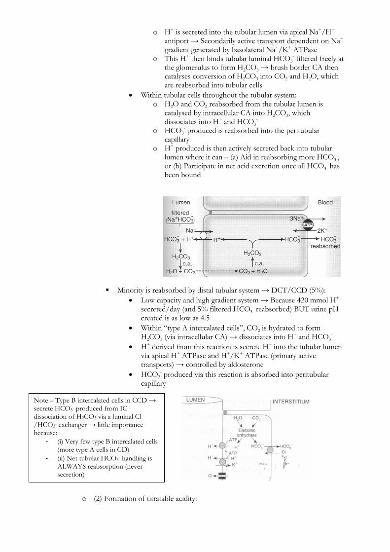
o H+ is secreted into the tubular lumen via apical Na+/H+ antiport → Secondarily active transport dependent on Na+ gradient generated by basolateral Na+/K+ ATPase
o This H+ then binds tubular luminal HCO3- filtered freely at
the glomerulus to form H2CO3 → brush border CA then catalyses conversion of H2CO3 into CO2 and H2O, which are reabsorbed into tubular cells
Within tubular cells throughout the tubular system: o H2O and CO2 reabsorbed from the tubular lumen is
catalysed by intracellular CA into H2CO3, which dissociates into H+ and HCO3
- o HCO3
- produced is reabsorbed into the peritubular capillary
o H+ produced is then actively secreted back into tubular lumen where it can – (a) Aid in reabsorbing more HCO3
-, or (b) Participate in net acid excretion once all HCO3
- has been bound
Minority is reabsorbed by distal tubular system → DCT/CCD (5%): Low capacity and high gradient system → Because 420 mmol H+
secreted/day (and 5% filtered HCO3- reabsorbed) BUT urine pH
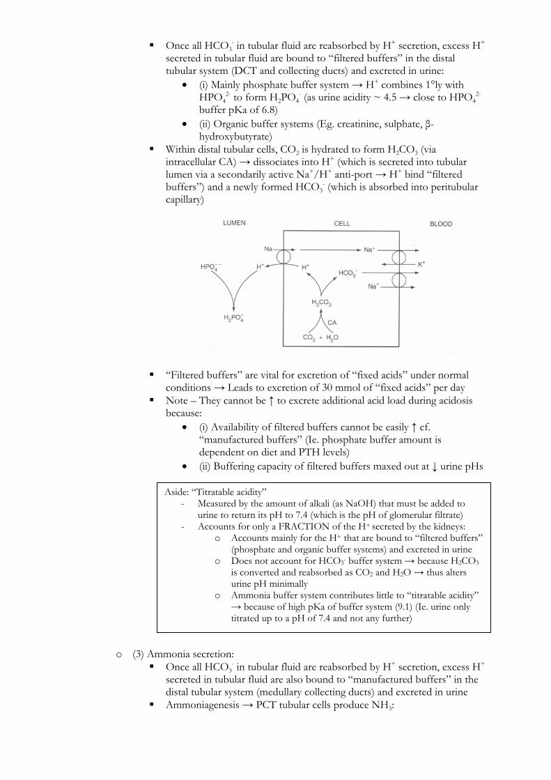
created is as low as 4.5 Within “type A intercalated cells”, CO2 is hydrated to form
H2CO3 (via intracellular CA) → dissociates into H+ and HCO3-
H+ derived from this reaction is secrete H+ into the tubular lumen via apical H+ ATPase and H+/K+ ATPase (primary active transports) → controlled by aldosterone
HCO3- produced via this reaction is absorbed into peritubular
capillary
o (2) Formation of titratable acidity:
Note – Type B intercalated cells in CCD → secrete HCO3- produced from IC dissociation of H2CO3 via a luminal Cl-/HCO3- exchanger → little importance because:
- (i) Very few type B intercalated cells (more type A cells in CD)
- (ii) Net tubular HCO3- handling is ALWAYS reabsorption (never secretion)
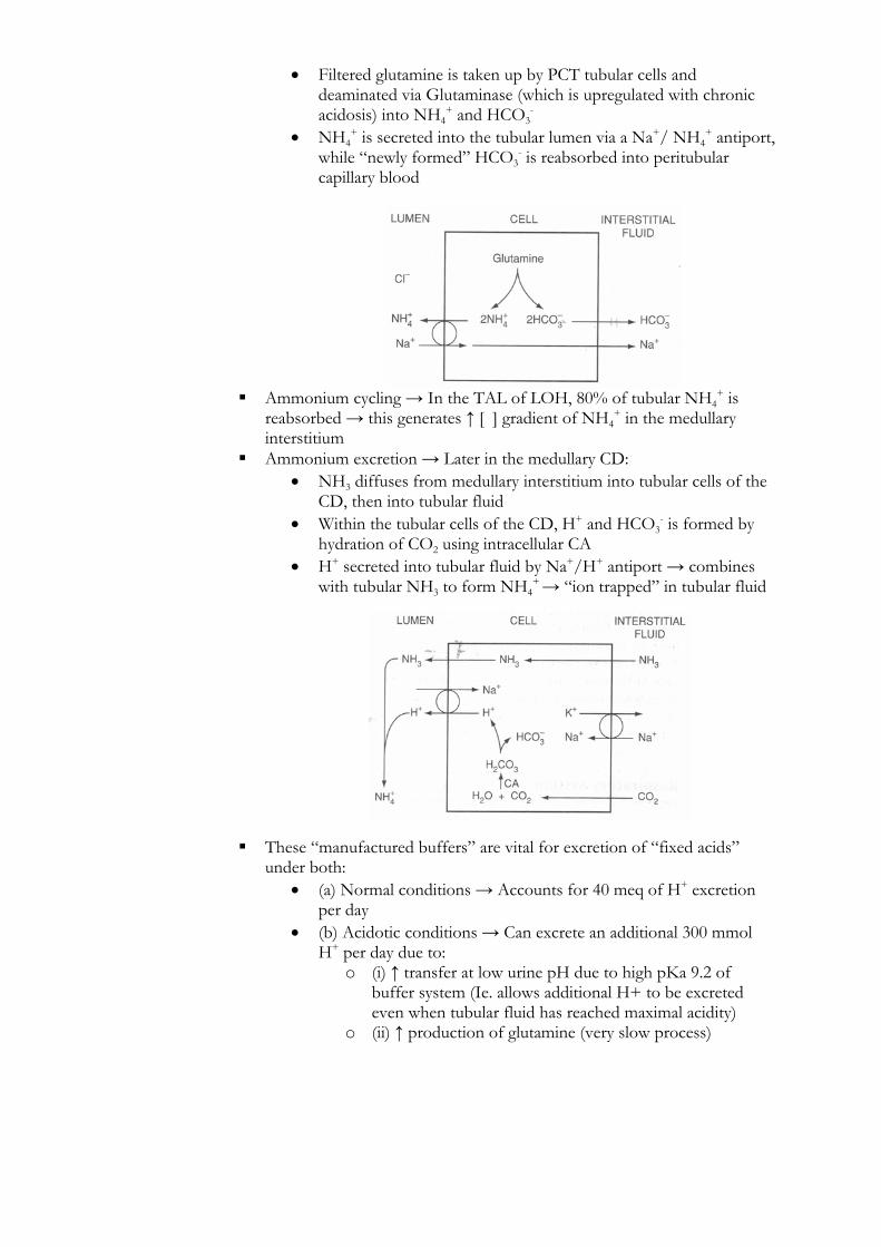
Once all HCO3- in tubular fluid are reabsorbed by H+ secretion, excess H+
secreted in tubular fluid are bound to “filtered buffers” in the distal tubular system (DCT and collecting ducts) and excreted in urine:
(i) Mainly phosphate buffer system → H+ combines 1°ly with HPO4
2- to form H2PO4- (as urine acidity ~ 4.5 → close to HPO4
2- buffer pKa of 6.8)
(ii) Organic buffer systems (Eg. creatinine, sulphate, β-hydroxybutyrate)
Within distal tubular cells, CO2 is hydrated to form H2CO3 (via intracellular CA) → dissociates into H+ (which is secreted into tubular lumen via a secondarily active Na+/H+ anti-port → H+ bind “filtered buffers”) and a newly formed HCO3
- (which is absorbed into peritubular capillary)
“Filtered buffers” are vital for excretion of “fixed acids” under normal conditions → Leads to excretion of 30 mmol of “fixed acids” per day
Note – They cannot be ↑ to excrete additional acid load during acidosis because:
(i) Availability of filtered buffers cannot be easily ↑ cf. “manufactured buffers” (Ie. phosphate buffer amount is dependent on diet and PTH levels)
(ii) Buffering capacity of filtered buffers maxed out at ↓ urine pHs
o (3) Ammonia secretion: Once all HCO3
- in tubular fluid are reabsorbed by H+ secretion, excess H+ secreted in tubular fluid are also bound to “manufactured buffers” in the distal tubular system (medullary collecting ducts) and excreted in urine
Ammoniagenesis → PCT tubular cells produce NH3:
Aside: “Titratable acidity” - Measured by the amount of alkali (as NaOH) that must be added to
urine to return its pH to 7.4 (which is the pH of glomerular filtrate) - Accounts for only a FRACTION of the H+ secreted by the kidneys:
o Accounts mainly for the H+ that are bound to “filtered buffers” (phosphate and organic buffer systems) and excreted in urine
o Does not account for HCO3- buffer system → because H2CO3 is converted and reabsorbed as CO2 and H2O → thus alters urine pH minimally
o Ammonia buffer system contributes little to “titratable acidity” → because of high pKa of buffer system (9.1) (Ie. urine only titrated up to a pH of 7.4 and not any further)
Filtered glutamine is taken up by PCT tubular cells and deaminated via Glutaminase (which is upregulated with chronic acidosis) into NH4
+ and HCO3-
NH4+ is secreted into the tubular lumen via a Na+/ NH4
+ antiport, while “newly formed” HCO3
- is reabsorbed into peritubular capillary blood
Ammonium cycling → In the TAL of LOH, 80% of tubular NH4+ is
reabsorbed → this generates ↑ [ ] gradient of NH4+ in the medullary
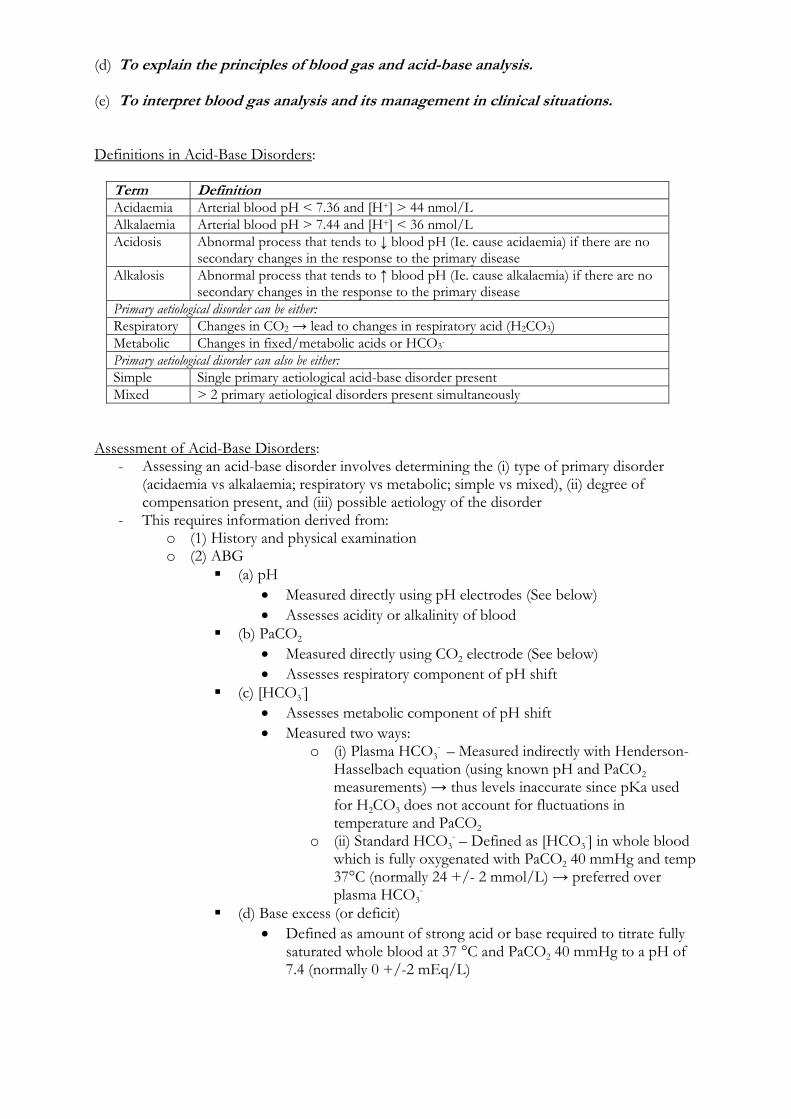
interstitium Ammonium excretion → Later in the medullary CD:
NH3 diffuses from medullary interstitium into tubular cells of the CD, then into tubular fluid
Within the tubular cells of the CD, H+ and HCO3- is formed by
hydration of CO2 using intracellular CA H+ secreted into tubular fluid by Na+/H+ antiport → combines
with tubular NH3 to form NH4+ → “ion trapped” in tubular fluid
These “manufactured buffers” are vital for excretion of “fixed acids” under both:
(a) Normal conditions → Accounts for 40 meq of H+ excretion per day
(b) Acidotic conditions → Can excrete an additional 300 mmol H+ per day due to:
o (i) ↑ transfer at low urine pH due to high pKa 9.2 of buffer system (Ie. allows additional H+ to be excreted even when tubular fluid has reached maximal acidity)
o (ii) ↑ production of glutamine (very slow process)

- Minimum urinary pH: o Only a small amount of H+ can be excreted in its free form → because active
transport of H+ secretion is inhibited at high urinary [H+] → thus lowest urinary pH achieved is 4.4
o As a result, H+ secretion and excretion in urine is dependent on binding to “urinary buffers” → (i) HCO3
- buffer (pKa 6.1), (ii) HPO42- buffer (pKa 6.8), and
(iii) NH3 buffer (pKa 9.1) o In the absence these urinary buffer systems → urine pH of 4.4 would be reached
very rapidly and any further H+ secretion would cease Clinical Effects of Acid-Base Changes:
System Acidosis Alkalosis CVS - Direct –ve inotropic effect:
- Due to ↓ slow inward Ca2+ current and ↓ Ca2+ release from SR
- Initially opposed by medullary catecholamine response → until pH 7.2 then –ve inotropy
- ↑ SNS activity (medullary catecholamine release): - Offsets –ve inotropy - BUT ↑ cardiac arrhythmias, ↑ SVR and
renal/splanchnic vasoconstriction - Cardiac arrhythmias:
- Due to ↓ IC [K+] (↑ RMP of pacemaker cells) and ↑ adrenal medulla catecholamine release
- Vascular effect - Mild acidosis → ↑ SVR and
renal/splanchnic vasoconstriction (due to medullary SNS response)
- ↑ acidosis → Vasodilation (skin, skeletal muscle, heart) and ↓ SVR
- Pulmonary vasoconstriction → HTN
- ↑ coronary VC and ↑ SVR
Respiratory - ↑ MV → ↑ response with respiratory acidosis cf. metabolic acidosis b/c CO2 more permeable than H+ at BBB - Bronchodilation (due to hypercapnoea) - Right shift of O2 HDC → ↑ O2 tissue delivery
- Opposite effects to acidosis
CNS Impairs LOC due to changes in CBF and ICP Epilepsy GIT - Splanchnic vasoconstriction
- ↓ GIT motility
Electrolyte - ↑ free ionised serum Ca2+ → due H+ competing for –ve binding on albumin (chronically, 2° to Ca2+
mobilisation from bone) - ↑ serum K+ → EC H+ exchanged for IC K+ (0.6 mmol ↑ [K+] per 0.1 ↓ pH)
- Opposite effects to acidosis
Note: Control of H+ secretion and excretion is ↑ by the following factors: - (1) Raised PaCO2 - (2) High EC [H+] → Stimulates glutamine production - (3) K+ and Cl- depletion → ↑ renal HCO3- reabsorption - (4) Inhibition of CA → H+ secretion is inhibited - (5) Aldosterone (and cortisol) → Upregulates H+ and H+/K+
ATPases - (6) ECF volume depletion → ↑ renal HCO3- reabsorption - (7) ↓ GFR → ↓ filtration of HCO3-
(d) To explain the principles of blood gas and acid-base analysis. (e) To interpret blood gas analysis and its management in clinical situations. Definitions in Acid-Base Disorders:
Term Definition Acidaemia Arterial blood pH < 7.36 and [H+] > 44 nmol/L Alkalaemia Arterial blood pH > 7.44 and [H+] < 36 nmol/L Acidosis Abnormal process that tends to ↓ blood pH (Ie. cause acidaemia) if there are no
secondary changes in the response to the primary disease Alkalosis Abnormal process that tends to ↑ blood pH (Ie. cause alkalaemia) if there are no
secondary changes in the response to the primary disease Primary aetiological disorder can be either: Respiratory Changes in CO2 → lead to changes in respiratory acid (H2CO3) Metabolic Changes in fixed/metabolic acids or HCO3- Primary aetiological disorder can also be either: Simple Single primary aetiological acid-base disorder present Mixed > 2 primary aetiological disorders present simultaneously
Assessment of Acid-Base Disorders:
- Assessing an acid-base disorder involves determining the (i) type of primary disorder (acidaemia vs alkalaemia; respiratory vs metabolic; simple vs mixed), (ii) degree of compensation present, and (iii) possible aetiology of the disorder
- This requires information derived from: o (1) History and physical examination o (2) ABG
(a) pH Measured directly using pH electrodes (See below) Assesses acidity or alkalinity of blood
(b) PaCO2 Measured directly using CO2 electrode (See below) Assesses respiratory component of pH shift
(c) [HCO3-]
Assesses metabolic component of pH shift Measured two ways:
o (i) Plasma HCO3- – Measured indirectly with Henderson-
Hasselbach equation (using known pH and PaCO2 measurements) → thus levels inaccurate since pKa used for H2CO3 does not account for fluctuations in temperature and PaCO2
o (ii) Standard HCO3- – Defined as [HCO3
-] in whole blood which is fully oxygenated with PaCO2 40 mmHg and temp 37°C (normally 24 +/- 2 mmol/L) → preferred over plasma HCO3
- (d) Base excess (or deficit)
Defined as amount of strong acid or base required to titrate fully saturated whole blood at 37 °C and PaCO2 40 mmHg to a pH of 7.4 (normally 0 +/-2 mEq/L)
Can be also used to measure metabolic component of pH shift → preferred over plasma HCO3
- Assumptions:
o (i) When pH of blood is normal, ratios and total [ ] of non-carbonic buffers are normal
o (ii) Blood behaves as simple HCO3- solution (Ie. controlled
by bicarbonate-carbonic acid buffer system) → so all buffering (and changes in blood pH/[H+]) is achieved by changes in HCO3
- levels → thus changes in [HCO3-]
reflects amount of acid/base added to blood o (iii) It is NOT affected by respiratory acid-base
disturbances → because changes in PaCO2 involves equal changes in plasma levels of H+ and HCO3
- from H2CO3 Clinical limitations:
o (i) It is an in vitro system with a set of assumptions o (ii) It does not account for extravascular buffers or
interactions of blood buffers with ISF/ICF buffers o (iii) Assess only blood buffers (33% of total body
buffering capacity) o (iv) Tends to overestimate acid-base changes of whole
body (e) PaO2
Measured directly using Clark electrode (See below) Assesses whether patient is hypoxaemic and has potential
respiratory disease (Ie. ↑ A-a PO2 gradient) When FiO2 is not known (Ie. assess if breathing supplemental O2)
→ utilise Alveolar gas equation to determine PAO2 → if PaO2 > PAO2, likely that patient is using supplemental O2)
(f) Temperature With ↓ temperature:
o (i) ↑ gas solubility (CO2 and O2) → ↓ PaCO2 (↓ 4.5% per ↓ 1°C) and ↓ PaO2
o (ii) ↑ pH Neutral H2O: pH ↑ 0.017 unit per ↓ 1°C Blood: pH ↑ 0.015 unit per ↓ 1°C (Rosenthal
correction factor) → Different from neutral H2O due to imidazole moieties of histidine in Hb
Consequence → Assessment of blood at 37°C from hypothermic patient can lead to falsely ↑ PO2 and PCO2 and falsely ↓pH → thus above “correction factors” are applied to avoid this
- Buffer base – Sum of buffer anions [ ] in blood (Hb, HCO3-,
protein, phosphate) in fully oxygenated blood (normally 45-60 mmol/L)
- Base excess – Increase in buffer base → indicates ↑ buffering capacity (Ie. due to ↓ metabolic acids or ↑ in buffer systems)
- Base deficits – Decrease in buffer base → indicates ↓ buffering capacity (Ie. due to ↑ metabolic acids or ↓ buffer systems)
(g) FiO2 (h) SaO2
o (3) Biochemistry laboratory results → used to determine: (a) Anion gap (AG):
AG represents all the unmeasured anions in plasma (sulphates, phosphates, organic acids, proteins) → normally 12 +/-2 mmol/L
(b) Osmolar gap (OG): OG is the difference between measured and calculated serum
osmolality (normally < 15 mOsm/kg) Assist in differentiating causes elevated AG metabolic acidosis (Ie.
↑ with presence of circulating intoxicants, such as methanol)
AG = [Na+ + K+] – [Cl- + HCO3-]
Nb. An AG arises because routine clinical electrolyte measurements include most cations (Na+ and K+) but only some anions (Cl- and HCO3-) → thus, several anions in plasma remain unmeasured. Since the law of electroneutrality states that sum of +ve charges is balanced by sum of –ve charges, the AG is the “apparent” difference between the total measured cation [ ] and total measured anion [ ]
Where “calculated” serum osmolality = 2x[Na+] + [urea] + BSL
OG = “Measured” serum osmolality – “Calculated” serum osmolality
Note – There are two means to correct for temperature changes: (i) α-stat hypothesis
- Refers to the theory that the degree of ionisation of imidazole groups should remains constant despite changes in temperature whilst keeping CO2 stores constant (Ie. pH changes with temp)
- So with ↓ temp → pKa of imidazole groups on proteins will ↓ → % unprotonated imidazole groups remain constant → thus, no change in CO2 stores
- Blood sample is heated to 37°C and is interpreted against values at that temperature irrespectively of what the patient’s temperature is
(ii) pH-stat hypothesis
- Refers to the theory that pH should remain constant despite changes in temperature
- To overcome ↑ CO2 with ↑ temp → CO2 is added to system to maintain a constant PCO2 ~ 40mmHg → thus, there is an overall ↑ in CO2 stores
- Blood sample is measured against normalised values at 37°C regardless of what the patient’s temperature is (as there is no change in pH with temp change)
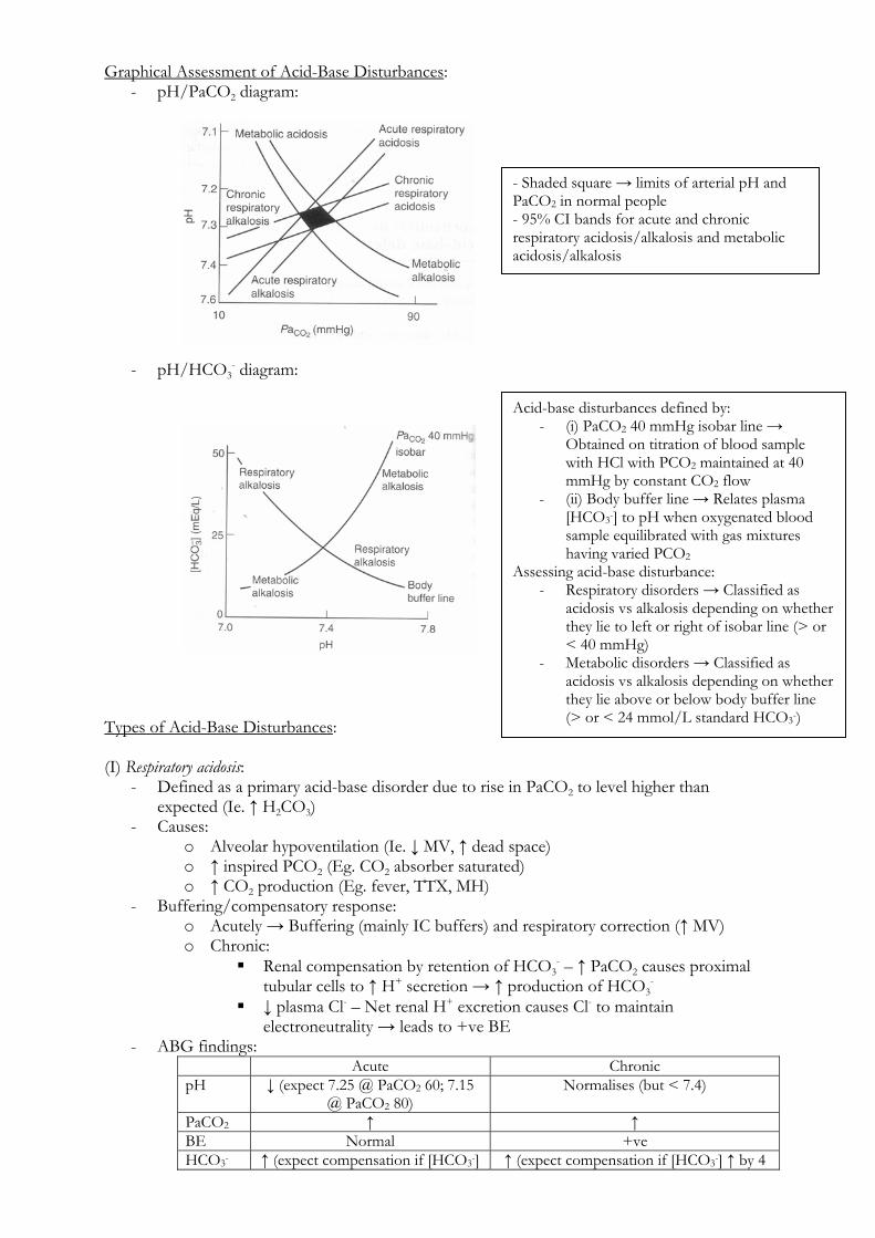
Graphical Assessment of Acid-Base Disturbances: - pH/PaCO2 diagram:
- pH/HCO3
- diagram: Types of Acid-Base Disturbances: (I) Respiratory acidosis:
- Defined as a primary acid-base disorder due to rise in PaCO2 to level higher than expected (Ie. ↑ H2CO3)
- Causes: o Alveolar hypoventilation (Ie. ↓ MV, ↑ dead space) o ↑ inspired PCO2 (Eg. CO2 absorber saturated) o ↑ CO2 production (Eg. fever, TTX, MH)
- Buffering/compensatory response: o Acutely → Buffering (mainly IC buffers) and respiratory correction (↑ MV) o Chronic:
Renal compensation by retention of HCO3- – ↑ PaCO2 causes proximal
tubular cells to ↑ H+ secretion → ↑ production of HCO3-
↓ plasma Cl- – Net renal H+ excretion causes Cl- to maintain electroneutrality → leads to +ve BE
- ABG findings: Acute Chronic pH ↓ (expect 7.25 @ PaCO2 60; 7.15
@ PaCO2 80) Normalises (but < 7.4)
PaCO2 ↑ ↑ BE Normal +ve HCO3- ↑ (expect compensation if [HCO3-] ↑ (expect compensation if [HCO3-] ↑ by 4
- Shaded square → limits of arterial pH and PaCO2 in normal people - 95% CI bands for acute and chronic respiratory acidosis/alkalosis and metabolic acidosis/alkalosis
Acid-base disturbances defined by: - (i) PaCO2 40 mmHg isobar line →
Obtained on titration of blood sample with HCl with PCO2 maintained at 40 mmHg by constant CO2 flow
- (ii) Body buffer line → Relates plasma [HCO3-] to pH when oxygenated blood sample equilibrated with gas mixtures having varied PCO2
Assessing acid-base disturbance: - Respiratory disorders → Classified as
acidosis vs alkalosis depending on whether they lie to left or right of isobar line (> or < 40 mmHg)
- Metabolic disorders → Classified as acidosis vs alkalosis depending on whether they lie above or below body buffer line (> or < 24 mmol/L standard HCO3-)
↑ by 1 mmol/L for each 10 mmHg ↑ PaCO2 above 40 mmHg
mmol/L for each 10 mmHg ↑ PaCO2 above 40 mmHg
- Treatment → Correct underlying cause (II) Respiratory alkalosis:
- Defined as a primary acid-base disorder due to fall in PaCO2 to a level lower than expected (Ie. ↓ H2CO3)
- Causes: o Invariably due to alveolar hyperventilation (Ie. ↑ MV, ↓ DS) o ↓ CO2 production (Eg. GA, hypothermia)
- Buffering/compensatory response: o Acutely → Buffering (mainly IC buffers) and respiratory correction (↓ MV → but
limited by need to maintain oxygenation!) o Chronic → Renal compensation by ↑ HCO3
- excretion and ↓ NH4+ excretion (Ie.
net H+ retention/HCO3- loss) → -ve BE and ↓ serum HCO3
- - ABG findings:
Acute Chronic pH ↑ (expect 7.5 @ PaCO2 30; 7.6 @
PaCO2 40) Normalises (but > 7.4)
PaCO2 ↓ ↓ BE Normal -ve HCO3- ↓ (expect compensation if [HCO3-]
↓ by 2 mmol/L for each 10 mmHg ↓ PaCO2 above 40 mmHg
↓ (expect compensation if [HCO3-] ↓ by 5 mmol/L for each 10 mmHg ↓ PaCO2
above 40 mmHg - Treatment → Correct underlying cause
(III) Metabolic acidosis:
- Defined as a primary acid-base disorder that causes plasma HCO3- to fall to a level lower
than expected due to either (i) an increase in metabolic/fixed acids, and/or (ii) loss of bases in blood
- Causes: o ↑ AG → Due to replacement of HCO3
- with fixed/metabolic acids (which are unmeasured anions) → MUD PILES
o Normal AG → Usually associated with hyperchloraemia → GI losses (diarrhoea, pancreatic fistula, external drainage of pancreatic/biliary secretions, uretero-enterostomy, obstructed ileal conduit), RTA, renal interstitial disease, mineralocorticoid deficiency, infusion of HCl/NH4Cl or CAi (acetazolamide)
o ↓ AG → Hypoproteinaemic states - Buffering/compensatory response:
o Acutely: Buffering (60% IC buffers; 40% EC buffers) Respiratory compensation
Initially – ↓ pH triggers peripheral chemoreceptors → hyperventilation → ↓ PaCO2 to partly normalise pH
BUT this ↓ brain ECF [H+] and causes central chemoreceptors to limit the ↑ in MV
Full effect of respiratory compensation requires 12-24 hrs – HCO3
- equilibrates across BBB and brain ECF [H+] ↑ → inhibition on MV by central chemoreceptors gradually removed
Nb. Respiratory compensation CANNOT excrete fixed acids! o Chronic → Renal compensation by excreting excess acid anions (equivalent to
reabsorption of HCO3-/excretion of H+)
- ABG findings: Acute Chronic pH ↓ Normalises (but < 7.4) PaCO2 Normal ↓ (expect compensation if PaCO2 is
1.5x[HCO3-] + 8) BE -ve -ve HCO3- ↓ ↓↓
- Treatment: o (i) Eliminate causative factor o (ii) IV NaCl → allows kidneys to excrete sufficient HCl to correct acidosis (if
acidaemia is not affecting C.O.) o (iii) IV HCO3
- (if acidaemia depressing C.O. → avoid vicious cycle of worsening CVS depression with increasing lactic acidosis)
o (iv) Dialysis (IV) Metabolic alkalosis:
- Defined as a primary acid-base disorder that causes plasma HCO3- to rise to a level higher
than expected due to either (i) an gain in bases, and/or (ii) loss of metabolic/fixed acids in blood
- Causes: o Loss of acids → Renal (hyperaldosteronism, Cushing’s, thiazide diuretics, severe
hypokalaemia, hypomagnesiuma, hypercalcaemia) or GIT (NG suctioning, severe vomiting)
o Increased base intake → NaHCO3 administration, metabolic conversion of exogenous organic ions (Eg. Lactate)
- Buffering/compensatory response: o Acutely → Buffering (70% EC buffers; 30% IC buffers) and respiratory
compensation (↓ MV) o Chronic → Renal compensation by excretion of excess HCO3
-/retention of H+ - ABG findings:
Acute Chronic pH ↑ Normalises (but > 7.4) PaCO2 Normal ↑ (expect compensation if PaCO2 is
0.7x[HCO3-] + 20) BE +ve +ve HCO3- ↑ ↑↑
- Treatment: o (i) Eliminate causative factor o (ii) Replace volume deficit with NaCl → allows excretion of NaHCO3 o (iii) IV NH4Cl (if very severe alkalosis or poor renal/cardiac function) o (iv) IV HCl avoided due to very low pH and need for administration by CVC only o (v) CAi (Eg. acetazolamide) o (vi) Spironolactone (to ↑ K+)
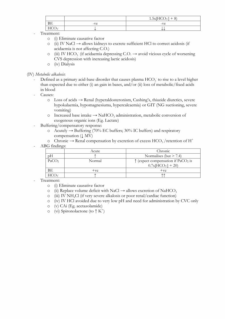
Aside: Measurement of pH, PCO2 and PO2 from ABG Measurement of pH: pH electrode (Glass-Calomel electrode)
- Consists of two ion-selective electrodes: o (i) Measuring electrode
Ag/AgCl surrounded by buffer solution in glass casing → glass bulb at electrode tip made of special pH-sensitive glass (-vely charged glass that is permeable to H+) that is in direct contact with arterial blood sample
H+ diffuses from blood sample through the glass membrane into buffer solution → buffer solution allows a pH (and [H+]) gradient to form across the glass membrane → produces an electrical potential difference that is dependent on the pH (and [H+]) gradient
o (ii) Reference electrode Hg/Hg2Cl2 (calomel) surrounded by saturated 0.1 M KCl solution (“salt
bridge”) that is separated from the arterial blood sample via a semi-permeable membrane
Maintains constant potential despite changes in arterial pH - These two electrodes are connected via blood to create an electrical circuit → electrical
potential difference produced is proportional to pH (and [H+]) gradient → 61.5 mV/pH unit - It has an accuracy of +/- 0.005 pH units - Issues:
o (i) Both electrodes must be: Kept at 37°C → due to temperature-dependent changes in pH
(acid/bases dissociate at ↑ temperatures → so pH ↓ 0.015 unit per ↑ 1°C → Rosenthal factor) and solubility of gases (Ie. CO2)
Kept clean (esp free from protein and cellular deposits) Calibrated with 2x PO4
3- buffer solutions of known pH o (ii) Delicate plastic membrane → damage leads to inaccurate results (due to
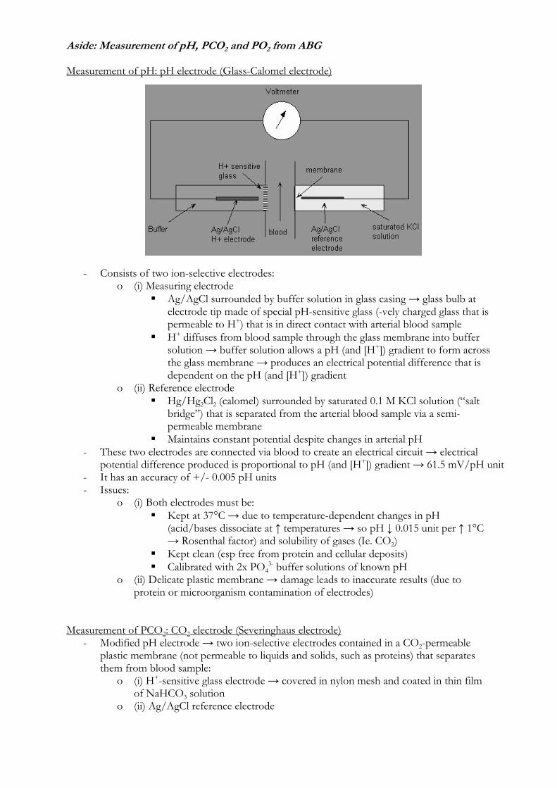
protein or microorganism contamination of electrodes) Measurement of PCO2: CO2 electrode (Severinghaus electrode)
- Modified pH electrode → two ion-selective electrodes contained in a CO2-permeable plastic membrane (not permeable to liquids and solids, such as proteins) that separates them from blood sample:
o (i) H+-sensitive glass electrode → covered in nylon mesh and coated in thin film of NaHCO3 solution
o (ii) Ag/AgCl reference electrode
- CO2 diffuses from blood sample across plastic membrane into the NaHCO3-coated nylon mesh → CO2 hydrated to form H+ and HCO3
- → glass electrode measures change in H+ (and pH) in NaHCO3 solution, which is proportional to changes in CO2 tension
- It has an accuracy of +/- 1 mmHg - Issues:
o (i) Slow response time (2-3 mins) → because CO2 needs to diffuse across membrane and react with H2O to form H+ and HCO3
- (Nb. Can be accelerated by
addition of CA) o (ii) Delicate plastic membrane → damage leads to inaccurate results o (iii) Both electrodes must be kept at 37°C, clean (Ie. free from protein deposits)
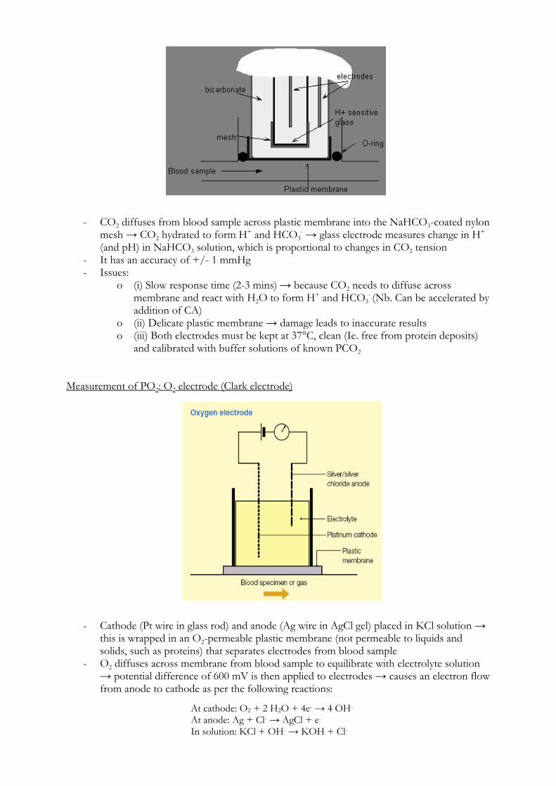
and calibrated with buffer solutions of known PCO2 Measurement of PO2: O2 electrode (Clark electrode)
- Cathode (Pt wire in glass rod) and anode (Ag wire in AgCl gel) placed in KCl solution → this is wrapped in an O2-permeable plastic membrane (not permeable to liquids and solids, such as proteins) that separates electrodes from blood sample
- O2 diffuses across membrane from blood sample to equilibrate with electrolyte solution → potential difference of 600 mV is then applied to electrodes → causes an electron flow from anode to cathode as per the following reactions:
At cathode: O2 + 2 H2O + 4e- → 4 OH- At anode: Ag + Cl- → AgCl + e- In solution: KCl + OH- → KOH + Cl-
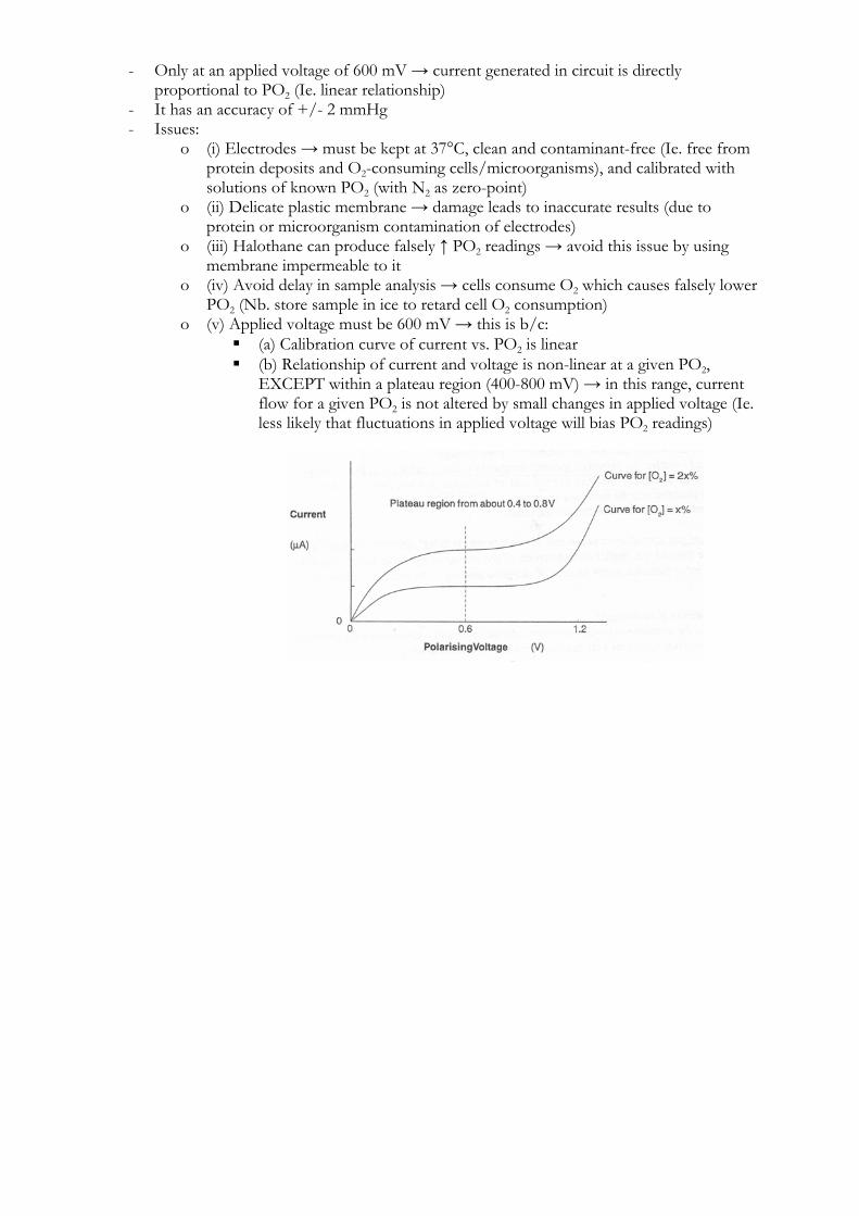
- Only at an applied voltage of 600 mV → current generated in circuit is directly proportional to PO2 (Ie. linear relationship)
- It has an accuracy of +/- 2 mmHg - Issues:
o (i) Electrodes → must be kept at 37°C, clean and contaminant-free (Ie. free from protein deposits and O2-consuming cells/microorganisms), and calibrated with solutions of known PO2 (with N2 as zero-point)
o (ii) Delicate plastic membrane → damage leads to inaccurate results (due to protein or microorganism contamination of electrodes)
o (iii) Halothane can produce falsely ↑ PO2 readings → avoid this issue by using membrane impermeable to it
o (iv) Avoid delay in sample analysis → cells consume O2 which causes falsely lower PO2 (Nb. store sample in ice to retard cell O2 consumption)
o (v) Applied voltage must be 600 mV → this is b/c: (a) Calibration curve of current vs. PO2 is linear (b) Relationship of current and voltage is non-linear at a given PO2,
EXCEPT within a plateau region (400-800 mV) → in this range, current flow for a given PO2 is not altered by small changes in applied voltage (Ie. less likely that fluctuations in applied voltage will bias PO2 readings)


