1 Acid Suppression Therapy: Neutralizing the Hype Author: Leslie Jackowski, B.Sc., M.B.B.S. Consultants: Jerry Avorn, M.D., Niteesh K. Choudhry, M.D., Ph.D., Michael Fischer, M.D., M.S. Reviewers: John Saltzman, M.D., György Baffy, M.D., Ph.D., Jennifer Potter, M.D., Laurie LaRusso, M.S, E.L.S. The Independent Drug Information Service (IDIS) is supported by the PACE Program of the Department of Aging of the Commonwealth of Pennsylvania and the Washington D.C. Department of Health. This material is provided by The Alosa Foundation, a nonprofit organization, which is not affiliated in any way with any pharmaceutical company. None of the authors accepts any personal compensation from any pharmaceutical company. These are general recommendations only; specific clinical decisions should be made by the treating physician based on an individual patient’s clinical condition. For more information, visit www.RxFacts.org
Transcript
1
Acid Suppression Therapy: Neutralizing the Hype
Author: Leslie Jackowski, B.Sc., M.B.B.S.
Consultants: Jerry Avorn, M.D., Niteesh K. Choudhry, M.D., Ph.D., Michael Fischer, M.D., M.S.
Reviewers: John Saltzman, M.D., György Baffy, M.D., Ph.D., Jennifer Potter, M.D., Laurie LaRusso, M.S, E.L.S.
The Independent Drug Information Service (IDIS) is supported by the PACE Program of the Department of Aging of the Commonwealth of Pennsylvania and the Washington D.C. Department of Health.
This material is provided by The Alosa Foundation, a nonprofit organization, which is not affiliated in any way with any pharmaceutical company. None of the authors accepts any personal compensation from any pharmaceutical company.
These are general recommendations only; specific clinical decisions should be made by the treating physician based on an individual patient’s clinical condition.
For more information, visit www.RxFacts.org
2
3
The Alosa Foundation Acid Suppression Therapy:
Neutralizing the Hype
Accreditation: This activity has been planned and implemented in accordance with the Essential Areas and policies of the Accreditation Council for Continuing Medical Education through the joint sponsorship of Harvard Medical School and The Alosa Foundation. The Harvard Medical School is accredited by the ACCME to provide continuing medical education for physicians.
Credit Designation: The Harvard Medical School designates this enduring material for a maximum of 1.5 AMA PRA Category 1 Credit™. Physicians should claim only the credit commensurate with the extent of their participation in the activity.
Activity Overview: The goal of the educational program is to help practitioners assess the comparative effectiveness and safety of acid-suppressive drugs; understand the evidence regarding appropriate therapy; weigh the benefits, risks, and value of treatment options; and improve the quality of prescribing and patient care. In addition to providing this evidence report, the education program uses an innovative approach, academic detailing, one-on-one educational sessions in physicians’ offices with trained outreach educators (pharmacists, nurses, physicians) who present the educational material interactively. Additionally, reference cards and patient education materials are provided.
Target Audience: The educational program is designed for primary care physicians practicing internal medicine, primary care, family medicine, and geriatrics, and other health care professionals who deliver primary care.
Learning Objectives: Upon completion of this activity, participants will be able to:
• Identify the causes of GI symptoms that require long-term use of PPIs. • Safely taper off unnecessary PPIs. • Assess the patient instead of resorting to PPI use early on. • Recognize when a test for H. pylori is indicated and be able to treat infection when positive. • Recognize “red flags” for potentially serious disease and refer those patients for additional
evaluation.
Disclosure Policy: Harvard Medical School (HMS) adheres to all ACCME Essential Areas, Standards, and Policies. It is HMS's policy that those who have influenced the content of a CME activity (e.g. planners, faculty, authors, reviewers and others) disclose all relevant financial relationships with commercial entities so that HMS may identify and resolve any conflicts of interest prior to the activity. These disclosures are provided in the activity materials along with disclosure of any commercial support received for the activity.
4
Additionally, faculty members have been instructed to disclose any limitations of data and unlabeled or investigational uses of products discussed.
Disclosures: This material is provided by The Alosa Foundation, a nonprofit organization which is not affiliated in any way with any pharmaceutical company. No commercial support has been received for this activity. None of the planners/authors have any financial relationships to disclose. The Independent Drug Information Service (IDIS) is supported by the PACE Program of the Department of Aging of the Commonwealth of Pennsylvania, and the Washington D.C. Department of Health.
Faculty and Planners: Leslie Jackowski, B.Sc., M.B.B.S. is a Senior Clinical Consultant with the Division of Pharmacoepidemiology and Pharmacoeconomics, Brigham and Women's Hospital, Harvard Medical School. Dr. Jackowski has no relevant financial relationships to disclose.
Jerry Avorn, M.D. is a Professor of Medicine at Harvard Medical School and Chief of the Division of Pharmacoepidemiology and Pharmacoeconomics at Brigham and Women's Hospital. An internist, he has worked as a primary care physician and geriatrician and has been studying drug use and its outcomes for over 25 years. Dr. Avorn has no relevant financial relationships to disclose. Niteesh K. Choudhry, M.D., Ph.D. is an Associate Professor of Medicine at Harvard Medical School and a hospitalist at Brigham and Women's Hospital. His research focuses on the use of medications to treat common chronic conditions. Dr. Choudhry has no relevant financial relationships to disclose. Michael Fischer, M.D., M.S. is an Assistant Professor of Medicine at Harvard Medical School and a primary care internist who studies cost-effective drug use in outpatient practices. Dr. Fischer has no relevant financial relationships to disclose.
Reviewers: John R. Saltzman, M.D. is an Associate Professor of Medicine at Harvard Medical School and the Director of Endoscopy at the Brigham and Women's Hospital. He is an Associate Editor of the Journal Gastrointestinal Endoscopy and the Section Editor for Diagnostic and Therapeutic Endoscopy for UpToDate. Dr. Saltzman has no relevant financial relationships to disclose.
György Baffy, M.D., Ph.D. is an Assistant Professor of Medicine at Harvard Medical School and practices in the Department of Medicine at the Brigham and Women’s Hospital. Dr. Baffy has no relevant financial relationships to disclose.
Jennifer Potter, M.D. is an Associate Professor of Medicine at Harvard Medical School and practices in the Department of Medicine at the Beth Israel Deaconess Medical Center. Dr. Potter has no relevant financial relationships to disclose.
Laurie LaRusso, M.S., E.L.S. is the Principal of Chestnut Medical Communications where she provides reviews of continuing medical education materials and medical writing services on a wide range of topics. Ms. LaRusso has no relevant financial relationships to disclose.
5
Media used: Printed educational material.
Instructions for Participation and Credit: There are no fees to participate in this activity. To receive credit, participants must (1) read the statements on target audience, learning objectives, and disclosures, (2) study the educational activity, and (3) complete the post-test and activity evaluation. To receive AMA PRA Category 1 Credit™, participants must receive a minimum score of 70% on the post-test. Tests and evaluations should be submitted to the Alosa Foundation via mail or fax. Mailing address: The Alosa Foundation 699 Boylston Street, Suite 2 Boston, MA 02116 Fax: 857-350-9155 The activity will take approximately 1.5 hours to complete. Activity publication date: July 1, 2012 Termination date: July 1, 2015 Please e-mail any questions to [email protected] or call (857) 350-9105.
6
Table of Contents Introduction .................................................................................................................................................. 7
Common causes of dyspepsia ..................................................................................................................... 7
Assessment of patients presenting with dyspepsia` .................................................................................. 11
Management of GERD .............................................................................................................................. 13
Management of peptic ulcer disease ......................................................................................................... 19
Management of non-ulcer dyspepsia ......................................................................................................... 22
Management of dyspepsia: Test-and-treat vs. empirical acid suppression ............................................... 28
Comparative effectiveness and safety ....................................................................................................... 29
Compliance and adverse drug reactions ................................................................................................... 34
Costs and comparative value of acid-suppressive drugs .......................................................................... 34
Appendix 1: The Los Angeles classification of esophagitis ....................................................................... 38
Appendix 2: Efficacy of acid-suppressive medications in GERD ............................................................... 39
7
Introduction Symptoms related to gastrointestinal acid production are among the most common in medicine. Approximately 25% of adults regularly experience heartburn.1 Such symptoms are usually just an occasional nuisance, but for some people they may be debilitating, or a sign of a more serious problem.
Histamine-2 receptor antagonists (H2 blockers) revolutionized the care of acid-related disease when they were first introduced in the 1970s, and are now available over-the-counter and in generic forms. Proton pump inhibitors (PPIs) are currently among the most widely used and heavily advertised medications in the world. More than 113 million prescriptions for PPIs are filled each year, making this class of drugs, at $14 billion in sales, the third highest seller in the United States.2 These medications are effective for the treatment of gastroesophageal reflux disease (GERD), prevention of ulcers induced by non-steroidal anti-inflammatory drugs (NSAIDs), the healing of peptic ulcers, as part of a regimen for Helicobacter pylori eradication, erosive esophagitis, Barrett’s esophagus, and Zollinger-Ellison syndrome, However, these indications do not account for the number of prescriptions written, and 50%-70% of PPI prescriptions may be for inappropriate indications.2
Over the past several years, some PPIs have become available over-the-counter, which has led to even wider advertising of PPIs. Clinicians now face difficult challenges when considering acid suppression options for their patients including deciding when to prescribe acid-suppressive medications, whether to choose a PPI or another agent, and how long to continue therapy. Considerations of patient costs, medication adherence, and uncertainty about what patients may be purchasing over-the-counter compound the complexity of these prescribing decisions, as do recent studies suggesting previously unrecognized risks associated with PPI treatment.2-5
This document presents an evidence-based approach for the evaluation and management of several common gastrointestinal complaints, including GERD, peptic ulcer disease (including NSAID-induced ulcers), and non-ulcer dyspepsia.
Common causes of dyspepsia The term dyspepsia includes a constellation of symptoms such as upper abdominal discomfort, heartburn, retrosternal pain, nausea, early satiety (sensation of fullness), acid regurgitation, and excessive belching.6 It is very common.1
8
Figure 1: Incidence of heartburn in adults
The three major causes of dyspepsia are GERD, peptic ulcer disease, and non-ulcer dyspepsia. The prevalence of the 3 conditions are 14-20%, 0.5-5%, and 2% respectively, although the figures are only approximations, as GERD has a somewhat nebulous definition and non-ulcer dyspepsia is a diagnosis of exclusion.7, 8 Symptoms mistaken for dyspepsia may also result from other conditions such as coronary artery disease, pericarditis, aortic dissection, pulmonary embolism, gallstones, and pancreatitis.
Gastroesophageal reflux disease
Definition The American Gastroenterological Association defines GERD as “a condition which develops when the reflux of stomach contents causes troublesome symptoms and/or complications.”9 GERD may involve esophageal injury (esophagitis).9
Pathogenesis The primary underlying mechanism is thought to be impaired lower esophageal sphincter (LES) function. The LES normally relaxes in response to esophageal peristalsis to allow the passage of food, liquid or saliva into the stomach. There are brief periods where the LES relaxes when there is no swallowing or esophageal peristalsis, known as transient LES relaxations (TLESRs). These are normal, and expose the esophagus to a small amount of acid after meals. Most patients with GERD have an increased frequency of TLESRs, exposing the esophagus to acid for longer periods. This increases the risk of symptoms and esophageal damage. A minority of patients have a permanent defect in the LES leading to a constant decrease in resting tone. These patients are more likely to have severe esophagitis and/or complications such as esophageal stricture.10, 11
25%
5%
0%
5%
10%
15%
20%
25%
30%
Monthly incidence Daily incidence
Incide
nce
9
Epidemiology and risk factors Gastroesophageal reflux disease is the most common gastrointestinal diagnosis recorded in outpatient clinics.7 Risk factors for GERD include:
• smoking • alcohol use • excessive weight • hiatal hernia • pregnancy • asthma • diabetes • reduced gastric motility • rarer conditions including scleroderma and Zollinger-Ellison syndrome
Peptic ulcer disease
Definition In peptic ulcer disease, there is a break in the mucosal lining of the stomach and/or proximal duodenum. Less commonly, ulcers can also occur in the lower esophagus, the distal duodenum, or the jejunum in gastric acid hypersecretory states such as Zollinger-Ellison syndrome.12 Ulcers smaller than 5 mm or without obvious depth are known as erosions.
Pathophysiology Peptic ulcers result from an imbalance between factors that damage the gastroduodenal mucosal lining and defense mechanisms that limit the injury. Normal mucosal defense involves a mucus bicarbonate layer, which forms a viscous gel over the gastric mucosa.
H. pylori infection in the gastric antrum leads to the release of gastrin, which stimulates excess acid secretion from the proximal acid-secreting mucosa in the fundus. The increased acid load damages the duodenal mucosa, causing ulceration and gastric metaplasia. The metaplastic mucosa can then become colonized by H. pylori.8 Chronic H pylori infection and inflammation throughout the stomach causes degradation of the mucus layer and death of gastric epithelial cells.8
NSAIDs cause gastric ulcers by direct topical injury, and indirectly by inhibiting the synthesis of prostaglandins needed for maintaining the integrity of the gastric mucosa. NSAIDs also increase bleeding risk through their antiplatelet activity.
In Zollinger-Ellison syndrome, a gastrin-secreting neuroendocrine tumor stimulates high levels of gastric acid secretion and subsequent peptic ulcer disease.
Epidemiology Lifetime prevalence of peptic ulcer in the U.S. is about 10%, and about 500,000 persons develop peptic ulcer disease in the U.S. each year.12, 13 In about 70% of patients it occurs between the ages of 25 and 64 years. The annual direct and indirect health care costs of the disease are estimated at about $10 billion. However, the incidence of peptic ulcers is declining, possibly as a result of the increasing use of PPIs and decreasing rates of H. pylori infection.12
10
Risk factors H. pylori infection and the use of aspirin and other NSAIDs are the major causes of peptic ulcer disease in the United States.8, 12 Critical illness, surgery, or hypovolemia leading to splanchnic hypoperfusion may result in gastroduodenal erosions or ulcers (stress ulcers); these may be silent or manifest with bleeding or perforation. Other risk factors for peptic ulcer disease include older age, low socio-economic status, smoking, a family history of ulcers, and excessive alcohol intake.8 Smoking also increases the risk of ulcer recurrence and slows healing.12
Most gastric ulcers are due to H. pylori infection or NSAIDs. Less common causes include gastric cancer, Zollinger-Ellison syndrome, some viral infections, and other medications. About 90% of duodenal ulcers are due to H. pylori infection, although this rate is decreasing in developed countries.14-16 Non-NSAID, non-H. pylori peptic ulcers can also occur, and usually heal with PPI therapy. NSAIDs and H. pylori independently increase the risk of peptic ulcer bleeding (see table below).17
Table 1. Risk factors for ulcer bleeding
Risk factor Increase in risk of ulcer bleeding
NSAIDs two times
H. pylori five times
NSAIDs and H. pylori six times (compared with patients who have neither risk factor)
Non-ulcer dyspepsia Non-ulcer dyspepsia is a term given to persistent dyspepsia when other diagnoses have been excluded and no other specific cause can be identified. After GERD, non-ulcer dyspepsia is the second-most common cause of upper GI symptoms and is the most frequent diagnosis reached after endoscopy. The cause of non-ulcer dyspepsia is not known, but is likely multifactorial.18
Other causes Dyspepsia is a common adverse effect of many medications, including aspirin, NSAIDs, COX-2 inhibitors, diuretics, antibiotics, antihypertensives, corticosteroids, and bisphosphonates. NSAIDs and COX-2 inhibitors can cause dyspepsia without peptic ulceration, but the mechanism by which these agents cause dyspepsia is not well defined.19
11
Assessment of patient presenting with dyspepsia An organized approach to the assessment and management of patients presenting with dyspepsia can make it easier to choose appropriate medical therapy, to understand when to stop therapy, and to communicate effectively with patients about these management decisions. The first steps are to:
1. Characterize the dyspepsia. Location of discomfort/pain, onset, timing, radiation, aggravating factors, alleviating factors, associated symptoms, duration, and intensity.
2. Look for precipitating medications and associations. Identify whether the patient is taking medications that may cause or exacerbate dyspepsia, in particular aspirin, other NSAIDs, anticholinergic agents, theophylline, dopaminergic agents, oral bisphosphonates, corticosteroids, and calcium channel blockers.20 Define the relationship and timing of symptoms with foods. Identify the specific foods that elicit the symptoms.
3. Exclude serious non-GI causes of symptoms that may present as dyspepsia such as coronary artery disease, pericarditis, aortic dissection, and pulmonary embolism. Discomfort/pain that worsens with exertion or deep inspiration, or that radiates to the shoulders or arms, may reflect a cardiac or vascular cause.
4. Assess presence of alarm (“red flag”) symptoms/signs suggesting cancer, stricture or severe ulceration:
• dysphagia • hematemesis • gastrointestinal bleeding • change in bowel habit • anemia • odynophagia • previous GI malignancy or ulcer • recurrent vomiting • anorexia • unexplained weight loss • early satiety • abdominal mass • hepatomegaly • lymphadenopathy
Alarm features are relatively uncommon and occur in only a minority of patients. If any of these alarm features are present, refer for prompt upper endoscopy and possible biopsy. Additional diagnostic testing beyond esophagogastroduodenoscopy (EGD) such as endoscopic ultrasonography or 24-hour esophageal pH testing has a low yield in the initial assessment of dyspepsia in primary care.21
5. Consider testing for H. pylori in patients ≤55 years without alarm features (see page 23).
Patients with predominant or frequent (more than once a week) heartburn or acid regurgitation should be considered to have GERD until proven otherwise.21, 22 Peptic ulcer disease may present with gnawing or
12
burning, non-radiating, epigastric pain that is relieved by antacids, food, or milk. However, symptoms from different upper gastrointestinal problems have significant overlap, making it difficult to clinically distinguish between conditions in the patient first presenting with dyspepsia.21,23
Diagnosis of GERD GERD can present with a wide variety of clinical symptoms,11 but its cardinal symptoms are heartburn (a burning feeling in the epigastrium or central chest rising toward the neck) or acid regurgitation (a sour or bitter taste in the mouth).21, 22
Most patients with GERD will not require endoscopy, but for the subset of patients with GERD who also have alarm features, the American Gastroenterological Association 2008 guidelines for the management of GERD recommend endoscopy with biopsy.9 GERD may involve esophagitis in a minority of people and it is important to identify these patients, since a diagnosis of esophagitis has long-term treatment implications.
The Los Angeles Classification (see Appendix 1) is the most widely used method for describing reflux esophagitis. It provides a practical and standardized grading system, and is well-validated.24 The inconsistent use of the terms ‘ulcer’ and ‘erosion’ when applied to reflux esophagitis led to the adoption of the term ‘mucosal break’.24
Diagnosis of peptic ulcer disease The most specific symptoms that help rule in a diagnosis of peptic ulcer are:12
• episodic gnawing or burning epigastric pain • pain occurring two to five hours after meals or on an empty stomach • nighttime awakening because of abdominal pain, with relief on eating • pain relieved by food intake, antacids, or antisecretory agents
Less common symptoms include indigestion, vomiting, loss of appetite, intolerance of fatty foods, and heartburn. The physical examination is usually unremarkable.
Abdominal pain is absent in ≥30% of older patients with peptic ulcers. Postprandial epigastric pain is more likely to be relieved by food or antacids in patients with duodenal ulcers than in those with gastric ulcers. Weight loss precipitated by avoidance of food intake is characteristic of gastric ulcers.12
Prompt endoscopy is recommended for patients with symptoms highly suspicious for peptic ulcers who have alarm features or symptoms that do not respond to treatment. Some gastroenterologists also recommend endoscopy in all patients younger than age 55 with suspected ulcer symptoms, regardless of alarm features. Endoscopy is the gold standard for diagnosis.12
Diagnosis of non-ulcer dyspepsia Non-ulcer dyspepsia is a common diagnosis of exclusion, assigned to patients with persistent dyspepsia who have no evidence of structural disease (including at endoscopy) that is likely to explain the symptoms.
13
Bottom line: Exclude non-GI causes of dyspepsia, consider if symptoms may be drug-induced (especially NSAIDs), assess for alarm features, and consider testing for H. pylori or referral for endoscopy.
Management of GERD Lifestyle interventions Lifestyle modification can be very effective, are the foundation of GERD treatment, and may help avoid or reduce the need for medications. Lifestyle interventions include:7, 9, 25
• avoiding foods that worsen reflux, especially those that lower the tone of the lower esophageal sphincter or precipitate symptoms in a given patient (e.g., coffee, tea, other caffeinated drinks, chocolate, mint, fatty or fried foods)
• avoiding acidic foods that may precipitate heartburn (e.g., citrus, carbonated drinks, onions, tomatoes, spicy foods)
• encouraging lifestyle modification including: ⎯ weight loss for patients who are overweight (BMI 25.0–29.9) or obese (BMI ≥30.0), ⎯ smoking cessation, ⎯ discourage lying down for 2–3 hours after meals, ⎯ avoid excessive alcohol; ⎯ discourage use of clothing that constricts the waist; ⎯ encourage smaller and more frequent meals.
• elevating the head of the bed for patients troubled with heartburn or regurgitation when lying down.
Medications
Antacids Aluminium hydroxide, calcium carbonate and magnesium salts are inexpensive and provide quick relief. They may be used alone or in combination with other acid-suppressive drugs. Despite their time-honored place in therapy, evidence of efficacy from controlled clinical trials is lacking, perhaps because they are generally available over-the-counter, and came into widespread use before a time when randomized trials were required for marketing.
Acid-suppressive medications Acid suppression therapy should be initiated for GERD if lifestyle interventions fail to adequately control symptoms (persistent symptoms more than once per week). It is recommended for patients with GERD with or without esophagitis. A short course or as-needed use of acid suppression therapy is appropriate in patients with GERD without esophagitis when symptom control is the primary objective.9
14
Table 2: Standard doses of PPIs and H2 receptor antagonists
PPI H2 receptor antagonist
esomeprazole 20 mg once daily cimetidine 800 mg once daily
lansoprazole 30 mg once daily famotidine 40 mg once daily
omeprazole 20 mg once daily nizatidine 300 mg once daily
pantoprazole 40 mg once daily ranitidine 300 mg once daily
rabeprazole 20 mg once daily
PPIs work best when taken 15-30 minutes before a meal, and are best given in the morning if used once daily. A major reason for failure of PPIs is the improper use of these medications, with one study showing that only 46% of patients took their PPIs correctly.26 Twice-daily dosing of PPIs may improve symptom relief in patients with an inadequate response to once-daily dosing.9
Although there is limited evidence that adding a night-time H2 receptor antagonists to twice-daily PPI therapy improves symptoms, observational data and clinical experience suggest that use of an H2
receptor antagonist at bedtime can be beneficial, at least for some patients. A nighttime H2 receptor antagonist may be an option in patients who do not respond to twice daily PPI. If clinical tolerance has been encountered, then using the H2 receptor antagonist intermittently or on demand could theoretically be helpful.9, 27, 28
An important limitation of PPI therapy is that these agents take longer to provide symptom relief than H2 receptor antagonists or antacids; over 24 hours are required for PPIs to fully suppress acid production, while antacids relieve symptoms within minutes and H2 receptor antagonists relieve symptoms within an hour. In one study of GERD patients randomized to PPI therapy, the median time to first symptom relief was 2 days and the median time to sustained symptom relief was >10 days.29 Therefore, antacids and H2 receptor antagonists generally provide quicker symptom relief than PPIs (see also comparative effectiveness of PPIs on page 29).
Although PPIs take longer to begin working, once they do so, they are likely to be more effective than other acid suppressive drugs. A recent comprehensive Cochrane review examined the effect of short-term treatment with medications for heartburn symptoms. It found that in the empirical treatment of GERD, the risk reductions for heartburn remission were as follows:1
Table 3: Heartburn remission in GERD
Drug therapy Relative risk reduction compared to placebo
PPIs 63%
H2 receptor antagonists 23%
Prokinetic agents Not significant
15
In a direct comparison, PPIs were 34% more effective than H2 receptor antagonists (relative risk, 0.66; 95% CI, 0.60 to 0.73) and 47% more effective than prokinetic agents (relative risk 0.53; 95% CI 0.32 to 0.87).1
Medication therapy for patients with esophagitis PPIs are also more effective than H2 receptor antagonists for healing esophagitis. A Cochrane review found that the healing rate of esophagitis after 4-8 weeks of treatment with standard dose PPI was 84% compared to 29% with placebo.10 The number needed to treat was 1.7 (95% CI, 1.5 to 2.1), a very small number for benefit. Higher doses of PPIs (twice the standard dose, given once daily) were more effective than standard doses in healing esophagitis.
Patients with esophagitis have high rates of recurrence of erosive disease if they do not continue acid suppression therapy.9 Several clinical trials have shown that:9
• recurrence of erosive esophagitis is dramatically reduced with daily PPI treatment compared to placebo;
• patients taking H2 receptor antagonists are almost twice as likely to have recurrent erosive disease compared with patients taking PPIs; and
• patients with erosive esophagitis who are healed with PPI therapy have higher recurrence rates of erosive disease if they continue with on-demand therapy, compared to continuous therapy.
A diagnosis of esophagitis should be confirmed by endoscopy and long-term PPI therapy should be used for such patients.9 Long-term therapy should be titrated down to the lowest effective dose based on symptom control.9 Less than daily dosing of PPI maintenance therapy in patients with GERD who have previously had severe esophagitis is not recommended.9
For further data on the efficacy of acid-suppressive medications, see Appendix 2.
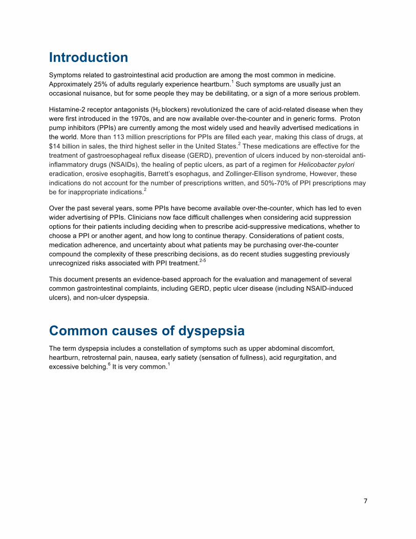
Maintenance therapy Many patients who do not have esophagitis or other indications for long-term PPI therapy (e.g. Zollinger-Ellison syndrome) can tolerate lowering the dose or discontinuing PPI therapy after a sufficient course.9, 30 Patients on long-term PPI treatment produce very high levels of gastrin in response to the lower acidity of the stomach, and when PPIs are removed the level of gastric acid secretion can be very high. Because of this, some patients will develop significant symptoms of “rebound” acid hypersecretion. However, even when symptoms occur, their duration is generally brief.31 For many patients, a tapering regimen including the use of H2 receptor antagonists and antacids can control rebound symptoms and provide the best chance of successfully stopping PPIs. A suggested tapering regimen is provided in the figure below (and as a tear-off pad for patients).
16
Figure 2: Tapering down PPIs
Role of H. pylori eradication Routine testing for H. pylori is not recommended in GERD, but testing may be considered in patients receiving long-term maintenance treatment with PPIs (because long-term acid suppression may cause atrophic gastritis, and H. pylori eradication slows its progression).17 This approach is controversial and is recommended by European but not American guidelines.9, 17
Despite previous thought to the contrary, eradication of H. pylori infection does not cause or exacerbate GERD, improve symptoms, or affect the outcome of PPI therapy.17, 32, 33 Therefore, although routine testing is not suggested, treatment of H. pylori infection should not be withheld in patients with GERD.33
See page 23 for further details on tests used to detect H. pylori and recommended eradication regimens.
This tear-off pad will be provided to physicians so they can “prescribe” a tapering plan to their patients.
17
Follow-up The American Gastroenterological Association 2008 guidelines for the follow up of patient after initial treatment of GERD recommend:9
• Assess and reinforce lifestyle modification (see above). Endoscopy to evaluate patients who have not responded to an empirical trial of twice-daily PPI therapy.
• Manometry to evaluate patients who have not responded to an empirical trial of twice-daily PPI therapy and have normal findings on endoscopy.
• Ambulatory impedance-pH, catheter pH, or wireless pH monitoring (PPI therapy withheld for 7 days) to evaluate patients who have not responded to an empirical trial of PPI therapy, have normal findings on endoscopy, and have no major abnormality on manometry.
There is insufficient evidence to recommend for or against combined impedance-pH, catheter pH, or wireless pH esophageal monitoring studies performed while taking PPIs. Routine endoscopy to assess disease progression is not recommended.9
Patients who do not achieve adequate symptom control with standard approaches or are intolerant of medications might benefit from endoscopic or surgical approaches for which referral will be needed.
Bottom line: If lifestyle measures inadequately control symptoms in patients without esophagitis, trial either (i) a short course of a PPI, or (ii) as needed antacid and/or H2 receptor antagonist. If symptoms persist, long term PPI therapy may be needed. Patients with proven esophagitis should be treated with long-term PPI therapy titrated down to the lowest effective dose based on symptom control. Patients with severe esophagitis should receive long-term daily PPI maintenance therapy (see algorithm below).
18
Management algorithm
Figure 3: Approach to patients with GERD symptoms
19
Management of peptic ulcer disease Key recommendations for the treatment of peptic ulcers are as follows:12
• Patients should be advised to stop smoking and avoid excessive alcohol. • Discontinue aspirin and NSAIDs if possible; if these agents need to be continued, consider
adding a PPI, double-dose H2 receptor antagonist, or misoprostol. • If H. pylori is present, eradicate it to assist in healing and reduce the risk of ulcer recurrence. • PPIs provide healing rates and symptom relief superior to other acid suppression therapies.
An algorithm for the management of peptic ulcers in primary care is provided in the figure below.
20
Figure 3: Approach to patients with dyspepsia not consistent with GERD
Patients who do not achieve adequate symptom control with standard approaches or are intolerant of medications might benefit from endoscopic or surgical approaches for which referral will be needed.
21
Bleeding ulcer Upper GI bleeding occurs in 15-20% of patients with peptic ulcer disease. In older persons, 20% of bleeding episodes result from asymptomatic ulcers. Patients may present with hematemesis (bright red or “coffee ground”), melena, anemia, orthostasis, or syncope.12
In stable patients with GI bleeding, ulcer-causing medications should be discontinued and a PPI initiated.12 For patients hospitalized with a bleeding ulcer, intravenous PPIs can reduce transfusion requirements, need for surgery, and duration of hospitalization.12 EGD should be performed within 24 hours. Start oral PPIs as soon as the patient can resume oral intake.12
A meta-analysis of 1157 patients from 7 clinical trials examined the effectiveness of high-dose PPIs vs. standard/low-dose PPIs in patients with bleeding peptic ulcer, to assess the effect on re-bleeding, surgical intervention, and mortality. High-dose PPIs and standard/low-dose PPIs did not significantly differ in their effects on these outcomes.34 However, in clinical practice, patients at high-risk for further bleeding are often treated with continuous infusion high-dose PPIs for 72 hours before being switched to a standard-dose oral PPI.
H. pylori testing should be performed and eradication therapy prescribed if infection is present. Treatment of H. pylori infection is more effective than acid suppression therapy without eradication for preventing recurrent bleeding.35 Surgery may be required in continued or recurrent bleeding.
If continued administration of aspirin or NSAIDs is required, add concurrent misoprostol or proton pump inhibitor.12
Role of acid suppression therapy for primary and secondary prevention of NSAID-induced ulcers A detailed discussion of reducing the risk of NSAID-induced ulcers is provided in the IDIS evidence document on pain management, available at www.RxFacts.org. In summary:
• NSAIDs (alone or in combination with aspirin) significantly increase the risk of upper GI complications.
• To reduce NSAID-associated GI complications, use the lowest NSAID dose for the shortest possible time, and/or add a PPI, double-dose H2 receptor antagonist, or misoprostol to the regimen.
• COX-2 inhibitors have significantly less risk of GI complications than non-selective NSAIDs, but concomitant use of aspirin with a COX-2 inhibitor produces an ulcer risk that is the same as a non-selective NSAID.
• PPIs are superior to regular-dose H2 receptor antagonists in the primary and secondary prevention of NSAID- or aspirin-associated ulcers, but PPIs are equivalent to double-dose H2 receptor antagonists for primary prevention.
• Non-selective NSAID + PPI is as effective as a COX-2 inhibitor for primary and secondary prevention of GI complications. A COX-2 inhibitor + PPI reduces recurrent ulcer risk more than a COX-2 inhibitor alone.
• Misoprostol is effective in both primary and secondary prevention of NSAID-associated ulcers. • Concomitant use of steroids or anticoagulants increases the risk of GI complications.
22
Role of H. pylori testing/eradication in preventing NSAID-induced ulcers A recent report of The European Helicobacter Study Group found that:17
• In naive NSAID users, H. pylori eradication reduces the risk of peptic ulcer and bleeding, but does not completely prevent NSAID related ulcer disease in chronic NSAID users.
• For chronic NSAID users with peptic ulcer and/or ulcer bleeding, PPI maintenance treatment is more effective than H. pylori eradication in preventing ulcer recurrence and/or bleeding.
• Patients who have a GI bleed while receiving long-term aspirin should be tested for H. pylori and, if positive, receive eradication therapy.
Eradication of H. pylori infection can provide a long-term cure in patients with ulcers that are not associated with the use of NSAIDs.8 Regardless of whether or not a patient is taking an NSAID, all patients with a peptic ulcer should be tested for H. pylori and treated with eradication therapy if infected (see
23
Figure 3).33
Bottom line: Most peptic ulcers are caused by H. pylori and/or NSAIDs. Treat confirmed peptic ulcers by discontinuing NSAIDs, testing/eradicating H. pylori, and 4-8 weeks of PPI therapy; test to confirm eradication of H. pylori (see page 23). Options for the prevention of NSAID-induced ulcers include stopping the NSAID, switching to a COX-2 or a different class of analgesic, or adding a PPI, double-dose H2 receptor antagonist, or misoprostol.
Management of non-ulcer dyspepsia Lifestyle interventions Lifestyle interventions for non-ulcer dyspepsia have not been well studied and specific recommendations cannot be made.
Acid suppression therapy A Cochrane review found that H2 receptor antagonists and PPIs were significantly more effective than placebo for the treatment of non-ulcer dyspepsia (relative risk reductions of 23% and 13% respectively), while sucralfate and antacids were not.36 PPIs were not significantly more effective than H2 receptor antagonists in a direct comparison of the 2 medication classes (relative risk of symptom response 0.93; 95% CI, 0.84-1.02). There appears to be no statistically significant difference between low- and standard-dose PPI therapy in these patients (relative risk of persisting symptoms on standard-dose PPI compared to low-dose PPI, 0.98; 95% CI, 0.92-1.04).36
H. pylori eradication Eradication of H. pylori can cure non-ulcer dyspepsia, with a number needed to treat of 12-15 patients.17,
37, 38 A Cochrane review found that eradication of H. pylori infection in non-ulcer dyspepsia had a statistically significant clinical benefit (therapeutic gain of eradication over placebo = 8%, relative risk of remaining symptomatic, 0.91; 95% CI, 0.86–0.95; number needed to treat = 15).39 This compares favorably with other available treatments for non-ulcer dyspepsia.17
A Cochrane review examined 3 trials that compared H. pylori eradication with other pharmacological therapies.37 H. pylori eradication was associated with a significant reduction in symptom scores compared to H2 receptor antagonist or sucralfate therapy, and a non-significant reduction compared to metoclopramide.
24
Other options The management of non-ulcer dyspepsia is difficult if initial acid suppression therapy and H. pylori eradication fails.21 Prokinetic agents may be useful if there is an underlying motility disorder such as gastroparesis.21, 22, 36, 40 Antidepressant therapy, or psychological treatments (psychotherapy, cognitive behavioral therapy, relaxation therapy and hypnosis) are sometimes tried, although their benefits are not well established.
Bottom line: Non-ulcer dyspepsia is a diagnosis of exclusion. Acid suppression therapy and H. pylori eradication can be helpful.
Helicobacter pylori infection Epidemiology H. pylori infection is usually contracted in the first few years of life and persists life-long unless treated.8 Approximately 30–40% of the U.S. population is infected with H. pylori.33 Infection rates in children are decreasing, and it is likely that the prevalence of H. pylori in the United States will continue to fall in coming years.33
Disease states associated with H. pylori infection Infection is associated with a number of disease states, as shown in the figure below. Duodenal or gastric ulcers occur in 1-10% of infected patients, gastric cancer in 0.1-3%, and gastric mucosa-associated lymphoid-tissue (MALT) lymphoma in <0.01%.8 However, the vast majority of patients with H. pylori infection do not have any related clinical disease, and routine testing is not recommended.8
25
Figure 4: Disease states associated with H. pylori infection15
The prevalence of H. pylori infection may be declining in the population and in peptic ulcer, especially in younger patients. In the United States and parts of Europe, the prevalence of H. pylori in PUD (with NSAID use excluded) now ranges from 50 to 75% and continues to fall rapidly.41, 42
Whom to test Diagnosing and treating H. pylori infection can cure some patients with peptic ulcer disease and may eliminate the need for lifelong drug therapy.35 The American College of Gastroenterology recommends that testing for H. pylori should be performed in patients with a number of conditions, including:33
• an active gastric or duodenal ulcer • a history of active gastric or duodenal ulcer not previously treated for H. pylori infection • uninvestigated dyspepsia in adults under 55 years without alarm features (the ‘test-and-treat’
strategy)
Bottom line: H. pylori infection is associated with a number of disease states, including peptic ulcer disease. Testing for H. pylori is indicated in a number of clinical circumstances, but the majority of patients with H. pylori infection do not have any related clinical disease, and routine screening is not recommended.
26
Diagnostic testing When endoscopy is not indicated, non-invasive testing can be performed with the urea breath test, fecal antigen test, or antibody testing (serology).8 The urea breath test (UBT) and fecal antigen test (FAT) are the most accurate non-invasive diagnostic tools, each having a sensitivity and specificity of about 95%.8,
21, 33 For both the breath test and the fecal antigen test, the patient should stop their PPI 2 weeks before testing, stop their H2 receptor antagonists for 24 hours before testing, and should avoid taking antibiotics for 4 weeks before testing.8, 33 All these medications can suppress the infection and cause false negatives. These requirements make the UBT and FAT more difficult to use in routine practice.
Biopsy is the diagnostic test of choice for diagnosing H. pylori for patients who undergo endoscopy.33 Options for H. pylori testing with biopsy include the rapid urease test, histology, bacterial culture, and polymerase chain reaction.33
Serum antibody testing has lower sensitivity (85%) and specificity (79%) than UBT or FAT.8, 21, 33 The positive predictive value (PPV) of antibody testing varies significantly with H. pylori prevalence; in areas with a H. pylori prevalence less than 20%, the PPV of antibody testing is about 50%.33 The negative predictive value (NPV) of IgG antibody testing has been reported as 94%-100% (depending on the test and cut-off values used).43 The low PPV of antibody testing means that there may be some overuse of antibiotic based eradication regimens, but antibody testing is often the most practical option in primary care settings, where UBT or FAT may not be immediately available, or when patients have difficulty stopping their PPI for 2 weeks. The UBT and FAT have high positive (and negative) predictive values irrespective of H. pylori prevalence.33
Bottom line: Non-invasive tests for H. pylori used in primary care include the urea breath test, stool antigen test, and antibody testing. PPIs should be stopped for at least 2 weeks before the UBT and FAT. Although antibody testing is less accurate than the UBT and FAT, it does not require cessation of acid suppressive therapy and is often a practical option.
27
Eradication therapy Various drug regimens are used to treat H. pylori infection. The most commonly recommended first-line regimen for eradicating H. pylori is triple therapy for 10-14 days as shown below:8, 21, 33
Figure 5: Triple therapy for eradication of H. pylori
PPI, standard dose twice daily,* with
amoxicillin 1000 mg twice daily, and
clarithromycin 500 mg twice daily
Single-‐script triple therapy (Prevpac) is available
Metronidazole (500 mg twice daily) may be substituted for amoxicillin in penicillin-‐allergic patients
*A meta-‐analysis found that the various PPIs have similar efficacies for H. pylori eradication in triple therapy.44 Standard does of PPIs are provided in Table 2 on page 14.
An alternative first-line treatment is bismuth-based quadruple therapy, and 2 regimens are shown below. Both involve four-times-daily dosing.8, 33
Figure 6: Quadruple therapies for eradication of H. pylori
available as single script: Pylera; 3 capsules taken 4 times daily for 10 days
#Ranitidine 150 mg, or cimetidine 400 mg, or famotidine 20 mg; twice daily
Continuation of acid suppression therapy after treatment of infection is not necessary unless symptoms persist.
Comparative efficacy and cost of eradication therapies Eradication rates reported with 5 medication regimens are shown in the table below.8, 33, 45-47 A clinical trial comparing the efficacy of triple therapy with bismuth -based quadruple therapy found no significant difference for eradication rates between the 2 therapies (83% vs. 88% respectively; p=0.29). Although eradication rates are similar with different regimens, there are substantial differences in cost. Prevpac, Helidac, and Pylera are single script therapies that may be simpler to use than multiple individual medications, but are much more expensive.
28
Table 3: Comparative efficacy and cost of eradication therapies
Drug regimen Eradication rates
Duration of therapy
Approximate cost of single-script therapy* (brand name)
Approximate cost using individual
scripts and generics
Triple therapy (with amoxicillin)
70-94% 14 days $760 (Prevpac) $150
Triple therapy (with metronidazole)
70-85% 14 days Not available $135
Quadruple therapy
(bismuth subsalicylate-based)
75-90% 14 days $530 (Helidac) $50
Quadruple therapy
(bismuth subcitrate potassium-based)
88-93% 10 days $450 (Pylera) Not available^
Sequential therapy# 84-93% 10 days Not available $160
Prices from www.drugs.com June 2011. Prices may vary with discounts.
*Costs of Helidac and Pylera include the price of acid-suppression medication.
# PPI plus amoxicillin 1000 mg twice daily for 5 days, followed by PPI plus clarithromycin 500 mg twice daily and tinidazole 500 mg twice daily for 5 more days. Efficacy needs to be validated in the US before it can be recommended as a first-line therapy.8, 33
^Bismuth subcitrate potassium is not available in the US.
Confirming eradication American College of Gastroenterology guidelines suggest that eradication of infection should be confirmed in patients with an H. pylori associated ulcer and those with persistent dyspepsia following the test-and-treat strategy for uninvestigated dyspepsia.33 However, data from clinical trials are lacking to guide management of patients whose symptoms persist after completion of H. pylori eradication therapy for uninvestigated dyspepsia; other valid management options are a 4 week trial of PPI therapy or endoscopy.8
Non-invasive tests (UBT or FAT) can be used to confirm eradication, unless repeat endoscopy is indicated e.g., in patients with gastric ulcer. H. pylori eradication should be confirmed no sooner than 4 weeks after the completion of eradication therapy to avoid false negative results due to temporary suppression of H. pylori.8, 17, 33 Antibody testing is not suitable to confirm eradication, because antibody titers can remain elevated for many months following successful eradication of H. pylori.8, 33
29
Managing persistent infection Failure to eradicate H. pylori infection may be due to poor adherence and/or resistance to clarithromycin and/or metronidazole. The choice of treatment following failure of eradication is guided by the initial therapy, and 2 options are as follows:8, 33
• If initial therapy did not include a bismuth salt: bismuth-based quadruple therapy for 14 days; or • if initial therapy was with PPI/amoxicillin/clarithromycin: PPI with metronidazole and tetracycline
Antibiotics used for a second attempt at eradication should be different to those in the initial regimen. Patients in whom H. pylori infection persists after a second course of treatment should be referred to a specialist for biopsy, culturing, and antibiotic sensitivity testing.
Bottom line: Triple therapy with PPI/amoxicillin/clarithromycin is effective at eradicating H. pylori infection. Eradication should be confirmed no sooner than 4 weeks after the completion of eradication therapy in patients with a peptic ulcer and/or persistent dyspepsia.
Management of dyspepsia: Test-and-treat vs. empirical acid suppression Primary care physicians are often required to treat patients with uninvestigated dyspepsia. A “test-and-treat” strategy for H. pylori or empirical acid suppression therapy is recommended by both the American College of Gastroenterology and the American Gastroenterological Association for the management of uninvestigated dyspepsia in adults under 55 years without alarm features and without obvious GERD.21, 22
The test-and-treat option is preferred in populations with a moderate to high prevalence of H. pylori infection (≥10%), especially recent immigrants from developing countries. The overall prevalence of H. pylori infection in the U.S. is about 30-40%, with rates in the elderly being higher than in younger patients.21, 33 If symptoms persist despite successful eradication, a 4 week trial of PPIs may be tried. If that fails, endoscopy is a reasonable option.21
In areas of low H. pylori prevalence (<10%), empirical PPI therapy for 4-8 weeks is a reasonable approach, followed by tapering of PPI.21 If initial acid suppression treatment fails to manage symptoms after 2–4 weeks, one can change drug class or increase the dose. If the patient fails to respond or relapses rapidly after stopping acid suppression therapy, then the test-and-treat strategy should be tried before considering EGD.21
Bottom line: Testing and treating for H. pylori or empirical acid suppression is a reasonable strategy for many patients for the initial management of dyspepsia. If the first choice fails to adequately control symptoms, the alternate strategy may be tried.
30
Comparative effectiveness and safety Comparative effectiveness of PPIs A number of studies have demonstrated that the different PPIs have similar efficacies in the treatment of GERD, peptic ulcers, H. pylori eradication, non-ulcer dyspepsia, or Zollinger-Ellison syndrome.48-52 There are no clinically meaningful differences between most of the PPIs in efficacy, pharmacokinetics, pharmacodynamics, interactions with food, and potential for drug interactions. Therefore, the choice of one PPI over another will rarely depend on clinical differences.53 More important considerations may be the patient’s insurance coverage, cost, and whether a PPI is necessary at all (and for how long).
A comparison of the effectiveness of PPIs in patients with esophagitis illustrates their clinical equivalence in 4-week healing rates:54
Figure 7: Healing rates of PPIs
Omeprazole is a mixture of its 2 optical isomers, S-omeprazole (esomeprazole) and R-omeprazole. Esomeprazole reaches higher plasma concentrations than omeprazole after equivalent doses.55, 56
Esomeprazole at a high dose of 40 mg once daily is more effective than other PPIs at standard doses (omeprazole 20 mg or lansoprazole 30 mg once daily) for healing esophagitis,57-59 but the benefit is small and has not been demonstrated in all studies.60
31
A number of studies have compared esomeprazole with other PPIs, and although differences favoring esomeprazole have been reported, their magnitude has been variable and is of uncertain clinical significance.61 There is no conclusive evidence that esomeprazole is more effective than other PPIs for patients with GERD who do not have esophagitis, particularly if equivalent doses are used. A recent clinical trial found that in patients with uninvestigated GERD, rabeprazole 20 mg once daily was non-‐inferior to esomeprazole 40 mg once daily for the relief of regurgitation and heartburn.62
Bottom line: In comparable doses, PPIs have similar clinical efficacy in the treatment of gastrointestinal diseases when acid suppression is indicated. Although esomeprazole has been shown to be more effective than other PPIs in some (but not all) trials, the benefit is of uncertain clinical significance.
Serious adverse effects of PPIs A review of the literature supported by the federal Agency for Healthcare Quality and Research concluded that while PPIs are more effective in managing conditions in which total acid suppression is necessary, the PPIs also caused a substantially higher incidence of side effects than H2 receptor antagonists.63 The most commonly cited side effects were headache, diarrhea, and abdominal pain. Reducing PPI dose may help overcome some side effects; switching to another PPI is sometimes attempted, but is not well studied. Other important PPI side effects are discussed below.
Clostridium difficile-associated diarrhea A case-control study found that patients taking PPIs had nearly 3 times the risk of Clostridium difficile-associated disease compared to patients not taking acid suppression therapy, and those taking H2 receptor antagonists had approximately 2 times the risk.64 The study raised important concerns about the relationship between gastric acid suppression and the incidence of this sometimes-serious complication. Several (but not all) observational studies of this relationship have also reported an increased risk of C. difficile-associated disease with PPI use.65-73
A recent cohort study examined acid suppression therapy in >100,000 patients discharged from the hospital during a 5-year period.3 After adjustment for confounders such as comorbid conditions, age, and antibiotics, the risk of nosocomial C. difficile infection increased with increasing levels of acid suppression, as shown below.
32
Table 4: Risk of C. difficile infection with acid suppression
Patient group Relative risk of C. difficile infection
No acid suppression 1.0 (baseline)
H2 receptor antagonist only 1.53 (95% CI, 1.12-2.10)
PPI therapy (once daily) 1.74 (95% CI, 1.39-2.18)
PPI therapy (more than once daily) 2.36 (95% CI, 1.79-3.11)
A retrospective cohort study of >1000 patients found that PPI use during the treatment of C. difficile infection was associated with a 42% increased risk of recurrence compared to non-use (HR 1.42; 95% CI, 1.11-1.82).4
The US Food and Drug Administration (FDA) has reported that the risk of C. difficile infection or disease, including C. difficile-associated diarrhea, ranges from 1.4 to 2.7 times higher among patients with PPI exposure compared to those without PPI exposure (http://www.fda.gov/Drugs/DrugSafety/ucm290510.htm). The relationship between the risk of C. difficile infection and PPI dose and duration of use is uncertain. The FDA recommends that providers prescribe the lowest dose and shortest duration of PPI therapy appropriate to the condition being treated.
Interstitial nephritis One systematic review found 60 cases of PPI-associated acute interstitial nephritis with PPI use (59 confirmed by renal biopsy).74 The mean treatment duration before diagnosis was 13 weeks, and the average recovery time was 35 weeks. One patient required permanent dialysis, and there were no deaths. The review concluded that PPI-related interstitial nephritis is rare, idiosyncratic, and unpredictable.74 All PPIs have been associated with interstitial nephritis (indicating a class effect) and PPIs may now be the most common cause of drug-induced acute interstitial nephritis.75, 76
Pneumonia Several case-control studies have reported an increase in the risk of community- and hospital-acquired pneumonia associated with PPIs and H2 receptor antagonists, although the association may be confounded by the underlying indications for the drug.77-83 A recent meta-analysis found an increased risk of community acquired pneumonia associated with PPI use (odds ratio 1.36; 95% CI, 1.12-1.65), but significant heterogeneity existed among the 6 studies included.84
Osteoporosis and fractures PPIs may decrease calcium absorption, but it is unclear if an association exists between PPI use and fractures. A systematic review of observational studies found an association between PPI use and increased risk of hip and vertebral fractures, but could not establish a causal relationship.85
One recent study examined the effect of PPI use on fracture in a prospective analysis of >130,000 postmenopausal women without a history of hip fracture, enrolled in the Women’s Health Initiative
33
studies.86 Bone mineral density (BMD) measurements at baseline were not significantly different between PPI users and non-users. Use of PPIs was associated with a small negative effect on 3-year BMD change at the hip (P=.05). However, this association was not present after examining a longer follow-up of up to 6 years, or at other body sites. The hazard ratios (HR) for fractures with PPI use were:
There is insufficient evidence to recommend calcium supplementation or bone density studies solely because of PPI use.9 However, a prudent strategy in older adults who require long-term PPI therapy would be to use the lowest effective dose, assess dietary calcium intake, and add calcium and Vitamin D supplements when necessary. Calcium citrate does not require acid for absorption, and does not need to be taken with meals. It is preferred in patients on acid-suppressive therapies
Reduced efficacy of clopidogrel There has been some concern that the concomitant use of a PPI might decrease the platelet inhibitory effect of clopidogrel (Plavix), because both drugs are metabolized by CYP 2C19.87 Two retrospective studies have found rates of re-hospitalization for acute coronary syndrome in patients treated with clopidogrel and a PPI to be increased by about 25%, compared to those treated with clopidogrel alone.88,
89 Of note, the subgroup of patients in one of these studies who received pantoprazole, which does not inhibit CYP 2C19, did not have a higher rate of adverse cardiovascular events.89 By contrast, one prospective study showed that PPIs did not affect clinical response to clopidogrel.90 A large retrospective study found a slightly increased risk of MI or death in older patients initiating both clopidogrel and a PPI, although the risk was unlikely to be of major clinical relevance.91
The Clopidogrel and the Optimization of Gastrointestinal Events Trial (COGENT) examined the effect of omeprazole on dual antiplatelet therapy with clopidogrel and aspirin.92 The primary gastrointestinal end point was a composite of overt or occult bleeding, symptomatic gastroduodenal ulcers or erosions, obstruction, or perforation. The primary cardiovascular end point was a composite of death from cardiovascular causes, nonfatal myocardial infarction, revascularization, or stroke. The trial was terminated prematurely due to loss of funding. Results were as follows:
34
Table 5: Results of the COGENT trial
Outcome
Rate in placebo group
Rate in omeprazole
group Relative risk
reduction Hazard ratio
Composite of GI events
2.9% 1.1% 66% 0.34; 95% CI, 0.18 to 0.63; P<0.001
Overt upper GI bleed
1.2% 0.2% 87% 0.13; 95% CI, 0.03 to 0.56; P=0.001
Composite of cardiovascular events
5.7% 4.9% Not significant 0.99; 95% CI, 0.68 to 1.44; P=0.96
The two groups did not differ significantly in the rate of serious adverse events.
The COGENT study demonstrated a clear GI benefit to PPI use with clopidogrel/aspirin, and did not provide any evidence for an increased cardiovascular risk, though it could not completely exclude it.92-94 The clinical significance of a PPI-clopidogrel interaction remains somewhat controversial.94 For clinicians concerned about this possible risk, one strategy while awaiting more definitive data would be to limit the use of PPIs to those clopidogrel-treated patients at higher risk of adverse gastrointestinal events, and/or use a PPI that is not metabolized by the CYP enzyme (e.g. pantoprazole).
Bottom line: While PPIs are more effective than H2 receptor antagonists, they also have more side effects, the most common of which are headache, diarrhea, and abdominal pain. PPIs also appear to cause a higher risk of serious adverse effects such as C. difficile-associated disease, interstitial nephritis, and pneumonia. The clinical significance of a PPI-clopidogrel interaction remains unclear.
35
Compliance and adverse drug reactions Patients taking multiple medications are more likely to have problems with compliance and adverse drug reactions. They may also be less able to afford the cost of their drug regimens. Prescribing unnecessary acid-suppressive drugs can put patients at increased risk of omitting other drugs that are essential for treating chronic conditions.95 Complex drug regimens can also make older patients confused about their medication schedules. A review of risk factors for adverse drug reactions (ADRs) in elderly patients found that the absolute number of concurrently used medications was the most important independent predictor for ADRs.96 This is one more reason to try to discontinue unnecessary PPI therapy if possible.
Costs and comparative value of acid-suppressive drugs The table below shows therapeutic options for several conditions. Cost of medications can be a barrier to patient adherence and persistence, and the price of acid-suppressive drugs varies widely. Several (but not all) PPIs and all H2 receptor antagonists are available as prescription generics and/or over-the-counter. The costs of a 30-day supply of commonly used daily doses of acid-suppressive medications are provided in Figure 8. A comparison of the efficacy, safety and cost of acid-suppressive drugs for GERD is provided in Figure 9.
Table 6: Choice of acid-suppressive medications
GERD (without esophagitis)
GERD (with esophagitis)
Non-ulcer dyspepsia
NSAID-induced ulcers
• Standard dose PPI more effective than H2RA for resolution of heartburn.
• Twice daily PPI if once daily PPI ineffective.
• Antacids and H2RAs may provide quicker symptom relief than PPIs.
• Titrate down to lowest effective dose of PPI, and consider switching to H2RA and/or antacid.
• PPI better than H2RA for healing esophagitis.
• PPI better than H2RA for resolution of heartburn.
• High dose PPI better than standard dose PPI for healing esophagitis.
• Daily PPI better than H2RA or prn PPI for preventing recurrence of erosive disease.
• PPI or H2RA.
• Antacid no better than placebo.
Options include: • use the lowest
NSAID dose for the shortest possible time
• stop NSAID • change to COX-2 • change to analgesic
from another drug class
• add PPI or double dose H2RA
• add misoprostol
H2RA = H2 receptor antagonist
36
Figure 8: Monthly cost of acid-suppressive medications*
*Listed doses reflect dosing equivalents. Drug prices obtained from Walmart Pharmacy, Walgreens Pharmacy, and GoodRx.com, June 2012.
$14
$15
$162
$58
$4
$4
$4
$2
$234
$190
$25
$196
$24
$282
$20
$188
$201
$0 $50 $100 $150 $200 $250 $300
rabeprazole (Aciphex) 20 mg
pantoprazole (Protonix) 40 mg
OTC omeprazole (Prilosec) 20 mg
omeprazole (Prilosec) (DR) 20 mg
OTC omeprazole/sodium bicarbonate (Zegerid) 20 mg
omeprazole/sodium bicarbonate (Zegerid) 20 mg
OTC lansoprazole (Prevacid 24HR) 15 mg
lansoprazole (Prevacid) (DR) 15 mg
esomeprazole (Nexium) (DR) 20 mg
raniKdine (Zantac) 150 mg
famoKdine (Pepcid) 20 mg
cimeKdine (Tagamet) 800 mg
Antacids
Brand name
Generic
37
Figure 9: Comparative efficacy, safety and cost of acid-neutralizing/suppressive drugs for GERD
Green: Best outcome Yellow: Intermediate Red: Problem
Therapy Efficacy
Adverse effect profile Cost Overall value
Antacids
H2 receptor antagonists
cimetidine (generics, Tagamet)
famotidine (generics, Pepcid)
nizatidine (generics, Axid)
ranitidine (generics, Zantac)
Proton pump inhibitors
esomeprazole (Nexium)
lansoprazole (generics, Prevacid)
omeprazole (generics, Prilosec)
pantoprazole (generics, Protonix)
rabeprazole (Aciphex)
38
Glossary of terms Antacid a medication that neutralizes gastric acid
ACG American College of Gastroenterology
AGA American Gastroenterological Association
COX cyclooxygenase
EGD esophagogastroduodenoscopy
Dyspepsia
a term that embraces a constellation of symptoms, including upper abdominal discomfort, heartburn, retrosternal pain, epigastric pain, nausea, early satiety (sensation of fullness), acid regurgitation, excessive belching, and water brash (patient’s mouth suddenly fills with saliva)
Non-ulcer dyspepsia a term given to a persistent dyspepsia where other diagnoses have been excluded and where an organic cause cannot be identified
GERD gastro-esophageal reflux disease; reflux of stomach contents into the esophagus
H2 receptor antagonists histamine receptor antagonists; a class of acid-suppressive drugs
H. pylori Helicobacter pylori bacterium which colonizes the upper GI tract and can cause a number of diseases including peptic ulcers, MALT lymphoma, and gastric cancer
PPIs proton pump inhibitors; a class of acid-suppressive drugs
PPV positive predictive value; the probability that a positive test result is true
PUD peptic ulcer disease – ulceration of the gastric or duodenal mucosa
Zollinger-Ellison syndrome a gastric secreting tumor causing hypersecretion of gastric acid
39
Appendix 1. The Los Angeles classification of esophagitis
Reproduced with permission from: Dent, J. Endoscopic grading of reflux esophagitis: The past, present and future. Best Practice & Research Clinical Gastroenterology 2008;22(4):585–599.
40
Appendix 2: Efficacy of acid-suppressive medications in GERD The following figure summarizes data from clinical trials and meta-analyses relating to the efficacy of acid-suppressive medications used to treat GERD.7
Treatment Data on the Use of PPIs and H2 receptor antagonists (H2RA) for GERD*
Healing of esophagitis
Proton-pump inhibitor
Superior to placebo (83% vs. 18%) at 8 wk; NNT, 1.7
Superior to H2RA (83% vs. 18%); relative risk, 0.51
Superior to H2RA (84% vs. 52%); relative risk, 0.51
Significant dose–response effect at 4 wk
Low dose vs. standard dose once daily: NNT, 10
Standard dose vs. high dose once daily: NNT, 25
H2RA
Superior to placebo (41% vs. 20%) at 6 wk; NNT, 5
No significant dose–response effect (standard dose vs. high dose twice daily)
Resolution of heartburn†
Patients with esophagitis
Proton-pump inhibitor superior to placebo (56% vs. 8%) at 4 wk; NNT, 2 to 3
Proton-pump inhibitor superior to H2RA (77% vs. 48%) at 4 to 12 wk
H2RA superior to placebo (56% vs. 45%) at 12 wk
No significant dose–response effect for proton-pump inhibitor at 4 wk
Low dose vs. standard dose once daily: 75% vs. 79%
Standard dose vs. high dose once daily: 73% vs. 76%
Patients without known esophagitis
Proton-pump inhibitor superior to placebo (36.7% vs. 9.5%); NNT, 3 to 4
Proton-pump inhibitor superior to H2RA (61% vs. 40%); NNT, 5
41
H2RA superior to placebo (relative risk, 0.77; 95% CI, 0.60 to 0.99)
No significant dose–response effect for H2RA at 8 wk
Standard dose vs. high dose twice daily: 45.8% vs. 44.8%
Maintenance therapy‡
Remission of esophagitis
Proton-pump inhibitor superior to placebo (93% vs. 29%)
Low dose of proton-pump inhibitor effective in 35% to 95% of patients
Remission of heartburn (without esophagitis)
Acceptable symptom control with low-dose, intermittent therapy with Proton-pump inhibitor in 83% to 92% of patients
*Relative risk refers to the probability of treatment failure in the active-treatment group. NNT denotes number of patients needed to treat to benefit one patient.
† Resolution of heartburn is generally defined as no symptoms for 7 days.
‡ The duration of maintenance therapy was 6 to 12 months.
Figure adapted with permission from: Kahrilas PJ. Clinical practice. Gastroesophageal reflux disease. N Engl J Med. Oct 16 2008;359(16):1700-1707.
42
References 1. van Pinxteren B, Numans ME, Bonis PA, Lau J. Short-term treatment with proton pump inhibitors,
H2-receptor antagonists and prokinetics for gastro-oesophageal reflux disease-like symptoms and endoscopy negative reflux disease. Cochrane Database Syst Rev. 2006;3:CD002095.
2. Katz MH. Failing the acid test: benefits of proton pump inhibitors may not justify the risks for many users. Arch Intern Med. 2010;170(9):747-748.
3. Howell MD, Novack V, Grgurich P, et al. Iatrogenic gastric acid suppression and the risk of nosocomial Clostridium difficile infection. Arch Intern Med. May 10;170(9):784-790.
4. Linsky A, Gupta K, Lawler EV, Fonda JR, Hermos JA. Proton pump inhibitors and risk for recurrent Clostridium difficile infection. Arch Intern Med. May 10;170(9):772-778.
5. Stockl KM, Le L, Zakharyan A, et al. Risk of Rehospitalization for Patients Using Clopidogrel With a Proton Pump Inhibitor. Arch Intern Med. 2010;170(8):704-710.
6. Delaney B, Ford AC, Forman D, Moayyedi P, Qume M. Initial management strategies for dyspepsia. Cochrane Database Syst Rev. 2005(4):CD001961.
7. Kahrilas PJ. Clinical practice. Gastroesophageal reflux disease. N Engl J Med. Oct 16 2008;359(16):1700-1707.
9. Kahrilas PJ, Shaheen NJ, Vaezi MF, et al. American Gastroenterological Association Medical Position Statement on the management of gastroesophageal reflux disease. Gastroenterology. Oct 2008;135(4):1383-1391.
10. Khan M, Santana J, Donnellan C, Preston C, Moayyedi P. Medical treatments in the short term management of reflux oesophagitis. Cochrane Database Syst Rev. 2007(2):CD003244.
11. Kahrilas PJ. GERD pathogenesis, pathophysiology, and clinical manifestations. Cleve Clin J Med. Nov 2003;70 Suppl 5:S4-19.
12. Ramakrishnan K, Salinas RC. Peptic ulcer disease. Am Fam Physician. Oct 1 2007;76(7):1005-1012.
13. Sonnenberg A, Everhart JE. The prevalence of self-reported peptic ulcer in the United States. Am J Public Health. Feb 1996;86(2):200-205.
14. Ciociola A, Mcsorley D, Turner K, et a. Heliobacter pylori infection rates in duodenal ulcer patients in the United States may be lower than previously estimated. Am J Gastroenterol. 1999;94:1834.
15. Marshall BJ. The 1995 Albert Lasker Medical Research Award. Helicobacter pylori. The etiologic agent for peptic ulcer. JAMA. Oct 4 1995;274(13):1064-1066.
16. Chiorean M, III GL, Zinsmeister A, et a. Changing rates of Heliobacter pylori testing and treatment in patients with peptic ulcer disease. Am J Gastroenterol. 2002;97:3015.
17. Malfertheiner P, Megraud F, O'Morain C, et al. Current concepts in the management of Helicobacter pylori infection: the Maastricht III Consensus Report. Gut. Jun 2007;56(6):772-781.
18. Moayyedi P, Delaney BC, Vakil N, Forman D, Talley NJ. The efficacy of proton pump inhibitors in nonulcer dyspepsia: a systematic review and economic analysis. Gastroenterology. Nov 2004;127(5):1329-1337.
19. Hollenz M, Stolte M, Leodolter A, Labenz J. NSAID-associated dyspepsia and ulcers: a prospective cohort study in primary care. Dig Dis. 2006;24(1-2):189-194.
20. Katelaris P, Holloway R, Talley N, Gotley D, Williams S, Dent J. Gastro-oesophageal reflux disease in adults: Guidelines for clinicians. J Gastroenterol Hepatol. Aug 2002;17(8):825-833.
21. Talley NJ, Vakil N. Guidelines for the management of dyspepsia. Am J Gastroenterol. Oct 2005;100(10):2324-2337.
22. Talley NJ. American Gastroenterological Association medical position statement: evaluation of dyspepsia. Gastroenterology. Nov 2005;129(5):1753-1755.
23. Logan R, Delaney B. ABC of the upper gastrointestinal tract: implications of dyspepsia for the NHS. BMJ. Sep 22 2001;323(7314):675-677.
24. Dent J. Endoscopic grading of reflux oesophagitis: the past, present and future. Best Pract Res Clin Gastroenterol. 2008;22(4):585-599.
43
25. DeVault KR, Castell DO. Updated guidelines for the diagnosis and treatment of gastroesophageal reflux disease. Am J Gastroenterol. Jan 2005;100(1):190-200.
26. Gunaratnam NT, Jessup TP, Inadomi J, Lascewski DP. Sub-optimal proton pump inhibitor dosing is prevalent in patients with poorly controlled gastro-oesophageal reflux disease. Aliment Pharmacol Ther. May 15 2006;23(10):1473-1477.
27. Wang Y, Pan T, Wang Q, Guo Z. Additional bedtime H2-receptor antagonist for the control of nocturnal gastric acid breakthrough. Cochrane Database Syst Rev. 2009(4):CD004275.
28. Rackoff A, Agrawal A, Hila A, Mainie I, Tutuian R, Castell DO. Histamine-2 receptor antagonists at night improve gastroesophageal reflux disease symptoms for patients on proton pump inhibitor therapy. Dis Esophagus. 2005;18(6):370-373.
29. Monnikes H, Pfaffenberger B, Gatz G, Hein J, Bardhan KD. Novel measurement of rapid treatment success with ReQuest: first and sustained symptom relief as outcome parameters in patients with endoscopy-negative GERD receiving 20 mg pantoprazole or 20 mg esomeprazole. Digestion. 2005;71(3):152-158.
30. Inadomi JM, McIntyre L, Bernard L, Fendrick AM. Step-down from multiple- to single-dose proton pump inhibitors (PPIs): a prospective study of patients with heartburn or acid regurgitation completely relieved with PPIs. Am J Gastroenterol. Sep 2003;98(9):1940-1944.
31. Qvigstad G, Waldum H. Rebound hypersecretion after inhibition of gastric acid secretion. Basic Clin Pharmacol Toxicol. May 2004;94(5):202-208.
32. Yaghoobi M, Farrokhyar F, Yuan Y, Hunt RH. Is there an increased risk of GERD after Helicobacter pylori eradication?: a meta-analysis. Am J Gastroenterol. May;105(5):1007-1013; quiz 1006, 1014.
33. Chey WD, Wong BC. American College of Gastroenterology guideline on the management of Helicobacter pylori infection. Am J Gastroenterol. Aug 2007;102(8):1808-1825.
34. Wang CH, Ma MH, Chou HC, et al. High-dose vs non-high-dose proton pump inhibitors after endoscopic treatment in patients with bleeding peptic ulcer: a systematic review and meta-analysis of randomized controlled trials. Arch Intern Med. 2010;170(9):751-758.
35. Gisbert JP, Khorrami S, Carballo F, Calvet X, Gene E, Dominguez-Munoz JE. H. pylori eradication therapy vs. antisecretory non-eradication therapy (with or without long-term maintenance antisecretory therapy) for the prevention of recurrent bleeding from peptic ulcer. Cochrane Database Syst Rev. 2004(2):CD004062.
36. Moayyedi P, Soo S, Deeks J, Delaney B, Innes M, Forman D. Pharmacological interventions for non-ulcer dyspepsia. Cochrane Database Syst Rev. 2006(4):CD001960.
37. Moayyedi P, Soo S, Deeks J, et al. Eradication of Helicobacter pylori for non-ulcer dyspepsia. Cochrane Database Syst Rev. 2006(2):CD002096.
38. Moayyedi P, Soo S, Deeks J, et al. Systematic review and economic evaluation of Helicobacter pylori eradication treatment for non-ulcer dyspepsia. Dyspepsia Review Group. BMJ. Sep 16 2000;321(7262):659-664.
39. Moayyedi P, Deeks J, Talley NJ, Delaney B, Forman D. An update of the Cochrane systematic review of Helicobacter pylori eradication therapy in nonulcer dyspepsia: resolving the discrepancy between systematic reviews. Am J Gastroenterol. Dec 2003;98(12):2621-2626.
40. Soo S, Moayyedi P, Deeks J, Delaney B, Lewis M, Forman D. Psychological interventions for non-ulcer dyspepsia. Cochrane Database Syst Rev. 2005(2):CD002301.
41. Chiorean MV, Locke GR, 3rd, Zinsmeister AR, Schleck CD, Melton LJ, 3rd. Changing rates of Helicobacter pylori testing and treatment in patients with peptic ulcer disease. Am J Gastroenterol. Dec 2002;97(12):3015-3022.
42. Ciociola AA, McSorley DJ, Turner K, Sykes D, Palmer JB. Helicobacter pylori infection rates in duodenal ulcer patients in the United States may be lower than previously estimated. Am J Gastroenterol. Jul 1999;94(7):1834-1840.
43. Chen TS, Li FY, Chang FY, Lee SD. Immunoglobulin G antibody against Helicobacter pylori: clinical implications of levels found in serum. Clin Diagn Lab Immunol. Sep 2002;9(5):1044-1048.
44. Vergara M, Vallve M, Gisbert JP, Calvet X. Meta-analysis: comparative efficacy of different proton-pump inhibitors in triple therapy for Helicobacter pylori eradication. Aliment Pharmacol Ther. Sep 15 2003;18(6):647-654.
45. O'Morain C, Borody T, Farley A, et al. Efficacy and safety of single-triple capsules of bismuth biskalcitrate, metronidazole and tetracycline, given with omeprazole, for the eradication of
44
Helicobacter pylori: an international multicentre study. Aliment Pharmacol Ther. Feb 2003;17(3):415-420.
46. Laine L, Hunt R, El-Zimaity H, Nguyen B, Osato M, Spenard J. Bismuth-based quadruple therapy using a single capsule of bismuth biskalcitrate, metronidazole, and tetracycline given with omeprazole versus omeprazole, amoxicillin, and clarithromycin for eradication of Helicobacter pylori in duodenal ulcer patients: a prospective, randomized, multicenter, North American trial. Am J Gastroenterol. Mar 2003;98(3):562-567.
47. Office of Vermont Health Access. Therapeutic Class Review: combination products for helicobacter pylori 2008. Available at: http://ovha.vermont.gov/advisory-boards/e12-ppi-combination-products.ovha.02.10.09.pdf.
48. Carswell CI, Goa KL. Rabeprazole: an update of its use in acid-related disorders. Drugs. 2001;61(15):2327-2356.
49. Langman MJ. Which PPI? Gut. Aug 2001;49(2):309-310. 50. Stedman CA, Barclay ML. Review article: comparison of the pharmacokinetics, acid suppression
and efficacy of proton pump inhibitors. Aliment Pharmacol Ther. Aug 2000;14(8):963-978. 51. Vanderhoff BT, Tahboub RM. Proton pump inhibitors: an update. Am Fam Physician. Jul 15
2002;66(2):273-280. 52. Welage LS, Berardi RR. Evaluation of omeprazole, lansoprazole, pantoprazole, and rabeprazole
in the treatment of acid-related diseases. J Am Pharm Assoc (Wash). Jan-Feb 2000;40(1):52-62; quiz 121-123.
53. Thomson AB. Are the orally administered proton pump inhibitors equivalent? A comparison of lansoprazole, omeprazole, pantoprazole, and rabeprazole. Curr Gastroenterol Rep. Dec 2000;2(6):482-493.
54. McDonagh MS, Carson S for the Oregon Evidence-based Practice Center. Drug class review on proton pump inhibitors. April 2004.
55. Lindberg P, Keeling D, Fryklund J, Andersson T, Lundborg P, Carlsson E. Review article: Esomeprazole--enhanced bio-availability, specificity for the proton pump and inhibition of acid secretion. Aliment Pharmacol Ther. Feb 15 2003;17(4):481-488.
56. Andersson T, Rohss K, Bredberg E, Hassan-Alin M. Pharmacokinetics and pharmacodynamics of esomeprazole, the S-isomer of omeprazole. Aliment Pharmacol Ther. Oct 2001;15(10):1563-1569.
57. Castell DO, Kahrilas PJ, Richter JE, et al. Esomeprazole (40 mg) compared with lansoprazole (30 mg) in the treatment of erosive esophagitis. Am J Gastroenterol. Mar 2002;97(3):575-583.
58. Kahrilas PJ, Falk GW, Johnson DA, et al. Esomeprazole improves healing and symptom resolution as compared with omeprazole in reflux oesophagitis patients: a randomized controlled trial. The Esomeprazole Study Investigators. Aliment Pharmacol Ther. Oct 2000;14(10):1249-1258.
59. Richter JE, Kahrilas PJ, Johanson J, et al. Efficacy and safety of esomeprazole compared with omeprazole in GERD patients with erosive esophagitis: a randomized controlled trial. Am J Gastroenterol. Mar 2001;96(3):656-665.
60. Zheng RN. Comparative study of omeprazole, lansoprazole, pantoprazole and esomeprazole for symptom relief in patients with reflux esophagitis. World J Gastroenterol. Feb 28 2009;15(8):990-995.
61. Vachhani R, Olds G, Velanovich V. Esomeprazole: a proton pump inhibitor. Expert Rev Gastroenterol Hepatol. Feb 2009;3(1):15-27.
62. Eggleston A, Katelaris PH, Nandurkar S, Thorpe P, Holtmann G. Clinical trial: the treatment of gastro-oesophageal reflux disease in primary care--prospective randomized comparison of rabeprazole 20 mg with esomeprazole 20 and 40 mg. Aliment Pharmacol Ther. May 1 2009;29(9):967-978.
63. Comparative effectiveness of management strategies for Gastroesophageal Reflux Disease. AHRQ Pub No. 06-CH003. 1 Dec 2005. Available at: http://effectivehealthcare.ahrq.gov/ehc/products/1/42/GERDExecSum.pdf.
64. Dial S, Delaney JA, Barkun AN, Suissa S. Use of gastric acid-suppressive agents and the risk of community-acquired Clostridium difficile-associated disease. JAMA. Dec 21 2005;294(23):2989-2995.
45
65. Aseeri M, Schroeder T, Kramer J, Zackula R. Gastric acid suppression by proton pump inhibitors as a risk factor for clostridium difficile-associated diarrhea in hospitalized patients. Am J Gastroenterol. Sep 2008;103(9):2308-2313.
66. Al-Tureihi FI, Hassoun A, Wolf-Klein G, Isenberg H. Albumin, length of stay, and proton pump inhibitors: key factors in Clostridium difficile-associated disease in nursing home patients. J Am Med Dir Assoc. Mar-Apr 2005;6(2):105-108.
67. Cunningham R, Dale B, Undy B, Gaunt N. Proton pump inhibitors as a risk factor for Clostridium difficile diarrhoea. J Hosp Infect. Jul 2003;54(3):243-245.
68. Dalton BR, Lye-Maccannell T, Henderson EA, Maccannell DR, Louie TJ. Proton pump inhibitors increase significantly the risk of Clostridium difficile infection in a low-endemicity, non-outbreak hospital setting. Aliment Pharmacol Ther. Mar 15 2009;29(6):626-634.
69. Dial S, Alrasadi K, Manoukian C, Huang A, Menzies D. Risk of Clostridium difficile diarrhea among hospital inpatients prescribed proton pump inhibitors: cohort and case-control studies. CMAJ. Jul 6 2004;171(1):33-38.
70. Dial S, Delaney JA, Schneider V, Suissa S. Proton pump inhibitor use and risk of community-acquired Clostridium difficile-associated disease defined by prescription for oral vancomycin therapy. CMAJ. Sep 26 2006;175(7):745-748.
71. Jayatilaka S, Shakov R, Eddi R, Bakaj G, Baddoura WJ, DeBari VA. Clostridium difficile infection in an urban medical center: five-year analysis of infection rates among adult admissions and association with the use of proton pump inhibitors. Ann Clin Lab Sci. Summer 2007;37(3):241-247.
72. Lowe DO, Mamdani MM, Kopp A, Low DE, Juurlink DN. Proton pump inhibitors and hospitalization for Clostridium difficile-associated disease: a population-based study. Clin Infect Dis. Nov 15 2006;43(10):1272-1276.
73. Yearsley KA, Gilby LJ, Ramadas AV, Kubiak EM, Fone DL, Allison MC. Proton pump inhibitor therapy is a risk factor for Clostridium difficile-associated diarrhoea. Aliment Pharmacol Ther. Aug 15 2006;24(4):613-619.
74. Sierra F, Suarez M, Rey M, Vela MF. Systematic review: Proton pump inhibitor-associated acute interstitial nephritis. Aliment Pharmacol Ther. Aug 15 2007;26(4):545-553.
75. Brewster UC, Perazella MA. Proton pump inhibitors and the kidney: critical review. Clin Nephrol. Aug 2007;68(2):65-72.
76. Harmark L, van der Wiel HE, de Groot MC, van Grootheest AC. Proton pump inhibitor-induced acute interstitial nephritis. Br J Clin Pharmacol. Dec 2007;64(6):819-823.
77. Rodriguez LA, Ruigomez A, Wallander MA, Johansson S. Acid-suppressive drugs and community-acquired pneumonia. Epidemiology. Nov 2009;20(6):800-806.
78. Roughead EE, Ramsay EN, Pratt NL, Ryan P, Gilbert AL. Proton-pump inhibitors and the risk of antibiotic use and hospitalisation for pneumonia. Med J Aust. Feb 2 2009;190(3):114-116.
79. Myles PR, Hubbard RB, McKeever TM, Pogson Z, Smith CJ, Gibson JE. Risk of community-acquired pneumonia and the use of statins, ace inhibitors and gastric acid suppressants: a population-based case-control study. Pharmacoepidemiol Drug Saf. Apr 2009;18(4):269-275.
80. Herzig SJ, Howell MD, Ngo LH, Marcantonio ER. Acid-suppressive medication use and the risk for hospital-acquired pneumonia. JAMA. May 27 2009;301(20):2120-2128.
81. Sarkar M, Hennessy S, Yang YX. Proton-pump inhibitor use and the risk for community-acquired pneumonia. Ann Intern Med. Sep 16 2008;149(6):391-398.
82. Gulmez SE, Holm A, Frederiksen H, Jensen TG, Pedersen C, Hallas J. Use of proton pump inhibitors and the risk of community-acquired pneumonia: a population-based case-control study. Arch Intern Med. May 14 2007;167(9):950-955.
83. Laheij RJ, Sturkenboom MC, Hassing RJ, Dieleman J, Stricker BH, Jansen JB. Risk of community-acquired pneumonia and use of gastric acid-suppressive drugs. JAMA. Oct 27 2004;292(16):1955-1960.
84. Johnstone J, Nerenberg K, Loeb M. Meta-analysis: proton pump inhibitor use and risk of community acquired pneumonia. Aliment Pharmacol Ther. Mar 4.
85. Ngamruengphong S, Leontiadis GI, Radhi S, Dentino A, Nugent K. Proton Pump Inhibitors and Risk of Fracture: A Systematic Review and Meta-Analysis of Observational Studies. American Journal of Gastroenterology. April 2011.
46
86. Gray SL, LaCroix AZ, Larson J, et al. Proton pump inhibitor use, hip fracture, and change in bone mineral density in postmenopausal women: results from the Women's Health Initiative. Arch Intern Med. 2010;170(9):765-771.
87. Freedman JE, Hylek EM. Clopidogrel, genetics, and drug responsiveness. N Engl J Med. Jan 22 2009;360(4):411-413.
88. Ho PM, Maddox TM, Wang L, et al. Risk of adverse outcomes associated with concomitant use of clopidogrel and proton pump inhibitors following acute coronary syndrome. JAMA. Mar 4 2009;301(9):937-944.
89. Juurlink DN, Gomes T, Ko DT, et al. A population-based study of the drug interaction between proton pump inhibitors and clopidogrel. CMAJ. 2009;180:Online 1-7.
90. Simon T, Verstuyft C, Mary-Krause M, et al. Genetic determinants of response to clopidogrel and cardiovascular events. N Engl J Med. Jan 22 2009;360(4):363-375.
91. Rassen JA, Choudhry NK, Avorn J, Schneeweiss S. Cardiovascular outcomes and mortality in patients using clopidogrel with proton pump inhibitors after percutaneous coronary intervention or acute coronary syndrome. Circulation. Dec 8 2009;120(23):2322-2329.
92. Bhatt DL, Cryer BL, Contant CF, et al. Clopidogrel with or without Omeprazole in Coronary Artery Disease. N Engl J Med. Nov 11;363(20):1909-1917.
93. Southworth MR, Temple R. Interaction of clopidogrel and omeprazole. N Engl J Med. Nov 11;363(20):1977.
94. Abraham NS, Hlatky MA, Antman EM, et al. ACCF/ACG/AHA 2010 Expert Consensus Document on the Concomitant Use of Proton Pump Inhibitors and Thienopyridines: A Focused Update of the ACCF/ACG/AHA 2008 Expert Consensus Document on Reducing the Gastrointestinal Risks of Antiplatelet Therapy and NSAID Use. Circulation. Nov 8.
95. Steinman MA, Sands LP, Covinsky KE. Self-restriction of medications due to cost in seniors without prescription coverage. J Gen Intern Med. Dec 2001;16(12):793-799.
96. Atkin PA, Veitch PC, Veitch EM, Ogle SJ. The epidemiology of serious adverse drug reactions among the elderly. Drugs & Aging. 1999;14:141-152.