Page 1
Acquired heart disease in low-income and middle-income countries
Curry, C., Zuhlke, L., Mocumbi, A., & Kennedy, N. (2017). Acquired heart disease in low-income and middle-income countries. DOI: 10.1136/archdischild-2016-312521
Published in:Archives of Disease in Childhood
Document Version:Peer reviewed version
Queen's University Belfast - Research Portal:Link to publication record in Queen's University Belfast Research Portal
Publisher rights© Article author(s) 2017. All rights reserved. This work is made available online in accordance with the publisher’s policies. Please refer toany applicable terms of use of the publisher.
General rightsCopyright for the publications made accessible via the Queen's University Belfast Research Portal is retained by the author(s) and / or othercopyright owners and it is a condition of accessing these publications that users recognise and abide by the legal requirements associatedwith these rights.
Take down policyThe Research Portal is Queen's institutional repository that provides access to Queen's research output. Every effort has been made toensure that content in the Research Portal does not infringe any person's rights, or applicable UK laws. If you discover content in theResearch Portal that you believe breaches copyright or violates any law, please contact [email protected] .
Download date:08. Sep. 2018
Page 2
Acquired heart disease in Low and Middle Income Countries
Introduction
In low and middle-income countries (LMIC) the child health focus has typically been on the high
burden of communicable diseases. While implementation of the millennium development goals has
seen the under-five mortality decline by more than half between 1990 and 2015, further reductions
require more focus on neglected and non-communicable diseases1. In recent years there has been
increasing interest in the burden associated with congenital and acquired heart disease.
There are little reliable data concerning the spectrum and prevalence of paediatric cardiac disease in
LMIC, but enough to know that the burden is considerable with patients typically presenting with
advanced disease2. A small number of studies have characterised the spectrum of acquired heart
disease in specific populations in LMIC. One study in Malawi found that acquired heart disease
accounted for 44.4% of pathology presenting to an urban paediatric cardiology clinic, predominantly
rheumatic heart disease and dilated cardiomyopathy3. Multiple studies have confirmed RHD as the
leading cause of heart disease in children in developing countries4. Indeed, the burden of disease is
significant in comparison with other better studied and funded diseases in LMIC. For example 314,
000 people die per annum with RHD, which is similar to the number of deaths due to neonatal
sepsis (351,000) or as a result of congenital heart disease (303,000)5. It has been estimated that
RHD has a mortality about 50% of malaria, yet receives only 0.07% of global health funding6.
Studies In endemic parts of Africa and Asia have shown that HIV and TB are a significant cause of
childhood cardiac disease while endomyocardial fibrosis is important in specific, high prevalence
areas of Africa, Asia and South America7,8.
In this review, therefore, we will concentrate on the significant causes of acquired heart disease in
children in LMIC; rheumatic heart disease, tropical endomyocardial fibrosis, dilated cardiomyopathy
(including HIV) and tuberculous pericarditis.
Rheumatic Heart Disease
Page 3
While rheumatic heart disease (RHD) ceased to be a public health concern in most developed
countries decades ago, it remains the largest cardiac cause of morbidity and mortality in children,
adolescents and young adults in LMIC worldwide. Most recent figures estimate that there are nearly
33 million people with rheumatic heart disease globally, accounting for around 275, 000 deaths per
year 9. A recent systematic review and meta-analysis of population based studies across Oceania,
Asia, Africa, Latin America and Europe found evidence of clinically manifest disease in 2.7 per 1000
children and clinically silent disease in 21 per 1000 children in endemic countries10.
The development of RHD is associated with severe or multiple episodes of acute rheumatic fever
(ARF) which peak between the ages of five and fourteen11. Group A streptococcal infection of the
pharynx leads to an autoimmune response characterized by various combinations of fever, joint pain
and swelling, carditis, chorea and skin manifestations. Clinical diagnosis is guided by the Jones
criteria, updated in 2015, and streptococcal serology which is often not available in LMIC11. The
development of chronic cardiac complications is highly preventable with the use of antibiotics as
primary and secondary prophylaxis11. Without effective treatment of repeated episodes, the
development of cross-reactive immune complexes and inflammation of the heart valves and
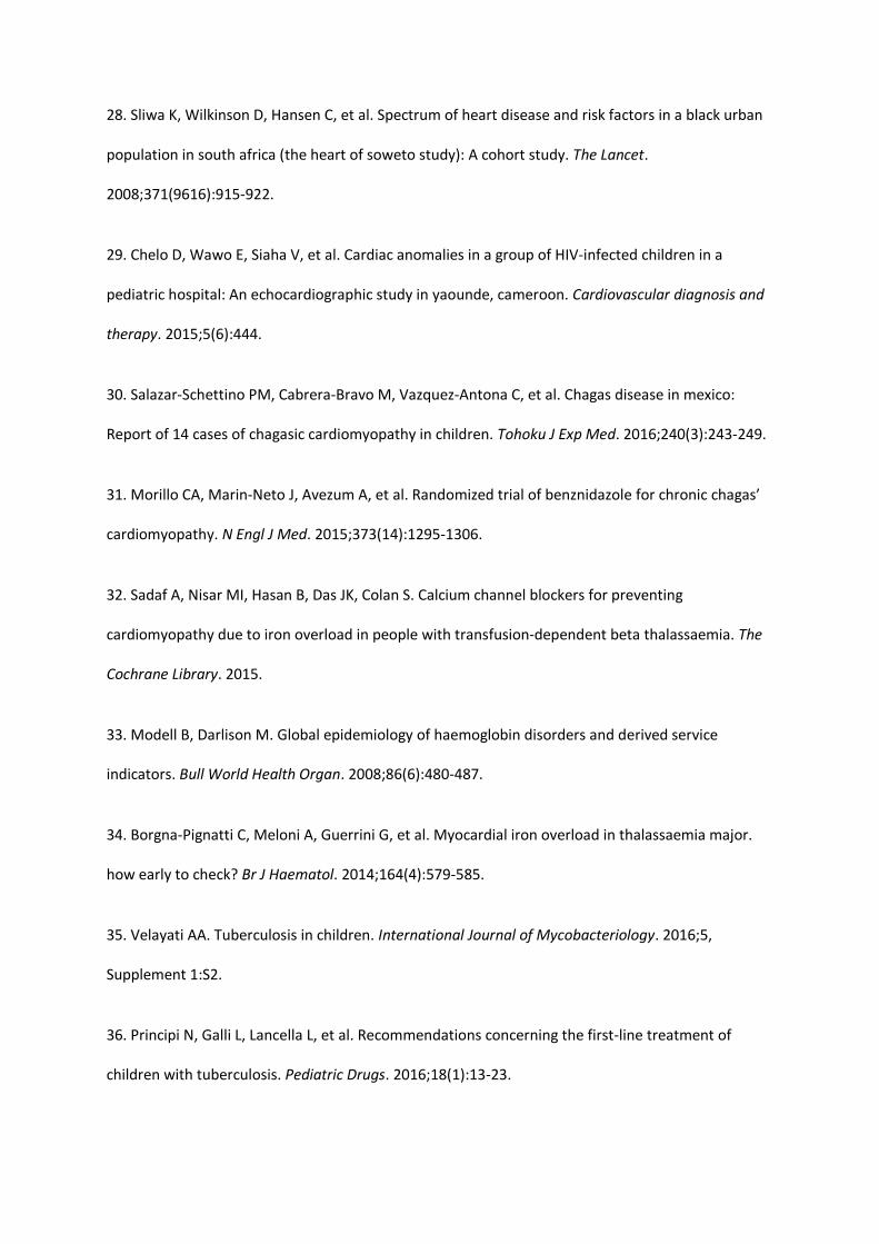
myocardium results in permanent valvular damage and the development of RHD (Figure 1). Valvular
manifestations are typically mitral and/or aortic regurgitation with stenosis in long-standing cases,
leading to the development of cardiac failure and increased risk of embolic stroke, endocarditis and
atrial fibrillation11. Barriers to the implementation of primary and secondary prevention can include
limited access to primary care, lack of healthcare workers, expense of microbiological diagnosis or
echocardiography, poor community awareness and lack of recognition of ARF by clinicians. For
example, in one cohort study of 309 newly diagnosed patients with RHD in Uganda none had a
confirmed history or ARF12.
Successful management of RHD hinges on secondary penicillin prophylaxis to prevent disease
progression, specialist review and serial cardiac imaging, appropriate prescription and monitoring of
Page 4
anticoagulation and timely referral for cardiac surgery11. Unfortunately, in many LMIC access to
these services is poor and many patients present late in the disease course with established RHD,
advanced cardiac failure, embolic stroke, infective endocarditis or symptomatic arrhythmias11. The
REMEDY study, an international hospital based registry of 3,343 patients with symptomatic RHD in
12 African countries, India and Yemen, has highlighted the ongoing challenges in the management of
RHD in LMIC9,13. Only 54.8% of patients were on secondary penicillin prophylaxis. Appropriate use of
oral anticoagulation was variable, with low rates even amongst patients with mitral stenosis or
established left atrial thrombus. Monitoring of warfarin prescription provided challenges, with 12.2%
having no INR monitoring and 34% monitored less than 3 times in 6 months. Only 10.3% of patients
requiring surgery or percutaneous procedures received intervention, most of whom were in upper-
middle-income countries9. At two year follow up 16.9% of the cohort had died at a median age of
28.79.
Earlier detection and management of ARF and RHD in LMIC is clearly critical to reducing the
burden of disease. Multiple studies using portable echocardiography in school-aged children have
detected a high prevalence of latent, pre-clinical disease, raising the possibility of successful
secondary prophylaxis in these children before complications occur11. One study in Fiji showed that
nurse led echocardiographic screening had good sensitivity and specificity in detecting mitral
regurgitation in RHD14. However it remains unclear which children with latent disease will benefit
from prophylaxis as a proportion of those with borderline disease improve to normal after medium-
term follow-up15. Further large-scale trails are required before recommending the implementation
of a potentially very costly and skill intensive intervention. Targeted screening of index cases may
be a useful approach to identify those at risk of developing symptomatic disease16.
The findings of REMEDY reinforce the fact that RHD is a disease of poverty and social injustice.
Those affected are predominantly young, largely unemployed and two thirds are female9. It is
known that household overcrowding, rural location and under-nutrition are all associated with
Page 5
increased risk of ARF and RHD11. Education beyond primary school is associated with significantly
decreased risk of mortality9. Encouragingly there has been a renewed interest in tackling RHD in
recent years, with the World Heart Federation calling for a 25% reduction in premature mortality
from RHD by 2025 and the Social Cluster of the Africa Union Commission laying out key priorities for
tackling RHD in 201517. Central to these efforts will be development of robust surveillance
programmes for RHD, increased access to primary and secondary prophylaxis and the establishment
of cardiac surgical services in LMIC. Improvement of the social determinants of health in LMIC, by
tackling living conditions and overcrowding, is an over-riding challenge in countries with high
prevalence of RHD.
After many years of research there is renewed hope of the development of an effective group A
streptococcal vaccine to aid primary prevention of RHD. Challenges in development have included
the plethora of group A streptococcal strains, the risk of immune reactivity to the vaccine and
commercial viability18.
Tropical endomyocardial fibrosis
Endomyocardial fibrosis (EMF) is the most common cause of restrictive cardiomyopathy worldwide,
characterised by deposition of fibrous tissue in the endomyocardium19. This neglected disease
typically affects poor, rural populations in tropical LMIC, with geographically distinct pockets of high
prevalence in Africa, Asia and South America. In contrast to rheumatic heart disease, the main
challenges in managing EMF are a lack of knowledge of the origin and pathogenesis of the disease
with no specific treatment or evidence based prevention strategies currently available19.
Although first described in 1948, the pathogenesis of the disease remains unclear. Proposed, but
unproven disease mechanisms include hypereosinophilia, infection, autoimmune, genetic, dietary
and geochemical factors19. The occurrence of EMF in small numbers of people from Europe and
North America after short stays in endemic regions supports the role of an infectious or other
environmental cause, however no specific trigger has been identified19. Familial occurrence and high
Page 6
incidence among certain ethnic groups has indicated a genetic susceptibility to the disease19.
Critically, EMF cannot be explained by a single cause in all areas where it has been reported. It is
likely to be triggered by multiple independent environmental factors acting on individuals with a
genetic predisposition19.
An estimated 10 million people are affected by EMF worldwide although geographic distribution in
affected countries is not uniform20. In endemic areas of Africa EMF is the second most common
cause of admission for acquired heart disease (after rheumatic heart disease) and accounts for 20%
of all cases of heart failure21. Few systematic studies of prevalence in the community exist, but a
community based study in a rural area of Mozambique using portable echocardiography found
evidence of EMF in 19.8% of the general population, and 28.1% of those aged 10-19 years22.
Children and adolescents are predominantly affected, with over half of cases seen in the first decade
of life21.
If recognised, the initial clinical features of EMF are a febrile illness associated with pericarditis and
eosinophilia, dyspnoea, itching and periorbital swelling19. This is followed by ventricular thrombosis
affecting usually the apices and subvalvular apparatus which evolves to form endocardial fibrosis
typical of the advanced stage of the disease. The resulting impedance of ventricular filling and valve
distortion leads to restrictive physiology with atrioventricular regurgitation, and the typical
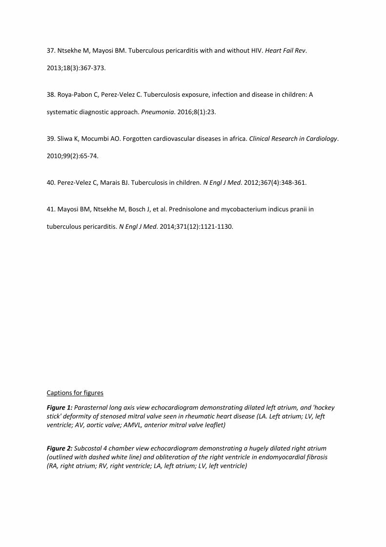
appearance of small ventricles with severely dilated atria (Figure 2). Echocardiography is the
mainstay of diagnosis8. The majority of patients present late with features of longstanding cardiac
failure, clubbing, growth retardation, testicular atrophy, pubertal delay and cachexia. Marked ascites
out of proportion to peripheral oedema is typical, leading some authors to hypothesise EMF to be a
systemic syndrome with associated peritoneal inflammation19. Atrial fibrillation occurs in greater
that 30% of cases, and other conduction abnormalities are common19.
No specific treatment has been developed to treat EMF. Medical management is based on the
symptomatic treatment of heart failure, arrhythmias and anticoagulation where indicated19. Short
Page 7
courses of steroids have been used to suppress eosinophilia in the acute phase of the disease,
although there is a lack of evidence to support their use19. Surgical intervention has been shown to
improve symptoms and improve survival in EMF, most commonly using targeted endocardial
resection combined with valve repair or replacement23. However, access to cardiac surgery in
endemic regions is extremely limited, with only a handful of sub-Saharan African countries having
independent cardiac surgery programmes24. Research into surgical intervention is limited to a small
number of studies in patients with advanced disease21.
The prognosis of EMF remains extremely poor, with 75% of patients dying within 2 years of
diagnosis19. Death is typically due to progressive heart failure, pulmonary embolism or fatal
ventricular arrhythmia21. There is an urgent need for further research into the aetiology and
mechanisms of the disease in order to develop effective treatment and prevention strategies.
Dilated Cardiomyopathy
In developed countries dilated cardiomyopathies (DCM) account for about half of all childhood heart
transplants and population cohort studies have found an annual incidence of cardiomyopathy of up
to 1.24 per 100,000 children under the age of ten25. Epidemiological data is lacking in developing
countries, but the disease burden is thought to be significant given the association of
cardiomyopathies with malnutrition and infectious disease26. One tertiary centre in Nigeria recorded
cardiomyopathy in nearly 3% of children accessing cardiac services in South-west Nigeria27. A lack of
specialist investigations poses significant problems to the diagnosis and management of paediatric
DCM in LMIC. The cause in most children is not known.
HIV associated cardiomyopathy is an important known cause. The Heart of Soweto study found that
HIV associated DCM was the most common cardiac diagnosis amongst HIV positive patients28. In one
Ugandan study, cardiac abnormalities were present in 50% of newly diagnosed HIV positive
children29. Direct infection of myocytes with HIV triggering an autoimmune response is thought to
underlie HIV cardiomyopathy, although nutritional deficiencies, and opportunistic infections are also
Page 8
implicated29. HIV cardiomyopathy is an indication to start anti-retroviral therapy, independent of
CD4 count, and there is justification for routine echocardiography in HIV-positive children in order to
identify pre-symptomatic cardiac disease. However in areas where early antiretroviral treatment is
available, the incidence is falling.
In Latin America, Chagas disease caused by Trypanosoma cruzi is known to be a significant infective
cause of DCM. Although usually observed in chronic disease 10-20 years after initial infection, a
recent epidemiological study of more than 3000 children in Mexico identified 14 children with pre-
symptomatic chagasic cardiomyopathy30. It is unclear if antiparasitic treatment improves the
prognosis once the disease has developed31.
β thalassemia is a common inherited blood disorder in the Indian sub-continent, south-east and
central Asia, Southern China, Mediterranean, North Africa and the Middle East, characterised by
severe anaemia requiring regular blood transfusions32. These children are at risk of Iron-overload
related cardiomyopathy in LMIC. A public health review in 2008 estimated over 25000 annual
births of transfusion dependent thalassemia worldwide, mostly in LMIC33. While only 11.7% of
children who require transfusions receive them, those who do carry a significant risk of developing
cardiac iron overload and cardiomyopathy with a subsequent rapid decrease in myocardial
function and death33. This has been demonstrated in children under the age of 10 despite
receiving chelation therapy34. The high cost of managing these patients presents a difficult
challenge to health systems in LMIC.
Tuberculous Pericarditis
WHO figures in 2015 estimated that globally, 1 million children are infected with TB, predominantly
in LMIC in Asia and Sub-Saharan Africa, accounting for more than 136,000 deaths each year35. TB is
one of the most common causes of pericardial effusion in TB endemic countries, with approximately
Page 9
1-4% of children with TB developing pericarditis36. The HIV pandemic has dramatically changed the
epidemiology, manifestation and treatment options for TB pericarditis. In one large study in South
Africa, 83% of patients aged 15-29 undergoing pericardiocentesis for large pericardial effusions had
TB, with over 50% of patients co-infected with HIV37. HIV predisposes patients to more disseminated
disease, with increased risk of pericardial and myocardial involvement37,38.
In children, TB pericardial disease has three main presentations: pericardial effusion (most
common), constrictive pericarditis, and a combination known as effusive-constrictive disease. Most
frequently, the pericardium is infiltrated from an infected contiguous subcarinal lymph node38. Once
in the pericardium, an inflammatory process with granuloma formation results in the production of a
fibrinous exudate37.
Importantly, the clinical presentation is variable and often non-specific. However, given the poor
availability and sensitivity of microbiological diagnosis in LMIC, the diagnosis is usually based on
clinical signs alone. A high index of suspicion is required in endemic areas39. Children typically
present with signs and symptoms of heart failure, including persistent cough, dyspnoea, chest pain
and hepatomegaly in addition to fever, night sweats and failure to thrive38. Chest radiography may
show cardiomegaly with a globular silhouette, while echocardiography can confirm the presence of
effusions, often associated with fibrinous threads and a “bread and butter” appearance of the
visceral pericardium and may identify associated mediastinal lymphadenopathy38. Valvular disease
is not a typical feature of TB pericarditis however the myocardium is often affected adding to the
complexity of the disease. The following features of pericardial fluid suggest TB: a predominantly
lymphocytic cell count, elevated protein and LDH level and glucose 3.0–5.5 mmol/L are suggestive
but not diagnostic of TB38.
Specific research in the management of childhood tuberculous pericarditis is lacking, and adult
management regimens are followed 40. A typical approach is pericardiocentesis if there is evidence
of tamponade, followed by an initial regimen of rifampicin, isoniazid, pyrazinamide and ethambutol
Page 10
for at least two months followed by isoniazid and rifampicin for a further four months. Pericardial
TB management in children is complicated by the lack of suitable formulations of medications for
the first years of life. The multicentre IMPI trial showed that the use of adjunctive high dose steroids
reduced the incidence of constrictive pericarditis and frequency of hospitalisation in adults, but was
associated with an increased risk of malignancy in patients with HIV41. No similar study in children
exists. Surgical resection of the pericardium is indicated in patients with persistent constrictive
symptoms following anti-TB chemotherapy37.
Conclusions
The burden of illness associated with acquired cardiac disease in children in LMIC is significant and
may be equivalent to that of congenital heart disease. Rheumatic heart disease, endomyocardial
fibrosis, cardiomyopathy (including HIV cardiomyopathy) and TB are the most important causes. All
are associated with poverty with the neediest children having the least access to care. The
associated mortality and morbidity is high. While detailed analysis of the cost burden of and
funding for these diseases is beyond the scope of this review there is a clear disparity between the
burden of disease and current research. There is an urgent need to improve cardiac care in LMIC,
particularly in sub-Saharan Africa and parts of South-East Asia where the burden is highest.
References
1. You D, Hug L, Ejdemyr S, et al. Global, regional, and national levels and trends in under-5 mortality
between 1990 and 2015, with scenario-based projections to 2030: A systematic analysis by the UN
inter-agency group for child mortality estimation. The Lancet. ;386(10010):2275-2286.
2. Hewitson J, Zilla P. Children’s heart disease in sub-saharan africa:Challenging the burden of
disease. SA Heart. 2010;7.
3. Kennedy N, Miller P. The spectrum of paediatric cardiac disease presenting to an outpatient clinic
in malawi. BMC Research Notes. 2013;6(1):53.
Page 11
4. Steer AC, Carapetis JR, Nolan TM, Shann F. Systematic review of rheumatic heart disease
prevalence in children in developing countries: The role of environmental factors. J Paediatr Child
Health. 2002;38(3):229-234.
5. Wang H, Naghavi M, Allen C, et al. Global, regional, and national life expectancy, all-cause
mortality, and cause-specific mortality for 249 causes of death, 19802015: A systematic analysis for
the global burden of disease study 2015. The Lancet. ;388(10053):1459-1544.
6. Watkins DA, Zuhlke LJ, Engel ME, Mayosi BM. Rheumatic fever: Neglected again. Science.
2009;324(5923):37. http://science.sciencemag.org/content/324/5923/37.2.abstract. doi:
10.1126/science.324.5923.37b.
7. Ellis J, Martin R, Wilde P, Tometzki A, Senkungu J, Nansera D. Echocardiographic, chest X-ray and
electrocardiogram findings in children presenting with heart failure to a ugandan paediatric ward.
Trop Doct. 2007;37(3):149-150.
8. Grimaldi A, Mocumbi AO, Freers J, et al. Tropical endomyocardial fibrosis: Natural history,
challenges, and perspectives. Circulation. 2016;133(24):2503-2515.
9. Zhlke L, Karthikeyan G, Engel ME, et al. Clinical outcomes in 3343 children and adults with
rheumatic heart disease from 14 low-and middle-income CountriesClinical perspective. Circulation.
2016;134(19):1456-1466.
10. Rothenbühler M, O'Sullivan C,J., Stortecky S, et al. Active surveillance for rheumatic heart disease
in endemic regions: A systematic review and meta-analysis of prevalence among children and
adolescents. The Lancet Global Health. ;2(12):e726.
11. Carapetis JR, Beaton A, Cunningham MW, et al. Acute rheumatic fever and rheumatic heart
disease. Nat Rev Dis Primers. 2016;2:15084.
Page 12
12. Okello E, Wanzhu Z, Musoke C, et al. Cardiovascular complications in newly diagnosed rheumatic
heart disease patients at mulago hospital, uganda. Cardiovascular journal of Africa. 2013;24(3):80.
http://www.ncbi.nlm.nih.gov/pubmed/23736132.
13. Zhlke L, Engel ME, Karthikeyan G, et al. Characteristics, complications, and gaps in evidence-
based interventions in rheumatic heart disease: The global rheumatic heart disease registry (the
REMEDY study). Eur Heart J. 2015;36(18):1115-1122.
14. Colquhoun SM, Carapetis JR, Kado JH, et al. Pilot study of nurse-led rheumatic heart disease
echocardiography screening in fiji--a novel approach in a resource-poor setting. Cardiol Young.
2013;23(4):546-552.
15. Beaton A, Okello E, Aliku T, et al. Latent rheumatic heart disease: Outcomes 2 years after
echocardiographic detection. Pediatr Cardiol. 2014;35(7):1259-1267.
16. Zuhlke L, Engel ME, Lemmer CE, et al. The natural history of latent rheumatic heart disease in a 5
year follow-up study: A prospective observational study. BMC Cardiovasc Disord. 2016;16:3.
17. Watkins D, Zuhlke L, Engel M, et al. Seven key actions to eradicate rheumatic heart disease in
africa: The addis ababa communiqu: Cardiovascular topics. Cardiovascular journal of Africa.
2016;27(3):184-187.
18. de Dassel JL, Ralph AP, Carapetis JR. Controlling acute rheumatic fever and rheumatic heart
disease in developing countries: Are we getting closer? Curr Opin Pediatr. 2015;27(1):116-123.
19. Grimaldi A, Mocumbi A, Freers J, et al. Tropical endomyocardial fibrosis: Natural history,
challenges, and perspectives. Circulation. 2016;133(24):2503-2515.
http://ovidsp.ovid.com/ovidweb.cgi?T=JS&NEWS=n&CSC=Y&PAGE=fulltext&D=ovft&AN=00003017-
201606140-00015. doi: 10.1161/CIRCULATIONAHA.115.021178.
Page 13
20. Yacoub S, Kotit S, Mocumbi AO, Yacoub MH. Neglected diseases in cardiology: A call for urgent
action. Nat Clin Pract Cardiovasc Med. 2008;5(4):176-177.
21. Mocumbi AO, Yacoub S, Yacoub MH. Neglected tropical cardiomyopathies: II. endomyocardial
fibrosis: Myocardial disease. Heart. 2008;94(3):384-390.
22. Mocumbi AO, Ferreira MB, Sidi D, Yacoub MH. A population study of endomyocardial fibrosis in a
rural area of mozambique. N Engl J Med. 2008;359(1):43-49.
23. Mocumbi AO, Sidi D, Vouhe P, Yacoub M. An innovative technique for the relief of right
ventricular trabecular cavity obliteration in endomyocardial fibrosis. J Thorac Cardiovasc Surg.
2007;134(4):1070-1072.
24. Zühlke L, Mirabel M, Marijon E. Congenital heart disease and rheumatic heart disease in africa:
Recent advances and current priorities. Heart. 2013.
http://heart.bmj.com/content/early/2013/05/16/heartjnl-2013-303896.abstract. doi:
10.1136/heartjnl-2013-303896.
25. Nugent AW, Daubeney PEF, Chondros P, et al. The epidemiology of childhood cardiomyopathy in
australia. N Engl J Med. 2003;348(17):1639-1646.
26. Sliwa K, Damasceno A, Mayosi BM. Epidemiology and etiology of cardiomyopathy in africa.
Circulation. 2005;112(23):3577-3583.
27. Animasahun BA, Madise-Wobo AD, Ogunkunle OO, Gbelee OH, Oke DA. Cardiomyopathies
among children attending a tertiary hospital in south-western nigeria. J Clin Exp Res Cardiol.
2015;2(3):302.
Page 14
28. Sliwa K, Wilkinson D, Hansen C, et al. Spectrum of heart disease and risk factors in a black urban
population in south africa (the heart of soweto study): A cohort study. The Lancet.
2008;371(9616):915-922.
29. Chelo D, Wawo E, Siaha V, et al. Cardiac anomalies in a group of HIV-infected children in a
pediatric hospital: An echocardiographic study in yaounde, cameroon. Cardiovascular diagnosis and
therapy. 2015;5(6):444.
30. Salazar-Schettino PM, Cabrera-Bravo M, Vazquez-Antona C, et al. Chagas disease in mexico:
Report of 14 cases of chagasic cardiomyopathy in children. Tohoku J Exp Med. 2016;240(3):243-249.
31. Morillo CA, Marin-Neto J, Avezum A, et al. Randomized trial of benznidazole for chronic chagas’
cardiomyopathy. N Engl J Med. 2015;373(14):1295-1306.
32. Sadaf A, Nisar MI, Hasan B, Das JK, Colan S. Calcium channel blockers for preventing
cardiomyopathy due to iron overload in people with transfusion‐dependent beta thalassaemia. The
Cochrane Library. 2015.
33. Modell B, Darlison M. Global epidemiology of haemoglobin disorders and derived service
indicators. Bull World Health Organ. 2008;86(6):480-487.
34. Borgna‐Pignatti C, Meloni A, Guerrini G, et al. Myocardial iron overload in thalassaemia major.
how early to check? Br J Haematol. 2014;164(4):579-585.
35. Velayati AA. Tuberculosis in children. International Journal of Mycobacteriology. 2016;5,
Supplement 1:S2.
36. Principi N, Galli L, Lancella L, et al. Recommendations concerning the first-line treatment of
children with tuberculosis. Pediatric Drugs. 2016;18(1):13-23.
Page 15
37. Ntsekhe M, Mayosi BM. Tuberculous pericarditis with and without HIV. Heart Fail Rev.
2013;18(3):367-373.
38. Roya-Pabon C, Perez-Velez C. Tuberculosis exposure, infection and disease in children: A
systematic diagnostic approach. Pneumonia. 2016;8(1):23.
39. Sliwa K, Mocumbi AO. Forgotten cardiovascular diseases in africa. Clinical Research in Cardiology.
2010;99(2):65-74.
40. Perez-Velez C, Marais BJ. Tuberculosis in children. N Engl J Med. 2012;367(4):348-361.
41. Mayosi BM, Ntsekhe M, Bosch J, et al. Prednisolone and mycobacterium indicus pranii in
tuberculous pericarditis. N Engl J Med. 2014;371(12):1121-1130.
Captions for figures
Figure 1: Parasternal long axis view echocardiogram demonstrating dilated left atrium, and 'hockey stick' deformity of stenosed mitral valve seen in rheumatic heart disease (LA. Left atrium; LV, left ventricle; AV, aortic valve; AMVL, anterior mitral valve leaflet)
Figure 2: Subcostal 4 chamber view echocardiogram demonstrating a hugely dilated right atrium (outlined with dashed white line) and obliteration of the right ventricle in endomyocardial fibrosis (RA, right atrium; RV, right ventricle; LA, left atrium; LV, left ventricle)