Çankaya University Journal of Science and Engineering
Volume 15, No. 2 (2018) 063-075
Date Received: 17/09/2018
Date Accepted: 23/11/2018
ISSN 2564 – 7954 © 2018 Çankaya University
Acrolein-induced Histopathological
Alterations in the Liver of Goldfish, Carassius
auratus (Linnaeus, 1758)
Sezgi Arman1, Sema İşisağ Üçüncü2
1Department of Biology, Sakarya University,Turkey,
2Department of Biology, Ege University, Turkey.
e-mail: [email protected], [email protected]
Abstract: The present study was conducted to examine the potential histopathological changes caused by a
herbicide, acrolein, in the liver of Carassius auratus (goldfish). Fish were exposed to 1, 5 and 25 µg/L
acrolein for 96 h. Liver tissues were removed, fixed with Bouin’s fluid and embedded in paraffin. 5 µm
serial sections were stained with hematoxylin eosin and the samples were investigated by light microscopy.
Acrolein treatment gave rise to sinusoidal dilatation and congestion, vacuolar degeneration, hemorrhage,
lymphocyte infiltration, presence of enlarged melanomacrophagic centers, cloudy swelling, nucleolus
absence, and necrosis. These results are important for paying attention to acrolein usage limits and its
contamination in the aquatic environment.
Keywords: Acrolein, Carassius auratus, goldfish, herbicide, histopathology, liver.
1. Introduction
Pesticides are kind of agrochemicals that have been widely used to control pests. They are
described as any substance(s) that kills, prevents, mitigates or restricts the population size of
unwanted organisms. There are over 500 compounds are registered as pesticides world-wide
[1,2]. Classification of pesticides can be variable, however the type that based on the target
organisms (e.g. insecticides, herbicide) is may be the most used. Herbicides are designed to
control plants that they are the most commonly used pesticide type compared with insecticides
64 S.Arman & S. I. Ucuncu
and fungicides [3]. However, it’s reported that many herbicides adversely affect various non-
target vertebrate organisms [4-8]. These chemicals are generally show mobility between
different environmental phases and enter into aquatic ecosystems via direct use or drift, wash
off and drain from the agricultural sites and/or the application area [9].
Acrolein (C3H4O) is the most reactive and highly electrophilic α,β-unsatured aldehyde that is
generated from the burning processes of gasoline, fuels, cigarettes, woods, plastics [10]. It is
directly injected below the surface of irrigation canals as a herbicide to control submerged and
floating weeds; the concentration of acrolein in the irragation systems can be reached up to 15
ppm [11-13]. It can be also produced endogenously through lipid peroxidation in various
tissues by normal cellular metabolism [14,15].
The toxic effects of acrolein can emerged following inhalation, ingestion, dermal exposure or
systemically after absorption [10]. Due to it’s high reactivity, acrolein shows its toxicity
through rapid binding and depleting the cellular thiols or other nucleophiles like glutathione
(GSH). Reactions with thiols may affect gene expression or acrolein can directly interact with
genes and transcription factors [16,17]. It leads to genotoxicity and cytotoxicity [18],
including liver damage and hepatocyte death [19].
The liver is a multifunctional internal organ that is responsible for various vital processes such
as bile secretion, metabolism of carbohydrates, proteins and fats; production of vitellogenin,
detoxification and inactivation [20,21].
The aim of this study was to investigate the adverse effects of acrolein exposure on the liver
tissue of goldfish, Carassius auratus.
2. Materials and Methods
Goldfish were obtained from a commercial supplier. Fish were 3.7-4.2 cm in lenght and 3.87-
5.8 g in weight. They were acclimated in our laboratory for two weeks before the treatment.
CUJSE 15, No. 2 (2018) 65
Specimens were maintained in a 100 L glass tank with dechlorinated tap water at 26±2 ºC and
in natural daylight and darkness. Fish were fed with Sera Goldy twice a day.
Acrolein (analytical standard) (CAS No: 107-02-8) was purchased from Sigma-Aldrich.
Sublethal concentrations were determined as 1, 5 and 25 µg/L and they were prepared by
diluting a more concentrated stock solution (100 mg/L). Specimens were divided into four
groups randomly and five fish were used for each experimental and chemical-free control
groups in separate tanks. Test solutions were not renewed and a static toxicity test system was
conducted for 96 h. After the treatment, all the fish were anaesthetized with MS222 (tricaine
methanesulfonate) (Sigma), livers were removed and fixed in Bouin’s fluid for 24 hr.
Specimens were dehydrated in ethanol, treated with xylol and embedded in paraffin. 5 µm
cross sections were stained with Mayer’s hematoxylin eosin (H-E). Histopathological
alterations were examined by light microscopy and the images were taken with Zeiss Axio
Scope A1 equipped with Zeiss Axiocam ERc5s.
3. Results
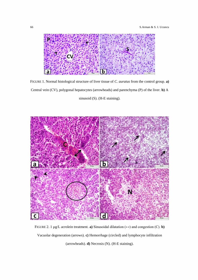
Control samples exhibited normal histological structure and no histopathological alteration
was observed. Hepatocytes were the parenchymal cells of the liver which were polygonal in
shape. They were clearly observed around the central veins (Fig 1a). Sinusoids were the
capillary networks lined with endothelial cells and surrounded by hepatocytes (Fig 1b).
The samples of 1 µg/L acrolein treatment group showed sinusoidal dilatation and congestion
(Fig. 2a), vacuolar degeneration of hepatocytes (Fig 2b), hemorrhage, lymphocyte infiltration
(Fig 2c) and necrosis (Fig 2d).
66 S.Arman & S. I. Ucuncu
FIGURE 1. Normal histological structure of liver tissue of C. auratus from the control group. a)
Central vein (CV), polygonal hepatocytes (arrowheads) and parenchyma (P) of the liver. b) A
sinusoid (S). (H-E staining).
FIGURE 2. 1 µg/L acrolein treatment. a) Sinusoidal dilatation (↔) and congestion (C). b)
Vacuolar degeneration (arrows). c) Hemorrhage (circled) and lymphocyte infiltration
(arrowheads). d) Necrosis (N). (H-E staining).
CUJSE 15, No. 2 (2018) 67
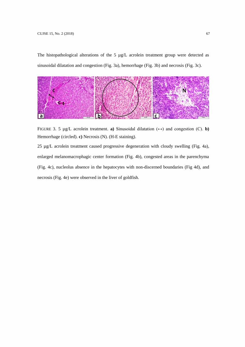
The histopathological alterations of the 5 µg/L acrolein treatment group were detected as
sinusoidal dilatation and congestion (Fig. 3a), hemorrhage (Fig. 3b) and necrosis (Fig. 3c).
FIGURE 3. 5 µg/L acrolein treatment. a) Sinusoidal dilatation (↔) and congestion (C). b)
Hemorrhage (circled). c) Necrosis (N). (H-E staining).
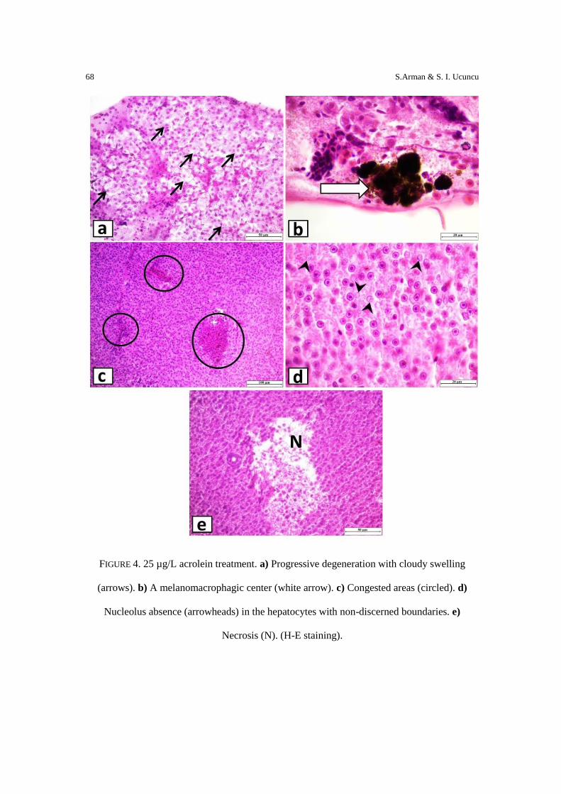
25 µg/L acrolein treatment caused progressive degeneration with cloudy swelling (Fig. 4a),
enlarged melanomacrophagic center formation (Fig. 4b), congested areas in the parenchyma
(Fig. 4c), nucleolus absence in the hepatocytes with non-discerned boundaries (Fig 4d), and
necrosis (Fig. 4e) were observed in the liver of goldfish.
68 S.Arman & S. I. Ucuncu
FIGURE 4. 25 µg/L acrolein treatment. a) Progressive degeneration with cloudy swelling
(arrows). b) A melanomacrophagic center (white arrow). c) Congested areas (circled). d)
Nucleolus absence (arrowheads) in the hepatocytes with non-discerned boundaries. e)
Necrosis (N). (H-E staining).
CUJSE 15, No. 2 (2018) 69
4. Discussion
Liver is a helpful internal structure to observe the toxicity of environmental pollutants. Our
results clearly indicated that acrolein exposure caused distinct histopathological alterations in
the liver of C. auratus. Several studies on investigation the effects of herbicides to non-target
organisms especially the teleost fish have been conducting for a long time. However, there is
only limited studies that have focused on the adverse effects of acrolein on various tissues of
freshwater teleosts [22].
Acrolein gave rise to sinusoidal dilatation and congestion, vacuolar degeneration of
hepatocytes, hemorrhage, lymphocyte infiltration, cloudy swelling, enlarged
melanomacrophagic center formation, nucleolus absence and necrosis in the goldfish liver.
Figueiredo-Fernandes et al. [23] revealed that paraquat caused vacuolization like
parenchymatic alteration, increase of melanomacrophage aggregates and eosinophilic granular
cells, and necrosis in the liver of Oreochromis niloticus. It was reported that clomazone
caused hepatocyte vacuolization in Rhamdia quelen [24]. Peebua et al. [25] stated that O.
niloticus treated with alachlor showed hydropic swelling of hepatocytes and vacuolization in
the liver. Yerbimat exposure induced increasing vacuolization in the hepatic cells and fibrosis
in Goodea atripinnis [26]. Mela et al. [27] noted that atrazine treatment resulted in leukocyte
infiltration, hepatocyte vacuolization, increase in melanomacrophage numbers and necrosis in
R. quelen. 2,4-D exposed Poecilia vivipara liver showed hepatocyte vacuolization, nuclear
swelling and micronuclei [28].
Glyphosate hepatotoxicty in fish have been frequently examined histologically by several
authors. In Cyprinus carpio sinusoidal congestion and early fibrosis were observed [29].
Jiraungkoorskul et al. [30] reported hydropic swelling of hepatocytes with some pyknotic
nuclei and severe leucocyte infiltration in the liver of O. niloticus. Ayoola [31] also noted
vacuolization of hepatocytes and necrosis in the same species. Vacuoles in the cytoplasm,
hyperemia, cytoplasmic and nuclear degeneration and hypertrophy, pyknotic nuclei were
70 S.Arman & S. I. Ucuncu
determined in Prochilodus lineatus [32]. The liver of glyphosate treated Piaractus
mesopotamicus showed enlargement of sinusoids, hepatocyte hypertrophy, lipid droplets,
peripherally located nuclei, nuclear deformation and degeneration, absence of nucleoli, and
necrosis [33].
The findings of the current paper and the results of the previous histopathological studies are
majorly similar to each other that it shows once again the alterations are not chemical specific.
Hepatic sinusoidal dilatation and congestion might be related to venous outflow impairment
and it could be observed by inflammatory diseases [34]. Brancatelli et al. [35] indicated that
sinusoidal dilatation could be caused by not only hepatic venous outflow obstruction but also
it could be associated with pericardial disease, heart failure, and extrahepatic inflammatory
conditions. Vacuolization of the hepatocytes might be due to the imbalance rate conditions of
the synthesized and released materials by the hepatocytes [36,30]. Abdel-Moneim et al. [37]
noted that vacuolization might be resulted from lipid dystrophies. Hemorrhage is arised
internal or externally as a result of the injury of a blood vessel. Lymphoid cell infiltration
might be caused by a response to inflammation or necrosis [27]. Cloudy swelling is occured
when the parenchymal cells are disable to maintain the ionic and fluid homeostasis or it may
be due to cytoplasmic degeneration and macromolecular crowding caused by leakage of
lysosomal hydrolytic enzymes [38,39]. Increased melanomacrophagic centers are thought to
be related with biotransformation capacity of the liver [40,23]. It’s also emphasized that
melanomacrophagic aggregates might be associated with degenerative necrotic conditions
[41]. Shiogiri et al. [33] noted that deformation of cellular membranes, degenerative nuclei
and absence of the nucleoli indicated that the beginning of necrosis in consequence of
chemical exposure. Necrosis is mainly associated with oxidative stress [42,37,27] and
oxidative stress is related with cellular damage which may be due to the free radicals react
with the lipids of the cell membrane and affect its structure irreversibly [43,26].
CUJSE 15, No. 2 (2018) 71
The histopathological alterations observed in the liver of goldfish exposed to acrolein might
be probably caused by cytotoxic and highly electrophilic character of the chemical by
depleting cellular nucleophiles such as GSH and leave the hepatocytes vulnerable to oxidative
damage. Liver is responsible for detoxification of xenobiotics and has many other metabolic
functions. Such effects on the liver may lead to malfunctions and metabolical disorders in the
organism. These results should pay attention to acrolein contamination and usage limits for
environmental safety.
Acknowledgements
This study was represented as a poster presentation in International Congress of Health and
Environment (2017) in Adana-Turkey.
References
[1] G. R. Van der Hoff, P. van Zoonen, Trace analysis of pesticides by gas
chromatography, Journal of Chromatography A, 843(1), (1999), 301-322.
[2] F. E. Ahmed, Analyses of pesticides and their metabolites in foods and drinks,
TrAC - Trends in Analytical Chemistry, 20(11), (2001), 649–661.
[3] J. Cooper, H. Dobson, September. The benefits of pesticides to mankind and the
environment, Crop Protection, 26(9), (2007), 1337-1348.
[4] M. S. Butchiram, K. S. Tilak, P. W. Raju, Studies on histopathological changes in
the gill, liver and kidney of Channa punctatus (Bloch) exposed to alachlor, Journal of
Environmental Biology, 30(2), (2009), 303–306.
[5] J. M. Pérez-Iglesias, S. Soloneski, N. Nikoloff, G. S. Natale, M. L. Larramendy,
Toxic and genotoxic effects of the imazethapyr-based herbicide formulation Pivot H®
on montevideo tree frog Hypsiboas pulchellus tadpoles (Anura, Hylidae),
Ecotoxicology and Environmental Safety, 119, (2015), 15-24.
[6] J. P. Myers, M. N. Antoniou, B. Blumberg, L. Carroll, T. Colborn, L. G. Everett,
... , L. N. Vandenberg, Concerns over use of glyphosate-based herbicides and risks
72 S.Arman & S. I. Ucuncu
associated with exposures: a consensus statement, Environmental Health, 15(1),
(2016), 19.
[7] M. Ehrsam, S. A. Knutie, J. R. Rohr, The herbicide atrazine induces hyperactivity
and compromises tadpole detection of predator chemical cues, Environmental
Toxicology and Chemistry, 35(9), (2016), 2239-2244.
[8] D. B. Lindenmayer, J. Wood, C. MacGregor, R. J. Hobbs, J. A. Catford, Non‐
target impacts of weed control on birds, mammals, and reptiles, Ecosphere, 8(5),
(2017).
[9] J. Stanley, G. Preetha, Pesticide toxicity to non-target organisms, Springer, (2016),
502 p.
[10] R. J. Henning, G. T. Johnson, J. P. Coyle, R. D.Harbison, Acrolein Can Cause
Cardiovascular Disease: A Review, Cardiovascular Toxicology, 17(3), (2017), 227-
236.
[11] L. L. C. Alligare, Magnacide H herbicide application and safety manual. (2013),
(URL:http://www.alligarellc.com/assets/pdf/MAGNACIDE_H_Manual.pdf). (Date
accessed: April 2018).
[12] D. Shaner, Herbicide handbook. 10th Edition, Weed Science Society of America,
Lawrence, (2014), 513 p.
[13] K. D. King, Acrolein : Environmental fate and ecotoxicology, (2016), (URL:
http://www.cdpr.ca.gov/docs/emon/pubs/fatememo/acrolein.pdf). (Date accessed:
April 2018).
[14] K. Uchida, M. Kanematsu, Y. Morimitsu, T. Osawa, N. Noguchi, E. Niki,
Acrolein is a product of lipid peroxidation reaction. Formation of free acrolein and its
conjugate with lysine residues in oxidized low density lipoproteins, The Journal of
Biological Chemistry, 273, (1998), 16058–16066.
[15] M. K. Mohammad, D. Avila, J. Zhang, S. Barve, G. Arteel, C. McClain, S. Joshi-
Barve, Acrolein cytotoxicity in hepatocytes involves endoplasmic reticulum stress,
mitochondrial dysfunction and oxidative stress, Toxicology and Applied
Pharmacology, 265(1), (2012), 73–82.
[16] J. P. Kehrer, S. S. Biswal, The molecular effects of acrolein, Toxicological
Sciences, 57(1), (2000), 6–15.
CUJSE 15, No. 2 (2018) 73
[17] M. J. Randall, M. Hristova, A. Van Der Vliet, Protein alkylation by the α,β-
unsaturated aldehyde acrolein. A reversible mechanism of electrophile signaling?,
FEBS Letters, 587(23), (2013), 3808–3814.
[18] H. Esterbauer, R. J. Schaur, H. Zollner, Chemistry and biochemistry of 4-
hydroxynonenal, malonaldehyde and related aldehydes, Free Radical Biology and
Medicine, 11, (1991), 81–128.
[19] L. Sun, C. Luo, J. Long, D. Wei, J. Liu, Acrolein is a mitochondrial toxin: effects
on respiratory function and enzyme activities in isolated rat liver mitochondria,
Mitochondrion, 6(3), (2006), 136-142.
[20] H. Akiyoshi, A. Inoue, Comparative histological study of teleost livers in relation
to phylogeny, Zoological Science, 21(8), (2004), 841–850.
[21] C. F. Sales, R. F. Silva, M. G. C. Amaral, F. F. T. Domingos, R. I. M. A. Ribeiro,
R. G. Thomé, H. B. Santos, Comparative histology in the liver and spleen of three
species of freshwater teleost, Neotropical Ichthyology, 15(1), (2017).
[22] S. Arman, S. I. Üçüncü, Histopathological changes in the gill and kidney tissues
of Carassius auratus exposed to acrolein, Journal of Environmental Biology, 38(2),
(2017), 263.
[23] A. Figueiredo-Fernandes, A. Fontaínhas-Fernandes, E. Rocha, M. A. Reis-
Henriques, The effect of paraquat on hepatic EROD activity, liver, and gonadal
histology in males and females of Nile tilapia, Oreochromis niloticus, exposed at
different temperatures, Archives of Environmental Contamination and Toxicology,
51(4), (2006), 626–632.
[24] M. Crestani, C. Menezes, L. Glusczak, D. dos Santos Miron, R. Spanevello, A.
Silveira, … ,V. L. Loro, Effect of clomazone herbicide on biochemical and
histological aspects of silver catfish (Rhamdia quelen) and recovery pattern,
Chemosphere, 67(11), (2007), 2305–2311.
[25] P. Peebua, M. Kruatrachue, P. Pokethitiyook, S. Singhakaew, Histopathological
alterations of Nile tilapia, Oreochromis niloticus in acute and subchronic alachlor
exposure, Journal of Environmental Biology, 29(3), (2008), 325–331.
[26] E.Ortiz-Ordoñez, E. Uría-Galicia, R. A. Ruiz-Picos, A. G. Sánchez Duran, Y.
Hernández Trejo, J. E. Sedeño-Díaz, E. López-López, Effect of Yerbimat herbicide
on lipid peroxidation, catalase activity, and histological damage in gills and liver of
74 S.Arman & S. I. Ucuncu
the freshwater fish Goodea atripinnis, Archives of Environmental Contamination and
Toxicology, 61(3), (2011), 443–452.
[27] M. Mela, I. C. Guiloski, H. B. Doria, M. A. F. Randi, C. A. De Oliveira Ribeiro,
L. Pereira, …, H. C. Silva de Assis, Effects of the herbicide atrazine in neotropical
catfish (Rhamdia quelen), Ecotoxicology and Environmental Safety, , 93, (2013a), 13–
21.
[28] A. F. Vigário, S. M. T. Sabóia-Morais, Effects of the 2,4-D herbicide on gills
epithelia and liver of the fish Poecilia vivipara, Pesquisa Veterinaria Brasileira, ,
34(6), (2014), 523–528.
[29] N. K. Nešković, V. Poleksić, I. Elezovíc, V. Karan, M. Budimir, Biochemical
and histopathological effects of glyphosate on carp, Cyprinus carpio L., Bulletin of
Environmental Contamination and Toxicology, 56(2), (1996), 295–302.
[30] W. Jiraungkoorskul, E. S. Upatham, M. Kruatrachue, S. Sahaphong, S. Vichasri-
Grams, P. Pokethitiyook, Biochemical and histopathological effects of glyphosate
herbicide on nile tilapia (Oreochromis niloticus), Environmental Toxicology, 18(4),
(2003), 260–267.
[31] S. O. Ayoola, Toxicity of glyphosate herbicide on Nile tilapia (Oreochromis
niloticus) juvenile, African Journal of Agricultural Research, 3(12), (2008), 825–834.
[32] V. do C. Langiano, C. B. R. Martinez, Toxicity and effects of a glyphosate-based
herbicide on the Neotropical fish Prochilodus lineatus, Comparative Biochemistry and
Physiology - C Toxicology and Pharmacology, 147(2), (2008), 222–231.
[33] N. S. Shiogiri, M. G. Paulino, S. P. Carraschi, F. G. Baraldi, C. da Cruz, M. N.
Fernandes, Acute exposure of a glyphosate-based herbicide affects the gills and liver
of the Neotropical fish, Piaractus mesopotamicus, Environmental Toxicology and
Pharmacology, 34(2), (2012), 388–396.
[34] S. Kakar, P. S. Kamath, L. J. Burgart, Sinusoidal dilatation and congestion in
liver biopsy: is it always due to venous outflow impairment?, Archives of Pathology
and Laboratory Medicine, 128(8), (2004), 901–904.
[35] G. Brancatelli, A. Furlan, A. Calandra, M. D. Burgio, Hepatic sinusoidal
dilatation, Abdominal Radiology, (2018), 1-12.
[36] W. H. Gingerich, Hepatic toxicology of fishes, Aquatic Toxicology, (1982), 1:55-
105.
CUJSE 15, No. 2 (2018) 75
[37] A. M. Abdel-Moneim, M.A. Al-Kahtani, O. M. Elmenshawy, Histopathological
biomarkers in gills and liver of Oreochromis niloticus from polluted wetland
environments, Saudi Arabia, Chemosphere, 88(8), (2012), 1028–1035.
[38] U. Del Monte, Swelling of hepatocytes injured by oxidative stress suggests
pathological changes related to macromolecular crowding, Medical Hypotheses, 64(4),
(2005), 818-825.
[39] M. A. K. Abdelhalim, B. M. Jarrar, Gold nanoparticles induced cloudy swelling
to hydropic degeneration, cytoplasmic hyaline vacuolation, polymorphism,
binucleation, karyopyknosis, karyolysis, karyorrhexis and necrosis in the liver, Lipids
in Health and Disease, 10(1), (2011), 166.
[40] M. Pacheco, M. A. Santos, Biotransformation, genotoxic and histopath- ological
effects of environmental contaminants in European eel (Anguilla anguilla L.),
Ecotoxicology and Environmental Safety, 53, (2002), 331–347.
[41] M. Mela, I.,C. Guiloski, H.,B. Doria, I.,S. Rabitto, C.,A. Silva, A. C. Maraschi,
V. Prodocimo, C. A. Freire, M. A. F. Randi, C. A. Oliveira Ribeiro, H. C. Silva de
Assis, Risks of waterborne copper exposure to a cultivated freshwater neotropical
catfish (Rhamdia quelen), Ecotoxicology and Environmental Safety, 88, (2013b), 108–
116.
[42] A. Avci, M. Kaàmaz, I. Durak, Peroxidation in muscle and liver tissues from fish
in a contaminated river due to a petroleum refinery industry, Ecotoxicology and
Environmental Safety, 460, (2005), 101–105.
[43] X. Zhu, L. Zhu, Y. Lang, Y. Chen, Oxidative stress and growth inhibition in the
freshwater fish Carassius auratus induces by chronic exposure to sublethal fullerene
aggregates, Environmental Toxicology and Chemistry, 27(9), (2008), 1979–1985.