ACS/ASE Medical Student Core Curriculum Intra-Abdominal and Retroperitoneal Masses American College of Surgeons Division of Education Page 1 of 36 Blended Surgical Education and Training for Life ® INTRA-ABDOMINAL AND RETROPERITONEAL MASSES I. Hepatic Masses One of the principal organs where intra-abdominal masses occur is the liver. In the majority of cases, those masses remain undetected, until their often incidental discovery on abdominal imaging (e.g., CT, US). Hepatic masses are classically divided into cystic and solid which should be distinguished from diffuse enlargement of the liver. Hepatomegaly Hepatomegaly (enlarged liver) is common and occurs as a result of a variety of disease processes (see table). It is important to distinguish true hepatomegaly from situations in which a normal liver may be palpable below the right costal margin such as in thin people, during deep inspiration, in the setting of a right pleural effusion, or when emphysema results in hyperinflation of the chest with diaphragmatic descent and downward displacement of the liver. In addition, anatomic variations of the liver that may be confused with hepatomegaly on imaging include Riedel’s lobe (downward, tongue-like projection of the right lobe), “beaver-tailed liver” (elongated left lobe of the liver), and projection of a papillary process from the caudate lobe (segment I). Conditions that can cause Hepatomegaly Hepatitis Infections Acute and chronic viral hepatitis Other viral infections (e.g., EBV, CMV) Bacterial infections (e.g., Leptospirosis, Brucellosis) Parasitic infections (e.g., Leishmaniasis, Malaria) Ischemia Ischemic hepatitis ("shock liver") Toxins Alcohol, drug induced hepatitis Steatosis Alcoholic steatohepatitis Nonalcoholic steatohepatitis (NASH) Medications Drug induced liver injury (DILI) Immune mediated hepatitis Autoimmune hepatitis Copper deposition Wilson disease Storage disorders Glycogen Glycogen storage disorders Diabetes mellitus Lipid Alcoholic fatty liver disease Nonalcoholic fatty liver disease Acute fatty liver disease of pregnancy Gaucher’s disease Niemann-Pick disease Protein Alpha-1 antitrypsin deficiency Iron Hemochromatosis
Transcript
ACS/ASE Medical Student Core Curriculum Intra-Abdominal and Retroperitoneal Masses
American College of Surgeons Division of Education Page 1 of 36 Blended Surgical Education and Training for Life®
INTRA-ABDOMINAL AND RETROPERITONEAL MASSES
I. Hepatic Masses
One of the principal organs where intra-abdominal masses occur is the liver. In the majority of cases, those masses remain undetected, until their often incidental discovery on abdominal imaging (e.g., CT, US). Hepatic masses are classically divided into cystic and solid which should be distinguished from diffuse enlargement of the liver.
Hepatomegaly
Hepatomegaly (enlarged liver) is common and occurs as a result of a variety of disease processes (see table). It is important to distinguish true hepatomegaly from situations in which a normal liver may be palpable below the right costal margin such as in thin people, during deep inspiration, in the setting of a right pleural effusion, or when emphysema results in hyperinflation of the chest with diaphragmatic descent and downward displacement of the liver. In addition, anatomic variations of the liver that may be confused with hepatomegaly on imaging include Riedel’s lobe (downward, tongue-like projection of the right lobe), “beaver-tailed liver” (elongated left lobe of the liver), and projection of a papillary process from the caudate lobe (segment I).
Protein Alpha-1 antitrypsin deficiency Iron Hemochromatosis
ACS/ASE Medical Student Core Curriculum Intra-Abdominal and Retroperitoneal Masses
American College of Surgeons Division of Education Page 2 of 36 Blended Surgical Education and Training for Life®
Liver cysts
Cystic lesions of the liver are a frequently encountered problem in everyday clinical practice. About 5% of the population is found to have one or more hepatic cysts at some point in their lives. Thus, it is essential for the clinician to be familiar with the assessment and management of these lesions. The differential diagnosis of hepatic cysts is listed in the table below.
Impaired venous outflow
Cardiac Right heart failure Constrictive pericarditis
ACS/ASE Medical Student Core Curriculum Intra-Abdominal and Retroperitoneal Masses
American College of Surgeons Division of Education Page 3 of 36 Blended Surgical Education and Training for Life®
It is important to rule out malignancy and infection during workup of these lesions. A thorough history and physical examination and abdominal imaging contribute to the diagnosis of these lesions. The majority of liver cysts are asymptomatic. Larger cysts are more likely to become symptomatic, due to compression of adjacent structures and stretching of the liver capsule. Before attributing the symptoms to a simple cyst, alternative diagnoses should be excluded after careful evaluation. Symptoms include abdominal discomfort or pain, early satiety, nausea, palpable abdominal mass, hepatomegaly or jaundice (due to extrinsic compression of the bile ducts). If the patient is febrile, this should raise suspicion of a pyogenic liver abscess or some other type of infection. Occasionally, cystic lesions of the liver may first present due to complications such as spontaneous intracystic hemorrhage, rupture into the peritoneal cavity or bile duct, compression of the biliary tree, infection and, in the case of echinococcal infection, anaphylactic shock. In addition, very large cysts may cause pressure-related atrophy of the surrounding hepatic parenchyma. During history-taking, the patient should be asked about past medical history of Polycystic Kidney Disease or family history of PCLD. Also, if contact with livestock, recent foreign travel or drinking of unfiltered water is mentioned, the risk for Echinococcus or Entamoeba infection is increased. Radiologic imaging is vital for the assessment of cyst size, type of cyst, location within the liver and the anatomic relations with biliary and vascular structures. Ultrasound is probably the most helpful initial test, because, most of the times, it can differentiate a simple cyst from other cystic lesions. It is inexpensive and carries a sensitivity and specificity of more than 90%, in the hands of an experienced operator. The use of CT and MRI is reserved for more complicated cases, due to their superiority at evaluating the location and surrounding structures of the cyst. The imaging characteristics for each type of liver cyst are summarized in the table below.
Diagnosis Imaging Characteristics
Simple cyst
Single or multiple. Round or oval shape. Unilocular. Filled with clear fluid. Homogeneous cyst content. Thin walls. No septations, nodules or daughter cysts. Usually, size <3 cm although size may vary greatly.
On US, anechoic with posterior acoustic enhancement
On CT, low fluid density/water attenuation, no enhancement with IV contrast
If intracystic bleeding then nonhomogeneous cyst content and fluid is brown on aspiration.
Complex, multilocular cystic lesion with septations, microcalcification, nodules and nonhomogeneous cyst content. Contrast enhancement of septations
Mucinous Cystic Neoplasm with associated Invasive Carcinoma (Cystadenocarcinoma)
Complex, multilocular cystic lesion with septations, microcalcification, nodules and nonhomogeneous cyst content. Contrast enhancement of septations
Cystic Hepatocellular Carcinoma (HCC)
Complex, hypervascular cystic lesion with portal venous washout
Cystic Metastasis Cystic lesion with thick or irregular wall and contrast enhancement. Single or multiple
ACS/ASE Medical Student Core Curriculum Intra-Abdominal and Retroperitoneal Masses
American College of Surgeons Division of Education Page 4 of 36 Blended Surgical Education and Training for Life®
The value of laboratory testing is limited in the evaluation of simple hepatic cysts. Liver function tests are usually normal unless the biliary system or other hepatic structures are compressed. When parasitic infection is suspected, however, serum studies for Echinococcus spp or stool studies for Entamoeba spp should promptly be obtained. Also, in the presence of a pyogenic liver abscess, WBC count elevation and positive blood cultures may help in the diagnosis. Finally, no biochemical marker has the ability to reliably diagnose malignant cysts or cysts with malignant potential.
Decisions regarding the management and treatment of hepatic cysts mainly depend on the underlying cause. Simple, cysts that remain asymptomatic do not require treatment and require a periodical follow-up with ultrasonography only when >4 cm. A simple cyst that is symptomatic requires intervention. The interventional therapy of choice is sclerotherapy, whereas the surgical procedure of choice is wide unroofing (fenestration). Fenestration usually proves to be curative and is preferably performed laparoscopically.
In Polycystic Liver Disease (PCLD) therapeutic intervention should not be considered unless the patient becomes symptomatic. In those cases, the treatment is surgical. Depending on the severity of PCLD, the operation may be open fenestration, combined resection and fenestration or liver transplantation.
Small and not complicated echinococcal cysts can be effectively treated with antihelminthic drug monotherapy. Larger or complicated echinococcal cysts require a combination of medical with interventional or surgical techniques. Amebic liver abscesses can almost always be treated with metronidazole monotherapy whereas with pyogenic liver abscesses a combination of antibiotic therapy with percutaneous drainage is typically required.
Cystadenomas must always be surgically removed, because of their malignant potential. Simple enucleation may prove to be sufficient, otherwise partial hepatectomy is needed. On the other hand, cystadenocarcinomas always require hepatic resection (lobectomy, partial hepatectomy). The treatment for cystic HCC or metastasis is the same, as if those lesions where solid masses.
Hemangiomas
Hemangiomas are congenital vascular malformations and are the most common benign masses of the liver. Under the microscope, hemangiomas will appear as cavernous spaces which are lined by vascular endothelium and divided by fibrous septations. Despite their vascular origin, it is rare that hemangiomas rupture or bleed spontaneously. Hemangiomas have no malignant potential. Most are small, though they can grow to be greater than 10 cm in size and may cause pain or compression of local organs. On CT scan with IV contrast, hemangiomas typically demonstrate asymmetric peripheral enhancement followed by a slowly progressive peripheral to central enhancement. Imaging can be diagnostic, and in fact, most hemangiomas are discovered incidentally on scans performed for unrelated reasons. Biopsy is not recommended due to the risk of iatrogenic bleeding. The vast majority of patients with hemangiomas remain asymptomatic and do not require intervention.
Hepatic adenomas
Hepatic adenomas are benign solid tumors. Long-term estrogen exposure corresponds to an increased risk of developing hepatic adenoma. For this reason, these rare tumors occur most commonly in premenopausal women who are between 30 and 50 years of age and who have a long-standing history of oral contraceptive use. On gross inspection, adenomas are soft and appear tan to light brown in color. On microscopic examination, adenomas are composed of swollen hepatocytes filled with glycogen. Adenomas lack bile ducts and Kupffer cells that would be characteristic of normal liver tissue. On CT imaging, an adenoma will appear as a well-circumscribed, hypo-intense lesion. Adenomas carry the risk of sudden spontaneous bleeding as well as malignant
ACS/ASE Medical Student Core Curriculum Intra-Abdominal and Retroperitoneal Masses
American College of Surgeons Division of Education Page 5 of 36 Blended Surgical Education and Training for Life®
transformation. Discontinuation of oral contraceptives may lead to involution of some adenomas, but for adenomas larger than 4 cm surgical resection is recommended.
Focal nodular hyperplasia
Focal nodular hyperplasia (FNH) is the quite descriptive name of the second most common benign lesion of the liver. FNH is a hyperplastic proliferation of liver cells in response to an arterial malformation. Fibrous scarring of blood vessels at the center of these lesions account for the classic appearance of FNH on CT imaging: that is, a well-circumscribed mass with central stellate scar. On CT scan with IV contrast compared to normal liver parenchyma FNH appears hyperintense on arterial phase but isodense on venous phase, respectively. Like adenomas, FNH is more common in women of reproductive age. Unlike adenomas, it is rare for an FNH lesion to rupture or bleed, there is no association between FNH and use of oral contraceptives, and FNH has no known potential for malignant transformation. FNH may be managed with reassurance and observation.
Hepatocellular carcinoma
Hepatocellular carcinoma (HCC) should be considered in the differential diagnosis of any liver mass. It is the most common primary liver malignancy in the world (this distinction excludes metastatic lesions to the liver, as metastatic colorectal cancer is the most common tumor of the liver overall). An estimated 70-90% of HCC arises in the setting of cirrhosis, so patients with hepatitis B, hepatitis C, alcoholic cirrhosis, non-alcoholic steatohepatitis (NASH), or other chronic liver disease are at increased risk. As a result, multiple guidelines recommend serial ultrasound surveillance of adults with chronic liver disease. If a patient with cirrhosis presents with a liver mass that is larger than 1 cm, the physician should pursue a definitive diagnosis to rule out HCC. It is also important to note that even in the absence of cirrhosis, patients who harbor chronic viral hepatitis infection still have an elevated risk of developing HCC.
HCC tends to have a hypervascular blood supply from the hepatic artery. On CT scan with IV contrast, there will be diffuse enhancement of the lesion with arterial phase contrast, followed by washout during delayed venous images. Alpha-fetoprotein (AFP) may already be chronically elevated in patients with underlying cirrhosis, however, an AFP level above 500 mg/dL should raise concern for the presence of HCC.
Resection or liver transplant are the two primary surgical treatments for HCC. This tumor is prone to vascular invasion, particularly into the portal venous circulation; the larger the size of the tumor, the greater the risk of vascular spread. Resection is typically limited to patients with small single lesions, limited or no cirrhosis, and no evidence of extrahepatic disease. Lung and bone are the most common sites of metastases. Given the frequency of underlying chronic liver dysfunction, fewer than one third of patients with HCC are candidates for resection. Following successful resection, 5-year survival is 40%, and the majority of recurrences will occur in the remnant liver, which again reflects the toll of underlying liver disease in the majority of these patients.
For patients with more advanced cirrhosis, or with larger disease burden, but still without evidence of extrahepatic disease, liver transplant can treat both their cancer and their underlying liver dysfunction. Multiple grading systems have been developed to determine candidacy and project survival benefit of liver transplantation for patients with HCC. Among them, the Milan Criteria and UCSF Criteria frequently guide patient selection for transplantation in the United States. Reported 4 year survival rates are above 75% when these criteria are used.
ACS/ASE Medical Student Core Curriculum Intra-Abdominal and Retroperitoneal Masses
American College of Surgeons Division of Education Page 6 of 36 Blended Surgical Education and Training for Life®
Milan Criteria Single tumor <5 cm; OR, up to 3 tumors, each < 3 cm No extrahepatic metastases No vascular invasion
Non-surgical therapies can be combined with surgery to reduce tumor burden and destroy additional malignant cells. These therapies include transarterial chemoembolization (TACE) and radio frequency ablation (RFA). TACE attempts to induce ischemia by cutting off local blood supply to the tumor, while at the same time delivering chemotherapy directly to the site of the tumor. RFA and other ablative techniques aim to destroy tumor cells locally through application of thermal energy. It has been demonstrated that overall survival is significantly better in patients who undergo both TACE and RFA in combination than in patients who undergo either therapy alone.
Patients who are neither a candidate for resection nor liver transplant may receive some survival benefit from the tyrosine kinase inhibitor Sorafenib.
Treatment options for HCC Surgical resection Liver transplantation Transarterial chemoembolization (TACE) Radio frequency ablation (RFA) Cryoablation Ethanol ablation Microwave ablation Systemic chemotherapy (note this has limited benefit) Supportive care
Cholangiocarcinoma
Cholangiocarcinoma is the second-most common primary malignancy of the liver after HCC. Cholangiocarcinoma results from malignant transformation of the biliary epithelium into adenocarcinoma, and as with HCC, underlying chronic inflammation plays a predisposing role. Cirrhosis, sclerosing cholangitis, choledochal cysts, and parasitic infections all are risk factors for the development of cholangiocarcinoma. The anatomic location of these tumors greatly influences their clinical presentation and the difficulty in treating them. Accordingly, important distinctions exist between perihilar cholangiocarcinoma and intrahepatic cholangiocarcinoma.
Most often, cholangiocarcinoma presents with weight loss, abdominal pain, and progressive jaundice secondary to obliteration of the bile ducts. From a clinical diagnosis standpoint, the difference between the progressive course of these symptoms and the typical waxing and waning symptoms of gallstone disease is worth noting. It is less common that cholangiocarcinoma presents as an abdominal mass on exam or imaging; in instances when cholangiocarcinoma presents as a mass in the absence of biliary symptoms, the tumor tends to occur in the periphery of the liver where it is less likely to cause biliary obstruction.
Surgical resection with negative margin remains the only cure for cholangiocarcinoma, and achieving this is rare. Given the close relationship of the biliary tree to the porta hepatis, one-third of patients will have local portal or lymphatic invasion at the time of surgery. Only half of patients who undergo successful resection are still alive at 5 years. Given high recurrence rates, liver transplantation is not an acceptable treatment option for cholangiocarcinoma as it is with HCC.
ACS/ASE Medical Student Core Curriculum Intra-Abdominal and Retroperitoneal Masses
American College of Surgeons Division of Education Page 7 of 36 Blended Surgical Education and Training for Life®
Metastatic disease
Metastasis, as previously noted, is the most common liver tumor overall. In particular, metastatic colorectal cancer (CRC) to the liver is more common than any primary liver malignancy. This is a result of the colon’s direct outflow to the liver via the portal venous system. Consider the following statistics, which provide more context for this disease: Worldwide, there are more than 1 million people who are living with CRC. In the United States, CRC is the third most common malignancy, and more than 140,000 people receive a diagnosis of CRC annually. Of these individuals, 25–30% will also develop metastatic disease to the liver. CT imaging is used to evaluate metastatic disease in all patients diagnosed with CRC. Metastatic liver lesions appear hypo-intense with administration of venous phase contrast. CT does not reliably detect lesions smaller than 1 cm.
Surgical resection of hepatic metastases is the only strategy with potential for cure and is the preferred approach whenever possible. Appropriate selection is important to confer patients with greatest survival benefit and least morbidity. The healthcare team and indeed the surgeon must consider the size, number, and location of lesions within the liver. Proximity of lesions to vital structures, e.g., arteries, veins, and the biliary tree, dictate resectability. PET is useful as it is even more sensitive than CT imaging for detecting hepatic metastases. Metastatic lesions appear hypo-intense on T1 weighted images, and hyper-intense on T2 weighted images, compared to surrounding liver parenchyma. PET can detect lesions smaller than 1 cm.
The goal of hepatic resection is to achieve completely disease-free margins while leaving the patient with a sufficient functional liver remnant. Other prognostic factors to consider include the presence or absence of metastatic foci beyond the liver; node status of the primary colorectal tumor; carcinoembryonic antigen (CEA) level; and, if disease is recurrent, the length of the preceding disease-free interval. After hepatic resection for CRC, two-thirds of patients will have recurrent disease, and long-term survival is 30–40% at 5 years.
The anatomic extent of disease, underlying liver dysfunction, or other medical comorbidities may prohibit operative intervention. In such cases when patients are not candidates for hepatic resection, RFA is an alternative therapeutic option to consider. However, the benefits of RFA and other forms of ablative therapy in unresectable metastatic liver disease remains poorly defined.
In reality, almost any cancer can metastasize to the liver. Non-CRC malignancies to also be aware of include neuroendocrine, breast, renal, and other GI tumors. For neuroendocrine tumors, treatment strategies are aimed at controlling symptoms and increasing survival. Surgery may be limited to debulking procedures instead of complete resection. As a final point, it is important to remember that liver involvement of any metastases, regardless of primary origin, represents just one component of a systemic disease process, the treatment of which requires a collaborative, multidisciplinary approach.
II. Splenic Masses
For splenic masses, it is important to define “hypersplenism” and “splenomegaly”. These are key terms with nuanced but foundationally important differences in meaning. Their use reflects the fact that splenic disorders are categorized as functional or anatomical.
Splenomegaly
Splenomegaly is anatomic enlargement of the spleen and can result from a wide variety of etiologies. Conditions which can cause splenomegaly include a host of malignant, infectious, and other conditions and are listed in the table that follows:
ACS/ASE Medical Student Core Curriculum Intra-Abdominal and Retroperitoneal Masses
American College of Surgeons Division of Education Page 8 of 36 Blended Surgical Education and Training for Life®
Conditions with potential to cause splenomegaly Malignant and myeloproliferative disorders:
Portal hypertension Metastases (lung cancer most common but still rare) Felty’s syndrome Idiopathic
In cases of massive splenomegaly, local mass effect can lead to left upper quadrant discomfort and early satiety. Remember that under normal circumstances, the spleen is rarely palpable on physical examination, and splenomegaly secondary to malignant and myeloproliferative disorders listed above is unlikely to be painful on palpation. Tenderness on splenic palpation should heighten suspicion for an infectious or hemorrhagic etiology.
Hypersplenism
Hypersplenism is excessive function of the spleen. A detailed review of splenic physiology is beyond the scope of this section, but the consequences of splenic dysfunction are inextricable from the spleen’s activities as an organ of the lymphatic and circulatory systems, the reticuloendothelial system, and the broader immune system. One of the spleen’s most specific roles is that of antibody production and the sequestration and destruction of old blood components. Hypersplenism is characterized by cytopenia on a background of normal bone marrow response. In other words,
ACS/ASE Medical Student Core Curriculum Intra-Abdominal and Retroperitoneal Masses
American College of Surgeons Division of Education Page 9 of 36 Blended Surgical Education and Training for Life®
hematopoiesis will manufacture normal blood precursors and an appropriate amount of blood components, but the patient will demonstrate one or more combination of anemia, leukopenia, and thrombocytopenia as a result of the hyperactivity of the spleen. Clinical presentation naturally follows the specific disorder. Thrombocytopenia manifests as petechiae, ecchymoses, and spontaneous or excessive bleeding. Leukopenia leads to increased susceptibility to infectious pathogens. Anemia presents with malaise, fatigue, shortness of breath, and other sequelae of decreased oxygen carrying capacity.
Ultrasound is a useful imaging modality for evaluation of the spleen. It is non-invasive, does not expose the patient to radiation, and can identify splenomegaly, bleeding in trauma, or other splenic lesions. Doppler can determine splenic artery and splenic vein patency. CT imaging has particular uses in cases of splenomegaly. CT volumetry can be used to measure the true size of an enlarged spleen; this can in turn inform pre-operative planning for an open versus a laparoscopic approach to splenectomy. CT can also identify accessory splenic tissue. Given there is a 10–30% incidence of accessory spleen in the general population, this extra aid can lessen the likelihood of incomplete splenectomy. In patients with massive splenomegaly that would otherwise prohibit a minimally invasive approach, angiography with embolization can be used to facilitate involution of the spleen and allow attempts at laparoscopic surgery before open surgery. Embolization is also commonly used to control bleeding in splenic trauma. Nuclear imaging has no definitive benefit before splenectomy; however, in cases of thrombocytopenia, radiolabeled platelets have been used to confirm the spleen as the site of platelet sequestration.
For many of the disorders discussed above, splenectomy can be an appropriate therapeutic intervention. Splenectomy relives early satiety, weight loss, and abdominal pain and distention caused by splenomegaly. In the treatment of hypersplenism, the goal of splenectomy in the treatment of hypersplenism is to induce a hyposplenic state. The consequences of hyposplenism, while not insignificant, can be far more manageable than those of excessive hypersplenism.
Though not discussed in this section on abdominal masses, splenectomy can also reduce the morbidity of autoimmune disorders (e.g., ITP, TTP, MAHA), hemoglobinopathies (e.g., sickle cell, thalassemia), enzyme deficiencies (e.g., PK, G6PD), and hereditary abnormalities in red blood cell structure.
The feared life-threatening complication following splenectomy is overwhelming post-splenectomy infection (OPSI). Given the spleen’s important role in immune defense, specifically, its opsonization and clearance of encapsulated bacterial species, post-splenectomy, the risk of OPSI lasts the remainder of a patient’s lifetime. Those who are most susceptible are children under the age of 5. The incidence of OPSI in adults is 1% but is as high as 5% in children. Classic culprit pathogens are group A Streptococcus, Streptococcus pneumonia, Haemophilus influenza type B, and Neisseria meningitides, all of which have the potential to flourish in the spleen’s absence and cause rapid progression to sepsis. The mortality rate for OPSI is 50%. As such, all patients undergoing splenectomy should receive peri-operative vaccinations against H. influenza, pneumococcus, and meningococcus, with booster vaccination against meningococcus at 5 years. Children under the age of 16 should continue daily prophylaxis with penicillin for at least 2 years. When OPSI does develop, quick recognition, early and aggressive resuscitation, and broad-spectrum antibiotic coverage are imperative.
III. Pancreatic Masses
Acute Pancreatitis
Acute pancreatitis is the most common gastro-intestinal cause of hospitalization amongst adults in the United States. Worldwide, the incidence of acute pancreatitis varies greatly by region and
ACS/ASE Medical Student Core Curriculum Intra-Abdominal and Retroperitoneal Masses
American College of Surgeons Division of Education Page 10 of 36 Blended Surgical Education and Training for Life®
according to many etiologic factors. Estimates of the annual incidence of acute pancreatitis range from less than 10 cases to more than 45 cases per 100,000 persons. African Americans face two to four times the risk compared to white Americans. The overwhelming majority of cases of acute pancreatitis (>80%) are due to either gallstones or alcohol. (For Step I, you no doubt have memorized or will memorize the “I GET SMASHED” mnemonic. While it is important to be aware of both the common and the uncommon causes of pancreatitis, and while it is quixotic to fantasize about diagnosing a scorpion sting, there are practical reasons to focus your study time on the management of the more common etiologies. We will do that in this section, as this will serve the majority of patients for whom you will provide care on the wards.) Other independent risk factors associated with acute pancreatitis include increasing age and history of smoking. While gallstone pancreatitis is more common in females and alcohol pancreatitis is more common in males, gender itself does not appear to levy an independent impact on risk one way or the other.
As an aspiring physician, your cause for concern in any patient with acute pancreatitis is the potential for the development of pancreatic necrosis leading to multiple organ failure and death. Fortunately, the mortality rate for an episode of acute pancreatitis is low at less than 1%, but morbidity can still be significant. One third of patients who suffer an episode of acute pancreatitis will have a recurrent episode at some point in their future, and 1 in 10 will go on to develop chronic pancreatitis. In addition, the financial cost of acute pancreatitis on the healthcare system is substantial—into the billions of dollars annually according to one study. Strategies for early recognition and appropriate management of acute pancreatitis are well-established, but they are also nuanced in ways than can be confusing to medical students. It is the aim of this section to present some of the most important concepts in a way that improves your understanding of acute pancreatitis and clarifies diagnosis and treatment strategies. In particular, you should be aware that surgery is reserved for patients for whom other interventions have failed—a fact that may initially seem counter-intuitive.
Acute pancreatitis is, conceptually, a pathologic condition in which the exocrine pancreas auto-digests itself. Inappropriate activation of digestive enzymes—enzymes which under normal conditions remain inactive until they reach the intestinal brush-border—leads to acinar cell injury and destruction of pancreatic and peripancreatic tissues. Study of the biochemical mechanisms that lead from initial insult to inappropriate enzyme activation are ongoing but are beyond the scope of this section. However, the associated inflammatory cascade set in motion by pancreatic insult is an important mediator in acute pancreatitis, and this is important to keep in mind. Moreover, the aspiring physician should come to recognize the inflammatory process across many disease states.
In acute pancreatitis, the inflammatory response to enzymatic auto-injury causes increased vascular permeability within pancreatic tissues. On gross inspection, the inflamed pancreas will appear edematous; under the microscope, pancreatic tissue will show inflammatory infiltrates and interstitial edema. If the inflammatory response is more substantial still, thrombosis and hemorrhage will clog the pancreatic microcirculation, leading in turn to hypoperfusion and tissue necrosis, which is undesirable.
Acute pancreatitis does not present as an abdominal mass per se, but rather it is more typical for a patient to have general abdominal distention and pain. (Certain sequelae of acute pancreatitis can present as discrete masses, and these are discussed later in this section). Patients with acute pancreatitis will present complaining of acute onset nausea, vomiting, and persistent epigastric abdominal pain. The classic description is pain that radiates to the upper back. On palpation, patients are tender across the upper abdomen. Two interesting but rare clinical signs to look for are Grey Turner’s sign and Cullen’s sign. If hemorrhage and indeed the pancreatitis itself are severe, blood can dissect into the retroperitoneal tissues (remember that the pancreas is a retroperitoneal organ!) and cause a ecchymoses along the patient’s flanks (Grey Turner’s sign). Similarly, blood can dissect anteriorly into the falciform ligament to the umbilicus. This periumbilical ecchymoses is Cullen’s sign. More often the patient will present with fever, tachycardia, or other signs of systemic inflammatory response. As always, you should conduct a thorough history and a detailed physical
ACS/ASE Medical Student Core Curriculum Intra-Abdominal and Retroperitoneal Masses
American College of Surgeons Division of Education Page 11 of 36 Blended Surgical Education and Training for Life®
exam. Ask the patient about any prior episodes of pancreatitis, and when attempting to determine the etiology, ask about family history, and about personal history of gallstones, alcohol use, recent abdominal trauma, or recent upper GI instrumentation (namely, endoscopic retrograde cholangiopancreatography). Pancreatitis occurs after 1% to 5% of ERCPs, and ERCP is the third-most common cause of pancreatitis after alcohol and gallstones.
No single blood test is diagnostic of pancreatitis. In our observation, what tends to trip students up is that while elevated lipase and amylase are classically associated with pancreatitis, they are not specific to pancreatitis. That said, these laboratory studies are an important aid to your clinical diagnosis. An elevation of the serum amylase to 1.5 to 3 times the upper limit of normal and an elevation of the serum lipase to 3 to 5 times the upper limit of normal are frequently seen with acute pancreatitis. You would also expect to see a leukocytosis and/or a “left-shift” (meaning, >10% bands on complete blood count with differential, indicating the bone marrow is producing more neutrophils in response to an ongoing acute inflammatory insult). Patients suffering acute pancreatitis experience significant volume loss, and here again inflammation drives illness: increased vascular permeability causes major fluid shifts. Blood tests are likely to reveal elevated BUN and creatinine, an indication of the extent of pre-renal insult.
Believe it or not, routine use of CT imaging is not recommended in the diagnosis of acute pancreatitis. If you have done your due diligence and the aforementioned diagnostic indicators are present, you do not need imaging to make the call.
Once a diagnosis is made, the immediate next concerns are prognosis and management. In the 1970s, Ranson et al. developed a useful scoring system for pancreatitis that is still in wide use today. There are several key points to emphasize for the medical student, especially if this is your first time learning about Ranson’s Criteria. One, Ranson’s Criteria are not used to diagnose pancreatitis; rather, they are used as prognostic indicators to predict the severity of pancreatitis once a diagnosis of pancreatitis has already been made. Two—and this may surprise you—serum amylase and lipase are not components of Ranson’s Criteria (see Table). Three, Ranson’s Criteria are properly applied once upon admission, and again within 48 hours of admission. If fewer than 3 of Ranson’s Criteria are met, the prognosis is favorable for a mild course. If more than 6 of Ranson’s Criteria are met, the prognosis is severe and the mortality risk is 50%!
Ranson’s Criteria Upon admission Within 48 hours of admission Blood glucose >200 mg/dL
Age >55 years
Serum LDH >350 IU/L
Serum AST >250 U/dL
WBC >16,000/mm3
Serum calcium <8 mg/dL
Hematocrit ↓ by more than 10 points
PaO2 <60 mmHg
Base deficit >4 mEq/L
BUN ↑ by more than 5mg/dL
Est. fluid sequestration >6 L
Two useful mnemonics to memorize Ranson’s Criteria are GA LAW (“Georgia Law”) and Ca HOBBS (“Calvin & Hobbes”), wherein G=glucose, A=age, L=LDH, A=AST, W=WBC; and, Ca=calcium, H=hematocrit, O=PaO2, B=base deficit, B=BUN, and S=sequestration.
Because morbidity and mortality of acute pancreatitis is determined by the extent of systemic inflammation (again, inflammation lies at the root of this disease process), additional prognostic scoring systems are useful in the assessment of acute pancreatitis. There are many, but two that we
ACS/ASE Medical Student Core Curriculum Intra-Abdominal and Retroperitoneal Masses
American College of Surgeons Division of Education Page 12 of 36 Blended Surgical Education and Training for Life®
would strongly recommend you read about elsewhere include the Sequential Organ Failure Assessment (SOFA) and the Acute Physiological Assessment and Chronic Health Evaluation (APACHE II). Neither is specific to acute pancreatitis per se, but rather can be an aid to the surgeon when a patient with acute pancreatitis meets SIRS criteria. As a point of comparison to Ranson’s Criteria, the APACHE II was conceived to assess patients in intensive care settings, is not bound by chronologic constraints (meaning it can be applied at any point during admission), and it considers age, vital signs, and measures of organ dysfunction in order to predict disease severity.
The aspiring physician should also be aware of the 2012 Atlanta Classification, which can be used to describe morphological characteristics of acute pancreatitis. It is helpful to appreciate that the classification makes key distinctions among morphologies based on the clinical timeline of presentation and findings on CT imaging—namely, the presence or absence of necrosis (see Matrix). CT imaging becomes useful when necrosis is suspected. Recall, necrosis develops when inflammation compromises microvascular blood-flow, therefore necrotic tissue does not enhance with IV contrast.
Duration of pancreatitis ≤ 4 weeks > 4 weeks
Nec
rosi
s No Acute peripancreatic fluid collection Pancreatic pseudocyst
Yes Acute necrotic collection Walled-off necrosis
Once necrosis is present in acute pancreatitis, the most important ensuing development for the patient will be whether or not the necrotic tissue then becomes infected. The presence or absence of infected necrosis significantly impacts morbidity, mortality, and management strategy. This brings us to another counter-intuitive point in the management of acute pancreatitis: prophylactic antibiotics are not recommended in order to prevent infected necrosis. There is no evidence that supports the use of prophylactic antibiotics. Lack of demonstrated benefit, coupled with concerns for the development of resistant organisms, provides extra incentive to use antibiotics appropriately. Antibiotics should only be administered once an infection is identified.
It may also come as a surprise that necrosis is not an immediate indication for surgery. In fact, it has been demonstrated that the mortality rate declines the longer operative intervention can be delayed. Most patients with pancreatic necrosis recover without surgery so long as that necrosis does not become infected.
So then, you may find yourself asking, what management strategies are useful in acute pancreatitis? First and foremost, as soon as a diagnosis of acute pancreatitis is made, fluid resuscitation becomes the single-most important priority. Due to the pancreas’ retroperitoneal location, the degree of fluid sequestration can be hidden and, as a result, not appreciated by the clinician. This puts patients at great risk for hypovolemic shock. Lactated Ringer’s should be administered (taking into consideration the patient’s baseline cardiac and renal function), with the aim of restoring blood pressure and urine output. Resuscitation is often aided by placement of a central line for more rapid infusion of crystalloid and by placement of a Foley catheter for strict monitoring of urine output. Electrolytes should be followed closely and maintained within normal limits. Additional physiologic support should be provided as needed, as with any patient presenting with or at risk for developing shock.
Early, adequate nutrition improves survival and recovery. This should be provided enterally whenever possible. For a long time, it was believed that the best strategy was to avoid enteral feeding in order to “rest” the pancreas. However, we now know that this is not helpful, and may in fact be harmful. Stimulation and maintenance of enterocytes may help maintain an important
ACS/ASE Medical Student Core Curriculum Intra-Abdominal and Retroperitoneal Masses
American College of Surgeons Division of Education Page 13 of 36 Blended Surgical Education and Training for Life®
mucosal barrier against bacterial translocation. Patients who are able to do so should be allowed to eat. Alternatively, gastric and/or jejunal feeding tubes can serve as a bridge for providing enteral nutrition until a patient is able to eat on his or her own. The bottom line is that enteral feeding should always be considered before resorting to parenteral nutrition.
Once a patient is initially resuscitated, the surgeon then turns his or her attention to the inciting cause and any complications that may develop. In cases of gallstone pancreatitis, stable, unobstructed patients who can tolerate surgery may undergo cholecystectomy during the index admission. For unstable patients with more severe disease, it is best to wait 4-6 weeks; it is typically much safer to operate on these patients once they have “cooled off” and are recovered from their initial critical illness. For patients in whom the surgeon suspects ongoing biliary obstruction, ERCP with sphincterotomy can be used to relieve obstruction. The patient is then managed based upon the severity of their ongoing disease process, as above. In patients with obstruction, acute pancreatitis, and concern for ascending biliary infection (i.e., cholangitis), ERCP with sphincterotomy becomes an emergently necessary procedure. In these very sick patients, obstruction must be relieved in order to decompress the infected biliary tree.
As alluded to in our brief mention of the Atlanta Classification, there are several common sequelae of acute pancreatitis. Of consequence, these complications include development of peripancreatic fluid collections, pseudocysts, and as previously referenced, infected necrosis. The latter, infected necrosis, is the most life-threatening complication. Mortality reaches 40%. In a patient with suspected infected necrosis, CT can confirm the presence of gas. Clinically, any patient with SIRS, failure to achieve adequate tissue perfusion with resuscitation, or persistent ileus on top of acute pancreatitis should raise concern. Procedural interventions include radiological, endoscopic, or surgical drainage and debridement. However, there is good evidence that delaying intervention at least 4 to 6 weeks from initial onset allows for more complete demarcation of necrotic tissue; this, in turn, aids in resection and has been shown to lead to decreased morbidity and mortality. Even then, intervention should be reserved to patients in whom organ failure persists despite aggressive attempts at medical management. Operating on an actively inflamed, necrotic pancreas is undeniably a worst-case scenario.
Peripancreatic fluid collections that do not become necrotic or infected typically resolve spontaneously. They can be followed with serial imaging to confirm resolution. If peripancreatic fluid collections persist, the tissue around the fluid collection can mature into a thick, fibrotic capsule. This capsule does not contain epithelial lining characteristic of true cysts, and is thus termed a pancreatic pseudocyst. Pseudocysts can grow large enough to cause local mass effect. If sufficiently large, these post-pancreatitis fluid collections and pseudocysts can present as clinically palpable masses. Patients present with abdominal pain, nausea, vomiting, and early satiety. The management of pseudocyst requires drainage, either percutaneously, endoscopically, or surgically. CT scan can help determine when the pseudocyst wall is sufficiently mature to allow for intervention. In surgical drainage, the cyst wall is opened (called “cystotomy”). A corresponding opening is made in the stomach (“gastrotomy”). The wall of the cyst is then sewn into continuity with the wall of the stomach (“cystogastrotomy”), which allows the cyst to actively decompress into the GI tract.
In summary, the management of acute pancreatitis is an exercise in persistent medical management and keen, experienced surgical judgement. The decision to operate on these patients comes with great risk and is never taken lightly.
Cystic Masses of the Pancreas
There is a wide variety of pathologic processes that cause the formation of pancreatic cysts. The table below provides a simplified outline of the classification of pancreatic cystic lesions.
ACS/ASE Medical Student Core Curriculum Intra-Abdominal and Retroperitoneal Masses
American College of Surgeons Division of Education Page 14 of 36 Blended Surgical Education and Training for Life®
Given that the management of a pancreatic cyst is dependent on its type, it is imperative to be familiar with the distinctive characteristics of different cystic lesion types. More specifically, it is important to distinguish between the three major categories and between the different types of Pancreatic Cystic Neoplasms (PCNs) especially since the latter have the potential to be malignant, with each subtype having a different rate of malignant transformation. On the other hand, non-neoplastic pancreatic cysts (NNPCs) are rare and do not have any malignant potential, as their name indicates. Finally, inflammatory fluid collections are mainly associated with pancreatitis and usually present in a different clinical context and less often as an incidentally discovered abdominal mass.
A considerable number of patients with NNPCs and PCNs are asymptomatic prior to the cyst discovery. If symptoms are present, they are mainly caused by the mass effect from a larger cyst on adjacent structures. Such symptoms include abdominal discomfort or pain, nausea/vomiting, and early satiety. If the mass compresses the main pancreatic duct or one of its branches, it might cause obstruction, leading to pancreatitis. Obstruction of the main pancreatic duct can also lead to insufficiency of the exocrine pancreas. Finally, the obstruction of the common bile duct causes jaundice and other symptoms of hyperbilirubinemia. In general, there are no specific signs or symptoms that suggest a particular diagnosis.
The first step towards diagnosis relies on abdominal imaging (CT or MRI). Radiologic features of interest include the size and density of the lesion, its growth rate, wall characteristics such as nodules, septations, or calcifications, and the relationship between the lesion and the pancreatic duct. EUS-guided FNA and analysis of cyst fluid or ERCP can further help clarify the diagnosis.
ACS/ASE Medical Student Core Curriculum Intra-Abdominal and Retroperitoneal Masses
American College of Surgeons Division of Education Page 15 of 36 Blended Surgical Education and Training for Life®
The following table provides the most important radiographic characteristics of PCNs.
The primary therapeutic dilemma when managing these patients is choosing between surgically removing the lesion or just observing it. Decision making takes into account the type of the lesion, its malignant potential and whether it causes symptoms or not. Non-neoplastic pancreatic cysts (NNPCs) and serous cystic tumor do not need to be removed unless they cause symptoms to the patient. On the other hand, mucinous cystic neoplasms (MCN) and solid pseudopapillary neoplasms (SPN) should be removed because they carry a significant malignant potential. Finally, when managing a patient with IPMN, another factor to consider is its anatomic relationship with the main pancreatic duct. If the IPMN causes dilation of the main pancreatic duct (Main-duct IPMN), it should be resected. The IPMN should also be resected if the lesion is suspicious for malignancy or causes symptoms. On the other hand, in asymptomatic, branch-duct IPMNs, with no suspicious features on imaging, close monitoring may be appropriate.
Solid Masses of the Pancreas
Solid masses of the pancreas are also common and can be attributed to a plethora of different pathologic causes, neoplastic and non-neoplastic. The two major groups of solid pancreatic tumors are the tumors of the exocrine pancreas and pancreatic neuroendocrine tumors (tumors of the endocrine pancreas). A special group is the periampullary tumors whose origin can be pancreatic or non-pancreatic.
Diagnosis Imaging Characteristics
Serous cystic tumor
Hypervascular lesions with septations, central calcification, central scar or sunburst calcification
Microcystic (“Honeycomb” or “Sponge”) or oligocystic appearance
Mucinous cystic neoplasm (MCN)
Round, well encapsulated, unilocular or septated cyst +/ wall calcifications
If a solid component is present, it may suggest malignancy
Intraductal papillary mucinous neoplasm (IPMN)
IPMNs communicate with the pancreatic ductal system
Main-duct IPMN: Dilated main pancreatic duct +/ parenchymal atrophy
Branch-duct IPMN: Dilated pancreatic duct branch or branches
Mixed-type IPMN: Combines features of Main- and Branch-duct IPMN
If a solid component is present, it may suggest malignancy
Solid pseudopapillary neoplasm (SPN)
Large mass, solid and cystic components, hemorrhage, necrosis +/ calcifications
Most commonly found in the body or tail of the pancreas
ACS/ASE Medical Student Core Curriculum Intra-Abdominal and Retroperitoneal Masses
American College of Surgeons Division of Education Page 16 of 36 Blended Surgical Education and Training for Life®
When trying to diagnose the cause of a solid mass found in the pancreas, it is crucial to be familiar with the key clinical, laboratory, and radiographic characteristics of pancreatic ductal adenocarcinoma (PDA), since this disease is responsible for the vast majority of solid pancreatic masses. For the rest of diseases, it is important to know key diagnostic features and, especially, those features that are hallmarks of a particular disease.
Pancreatic Ductal Adenocarcinoma (PDA)
The most prominent pancreatic malignancy is PDA, accounting for approximately 90% of malignant pancreatic tumors. It is the tenth most prevalent type of cancer, overall, and the fourth leading cause of cancer-related deaths. Risk factors for developing PDA are obesity, type II diabetes, smoking, alcohol abuse, chronic pancreatitis, family history of PDA or hereditary syndromes (e.g., von Hippel-Lindau, Peutz-Jeghers, HNPCC).
Patients with PDA will be either asymptomatic or have a slow onset of nonspecific symptoms. The symptom that is almost always present is weight loss. Other common symptoms are weakness, anorexia, nausea (or vomiting) and pain. Pancreatic cancer is traditionally described as a mass that causes painless jaundice. Contrary to the classical teaching, pain is actually common, regardless of
Solid Pancreatic Lesions
Periampullary tumors* Ductal Adenocarcinoma Distal Bile Duct Adenocarcinoma (Cholangiocarcinoma) Adenocarcinoma of the Ampulla of Vater Duodenal Adenocarcinoma
Tumors of the Exocrine pancreas Ductal adenocarcinoma Acinar cell carcinoma Adenosquamous carcinoma Giant Cell Carcinoma Solid Pseudopapillary Tumor Pancreatoblastoma
Tumors of the Endocrine pancreas / Pancreatic (Neuro)Endocrine Tumors (PET or PNET)
Other tumor types Autoimmune pancreatitis Primary pancreatic lymphoma Metastatic lesions to the pancreas from other sites Intrapancreatic Accessory Spleen
ACS/ASE Medical Student Core Curriculum Intra-Abdominal and Retroperitoneal Masses
American College of Surgeons Division of Education Page 17 of 36 Blended Surgical Education and Training for Life®
the tumor location within the pancreas. The main sites of pain are the back and the abdomen. A typical description is that of discomfort or a dull, constant ache in the epigastrium that directly radiates to the back. Also, improvement of the epigastric pain with leaning forward (Ingelfinger sign) is characteristic, although not always present. Severe abdominal pain is not usual and its presence indicates advanced disease and tumor invasion of the retroperitoneal (celiac) nerve plexus.
Patients with PDA in the head of the pancreas (or with another periampullary tumor) will typically present with signs and symptoms of bile duct obstruction, such as jaundice, pruritus, steatorrhea and dark urine. Steatorrhea might also be present if the pancreatic duct alone is obstructed. Obstruction of the pancreatic duct can manifest with acute pancreatitis or exocrine pancreas insufficiency. Endocrine pancreas insufficiency, due to the destruction of the islets of Langerhans, can also occur. About 15% of patients will have a history of being diagnosed with diabetes within the year prior to the PDA discovery. It is essential to consider PDA, as the underlying cause, in patients with new-onset diabetes or acute pancreatitis (especially if there is no history of alcohol or gallstones).
Because of the diverse clinical manifestations of PDA, a thorough history and physical examination are vital. While examining the patient, signs of jaundice, palpable abdominal mass, palpable non-tender gallbladder (Courvoisier’s sign) and hepatomegaly should be sought. Also, physical findings of advanced disease include enlarged left supraclavicular (Virchow’s) node, enlarged periumbilical node (Sister Mary Joseph sign) and pelvic drop metastases (Blumer’s shelf).
Most routinely measured laboratory values are not impaired. In cases of bile duct obstruction from a PDA in the head of the pancreas, serum bilirubin (total and conjugated), alkaline phosphatase (ALP) and γ-glutamyl transferase (γGT) might be elevated. From tumor markers, CA19-9 is characteristically elevated, but CEA levels are also frequently elevated. Although supportive of the diagnosis, the role of CA19-9 measurement is not to confirm the presence of PDA, since its sensitivity and specificity are limited. If CA19-9 is elevated because of PDA, then it is used for monitoring the disease progression following treatment.
Radiologic imaging plays an important role in the diagnosis, staging, treatment planning and follow-up of patients with pancreatic cancer. The initial test of choice is CT, more specifically a “pancreatic protocol CT,” that includes administration of IV contrast, thin cuts and triphasic acquisition of cross-sectional images. PDA usually appears as a hypodense (hypoattenuating) lesion that disrupts the normal architecture of the pancreas. In PDAs of the head of the pancreas, as well as in other periampullary tumors, there might be varying degrees of pancreatic or common bile duct dilatation. If both ducts are dilated, a “double-duct” sign may be present. Endoscopic evaluation using endoscopic ultrasound (EUS) or endoscopic retrograde cholangiopancreatography (ERCP) with tissue sampling are also typically part of the diagnostic process.
The mainstay of treatment for pancreatic cancer is surgery. Unfortunately, only 15-20% of patients with PDA qualify for surgery, mostly because of distant metastatic disease at the time of diagnosis. Local unresectability is usually due to vascular invasion. For resectable PDAs of the pancreas head, the operation of choice is pancreaticoduodenectomy, more famously known as the Whipple procedure. On the other hand, for PDAs of the body or tail of the pancreas, the preferred option is distal pancreatectomy.
Available non-surgical treatment modalities for PDA are chemotherapy and radiotherapy (used before and/or after surgery).
Acinar Cell Carcinoma (ACC)
Patients with ACC are almost always symptomatic. A key feature of ACC is the ability to produce pancreatic enzymes, causing elevated serum levels of lipase, amylase, trypsin, etc. In 10-15% of cases, the ability to release lipase can cause the lipase hypersecretion syndrome, a paraneoplastic
ACS/ASE Medical Student Core Curriculum Intra-Abdominal and Retroperitoneal Masses
American College of Surgeons Division of Education Page 18 of 36 Blended Surgical Education and Training for Life®
syndrome characterized by polyarthralgia, subcutaneous fat necrosis with erythema nodosum, and eosinophilia (also known as Schmid’s triad). Upon imaging of the pancreas, ACC can appear as a solid or cystic mass and does not have any specific radiographic characteristics that can help us distinguish it from PDA. The treatment of choice for ACC is surgical resection.
Pancreatic Neuroendocrine Tumors (PNET)
PNETs represent approximately 3% of all pancreatic tumors and originate from pluripotent stem cells within the pancreas. They can be separated into two groups, functional and non-functional. The masses that are functional produce and secrete hormones, causing various clinical syndromes named after the hormone they overproduce. Non-functional tumors do not produce hormones. The table below describes the typical types of neuroendocrine tumors.
Depending on their clinical presentation, patients should undergo appropriate serum hormone testing, to confirm hormone hypersecretion. In addition, abdominal imaging, using CT or MRI, should be obtained in all patients, regardless of the clinical presentation. On CT scan with IV contrast enhancement, PNETs typically appear as hypervascular lesions on arterial phase. EUS is superior to CT at locating PNETs and enables biopsy of the lesion and should be considered.
The primary method of treatment is surgery. All PNETs, except insulinoma, carry a considerable risk of becoming malignant and should thus be resected. Removal of a functional tumor will also lead to the relief of hormone-associated symptoms. Besides surgery, certain patients require the addition of medical therapy.
Diagnosis Clinical Features
Gastrinoma (Zollinger-Ellison syndrome)
Peptic ulcer disease, esophagitis, diarrhea
Insulinoma
Symptoms associated with hypoglycemia: Neuroglycopenic (headaches, confusion, and visual disturbances) or autonomic (tachycardia, palpitations, sweating, and tremors)
Relief of symptoms after glucose administration
Weight gain
Whipple’s triad: Neuroglycopenia, ↓ plasma glucose and relief of symptoms after glucose administration
Nonfunctional Asymptomatic or non-specific symptoms
ACS/ASE Medical Student Core Curriculum Intra-Abdominal and Retroperitoneal Masses
American College of Surgeons Division of Education Page 19 of 36 Blended Surgical Education and Training for Life®
IV. Retroperitoneal Masses
The majority of the neoplasms found within the retroperitoneal space are malignant (80%). Malignant neoplasms in the retroperitoneum may arise from:
• Retroperitoneal organs, such as the kidney, adrenal, colon, or pancreas • Soft tissue of the retroperitoneum (e.g., sarcomas and desmoid tumors) • Retroperitoneal lymphatic system (e.g., lymphoma) • Development of a primary germ cell neoplasm from embryonic rest cells • Malignant neoplasms that originate from remote primary locations and metastasize into the
retroperitoneum (e.g., testicular cancer)
Retroperitoneal Sarcoma
Retroperitoneal sarcomas (RPS) are the most common primary malignant neoplasms of the retroperitoneum and comprise approximately 1/3 of all retroperitoneal masses. They also account for 10-15% of all Soft Tissue Sarcomas (STS). The most common histologic types of RPS in Adults are Liposarcoma (50%), Leiomyosarcoma (25%) and undifferentiated/unclassified soft tissue sarcoma (formerly known as malignant fibrous histiocytoma - MFH). Liposarcomas can either be well-differentiated or Dedifferentiated. Well-differentiated (low-grade) liposarcomas are the majority and can recur locally but rarely metastasize. On the other hand, dedifferentiated liposarcomas are high-grade tumors that have higher local recurrence rates and, also, the propensity to metastasize. Leiomyosarcomas arise from the inferior vena cava, its tributaries, or any small vessel.
The most common clinical scenario is for RPS to be an incidentally discovered mass in a patient that received abdominal imaging for a medically unrelated reason. RPS is usually asymptomatic or produces few, atypical symptoms, until it is big enough to compress or encase surrounding structures. As a result, 70% of RPS are > 20cm when first discovered. The signs and/or symptoms related to mass effect are mainly referred to the lower extremities (weakness, paresthesia, dysesthesia, edema, varicosities etc.). Non-specific symptoms include early satiety, abdominal discomfort and nausea/vomiting. Finally, there can be symptoms related to specific histologic types (e.g., paraneoplastic hypoglycemia caused by leiomyosarcoma) or caused by metastatic disease to distant sites.
The preferred imaging study for diagnosis and staging of RTS is contrast-enhanced CT scan of the abdomen and pelvis. Chest CT should be performed simultaneously, to rule out metastatic disease to the lungs. The radiographic appearance of the primary tumor on a CT scan can offer clues as to the histologic subtype and grade of the tumor, although imaging alone is diagnostic of the specific sarcoma histology in very few instances. MRI with gadolinium is an alternative, mainly in patients allergic to iodinated contrast agents, in case of pelvic involvement and in masses with equivocal muscle, bone, or foraminal involvement on CT. Neither PET nor PET/CT are routinely used during the initial evaluation and staging of RTS. The use of image-guided percutaneous core needle biopsy is considered safe and very useful. Tumor seeding of the biopsy tract is rare and the risk of complications is very small. In addition, the information obtained can significantly determine the need for and extent of surgery, as well as the choice of treatment modalities.
When it comes to the treatment of RPS, there is no "one size fits all". Factors affecting the choice of treatment are resectability, tumor size, grade of differentiation and histology. Surgical resection is probably the only potentially curative treatment method. The goal of surgery should be an aggressive multiorgan resection, with removal of involved surrounding organs and retroperitoneal fat en bloc with the tumor, in an effort to achieve microscopically clear resection margins (R0 resection). The most common organs removed are the kidneys, colon, spleen, and pancreas, adrenals. The use of non-surgical therapy (radiotherapy, chemotherapy; either adjuvant or neoadjuvant) is debated and there is no consensus as to the best approach for all patients.
ACS/ASE Medical Student Core Curriculum Intra-Abdominal and Retroperitoneal Masses
American College of Surgeons Division of Education Page 20 of 36 Blended Surgical Education and Training for Life®
The main prognostic factors for RTS are surgical margin/resectability, grade of differentiation, histologic subtype, and the presence or absence of metastatic disease.
Retroperitoneal sarcomas have substantially worse prognosis than soft tissue sarcomas located in other sites, such as the extremities or trunk. That is because they are typically larger at diagnosis and anatomically situated in a way that makes wide resection challenging. What is more, the surrounding normal tissues (liver, kidney, stomach, intestines, spinal cord) have low tolerance for radiation therapy, making the delivery of adequate radiotherapy challenging. Finally, metastases (most frequently to the lungs and liver) are present at the time of diagnosis in approximately 10% of cases.
Adrenal Incidentaloma
The widespread use of computed tomography (CT) and magnetic resonance imaging (MRI) in clinical practice has led to the incidental discovery of various lesions throughout the body. Locations where mass lesions are frequently found are the adrenals and we commonly refer to such lesions as “adrenal incidentalomas”.
Adrenal incidentalomas are found in approximately 5% of abdominal CT scans and their prevalence is higher on older, obese, hypertensive & diabetic patients. Bilateral masses are found in 10-15% of cases.
The differential for an adrenal mass is broad, given the variety of cell types present in the adrenal gland.
ACS/ASE Medical Student Core Curriculum Intra-Abdominal and Retroperitoneal Masses
American College of Surgeons Division of Education Page 21 of 36 Blended Surgical Education and Training for Life®
Most adrenal incidentalomas are nonfunctioning lesions, with the majority of them being benign adrenal cortical adenomas. Only 10-15% of adrenal adenomas are associated with hormone hypersecretion. Between functional lesions, Cortisol-producing adenoma is the most common (6.4%), followed by Pheochromocytoma (3.1%) and Aldosteronoma (Conn’s adenoma). Amongst primary adrenal carcinomas, 60% are functional. Malignancy is an uncommon cause of adrenal incidentaloma in patients without a known diagnosis of cancer.
Upon incidental discovery of an adrenal mass, the clinician must raise two important questions:
1. Is it functional? 2. Is it malignant?
1. Is it functional?
A complete history and physical examination will reveal clinical features that are suggestive of increased adrenal function.
Appropriate laboratory testing should be performed if the patient has clinical features that are suggestive of hormonal hypersecretion. However, since subclinical Cushing's syndrome and pheochromocytoma are sufficiently common, all patients with an adrenal incidentaloma should be tested for these disorders. In addition, hypertensive patients should be evaluated for an Aldosteronoma even if the serum potassium concentration is normal.
Hypersecretion syndrome Clinical features
Cushing's syndrome
Weight gain, central obesity, moon facies, buffalo hump, proximal muscle wasting, purple striae on the abdomen and extremities (due to skin thinning), easy bruising, poor wound healing, facial hirsutism, amenorrhea, depression, hypertension, type 2 diabetes, dyslipidemia, osteoporosis
Subclinical Cushing's syndrome
Patients may have one or more of the effects of continuous ACTH-independent cortisol secretion, including hypertension, impaired glucose tolerance or type 2 diabetes, atherosclerosis and a high incidence of vertebral fractures
ACS/ASE Medical Student Core Curriculum Intra-Abdominal and Retroperitoneal Masses
American College of Surgeons Division of Education Page 22 of 36 Blended Surgical Education and Training for Life®
2. Is it malignant?
The radiographic characteristics of an incidentaloma on CT or MRI can help determine whether it is benign or malignant.
Imaging characteristics that suggest adrenal carcinoma or metastases include: irregular shape, inhomogeneous density, high unenhanced CT attenuation values (>20 HU), delayed contrast medium washout (e.g., <50% at 10 minutes), diameter >4 cm, and tumor calcification.
Image-guided fine needle aspiration (FNA) may be indicated, especially in a patient with a known primary malignancy elsewhere, who has a newly discovered adrenal mass that has an imaging phenotype consistent with metastatic disease. Before performing the FNA, it is crucial to exclude pheochromocytoma with biochemical testing, FNA on a pheochromocytoma may result in hemorrhage and hypertensive crisis.
If the answer to the questions above is YES, then surgery is the treatment of choice. More specifically, surgical removal of masses larger than 4 cm should be considered to avoid missing adrenal carcinomas given that the larger the mass is the higher the risk of carcinoma gets, particularly in younger patients. Moreover, adrenal incidentalomas that have a suspicious imaging phenotype should be resected. Finally, all patients with biochemical documentation of hormone hypersecretion are candidates for adrenalectomy.
Diagnosis Laboratory testing
Cushing’s syndrome
or Subclinical Cushing's
1st Test:
Low-dose (1mg) overnight Dexamethasone Suppression Testing — DST (1 mg DM at 11 PM, 8 AM plasma cortisol) If positive, confirm with:
Hyperaldosteronism Plasma Aldosterone Concentration-to-Plasma Renin Activity ratio (PAA/PRA ratio)
Serum potassium
ACS/ASE Medical Student Core Curriculum Intra-Abdominal and Retroperitoneal Masses
American College of Surgeons Division of Education Page 23 of 36 Blended Surgical Education and Training for Life®
Lymphoma
The retroperitoneum is a relatively rare location for lymphomas to develop.
Patients with retroperitoneal lymphomas can be asymptomatic or present with the same atypical symptoms other retroperitoneal masses do, such as early satiety, abdominal discomfort and nausea/vomiting. These patients can also manifest lymphoma-related B-symptoms, including fever, night sweats and sudden weight loss. Those systemic symptoms usually indicate a worse prognosis. Finally, obstruction of retroperitoneal lymphatics by a retroperitoneal lymphoma may cause rupture of major lymphatic channels and leakage of chyle into the peritoneal cavity, which can lead to Chylous Ascites.
ACS/ASE Medical Student Core Curriculum Intra-Abdominal and Retroperitoneal Masses
American College of Surgeons Division of Education Page 24 of 36 Blended Surgical Education and Training for Life®
During diagnostic evaluation, a thorough history and physical examination should be obtained. Laboratory testing should include measurement of serum Lactate Dehydrogenase (LDH) levels, because elevated levels may be suggestive of lymphoma. Cross-sectional imaging methods (CT, MRI) are important because they can provide valuable diagnostic information.
The role of surgery is limited in lymphoma as there are preferred radiation and chemotherapy treatment modalities which, depending upon the type of lymphoma, can significantly improve survival. Surgery may sometimes be required to acquire tissue biopsy for diagnosis.
V. Aortic Aneurysm
The differential diagnosis of palpable abdominal masses should always include Abdominal Aortic Aneurysms (AAA). AAA is a segmental, full-thickness dilation of the abdominal aorta that is 50% greater than the standard diameter. As a general rule of thumb, an abdominal aorta with diameter ≥3.0 cm is considered aneurysmal.
AAA is a very common condition. The estimated prevalence in developed countries is 2-8%, with men older than 50 being the ones mainly affected. Important risk factors are male gender, advancing age, smoking, family history of AAA, hypertension, atherosclerosis, connective tissue disorders (e.g., Marfan and Ehlers-Danlos syndromes) and presence of arterial aneurysms in other positions (e.g., iliac, femoral, popliteal, intracranial). Being acquainted with the risk factors is necessary, in order both to prevent and diagnose AAA.
AAA can be asymptomatic, symptomatic or present with rupture. The majority of patients with AAA are asymptomatic. Patients with symptomatic, non-ruptured AAA present most commonly with abdominal, back or flank pain. Another symptom may be limb ischemia (acute or chronic), which is caused by thromboembolism originating from the aneurysm. Also, if inflammation or infection is the underlying etiology of the aneurysm, systemic manifestations like fever and malaise may be present. In this group of patients, the development of symptoms can be attributed to a rapidly expanding aneurysm, a large aneurysm compressing surrounding structures or an aneurysm caused by inflammation or infection. Early identification of symptomatic AAA is important as they are associated with increased risk for aneurysm rupture.
The classic clinical triad of ruptured AAA is acute, severe pain (in the abdomen, back or flank), hypotension and a pulsatile, palpable abdominal mass. In addition, if an extensive retroperitoneal hematoma forms, it can manifest with signs of retroperitoneal hemorrhage and extravasation of blood into the subcutaneous tissues. Those signs are flank ecchymosis (Grey-Turner’s sign), periumbilical ecchymosis (Cullen's sign), ecchymosis of the proximal thigh (Fox’s sign) and, in males, discoloration of the scrotum (Bryant's sign).
Most asymptomatic AAA are discovered accidentally, either as a palpable abdominal mass (that needs to be further evaluated with imaging) or as an incidental finding on abdominal imaging. When evaluating a patient’s risk of having or developing an AAA, it is essential to take a detailed medical history and seek all the risk factors mentioned above. If clinical suspicion for AAA exists, the next step is to obtain radiographic confirmation typically with a CT (imaging modality of choice) or ultrasonography.
Once identified, the choice whether or not to surgically repair an asymptomatic AAA is determined by the aneurysm’s rupture risk and the patient’s surgical risk. Factors that increase rupture risk are aneurysm diameter >5.5 cm, coexisting peripheral aneurysm or peripheral artery disease, recent operation, current smoking, uncontrolled hypertension and female gender. If surgery is decided, the repair can be open or endovascular.
ACS/ASE Medical Student Core Curriculum Intra-Abdominal and Retroperitoneal Masses
American College of Surgeons Division of Education Page 25 of 36 Blended Surgical Education and Training for Life®
In symptomatic patients, accurate identification of a ruptured AAA from non-ruptured AAA or other conditions that mimic the AAA symptoms (e.g., renal colic, perforated viscus, diverticulitis, ischemic bowel) is essential. The majority of patients with ruptured AAA (70-80%), present to the ER unaware of having an aneurysm and may not have experienced preceding symptoms. In addition, the classic clinical triad of ruptured AAA (severe abdomen, back or flank pain, hypotension and a pulsatile, palpable abdominal mass) exists only in 50% of patients. Ruptured AAA diagnosis can, therefore, be challenging and requires a high level of suspicion.
The management of the symptomatic patient is dependent on the patient’s hemodynamic status. With a hemodynamically stable patient an abdominal CT scan with IV contrast should be pursued. If a ruptured AAA is identified, the patient should undergo emergent aneurysm repair (open or endovascular). If the aneurysm detected is not ruptured, then elective repair of the aneurysm (open or endovascular) should be strongly considered due to the increased risk for rupture. When the patient is hemodynamically unstable, a focused ultrasound (US) assessment of the aorta at the bedside may provide the diagnosis. If past medical history of AAA is known, suspicion for rupture is high, and ultrasound is not available, then the hemodynamically unstable patient must be taken immediately to the operating room for resuscitation, control of bleeding and repair of the ruptured aneurysm.
Special Considerations: Pediatrics
Introduction
Children of any age can present with a palpable abdominal mass. A palpable abdominal mass in a child requires immediate diagnostic evaluation. The differential diagnosis is broad and is based on the location of the mass, the age of the child and the sex of the child. The age at presentation is broad, ranging from prenatally identified masses to masses identified by increasing abdominal girth noticed by the child, caretaker, or providers. The mass may be due to organomegaly, congenital masses, tumors or infection.
Organomegaly
1. Hepatomegaly
Hepatomegaly is defined as a liver edge palpable at 3.5 cm below the costal margin in a newborn and more than 2 cm below the costal margin in older infants and children. Hepatomegaly may be due to infection (TORCH, neonatal hepatitis), storage diseases, infiltration with tumor (primary tumors, most common benign hemangioma, most common malignant hepatoblastoma), congestion due to increased inflow (right sided heart failure, restrictive pericarditis), or outflow obstruction, (Budd Chiari Syndrome).
A. TORCH syndrome refers to a group of congenitally acquired infections in a developing fetus that can cause a constellation of symptoms after birth. These agents are Toxoplasmosis, Other viruses (parvovirus B 19, enteroviruses) Rubella, Cytomegalovirus and Herpes Simplex. Infected newborns can display fever, poor feeding, red/purple spots under the skin due to small hemorrhages, enlargement of the liver and spleen, jaundice, hearing impairment, and abnormalities of the eyes. Treatment consists of antiviral therapy and supportive care.
B. Neonatal Hepatitis is inflammation of the liver occurring during the first few months of life. Neonatal hepatitis can be a cause of hepatomegaly in these young infants. In 20% of cases of neonatal hepatitis, hepatitis B or C are isolated as the cause and are believed to be contracted from the mothers at birth. The other 80% have no specific virus identified. Other possible viruses include cytomegalovirus, rubella, and hepatitis A. Presenting signs and symptoms of neonatal hepatitis include fever, vomiting, diarrhea, jaundice, and
ACS/ASE Medical Student Core Curriculum Intra-Abdominal and Retroperitoneal Masses
American College of Surgeons Division of Education Page 26 of 36 Blended Surgical Education and Training for Life®
enlargement of liver. Complications can include encephalitis, cirrhosis of the liver, deficiency of fat soluble vitamins (A, D, E, K), and cholestasis. Treatment is primarily supportive and includes vitamin supplementation, phenobarbital to promote flow of bile, feeding with a low fat formula, and acetaminophen for fever control. Infants with hepatitis A infection generally improve within 6 months. Infants with hepatitis B and C infection often progress to having a chronic form of the infection. Infants who develop cirrhosis eventually go on to require liver transplantation.
2. Splenomegaly
Splenomegaly is defined as enlargement of the spleen. In adults, splenic enlargement would manifest as a spleen that is palpable below the left costal margin and would correlate to a spleen larger than 11 cm in cranio-caudal dimension. In a newborn, the normal spleen tip can often be palpated at 1-2 cm below the left costal margin. A spleen palpated below 2 cm likely represents enlargement and should prompt investigation. Possible causes of splenic enlargement include hematologic diseases (hereditary spherocytosis, elliptocytosis, and G6PD deficiency) infiltration with tumor (leukemia or lymphoma), congestion (portal hypertension) and infection (Cytomegalovirus, Epstein Barr Virus), hemoglobinopathies (sickle cell anemia, thalassemia), and extramedullary hematopoiesis). Initial work-up depends on the clinical scenario, but can include ultrasound, CT, CBC with examination of the peripheral blood smear, liver function tests, and viral titers.
Treatment is focused on addressing the underlying cause. This can include supportive care, chemotherapy, antibiotics, and in some cases, removal of the spleen.
Congenital Anomalies
Infants are more likely than older children to have congenital malformations as a cause for an abdominal mass. In neonates, congenital anomalies are the leading cause of an abdominal mass.
• Hydrocolpos or hydrometrocolpos from distention of the vagina from developmental obstruction at the introitus
• Urachal cyst from persistence of the urachus • Omphalomesenteric cyst due to persistence of the omphalomesenteric duct • Choledochal cyst due to abnormality of development of the extrahepatic biliary tree • GI duplication cyst due to duplication of GI tract in development • Omental and mesenteric cyst due to abnormalities of lymphatic development, • Hemangioendothelioma due to abnormalities in development of blood vessels • Lymphangioma cysts/masses due to abnormal development of lymphatics
Neoplasms
The most common pediatric abdominal masses are listed in the following tables. Older children are more likely to have neoplasm as a cause of an abdominal mass when compared to infants. Of these, the most common benign tumor of the abdomen in a child is hemangioma of the liver, and the most common malignant tumor is neuroblastoma.
Diagnostic Evaluation
History
A thorough history should be taken and should include: how and when was the mass identified (parents, other caretakers, primary physician, or seen on diagnostic imaging for another reason). The presence of associated symptoms such as fever, pain, hematuria, vomiting, numbness or weakness in the lower extremities, the possibility of pregnancy, and history of trauma should also be included. Depending on the location and size of the mass, symptoms may be caused by mass effect or compression of surrounding organs, such as a liver mass compressing the stomach, causing
ACS/ASE Medical Student Core Curriculum Intra-Abdominal and Retroperitoneal Masses
American College of Surgeons Division of Education Page 27 of 36 Blended Surgical Education and Training for Life®
vomiting or dysuria due to compression of the bladder by a pelvic mass. A family history of genetic or familial syndromes should also be explored.
Physical Exam
A comprehensive physical exam should be performed. This should include general findings such as lethargy, cachexia, and skin lesions. A thorough exam focusing on the abdomen can suggest the organ of origin based on location and should discern location, mobility, and presence of tenderness.
Laboratory Testing
All children presenting with abdominal masses should have the following labs: complete blood count, electrolytes, liver function tests, lactate dehydrogenase, uric acid, and urinalysis. Other laboratory tests can be performed, depending on the differential diagnosis. These include:
• urinary catecholamines vanillylmandelic acid (VMA) and homovanillic acid (HVA) for neuroblastoma or pheochromocytoma
• ferritin and neuron specific enolase for neuroblastoma • alpha-fetoprotein for germ cell tumor and hepatoblastoma, • quantitative beta human chorionic gonadotropin for germ cell tumor • estradiol, CA-125, CA 19-9, and carcinoembryonic antigen (CEA) for ovarian tumors
Imaging
The most helpful first study is a complete abdominal ultrasound. This can determine the size, nature (cystic or sold), organ of origin, and possible involvement of vascular structures. Ultrasound may be followed by CT or MRI of the abdomen and pelvis, depending on the findings. If a neuroblastoma or pheochromocytoma is suspected, a MIBG (metaiodobenzylguanidine) scan should be obtained.
ACS/ASE Medical Student Core Curriculum Intra-Abdominal and Retroperitoneal Masses
American College of Surgeons Division of Education Page 28 of 36 Blended Surgical Education and Training for Life®
Table 1 - Abdominal Masses in Infants (Adapted from American Pediatric Surgical Association NAT; Abdominal masses, 2016) Organ/Mass Treatment Prognosis
Kidney
Hydronephrosis Surveillance US, +/- UPJ repair Good
Multicystic Dysplastic Kidney Surveillance US, evaluation contralateral kidney Good if unilateral; fatal if bilateral
Mesoblastic Nephroma/ Mesenchymal Cells Resection Good (benign)
Wilms Tumor Resection +/- chemo and RT Depends on stage, Overall survival 85%
Adrenal Gland
Adrenal Hemorrhage Observation w/ serial US Good, (most spontaneously resolve)
Neuroblastoma Resection +/- chemo and RT Variable depends on stage
Gastrointestinal Tract
Duplication cyst Excision Good
Mesenteric cyst Excision Good
Omphalomesenteric remnant Excision Good
Hepatobiliary System
Hepatomegaly Treat underlying cause (neonatal hepatitis, chf) Variable
Hemangioendothelioma Observe if asymptomatic Good
Mesenchymal Hamartoma Resection Good
Hepatoblastoma Resection +/-chemo Depends on stage
Choledochal Cyst Resection W/GI-biliary reconstruction Good
Urologic-Gynecologic Structures
Ovarian Cyst * Surveillance if < 5 cm or torsion Drainage/Resection if > 5 cm
Good; consider ovarian preservation
Hydrocolpos/hydrometrocolpos Drain and relieve obstruction Good
Urachal Cyst Excision, treat infection Good
Soft Tissue/Germ Cell Line
Sacralcoccygeal Teratoma Excision +/_ chemo Good if benign, variable if malignant
Retroperitoneal Teratoma Excision +/_ chemo Good if benign, variable if malignant
Sarcoma Excision +/_ chemo and RT Depends on stage
ACS/ASE Medical Student Core Curriculum Intra-Abdominal and Retroperitoneal Masses
American College of Surgeons Division of Education Page 29 of 36 Blended Surgical Education and Training for Life®
Table 2 - Abdominal Masses in Children (Adapted from American Pediatric Surgical Association NAT; Abdominal masses, 2016) Organ/Mass Treatment Prognosis
Kidney
Wilms Tumor Excision +/- chemo and RT Depends on stage, Overall survival 85%
Hydronephrosis US Surveillance, treat underlying cause Good
Cystic Disease US surveillance +/- removal Good if unilateral, fatal if bilateral
Renal Cell Carcinoma Radical nephrectomy +/- chemo or targeted immunotherapy Variable, depending on stage
Lymphoma Resection, chemo, +/- RT Variable depending on type and stage
Angiolipoma Resection Good (benign)
Adrenal Gland
Neuroblastoma Resection +/- chemo and RT Variable depending on stage
Adrenocortical Carcinoma Adrenalectomy +/- chemo Better than adults
Pheochromocytoma Alpha and beta block followed by resection
Good if benign, variable if malignant
Gastrointestinal Tract
Abscess Drain, broad spectrum antibiotics, treat underlying cause (perforated appendix?)
Good
Intussusception * Reduce (radiographic or surgical) or bowel resection Good
Lymphoma Resection +/- chemo and RT Variable depending on stage and type
Hepatobiliary System
Hepatomegaly Treat underlying cause Variable depending on cause
Hepatoblastoma Resection +/- chemo Variable depending on stage
Hepatocellular Carcinoma Resection +/- chemo Variable depending on stage
Mesenchymal Hamartoma Resection Good; benign
Choledochal Cyst Resection with GI HB reconstruction Good
ACS/ASE Medical Student Core Curriculum Intra-Abdominal and Retroperitoneal Masses
American College of Surgeons Division of Education Page 30 of 36 Blended Surgical Education and Training for Life®
Organ/Mass Treatment Prognosis
Pancreas
Pancreatic Pseudocyst Observation, Drainage Good
Pancreatoblastoma Resection Variable depending on stage
Pseudopapillary Epithelial Neoplasm Resection Good (benign)
Urologic-Gynecologic System
Ovarian Cyst * Observation if < 5 cm, resection (consider ovarian preservation) Good
Ovarian Mass Resection (consider ovarian preservation) +/- chemo
Good if benign, variable if malignant
Hydrocolpos/Hydrometrocolpos Drain and relieve underlying obstruction Good
Soft Tissue/Germ Cell Line
Teratoma Resection +/- chemo Good if benign, variable if malignant
Sarcoma Resection +/- chemo and RT Variable, depending on stage and type
*Should refer to Abdominal Masses module for additional information.
Questions
1. A 52-year-old woman with no past medical history is referred to the surgery clinic after she was found to have a 2.3cm left adrenal mass on a CT scan performed following a minor motor vehicle accident. What is the best next step in management of the incidental adrenal mass in this patient?
A. Magnetic resonance imaging (MRI) B. CT-guided fine-needle aspiration (FNA) biopsy C. Biochemical workup D. Observation and follow-up CT in 6 months E. Observation and follow-up CT in 12 months
2. A 71-year-old man is found to have a 3.6cm right adrenal mass on a CT performed for
nonspecific abdominal pain. On review of the CT scan, which of the following suggests a benign lesion? A. Mass larger than 3cm B. Less than 10 Hounsfield units (HU) C. Unilateral lesion D. Heterogeneous enhancement E. Contrast washout of less than 50% in 10 minutes
ACS/ASE Medical Student Core Curriculum Intra-Abdominal and Retroperitoneal Masses
American College of Surgeons Division of Education Page 31 of 36 Blended Surgical Education and Training for Life®
3. A 60-year-old patient is admitted to the hospital with an episode of acute pancreatitis. He returns to clinic 3 weeks later and a CT scan has been performed that reveals a 6cm peri-pancreatic fluid collection abutting the posterior wall of the stomach. He is otherwise asymptomatic, is eating, and does not have clinical signs of infection. What is the recommended treatment? A. Expectant management without intervention B. Nothing by mouth and total parenteral nutrition C. Percutaneous catheter drainage of the fluid collection D. Endoscopic transgastric drainage of the fluid collection E. Repeat imaging in 4-6 weeks and surgery for internal drainage if the collection persists
4. An otherwise healthy and asymptomatic 26-year-old white woman who takes no medications is found to have a 4.0cm hypervascular lesion with a central scar in the left lobe of her liver on a CT scan performed after a motor vehicle collision. On delayed images, the central scar has more contrast retained compared to the surrounding liver parenchyma. She has no surgical history and takes no medications. Transaminase and alpha-fetoprotein levels are normal. Which of the following is the most appropriate management of this patient? A. Open liver resection B. Open surgical biopsy C. Chemoembolization D. Hepatic artery embolization E. Observation
5. A 45-year-old woman with Felty’s syndrome underwent splenectomy eight years ago. What would examination of her peripheral blood smear be most likely to reveal? A. Increased circulating reticulocytes B. High levels of properdin and tuftsin C. No change in the level of antibodies needed to clear organisms compared to pre-
splenectomy levels D. Circulating spur cells, target cells, and stippled erythrocytes E. Howell-Jolly bodies
ACS/ASE Medical Student Core Curriculum Intra-Abdominal and Retroperitoneal Masses
American College of Surgeons Division of Education Page 32 of 36 Blended Surgical Education and Training for Life®
Answers
1. A 52-year-old woman with no past medical history is referred to the surgery clinic after she was found to have a 2.3cm left adrenal mass on a CT scan performed following a minor motor vehicle accident. What is the best next step in management of the incidental adrenal mass in this patient?
A. Magnetic resonance imaging (MRI) B. CT-guided fine-needle aspiration (FNA) biopsy C. Biochemical workup D. Observation and follow-up CT in 6 months E. Observation and follow-up CT in 12 months
Comments: Even without specific symptoms, the first step in management of an incidental adrenal mass (incidentaloma) is to complete a biochemical evaluation for a functional tumor, as this is an indication for surgery. If the lesion is non-functional, the next step is to determine whether the lesion is benign or malignant. If the CT was performed with contrast, this can be determined with CT alone, and MRI scans can give some additional information as well. If the lesion has no features concerning for malignancy on imaging, then it can be observed over time for growth, typically in 6-12 months.
2. A 71-year-old man is found to have a 3.6cm right adrenal mass on a CT performed for
nonspecific abdominal pain. On review of the CT scan, which of the following suggests a benign lesion? A. Mass larger than 3cm B. Less than 10 Hounsfield units (HU) C. Unilateral lesion D. Heterogeneous enhancement E. Contrast washout of less than 50% in 10 minutes
Comments: Once the mass has been determined to be non-functional, the next step is to determine whether the lesion is benign or malignant. There are a number of features that can assist the physician in making this determination. Masses larger than 4cm are more likely to be malignant, as are those with irregular margins or which are heterogeneously enhancing. Hounsfield units (HU) on CT scan are a measure of the density of the tissue. Adrenal masses that measure 10 HU on unenhanced CT are indicative of benign lesions. HU can also be measured before and after a contrast bolus to assess the rapidity of washout, with typical benign lesions having rapid washout (>50% of the initial attenuation value lost at 10 minutes). Malignant lesions and pheochromocytoma tend to have washout values less than 50%, with a sensitivity and specificity of 98% and 92%, respectively.
3. A 60-year-old patient is admitted to the hospital with an episode of acute pancreatitis. He
returns to clinic 3 weeks later and a CT scan has been performed that reveals a 6cm peri-pancreatic fluid collection abutting the posterior wall of the stomach. He is otherwise asymptomatic, is eating, and does not have clinical signs of infection. What is the recommended treatment? A. Expectant management without intervention B. Nothing by mouth and total parenteral nutrition C. Percutaneous catheter drainage of the fluid collection
ACS/ASE Medical Student Core Curriculum Intra-Abdominal and Retroperitoneal Masses
American College of Surgeons Division of Education Page 33 of 36 Blended Surgical Education and Training for Life®
D. Endoscopic transgastric drainage of the fluid collection E. Repeat imaging in 4-6 weeks and surgery for internal drainage if the collection persists
Comments: Approximately 20% of patients with acute pancreatitis can develop a peripancreatic fluid collection, but the majority of these resolve spontaneously without intervention. If the patient is clinically stable, is tolerating enteral nutrition, and does not have evidence of infection, expectant management is indicated, and the collection can be monitored with repeat imaging (US or CT in 1-3 months). If the patient is symptomatic or unable to tolerate enteral nutrition, then post-pyloric feedings or parenteral nutrition can be initiated for several weeks to allow resolution or for the collection to mature into a pseudocyst. If the patient has a complicated or infected fluid collection, it may require early intervention. External drainage by a percutaneous method is often employed if the infected fluid is thin, but operative drainage is often necessary in the setting of necrotic debris.
4. An otherwise healthy and asymptomatic 26-year-old white woman who takes no medications
is found to have a 4.0cm hypervascular lesion with a central scar in the left lobe of her liver on a CT scan performed after a motor vehicle collision. On delayed images, the central scar has more contrast retained compared to the surrounding liver parenchyma. She has no surgical history and takes no medications. Transaminase and alpha-fetoprotein levels are normal. Which of the following is the most appropriate management of this patient? A. Open liver resection B. Open surgical biopsy C. Chemoembolization D. Hepatic artery embolization E. Observation
Comments: This patient’s presentation is classic for focal nodular hyperplasia (FNH), which is a benign tumor of the liver that primarily occurs in women of childbearing age (3-5th decades of life). They are usually asymptomatic and are found incidentally on imaging or at the time of laparotomy for other indications. FNH lesions are thought to have minimal risk for malignant transformation. Given the small size, patient’s lack of symptoms and negligible risk for malignancy, observation is appropriate for this patient. FNH must be differentiated from the other hepatic mass commonly found in women of childbearing age – hepatic adenoma (HA). HA is associated with oral contraceptive and anabolic steroid use and are often symptomatic. HAs can rupture and lead to profound hemorrhage in a substantial proportion of patients and have a risk for malignant transformation, which is why they are usually resected.
5. A 45-year-old woman with Felty’s syndrome underwent splenectomy eight years ago. What
would examination of her peripheral blood smear be most likely to reveal? A. Increased circulating reticulocytes B. High levels of properdin and tuftsin C. No change in the level of antibodies needed to clear organisms compared to pre-
splenectomy levels D. Circulating spur cells, target cells, and stippled erythrocytes E. Howell-Jolly bodies
Comments: Following splenectomy, altered erythrocytes that would normally be cleared from circulation are commonly observed in blood smears. These include spur cells, target cells and cells with stippling or Howell-Jolly bodies (abnormal cytoplasmic inclusions). If these cells were absent, it would indicate the presence of an accessory spleen. Properdin and
ACS/ASE Medical Student Core Curriculum Intra-Abdominal and Retroperitoneal Masses
American College of Surgeons Division of Education Page 34 of 36 Blended Surgical Education and Training for Life®
tuftsin are opsonins made in the spleen which activate the alternative pathway for complement activation and enhance the phagocytic activity of granulocytes, respectively. As the spleen is also the initial site of IgM production following bacterial infection, post-splenectomy patients require relatively higher levels of antibodies to clear infections compared to patients with a functioning spleen.
References
I. Hepatic Masses
1. Ferrone CR. In; Cameron J, Cameron A eds. Cystic Disease of the Liver. Current Surgical Therapy. 11e. p307-310. Philadelphia, PA: Saunders Elsevier; 2014
2. Lunsford KE, Kaldas FM. In; Cameron J, Cameron A eds. The Management of Cystic Disease of the Liver. Current Surgical Therapy. 12e. p335-343. Philadelphia, PA: Elsevier; 2017
3. Soares KC, Pawlik TM. In; Cameron J, Cameron A eds. The Management of Cystic Disease of the Liver. Current Surgical Therapy. 12e. p349-354. Philadelphia, PA: Elsevier; 2017
4. Poruk KE, Weiss MJ. In; Cameron J, Cameron A eds. The Management of Benign Liver Lesions. Current Surgical Therapy. 12e. p355-357. Philadelphia, PA: Elsevier; 2017
5. Schulick RD, Gleisner AL. In; Cameron J, Cameron A eds. The Management of Malignant Liver Tumors. Current Surgical Therapy. 12e. p360-365. Philadelphia, PA: Elsevier; 2017
6. Subramanian A, Gurakar A, Klein A, Cameron A. In; Mulholland MW, Lillemoe KD, Doherty GM, Maier R V, Simeone DM, Upchurch Jr. GR, eds. Hepatic Infection and Acute Hepatic Failure. Greenfield’s surgery: Scientific principles and practice. 5e. p888-892. Philadelphia, PA: Wolters Kluwer Health/Lippincott Williams & Wilkins; 2011
7. Cheng EY, Zarrinpar A, Geller DA, Goss JA, Busuttil RW. In; Brunicardi FC, Andersen DK, Billiar TR, Dunn DL, Hunter JG, Matthews JB, Pollock RE. eds. Liver. Schwartz's Principles of Surgery. 10e. p1263-1307. New York, NY: McGraw-Hill; 2015
8. Curry MP, Bonder A. Hepatomegaly: Differential diagnosis and evaluation. Chopra S ed. UpToDate. Waltham, MA: UpToDate Inc. http://www.uptodate.com
9. Regev A, Reddy KR. Diagnosis and management of cystic lesions of the liver. Chopra S ed. UpToDate. Waltham, MA: UpToDate Inc. http://www.uptodate.com
10. Chapman WC, Alseidi AA, Hiatt JR, Nauta RJ, Stone MD. In; Lawrence PF, Bell RM, Dayton MT, Hebert JC, Ahmed MI, eds. eds. Liver. Essentials of General Surgery. 5e. Wolters Kluwer Health/Lippincott Williams & Wilkins; 2013
11. Goussous N, Patel ST, Cunningham SC. In; Cameron J, Cameron A eds. The Management of Bile Duct Cancer. Current Surgical Therapy. 12e. p459-473. Philadelphia, PA: Elsevier; 2017
12. Pham TH, Hunter JG. In; Cameron J, Cameron A eds. Bile Duct Carcinoma. Current Surgical Therapy. 12e. p1335-1338. Philadelphia, PA: Elsevier; 20173
13. Hines OJ, Bingener J, Cason FD, Edwards M, Hopkins MA. In; Lawrence PF, Bell RM, Dayton MT, Hebert JC, Ahmed MI, eds. eds. Biliary Tract. Essentials of General Surgery. 5e. Wolters Kluwer Health/Lippincott Williams & Wilkins; 2013
II. Splenic Masses
1. Park AE, Targarona EM, Belyansky I. In; Brunicardi FC, Andersen DK, Billiar TR, Dunn DL, Hunter JG, Matthews JB, Pollock RE. eds. Spleen. Schwartz's Principles of Surgery. 10e. p1423-1448. New York, NY: McGraw-Hill; 2015
2. Bellistri JP, Muscarella P. In; Cameron J, Cameron A eds. Splenectomy for Hematologic Disorders. Current Surgical Therapy. 12e. p603-610. Philadelphia, PA: Elsevier; 2017
3. Wiesel O, Fisichella PM. In; Cameron J, Cameron A eds. The Management of Cysts, Tumors, and Abscesses of the Spleen. Current Surgical Therapy. 12e. p610-615. Philadelphia, PA: Elsevier; 2017
ACS/ASE Medical Student Core Curriculum Intra-Abdominal and Retroperitoneal Masses
American College of Surgeons Division of Education Page 35 of 36 Blended Surgical Education and Training for Life®
4. Herbert JC, Andrales GL, Forgione P, Sartorelli KH, Widmann WD. In; Lawrence PF, Bell RM, Dayton MT, Hebert JC, Ahmed MI, eds. Spleen and Lymph Nodes. Essentials of General Surgery. 5e. Wolters Kluwer Health/Lippincott Williams & Wilkins; 2013
III. Pancreatic Masses
1. LaFace A, Davis D, Velanovich V. In; Cameron J, Cameron A eds. The Management of Acute Pancreatitis. Current Surgical Therapy. 12e. p489-493. Philadelphia, PA: Elsevier; 2017
2. Albert SP, Bloomston M, Ellison EC. In; Cameron J, Cameron A eds. The Management of Pancreatic Islet Cell Tumors Excluding Gastrinomas. Current Surgical Therapy. 11e. p506-510. Philadelphia, PA: Saunders Elsevier; 2014
3. Griffin JF, Poruk KE, Wolfgang CL. In; Cameron J, Cameron A eds. The Management of Periampullary Cancer. Current Surgical Therapy. 12e. p538-548. Philadelphia, PA: Elsevier; 2017
4. Datta J, Vollmer Jr. CM. In; Cameron J, Cameron A eds. Unusual Pancreatic Tumors. Current Surgical Therapy. 12e. p565-571. Philadelphia, PA: Elsevier; 2017
5. Lou I, Chen H. In; Cameron J, Cameron A eds. The Management of Pancreatic Islet Cell Tumors Excluding Gastrinomas. Current Surgical Therapy. 12e. 580-585. Philadelphia, PA: Elsevier; 2017
6. Kennedy EP, Brody JR, Yeo CJ. In; Mulholland MW, Lillemoe KD, Doherty GM, Maier R V, Simeone DM, Upchurch Jr. GR, eds. Neoplasms of the Endocrine Pancreas. Greenfield’s surgery: Scientific principles and practice. 5e. p857-868. Philadelphia, PA: Wolters Kluwer Health/Lippincott Williams & Wilkins; 2011
7. Fisher WE, Andersen DK, Windsor JA, Saluja AK, Brunicardi FC. In; Brunicardi FC, Andersen DK, Billiar TR, Dunn DL, Hunter JG, Matthews JB, Pollock RE. eds. Pancreas. Schwartz's Principles of Surgery. 10e. p1341-1422. New York, NY: McGraw-Hill; 2015
8. Khalid A, McGrath K. Classification of pancreatic cysts. Saltzman JR ed. UpToDate. Waltham, MA: UpToDate Inc. http://www.uptodate.com
9. Fernandez-Del Castillo C. Clinical manifestations, diagnosis, and staging of exocrine pancreatic cancer. Tanabe KK, Howell DA eds. UpToDate. Waltham, MA: UpToDate Inc. http://www.uptodate.com
10. Fernandez-Del Castillo C, Jimenez RE. Overview of surgery in the treatment of exocrine pancreatic cancer and prognosis. Ashley SW, Tanabe KK. UpToDate. Waltham, MA: UpToDate Inc. http://www.uptodate.com
11. Ryan DP, Mamon H. Treatment for potentially resectable exocrine pancreatic cancer. Goldberg RM, Willett CG eds. UpToDate. Waltham, MA: UpToDate Inc. http://www.uptodate.com
12. Goldin SB, Avgerinos D, Parikh AA, Shabahang M. In; Lawrence PF, Bell RM, Dayton MT, Hebert JC, Ahmed MI, eds. Pancreas. Essentials of General Surgery. 5e. Wolters Kluwer Health/Lippincott Williams & Wilkins; 2013
IV. Retroperitoneal Masses
1. Raut CP, Gronchi A. In; Cameron J, Cameron A eds. Retroperitoneal Soft Tissue Sarcoma. Current Surgical Therapy. 11e. p714-716. Philadelphia, PA: Saunders Elsevier; 2014
2. Kirane A, Brennan MF. In; Cameron J, Cameron A eds. The Management of Soft Tissue Sarcoma. Current Surgical Therapy. 12e. p827-835. Philadelphia, PA: Elsevier; 2017
3. Brunt LM, Salles A. In; Cameron J, Cameron A eds. Adrenal Incidentaloma. Current Surgical Therapy. 12e. p749-755. Philadelphia, PA: Elsevier; 2017
4. Olson Jr. JA, Scheri RP. In; Mulholland MW, Lillemoe KD, Doherty GM, Maier R V, Simeone DM, Upchurch Jr. GR eds. Adrenal Gland. Greenfield’s surgery: Scientific principles and practice. 5e. p1325-1345. Philadelphia, PA: Wolters Kluwer Health/Lippincott Williams & Wilkins; 2011
5. Wong SL. In; Mulholland MW, Lillemoe KD, Doherty GM, Maier R V, Simeone DM, Upchurch Jr. GR, eds. Sarcomas of Soft Tissue and Bone. Greenfield’s surgery: Scientific principles and practice. 5e. p2018-2038. Philadelphia, PA: Wolters Kluwer Health/Lippincott Williams & Wilkins; 2011
ACS/ASE Medical Student Core Curriculum Intra-Abdominal and Retroperitoneal Masses
American College of Surgeons Division of Education Page 36 of 36 Blended Surgical Education and Training for Life®
6. Cormier JN, Gronchi A, Pollock RE. In; Brunicardi FC, Andersen DK, Billiar TR, Dunn DL, Hunter JG, Matthews JB, Pollock RE. eds. Soft Tissue Sarcoma. Schwartz's Principles of Surgery. 10e. p1465-1493. New York, NY: McGraw-Hill; 2015
7. Livhits MJ, Reid CM, Yeh MW. In; de Virgilio C, Frank PN, Grigorian A, eds. Incidentally Discovered Adrenal Mass on CT Scan. Surgery: A Case Based Clinical Review. 1e. p77-86. New York, NY: Springer New York; 2015
8. Mullen JT, DeLaney TF. Clinical features, evaluation, and treatment of retroperitoneal soft tissue sarcoma. Maki R, Pollock RE eds. UpToDate. Waltham, MA: UpToDate Inc. http://www.uptodate.com
9. Young WF, Kebebew E. The adrenal incidentaloma. Lacroix A, Carty SE eds. UpToDate. Waltham, MA: UpToDate Inc. http://www.uptodate.com
V. Aortic Aneurysm
1. Lin PH, Poi MJ, Matos J, Kougias P, Bechara C, Chen C. In; Brunicardi FC, Andersen DK, Billiar TR, Dunn DL, Hunter JG, Matthews JB, Pollock RE. eds. Abdominal aortic aneurysm. Schwartz's Principles of Surgery. 10e. p850-859. New York, NY: McGraw-Hill; 2015
2. Dalman RL, Mell M. Overview of abdominal aortic aneurysm. Eidt JF, Mills Sr. JL eds. UpToDate. Waltham, MA: UpToDate Inc. http://www.uptodate.com
3. Jim J, Thompson RW. Clinical features and diagnosis of abdominal aortic aneurysm. Eidt JF, Mills Sr. JL, Creager MA eds. UpToDate. Waltham, MA: UpToDate Inc. http://www.uptodate.com
Authors/Contributors
Loretto Glynn, MD, FACS (Section Editor) Parkview Regional Medical Center, Fort Wayne, IN
Dimitrios Stefanidis, MD, FACS (Goals and Objectives Author) Indiana University School of Medicine, Indianapolis, IN
John R. Martin, MD (Content Author) Indiana University School of Medicine, Indianapolis, IN
Panagiotis Kapsampelis, MD (Content Author) Laboratory of Experimental Surgery and Surgical Research “N.S. Christeas” (LESSR)
School of Medicine National and Kapodistrian University of Athens, Athens, Greece Dimitrios Stefanidis, MD, FACS (Content Author) Indiana University School of Medicine, Indianapolis, IN
Brenessa Lindeman, MD (Assessment Consultant) University of Alabama Birmingham Health System, Birmingham, AL


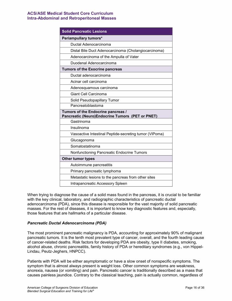


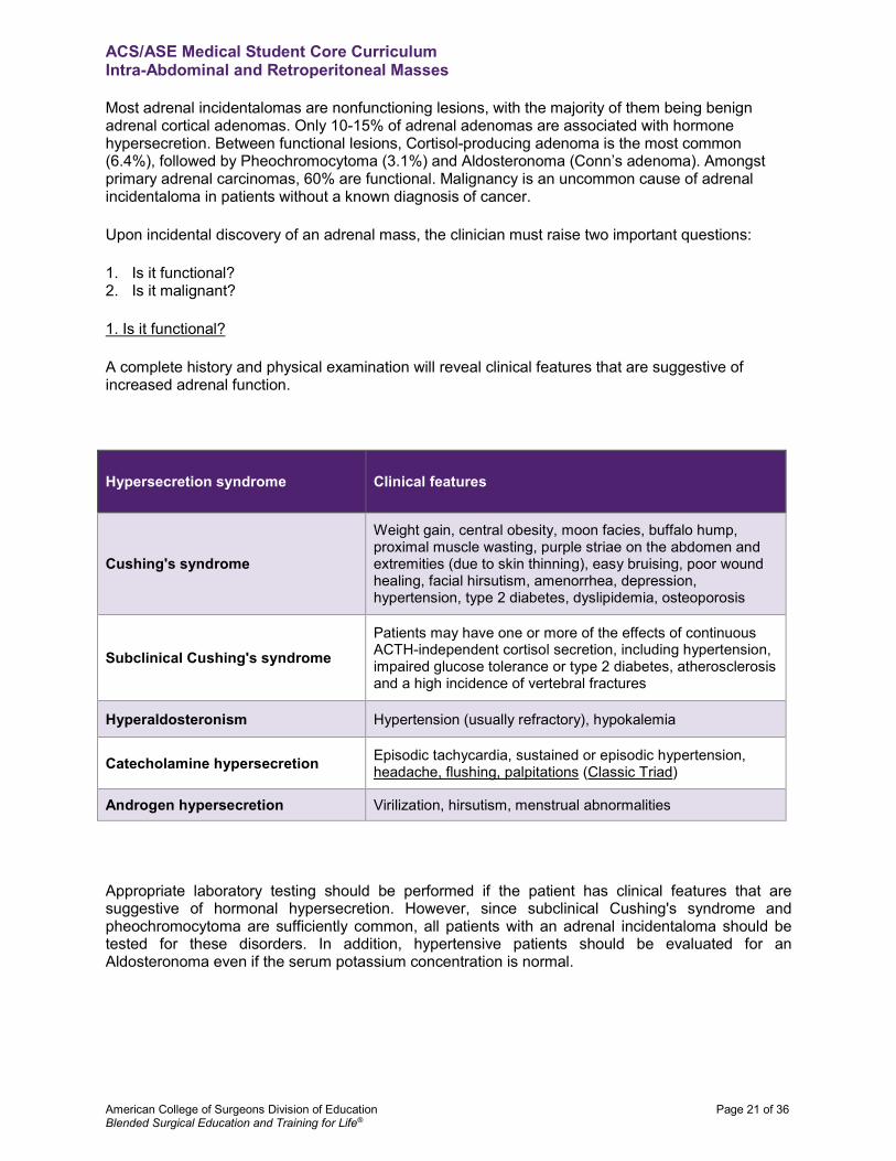


![Intra-Abdominal and Abdominal Wall Desmoid Fibromatosis · intra-abdominal and involving the small bowel mesentery [2]. TREATMENT Surgery Margin-negative resection has historically](https://static.documents.pub/doc/80x56/5e5a290071d21b380f5b7e74/intra-abdominal-and-abdominal-wall-desmoid-fibromatosis-intra-abdominal-and-involving.jpg)


