229 10 Actin and Actin-Binding Proteins in Cancer Progression and Metastasis Marleen Van Troys, Joël Vandekerckhove, and Christophe Ampe Introduction With over 10 million cases and over 5.7 million deaths a year (GLOBOCAN data 2002; Ferlay et al. 2004), cancer presents a major health problem worldwide. Dealing with cancer, both from a clinical and from a fundamen- tal scientific view, is complicated by the extensive diversity within this disease. Based on their origin, more than 100 different human cancer types have been described and, within one organ, distinct subtypes can occur. In addition, tumors may progress to a malignant state that manifests itself mainly when tumor cells spread out from the primary lesion (or neoplasm) and metasta- size, i.e., colonize distant sites of the patient’s body. Metastatic cancer, accounts for 90% of cancer-related lethality (Sporn 1996) and hence forms the primary determining factor in patient outcome. Therefore a detailed molecular understanding of tumor cell spread is required to render treatment of cancer more specific and efficient. A cancer cell exploits defects in normal cellular regulatory circuits. As out- lined below, tumor cell migration and adhesion and tumor cell interactions with host extracellular matrix (ECM) and with host cells are important features during the switch to the metastatic state. The actin cytoskeleton is a central player in these processes and consequently it is necessarily involved in many aspects of cancer and cancer progression (Lambrechts, Van Troys and Ampe 2004). In this chapter we present an overview of current knowledge on deregu- lations within the actin cytoskeleton during motility events in this disease. In a cancer cell both actin itself and members within the large group of actin-binding proteins (ABPs) may be affected. The differential func- tioning of the actin system is based on (1) the presence of mutant proteins (actins, ABPs), (2) altered expression levels of their genes, and/or (3) their altered activation status as a consequence of altered upstream signaling. In addition, ABPs are without doubt proving to surpass their original name. Next to their capacity to reorganize the actin cytoskeleton, many ABPs
Transcript
229
10Actin and Actin-Binding Proteins in Cancer Progression and Metastasis
Marleen Van Troys, Joël Vandekerckhove, and Christophe Ampe
Introduction
With over 10 million cases and over 5.7 million deaths a year (GLOBOCANdata 2002; Ferlay et al. 2004), cancer presents a major health problemworldwide. Dealing with cancer, both from a clinical and from a fundamen-tal scientific view, is complicated by the extensive diversity within this disease.Based on their origin, more than 100 different human cancer types have beendescribed and, within one organ, distinct subtypes can occur. In addition,tumors may progress to a malignant state that manifests itself mainly whentumor cells spread out from the primary lesion (or neoplasm) and metasta-size, i.e., colonize distant sites of the patient’s body. Metastatic cancer,accounts for 90% of cancer-related lethality (Sporn 1996) and hence formsthe primary determining factor in patient outcome. Therefore a detailedmolecular understanding of tumor cell spread is required to render treatmentof cancer more specific and efficient.
A cancer cell exploits defects in normal cellular regulatory circuits. As out-lined below, tumor cell migration and adhesion and tumor cell interactionswith host extracellular matrix (ECM) and with host cells are importantfeatures during the switch to the metastatic state. The actin cytoskeleton is acentral player in these processes and consequently it is necessarily involved inmany aspects of cancer and cancer progression (Lambrechts, Van Troys andAmpe 2004).
In this chapter we present an overview of current knowledge on deregu-lations within the actin cytoskeleton during motility events in this disease.In a cancer cell both actin itself and members within the large group ofactin-binding proteins (ABPs) may be affected. The differential func-tioning of the actin system is based on (1) the presence of mutant proteins(actins, ABPs), (2) altered expression levels of their genes, and/or(3) their altered activation status as a consequence of altered upstreamsignaling.
In addition, ABPs are without doubt proving to surpass their originalname. Next to their capacity to reorganize the actin cytoskeleton, many ABPs
230 M. Van Troys et al.
have additional partners and/or appear to be involved in functions notdirectly related to regulating the actin cytoskeleton. Many can also reside inthe nucleus where they may affect gene expression. Therefore these novelproperties of ABPs may additionally turn out relevant in the contributionsof these proteins to cancer cell motility and cancer progression. Although weare far from a complete understanding at the molecular level, the currentdata reveal that components of the actin system hold significant potential astargets in future cancer therapies.
Cell Migration Depends on a Dynamic Actin System
Cell motility and migration are pivotal to eukaryotic life (Pollard and Borisy2003). Many unicellular eukaryotes migrate to nutrient sources in a chemo-tactic way, i.e., attracted by factors secreted by the organism they feed on.In multicellular organisms, proper development is impossible withoutcompletion of multiple migratory steps during embryogenesis and morpho-genesis to lay down the body plan (Keller 2005) and accomplish the wiring ofthe nervous, vascular, and lymphatic systems (Weinstein 2005). After devel-opment, cell migration is limited since differentiated cells are confined withintissues by cell–cell adhesion or by interaction with ECM proteins. At thisstage, cells that do display locomotion mainly act in response to potentialharm. Fibroblasts and epithelial cells locally migrate during wound repair,and locomotion of white blood cells across vessel walls and within tissues isessential in immune surveillance.
In a strikingly similar way, malignant cancer cells are able to move out ofthe primary tumor and beyond the boundaries of the tissue or organ wherethe tumor initially developed (Mareel and Leroy 2003). Given the essentialcontribution of cell migration in tumor cell malignancy, we first briefly dis-cuss the functioning of the actin-based machinery during normal cellmigration. This also serves as an introduction to the function of key play-ers of the actin system putatively deregulated during aberrant motility oftumor cells.
The Actin Machinery Drives Cell MigrationA well-described example of migrating cells is that of fibroblasts moving ina random or (semi)directional fashion over a two-dimensional surface, amigratory mode that is termed mesemchymal (Fig. 1). Various aspects of thistype of migration have been studied in detail (see also Vicente-Manzanares,Webb and Horwitz 2005). Mesemchymal cells adopt an elongated spindle-like shape, are internally polarized (Franca-Koh and Devreotes 2004;Gamba et al. 2005; Nishiya et al. 2005), and succeed in moving or sliding in
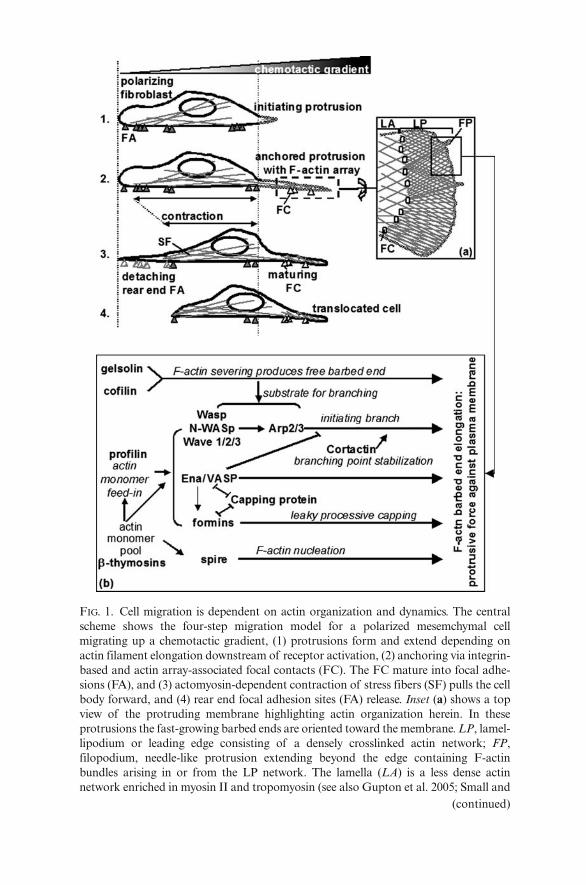
FIG. 1. Cell migration is dependent on actin organization and dynamics. The centralscheme shows the four-step migration model for a polarized mesemchymal cellmigrating up a chemotactic gradient, (1) protrusions form and extend depending onactin filament elongation downstream of receptor activation, (2) anchoring via integrin-based and actin array-associated focal contacts (FC). The FC mature into focal adhe-sions (FA), and (3) actomyosin-dependent contraction of stress fibers (SF) pulls the cellbody forward, and (4) rear end focal adhesion sites (FA) release. Inset (a) shows a topview of the protruding membrane highlighting actin organization herein. In theseprotrusions the fast-growing barbed ends are oriented toward the membrane. LP, lamel-lipodium or leading edge consisting of a densely crosslinked actin network; FP,filopodium, needle-like protrusion extending beyond the edge containing F-actinbundles arising in or from the LP network. The lamella (LA) is a less dense actinnetwork enriched in myosin II and tropomyosin (see also Gupton et al. 2005; Small and
(continued)
232 M. Van Troys et al.
the “chosen” direction by repeating a four-step process (Fig. 1) of (1) mem-brane protrusion at the front of the cell, (2) substrate adhesion of this lead-ing edge, (3) cell body contraction, and (4) detachment of the rear end of thecell (Lauffenburger and Horwitz 1996; Mitchison and Cramer 1996).
It is widely accepted and well documented that the actin cytoskeleton con-stitutes essential driving forces during all steps of this process as discussednext (Lambrechts, Van Troys and Ampe 2004). Within developing membraneprotrusions at the front of a polarized cell (step 1, Fig. 1), an active molecu-lar machinery tightly couples unidirectional actin filament elongation toforces pushing locally against the membrane (Fig. 1, inset a).
Unidirectional growth is an inherent property of actin filaments, related tothe head-to-tail association of actin monomers and to the associated hydrol-ysis of ATP bound to the actin protomers. This results in a fast-growing(barbed) actin filament end and a slow-growing (pointed) end. Superimposedon this property and essential to the mechanistic coupling to produce force isthe balanced activity of ABPs. These are recruited to and/or activated at thesite of protrusion, for instance as a response to external chemotactic signals,where they control the growth of actin filaments and organize them insupramolecular structures.
Typical protrusions that form in a two-dimensional context are lamellipo-dia, thin veil-like structures that contain dense, branched actin filament net-works, and filopodia, finger-like structures that contain parallel bundles ofactin filaments (Svitkina and Borisy 1999; Small et al. 2002; Faix and Rottner2006) (Fig. 1).
Figure 1 (inset b) shows ABPs that have been demonstrated to cooperatein locally generating protrusion. It is already worth noting that most of theseproteins display altered expression in tumor cells (see below). These ABPs
FIG. 1. (continued) Resch 2005). Inset (b) lists actin-binding proteins that can induce ormodulate barbed end elongation and that are present in LP or FP or both (for refer-ences see main text). Functional interaction between cofilin (DesMarais et al. 2004) orbetween gelsolin (Falet et al. 2002) and Arp2/3 has been demonstrated but these pro-teins may also produce F-actin ends compatible with Ena/Vasp activity. The connec-tion drawn between Ena/VASP Arp2/3-activity is based upon the observation thatVASP can reduce the frequency of Arp2/3-dependent branch formation in vitro (Skobleet al. 2001). The line drawn between capping protein and Ena/VASP-activity points attheir competitive activity at the barbed end, i.e., Ena/VASP proteins exert anticappingactivity as described by Barzik et al. (2005). A similar competitive effect betweenformin and capping protein is described in yeast (Kovar, Wu and Pollard 2005). Theactivity of the Spire protein is described by Schumacher et al. (2004) and Quinlan et al.(2005), the connection between formins and VASP by Grosse et al. (2003) andSchirenbeck et al. (2005)
10. Actin and Actin-Binding Proteins in Cancer Progression 233
display a wide range of activities on actin (for references see Van Troys,Vandekerckhove and Ampe 1999; Huff et al. 2001; Small et al. 2002; dosRemedios et al. 2003; Pollard and Borisy 2003; Lambrechts, Van Troys andAmpe 2004; Zigmond 2004; Polet et al. 2006).
β-Thymosins (see chapter by An et al.) and profilins (see chapter byMoens) are the most ubiquitous actin monomer-sequestering proteins andare important for feeding actin monomers to elongating ends. Cofilins (seechapter by Maloney et al.) and gelsolin (see chapter by Burtnick andRobinson) can act as filament-severing proteins. Their fragmenting capacitiesnot only promote depolymerization but also provide free barbed ends thatmay form substrates for other ABPs (such as the WASP or Wave proteins,Ena/VASP proteins, formins, and Spire) resulting in barbed end elongation.In addition, Ena/VASP proteins (Lambrechts et al. 2000) and also the Arp2/3complex have actin filament-nucleating properties with the feature thatArp2/3 generates branches on existing filaments. Finally, also barbed endcapping activity (by capping protein, CapG, or gelsolin) is essential in actindynamics as it competes with the elongation promoting ABPs (Barzik et al.2005; Kovar, Wu and Pollard 2005).
On top of these activities, the formation of supramolecular actin filament-based structures in protrusions requires ABPs acting at interfilament con-nections. These include cortactin which stabilizes Arp2/3-induced branchingpoints (Bryce et al. 2005); α-actinin, filamin, and EPLIN which are presentin crosslinked networks (Flanagan et al. 2001; Maul et al. 2003); fascin andplastin/fimbrin which bundle filaments (Delanote, Vandekerckhove andGettemans 2005; Giganti et al. 2005; Hashimoto, Skacel and Adams 2005);and tropomyosin which stabilizes them (Gunning et al. 2005).
For many of these ABPs some type of upstream signaling pathway hasbeen documented. In particular, signaling cascades acting via small GTPasesof the Rho-family and polyphosphoinositide (PPI)-modulating enzymesappear to be important (for details see Yin and Janmey 2003; Raftopoulouand Hall 2004; Stradal and Scita 2006).
The second step in the two-dimensional-migration model involvesanchorage of newly formed protrusions to substrate molecules. In a physi-ological context, this will in most cases imply adhesion of β1 and/or β3integrin transmembrane receptors to ECM proteins via the formation oftransient focal contacts (FC) at the base of the dense lamellipodial actinnetwork (Fig. 1). In these focal contacts, different anchor proteins aresequentially recruited and become involved in connecting the actincytoskeleton to the membrane (Zaidel-Bar et al. 2003). Focal contacts aretransient structures: as the lamellipod protrudes they either disassembleand are replaced by novel contacts positioned more anteriorly, or theymature into the more rigid focal adhesions (Ballestrem et al. 2001;Kaverina, Krylyshkina and Small 2002; Zaidel-Bar et al. 2003) (Fig. 1). Itshould be noted that too rigid adhesion negatively correlates with migra-tory speed (Huttenlocher, Ginsberg and Horwitz 1996).
234 M. Van Troys et al.
Cell body contraction, the third essential step in cell locomotion, is alsoactin dependent. Stress fibers, that consist of myosin II decorated actin bun-dles and span the cell body of substrate adherent cells (Fig. 1), contractdownstream of Rho and myosin light chain kinase. This shortening of stressfibers drags the trailing cell body forward and mechanically stresses rear cellattachments. Their detachment, e.g., by action of the intracellular proteasecalpain (Franco and Huttenlocher 2005), results in cell translocation (step 4).
Different Cell Types Migrate via Different Modes
Next to this mesemchymal- or fibroblast-like migration, other migratory modesor adaptations have been described that are less strongly dependent on integrinadhesion. Epidermal keratinocytes are rapidly moving cells in which the cou-pling between actin-based protrusion, adhesion, and retraction appears opti-mized: their cell body closely follows the protruding lamellipodium, they haveno lagging tail and form less adhesion sites (Anderson, Wang and Small 1996).Leukocytes (lymphocytes, neutrophils) also display a faster crawling mode on atwo-dimensional substrate. Their migration is termed “amoeboid” based on thesimilarity to migration by unicellular organisms like Dictyostelium discoideum(Condeelis 1993; Friedl, Borgmann and Brocker 2001). Lack of stress fibers, thepresence of cortical actin and the establishment of weak transient contacts withthe surface characterize this migration mode.
In general, cells adopting amoeboid movement display fast morphodynamicsas they continuously change shape based on cortical actin filament polymeriza-tion and actin–myosin-based contraction. The differences between mesemchy-mal and amoeboid locomotion manifest themselves even more strongly in athree-dimensional context and are relevant to cancer cell migratory behavior.
In addition, it is well documented, though mechanistically less well under-stood, that specific cell types such as epithelial monolayers during gastrulationor wound healing move as one functional group. During this collective migration(reviewed by Friedl, Hegerfeldt and Tusch 2004) as sheets, strands, or clusters,cell–cell contacts are retained via cell adhesion molecules, a.o. cadherins. Motilityaspects such as protrusion, traction force, and retraction are apparently executedby different subsets of cells within the motile group, however with a high degreeof synchronization in each subset (Friedl, Hegerfeldt and Tusch 2004).
Cancer Cell Migration
Variations on the Theme of Normal Cell MigrationFrom histopathological images of human tumors of different degrees of pro-gression, it has been evident for many decades that not all invasive tumorsprovide an identical picture. Indeed, some form protruding sheets or strands
10. Actin and Actin-Binding Proteins in Cancer Progression 235
still attached to the primary tumor (collective migration mode), for examplein melanoma (Friedl 2004) and colorectal carcinoma (Nabeshima et al. 1999)whereas in other tumors, cells detach as single cells of varying morphologies.These single cells can be fibroblast-like or have a rounded shape. The latter isusually correlated with a stronger level of dedifferentiation (Thiery 2002).
It can be inferred that, similar to normal cells, cancer cell migration modes arealso the result of a balance between forward movement and cell–cell and/orcell–substrate adhesion. Obviously, in motile cancer cells these complex equilib-ria are deregulated in comparison to noncancerous cells in differentiated tissue.From the normal migration modes described above, it is evident that this bal-ance is based upon the actions of integrins (cell–substrate adhesion), cadherins(cell–cell adhesion), and the actin system that are intricately linked by intracel-lular signaling pathways (Brunton, MacPherson and Frame 2004; Burridge andWennerberg 2004; Huber, Kraut and Beug 2005; Nishiya et al. 2005).
The actin system functions downstream of these two adhesion systems butconversely can itself influence adhesion (Chu et al. 2004; Scott et al. 2005;Wiesner, Legate and Fassler 2005; Yamada et al. 2005b), and all three systemsare themselves controlled by growth factor receptor signaling (references inBrunton, MacPherson and Frame 2004).
In a cancer cell, deregulation of either system can be “beneficial” to exert inva-sion as already evident from the identification of invasion-modulating proteinswithin these systems such as E-cadherin, focal adhesion components Src or focaladhesion kinase (FAK), the actin regulator RhoC, the ABP cortactin, etc. Thealterations observed within the actin system in tumor cells are considered indetail below. Full understanding of cancer cell movement, however, requires toalso take into account the microenvironments in which these cells move becausethese provide input for actin-based migration during tumor cell invasion.
Role of Matrix and Host Cells in Tumor Cell Invasion
Activating Signaling Pathways to the Actin CytoskeletonIn vivo, migrating cells are confronted with a three-dimensional stromallattice consisting of protein fibers (mainly collagen) and embedded cells.Consequently, migrating cancer cells are presented with a physical barrier.The density and composition of this ECM varies depending on thelocation. The most tightly packed ECM is the thin acellular layer formedby the basement membrane that, e.g., underlies all epithelia and endothe-lia. As the majority of tumors originate in epithelial lining (epidermal,endodermal, endothelial, glandular), disruption of the basement mem-brane is, in most cases, a prerequisite for tumor cell invasion. It appears,however, that escaping tumor cells are optimally equipped to face (and/orexploit) this degree of environmental variation and complexity during thevarious steps of the invasive process.
236 M. Van Troys et al.
Figure 2 gives a general overview of the consecutive steps of basementmembrane disruption, local initial invasion, vessel intravasation, transport,vessel extravasation, and homing, that together form the metastatic processthat malignant cells need to follow to ultimately give rise to a secondarytumor in a distant organ (see legend for details).
FIG. 2. Metastasis is a multistep process. The hallmarks of a grown tumor within dif-ferentiated tissue are self-sufficient growth and limitless replicative potential, escapefrom programmed cell death and sustained angiogenesis (Hanahan and Weinberg2000). Malignancy is characterized by the capacity of cells within the tumor to initi-ate the invasive and metastatic process. By loss of homotypic cell–cell adhesion,malignant cells detach from the primary tumor mass, disrupt the basement membrane(BM), and migrate through surrounding tissue and along protein fibers toward bloodor lymphatic vessels. Intracellular rearrangements in the actin cytoskeleton and extra-cellular matrix (ECM) degradation by proteolytic enzymes from the tumor cells (inthe mesemchymal migration mode, see text) enable this migratory step. The tumorcells also activate or stimulate secretion of growth factors by host cells such as myofi-broblasts (MF) and macrophages (MP). This facilitates (chemotactic) tumor cell inva-sion and putatively also the intravasation through the basement membrane andendothelial layer of the vessel. At a distant site, upon extravasation, a reverse processoccurs that again involves cytoskeletal rearrangements. Tumor cells adhere to the cap-illary endothelium, penetrate and migrate through the extravascular tissue where theyagain create a growth permissive microenvironment a.o. by attracting blood supply.This finally results in a secondary lesion or metastasis
10. Actin and Actin-Binding Proteins in Cancer Progression 237
During the last decade, several research groups have focused on describingand understanding how tumor cells find their way through the matrix duringthe crucial initial migratory steps of this process. It is now evident that actin-based motility and adhesion are important features during this onset ofmalignancy. Important progress has been realized in this area via the devel-opment or optimization of novel real-time imaging techniques. Visualizing,at single cell resolution, the movements of individual normal and cancer cells(from established cell lines or cancer tissue explants) in reconstituted matri-ces of controlled composition has clarified fundamental aspects of migrationin dense protein networks (Friedl et al. 1997; Maaser et al. 1999; Friedl andBrocker 2000; Hegerfeldt et al. 2002).
Using multiphoton-based intravital imaging, the behavior of invasive cellsin and near a primary mammary tumor and the vessels surrounding it havebeen recorded in situ in living animals, providing fascinating high-resolutionmovies of malignant cells in action (Farina et al. 1998; Ahmed et al. 2002;Condeelis and Segall 2003; Wyckoff et al. 2004; Condeelis, Singer and Segall2005; Yamaguchi, Wyckoff and Condeelis 2005). Simultaneously, thesetechnologies allow dynamic imaging of ECM scaffolds and hence, visualizehow migrating cancer cells interact with collagen fibers (Condeelis andSegall 2003; Wolf et al. 2003a). In addition, the interaction of tumor cellswith host cells in the stromal tissue has also been recorded in situ either usingdual fluorescence imaging in which host and tumor display different fluo-rescence (Yang et al. 2004b; Hoffman 2005), or employing specific trans-plantation chambers (Skobe et al. 1997; Bajou et al. 1998) or by employinga chemotactic in vivo invasion assay (Wyckoff et al. 2004; Condeelis, Singerand Segall 2005).
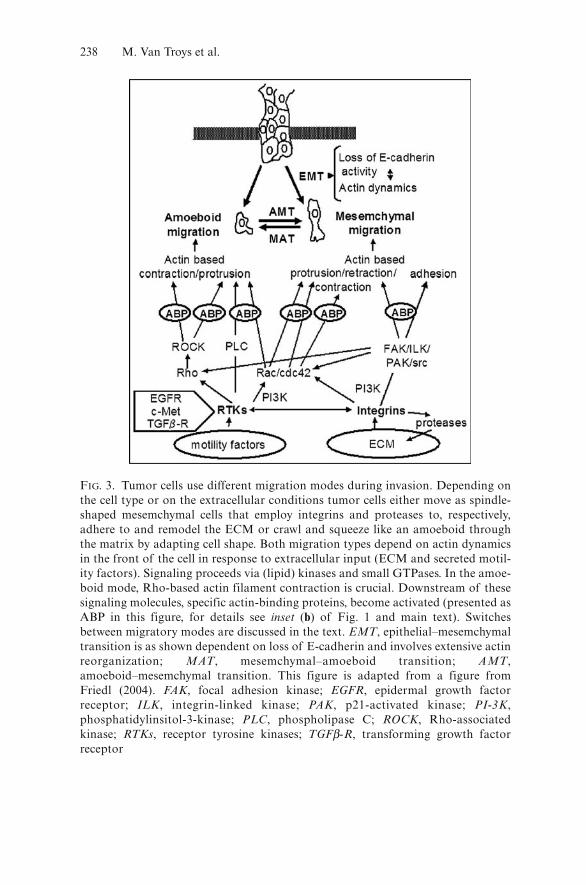
Figure 3 illustrates the different migratory modes observed for invasivecancer cells and some of the characteristic key effector molecules upstreamof actin dynamics. Cells moving through stroma in a mesemchymal fashionutilize the multistep process described above but in addition remodelthe matrix (Friedl and Wolf 2003). When their exploratory protrusionscontact the matrix fibers, proteases such as matrix metalloproteinases(MMP and MT-MMPs) and serine proteases become enriched at theintegrin adhesion sites. Concomitantly actin, ABPs, and signaling mole-cules are recruited to the intracellular side of the focal contacts (Wolf andFriedl 2005).
Hot spots of matrix degradation near the cell surface are located at thecontact sites of invadopodia (Friedl 2004). The latter are actin rich mem-brane extensions that protrude deep into the matrix and are typicallyformed by aggressive tumor cells (Buccione, Orth and McNiven 2004;McNiven, Baldassarre and Buccione 2004). Invadopodia are reminiscentof podosomes. These are protrusions present on differentiated cells withhigh matrix penetrating or degrading capacity (macrophages, dendriticcells, osteoclast; reviewed in Linder and Kopp 2005). EGF-inducedformation of podosomes requires the integrity of an activated actin
238 M. Van Troys et al.
FIG. 3. Tumor cells use different migration modes during invasion. Depending onthe cell type or on the extracellular conditions tumor cells either move as spindle-shaped mesemchymal cells that employ integrins and proteases to, respectively,adhere to and remodel the ECM or crawl and squeeze like an amoeboid throughthe matrix by adapting cell shape. Both migration types depend on actin dynamicsin the front of the cell in response to extracellular input (ECM and secreted motil-ity factors). Signaling proceeds via (lipid) kinases and small GTPases. In the amoe-boid mode, Rho-based actin filament contraction is crucial. Downstream of thesesignaling molecules, specific actin-binding proteins, become activated (presented asABP in this figure, for details see inset (b) of Fig. 1 and main text). Switchesbetween migratory modes are discussed in the text. EMT, epithelial–mesemchymaltransition is as shown dependent on loss of E-cadherin and involves extensive actinreorganization; MAT, mesemchymal–amoeboid transition; AMT,amoeboid–mesemchymal transition. This figure is adapted from a figure fromFriedl (2004). FAK, focal adhesion kinase; EGFR, epidermal growth factorreceptor; ILK, integrin-linked kinase; PAK, p21-activated kinase; PI-3K,phosphatidylinsitol-3-kinase; PLC, phospholipase C; ROCK, Rho-associatedkinase; RTKs, receptor tyrosine kinases; TGFβ-R, transforming growth factorreceptor
10. Actin and Actin-Binding Proteins in Cancer Progression 239
machinery containing in particular Arp2/3, N-WASp and its regulatorNck, cdc42, WIP, cortactin, and dynamin (McNiven, Baldassarre andBuccione 2004; Yamaguchi et al. 2005). Sustained podosome stability isrequired for efficient matrix degradation and depends on cofilin activity(Yamaguchi et al. 2005). Taken together, three-dimensional-mesemchymalmovements depend on proteolysis, integrin adhesion, and actin dynamics(Maaser et al. 1999; Wolf et al. 2003a) (Fig. 3). In tumors this type of migra-tion is observed in, e.g., fibrosarcomas, in gliomas, in progressed epithelialcancers (after epithelial–mesemchymal transition, see below), and in the cellsat the invasive front of tumors protruding via collective movement (Friedl,Hegerfeldt and Tusch 2004).
However, pericellular proteolysis is not a prerequisite to cross ECM-barriers.Using an amoeboid-like, leukocyte type migration mode, certain tumor cells (e.g.,lymphomas) display morphological adaptation to preformed matrices and areable to squeeze through preexisting holes in the matrix (Verschueren et al. 1994;Wolf et al. 2003b). In their escape from the primary tumor, amoeboid metastaticmammary carcinoma cells (MtLn3) use existing collagen fibers as tracks to movefast in the direction of blood vessels (Wang et al. 2002). In vitro, amoeboid tumorcell migration is protease independent and largely integrin independent (Fig. 3).It, however, requires functional Rho–ROCK signaling to control cortical actinfilaments, a.o. for myosin II-based filament contraction (Sahai and Marshall2003) and for cofilin-based filament remodeling (DesMarais et al. 2004).
Tumor cells not only display different morphologies and migratory modes,but they can also switch between these modes (Fig. 3). It is important to realizethat this mimics (or mimics the reverse of) similar physiological transitionsoccurring during development and cell differentiation (reviewed in Thiery 2003;Friedl 2004). One such switch in morphology and motility is the epithelial tomesenchymal transition (EMT) that is considered as a point of no return in theprogression of epithelial cancers (Thiery 2002). EMT is mainly characterized byloss of basal–apical polarity and of E-cadherin-based cell–cell adhesion (Fig. 3).
The various molecular mechanisms that can induce this switch are beingelucidated. These include genetic and epigenetic changes and transcriptionalcontrol (reviewed by Berx and Van Roy 2001; Thiery 2003; Huber, Kraut andBeug 2005). Also, considerable cytoskeletal reorganization is involved duringEMT and, interestingly, the expression levels of a number of actinpolymerization-modulating ABPs (gelsolin, capping protein, etc.) are alteredby activation of Snail – a transcriptional repressor-inducing epithelial dedif-ferentiation (De Craene et al. 2005).
Next to this EMT switch, it has recently been demonstrated that, uponaltering environmental conditions counteracting one migratory mode, vari-ous cancer cells can keep moving by switching from mesemchymal to amoe-boid movement (MTA) or vice versa (ATM; Fig. 3) (Sahai and Marshall2003; Wolf et al. 2003a; Wolf and Friedl 2005). This was observed in vitroupon addition of either protease-, integrin-, or Rho/ROCK-blocking factors(Sahai and Marshall 2003; Wolf et al. 2003a). Marshall and colleagues
240 M. Van Troys et al.
recently showed that the Rho/ROCK-independent mesemchymal-like migra-tion employs Cdc42/MRCK (myotonic dystrophy kinase-related Cdc42-bind-ing kinase)-based signaling to control contractile processes (Wilkinson,Paterson and Marshall 2005). Since these escape mechanisms of malignantcells are relevant when applying specific therapeutics (e.g., MMP inhibitors),it may be more efficient to directly target the actin migration machinery inorder to switch off migration.
Tumor cell–stroma interactions go beyond mere proteolytic digestion ofthe matrix to provide room to move or the capacity to trespass barriers. Acancer cell actively modulates the stroma into an environment supportingefficient tumor progression and invasion by the secretion of factors thatpromote tumor cell growth as well as migration. Next to autocrine signal-ing (e.g., tumor cell secreted EGF signals to the actin cytoskeleton of thesame cells) there is considerable paracrine signaling. The tumor cells secretegrowth factors and cytokines that attract and activate host cells suchas fibroblast, myofibroblasts, macrophages, and endothelial cells (Fig. 2).In response these cells secrete (growth) factors facilitating tumor progres-sion and/or migration. This is extensively reviewed in Mueller and Fusenig(2004). An early example of this is the recruitment of endothelial cells viavascular endothelial GF (VEGF) secretion by tumor cells during neoan-giogenesis (reviewed in Carmeliet 2005) that is crucial both in early tumorgrowth and for invasion.
We will now expatiate on two recently reported examples that establish alink between host cell engagement and the functioning of the actin cytoskele-ton in the tumor cell. De Wever and colleagues (2004a,b) describe a paracrineloop between colon cancer cells and transforming GF-β (TGF-β)-activatedmyofibroblasts. Upon activation, the latter produce hepatocyte GF (HGF)and the ECM component tenascin that, respectively, mediate activation ofRac and inactivation of Rho in the tumor cells, conditions favoring actin-based mesemchymal migration.
The Condeelis group (Wyckoff et al. 2004) discovered in an orthotopicallygrown mammary tumor in rat a promigratory paracrine loop between the carci-noma cells and macrophages accumulating near the blood vessel at the tissueside. The tumor cells produce colony-stimulating factor (CSF), a known pro-moter of tumor progression (references in Mueller and Fusenig 2004), that acti-vates the infiltrating macrophages. In response, these cells produce EGF actingas chemoattractant for the tumor cells that then track with high directional per-sistence to the blood vessel. In an experimental set up consisting of macrophagesand a needle containing EGF placed in the tumor microenvironment, the tumorcells migrate into the needle and can be collected (Wang et al. 2004a). The tran-scriptome of these collected invasive tumor cells extensively differs fromthat of the bulk tumor and provides insight in the requirement for theinvasive switch: levels of mRNAs encoding for proteins that promote anantiproliferative, antiapoptotic, and highly motile state are altered.Accordingly, expression of many ABPs and their upstream regulators(Fig. 3) (see below) were found upregulated (Lorenz et al. 2004; Condeelis,
10. Actin and Actin-Binding Proteins in Cancer Progression 241
Singer and Segall 2005; Wang et al. 2005a; Yamaguchi, Wyckoff andCondeelis 2005) as a consequence of this host tumor interplay. These andother reported deregulations in the actin system in a tumor context arereviewed in detail below.
Deregulation Within the Actin System in tumor contexts
Given the tight link between actin dynamics and cell migration, a deregulatedactin system obviously forms a principle element of the oncogenic and, evenmore likely, the invasive proteome of cancer cells. As causes for deregulationwithin the actin system, one can distinguish (1) mutations, translocations, oramplifications of coding genes (Futreal et al. 2004), (2) epigenetic changesaffecting gene transcription, (3) changed mRNA levels, or (4) changed activ-ity of the expressed protein. More recently an example of translationalcontrol has also been reported.
Actin Expression in Cancer CellsOnly few mutations in genes encoding components of the actin cytoskeletonhave been reported in relation to cancer (see chapter by Sparrow and Laing).For β-actin two mutants have been described in tumor cells and their expressionwas associated with a neoplastic phenotype. Parental B16 mouse melanomacells abundantly express a β-actin mutant with a single point mutation R28L(Sadano et al. 1988) of which the expression level in sublines negatively corre-lates with invasiveness and metastasis (Sadano, Taniguchi and Baba 1990).
β-Actin G244D (Vandekerckhove et al. 1980) was identified in immortal-ized human fibroblasts that produce tumors in athymic mice (Leavitt et al.1987). In vitro, this mutant displays a polymerization defect and reducedbinding to the chaperone CCT (Rommelaere et al. 2004). Interestingly,expression of subunits of the latter complex, which is largely dedicated toactin and tubulin folding, is upregulated in diverse (invasive) cancers(Yokota et al. 2001; Rhodes et al. 2004; Shen, Ghosh and Chinnaiyan 2004;Wang et al. 2004a) and this may enhance production levels of actin isoforms.The physiological role of CCT-subunit overexpression in cancer is, however,unexplored and could, next to actin, also be related to other CCT-substratessuch as tubulins or the von Hippel–Lindau protein (pVHL) – a tumor sup-pressor connected to response to hypoxia (Pugh and Ratcliffe 2003).
In cancer cells, differential transcriptional control of actin gene isoformexpression is likely to occur since this is also an important feature ofnormal developmental programs and other pathologies (referencesin Chaponnier and Gabbiani 2004). Opposite expression level changes ofβ- and γ-actin have been reported in salivary gland adenocarcinoma as afunction of metastatic capacity (Suzuki et al. 1998). More recently γ-actin-level changes were reported in a drug-resistant acute lymphoblasticleukemia (Verrills et al. 2006).
242 M. Van Troys et al.
Microarray data and serial analysis of actin gene expression (SAGE) alsoreveal altered expression in cancer tissues compared to normal tissue, forexample β- and γ-nonmuscle or α-smooth muscle actin. Similar observationshave been made for tumor material at different stages of progression althoughconsistent trends have been hard to define. In colon adenocarcinoma cell lineshowever, β-actin level and localization correlate with metastatic capacity(Nowak et al. 2005).
The zipcode-binding protein 1 (ZBP1) is an interesting modulator of actinexpression. It binds the 3′ UTR of β-actin mRNA and assures its transportto sites of high actin dynamics in the cell periphery (Ross et al. 1997; Zhanget al. 2001; Oleynikov and Singer 2003). It was recently shown that the asso-ciation of ZBP1 with β-actin mRNA represses its translation (Huttelmaieret al. 2005). However upon phosphorylation of ZBP1 by the exclusivelyperipheral Src kinase, ZBP1 binding is inhibited and β-actin protein is syn-thesized (Huttelmaier et al. 2005).
Interestingly, ZBP1-expression is downregulated in metastatic vs. non-metastatic breast tumors (Wang et al. 2002, 2004a). This is in line withobservations that metastatic breast cancer cell lines have less peripherallylocated β-actin than their nonmetastatic counterparts (Shestakova et al.1999). Restoring ZBP1-levels reduces invasiveness and the metastatic abilityof the tumor cells (Wang et al. 2004a), indicating that polarization in β-actinsynthesis by translational control negatively correlates with efficient invasion.This mechanism also provides a possible additional pathway from the Srconcogene to actin dynamics and cell migration but this has not beenexplored.
Gene Mutation and Deregulated Gene Transcription of ABPs in TumorsActin-Binding Proteins as Oncogenes?
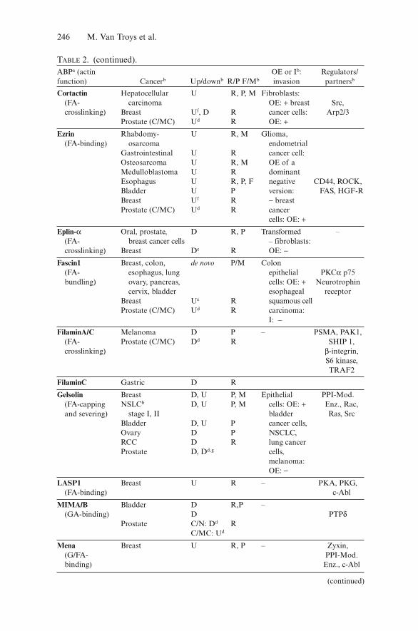
Table 1 lists known genes coding for ABPs that have frequently beenobserved to display (a) genetic defect(s) in cancer cells suggesting causal con-tribution to oncogenesis or malignant tumor progression. All but two areextracted from a recently published “cancer census” that contains 291 “can-cer genes” and is built using strict criteria. A cancer gene is assigned as suchif it carries at least one germ line mutation and/or more than five unambigu-ous somatic mutations in at least two independently analyzed primarytumors (Futreal et al. 2004).
For most ABP-genes in Table 1 a dominant somatic mutation resulting froma chromosomal translocation is the mutational defect that underlies their iden-tification as a potential cancer gene. These rearrangements result in chimeras(two genes are involved) in which the activity of the gene product is altered,favoring oncogenesis. Intriguingly, all of these ABPs bind filamentous actinand in a number of cases the fusion protein retains F-actin-binding capacity.
10. Actin and Actin-Binding Proteins in Cancer Progression 243
This is the case for the actin-binding tyrosine kinase (c-Abl), a well-recognizedprotooncogene. In a large set of human leukemias, the c-Abl tyrosine kinase isconstitutively activated by fusion to sequences encoded by the breakpoint clusterregion (bcr) gene (Gotoh and Broxmeyer 1997) (Table 1). In primary chronicmyelogenous leukemia (CML) cells this causes, next to abnormal proliferation,motility defects such as increased spontaneous and persistent movement,decreased adhesion and decreased chemotaxis to stroma-derived factor-1α(Salgia et al. 1997). The F-actin-binding capacity of the Abl-kinase in theBCR/Abl-fusion has been implicated in the altered motile phenotype ofthese cancer cells (Ramaraj et al. 2004). In addition, many studies point toa regulatory role for Abl in actin remodeling both by small GTPase acti-vation or by interacting with and modulating the activity of ABPs impor-tant in actin dynamics (e.g., Abl-kinase interactor 1, Ena/VASP proteins,WASp/Wave proteins; reviewed in Hernandez et al. 2004). Also in themyosin heavy chain (MHC) 11 fusion to the beta subunit of the transcrip-tion factor core-binding factor (Table 1) the F-actin-binding capacity ofMHC11 is important. The dominant negative effect of this oncogenicfusion results from the cytosolic sequestering on actin filaments of the
TABLE 1. Actin-binding protein genes mutated in cancera.ABP Gene Mutated inb Typec Actin-binding
Abl c-abl CML, ALL T FA-bindingLASP1 LASP1 AML T FA-bindingL-plastin LCP1 nHL T FA-bundlingMoesin MSN ALCL T FA-bindingMyosin heavy chain 11 MYH11 AML T FA-bindingMyosin heavy chain 9 MYH9 ALCL T FA-bindingTropomyosin 3 TMP3 Papillary thyroid, T FA-binding
ALCL, IMTTropomyosin 4 TMP4 ALCL T FA-bindingNF2 NF2 NF type 2, T, O FA-binding
mesothelioma,melanoma
WASp WAS Lymphoma, O G/FA-bindingWiskott–Aldrich syndrome
Cortactin CCTNa Breast cancer, HNSCC A FA-crosslinkingMyopodin SYNPO2 Prostate cancer D FA-bundling
aObtained from the Wellcome Trust Sanger Institute Cancer Genome Project web site(http://www.sanger.ac.uk/genetics/CGP) and from Futreal et al. (2004), except for CCTN (thecortactin gene also termed EMS1) and SYNPO2.bAML acute myelogenous leukemia, ALCL anaplastic large cell lymphoma, ALL acute lympho-cytic leukemia, CML chronic myeloid leukemia, IMT inflammatory myofibroblastic tumor, NFneurofibromatosis, nHL non-Hodgkin lymphoma.cA amplification, D deletion, O deletions, missense and nonsense mutations, splice site muta-tions, and others, T translocation (see http://www.sanger.ac.uk/genetics/CGP/ cancer census).
244 M. Van Troys et al.
heterodimeric transcription factor via the fusion (Adya et al. 1998;Lukasik et al. 2002).
For the other ABP-genes that are involved in chromosomal translocations(Table 1) it is not yet clear whether their normal function in the actin systemis a determining property of the fusion proteins in oncogenesis. They arefused to genes that are frequently rearranged and/or mutated in specifictumors and that are by themselves already associated with malignancy. TheABP LASP1 (LIM and SH3 protein) is only one of more than 30 fusion part-ners of the histon methyl transferase mixed lineage leukemia gene in acutemyeloid leukemia (Strehl et al. 2003). L-plastin (or L-fimbrin) is fused to thenuclear transcriptional repressor B-cell lymphoma 6 (BCL6) in non-Hodgkinlymphoma (Galiegue-Zouitina et al. 1999).
Moesin, nonmuscle MHC9, and tropomyosins 3 and 4 form an activatingfusion with the oncogenic receptor tyrosine kinase–anaplastic lymphomakinase (ALK–RTK) in anaplastic large cell lymphoma and/or inflammatorymyofibroblastic tumors (Pulford et al. 2004). For the F-actin-bindingtropomyosins, it is hypothesized that their coiled-coil self-associated domainenables ligand-independent oligomerization and hence activation of theALK receptor in the oncogenic fusions (Lawrence et al. 2000).
Based on these last examples one might assume that the function of theABP-counterpart in observed oncogenic fusions is based on more general fea-tures of these proteins or of their genes (such as high gene promoter activity,cytosolic gene expression, or protein dimerization capacity). However it isintriguing that for most ABP listed in Table 1, altered expression levels of theunfused ABP-genes are also reported in one or more cancer types (see alsoTable 2). This may indicate that their capacity for organizing or regulating theactin cytoskeleton is an additional important feature of the fused oncogene or,conversely, the fusion may interfere with the action of the nonfused ABP or ofother ABPs. It is conceivable that the ALK–RTK-fused tropomyosins interfereby competition with normal oligomerization of tropomyosins (Lawrence et al.2000) resulting in aberrant functioning of the latter. This could induce asimilar effect as the reported downregulation of specific tropomyosin isoformsin transformed cells (Novy et al. 1993; Varga et al. 2005) and their tumorsuppression effect (Prasad et al. 1999).
The genes for the ABPs neurofibromatosis type 2 (NF2) and WAS displaya range of inheritable mutations that result in loss of function of the ABP(Table 1). The WAS gene codes for the ABP WASp that is exclusivelyexpressed in hematopoietic cells. Loss of WASp-expression is the cause of theimmunodeficiency diseases Wiskott–Aldrich syndrome and X-linked throm-bocytopenia. Lymphocytes from these patients show impaired actin remod-eling and motility in response to stimuli since WASp is an importantmodulator of actin dynamics (Fig. 1) (Zicha et al. 1998).
The WAS gene is assigned as cancer gene because WAS patients have anincreased risk of malignancy, most notably non-Hodgkins lymphoma, but theunderlying mechanism is still unclear.
10. Actin and Actin-Binding Proteins in Cancer Progression 245
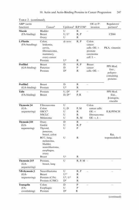
Cortactin Hepatocellular U R, P, M Fibroblasts:(FA- carcinoma OE: + breast Src,crosslinking) Breast Uf, D R cancer cells: Arp2/3
Prostate (C/MC) Ud R OE: +Ezrin Rhabdomy- U R, M Glioma,
(FA-binding) osarcoma endometrial Gastrointestinal U R cancer cell:Osteosarcoma U R, M OE of a Medulloblastoma U R dominant Esophagus U R, P, F negative CD44, ROCK,Bladder U P version: FAS, HGF-RBreast Uf R − breast Prostate (C/MC) Ud R cancer
cells: OE: +Eplin-α Oral, prostate, D R, P Transformed –
(FA- breast cancer cells – fibroblasts:crosslinking) Breast De R OE: −
Fascin1 Breast, colon, de novo P/M Colon(FA- esophagus, lung epithelial PKCα p75bundling) ovary, pancreas, cells: OE: + Neurotrophin
cervix, bladder esophageal receptorBreast Ue R squamous cellProstate (C/MC) Ud R carcinoma:
I: −FilaminA/C Melanoma D P – PSMA, PAK1,
(FA- Prostate (C/MC) Dd R SHIP 1,crosslinking) β-integrin,
S6 kinase,TRAF2
FilaminC Gastric D R
Gelsolin Breast D, U P, M Epithelial PPI-Mod.(FA-capping NSLCb D, U P, M cells: OE: + Enz., Rac,and severing) stage I, II bladder Ras, Src
Bladder D, U P cancer cells,Ovary D P NSCLC,RCC D R lung cancer Prostate D, Dd,g cells,
melanoma:OE: −
LASP1 Breast U R – PKA, PKG,(FA-binding) c-Abl
MIMA/B Bladder D R,P –(GA-binding) D PTPδ
Prostate C/N: Dd RC/MC: Ud
Mena Breast U R, P – Zyxin,(G/FA- PPI-Mod.binding) Enz., c-Abl
(continued)
10. Actin and Actin-Binding Proteins in Cancer Progression 247
Tropomyosin 1 Breast D, Uc R Breast –(FA-binding) Colon D R cancer cells,
Bladder, D P transformed esophagus, fibroblasts:neuroblastoma OE: −
Prostate Dd,g R –
Tropomyosin 4/5 Bladder(5), U P – –(FA-binding) Esophagus(4)
Breast (4,5) Uc RVASP Lung (C/N) U P – Zyxin,
(G/FA- PPI-Mod.binding) Enz., PKA,
PKG
Vinculin Melanoma, D P, M –(FA-binding) breast, salivary
gland PPI-Mod.Breast, U, Uc P, Ri Enz., α-actinin,
rhabdomyo talinsarcoma
Prostate Dd R
N-WASp Liver, prostate U, Ud R –(G/FA-binding)
WAVE2 Melanoma U P, M Melanoma(G/FA- Prostate Dd R cells: OE: +,binding) I: −
WAVE3 Breast Uc R Breast(G/FA- Prostate Dd R cancer cells:binding) I: −
aα-actinin (Honda et al. 1998, 2005; Clark et al. 2000; Nikolopoulos et al. 2000); Arp2 and Arp3(Kaneda et al. 2004; Otsubo et al. 2004); Calponin (Takeoka et al. 2002; Islam et al. 2004; Lener,Burgstaller and Gimona 2004); CapG (De Corte, Gettemans and Vandekerckhove 1997; VanGinkel et al. 1998; Lal et al. 1999; De Corte et al. 2004); capping protein (Smith-Beckerman et al.2005); cofilin (Gunnersen et al. 2000; Martoglio et al. 2000; Unwin et al. 2003; Ding et al. 2004;Lee et al. 2005b; Smith-Beckerman et al. 2005; Yap et al. 2005; Dang, Bamburg and Ramos2006); cortactin (Patel et al. 1998; Wang et al. 2002; Chuma et al. 2004); ezrin (Ohtani et al. 1999;Wick et al. 2001; Park et al. 2003; Shen et al. 2003; Khanna et al. 2004; Koon et al. 2004; Yu et al.2004a; Elliott et al. 2005; Langbein et al. 2006); eplin (Song et al. 2002; Maul et al. 2003); fascin(Jawhari et al. 2003; Hashimoto, Skacel and Adams 2005; Kabukcuoglu et al. 2005; Roma andPrayson 2005; Tong et al. 2005; Xie et al. 2005; Yoder et al. 2005); filamin C (Flanagan et al.2001; Kaneda et al. 2002; Anilkumar et al. 2003); gelsolin (Chaponnier and Gabbiani 1989;Tanaka et al. 1995; Dosaka-Akita et al. 1998; Asch et al. 1999; Lee et al. 1999; Shieh et al. 1999;Fujita et al. 2001; Thor et al. 2001; De Corte et al. 2002; Sazawa et al. 2002; Yang et al. 2004a;Noske et al. 2005; Langbein et al. 2006); LASP1 (Tomasetto et al. 1995; Lin et al. 2004);
(continued)
Arp2/3,PPI-Mod.Enz., SH3-containingproteins,
Rho-GTPases
10. Actin and Actin-Binding Proteins in Cancer Progression 249
NF2 is a germ line-based disorder caused by loss of the tumor suppressorNF2 (merlin, schwannomin). These patients develop slow growing and usuallynonmalignant schwannomas, meningiomas, and medulloepithelioma(Gutmann 2001). Somatic mutations of the NF2 gene have also been reportedin neoplasms of nonneuroectodermal origin, such as malignant mesotheliomaand melanoma (Robinson, Musk and Lake 2005). Overexpression of NF2 in ratschwannoma cells inhibits their growth and impairs cell motility, adhesion, andspreading (Gutmann 2001). NF2 belongs to the ezrin/moesin/radixin proteinsthat bind actin filaments, interact with membrane lipids, with the hyaluronicacid transmembrane receptor CD44 and with HGF-regulated tyrosine kinasesubstrate (Gautreau et al. 2002; Scoles et al. 2002). Its activity is regulated byphosphorylation downstream of rac1 and cdc42 (Xiao et al. 2002).Consequently, NF2 is centrally positioned to influence cell adhesion, motility,and growth factor controlled cell growth as discussed in Gutmann (2001) andMcClatchey and Giovannini (2005).
Amplification of the cortactin gene (CCTN) is not included in the cancer cen-sus because the chromosomal aberration at 11q13 affects several genes (Futrealet al. 2004). It occurs in 13–36% of human carcinomas mainly in breast cancer,
TABLE 2. (continued).Mena (Di Modugno et al. 2004; Wang et al. 2004a); MIM (Lee et al. 2002; Nixdorf et al. 2004;Loberg et al. 2005); moesin (Carmeci et al. 1998; Martoglio et al. 2000; Langbein et al. 2006); L-Plastin (Lin et al. 1993; Delanote, Vandekerckhove and Gettemans 2005); profilin (Janke et al.2000; Wang et al. 2002; Wittenmayer et al. 2004; Gronborg et al. 2006); talin (Glukhova et al.1995; Everley et al. 2004); thymosin β4 (Yamamoto et al. 1993; Clark et al. 2000; Kobayashi et al.2002; Cha, Jeong and Kleinman 2003; Muller-Tidow et al. 2004; Wang et al. 2004b; Vigneswaranet al. 2005); thymosin β10 (Hall 1994; Santelli et al. 1999; Lee et al. 2001; Takano et al. 2002;Oien et al. 2003; Chiappetta et al. 2004; Alldinger et al. 2005); thymosin β15 (Bao, Loda andZetter 1998; Chakravatri et al. 2000); NB thymosin β (Yokoyama et al. 1996); transgelin (Qi et al.2005; Yeo et al. 2006); tropomyosin 1 (Prasad, Fuldner and Cooper 1993; Gimona, Kazzaz andHelfman 1996; Prasad et al. 1999; Yager et al. 2003; Pawlak et al. 2004; Qi et al. 2005; Varga et al.2005); tropomyosin 5 (Pawlak et al. 2004); VASP (Dertsiz et al. 2005); vinculin (Sadano, Inoueand Taniguchi 1992; Glukhova et al. 1995; Meyer and Brinck 1997); WAVE2 (Kurisu et al. 2005).bCancer cancer type in which upregulated (U) or downregulated (D) cancer level is observed onRNA (R) or protein (P) level, F the tumor suppression of promoting function has been validatedin a functional assay (in vitro or in vivo invasion), C/A carcinoma vs. adenoma, C/MC carcinomavs. metastatic carcinoma, C/N cancer vs. normal, FA F-actin, I forced downregulation of theABP (obtained by RNAi or antisense technology) in these cells leads to inhibition (−) or toinduction (+) of invasion, M correlated with metastasis, NSCLC nonsmall cell lung cancer,OSCC oral squamous cell carcinoma, OE overexpression of the ABP in these cells leads to inhi-bition (−) or to induction (+) of invasion, PPI-Mod. Enz. polyphosphoinositide-modulatingenzymes, RCC renal cell carcinoma, Regulators/partners limited to those already implicated incancer.cWang et al. (2004a).dYu et al. (2004b) only significant up- or downregulations are listed: significance criterion isdefault p-value < 0.05 (http://www.Oncomine.org).evan ’t Veer et al. (2002), as in footnote d.fWang et al. (2005b) as in footnote d.gRhodes et al. (2002).hDhaese et al. (in preparation)
250 M. Van Troys et al.
oral squamous cell carcinoma, and head and neck squamous cell carcinomas.This gene encodes the actin filament crosslinking protein cortactin (Table 1). Itsoverexpression enhances cell migration, invasion, and metastasis in vitro (Patelet al. 1998) and in vivo (Rodrigo et al. 2000; Li et al. 2001; Chuma et al. 2004).This is in line with the role of cortactin as an F-actin organizer and Arp2/3 acti-vator in the leading edge and in invadopodia of motile and invasive cells.
Li and coworkers (2001) demonstrated that the effect of cortactin overex-pression on metastatic capacity in vivo is dependent on its phosphorylation bythe protooncogene Src. In addition, a recent study demonstrated that cor-tactin overexpression attenuated ligand-induced EGFR downregulation(Timpson et al. 2005).
We also included in Table 1 the synaptopodin 2 gene (SYNPO2) coding forthe actin-bundling protein myopodin or synaptopodin 2, because it is com-pletely or partially deleted in 80% of malignant prostate cancers and stronglycorrelated with an invasive phenotype (Lin et al. 2001; Jing et al. 2004).Overexpression of myopodin in invasive prostate cancer cells suppressestumor growth and invasion in mice (Jing et al. 2004) supporting its role astumor suppressor in prostate cancer.
It is as yet not definitively determined if actin-binding is needed for thesuppressive activity. Myopodin can reside both in nucleus and cytoplasm viathe presence of a nuclear location signal (De Ganck et al. 2005) and its cyto-plasmic (or nonnuclear) localization has been correlated with invasiveness inbladder cancer (Sanchez-Carbayo et al. 2003).
Altered Transcription of Actin Binding Proteins in Cancer Progression:On the Road to Revealing their Causal Roles
Intensified sequencing of cancer genomes will probably lead to the discoveryof increased mutations in ABP-genes in cancer cells. However, it is alreadyevident that many genes coding for ABPs display altered transcription and/ortranslation in specific cancers. The underlying mechanisms may be eithergenetic or epigenetic changes. Epigenetic changes have, e.g., been shown forgelsolin where a decreased transcription level in breast cancer cells is depend-ent on either histon deacetylase activity (Mielnicki et al. 1999) or ATF1 tran-scription repressor binding to the gelsolin promoter (Dong et al. 2002).
Below we report on the presence of ABPs in invasion or metastasis-associ-ated expression profiles obtained through differential transcriptome or pro-teome profiling of normal and/or tumor samples (e.g., Clark et al. 2000;Rhodes et al. 2002). Deregulation on the level of upstream regulators of theseproteins will also affect ABP activities (see below).
Based upon fascinating technological developments, in the last decade thescientific community has been provided with a massive amount of differentialexpression data. Several of these profiling studies (van ’t Veer et al. 2002;Budhu et al. 2005; Weigelt et al. 2005) have recently contributed to the opinionthat the metastatic capacity of a primary tumor may already be present anddetectable in the (bulk) primary tumor in early phases of many cancer types.
10. Actin and Actin-Binding Proteins in Cancer Progression 251
This opposes the classical view that metastasis is an endpoint of tumor pro-gression. In contrast, this suggests either that metastasis occurs at all stages insome tumors as evidenced from studies focusing on micrometastases (forexample Coello et al. 2004; Klein 2004; Pantel and Woelfle 2004) or that sometumors are inherently metastatic (Weigelt et al. 2005).
Since actin-dependent migration and invasion are already required at the onsetof the metastatic process relevant data on deregulation of ABPs may thereforebe distilled from profiling data comparing primary tumors of different metasta-tic capacity. This optimistic view is, however, slightly attenuated by a more recenthypothesis, arising from diverse approaches. Several authors (Welch 2004;Brabletz et al. 2005; Condeelis, Singer and Segall 2005) converge on the fact thatthe “effector invasive proteome” manifests itself only in subsets of cancer cells ofthe primary tumor that are confronted with specific cues from the permissive andactivated tumor microenvironment. Invasive and metastatic activity is putativelya transient and reversible feature of a cancer cell and can only be optimallyderived upon mimicking the necessary input as has been performed by Condeelis,Singer and Segall (2005). They collected the invasive subpopulation of a mam-mary tumor using chemotaxis to an EGF-source, hereby mimicking the attrac-tion by EGF-producing tumor-associated macrophages. This hypothesis of atransiently altered actin profile obviously has important implications for definingand evaluating metastatic transcriptome/proteome signatures as a means ofdetermining prognosis. Indeed the optimal experimental set up is required to pin-point key modulators of the actin system in invasive tumors and define what wemight term the actual “effector actinome for invasion.”
Apart from profiling data, we can gain important insights into the causalroles of ABPs in tumor cell migration and invasion from directed functionalstudies where up- or downregulation of expression of a specific ABP isinduced in a specific (cancer) cell and the effect on invasion is monitored.
In Table 2 we compile data on altered ABP-transcription and expression lev-els in tumor cells with data from functional studies. This list is primarily based ona survey of the literature and analyses on protein level and on mRNA level areboth included. In addition, for each of these ABPs we inspected the changes inexpression level reported in a selected set of microarray-based analyses involvingprostate and breast cancer. Yu and colleagues (2004b) compared benign prostate,prostate carcinoma, and metastatic prostate cancer samples. Rhodes et al. (2002)presented a meta-analysis of four microarray-based studies of prostate progres-sion. van ’t Veer et al. (2002) and Wang et al. (2005b) studied profiles of breastcancer metastasis and prognosis, and Wang et al. (2004a) determined the differ-ential transcriptome of the invasive subpopulation of a mammary tumor in rat,collected via EGF-based chemotaxis. If the role of altered expression has beenstudied or verified in an invasion assays, the outcome is also indicated.
ABPs within Three Functional Groups Display Altered Expression Levels
Table 2 contains 32 different ABPs from 24 ABP families. The fact thatchanged expression of ABPs occurs in several cancer types suggests that
252 M. Van Troys et al.
alterations in the actin system are a general feature of a tumor cell. This dereg-ulated expression of ABPs in cancers is in agreement with the earliest observa-tions that the overall actin organization in cancer cells is strongly altered upontransformation (reviewed in Rao and Li 2004). It falls beyond the scope of thistext to discuss in detail all cancer-related data on the ABPs in Table 2.
Several reviews have recently addressed individual ABPs, e.g., cofilins andArp2/3 (Condeelis, Singer and Segall 2005; DesMarais et al. 2005), ezrin(Curto and McClatchey 2004; Hunter 2004), fascin (Hashimoto, Skacel andAdams 2005), plastins (Delanote, Vandekerckhove and Gettemans 2005),profilins (Polet et al. 2006), tropomyosins (Gunning et al. 2005), talin andvinculin (Critchley 2004), WASp and WAVEs (Chandrasekar et al. 2005), andsome of these ABPs are addressed in this volume.
The ABPs listed in Table 2 largely fall into three functional groups. A firstsmall set contains molecules (for example ezrin/moesin, talin, vinculin, α-actinins) that provide linkages between actin filaments and membrane-localizedproteins or protein complexes, suggesting they affect cell–cell or cell–substrateadhesion and adhesive responses in cancer cells. As a second functional set ofABPs in Table 2, we distinguish proteins involved in supramolecular organiza-tion of actin filaments, i.e., F-actin crosslinking, bundling, and stabilization. Wehere also include proteins such as Arp2, Arp3, cortactin that promote formationand stabilization of branches in dendritic actin filaments arrays in lamellipodialprotrusions in migrating cells (Svitkina and Borisy 1999). A recent analysis ofmicroarray data, focusing on the identification of functional protein modulesimportant in primary breast cancers destined to metastasize (Rhodes andChinnaiyan 2005), revealed the “Y-branching pathway of actin filaments” asthe most strongly enriched of all biological pathways (here a pathway asdefined by BioCarta, http://www.biocarta.com). This is consistent with theknowledge that the higher-order organization of actin filaments into arrays,bundles, or stress fibers is essential for protrusive and contractile processes ofa migrating cell.
A third set of ABPs with altered expression in human tumors is formed byABPs important in the dynamics of the actin polymerization cycle includes Arp2and 3 of the Arp2/3 complex, cortactin, CapG, CapZ (capping protein), cofilin1,gelsolin, profilins, β-thymosins, Ena/VASP proteins, N-WASp, and WAVEs.
Parallel Effector Paths for Protrusion Formation are Simultaneously Activated
Comparing this set with Fig. 1b illustrates that components of three differentpaths leading to filament elongation and to protrusion formation are stronglyrepresented in Table 2 and thus deregulated in cancer. Within one tumor type,deregulated expression is frequently apparent for proteins of more than oneof these paths.
From Table 2 it follows that expression of gelsolin, cofilin, CapZ, and thy-mosin β10 are altered in ovarian cancer whereas CapZ, CapG, profilin1, cor-tactin, gelsolin, N-WASp, and WAVE expression levels are changed in prostatecancer. Many of these protrusion-promoting factors are also part of the “inva-sion signature” of invasive chemotactic primary breast cancer cells (Wang et al.
10. Actin and Actin-Binding Proteins in Cancer Progression 253
2004a; see also Table 2). As discussed in Condeelis, Singer and Segall (2005),their coordinately upregulated expression can in breast cancer cells be expectedto enhance the effects on protrusive activity and thus on cancer cell motility.
For the nonbreast cancer types in Table 2 this coordinated deregulation of spe-cific ABPs is not readily extractable. In order to obtain independent insightwithin an additional tumor type, we exploited an advanced analysis tool com-bined with filtering for a specific functional module (Rhodes and Chinnaiyan2005; Segal et al. 2005) to provide screening across multiple studies (seehttp://www.oncomine.org). We compared significant signals from a set of sevenprofiling studies of progressive prostate cancer (Dhanasekaran et al. (2001),Welsh et al. (2001), La Tulippe et al. (2002), Luo et al. (2002), Singh et al. (2002),Jingh et al. (2004), Lapointe et al. (2004)). As relevant functional modules weselected the BioCarta pathways “Y-branching of actin filaments” (12 genes) and“Rac1 cell motility signaling pathway” (21 genes; http://www.biocarta.com) andon the KEGG-pathway-162 “Regulation of the actin cytoskeleton” (213 genes;http://www.genome.jp/kegg/) (Kanehisa et al. 2006).
The results are presented in Table 3 and suggest that similar as in invasivebreast cancer (Wang et al. 2004a; Rhodes and Chinnaiyan 2005) significantupregulation exists within the WASp/Wave–Arp2/3 path in conjunction with
KEGG-162 ARPC3/p21c VCL/vinculinDIAPH1/formin1 GSN/gelsolinCFL1/cofilin1 ACTN4/α-actinin 4ARPC1A/p41 Ac TMSB4X/thymosinβ4WASL/N-WASp ACTNA/α-actinin 1MYH10/myosin heavy chain 10 DIAPH2/formin2PFN2/profilin2 WAS/WASpWASFl/wave 1 MYH10/myosin heavy chain10ARPC1B/p41Bc TMSB4Y/thymosinβ4VIL2/ezrin PFN1/profilin1CFL1/cofilin1d VCL/vinculinDIAPH1/formin1d GSN/gelsolind
ARPC3/p21c,d ACTNA/α-actinin 1d
WAS/WASpd
ahttp://www.biocarta.com; KEGG-pathway-162: “Regulation of the actin cytoskeleton” (213genes; http://www.genome.jp/kegg/) (Kanehisa et al. 2006).bListing is made based upon the p-value obtained after advanced analysis(http://www.oncomine.org) as measure for significance; genes underlined: p-value < 0.005; genes inbold: p-value < 0.05; others: 0.05 < p-value < 0.12; n.s. no significant signals within these criteria.cNot ABPs but components of the F-actin-binding Arp2/3 complex (Machesky et al. 1994).dThe expression of these genes are measured and significant in 6/7 studies; all others in 4/7 studies.
254 M. Van Troys et al.
the activities of cofilin1 and formin1. Additionally isoforms of formins,profilins, and also WASp proteins are found to be either up- or downregu-lated, albeit only at lower significance. Downregulation in prostate cancer isevident for the actin filament-severing protein gelsolin, and for the focaladhesion component vinculin (Table 3). This largely follows the globalpicture derived from multiple cancers (Table 2).
Context Dependency and Isoform Specificities of Changed ABP Levels
A novel aspect of Table 3 is that it suggests that thymosin β4 and α-actinin1 and 4 are downregulated in prostate cancer. From Table 2, it isevident that for these proteins both up- and downregulation of expressionare observed depending on the tumor type. Both alterations correlated withan invasive phenotype. In mouse melanoma and fibrosarcoma cells that yieldlung metastasis, upregulation of thymosin β4 was observed (Kobayashi et al.2002), whereas in human colorectal carcinoma cells showing metastasis in theliver, this protein is downregulated (Yamamoto et al. 1993). Likewise, themRNA level of the homologous thymosin β10 is upregulated in many cancertypes (Santelli et al. 1999), but decreased in ovarian cancers (Lee et al. 2001).
This example is not unique. It is somewhat frustrating that up- and down-regulation in different tumor types is observed for 21 of the 34 proteins inTable 2. An obvious – but none the less important – explanation is that thealtered expression level of a particular ABP needs to be evaluated in light ofthe cancer cell context in which this change occurs. With regard to cancer cellmotility and invasive capacity, this context in first instance refers to theaccompanying changes in the actin cytoskeleton and, additionally, to othersystems involved in migration, adhesion, and survival.
This underscores the need for multidisciplinary approaches combining pro-filing studies and functional assays of invasion and metastasis (preferentiallyin vivo) within a same experimental system, an approach already taken by anumber of research groups. Especially for highly regulated ABPs, one needs totake into account both expression levels of the ABP and regulation by signal-ing events (see below) to evaluate causal contributions to invasive capacity.
The observed differential (either up or down) expression levels of the actin-sev-ering protein gelsolin in breast and nonsmall cell lung carcinoma have led to aninteresting additional hypothesis that an ABP may have different roles during dif-ferent stages of tumor progression (Yang et al. 2004a). In both cancer types, gel-solin is generally downregulated (Dosaka-Akita et al. 1998; Asch et al. 1999) butin a small subpopulation of these cancers gelsolin upregulation is observed (Thoret al. 2001; Yang et al. 2004a) and the latter is correlated with bad prognosis.
From functional assays it is extractable that downregulation of gelsolin maybe linked to tumor cell proliferation and antiapoptosis (Sazawa et al. 2002;Boccellino et al. 2004), which is thus favorable in early tumor stages. In contrast,gelsolin upregulation, as observed in later phases, contributes to enhancedcancer cell motility and invasion (Cunningham, Stossel and Kwiatkowski 1991;
10. Actin and Actin-Binding Proteins in Cancer Progression 255
Witke et al. 1995; De Corte et al. 2002). It will be interesting to investigatewhether also other ABPs similarly display tumor stage-specific roles.
Table 2 also illustrates that different isoforms of a particular ABP that usu-ally have similar in vitro actin-binding properties do not always display simi-lar changes in expression level in the same cancer type. Parallel functionalstudies of isoform activities in specific cancer cells are therefore needed toclearly demonstrate isoform-specific effects in cancer.
For tropomyosins (TM), isoform switching is already apparent in normal andtransformed cells. Extensive evidence exists that tropomyosin1 has tumor sup-pression activities. The phenotype of ras-, Kirsten virus-, and Src-transformedfibroblasts can be rescued by overexpression of tropomyosin1 (Braverman et al.1996; Gimona, Kazzaz and Helfman 1996; Prasad et al. 1999). Similarly inbreast cancer cells, malignant growth is reduced by restoring TM1 levels(Mahadev et al. 2002). Thus, it may be surprising that the analogous proteinTM5 is upregulated in urinary bladder carcinoma cells concomitant with TM1and 2 downregulation (Pawlak et al. 2004). This strongly suggests there aresubtle differences in F-actin-binding function or in the interaction with othertropomyosin partners or actin-modulating proteins (Gunning et al. 2005).
By analogy, expression of the four human β-thymosin isoforms, β4, β10, β15,and neuroblastoma (NB) display intriguing differences in cancer cells.Overexpression of these actin monomer-sequestering proteins in cultured cells forall isoforms leads to decreased in stress fibers and positively affects cancer cellmigration (Bao et al. 1996; Van Troys et al. 1996; Huff et al. 2001; Kobayashiet al. 2002; Cha, Jeong and Kleinman 2003; Dhaese et al. in preparation).
Thymosin β4 and 10 are ubiquitously coexpressed in normal cells and fre-quently in deregulated in human cancer cells (Table 2). However, thymosinβ15 expression in adults appears to be tumor-specific and may be a urinarybiomarker for prostate cancer (Bao et al. 1996; Hutchinson et al. 2005). NB-β-thymosin, originally claimed to be neuroblastoma-specific (Yokoyamaet al. 1996), is also expressed in different cancer types (Dhaese et al. in prepa-ration). The de novo expression of the β15 and NB isoforms in tumors maybe due to their differential transcriptional regulation. It is currently unclearwhether they also contribute differential properties to cancer cells. In thisregard, several studies suggest that upregulation of the thymosin β4 and β10in cancer cells (Table 2) is implicated in different aspects of tumor progres-sion such as angiogenesis and antiapoptosis, respectively (Lee et al. 2001;Cha, Jeong and Kleinman 2003).
A major clue in understanding these isoform-specific activities in a tumorcontext may lie in the fact that differential nonactin partners have recentlybeen described. Thymosin β4 interacts with integrin-linked kinase(ILK)/PINCH complex that acts upstream of the protein kinase actinvolved in apoptosis (Bock-Marquette et al. 2004). Thymosin β10 can bindto Ras (Lee et al. 2005a) and to the ABP, tropomodulin E (Rho et al. 2004).The Ras–thymosin β10 interaction inhibits the MAPK/Erk signaling path-way leading to decreased production of vascular endothelial growth factor
256 M. Van Troys et al.
(Lee et al. 2005a). Accordingly, overexpressing thymosin β10 in tumor cellsresults in decreased tumor growth and vascularization in a mouse xenograftexperiment (Lee et al. 2005a). Future studies are needed to fully elucidatethe isoform-specific effects of this highly homologous ABP-family that isfrequently deregulated in human tumors.
Altered ABP Levels in Function of their Nuclear and Non-actin Related Activities?
An additional aspect of ABP function is that many of them display bothcytoplasmic and nuclear localization. For example, thymosin β4 has beenobserved in the nucleus of different cell types and an active transport into thenucleus has been suggested (McCormack et al. 1999; Huff et al. 2004).Gettemans and coworkers (2005) list 11 ABPs with known nuclear localization.Most of these are also present in Table 1 or 2 (profilin, thymosin β4, cofilin,CapG, gelsolin, α-actinins, myopodin, plastin, filaminA) and consequentlydisplay altered expression in cancer.
The nuclear actin field has seen great advances in the last years (reviewed byPederson and Aebi 2005). Actin is involved in eukaryotic transcription basedon RNA polymerase data and the observation of polymeric actin structures inthe nucleus. The ABPs present in the nucleus may affect the dynamics of the“nuclear actin system,” may associate with nuclear polyphosphoinositides, oraffect the role of actin in transcription. In addition, a role in transcription isproposed for a number of the nuclear ABPs (e.g., by binding to steroid hor-mone receptors; references in Gettemans et al. 2005). Thus, in addition to thefunction of ABPs in cytosolic actin rearrangements during motility, ABPseffect the expression levels in the nuclei of cancer cells.
Numerous studies have demonstrated that by altering the level of a givenABP there are additional changes in the expression profile in the cell.Overexpression of thymosin β4 in normal and/or neoplastic cells inducesincreased expression of plasminogen inhibitor 1 (Al-Nedawi et al. 2004) andthe matrix component laminin 5 (Sosne et al. 2004), and results in increasedproduction and activation of MMPs (Blain, Mason and Duance 2002; Wanget al. 2004b; Sosne et al. 2005). It remains to be investigated whether the actininteraction of thymosin β4, its interactions with other cytosolic proteins, orits nuclear activities play a determining role in the properties of tumor cells.Interestingly, analogous questions are rising for other ABPs.
Altered Regulation of ABP Activity in Cancer CellsAs discussed above, ABPs can be seen as the final effectors of complex signalingpathways that result in actin cytoskeleton reorganization. Tables 2 and 3 demon-strate that changes in concentrations of ABPs are present in cancer vs. normalcells. A change in the overall cellular concentration of one or more ABPs willobviously result in aberrant local concentrations at sites of actin dynamics and inimbalanced relative concentrations of the other ABPs that act in concert.
10. Actin and Actin-Binding Proteins in Cancer Progression 257
We now go further into upstream signaling events in a cell that act on ABPactivity. These may exert similar effects as altering the effective concentrationof the ABP. Consequently there is a need to take these upstream regulatorsinto account in studies aimed at understanding the role of a specific ABP intumor invasion. It falls beyond the scope of this review to discuss this in greatdetail. However, we have selected a few examples to illustrate the wide vari-ety of different types of regulation, including protein adaptor molecules,polyphosphoinositides, kinase, and phosphatase activity and small GTPases.Table 2 (last column) lists signaling molecules that may act upstream ofABPs or partners of the listed ABP for which a role in tumorigenesis ortumor progression has been suggested.
Altered Expression Levels of Scaffolding Proteins
These acts to recruit ABPs to subcellular sites of action. For example, thefocal adhesions components zyxin (binding partner of α-actinin andEna/VASP proteins) and paxillin (a vinculin partner) display altered expres-sion levels in cancer cells (Sattler et al. 2000; Amsellem et al. 2005).
Membrane Lipids and Upstream Enzymes
Also, active lipid components (PI[4,5]P2, PI[3,4,5]P3) of cellular membranescan recruit ABPs, for example cofilin (see chapter by Maloney et al.), gelsolin(see chapter by Burtnick and Robinson), profilin (see chapter by Moens),vinculin, α-actinin, and talin to sites of high actin dynamics. ABP–lipid inter-actions also determine the activity of these ABPs (reviewed in Hilpela,Vartiainen and Lappalainen 2004) and recently, novel roles have been sug-gested for cell adhesion (e.g., for vinculin, Chandrasekar et al. 2005) and formigration (e.g., for cofilin, our unpublished data by Leyman et al.).
Upstream of these phospholipids are the many enzymes (lipid kinases,phosphatases, and phospholipases, generally termed PPI-modulating enzymesin Table 2) that modify the levels of these lipid signaling molecules (Yin andJanmey 2003; Niggli 2005). Many of these (e.g., PI3K, PTEN, PLCγ, SHIP)have disturbed expression or activity in tumor cells (e.g., Kassis et al. 1999;Steelman, Bertrand and McCubrey 2004; Wymann and Marone 2005).
Other Master Regulators
The Rho-GTPases are upstream master regulators of the actin cytoskeleton(Raftopoulou and Hall 2004), and are mutated or deregulated in tumor cells.The same holds for many of their regulators (GEFs, GAPs, or GDIs) or theirdirect effectors (e.g., ROCK and PAK isoforms) (see Futreal et al. 2004; alsosee microarray studies used in Table 2). It is noteworthy that PPI-modulatingenzymes, small GTPases, and their effectors are strongly represented in theprofile of the invasive subpopulation of breast cancer cells for which motilityis a main cellular property (Wang et al. 2004a).
258 M. Van Troys et al.
Phosphorylation
The activity of a number of ABPs is directly regulated by phosphorylation.Not surprisingly the kinases or phosphatases involved have been implicatedin tumorigenesis or tumor progression. Vasodilator-stimulated phosphopro-tein (VASP), that localizes to areas of focal contacts and the leading edge oflamellipodia, is a substrate for the cyclic adenosine monophosphate/cyclicguanosine monophosphate (cAMP/cGMP)-dependent protein kinases(PKA, PKG) (Chitaley et al. 2004). cAMP-dependent protein kinase phos-phorylation of EVL, a Mena/VASP relative, regulates its interaction withactin and SH3 domains (Lambrechts et al. 2000). Both PKA (mainly its reg-ulatory subunit RIA) (references in Bossis and Stratakis 2004) and recentlyalso PKG (Hou et al. 2006) have been implicated in tumor proliferation andinvasion in several cancer types. Gelsolin and cortactin are substrates of theSrc-proto-oncogene and phosphorylation affects their effect on invasivecapacity of cells in vivo or in vitro (De Corte, Gettemans and Vandekerckhove1997; Daly 2004).
LIM Kinase 1 or 2 expression and activities have been correlated with inva-sive capacity of tumor cells by a.o. (Davila et al. 2003; Suyama et al. 2004;Wang et al. 2004a; Bagheri-Yarmand et al. 2006). The LIM kinases inactivatecofilins and are themselves activated by ROCK and PAK isoforms (Edwardset al. 1999; Ohashi et al. 2000). An increasing number of studies point at arole of these kinases in tumor malignancy and all are considered as potentialtherapeutic targets.
Many more examples demonstrate that the complex signaling pathwaysthat drive actin reorganizations form a rich soil for potential deregulations incancer cells affecting the ABP-mediated activities that generate cell migration.
Conclusions
Actin-based cell migration relies on the balanced activity of specific ABPsthat drive the dynamics of the actin system and govern its spatial organiza-tion. These activities are highly regulated downstream of growth factorreceptors, cell–cell and cell–substrate adhesion receptors.
Profiling of patient tumor samples and functional studies in malignantcells demonstrate that cancer cells that migrate in the tumor microenviron-ment in the initial phase of invasion or home to distant sites during metasta-sis display a wide range of changes in their actin cytoskeleton. These changesare genetic or epigenetic in nature and may even only be transient at crucialtime points during the invasive process.
The alterations within the actin cytoskeleton vary widely from cancer typeto cancer type and we have only identified key genes of the actin system thatunderlie tumor cell motility. It is highly probable that different combinationsof changes can meet the demands of motile tumor cell.
10. Actin and Actin-Binding Proteins in Cancer Progression 259
Elucidating the motility requirements of a tumor cell on the molecularlevel is complicated by required insight in (1) alterations in upstream regula-tors (at various levels), (2) the activity state of actin-modulating proteins, and(3) the tight connection of factors involved in the actin motility system withother cellular functions such as cell–cell adhesion. In addition, we havedescribed examples of ABPs that interact with other partners that have othercellular properties that also contribute to tumorigenesis and progression.This versatile activity of ABPs is even more evident in recent studies sug-gesting an active (in)direct role for specific ABPs in nuclear processes includ-ing the regulation of gene expression.
Much experimental work is consequently needed to elucidate the relation-ship between alterations in the actin system and the unique properties oftumor cells. Multidisciplinary approaches and careful experimental designwill be crucial in deriving signatures that represent “actinomes” of invasivetumor cells and in identifying the common or cancer-specific features ofthese signatures.
Novel technological advances are needed such as intravital imaging oftumor cell activity within the complex host environment and “caught in theact” biosensor-based approaches to follow ABP activity in cells (Lorenz et al.2004; Nalbant et al. 2004; Yamada et al. 2005a).
The actin and ABPs will most certainly contribute to providing inhibitorsof the invasive steps of cancer that can be validated in a clinical setting.
Acknowledgments. This work was supported by grants FWO-G.0157.05 (toM.V.T. and C.A.), FWO-G0133.06 (to C.A.), BOF 01-J04806 (to M.V.T.),and IUAP-5/31 (to C.A.).
ReferencesAdya, N., Stacy, T., Speck, N. A. and Liu, P. P. 1998. The leukemic protein core binding
factor beta (CBFbeta)-smooth-muscle myosin heavy chain sequesters CBFalpha2into cytoskeletal filaments and aggregates. Mol. Cell. Biol. 18, 7432–7443.
Ahmed, F., Wyckoff, J., Lin, E. Y., Wang, W., Wang, Y., Hennighausen, L., Miyazaki,J., Jones, J., Pollard, J. W., Condeelis, J. S. and Segall, J. E. 2002. GFP expression inthe mammary gland for imaging of mammary tumor cells in transgenic mice.Cancer Res. 62, 7166–7169.
Al-Nedawi, K. N., Czyz, M., Bednarek, R., Szemraj, J., Swiatkowska, M.,Cierniewska-Cieslak, A., Wyczolkowska, J. and Cierniewski, C. S. 2004.Thymosin beta 4 induces the synthesis of plasminogen activator inhibitor 1 in cul-tured endothelial cells and increases its extracellular expression. Blood 103,1319–1324.
Alldinger, I., Dittert, D., Peiper, M., Fusco, A., Chiappetta, G., Staub, E., Lohr, M.,Jesnowski, R., Baretton, G., Ockert, D., Saeger, H. D., Grutzmann, R. andPilarsky, C. 2005. Gene expression analysis of pancreatic cell lines reveals genesoverexpressed in pancreatic cancer. Pancreatology 5, 370–379.
260 M. Van Troys et al.
Amsellem, V., Kryszke, M. H., Hervy, M., Subra, F., Athman, R., Leh, H., Brachet-Ducos, C. and Auclair, C. 2005. The actin cytoskeleton-associated protein zyxinacts as a tumor suppressor in Ewing tumor cells. Exp. Cell Res. 304, 443–456.
Anderson, K. I., Wang, Y. L. and Small, J. V. 1996. Coordination of protrusion andtranslocation of the keratocyte involves rolling of the cell body. J. Cell Biol. 134,1209–1218.
Anilkumar, G., Rajasekaran, S. A., Wang, S., Hankinson, O., Bander, N. H. andRajasekaran, A. K. 2003. Prostate-specific membrane antigen association with filaminA modulates its internalization and NAALADase activity. Cancer Res. 63, 2645–2648.
Asch, H. L., Winston, J. S., Edge, S. B., Stomper, P. C. and Asch, B. B. 1999. Down-regulation of gelsolin expression in human breast ductal carcinoma in situ with andwithout invasion. Breast Cancer Res. Treat. 55, 179–188.
Bagheri-Yarmand, R., Mazumdar, A., Sahin, A. A. and Kumar, R. 2006. LIM kinase1 increases tumor metastasis of human breast cancer cells via regulation of theurokinase-type plasminogen activator system. Int. J. Cancer 118, 2703–2710.
Bajou, K., Noel, A., Gerard, R. D., Masson, V., Brunner, N., Holst-Hansen, C.,Skobe, M., Fusenig, N. E., Carmeliet, P., Collen, D. and Foidart, J. M. 1998.Absence of host plasminogen activator inhibitor 1 prevents cancer invasion andvascularization. Nat. Med. 4, 923–928.
Ballestrem, C., Hinz, B., Imhof, B. A. and Wehrle-Haller, B. 2001. Marching at thefront and dragging behind: Differential alphaVbeta3-integrin turnover regulatesfocal adhesion behavior. J. Cell Biol. 155, 1319–1332.
Bao, L., Loda, M., Janmey, P. A., Stewart, R., Anand-Apte, B. and Zetter, B. R. 1996.Thymosin beta 15: A novel regulator of tumor cell motility upregulated in metastaticprostate cancer. Nat. Med. 2, 1322–1328.
Bao, L., Loda, M. and Zetter, B. R. 1998. Thymosin beta15 expression in tumor celllines with varying metastatic potential. Clin. Exp. Metastasis 16, 227–233.
Barzik, M., Kotova, T. I., Higgs, H. N., Hazelwood, L., Hanein, D., Gertler, F. B. andSchafer, D. A. 2005. Ena/VASP proteins enhance actin polymerization in the pres-ence of barbed end capping proteins. J. Biol. Chem. 280, 28653–28662.
Berx, G. and Van Roy, F. 2001. The E-cadherin/catenin complex: An important gate-keeper in breast cancer tumorigenesis and malignant progression. Breast CancerRes. 3, 289–293.
Blain, E. J., Mason, D. J. and Duance, V. C. 2002. The effect of thymosin beta4 onarticular cartilage chondrocyte matrix metalloproteinase expression. Biochem. Soc.Trans. 30, 879–882.
Boccellino, M., Giuberti, G., Quagliuolo, L., Marra, M., D’Alessandro, A. M.,Fujita, H., Giovane, A., Abbruzzese, A. and Caraglia, M. 2004. Apoptosis inducedby interferon-alpha and antagonized by EGF is regulated by caspase-3-mediatedcleavage of gelsolin in human epidermoid cancer cells. J. Cell. Physiol. 201, 71–83.
Bock-Marquette, I., Saxena, A., White, M. D., Dimaio, J. M. and Srivastava, D. 2004.Thymosin beta4 activates integrin-linked kinase and promotes cardiac cell migra-tion, survival and cardiac repair. Nature 432, 466–472.
Bossis, I. and Stratakis, C. A. 2004. PRKAR1A: Normal and abnormal functions.Endocrinology 145, 5452–5458.
Brabletz, T., Jung, A., Spaderna, S., Hlubek, F. and Kirchner, T. 2005. Opinion:Migrating cancer stem cells – An integrated concept of malignant tumour progres-sion. Nat. Rev. Cancer 5, 744–749.
10. Actin and Actin-Binding Proteins in Cancer Progression 261
Braverman, R. H., Cooper, H. L., Lee, H. S. and Prasad, G. L. 1996. Anti-oncogeniceffects of tropomyosin: Isoform specificity and importance of protein codingsequences. Oncogene 13, 537–545.
Brunton, V. G., MacPherson, I. R. and Frame, M. C. 2004. Cell adhesion receptors,tyrosine kinases and actin modulators: A complex three-way circuitry. Biochim.Biophys. Acta 1692, 121–144.
Bryce, N. S., Clark, E. S., Leysath, J. L., Currie, J. D., Webb, D. J. and Weaver, A. M.2005. Cortactin promotes cell motility by enhancing lamellipodial persistence.Curr. Biol. 15, 1276–1285.
Buccione, R., Orth, J. D. and McNiven, M. A. 2004. Foot and mouth: Podosomes,invadopodia and circular dorsal ruffles. Nat. Rev. Mol. Cell. Biol. 5, 647–657.
Budhu, A. S., Zipser, B., Forgues, M., Ye, Q. H., Sun, Z. and Wang, X. W. 2005. Themolecular signature of metastases of human hepatocellular carcinoma. Oncology69(Suppl. 1), 23–27.
Burridge, K. and Wennerberg, K. 2004. Rho and Rac take center stage. Cell 116,167–179.
Carmeci, C., Thompson, D. A., Kuang, W. W., Lightdale, N., Furthmayr, H. and Weigel,R. J. 1998. Moesin expression is associated with the estrogen receptor-negative breastcancer phenotype. Surgery 124, 211–217.
Carmeliet, P. 2005. VEGF as a key mediator of angiogenesis in cancer. Oncology69(Suppl. 3), 4–10.
Cha, H. J., Jeong, M. J. and Kleinman, H. K. 2003. Role of thymosin beta4 in tumormetastasis and angiogenesis. J. Natl Cancer Inst. 95, 1674–1680.
Chakravatri, A., Zehr, E. M., Zietman, A. L., Shipley, W. U., Goggins, W. B., Finkelstein,D. M., Young, R. H., Chang, E. L. and Wu, C. L. 2000. Thymosin beta-15 predicts fordistant failure in patients with clinically localized prostate cancer – Results from a pilotstudy. Urology 55, 635–638.
Chandrasekar, I., Stradal, T. E., Holt, M. R., Entschladen, F., Jockusch, B. M. andZiegler, W. H. 2005. Vinculin acts as a sensor in lipid regulation of adhesion-siteturnover. J. Cell Sci. 118, 1461–1472.
Chaponnier, C. and Gabbiani, G. 1989. Gelsolin modulation in epithelial and stromalcells of mammary carcinoma. Am. J. Pathol. 134, 597–603.
Chaponnier, C. and Gabbiani, G. 2004. Pathological situations characterized byaltered actin isoform expression. J. Pathol. 204, 386–395.
Chiappetta, G., Pentimalli, F., Monaco, M., Fedele, M., Pasquinelli, R.,Pierantoni, G. M., Ribecco, M. T., Santelli, G., Califano, D., Pezzullo, L. andFusco, A. 2004. Thymosin beta-10 gene expression as a possible tool in diagno-sis of thyroid neoplasias. Oncol. Rep. 12, 239–243.
Chitaley, K., Chen, L., Galler, A., Walter, U., Daum, G. and Clowes, A. W. 2004.Vasodilator-stimulated phosphoprotein is a substrate for protein kinase C. FEBSLett. 556, 211–215.
Chu, Y. S., Thomas, W. A., Eder, O., Pincet, F., Perez, E., Thiery, J. P. and Dufour, S.2004. Force measurements in E-cadherin-mediated cell doublets reveal rapid adhe-sion strengthened by actin cytoskeleton remodeling through Rac and Cdc42. J. CellBiol. 167, 1183–1194.
Chuma, M., Sakamoto, M., Yasuda, J., Fujii, G., Nakanishi, K., Tsuchiya, A., Ohta, T.,Asaka, M. and Hirohashi, S. 2004. Overexpression of cortactin is involved in motil-ity and metastasis of hepatocellular carcinoma. J. Hepatol. 41, 629–636.
262 M. Van Troys et al.
Clark, E. A., Golub, T. R., Lander, E. S. and Hynes, R. O. 2000. Genomic analysis ofmetastasis reveals an essential role for RhoC. Nature 406, 532–535.
Coello, M. C., Luketich, J. D., Litle, V. R. and Godfrey, T. E. 2004. Prognostic significanceof micrometastasis in non-small-cell lung cancer. Clin. Lung Cancer 5, 214–225.
Condeelis, J. 1993. Understanding the cortex of crawling cells: Insights fromDictyostelium. Trends Cell Biol. 3, 371–376.
Condeelis, J. and Segall, J. E. 2003. Intravital imaging of cell movement in tumours.Nat. Rev. Cancer 3, 921–930.
Condeelis, J., Singer, R. H. and Segall, J. E. 2005. The great escape: When cancer cellshijack the genes for chemotaxis and motility. Annu. Rev. Cell Dev. Biol. 21, 695–718.
Critchley, D. R. 2004. Cytoskeletal proteins talin and vinculin in integrin-mediatedadhesion. Biochem. Soc. Trans. 32, 831–836.
Cunningham, C. C., Stossel, T. P. and Kwiatkowski, D. J. 1991. Enhanced motility inNIH 3T3 fibroblasts that overexpress gelsolin. Science 251, 1233–1236.
Curto, M. and McClatchey, A. I. 2004. Ezrin, a metastatic detERMinant? Cancer Cell5, 113–114.
Daly, R. J. 2004. Cortactin signalling and dynamic actin networks. Biochem. J. 382,13–25.
Dang, D., Bamburg, J. R. and Ramos, D. M. 2006. Alphavbeta3 integrin and cofilinmodulate K1735 melanoma cell invasion. Exp. Cell Res. 312, 468–477.
Davila, M., Frost, A. R., Grizzle, W. E. and Chakrabarti, R. 2003. LIM kinase 1 isessential for the invasive growth of prostate epithelial cells: Implications in prostatecancer. J. Biol. Chem. 278, 36868–36875.
De Corte, V., Bruyneel, E., Boucherie, C., Mareel, M., Vandekerckhove, J. andGettemans, J. 2002. Gelsolin-induced epithelial cell invasion is dependent onRas–Rac signaling. EMBO J. 21, 6781–6790.
De Corte, V., Gettemans, J. and Vandekerckhove, J. 1997. Phosphatidylinositol 4,5-bisphosphate specifically stimulates PP60(c-src) catalyzed phosphorylation ofgelsolin and related actin-binding proteins. FEBS Lett. 401, 191–196.
De Corte, V., Van Impe, K., Bruyneel, E., Boucherie, C., Mareel, M.,Vandekerckhove, J. and Gettemans, J. 2004. Increased importin-beta-dependentnuclear import of the actin modulating protein CapG promotes cell invasion. J. CellSci. 117, 5283–5292.
De Craene, B., Gilbert, B., Stove, C., Bruyneel, E., van Roy, F. and Berx, G. 2005. Thetranscription factor snail induces tumor cell invasion through modulation of theepithelial cell differentiation program. Cancer Res. 65, 6237–6244.
De Ganck, A., Hubert, T., Van Impe, K., Geelen, D., Vandekerckhove, J., De Corte, V.and Gettemans, J. 2005. A monopartite nuclear localization sequence regulatesnuclear targeting of the actin binding protein myopodin. FEBS Lett. 579, 6673–6680.
De Wever, O., Nguyen, Q. D., Van Hoorde, L., Bracke, M., Bruyneel, E., Gespach, C.and Mareel, M. 2004a. Tenascin-C and SF/HGF produced by myofibroblastsin vitro provide convergent pro-invasive signals to human colon cancer cellsthrough RhoA and Rac. FASEB J. 18, 1016–1018.
De Wever, O., Westbroek, W., Verloes, A., Bloemen, N., Bracke, M., Gespach, C.,Bruyneel, E. and Mareel, M. 2004b. Critical role of N-cadherin in myofibroblastinvasion and migration in vitro stimulated by colon-cancer-cell-derived TGF-betaor wounding. J. Cell Sci. 117, 4691–4703.
Delanote, V., Vandekerckhove, J. and Gettemans, J. 2005. Plastins: Versatile modula-tors of actin organization in (patho)physiological cellular processes. ActaPharmacol. Sin. 26, 769–779.
10. Actin and Actin-Binding Proteins in Cancer Progression 263
Dertsiz, L., Ozbilim, G., Kayisli, Y., Gokhan, G. A., Demircan, A. and Kayisli, U. A.2005. Differential expression of VASP in normal lung tissue and lung adenocarci-nomas. Thorax 60, 576–581.
DesMarais, V., Ghosh, M., Eddy, R. and Condeelis, J. 2005. Cofilin takes the lead.J. Cell Sci. 118, 19–26.
DesMarais, V., Macaluso, F., Condeelis, J. and Bailly, M. 2004. Synergistic interactionbetween the Arp2/3 complex and cofilin drives stimulated lamellipod extension.J. Cell Sci. 117, 3499–3510.
Dhanasekaran, S. M., Barrette, T. R., Ghosh, D., Shah, R., Varambally, S., Kurachi,K., Pienta, K. J., Rubin, M. A. and Chinnaiyan, A. M. 2001. Delineation of prog-nostic biomarkers in prostate cancer. Nature 412, 822–826.
Di Modugno, F., Bronzi, G., Scanlan, M. J., Del Bello, D., Cascioli, S., Venturo, I.,Botti, C., Nicotra, M. R., Mottolese, M., Natali, P. G., Santoni, A., Jager, E. andNistico, P. 2004. Human Mena protein, a serex-defined antigen overexpressed inbreast cancer eliciting both humoral and CD8+ T-cell immune response. Int.J. Cancer 109, 909–918.
Ding, S. J., Li, Y., Shao, X. X., Zhou, H., Zeng, R., Tang, Z. Y. and Xia, Q. C. 2004.Proteome analysis of hepatocellular carcinoma cell strains, MHCC97-H andMHCC97-L, with different metastasis potentials. Proteomics 4, 982–994.
Dong, Y., Asch, H. L., Ying, A. and Asch, B. B. 2002. Molecular mechanism of tran-scriptional repression of gelsolin in human breast cancer cells. Exp. Cell Res. 276,328–336.
Dosaka-Akita, H., Hommura, F., Fujita, H., Kinoshita, I., Nishi, M., Morikawa, T.,Katoh, H., Kawakami, Y. and Kuzumak, N. 1998. Frequent loss of gelsolin expres-sion in non-small cell lung cancers of heavy smokers. Cancer Res. 58, 322–327.
Edwards, D. C., Sanders, L. C., Bokoch, G. M. and Gill, G. N. 1999. Activation ofLIM-kinase by Pak1 couples Rac/Cdc42 GTPase signalling to actin cytoskeletaldynamics. Nat. Cell Biol. 1, 253–259.
Elliott, B. E., Meens, J. A., SenGupta, S. K., Louvard, D. and Arpin, M. 2005. Themembrane cytoskeletal crosslinker ezrin is required for metastasis of breast carci-noma cells. Breast Cancer Res. 7, 365–373.
Everley, P. A., Krijgsveld, J., Zetter, B. R. and Gygi, S. P. 2004. Quantitative cancerproteomics: Stable isotope labeling with amino acids in cell culture (SILAC) as atool for prostate cancer research. Mol. Cell Proteomics 3, 729–735.
Faix, J. and Rottner, K. 2006. The making of filopodia. Curr. Opin. Cell Biol. 18,18–25.
Falet, H., Hoffmeister, K. M., Neujahr, R., Italiano, J. E., Jr., Stossel, T. P.,Southwick, F. S. and Hartwig, J. H. 2002. Importance of free actin filament barbedends for Arp2/3 complex function in platelets and fibroblasts. Proc. Natl Acad. Sci.USA 99, 16782–16787.
Farina, K. L., Wyckoff, J. B., Rivera, J., Lee, H., Segall, J. E., Condeelis, J. S. andJones, J. G. 1998. Cell motility of tumor cells visualized in living intact primarytumors using green fluorescent protein. Cancer Res. 58, 2528–2532.
Ferlay, J., Bray, F., Pisani, P. and Parkin, D. M. 2004. GLOBOCAN 2002: CancerIncidence, Mortality and Prevalence Worldwide. IARCPress, Lyon.
Flanagan, L. A., Chou, J., Falet, H., Neujahr, R., Hartwig, J. H. and Stossel, T. P.2001. Filamin A, the Arp2/3 complex and the morphology and function of corticalactin filaments in human melanoma cells. J. Cell Biol. 155, 511–517.
Franca-Koh, J. and Devreotes, P. N. 2004. Moving forward: Mechanisms of chemoat-tractant gradient sensing. Physiology 19, 300–308.
264 M. Van Troys et al.
Franco, S. J. and Huttenlocher, A. 2005. Regulating cell migration: Calpains make thecut. J. Cell Sci. 118, 3829–3838.
Friedl, P. 2004. Prespecification and plasticity: Shifting mechanisms of cell migration.Curr. Opin. Cell Biol. 16, 14–23.
Friedl, P., Borgmann, S. and Brocker, E. B. 2001. Amoeboid leukocyte crawlingthrough extracellular matrix: Lessons from the Dictyostelium paradigm of cellmovement. J. Leukoc. Biol. 70, 491–509.
Friedl, P. and Brocker, E. B. 2000. The biology of cell locomotion within three-dimensional extracellular matrix. Cell. Mol. Life Sci. 57, 41–64.
Friedl, P., Hegerfeldt, Y. and Tusch, M. 2004. Collective cell migration in morpho-genesis and cancer. Int. J. Dev. Biol. 48, 441–449.
Friedl, P., Maaser, K., Klein, C. E., Niggemann, B., Krohne, G. and Zanker, K. S.1997. Migration of highly aggressive MV3 melanoma cells in 3-dimensional colla-gen lattices results in local matrix reorganization and shedding of alpha2 and beta1integrins and CD44. Cancer Res. 57, 2061–2070.
Friedl, P. and Wolf, K. 2003. Proteolytic and non-proteolytic migration of tumourcells and leucocytes. Biochem. Soc. Symp. 70, 277–285.
Fujita, H., Okada, F., Hamada, J., Hosokawa, M., Moriuchi, T., Koya, R. C. andKuzumaki, N. 2001. Gelsolin functions as a metastasis suppressor in B16–BL6mouse melanoma cells and requirement of the carboxyl-terminus for its effect. Int.J. Cancer 93, 773–780.
Futreal, P. A., Coin, L., Marshall, M., Down, T., Hubbard, T., Wooster, R., Rahman,N. and Stratton, M. R. 2004. A census of human cancer genes. Nat. Rev. Cancer 4,177–183.
Galiegue-Zouitina, S., Quief, S., Hildebrand, M. P., Denis, C., Detourmignies, L., Lai,J. L. and Kerckaert, J. P. 1999. Nonrandom fusion of L-plastin(LCP1) andLAZ3(BCL6) genes by t(3;13)(q27;q14) chromosome translocation in two cases ofB-cell non-Hodgkin lymphoma. Genes Chromosomes Cancer 26, 97–105.
Gamba, A., de Candia, A., Di Talia, S., Coniglio, A., Bussolino, F. and Serini, G.2005. Diffusion-limited phase separation in eukaryotic chemotaxis. Proc. NatlAcad. Sci. USA 102, 16927–16932.
Gautreau, A., Manent, J., Fievet, B., Louvard, D., Giovannini, M. and Arpin, M.2002. Mutant products of the NF2 tumor suppressor gene are degraded by theubiquitin–proteasome pathway. J. Biol. Chem. 277, 31279–31282.
Gettemans, J., Van Impe, K., Delanote, V., Hubert, T., Vandekerckhove, J. and DeCorte, V. 2005. Nuclear actin-binding proteins as modulators of gene transcription.Traffic 6, 847–857.
Giganti, A., Plastino, J., Janji, B., Van Troys, M., Lentz, D., Ampe, C., Sykes, C., andFriederich, E. 2005. Actin-filament cross-linking protein T-plastin increasesArp2/3-mediated actin-based movement. J. Cell Sci. 118, 1255–1265.
Gimona, M., Kazzaz, J. A. and Helfman, D. M. 1996. Forced expression oftropomyosin 2 or 3 in v-Ki-ras-transformed fibroblasts results in distinct pheno-typic effects. Proc. Natl Acad. Sci. USA 93, 9618–9623.
Glukhova, M., Koteliansky, V., Sastre, X. and Thiery, J. P. 1995. Adhesion systems innormal breast and in invasive breast carcinoma. Am. J. Pathol. 146, 706–716.
Gotoh, A. and Broxmeyer, H. E. 1997. The function of BCR/ABL and related proto-oncogenes. Curr. Opin. Hematol. 4, 3–11.
Gronborg, M., Kristiansen, T. Z., Iwahori, A., Chang, R., Reddy, R., Sato, N., Molina, H.,Jensen, O. N., Hruban, R. H., Goggins, M. G., Maitra, A. and Pandey, A. 2006.
10. Actin and Actin-Binding Proteins in Cancer Progression 265
Biomarker discovery from pancreatic cancer secretome using a differential proteomicapproach. Mol. Cell. Proteomics 5, 157–171.
Grosse, R., Copeland, J. W., Newsome, T. P., Way, M. and Treisman, R. 2003. A rolefor VASP in RhoA-Diaphanous signalling to actin dynamics and SRF activity.EMBO J. 22, 3050–3061.
Gunnersen, J. M., Spirkoska, V., Smith, P. E., Danks, R. A. and Tan, S. S. 2000.Growth and migration markers of rat C6 glioma cells identified by serial analysisof gene expression. Glia 32, 146–154.
Gunning, P. W., Schevzov, G., Kee, A. J. and Hardeman, E. C. 2005. Tropomyosin iso-forms: Divining rods for actin cytoskeleton function. Trends Cell Biol. 15, 333–341.
Gupton, S. L., Anderson, K. L., Kole, T. P., Fischer, R. S., Ponti, A., Hitchcock-DeGregori, S. E., Danuser, G., Fowler, V. M., Wirtz, D., Hanein, D. and Waterman-Storer, C. M. 2005. Cell migration without a lamellipodium: Translation of actindynamics into cell movement mediated by tropomyosin. J. Cell Biol. 168, 619–631.
Gutmann, D. H. 2001. The neurofibromatoses: When less is more. Hum. Mol. Genet.10, 747–755.
Hall, A. K. 1994. Amplification-independent overexpression of thymosin beta-10mRNA in human renal cell carcinoma. Ren. Fail. 16, 243–254.
Hanahan, D. and Weinberg, R. A. 2000. The hallmarks of cancer. Cell 100, 57–70.Hashimoto, Y., Skacel, M. and Adams, J. C. 2005. Roles of fascin in human carci-
noma motility and signaling: Prospects for a novel biomarker? Int. J. Biochem. CellBiol. 37, 1787–1804.
Hegerfeldt, Y., Tusch, M., Brocker, E. B. and Friedl, P. 2002. Collective cell movementin primary melanoma explants: Plasticity of cell–cell interaction, beta1-integrinfunction and migration strategies. Cancer Res. 62, 2125–2130.
Hernandez, S. E., Krishnaswami, M., Miller, A. L. and Koleske, A. J. 2004. How doAbl family kinases regulate cell shape and movement? Trends Cell Biol. 14, 36–44.
Hilpela, P., Vartiainen, M. K. and Lappalainen, P. 2004. Regulation of the actincytoskeleton by PI(4,5)P2 and PI(3,4,5)P3. Curr. Top. Microbiol. Immunol. 282,117–163.
Hoffman, R. M. 2005. In vivo cell biology of cancer cells visualized with fluorescentproteins. Curr. Top. Dev. Biol. 70, 121–144.
Honda, K., Yamada, T., Endo, R., Ino, Y., Gotoh, M., Tsuda, H., Yamada, Y., Chiba,H. and Hirohashi, S. 1998. Actinin-4, a novel actin-bundling protein associatedwith cell motility and cancer invasion. J. Cell Biol. 140, 1383–1393.
Honda, K., Yamada, T., Hayashida, Y., Idogawa, M., Sato, S., Hasegawa, F., Ino, Y.,Ono, M. and Hirohashi, S. 2005. Actinin-4 increases cell motility and promoteslymph node metastasis of colorectal cancer. Gastroenterology 128, 51–62.
Hou, Y., Gupta, N., Shoenlein, P., Wong, E., Martindale, R., Ganapathy, V. andBrowning, D. 2006. An anti-tumor role for cGMP-dependent protein kinase.Cancer Lett. 240, 60–68.
Huber, M. A., Kraut, N. and Beug, H. 2005. Molecular requirements forepithelial–mesenchymal transition during tumor progression. Curr. Opin. Cell Biol.17, 548–558.
Huff, T., Muller, C. S., Otto, A. M., Netzker, R. and Hannappel, E. 2001. beta-thymosins,small acidic peptides with multiple functions. Int. J. Biochem. Cell Biol. 33, 205–220.
Huff, T., Rosorius, O., Otto, A. M., Muller, C. S., Ballweber, E., Hannappel, E. andMannherz, H. G. 2004. Nuclear localisation of the G-actin sequestering peptidethymosin beta4. J. Cell Sci. 117, 5333–5341.
266 M. Van Troys et al.
Hunter, K. W. 2004. Ezrin, a key component in tumor metastasis. Trends Mol. Med.10, 201–204.
Hutchinson, L. M., Chang, E. L., Becker, C. M., Shih, M. C., Brice, M., DeWolf,W. C., Gaston, S. M. and Zetter, B. R. 2005. Use of thymosin beta15 as a urinarybiomarker in human prostate cancer. Prostate 64, 116–127.
Huttelmaier, S., Zenklusen, D., Lederer, M., Dictenberg, J., Lorenz, M., Meng, X.,Bassell, G. J., Condeelis, J. and Singer, R. H. 2005. Spatial regulation of beta-actintranslation by Src-dependent phosphorylation of ZBP1. Nature 438, 512–515.
Huttenlocher, A., Ginsberg, M. H. and Horwitz, A. F. 1996. Modulation of cellmigration by integrin-mediated cytoskeletal linkages and ligand-binding affinity.J. Cell Biol. 134, 1551–1562.
Islam, A. H., Ehara, T., Kato, H., Hayama, M. and Nishizawa, O. 2004. Loss ofcalponin h1 in renal angiomyolipoma correlates with aggressive clinical behavior.Urology 64, 468–473.
Janke, J., Schluter, K., Jandrig, B., Theile, M., Kolble, K., Arnold, W., Grinstein, E.,Schwartz, A., Estevez-Schwarz, L., Schlag, P. M., Jockusch, B. M. and Scherneck,S. 2000. Suppression of tumorigenicity in breast cancer cells by the microfilamentprotein profilin 1. J. Exp. Med. 191, 1675–1686.
Jawhari, A. U., Buda, A., Jenkins, M., Shehzad, K., Sarraf, C., Noda, M., Farthing, M.J., Pignatelli, M. and Adams, J. C. 2003. Fascin, an actin-bundling protein, modulatescolonic epithelial cell invasiveness and differentiation in vitro. Am. J. Pathol. 162, 69–80.
Jing, L., Liu, L., Yu, Y. P., Dhir, R., Acquafondada, M., Landsittel, D., Cieply, K.,Wells, A. and Luo, J. H. 2004. Expression of myopodin induces suppression oftumor growth and metastasis. Am. J. Pathol. 164, 1799–1806.
Kabukcuoglu, S., Ozalp, S. S., Oner, U., Acikalin, M. F., Yalcin, O. T. and Colak, E.2005. Fascin, an actin-bundling protein expression in cervical neoplasms. Eur.J. Gynaecol. Oncol. 26, 636–641.
Kaneda, A., Kaminishi, M., Sugimura, T. and Ushijima, T. 2004. Decreased expression ofthe seven ARP2/3 complex genes in human gastric cancers. Cancer Lett. 212, 203–210.
Kaneda, A., Kaminishi, M., Yanagihara, K., Sugimura, T. and Ushijima, T. 2002.Identification of silencing of nine genes in human gastric cancers. Cancer Res. 62,6645–6650.
Kanehisa, M., Goto, S., Hattori, M., Aoki-Kinoshita, K. F., Itoh, M., Kawashima,S., Katayama, T., Araki, M. and Hirakawa, M. 2006. From genomics to chemicalgenomics: New developments in KEGG. Nucleic Acids Res. 34, 354–357.
Kassis, J., Moellinger, J., Lo, H., Greenberg, N. M., Kim, H. G. and Wells, A. 1999.A role for phospholipase C-gamma-mediated signaling in tumor cell invasion. Clin.Cancer Res. 5, 2251–2260.
Kaverina, I., Krylyshkina, O. and Small, J. V. 2002. Regulation of substrate adhesiondynamics during cell motility. Int. J. Biochem. Cell Biol. 34, 746–761.
Keller, R. 2005. Cell migration during gastrulation. Curr. Opin. Cell Biol. 17, 533–541.Khanna, C., Wan, X., Bose, S., Cassaday, R., Olomu, O., Mendoza, A., Yeung, C.,
Gorlick, R., Hewitt, S. M. and Helman, L. J. 2004. The membrane-cytoskeletonlinker ezrin is necessary for osteosarcoma metastasis. Nat. Med. 10, 182–186.
Klein, C. A. 2004. Gene expression sigantures, cancer cell evolution and metastaticprogression. Cell Cycle 3, 29–31.
Kobayashi, T., Okada, F., Fujii, N., Tomita, N., Ito, S., Tazawa, H., Aoyama, T., Choi,S. K., Shibata, T., Fujita, H. and Hosokawa, M. 2002. Thymosin-beta4 regulates
10. Actin and Actin-Binding Proteins in Cancer Progression 267
motility and metastasis of malignant mouse fibrosarcoma cells. Am. J. Pathol. 160,869–882.
Koon, N., Schneider-Stock, R., Sarlomo-Rikala, M., Lasota, J., Smolkin, M., Petroni,G., Zaika, A., Boltze, C., Meyer, F., Andersson, L., Knuutila, S., Miettinen, M. andEl-Rifai, W. 2004. Molecular targets for tumour progression in gastrointestinal stro-mal tumours. Gut 53, 235–240.
Kovar, D. R., Wu, J. Q. and Pollard, T. D. 2005. Profilin-mediated competition betweencapping protein and formin Cdc12p during cytokinesis in fission yeast. Mol. Biol.Cell 16, 2313–2324.
Kurisu, S., Suetsugu, S., Yamazaki, D., Yamaguchi, H. and Takenawa, T. 2005. Rac-WAVE2 signaling is involved in the invasive and metastatic phenotypes of murinemelanoma cells. Oncogene 24, 1309–1319.
Lal, A., Lash, A. E., Altschul, S. F., Velculescu, V., Zhang, L., McLendon, R. E., Marra,M. A., Prange, C., Morin, P. J., Polyak, K., Papadopoulos, N., Vogelstein, B., Kinzler,K. W., Strausberg, R. L. and Riggins, G. J. 1999. A public database for gene expres-sion in human cancers. Cancer Res. 59, 5403–5407.
Lambrechts, A., Kwiatkowski, A. V., Lanier, L. M., Bear, J. E., Vandekerckhove, J.,Ampe, C. and Gertler, F. B. 2000. cAMP-dependent protein kinase phosphoryla-tion of EVL, a Mena/VASP relative, regulates its interaction with actin and SH3domains. J. Biol. Chem. 275, 36143–36151.
Lambrechts, A., Van Troys, M. and Ampe, C. 2004. The actin cytoskeleton in normaland pathological cell motility. Int. J. Biochem. Cell Biol. 6, 1890–1909.
Langbein, S., Lehmann, J., Harder, A., Steidler, A., Michel, M. S., Alken, P. andBadawi, J. K. 2006. Protein profiling of bladder cancer using the 2D-PAGE andSELDI-TOF-MS technique. Technol. Cancer Res. Treat. 5, 67–72.
Lapointe, J., Li, C., Higgins, J. P., van de Rijn, M., Bair, E., Montgomery, K., Ferrari, M.,Egevad, L., Rayford, W., Bergerheim, U., Ekman, P., DeMarzo, A. M., Tibshirani, R.,Botstein, D., Brown, P. O., Brooks, J. D. and Pollack, J. R. 2004. Gene expression pro-filing identifies clinically relevant subtypes of prostate cancer. Proc. Natl Acad. Sci.USA 101, 811–816.
LaTulippe, E., Satagopan, J., Smith, A., Scher, H., Scardino, P., Reuter, V. and Gerald,W. L. 2002. Comprehensive gene expression analysis of prostate cancer reveals dis-tinct transcriptional programs associated with metastatic disease. Cancer Res. 62,4499–4506.
Lauffenburger, D. A. and Horwitz, A. F. 1996. Cell migration: A physically integratedmolecular process. Cell 84, 359–369.
Lawrence, B., Perez-Atayde, A., Hibbard, M. K., Rubin, B. P., Dal, C. P., Pinkus, J. L.,Pinkus, G. S., Xiao, S., Yi, E., Fletcher, C. D. and Fletcher, J. A. 2000. TPM3-ALKand TPM4-ALK oncogenes in inflammatory myofibroblastic tumors. Am.J. Pathol. 157, 377–384.
Leavitt, J., Ng, S. Y., Varma, M., Latter, G., Burbeck, S., Gunning, P. and Kedes, L.1987. Expression of transfected mutant beta-actin genes: Transitions toward thestable tumorigenic state. Mol. Cell. Biol. 7, 2467–2476.
Lee, H. K., Driscoll, D., Asch, H., Asch, B. and Zhang, P. J. 1999. Downregulatedgelsolin expression in hyperplastic and neoplastic lesions of the prostate. Prostate40, 14–19.
Lee, Y. G., Macoska, J. A., Korenchuk, S. and Pienta, K. J. 2002. MIM, a potentialmetastasis suppressor gene in bladder cancer. Neoplasia 4, 291–294.
268 M. Van Troys et al.
Lee, Y. J., Mazzatti, D. J., Yun, Z. and Keng, P. C. 2005b. Inhibition of invasivenessof human lung cancer cell line H1299 by over-expression of cofilin. Cell Biol. Int.29, 877–883.
Lee, S. H., Son, M. J., Oh, S. H., Rho, S. B., Park, K., Kim, Y. J., Park, M. S. and Lee,J. H. 2005a. Thymosin {beta}(10) inhibits angiogenesis and tumor growth by inter-fering with Ras function. Cancer Res. 65, 137–148.
Lee, S. H., Zhang, W., Choi, J. J., Cho, Y. S., Oh, S. H., Kim, J. W., Hu, L., Xu, J., Liu, J. andLee, J. H. 2001. Overexpression of the thymosin beta-10 gene in human ovarian cancercells disrupts F-actin stress fiber and leads to apoptosis. Oncogene 20, 6700–6706.
Lener, T., Burgstaller, G. and Gimona, M. 2004. The role of calponin in the gene pro-file of metastatic cells: Inhibition of metastatic cell motility by multiple calponinrepeats. FEBS Lett. 556, 221–226.
Li, Y., Tondravi, M., Liu, J., Smith, E., Haudenschild, C. C., Kaczmarek, M. andZhan, X. 2001. Cortactin potentiates bone metastasis of breast cancer cells. CancerRes. 61, 6906–6911.
Lin, C. S., Park, T., Chen, Z. P. and Leavitt, J. 1993. Human plastin genes. Comparativegene structure, chromosome location and differential expression in normal and neo-plastic cells. J. Biol. Chem. 268, 2781–2792.
Lin, Y. H., Park, Z. Y., Lin, D., Brahmbhatt, A. A., Rio, M. C., Yates, J. R. andKlemke, R. L. 2004. Regulation of cell migration and survival by focal adhesiontargeting of Lasp-1. J. Cell Biol. 165, 421–432.
Lin, F., Yu, Y. P., Woods, J., Cieply, K., Gooding, B., Finkelstein, P., Dhir, R., Krill, D.,Becich, M. J., Michalopoulos, G., Finkelstein, S. and Luo, J. H. 2001. Myopodin, asynaptopodin homologue, is frequently deleted in invasive prostate cancers. Am.J. Pathol. 159, 1603–1612.
Linder, S. and Kopp, P. 2005. Podosomes at a glance. J. Cell Sci. 118, 2079–2082.Loberg, R. D., Neeley, C. K., Adam-Day, L. L., Fridman, Y., St John, L. N., Nixdorf,
S., Jackson, P., Kalikin, L. M. and Pienta, K. J. 2005. Differential expression analy-sis of MIM (MTSS1) splice variants and a functional role of MIM in prostate can-cer cell biology. Int. J. Oncol. 26, 1699–1705.
Lorenz, M., Yamaguchi, H., Wang, Y., Singer, R. H. and Condeelis, J. 2004. Imagingsites of N-wasp activity in lamellipodia and invadopodia of carcinoma cells. Curr.Biol. 14, 697–703.
Lukasik, S. M., Zhang, L., Corpora, T., Tomanicek, S., Li, Y., Kundu, M., Hartman,K., Liu, P. P., Laue, T. M., Biltonen, R. L., Speck, N. A. and Bushweller, J. H. 2002.Altered affinity of CBF beta-SMMHC for Runx1 explains its role in leukemogen-esis. Nat. Struct. Biol. 9, 674–679.
Luo, J. H., Yu, Y. P., Cieply, K., Lin, F., Deflavia, P., Dhir, R., Finkelstein, S.,Michalopoulos, G. and Becich, M. 2002. Gene expression analysis of prostate can-cers. Mol. Carcinog. 33, 25–35.
Maaser, K., Wolf, K., Klein, C. E., Niggemann, B., Zanker, K. S., Brocker, E. B. andFriedl, P. 1999. Functional hierarchy of simultaneously expressed adhesion recep-tors: Integrin alpha2beta1 but not CD44 mediates MV3 melanoma cell migrationand matrix reorganization within three-dimensional hyaluronan-containing colla-gen matrices. Mol. Biol. Cell 10, 3067–3079.
Machesky, L. M., Atkinson, S. J., Ampe, C., Vandekerckhove, J. and Pollard, T. D.1994. Purification of a cortical complex containing two unconventional actins fromAcanthamoeba by affinity chromatography on profilin-agarose. J. Cell Biol. 127,107–115.
10. Actin and Actin-Binding Proteins in Cancer Progression 269
Mahadev, K., Raval, G., Bharadwaj, S., Willingham, M. C., Lange, E. M.,Vonderhaar, B., Salomon, D. and Prasad, G. L. 2002. Suppression of the trans-formed phenotype of breast cancer by tropomyosin-1. Exp. Cell Res. 279, 40–51.
Mareel, M. and Leroy, A. 2003. Clinical, cellular, and molecular aspects of cancerinvasion. Physiol. Rev. 83, 337–376.
Martoglio, A. M., Tom, B. D., Starkey, M., Corps, A. N., Charnock-Jones, D. S. andSmith, S. K. 2000. Changes in tumorigenesis- and angiogenesis-related gene tran-script abundance profiles in ovarian cancer detected by tailored high density cDNAarrays. Mol. Med. 6, 750–765.
Maul, R. S., Song, Y., Amann, K. J., Gerbin, S. C., Pollard, T. D. and Chang, D. D.2003. EPLIN regulates actin dynamics by cross-linking and stabilizing filaments.J. Cell Biol. 160, 399–407.
McClatchey, A. I. and Giovannini, M. 2005. Membrane organization and tumorige-nesis – The NF2 tumor suppressor, Merlin. Genes Dev. 19, 2265–2277.
McCormack, S. A., Ray, R. M., Blanner, P. M. and Johnson, L. R. 1999. Polyaminedepletion alters the relationship of F-actin, G-actin and thymosin beta4 in migrat-ing IEC-6 cells. Am. J. Physiol. 276, 459–468.
McNiven, M. A., Baldassarre, M. and Buccione, R. 2004. The role of dynamin in theassembly and function of podosomes and invadopodia. Front. Biosci. 9,1944–1953.
Meyer, T. and Brinck, U. 1997. Immunohistochemical detection of vinculin in humanrhabdomyosarcomas. Gen. Diagn. Pathol. 142, 191–198.
Mielnicki, L. M., Ying, A. M., Head, K. L., Asch, H. L. and Asch, B. B. 1999.Epigenetic regulation of gelsolin expression in human breast cancer cells. Exp. CellRes. 249, 161–176.
Mitchison, T. J. and Cramer, L. P. 1996. Actin-based cell motility and cell locomotion.Cell 84, 371–379.
Mueller, M. M. and Fusenig, N. E. 2004. Friends or foes – Bipolar effects of thetumour stroma in cancer. Nat. Rev. Cancer 4, 839–849.
Muller-Tidow, C., Diederichs, S., Thomas, M. and Serve, H. 2004. Genome-widescreening for prognosis-predicting genes in early-stage non-small-cell lung cancer.Lung Cancer 45, S145–S150.
Nabeshima, K., Inoue, T., Shimao, Y., Kataoka, H. and Koono, M. 1999. Cohortmigration of carcinoma cells, differentiated colorectal carcinoma cells move ascoherent cell clusters or sheets. Histol. Histopathol. 14, 1183–1197.
Nalbant, P., Hodgson, L., Kraynov, V., Toutchkine, A. and Hahn, K. M. 2004.Activation of endogenous Cdc42 visualized in living cells. Science 305, 1615–1619.
Niggli, V. 2005. Regulation of protein activities by phosphoinositide phosphates.Annu. Rev. Cell Dev. Biol. 21, 57–79.
Nikolopoulos, S. N., Spengler, B. A., Kisselbach, K., Evans, A. E., Biedler, J. L. and Ross,R. A. 2000. The human non-muscle alpha-actinin protein encoded by the ACTN4gene suppresses tumorigenicity of human neuroblastoma cells. Oncogene 19, 380–386.
Nishiya, N., Kiosses, W., Han, J. and Ginsberg, M. 2005. An alpha4 integrin–pax-illin–Arf–GAP complex restricts Rac activation to the leading edge of migratingcells. Nat. Cell Biol. 7, 343–352.
Nixdorf, S., Grimm, M. O., Loberg, R., Marreiros, A., Russell, P. J., Pienta, K. J. andJackson, P. 2004. Expression and regulation of MIM (Missing In Metastasis), anovel putative metastasis suppressor gene, and MIM-B, in bladder cancer cell lines.Cancer Lett. 215, 209–220.
270 M. Van Troys et al.
Noske, A., Denkert, C., Schober, H., Sers, C., Zhumabayeva, B., Weichert, W., Dietel,M. and Wiechen, K. 2005. Loss of gelsolin expression in human ovarian carcino-mas. Eur. J. Cancer 41, 461–469.
Novy, R. E., Lin, J. L., Lin, C. S. and Lin, J. J. 1993. Human fibroblast tropomyosinisoforms: Characterization of cDNA clones and analysis of tropomyosin isoformexpression in human tissues and in normal and transformed cells. Cell Motil.Cytoskeleton 25, 267–281.
Nowak, D., Skwarek-Maruszewska, A., Zemanek-Zboch, M. and Malicka-Blaszkiewicz, M. 2005. Beta-actin in human colon adenocarcinoma cell lines withdifferent metastatic potential. Acta Biochim. Pol. 52, 461–468.
Ohashi, K., Nagata, K., Maekawa, M., Ishizaki, T., Narumiya, S. and Mizuno, K.2000. Rho-associated kinase ROCK activates LIM-kinase 1 by phosphorylation atthreonine 508 within the activation loop. J. Biol. Chem. 275, 3577–3582.
Ohtani, K., Sakamoto, H., Rutherford, T., Chen, Z., Satoh, K. and Naftolin, F. 1999.Ezrin, a membrane-cytoskeletal linking protein, is involved in the process of inva-sion of endometrial cancer cells. Cancer Lett. 147, 31–38.
Oien, K. A., Vass, J. K., Downie, I., Fullarton, G. and Keith, W. N. 2003. Profiling,comparison and validation of gene expression in gastric carcinoma and normalstomach. Oncogene 22, 4287–4300.
Oleynikov, Y. and Singer, R. H. 2003. Real-time visualization of ZBP1 association withbeta-actin mRNA during transcription and localization. Curr. Biol. 13, 199–207.
Otsubo, T., Iwaya, K., Mukai, Y., Mizokami, Y., Serizawa, H., Matsuoka, T. andMukai, K. 2004. Involvement of Arp2/3 complex in the process of colorectal car-cinogenesis. Mod. Pathol. 17, 461–467.
Pantel, K. and Woelfle, U. 2004. Micrometastasis in breast cancer and other solidtumors. J. Biol. Regul. Homeost. Agents 18, 120–125.
Park, P. C., Taylor, M. D., Mainprize, T. G., Becker, L. E., Ho, M., Dura, W. T.,Squire, J. and Rutka, J. T. 2003. Transcriptional profiling of medulloblastoma inchildren. J. Neurosurg. 99, 534–541.
Patel, A. S., Schechter, G. L., Wasilenko, W. J. and Somers, K. D. 1998.Overexpression of EMS1/cortactin in NIH3T3 fibroblasts causes increased cellmotility and invasion in vitro. Oncogene 16, 3227–3232.
Pawlak, G., McGarvey, T. W., Nguyen, T. B., Tomaszewski, J. E., Puthiyaveettil, R.,Malkowicz, S. B. and Helfman, D. M. 2004. Alterations in tropomyosin isoformexpression in human transitional cell carcinoma of the urinary bladder. Int.J. Cancer 110, 368–373.
Pederson, T. and Aebi, U. 2005. Nuclear actin extends, with no contraction in sight.Mol. Biol. Cell 16, 5055–5060.
Polet, D., Vandekerckhove, J., Ampe, C. and Lambrechts, A. 2006. Putting thebiochemistry of profilins in a cellular context. Curr. Top. Biochem. Res. 8,11–28.
Pollard, T. D. and Borisy, G. G. 2003. Cellular motility driven by assembly and disas-sembly of actin filaments. Cell 112, 453–465.
Prasad, G. L., Fuldner, R. A. and Cooper, H. L. 1993. Expression of transducedtropomyosin 1 cDNA suppresses neoplastic growth of cells transformed by the rasoncogene. Proc. Natl Acad. Sci. USA 90, 7039–7043.
Prasad, G. L., Masuelli, L., Raj, M. H. and Harindranath, N. 1999. Suppression ofsrc-induced transformed phenotype by expression of tropomyosin-1. Oncogene 18,2027–2031.
10. Actin and Actin-Binding Proteins in Cancer Progression 271
Pugh, C. W. and Ratcliffe, P. J. 2003. The von Hippel–Lindau tumor suppressor,hypoxia-inducible factor-1 (HIF-1) degradation and cancer pathogenesis. Semin.Cancer Biol. 13, 83–89.
Pulford, K., Lamant, L., Espinos, E., Jiang, Q., Xue, L., Turturro, F., Delsol, G. andMorris, S. W. 2004. The emerging normal and disease-related roles of anaplasticlymphoma kinase. Cell. Mol. Life Sci. 61, 2939–2953.
Qi, Y., Chiu, J. F., Wang, L., Kwong, D. L. and He, Q. Y. 2005. Comparative pro-teomic analysis of esophageal squamous cell carcinoma. Proteomics 5, 2960–2971.
Quinlan, M. E., Heuser, J. E., Kerkhoff, E. and Mullins, R. D. 2005. Drosophila Spireis an actin nucleation factor. Nature 433, 382–388.
Raftopoulou, M. and Hall, A. 2004. Cell migration: Rho GTPases lead the way. Dev.Biol. 265, 23–32.
Ramaraj, P., Singh, H., Niu, N., Chu, S., Holtz, M., Yee, J. K. and Bhatia, R. 2004.Effect of mutational inactivation of tyrosine kinase activity on BCR/ABL-inducedabnormalities in cell growth and adhesion in human hematopoietic progenitors.Cancer Res. 64, 5322–5331.
Rao, J. and Li, N. 2004. Microfilament actin remodeling as a potential target for can-cer drug development. Curr. Cancer Drug Targets 4, 345–354.
dos Remedios, C. G., Chhabra, D., Kekic, M., Dedova, I. V., Tsubakihara, M., Berry,D. A. and Nosworthy, N. J. 2003. Actin binding proteins: Regulation of cytoskele-tal microfilaments. Physiol. Rev. 83, 433–473.
Rho, S. B., Chun, T., Lee, S. H., Park, K. and Lee, J. H. 2004. The interaction betweenE-tropomodulin and thymosin beta-10 rescues tumor cells from thymosin beta-10mediated apoptosis by restoring actin architecture. FEBS Lett. 557, 57–63.
Rhodes, D. R., Barrette, T. R., Rubin, M. A., Ghosh, D. and Chinnaiyan, A. M. 2002.Meta-analysis of microarrays: Interstudy validation of gene expression profilesreveals pathway dysregulation in prostate cancer. Cancer Res. 62, 4427–4433.
Rhodes, D. R. and Chinnaiyan, A. M. 2005. Integrative analysis of the cancer tran-scriptome. Nat. Genet. 37(Suppl.), 31–37.
Rhodes, D. R., Yu, J., Shanker, K., Deshpande, N., Varambally, R., Ghosh, D.,Barrette, T., Pandey, A. and Chinnaiyan, A. M. 2004. Large-scale meta-analysis ofcancer microarray data identifies common transcriptional profiles of neoplastictransformation and progression. Proc. Natl Acad. Sci. USA 101, 9309–9314.
Robinson, B. W., Musk, A. W. and Lake, R. A. 2005. Malignant mesothelioma.Lancet 366, 397–408.
Rodrigo, J. P., Garcia, L. A., Ramos, S., Lazo, P. S. and Suarez, C. 2000. EMS1 geneamplification correlates with poor prognosis in squamous cell carcinomas of thehead and neck. Clin. Cancer Res. 6, 3177–3182.
Roma, A. A. and Prayson, R. A. 2005. Fascin expression in 90 patients with glioblas-toma multiforme. Ann. Diagn. Pathol. 9, 307–311.
Rommelaere, H., Waterschoot, D., Neirynck, K., Vandekerckhove, J. and Ampe, C.2004. A method for rapidly screening functionality of actin mutants and taggedactins. Biol. Proced. Online 6, 235–249.
Ross, A. F., Oleynikov, Y., Kislauskis, E. H., Taneja, K. L. and Singer, R. H. 1997.Characterization of a beta-actin mRNA zipcode-binding protein. Mol. Cell. Biol.17, 2158–2165.
Sadano, H., Inoue, M. and Taniguchi, S. 1992. Differential expression of vinculinbetween weakly and highly metastatic B16-melanoma cell lines. Jpn. J. Cancer Res.83, 625–630.
272 M. Van Troys et al.
Sadano, H., Taniguchi, S. and Baba, T. 1990. Newly identified type of beta actinreduces invasiveness of mouse B16-melanoma. FEBS Lett. 271, 23–27.
Sadano, H., Taniguchi, S., Kakunaga, T. and Baba, T. 1988. cDNA cloning and sequenceof a new type of actin in mouse B16 melanoma. J. Biol. Chem. 263, 15868–15871.
Sahai, E. and Marshall, C. J. 2003. Differing modes of tumour cell invasion have dis-tinct requirements for Rho/ROCK signalling and extracellular proteolysis. Nat.Cell Biol. 5, 711–719.
Salgia, R., Li, J. L., Ewaniuk, D. S., Pear, W., Pisick, E., Burky, S. A., Ernst, T., Sattler,M., Chen, L. B. and Griffin, J. D. 1997. BCR/ABL induces multiple abnormalitiesof cytoskeletal function. J. Clin. Invest. 100, 46–57.
Sanchez-Carbayo, M., Schwarz, K., Charytonowicz, E., Cordon-Cardo, C. andMundel, P. 2003. Tumor suppressor role for myopodin in bladder cancer: Loss ofnuclear expression of myopodin is cell-cycle dependent and predicts clinical out-come. Oncogene 22, 5298–5305.
Santelli, G., Califano, D., Chiappetta, G., Vento, M. T., Bartoli, P. C., Zullo, F.,Trapasso, F., Viglietto, G. and Fusco, A. 1999. Thymosin beta-10 gene overexpres-sion is a general event in human carcinogenesis. Am. J. Pathol. 155, 799–804.
Sattler, M., Pisick, E., Morrison, P. T. and Salgia, R. 2000. Role of the cytoskeletalprotein paxillin in oncogenesis. Crit. Rev. Oncog. 11, 63–76.
Sazawa, A., Watanabe, T., Tanaka, M., Haga, K., Fujita, H., Harabayashi, T.,Shinohara, N., Koyanagi, T. and Kuzumaki, N. 2002. Adenovirus mediated gelsolingene therapy for orthotopic human bladder cancer in nude mice. J. Urol. 168,1182–1187.
Schirenbeck, A., Arasada, R., Bretschneider, T., Schleicher, M. and Faix, J. 2005.Formins and VASPs may co-operate in the formation of filopodia. Biochem. Soc.Trans. 33, 1256–1259.
Schumacher, N., Borawski, J. M., Leberfinger, C. B., Gessler, M. and Kerkhoff, E.2004. Overlapping expression pattern of the actin organizers Spir-1 and formin-2in the developing mouse nervous system and the adult brain. Gene Expr. Patterns4, 249–255.
Scoles, D. R., Nguyen, V. D., Qin, Y., Sun, C. X., Morrison, H., Gutmann, D. H. andPulst, S. M. 2002. Neurofibromatosis 2 (NF2) tumor suppressor schwannomin and itsinteracting protein HRS regulate STAT signaling. Hum. Mol. Genet. 11, 3179–3189.
Scott, J. A., Shewan, A. M., den Elzen, N. R., Louriero, J. J., Gertler, F. B. and Yap,A. S. 2005. Ena/VASP proteins can regulate distinct modes of actin organization atcadherin-adhesive contacts. Mol. Biol. Cell 17, 1085–1095.
Segal, E., Friedman, N., Kaminski, N., Regev, A. and Koller, D. 2005. From signa-tures to models: Understanding cancer using microarrays. Nat. Genet. 37, S38–S45.
Shen, R., Ghosh, D. and Chinnaiyan, A. 2004. Prognostic meta-signature of breastcancer developed by two-stage mixture modeling of microarray data. BMCGenomics 5, 94.
Shen, Z., Xu, L., Chen, M., Li, E., Li, J., Wu, X. and Zeng, Y. 2003. Upregulatedexpression of Ezrin and invasive phenotype in malignantly transformed esophagealepithelial cells. World J. Gastroenterol. 9, 1182–1186.
Shestakova, E. A., Wyckoff, J., Jones, J., Singer, R. H. and Condeelis, J. 1999.Correlation of beta-actin messenger RNA localization with metastatic potential inrat adenocarcinoma cell lines. Cancer Res. 59, 1202–1205.
Shieh, D., Godleski, J., Herndon, J., Azuma, T., Mercer, H., Sugarbaker, D. andKwiatkowski, D. 1999. Cell motility as a prognostic factor in Stage I nonsmall celllung carcinoma: The role of gelsolin expression. Cancer 85, 47–57.
10. Actin and Actin-Binding Proteins in Cancer Progression 273
Singh, D., Febbo, P. G., Ross, K., Jackson, D. G., Manola, J., Ladd, C., Tamayo, P.,Renshaw, A. A., D’Amico, A. V., Richie, J. P., Lander, E. S., Loda, M., Kantoff, P.W., Golub, T. R. and Sellers, W. R. 2002. Gene expression correlates of clinicalprostate cancer behavior. Cancer Cell 1, 203–209.
Skobe, M., Rockwell, P., Goldstein, N., Vosseler, S. and Fusenig, N. E. 1997. Haltingangiogenesis suppresses carcinoma cell invasion. Nat. Med. 3, 1222–1227.
Skoble, J., Auerbuch, V., Goley, E. D., Welch, M. D. and Portnoy, D. A. 2001. Pivotalrole of VASP in Arp2/3 complex-mediated actin nucleation, actin branch-formationand Listeria monocytogenes motility. J. Cell Biol. 155, 89–100.
Small, J. V. and Resch, G. P. 2005. The comings and goings of actin: Coupling pro-trusion and retraction in cell motility. Curr. Opin. Cell Biol. 17, 517–523.
Small, J., Stradal, T., Vignal, E. and Rottner, K. 2002. The lamellipodium: Wheremotility begins. Trends Cell Biol. 12, 112–120.
Smith-Beckerman, D. M., Fung, K. W., Williams, K. E., Auersperg, N., Godwin, A.K. and Burlingame, A. L. 2005. Proteome changes in ovarian epithelial cellsderived from women with BRCA1 mutations and family histories of cancer. Mol.Cell. Proteomics 4, 156–168.
Song, Y., Maul, R. S., Gerbin, C. S. and Chang, D. D. 2002. Inhibition of anchorage-independent growth of transformed NIH3T3 cells by epithelial protein lost in neo-plasm (EPLIN) requires localization of EPLIN to actin cytoskeleton. Mol. Biol.Cell 13, 1408–1416.
Sosne, G., Christopherson, P. L., Barrett, R. P. and Fridman, R. 2005. Thymosin-beta4 modulates corneal matrix metalloproteinase levels and polymorphonu-clear cell infiltration after alkali injury. Invest. Ophthalmol. Vis. Sci. 46,2388–2395.
Sosne, G., Xu, L., Prach, L., Mrock, L. K., Kleinman, H. K., Letterio, J. J., Hazlett,L. D. and Kurpakus-Wheater, M. 2004. Thymosin beta 4 stimulates laminin-5 pro-duction independent of TGF-beta. Exp. Cell Res. 293, 175–183.
Sporn, M. B. 1996. The war on cancer. Lancet 347, 1377–1381.Steelman, L. S., Bertrand, F. E. and McCubrey, J. A. 2004. The complexity of PTEN:
Mutation, marker and potential target for therapeutic intervention. Expert Opin.Ther. Targets 8, 537–550.
Stradal, T. E. and Scita, G. 2006. Protein complexes regulating Arp2/3-mediated actinassembly. Curr. Opin. Cell Biol. 18, 4–10.
Strehl, S., Borkhardt, A., Slany, R., Fuchs, U. E., Konig, M. and Haas, O. A. 2003.The human LASP1 gene is fused to MLL in an acute myeloid leukemia witht(11;17)(q23;q21). Oncogene 22, 157–160.
Suyama, E., Wadhwa, R., Kawasaki, H., Yaguchi, T., Kaul, S. C., Nakajima, M. andTaira, K. 2004. LIM kinase-2 targeting as a possible anti-metastasis therapy.J. Gene Med. 6, 357–363.
Suzuki, H., Nagata, H., Shimada, Y. and Konno, A. 1998. Decrease in gamma-actinexpression, disruption of actin microfilaments and alterations in cell adhesion sys-tems associated with acquisition of metastatic capacity in human salivary glandadenocarcinoma cell clones. Int. J. Oncol. 12, 1079–1084.
Svitkina, T. M. and Borisy, G. G. 1999. Arp2/3 complex and actin depolymerizing fac-tor/cofilin in dendritic organization and treadmilling of actin filament array inlamellipodia. J. Cell Biol. 145, 1009–1026.
Takano, T., Hasegawa, Y., Miyauchi, A., Matsuzuka, F., Yoshida, H., Kuma, K. andAmino, N. 2002. Quantitative analysis of thymosin beta-10 messenger RNA in thyroidcarcinomas. Jpn. J. Clin. Oncol. 32, 229–232.
274 M. Van Troys et al.
Takeoka, M., Ehara, T., Sagara, J., Hashimoto, S. and Taniguchi, S. 2002. Calponinh1 induced a flattened morphology and suppressed the growth of human fibrosar-coma HT1080 cells. Eur. J. Cancer 38, 436–442.
Tanaka, M., Mullauer, L., Ogiso, Y., Fujita, H., Moriya, S., Furuuchi, K.,Harabayashi, T., Shinohara, N., Koyanagi, T. and Kuzumaki, N. 1995. Gelsolin: Acandidate for suppressor of human bladder cancer. Cancer Res. 55, 3228–3232.
Thiery, J. P. 2002. Epithelial–mesenchymal transitions in tumour progression. Nat.Rev. Cancer 2, 442–454.
Thiery, J. P. 2003. Epithelial–mesenchymal transitions in development and patholo-gies. Curr. Opin. Cell Biol. 15, 740–746.
Thor, A. D., Edgerton, S. M., Liu, S., Moore, D. H., II. and Kwiatkowski, D. J. 2001.Gelsolin as a negative prognostic factor and effector of motility in erbB-2-positive epi-dermal growth factor receptor-positive breast cancers. Clin. Cancer Res. 7, 2415–2424.
Timpson, P., Lynch, D. K., Schramek, D., Walker, F. and Daly, R. J. 2005. Cortactinoverexpression inhibits ligand-induced down-regulation of the epidermal growthfactor receptor. Cancer Res. 65, 3273–3280.
Tomasetto, C., Moog-Lutz, C., Regnier, C. H., Schreiber, V., Basset, P. and Rio, M. C.1995. Lasp-1 (MLN 50) defines a new LIM protein subfamily characterized by theassociation of LIM and SH3 domains. FEBS Lett. 373, 245–249.
Tong, G. X., Yee, H., Chiriboga, L., Hernandez, O. and Waisman, J. 2005. Fascin-1expression in papillary and invasive urothelial carcinomas of the urinary bladder.Hum. Pathol. 36, 741–746.
Unwin, R. D., Craven, R. A., Harnden, P., Hanrahan, S., Totty, N., Knowles, M.,Eardley, I., Selby, P. and Banks, R. E. 2003. Proteomic changes in renal cancer andco-ordinate demonstration of both the glycolytic and mitochondrial aspects of theWarburg effect. Proteomics 3, 1620–1632.
Van Ginkel, P. R., Gee, R. L., Walker, T. M., Hu, D. N., Heizmann, C. W. and Polans,A. S. 1998. The identification and differential expression of calcium-binding pro-teins associated with ocular melanoma. Biochim. Biophys. Acta 1448, 290–297.
Van Troys, M., Dewitte, D., Goethals, M., Carlier, M. F., Vandekerckhove, J. andAmpe, C. 1996. The actin binding site of thymosin beta 4 mapped by mutationalanalysis. EMBO J. 15, 201–210.
Van Troys, M., Vandekerckhove, J. and Ampe, C. 1999. Structural modules inactin-binding proteins: Towards a new classification. Biochim. Biophys. Acta1448, 323–348.
Vandekerckhove, J., Leavitt, J., Kakunaga, T. and Weber, K. 1980. Coexpression of amutant beta-actin and the two normal beta- and gamma-cytoplasmic actins in astably transformed human cell line. Cell 22, 893–899.
Varga, A. E., Stourman, N. V., Zheng, Q., Safina, A. F., Quan, L., Li, X., Sossey-Alaoui, K. and Bakin, A. V. 2005. Silencing of the tropomyosin-1 gene by DNAmethylation alters tumor suppressor function of TGF-beta. Oncogene 24,5043–5052.
van ‘t Veer, L. J., Dai, H., van de Vijver, M. J., He, Y. D., Hart, A. A., Mao, M., Peterse,H. L., van der Kooy, K., Marton, M. J., Witteveen, A. T., Schreiber, G. J., Kerkhoven,R. M., Roberts, C., Linsley, P. S., Bernards, R. and Friend, S. H. 2002. Gene expres-sion profiling predicts clinical outcome of breast cancer. Nature 415, 530–536.
Verrills, N. M., Liem, N. L., Liaw, T. Y., Hood, B. D., Lock, R. B. and Kavallaris, M.2006. Proteomic analysis reveals a novel role for the actin cytoskeleton in vincristineresistant childhood leukemia – An in vivo study. Proteomics 6, 1681–1694.
10. Actin and Actin-Binding Proteins in Cancer Progression 275
Verschueren, H., Dewit, J., De Braekeleer, J., Schirrmacher, V. and De Baetselier, P.1994. Motility and invasive potency of murine T-lymphoma cells: Effect of micro-tubule inhibitors. Cell Biol. Int. 18, 11–19.
Vicente-Manzanares, M., Webb, D. J. and Horwitz, A. R. 2005. Cell migration at aglance. J. Cell Sci. 118, 4917–4919.
Vigneswaran, N., Wu, J., Sacks, P., Gilcrease, M. and Zacharias, W. 2005. Microarraygene expression profiling of cell lines from primary and metastatic tongue squa-mous cell carcinoma: Possible insights from emerging technology. J. Oral Pathol.Med. 34, 77–86.
Wang, W. S., Chen, P. M., Hsiao, H. L., Wang, H. S., Liang, W. Y. and Su, Y. 2004b.Overexpression of the thymosin beta-4 gene is associated with increased invasion ofSW480 colon carcinoma cells and the distant metastasis of human colorectal car-cinoma. Oncogene 23, 6666–6671.
Wang, W., Goswami, S., Lapidus, K., Wells, A. L., Wyckoff, J. B., Sahai, E., Singer,R. H., Segall, J. E. and Condeelis, J. S. 2004a. Identification and testing of a geneexpression signature of invasive carcinoma cells within primary mammary tumors.Cancer Res. 64, 8585–8594.
Wang, W., Goswami, S., Sahai, E., Wyckoff, J. B., Segall, J. E. and Condeelis, J. S.2005a. Tumor cells caught in the act of invading: Their strategy for enhanced cellmotility. Trends Cell Biol. 15, 138–145.
Wang, Y., Klijn, J. G., Zhang, Y., Sieuwerts, A. M., Look, M. P., Yang, F., Talantov, D.,Timmermans, M., Meijer-van Gelder, M. E., Yu, J., Jatkoe, T., Berns, E. M., Atkins,D. and Foekens, J. A. 2005b. Gene-expression profiles to predict distant metastasis oflymph-node-negative primary breast cancer. Lancet 365, 671–679.
Wang, W., Wyckoff, J. B., Frohlich, V. C., Oleynikov, Y., Huttelmaier, S., Zavadil, J.,Cermak, L., Bottinger, E. P., Singer, R. H., White, J. G., Segall, J. E. and Condeelis,J. S. 2002. Single cell behavior in metastatic primary mammary tumors correlatedwith gene expression patterns revealed by molecular profiling. Cancer Res. 62,6278–6288.
Weigelt, B., Hu, Z., He, X., Livasy, C., Carey, L. A., Ewend, M. G., Glas, A. M.,Perou, C. M. and van ‘t Veer, L. J. 2005. Molecular portraits and 70-gene progno-sis signature are preserved throughout the metastatic process of breast cancer.Cancer Res. 65, 9155–9158.
Weinstein, B. M. 2005. Vessels and nerves: Marching to the same tune. Cell 120, 299–302.Welch, D. R. 2004. Microarrays bring new insights into understanding of breast can-
cer metastasis to bone. Breast Cancer Res. 6, 61–64.Welsh, J. B., Sapinoso, L. M., Su, A. I., Kern, S. G., Wang-Rodriguez, J., Moskaluk,
C. A., Frierson, H. F., Jr. and Hampton, G. M. 2001. Analysis of gene expressionidentifies candidate markers and pharmacological targets in prostate cancer.Cancer Res. 61, 5974–5978.
Wick, W., Grimmel, C., Wild-Bode, C., Platten, M., Arpin, M. and Weller, M. 2001.Ezrin-dependent promotion of glioma cell clonogenicity, motility and invasionmediated by BCL-2 and transforming growth factor-beta2. J. Neurosci. 21,3360–3368.
Wiesner, S., Legate, K. R. and Fassler, R. 2005. Integrin–actin interactions. Cell. Mol.Life Sci. 62, 1081–1099.
Wilkinson, S., Paterson, H. F. and Marshall, C. J. 2005. Cdc42-MRCK andRho–ROCK signalling cooperate in myosin phosphorylation and cell invasion.Nat. Cell Biol. 7, 255–261.
276 M. Van Troys et al.
Witke, W., Sharpe, A. H., Hartwig, J. H., Azuma, T., Stossel, T. P. and Kwiatkowski,D. J. 1995. Hemostatic, inflammatory and fibroblast responses are blunted in micelacking gelsolin. Cell 81, 41–51.
Wittenmayer, N., Jandrig, B., Rothkegel, M., Schluter, K., Arnold, W., Haensch, W.,Scherneck, S. and Jockusch, B. M. 2004. Tumor suppressor activity of profilinrequires a functional actin binding site. Mol. Biol. Cell 15, 1600–1608.
Wolf, K. and Friedl, P. 2005. Functional imaging of pericellular proteolysis in cancercell invasion. Biochimie 87, 315–320.
Wolf, K., Mazo, I., Leung, H., Engelke, K., von Andrian, U. H., Deryugina, E. I.,Strongin, A. Y., Brocker, E. B. and Friedl, P. 2003a. Compensation mechanism intumor cell migration: Mesenchymal–amoeboid transition after blocking of pericel-lular proteolysis. J. Cell Biol. 160, 267–277.
Wolf, K., Muller, R., Borgmann, S., Brocker, E. B. and Friedl, P. 2003b. Amoeboid shapechange and contact guidance: T-lymphocyte crawling through fibrillar collagen is inde-pendent of matrix remodeling by MMPs and other proteases. Blood 102, 3262–3269.
Wyckoff, J., Wang, W., Lin, E. Y., Wang, Y., Pixley, F., Stanley, E. R., Graf, T.,Pollard, J. W., Segall, J. and Condeelis, J. 2004. A paracrine loop between tumorcells and macrophages is required for tumor cell migration in mammary tumors.Cancer Res. 64, 7022–7029.
Wymann, M. and Marone, R. 2005. Phosphoinositide 3-kinase in disease: Timing,location and scaffolding. Curr. Opin. Cell Biol. 17, 141–149.
Xiao, G. H., Beeser, A., Chernoff, J. and Testa, J. R. 2002. p21-activated kinase linksRac/Cdc42 signaling to merlin. J. Biol. Chem. 277, 883–886.
Xie, J. J., Xu, L. Y., Zhang, H. H., Cai, W. J., Mai, R. Q., Xie, Y. M., Yang, Z. M., Niu,Y. D., Shen, Z. Y. and Li, E. M. 2005. Role of fascin in the proliferation and invasive-ness of esophageal carcinoma cells. Biochem. Biophys. Res. Commun. 337, 355–362.
Yager, M. L., Hughes, J. A., Lovicu, F. J., Gunning, P. W., Weinberger, R. P. and O’Neill,G. M. 2003. Functional analysis of the actin-binding protein, tropomyosin 1, inneuroblastoma. Br. J. Cancer 89, 860–863.
Yamada, A., Hirose, K., Hashimoto, A. and Iino, M. 2005a. Real-time imaging ofmyosin II regulatory light-chain phosphorylation using a new protein biosensor.Biochem. J. 385, 589–594.
Yamada, S., Pokutta, S., Drees, F., Weis, W. I. and Nelson, W. J. 2005b. Deconstructingthe cadherin–catenin–actin complex. Cell 123, 889–901.
Yamaguchi, H., Lorenz, M., Kempiak, S., Sarmiento, C., Coniglio, S., Symons, M.,Segall, J., Eddy, R., Miki, H., Takenawa, T. and Condeelis, J. 2005. Molecularmechanisms of invadopodium formation: The role of the N-WASP–Arp2/3 com-plex pathway and cofilin. J. Cell Biol. 168, 441–452.
Yamaguchi, H., Wyckoff, J. and Condeelis, J. 2005. Cell migration in tumors. Curr.Opin. Cell Biol. 17, 559–564.
Yamamoto, T., Gotoh, M., Kitajima, M. and Hirohashi, S. 1993. Thymosin beta-4expression is correlated with metastatic capacity of colorectal carcinomas.Biochem. Biophys. Res. Commun. 193, 706–710.
Yang, M., Reynoso, J., Jiang, P., Li, L., Moossa, A. R. and Hoffman, R. M. 2004b.Transgenic nude mouse with ubiquitous green fluorescent protein expression as ahost for human tumors. Cancer Res. 64, 8651–8656.
Yang, J., Tan, D., Asch, H. L., Swede, H., Bepler, G., Geradts, J. and Moysich, K. B.2004a. Prognostic significance of gelsolin expression level and variability in non-small cell lung cancer. Lung Cancer 46, 29–42.
10. Actin and Actin-Binding Proteins in Cancer Progression 277
Yap, C. T., Simpson, T. I., Pratt, T., Price, D. J. and Maciver, S. K. 2005. The motilityof glioblastoma tumour cells is modulated by intracellular cofilin expression in aconcentration-dependent manner. Cell Motil. Cytoskeleton 60, 153–165.
Yeo, M., Kim, D. K., Park, H. J., Oh, T. Y., Kim, J. H., Cho, S. W., Paik, Y. K. andHahm, K. B. 2006. Loss of transgelin in repeated bouts of ulcerative colitis-induced colon carcinogenesis. Proteomics 6, 1158–1165.
Yin, H. L. and Janmey, P. A. 2003. Phosphoinositide regulation of the actin cytoskele-ton. Annu. Rev. Physiol. 65, 761–789.
Yoder, B. J., Tso, E., Skacel, M., Pettay, J., Tarr, S., Budd, T., Tubbs, R. R., Adams,J. C. and Hicks, D. G. 2005. The expression of fascin, an actin-bundling motilityprotein, correlates with hormone receptor-negative breast cancer and a moreaggressive clinical course. Clin. Cancer Res. 11, 186–192.
Yokota, S., Yamamoto, Y., Shimizu, K., Momoi, H., Kamikawa, T., Yamaoka, Y.,Yanagi, H., Yura, T. and Kubota, H. 2001. Increased expression of cytosolic chap-eronin CCT in human hepatocellular and colonic carcinoma. Cell StressChaperones 6, 345–350.
Yokoyama, M., Nishi, Y., Yoshii, J., Okubo, K. and Matsubara, K. 1996.Identification and cloning of neuroblastoma-specific and nerve tissue-specificgenes through compiled expression profiles. DNA Res. 3, 311–320.
Yu, Y., Khan, J., Khanna, C., Helman, L., Meltzer, P. S. and Merlino, G. 2004a.Expression profiling identifies the cytoskeletal organizer ezrin and the develop-mental homeoprotein Six-1 as key metastatic regulators. Nat. Med. 10, 175–181.
Yu, Y. P., Landsittel, D., Jing, L., Nelson, J., Ren, B., Liu, L., McDonald, C., Thomas,R., Dhir, R., Finkelstein, S., Michalopoulos, G., Becich, M. and Luo, J. H. 2004b.Gene expression alterations in prostate cancer predicting tumor aggression and pre-ceding development of malignancy. J. Clin. Oncol. 22, 2790–2799.
Zaidel-Bar, R., Ballestrem, C., Kam, Z. and Geiger, B. 2003. Early molecular eventsin the assembly of matrix adhesions at the leading edge of migrating cells. J. CellSci. 116, 4605–4613.
Zhang, H. L., Eom, T., Oleynikov, Y., Shenoy, S. M., Liebelt, D. A., Dictenberg, J. B.,Singer, R. H. and Bassell, G. J. 2001. Neurotrophin-induced transport of a beta-actin mRNP complex increases beta-actin levels and stimulates growth cone motil-ity. Neuron 31, 261–275.
Zicha, D., Allen, W. E., Brickell, P. M., Kinnon, C., Dunn, G. A., Jones, G. E. andThrasher, A. J. 1998. Chemotaxis of macrophages is abolished in the Wiskott–Aldrichsyndrome. Br. J. Haematol. 101, 659–665.
Zigmond, S. H. 2004. Beginning and ending an actin filament: Control at the barbedend. Curr. Top. Dev. Biol. 63, 145–188.