31 Actinic lichen planus of unusual presentation Acta Dermatoven APA Vol 19, 2010, No 2 Case report Actinic lichen planus of unusual presentation A. Mebazaa, M. Denguezli, N. Ghariani, B. Sriha, C. Belajouza, and R. Nouira K E Y WORDS actinic lichen planus, Tunisia, sun exposure Actinic lichen planus (ALP) is a distinct variant of lichen planus mainly involving teenagers with an Asian racial profile. Three clinical types of ALP have been described: annular, pigmented, and dyschromic. We report an ALP with unusual presentation in a 56-year-old woman with no relevant medical history, which was clinically suggestive of actinic keratosis. Histological findings refined the diagnosis by showing typical aspects of lichen planus. This dermatosis, which is frequent in Tunisia because of sun exposure, may cause mainly aesthetic damage and requires adequate pho- toprotection. A BSTRACT Introduction Actinic lichen planus (ALP), also known as lichen planus tropicus, is a rare variant of LP that typically af- fects children or young adults with dark skin that live in tropical or subtropical regions (1, 2). This particular form of LP generally occurs on light-exposed areas. Three clinical types of ALP have been described: an- nular, pigmented, and dyschromic (1, 2). We report a case of ALP of unusual presentation, mimicking ac- tinic keratosis. A 56-year-old female patient with no relevant medical history presented with multiple erythemato- pigmented and squamous patches on the face that had slowly developed over the course of 1 year. No medi- cal history of medication use was noted. The patient had worked as a farm worker for 20 years and was chronically exposed to the sun. Dermatological exam- ination showed skin phototype IV. The skin showed signs of aging such as wrinkles and fine wrinkles, and there were multiple erythemato-pigmented, mildly squamous patches on the face that evoked actinic keratosis (Fig. 1). Examination of the patient’s nails and oral mucosa was normal. There was no lymphade- nopathy and the patient was generally well. Histological findings showed compact hyperkera- tosis, wedge-shaped hypergranulosis, saw-toothed hy- perplasia, coarse basal cell vacuolization, and civatte bodies. A bandlike inflammatory cell infiltrate in the pap- illary dermis invading the lower layers of the epider- mis with liquefaction of basal cells and presence of melanin in the dermis was found (Fig. 2). Direct im- munofluorescence of the exposed skin was negative. A diagnosis of actinic lichen planus was made and labo- ratory investigations revealed no inflammatory syn-
Transcript
31
Actinic lichen planus of unusual presentation
Acta Dermatoven APA Vol 19, 2010, No 2
C a s e r e p o r t
Actinic lichen planus of unusual presentation
A. Mebazaa, M. Denguezli, N. Ghariani, B. Sriha, C. Belajouza, and R. Nouira
K E YW O R D S
actinic lichen planus, Tunisia,
sun exposure
Actinic lichen planus (ALP) is a distinct variant of lichen planus mainly involving teenagers with an Asian racial profile. Three clinical types of ALP have been described: annular, pigmented, and dyschromic. We report an ALP with unusual presentation in a 56-year-old woman with no relevant medical history, which was clinically suggestive of actinic keratosis. Histological findings refined the diagnosis by showing typical aspects of lichen planus. This dermatosis, which is frequent in Tunisia because of sun exposure, may cause mainly aesthetic damage and requires adequate pho-toprotection.
A B S T R A C T
IntroductionActinic lichen planus (ALP), also known as lichen
planus tropicus, is a rare variant of LP that typically af-fects children or young adults with dark skin that live in tropical or subtropical regions (1, 2). This particular form of LP generally occurs on light-exposed areas. Three clinical types of ALP have been described: an-nular, pigmented, and dyschromic (1, 2). We report a case of ALP of unusual presentation, mimicking ac-tinic keratosis.
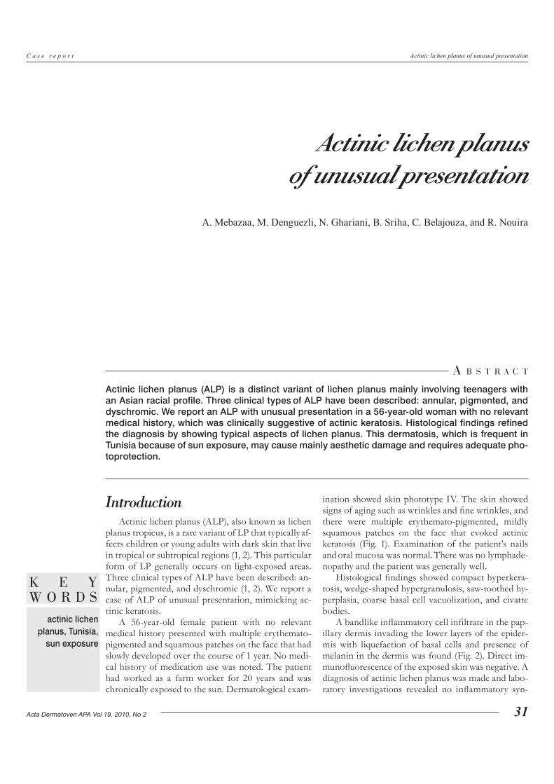
A 56-year-old female patient with no relevant medical history presented with multiple erythemato-pigmented and squamous patches on the face that had slowly developed over the course of 1 year. No medi-cal history of medication use was noted. The patient had worked as a farm worker for 20 years and was chronically exposed to the sun. Dermatological exam-
ination showed skin phototype IV. The skin showed signs of aging such as wrinkles and fine wrinkles, and there were multiple erythemato-pigmented, mildly squamous patches on the face that evoked actinic keratosis (Fig. 1). Examination of the patient’s nails and oral mucosa was normal. There was no lymphade-nopathy and the patient was generally well.
A bandlike inflammatory cell infiltrate in the pap-illary dermis invading the lower layers of the epider-mis with liquefaction of basal cells and presence of melanin in the dermis was found (Fig. 2). Direct im-munofluorescence of the exposed skin was negative. A diagnosis of actinic lichen planus was made and labo-ratory investigations revealed no inflammatory syn-
32
Actinic lichen planus of unusual presentation
Acta Dermatoven APA Vol 19, 2010, No 2
C a s e r e p o r t
drome, no antinuclear antibodies, no liver abnormali-ties, and negative hepatitis B and C virus serologies. The patient received topical corticosteroids of mild or intermediate level for a short time associated with sunblock. Her symptoms partially improved within 3 months with a relapse of pigmented lesions following sun exposure.
ALP is a distinct variant of lichen planus that af-fects mainly children and teenagers (1–4). A racial predilection to Asians with dark complexions and pa-tients living in tropical and subtropical countries has been noted (1–5).
The eruption usually appears during spring and summer, and improvement or complete remission takes place during the winter, leaving hyperpigmented patches. However, relapses may occur during subse-quent sunny seasons (1–5).
The most common form is the annular type, which consists of erythematous brownish plaques with an annular configuration, with or without atrophy. The pigmented type consists of hypermelanotic patches, with a melasma-like appearance. More rarely, the dys-chromic type is characterized by whitish pinhead and coalescent papules, mainly affecting the face, neck, and dorsal hands (1–3). Our case had an unusual presentation with small, mildly infiltrated pigmented patches mimicking actinic keratosis. Histological ex-amination refined the diagnosis by showing typical aspects of lichen planus.
The pathogenesis of ALP is still unknown. Sun-light appears to be the major precipitating factor, probably under the influence of genetic or other fac-tors (hormonal, toxic, or infectious factors, etc.). Hep-atitis viral infection (B and C) is also reported to be a trigger factor in the occurrence of ALP (3–7).
Several therapies have been tried with variable results, including bismuth, arsenic compounds, and
topical corticosteroid preparations. Treatment with antimalarial agents or intralesional corticosteroids combined with sunscreens has shown good results with prolonged remission (6, 7).
This dermatosis may cause significant aesthetic damage requiring prolonged care and adoption of photoprotection measures.
Figure 1. Multiple pigmented patches on the face.
Figure 3. A bandlike inflammatory cell infiltrate in the papillary dermis invading the lower layers of the epidermis with liquefaction of basal cells and presence of melanin in the dermis.