Activatable Near-Infrared Fluorescent Probe for In Vivo Imaging ofFibroblast Activation Protein-alphaJinbo Li,†,‡ Kai Chen,† Hongguang Liu,† Kai Cheng,† Meng Yang,† Jiping Zhang,§ Jonathan D. Cheng,§
Yan Zhang,*,‡ and Zhen Cheng*,†
†Molecular Imaging Program at Stanford (MIPS), Department of Radiology, and Bio-X Program, Canary Center at Stanford forCancer Early Detection, Stanford University, Stanford, California, 94305-5344, United States‡School of Chemistry and Chemical Engineering, Institute of Chemical Biology and Drug Innovation, Nanjing University, Nanjing210093, China§Department of Medical Oncology, Fox Chase Cancer Center, Philadelphia, Pennsylvania 19111, United States
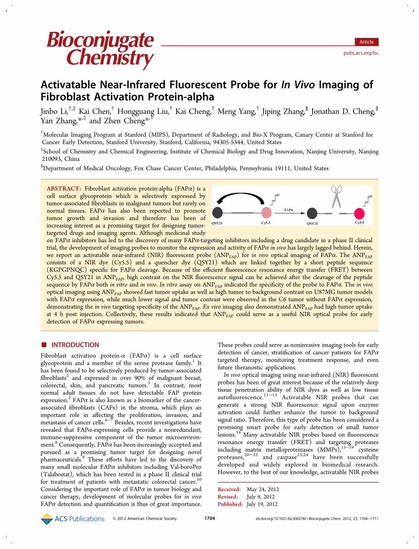
ABSTRACT: Fibroblast activation protein-alpha (FAPα) is acell surface glycoprotein which is selectively expressed bytumor-associated fibroblasts in malignant tumors but rarely onnormal tissues. FAPα has also been reported to promotetumor growth and invasion and therefore has been ofincreasing interest as a promising target for designing tumor-targeted drugs and imaging agents. Although medicinal studyon FAPα inhibitors has led to the discovery of many FAPα-targeting inhibitors including a drug candidate in a phase II clinicaltrial, the development of imaging probes to monitor the expression and activity of FAPα in vivo has largely lagged behind. Herein,we report an activatable near-infrared (NIR) fluorescent probe (ANPFAP) for in vivo optical imaging of FAPα. The ANPFAPconsists of a NIR dye (Cy5.5) and a quencher dye (QSY21) which are linked together by a short peptide sequence(KGPGPNQC) specific for FAPα cleavage. Because of the efficient fluorescence resonance energy transfer (FRET) betweenCy5.5 and QSY21 in ANPFAP, high contrast on the NIR fluorescence signal can be achieved after the cleavage of the peptidesequence by FAPα both in vitro and in vivo. In vitro assay on ANPFAP indicated the specificity of the probe to FAPα. The in vivooptical imaging using ANPFAP showed fast tumor uptake as well as high tumor to background contrast on U87MG tumor modelswith FAPα expression, while much lower signal and tumor contrast were observed in the C6 tumor without FAPα expression,demonstrating the in vivo targeting specificity of the ANPFAP. Ex vivo imaging also demonstrated ANPFAP had high tumor uptakeat 4 h post injection. Collectively, these results indicated that ANPFAP could serve as a useful NIR optical probe for earlydetection of FAPα expressing tumors.
■ INTRODUCTION
Fibroblast activation protein-α (FAPα) is a cell surfaceglycoprotein and a member of the serine protease family.1 Ithas been found to be selectively produced by tumor-associatedfibroblasts2 and expressed in over 90% of malignant breast,colorectal, skin, and pancreatic tumors.3 In contrast, mostnormal adult tissues do not have detectable FAP proteinexpression.2 FAPα is also known as a biomarker of the cancer-associated fibroblasts (CAFs) in the stroma, which plays animportant role in affecting the proliferation, invasion, andmetastasis of cancer cells.4−7 Besides, recent investigations haverevealed that FAPα-expressing cells provide a nonredundant,immune-suppressive component of the tumor microenviron-ment.8 Consequently, FAPα has been increasingly accepted andpursued as a promising tumor target for designing novelpharmaceuticals.9 These efforts have led to the discovery ofmany small molecular FAPα inhibitors including Val-boroPro(Talabostat), which has been tested in a phase II clinical trialfor treatment of patients with metastatic colorectal cancer.10
Considering the important role of FAPα in tumor biology andcancer therapy, development of molecular probes for in vivoFAPα detection and quantification is thus of great importance.
These probes could serve as noninvasive imaging tools for earlydetection of cancer, stratification of cancer patients for FAPαtargeted therapy, monitoring treatment response, and evenfuture theranostic applications.In vivo optical imaging using near-infrared (NIR) fluorescent
probes has been of great interest because of the relatively deeptissue penetration ability of NIR dyes as well as low tissueautofluorescence.11−13 Activatable NIR probes that cangenerate a strong NIR fluorescence signal upon enzymeactivation could further enhance the tumor to backgroundsignal ratio. Therefore, this type of probe has been considered apromising smart probe for early detection of small tumorlesions.14 Many activatable NIR probes based on fluorescenceresonance energy transfer (FRET) and targeting proteasesincluding matrix metalloproteinases (MMPs),15−19 cysteineproteases,20−22 and caspase23,24 have been successfullydeveloped and widely explored in biomedical research.However, to the best of our knowledge, activatable NIR probes
Received: May 24, 2012Revised: July 9, 2012Published: July 19, 2012
for FAPα in vivo imaging have not been reported despite thegreat potential of FAPα in tumor theranostics.The difficulty in the construction of activatable NIR probes
specific for FAPα lies in the fact that FAPα shares the samepeptide substrates for other postprolyl peptidases that arecapable of cleaving the Pro-Xxx amino acid bond.25 The mostclosely related prolyl peptidase family member of FAPα is thedipeptidyl peptidase-IV (DPPIV). The important substrate-binding domain and the key substrate-binding residues in FAPαand DPPIV are in very similar positions.26 However, systematicstructural and kinetic analysis of the substrate specificity ofFAPα27 and peptide substrate profiling revealed that FAPαpossesses endopeptidase activity.28 Then, peptide substratessuch as Ac-GPGP-2SBPO29 and Pyro-TSGPNQEQK(BHQ)9
were found able to discriminate FAPα from DPPIV. Therefore,it is possible for us to develop novel activatable NIR probeswhich can be selectively activated by FAPα and consequentlybe used as in vivo optical imaging probes for FAPα.Here, we report our work on the development of the novel
activatable NIR probe for FAPα (ANPFAP) and the successfulapplication of the probe for in vivo imaging of tumor with FAPαexpression. The FRET pair used in the probe is Cy5.5 andQSY21, which are linked by the peptide substrate(KGPGPNQC) specific for FAPα (Scheme 1). In vitro assayon the fluorescence activation by FAPα, DPPIV, and MMP-2proteins indicated the specificity of the probe for FAPα. In vivoimaging using the probe on two tumor models with andwithout FAPα expression showed high contrast from theFAPα-positive tumor model and much lower contrast from theFAPα-negative tumor model. Ex vivo imaging further revealedthe tissue distribution of the activated probe, which mayprovide important information on further modification andoptimization of the probe for in vivo imaging.
■ MATERIALS AND METHODSGeneral. All N-Fmoc-protected amino acids and rink amide
MBHA resin were purchased from CS Bio Company (MenloPark, CA). Cy5.5-monomaleimide was purchased from GEHealth Care (Piscataway, NJ). QSY21-NHS was purchasedfrom Invitrogen (Carlsbad, CA). All other standard chemicalswere purchased from Sigma-Aldrich Chemical Co. (St. Louis,MO). Recombinant FAPα and DPPIV were obtained fromR&D systems (Minneapolis, MN); recombinant MMP-2 waspurchased from EMD Millipore (Billerica, MA). The C6 andU87MG cells were obtained from American Type TissueCulture Collection (Manassas, VA). Female athymic nude micewere purchased from Charles River Laboratories (Boston, MA).
The peptide Lys-Gly-Pro-Gly-Pro-Asn-Gln-Cys was synthe-sized on an automatic peptide synthesizer (CS Bio) usingFmoc-protected amino acids and rink amide MBHA resin.Reversed-phase high-performance liquid chromatography (RP-HPLC) was performed on a Dionex Ultimate 3000 HPLCsystem (Sunnyvale, CA) for analysis and preparation ofpeptides and bioconjugates. Both semipreparative (Vydac,Hesperia, CA, 218TP510-C18, 10 mm × 250 mm) andanalytical (Dionex, Sunnyvale, CA, Acclaim120 C18, 4.6 mm ×250 mm) RP-HPLC columns were used. The mobile phase wassolvent A, 0.1% trifluoroacetic acid (TFA)/H2O, and solvent B,0.1% TFA/acetonitrile. Matrix-assisted laser desorption/ioniza-tion time-of-flight mass spectrometry (MALDI-TOF-MS,Framingham, MA) was performed by Stanford Protein andNucleic Acid Biotechnology Facility to verify the preparation ofcompounds. Absorption and emission spectra of ANPFAP weremeasured on an Agilent UV−visible ChemStation (AgilentTechnologies, Wilmington, DE) and an Inifite M1000spectrofluorometer (Tecan, Austria), respectively. In vivo andex vivo imaging were performed with an IVIS200 small animalimaging system (Caliper, Hopkinton, MA).
Synthesis of ANPFAP. The crude peptide Lys-Gly-Pro-Gly-Pro-Asn-Gln-Cys on the resin was first acetylated by anhydrousacetic anhydride with hydroxybenzotriazole (HOBt)/N,N-diisopropylethylamine (DIPEA) in dimethylformamide(DMF). Ac-Lys-Gly-Pro-Gly-Pro-Asn-Gln-Cys was thencleaved from the resin by addition of a 92.5/2.5/2.5/2.5 (v/v) mixture of TFA/triisopropylsilane/ethanedithiol/water for 2h at room temperature (RT) and then precipitated by coldanhydrous diethyl ether. The resulted peptide was dried andfurther purified by a preparative RP-HPLC. The flow rate was 4mL/min, with the mobile phase starting from 95% solvent Aand 5% solvent B (0−3 min) to 35% solvent A and 65% solventB at 33 min. Fractions containing the product were collectedand lyophilized. The target product was characterized byMALDI-TOF-MS and ready for use in the next step.The purified peptide (450 μg) was then conjugated with
Cy5.5-monomaleimide (300 μg) in 300 μL of phosphatebuffered saline (PBS, 0.1 M, pH = 7.4) with a molar ratio of 2:1for 2 h in the dark at RT. The Cy5.5-labeled peptide waspurified by RP-HPLC using the same flow rate and gradient asthat for the unmodified peptide listed above. The product wascharacterized by MALDI-TOF-MS and then used for the nextstep. Further conjugation with QSY21-NHS ester in dimethylsulfoxide (DMSO) and 2% DIPEA (1.5 equiv of QSY21-NHSester per equivalent of peptide) for 2 h resulted the finalproduct, ANPFAP. The probe was also purified by HPLC and
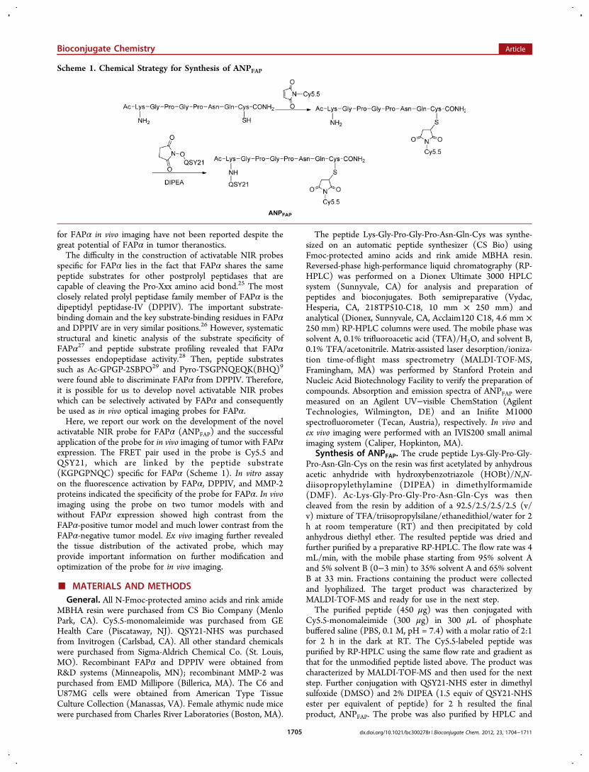
Scheme 1. Chemical Strategy for Synthesis of ANPFAP
lyophilized using the same conditions. ANPFAP was charac-terized by MALDI-TOF-MS and purity was confirmed byanalytical RP-HPLC using the same gradient as the preparativeruns.In Vitro Assay of ANPFAP Activation by FAPα. ANPFAP
was dissolved in water as a stock solution with a concentrationof 1 mM. For enzymatic assays, ANPFAP was mixed with FAPα,DPPIV, or MMP-2 and diluted with PBS to a finalconcentration of 10 μM. Different concentrations of FAPα(0.25 μg/mL, 0.5 μg/mL, 1 μg/mL) were used for dose-dependent study and 1 μg/mL of DPPIV and MMP-2 was usedto demonstrate the specificity of ANPFAP. All the assays wereperformed in triplicate on a 96-well plate, and the emissionspectra of ANPFAP were recorded at 37 °C every 10 min onspectrofluorometer for 1 h. The excitation wavelength was setat 675 nm and the emission spectra were recorded from 680 to800 nm. After 1 h incubation, samples were imagedimmediately by the IVIS200 imaging system for phantom study.Cell Culture and Animal Models. C6 and U87MG cells
were cultured in DMEM medium (Gibco, Carlsbad, CA),supplemented with 10% fetal bovine serum (FBS) (Gibco,Carlsbad, CA) and 1% penicillin−streptomycin (Gibco,Carlsbad, CA). Cells were maintained in a humidifiedatmosphere of 5% CO2 at 37 °C. C6 and U87MG tumormodels were developed by subcutaneous injection of 3 × 10⁶
and 5 × 10⁶ cells, respectively, into the right shoulder of femaleathymic nude mice. Tumor-bearing mice were subjected to invivo imaging studies when tumor volume reached 200−300mm3. All animal studies were performed according to theprotocol approved by the Stanford University AdministrativePanels on Laboratory Animal Care (APLAC).Western Blot Analysis. The expression of FAPα in C6 and
U87MG tumor tissues was analyzed by Western blot analysiswith anti-FAPα monoclonal antibody. Tumors were collectedright after the end of in vivo experiment and quickly frozen forpreservation. Before protein extraction, tumors were weighed,homogenized, and lysed with M-PER mammalian proteinextraction reagent (Thermo Fisher, Waltham, MA). Aftercentrifugation, clear cell lysates were transferred to new tubes.The protein concentrations were determined by using Bradfordreagent (Bio-Rad, Hercules, CA). The cell lysates (50 μg) wereloaded onto NuPAGE 4−12% Bis-Tris Gel gel (Invitrogen,Carlsbad, CA) and transferred to Immun-blot PVDFmembranes (Invitrogen, Carlsbad, CA). Anti-FAP monoclonalantibody was used at a 1:100 dilution. Anti-rabbit HRPconjugated secondary antibody was purchased from ThermoFisher (Waltham,MA) and used at a 1:5000 dilution. Actinantibody (Abcam, Cambridge, MA) was used at 1:3000 to
detect expression of actin as a control for protein loading. Allimmunoblots were analyzed by using the ECL detection system(Thermo Fisher, Waltham, MA).
In Vivo and Ex Vivo Imaging. For in vivo imaging, C6 andU87MG tumor-bearing mice (n = 5 for each group) wereinjected intravenously via tail vein with 1 nmol of ANPFAP in150 μL of PBS. NIRF imaging was then performed with theIVIS200 small animal imaging system. To acquire fluorescenceof ANPFAP in vivo, Cy5.5 filter (excitation 615−665 nm;emission 695−770 nm) and Cy5.5 background filter (580−610nm) were used. Identical illumination settings (lamp voltage,filters, f/stop, field of views, binning) were used for acquiring allimages. Fluorescence images were taken at 0.5, 1, 2, 3, and 4 hpost injection (p.i.) using a 2 s exposure time (f/stop = 1,binning = 4). Images were then analyzed using Living Image 3.2software (Caliper, Hopkinton, MA). The Cy5.5 backgroundfilter image was subtracted from the Cy5.5 filter image and thefluorescence emission was normalized to photons per secondper centimeter squared per steradian (p/s/cm2/sr). Forcomparison of tumor contrast, mean fluorescence intensitiesof the tumor area (T) at the right foreleg of the animal and ofthe area (N) at the right flank (normal tissue) were calculatedby the region-of-interest (ROI) function of Living Imagesoftware integrated with Igor (Wavemetrics, Lake Oswego,OR). Dividing T by N yielded the contrast between tumortissue and normal tissue.For ex vivo imaging, mice were sacrificed 4 h p.i. Tumor and
other major tissues were dissected, rinsed with PBS, dried withtissue paper, and then put on black paper. The ex vivo imageswere acquired immediately using IVIS200 system with the sameillumination setting as the in vivo imaging. Images were alsoprocessed with the same method as described above.
Statistical Analysis. All data are shown as mean ± SD(standard deviation). Difference between groups was assessedby Student’s t test. Statistical significance was considered if Pvalues are less than 0.05.
■ RESULTS
Design and Synthesis of the FAPα-Specific Probe. Todesign an optical probe suitable for in vivo imaging, the NIR dyeCy5.5 with maximum emission wavelength at 695 nm wasselected to label the peptide substrate. Since the emission ofCy5.5 was known to be efficiently quenched by the dyeQSY21,30 QSY21 was chosen as the FRET pair with Cy5.5 tolabel the peptide substrate. The selection of the peptidesequence (GPGPNQ) was based on the reported specific FAPαsubstrate sequences which could be cleaved by FAPα but notDPPIV.9,28,29 For efficient and site-specific labeling the peptide
Figure 1. (A) Absorption spectrum of QSY21, Cy5.5, and ANPFAP. (B) Fluorescence emission spectrum of Cy5.5 labeled substrate and ANPFAP.
substrate with Cy5.5 and QSY21, a lysine residue with a freeamine group on the side chain and a cysteine with a thiol groupwere inserted to the N and C terminal of the peptide,respectively. More specifically, Conjugation of the two dyeswith the peptide substrate was easily realized through thereaction of the cysteine thiol group with the Cy5.5 maleimideand the lysine amine with QSY21 NHS ester in ∼26% yield and>95% purity (Scheme 1). Successful conjugation of Cy5.5 andfurther modification with QSY21 were indicated by the peakwith m/z at 1880.6 (calculated as 1879.6) and 2544.6(calculated as 2544.9) on the MALDI-TOF-MS spectrum ofthe purified product, respectively. The UV spectra of Cy5.5,QSY21, and ANPFAP were shown in Figure 1A. There were twosignificant absorption bands centered at 620 and 676 nm,respectively, for ANPFAP. In addition, the strong fluorescentemission at ∼695 nm of the substrate labeled with Cy5.5 alonewas efficiently quenched by QSY21 as shown in thefluorescence spectrum of ANPFAP (Figure 1B).Sensitivity and Specificity of ANPFAP to FAPα.
Sensitivity of ANPFAP to FAPα was first examined in vitro.Upon incubation with FAPα at 37 °C, ANPFAP showedsignificant increase of the intensity of the fluorescence emissioncorresponding to that of Cy5.5. It indicated that the probecould be efficiently cleaved by FAPα to release the Cy5.5labeled residue and split the FRET pair. Fluorescent spectra ofANPFAP incubated with FAPα at different time points werecollected as shown in Figure 2A. It showed that the activationof ANPFAP was fast in kinetics, and an increase of 12-fold influorescence emission intensity at 695 nm could be achievedwithin 1 h. As a contrast, ANPFAP which was not incubated withany enzyme showed no increase in fluorescence emissionintensity. Besides, ANPFAP incubated with the same amount ofpostprolyl peptidase DPPIV and MMP-2 did not showsignificant activation on fluorescent emission either (Figure
2B). These results clearly suggested that the activation ofANPFAP was caused by the specific recognition and cleavage ofthe peptide substrate by FAPα.Dose-dependent activation of the ANPFAP was also observed
when the probe was incubated with different concentrations ofFAPα. Fluorescent spectra of ANPFAP incubated with differentamounts of FAPα at 37 °C for 1 h were recorded as shown inFigure 2C. The increase of the fluorescence intensity at 695 nmwas significant upon titration of FAPα concentration from 0.25μg/mL to 1 μg/mL (Figure 2D). Phantom study on theANPFAP incubated with FAPα at concentrations of 0, 0.25, 0.5,and 1 μg/mL also showed increased brightness of the probe(Figure 2D). The brightness of ANPFAP under optical imagingcorresponded well with the amount of FAPα presented withinthe target.
In Vivo Imaging. After successfully validating the specificityand sensitivity of ANPFAP in vitro, its potential for in vivoimaging was further investigated. For this purpose, the U87MGtumor model was developed because of the high expression ofFAPα in U87MG tumor. To evaluate the specificity of ANPFAPin vivo, another tumor model C6, which is not supposed toexpress FAPα, was also established. Expression of FAPα inthese two tumor models was measured, and it was found thatFAPα was highly expressed in U87MG tumors, while almost nodetectable expression in C6 tumor (Figure 3).On the basis of the expression of FAPα in these tumors, C6
and U87MG were used as negative and positive tumor models,respectively, for evaluation of the FAPα imaging ability ofANPFAP in vivo. ANPFAP was first injected into C6 and U87MGtumor-bearing mice through the tail vein. In vivo imaging wasrecorded over 4 h. Representative in vivo whole-body images ofmice at different time points (0.5, 1, 2, 3, and 4 h) were shownin Figure 4A. Strong fluorescent signals in FAPα positive tumorU87MG were observed, while much lower fluorescent signals
Figure 2. (A) Time-dependent fluorescence spectrum of 10 μM ANPFAP with 1 μg/mL FAPα at 37 °C. (B) Time-dependent fluorescence change of10 μM ANPFAP without FAPα, with 1 μg/mL FAPα, 1 μg/mL DPPIV, or 1 μg/mL MMP-2. (C) FAPα concentration-dependent fluorescencespectrum of 10 μM ANPFAP with different concentrations of FAPα after a 1 h incubation at 37 °C. (D) Fluorescence increase ratio of ANPFAP in thepresence of various concentrations of FAPα after a 1 h incubation. Insert: phantom study of activated ANPFAP.
were detected in FAPα negative tumor C6 in all thecorresponding time points (P < 0.05). Early activation ofANPFAP in U87MG tumor was observed at 0.5 h, and thefluorescent signals increased from 0.5 to 4 h p.i. Quantificationanalysis of fluorescent signals in tumors was calculated bydrawing ROI, and the results were shown in Figure 4B. Thefluorescence intensities of U87MG tumor increased after 0.5 hand reached maximum at 2 h p.i. and then remainedunchanged, while fluorescence intensities of C6 tumor didnot alter over the same time period. The fluorescence ratiosbetween tumor and normal tissue were also calculated andshown in Figure 4C. The tumor-to-normal tissue ratios ofU87MG increased rapidly from 0.5 h (2.218 ± 0.403) to 4 h(4.630 ± 0.728), suggesting accumulation of ANPFAP in tumors.In contrast, tumor-to-normal tissue ratios of C6 remainedconstant at all time points, and these ratios were significantlylower than the corresponding ratios obtained from the U87MGtumor model, highlighting the in vivo targeting specificity of theANPFAP.
Ex Vivo Imaging. To further confirm the in vivo imagingresults and specific activation and accumulation of ANPFAP intumors, ex vivo imaging of tumors and other normal organs wasperformed after in vivo imaging finished. Representative ex vivoimages of tumors and normal organs were shown in Figure 5A.Similarly to the in vivo imaging (Figure 4A), the U87MG tumorwas much brighter than the C6 tumor (P < 0.05), whichdemonstrated successful activation of ANPFAP by FAPα in vivo.Quantitative analysis of the fluorescent signals confirmed thatactivated ANPFAP was mainly located in the U87MG tumor, butnot in the FAPα negative tumor C6 (Figure 5B). Strong signalsfrom stomachs of both U87MG and C6 tumor bearing micewere also observed, which were likely from the autofluor-escence of remaining mouse chow. Last, it was noticed that theother major organs gave comparable signals (Figure 5B). Thetumor-to-normal tissue signal ratios were also calculated(Figure 5C), and a significant difference of the ratios betweenU87MG and C6 was clearly viewed (P < 0.05). All thesefindings were consistent with the in vivo imaging results,demonstrating that ANPFAP can be efficiently applied for in vivoimaging of FAPα.
■ DISCUSSION
Enzymes play important roles in many biological andphysiological processes, and sometimes, they can serve asvalid biomarkers for various diseases. Thus, imaging enzymeshelp to investigate enzyme function and even diagnose diseases.Unique catalytic activities of enzymes enable design of smartoptical probes that can be turned on upon meeting with theenzyme target. The self-quenching probe, Ac-GPGP-2SBPO,
Figure 3. Western blot analysis of tumor tissue lysates.
Figure 4. (A) In vivo fluorescent imaging of subcutaneous C6 and U87MG tumor-bearing mice at 0.5, 1, 2, 3, and 4 h post tail vein injection ofANPFAP (1 nmol). (B) Fluorescence ROI analysis of tumor and muscle in mice bearing C6 and U87MG tumor from 0.5 to 4 h p.i. (C) ROI analysisof tumor-to-normal tissue ratios of ANPFAP in mice bearing C6 and U87MG tumor from 0.5 to 4 h. Data are shown as mean ± SD (n = 5 per group),*P < 0.05.
demonstrates specificity to FAPα, and it could be activated inFAPα transfected cells.29 The dual functional probe, Pyro-TSGPNQEQK(BHQ), developed by Zheng et al. can imageFAPα and produce cytotoxicity simultaneously, by combinationof pyropheophorbide α acid (Pyro) as a photosensitizer as wellas a fluorophore whose fluorescence can be quenched byBHQ.9 The specificity of these probes to FAPα indicates theimportance of the peptide sequence used as a linker in theactivatable probes.Different from previous studies, a new specific peptide
substrate was selected by us according to the reportedsubstrates specific to FAPα.9,28,29 Unlike Ac-GPGP-2SBPO,29
which utilizes the strategy of self-quenching of the dye, ANPFAPemploys a quencher (QSY21) to make a NIR dye (Cy5.5)silent (Figure 1B), and the quenching efficiency was found tobe 94%. In the enzymatic assay, ANPFAP shows good sensitivityand selectivity toward FAPα (Figure 2). Approximately 12-foldfluorescence recovery of ANPFAP can be achieved afterincubation with 1 μg/mL of FAPα for 1 h (Figure 2B).Furthermore, for testing the specificity of ANPFAP, DPPIV,which also belongs to the serine protease family and has 50%amino acid similarity to FAPα, was selected. Coupledexpression of DPPIV and FAPα has been found in transformedastrocytic cells.31 The difference of these two enzymes mainlylies in their catalytic activities. FAPα is a strict endopeptidase,while DPPIV is a strict exopeptidase.25−28 On the basis of thehigh similarity between DPPIV and FAPα, DPPIV was chosen
as a specificity control for investigating the specificity ofANPFAP. When incubating ANPFAP with 1 μg/mL of DPPIV, noactivation of ANPFAP can be observed (Figure 2C). IncubatingANPFAP with another enzyme, MMP-2, also give similar results.Titration studies show nonlinear dose dependent activation ofANPFAP toward FAPα (Figure 2D). One possible reason is thatfluorescence recovery of ANPFAP has reached maximum at 1 hincubation with a higher concentration of FAPα such as 1 μg/mL (Figure 2A), while fluorescence intensities of the probeincubating with smaller amounts of FAPα for 1 h may not havefully recovered. Collectively, these in vitro assays demonstratethat ANPFAP is highly specific and sufficient to be used forfurther in vivo evaluation.Recently, FAPα was found to be expressed in U87MG cells,
making U87MG tumor model a good candidate for FAPα invivo imaging.31 The C6 tumor model which is FAPα negativewas also generated as a control. Western blot results confirmthat FAPα is indeed expressed in U87MG tumor but not in C6tumor (Figure 3). Then, the ability of ANPFAP for in vivoimaging was evaluated in these mice models throughintravenous tail vein injection of the probe. Despite limiteddelivery efficiency of Pyro-TSGPNQEQK(BHQ) after system-atic administration reported before,9 ANPFAP shows efficientaccumulation in U87MG tumor but not in C6 tumor (Figure4A). By quantitative analysis of fluorescence intensities in theU87MG tumor region, it can be seen that the signal increasesfrom 0.5 to 2 h and reaches maximum at 2 h (Figure 4B),
Figure 5. (A) Ex vivo imaging of tumor and normal tissue of C6 and U87MG mice after mice were sacrificed at 4 h post injection. The numeric labelfor each organ is as follows: 1, blood; 2, heart; 3, lung; 4, liver; 5, spleen; 6, pancreas; 7, stomach; 8, intestine; 9, kidney; 10, skin; 11, muscle; 12,bone; 13, tumor; 14, brain. (B) ROI analysis of fluorescent signals from tumors and normal tissues. (C) Tumor-to-normal tissue ratio of fluorescentintensities based on ROI analysis. Data are shown as mean ± SD (n = 5 per group), *P < 0.05.
indicating fast activation of the probe in vivo, and 2 h is a goodimaging time for future applications of ANPFAP. For C6 tumor,only a slight increase of the fluorescence intensities is observedfrom 0.5 to 1 h, which might be due to the influx of activatedprobes from blood into tumor through the enhancedpermeability and retention effect. Nevertheless, the differenceof fluorescence intensities between C6 tumor and U87MGtumor is significant (P < 0.05). It is worth noting that, since theprobe is not actively targeted, it may have different levels ofaccumulations in two tumor models, which may make somecontributions to the different fluorescence signals in tumors.Moreover, different from the absolute fluorescence intensities,tumor-to-normal tissue ratio of U87MG mice keeps increasingfrom 0.5 to 4 h (Figure 4C), suggesting a reduction ofbackground signals all the time. Ex vivo imaging also shows asignificant difference in tumor-to-normal tissue ratios betweenC6 tumor and U87MG tumor (Figure 5C), which is consistentwith the in vivo results. All these results demonstrate the highspecificity of ANPFAP and its high potential for serving as anNIR probe to image FAPα in vivo.Interestingly, signals from the mouse body were also
observed. Though autofluorescence was subtracted using theappropriate function of IVIS system, some signals could still becaused by autofluorescence of mice. An alternative imagingsystem, Maestro, may help further improve the imaging qualityby using spectral unmixing function. Another possible reason isthat FAPα can also be secreted from tumors, and soluble formsof FAPα have been found in bovine serum.32 Therefore, thesoluble FAPα expressed in the blood or other tissues couldcleave the probe to generate background signal. The ex vivoimaging study also confirms that, besides the high uptake ofANPFAP in U87MG tumor, background signals can be seen inthe liver, stomach, intestine, and kidney (Figure 5B). Specificactivation of probes in tumor is really hard to achieve andrepresents a common hurdle for enzyme imaging usingactivatable probes in this field, our finding here is really not asurprise. As an example, for imaging MMPs, a relatively highuptake of MMP activatable probes in organs beside tumor hasalso been observed.18,19 Regardless of the background signal,the encouraging thing is that we still can clearly visualize thetarget tumor. To further optimize the probe and overcome theproduction of nonspecific background signal, the activatableprobes could be encapsulated in polymers for protection duringthe circulation while being released when reaching tumors.It should be pointed out that some of the limitations of
optical imaging techniques can also hinder the use of ANPFAP invivo. For example, it is very hard to get quantitative informationabout FAPα, and the optical signals sometimes are misleading,as they can be easily affected by many other factors such asdepth of the probe presented in vivo, absorption and refractionof tissues, and so forth. To solve these problems, other imagingmodalities such as positron emission tomography (PET) andmagnetic resonance imaging (MRI) can be combined withfluorescence imaging. For example, MRI/NIRF activatableimaging of MMP-2 has been realized by loading Cy5.5 labeledMMP substrates onto iron oxide nanoparticles.33 It is thuspossible to use iron oxide nanoparticles to quench fluorescenceof the Cy5.5 labeled peptide substrate of FAPα and to realizeMR/NIRF imaging. An alternative strategy is to directly linkANPFAP with iron oxide nanoparticles, and then dual modalityimaging (NIRF/MRI) can be performed to image FAPα moreefficiently. One example is that prequenched MT1-MMP probewas anchored to magnetic nanocrystals for molecular imaging
of invasive cancer cells.34 It is expected that this approach couldbe adapted to improve the imaging efficiency of ANPFAP. Last,it is worth noting that the FRET based activatable probe itselfcould provide a target-dependent photoacoustic signal.35 Withthe photoacoustic signal, FAPα expressing tumors could bevisualized more easily and clearly. Therefore, while combiningfluorescence imaging with other imaging modalities, ANPFAPmay serve as a more efficient probe for better understanding ofthe distribution and function of FAPα in vivo.
■ CONCLUSIONAn FAPα specific NIRF probe ANPFAP, which is composed ofNIR dye Cy5.5, peptide substrate of FAPα, and a quencherQSY21, was designed and successfully synthesized. Fluorescentsignals of ANPFAP can be recovered in the presence of FAPα,while not by DPPIV or MMP-2, suggesting the high specificityof ANPFAP toward FAPα. In vivo imaging studies furtherdemonstrate that ANPFAP can be activated rapidly in FAPαpositive U87MG tumor but not in the negative C6 tumor.Overall, our study provides a new type of FAPα specific probethat can be used to image FAPα in vivo and to betterunderstand the roles of FAPα in vitro and in vivo.
■ ACKNOWLEDGMENTSThis work was supported in part by National Cancer Institute(NCI) 5R01 CA119053 (ZC) and the National Basic ResearchProgram of China (No. 2011CB935800). J. L. was supportedby a fellowship from the Chinese Scholarship Council (CSC).
■ ABBREVIATIONS:FAPα, fibroblast activation protein-alpha; NIR, near-infrared;FRET, fluorescence resonance energy transfer; p.i, postinjection; HPLC, high-performance liquid chromatography;ROI, regions of interest
■ REFERENCES(1) Rettig, W. J., Garin-Chesa, P., Beresford, H. R., Oettgen, H. F.,Melamed, M. R., and Old, L. J. (1988) Cell-surface glycoproteins ofhuman sarcomas: differential expression in normal and malignanttissues and cultured cells. Proc. Natl. Acad. Sci. U. S. A. 85, 3110−4.(2) Scanlan, M. J., Raj, B. K., Calvo, B., Garin-Chesa, P., Sanz-Moncasi, M. P., Healey, J. H., Old, L. J., and Rettig, W. J. (1994)Molecular cloning of fibroblast activation protein alpha, a member ofthe serine protease family selectively expressed in stromal fibroblasts ofepithelial cancers. Proc. Natl. Acad. Sci. U. S. A. 91, 5657−61.(3) Garin-Chesa, P., Old, L. J., and Rettig, W. J. (1990) Cell surfaceglycoprotein of reactive stromal fibroblasts as a potential antibodytarget in human epithelial cancers. Proc. Natl. Acad. Sci. U. S. A. 87,7235−9.(4) Kalluri, R., and Zeisberg, M. (2006) Fibroblasts in cancer. Nat.Rev. Cancer 6, 392−401.(5) Cheng, J. D., Dunbrack, R. L., Jr., Valianou, M., Rogatko, A.,Alpaugh, R. K., and Weiner, L. M. (2002) Promotion of tumor growthby murine fibroblast activation protein, a serine protease, in an animalmodel. Cancer Res. 62, 4767−72.
(6) Huang, Y., Simms, A. E., Mazur, A., Wang, S., Leon, N. R., Jones,B., Aziz, N., and Kelly, T. (2011) Fibroblast activation protein-alphapromotes tumor growth and invasion of breast cancer cells throughnon-enzymatic functions. Clin. Exp. Metastasis 28, 567−79.(7) Lee, H. O., Mullins, S. R., Franco-Barraza, J., Valianou, M.,Cukierman, E., and Cheng, J. D. (2011) FAP-overexpressingfibroblasts produce an extracellular matrix that enhances invasivevelocity and directionality of pancreatic cancer cells. BMC Cancer 11,245.(8) Kraman, M., Bambrough, P. J., Arnold, J. N., Roberts, E. W.,Magiera, L., Jones, J. O., Gopinathan, A., Tuveson, D. A., and Fearon,D. T. (2010) Suppression of antitumor immunity by stromal cellsexpressing fibroblast activation protein-alpha. Science 330, 827−30.(9) Lo, P. C., Chen, J., Stefflova, K., Warren, M. S., Navab, R.,Bandarchi, B., Mullins, S., Tsao, M., Cheng, J. D., and Zheng, G.(2009) Photodynamic molecular beacon triggered by fibroblastactivation protein on cancer-associated fibroblasts for diagnosis andtreatment of epithelial cancers. J. Med. Chem. 52, 358−68.(10) Narra, K., Mullins, S. R., Lee, H. O., Strzemkowski-Brun, B.,Magalong, K., Christiansen, V. J., McKee, P. A., Egleston, B., Cohen, S.J., Weiner, L. M., Meropol, N. J., and Cheng, J. D. (2007) Phase II trialof single agent Val-boroPro (Talabostat) inhibiting FibroblastActivation Protein in patients with metastatic colorectal cancer. CancerBiol. Ther. 6, 1691−9.(11) Petrovsky, A., Schellenberger, E., Josephson, L., Weissleder, R.,and Bogdanov, A., Jr. (2003) Near-infrared fluorescent imaging oftumor apoptosis. Cancer Res. 63, 1936−42.(12) Cheng, Z., Levi, J., Xiong, Z., Gheysens, O., Keren, S., Chen, X.,and Gambhir, S. S. (2006) Near-infrared fluorescent deoxyglucoseanalogue for tumor optical imaging in cell culture and living mice.Bioconjugate Chem. 17, 662−9.(13) Miao, Z., Ren, G., Liu, H., Jiang, L., and Cheng, Z. (2010)Cy5.5-labeled Affibody molecule for near-infrared fluorescent opticalimaging of epidermal growth factor receptor positive tumors. J.Biomed. Opt. 15, 036007.(14) Weissleder, R., Tung, C. H., Mahmood, U., and Bogdanov, A.,Jr. (1999) In vivo imaging of tumors with protease-activated near-infrared fluorescent probes. Nat. Biotechnol. 17, 375−8.(15) Pham, W., Choi, Y., Weissleder, R., and Tung, C. H. (2004)Developing a peptide-based near-infrared molecular probe for proteasesensing. Bioconjugate Chem. 15, 1403−7.(16) Chen, J., Tung, C. H., Allport, J. R., Chen, S., Weissleder, R., andHuang, P. L. (2005) Near-infrared fluorescent imaging of matrixmetalloproteinase activity after myocardial infarction. Circulation 111,1800−5.(17) Yoon, S. M., Myung, S. J., Ye, B. D., Kim, I. W., Lee, N. G., Ryu,Y. M., Park, K., Kim, K., Kwon, I. C., Park, Y. S., Park, C. S., Moon, D.H., Kim do, H., Do, M. Y., Byeon, J. S., Yang, S. K., and Kim, J. H.(2010) Near-infrared fluorescence imaging using a protease-specificprobe for the detection of colon tumors. Gut Liver 4, 488−97.(18) Zhu, L., Xie, J., Swierczewska, M., Zhang, F., Quan, Q., Ma, Y.,Fang, X., Kim, K., Lee, S., and Chen, X. (2011) Real-time videoimaging of protease expression in vivo. Theranostics 1, 18−27.(19) Zhu, L., Zhang, F., Ma, Y., Liu, G., Kim, K., Fang, X., Lee, S., andChen, X. (2011) In vivo optical imaging of membrane-type matrixmetalloproteinase (MT-MMP) activity. Mol. Pharm. 8, 2331−8.(20) Jaffer, F. A., Kim, D. E., Quinti, L., Tung, C. H., Aikawa, E.,Pande, A. N., Kohler, R. H., Shi, G. P., Libby, P., and Weissleder, R.(2007) Optical visualization of cathepsin K activity in atherosclerosiswith a novel, protease-activatable fluorescence sensor. Circulation 115,2292−8.(21) Kim, D. E., Kim, J. Y., Schellingerhout, D., Shon, S. M., Jeong, S.W., Kim, E. J., and Kim, W. K. (2009) Molecular imaging of cathepsinB proteolytic enzyme activity reflects the inflammatory component ofatherosclerotic pathology and can quantitatively demonstrate theantiatherosclerotic therapeutic effects of atorvastatin and glucosamine.Mol. Imaging 8, 291−301.(22) Abd-Elgaliel, W. R., Cruz-Monserrate, Z., Logsdon, C. D., andTung, C. H. (2011) Molecular imaging of Cathepsin E-positive tumors
in mice using a novel protease-activatable fluorescent probe. Mol.Biosyst. 7, 3207−13.(23) Bullok, K., and Piwnica-Worms, D. (2005) Synthesis andcharacterization of a small, membrane-permeant, caspase-activatablefar-red fluorescent peptide for imaging apoptosis. J. Med. Chem. 48,5404−7.(24) Barnett, E. M., Zhang, X., Maxwell, D., Chang, Q., and Piwnica-Worms, D. (2009) Single-cell imaging of retinal ganglion cell apoptosiswith a cell-penetrating, activatable peptide probe in an in vivoglaucoma model. Proc. Natl. Acad. Sci. U. S. A. 106, 9391−6.(25) Park, J. E., Lenter, M. C., Zimmermann, R. N., Garin-Chesa, P.,Old, L. J., and Rettig, W. J. (1999) Fibroblast activation protein, a dualspecificity serine protease expressed in reactive human tumor stromalfibroblasts. J. Biol. Chem. 274, 36505−12.(26) Edosada, C. Y., Quan, C., Wiesmann, C., Tran, T., Sutherlin, D.,Reynolds, M., Elliott, J. M., Raab, H., Fairbrother, W., and Wolf, B. B.(2006) Selective inhibition of fibroblast activation protein proteasebased on dipeptide substrate specificity. J. Biol. Chem. 281, 7437−44.(27) Aertgeerts, K., Levin, I., Shi, L., Snell, G. P., Jennings, A., Prasad,G. S., Zhang, Y., Kraus, M. L., Salakian, S., Sridhar, V., Wijnands, R.,and Tennant, M. G. (2005) Structural and kinetic analysis of thesubstrate specificity of human fibroblast activation protein alpha. J.Biol. Chem. 280, 19441−4.(28) Edosada, C. Y., Quan, C., Tran, T., Pham, V., Wiesmann, C.,Fairbrother, W., and Wolf, B. B. (2006) Peptide substrate profilingdefines fibroblast activation protein as an endopeptidase of strictGly(2)-Pro(1)-cleaving specificity. FEBS Lett. 580, 1581−6.(29) Lai, K. S., Ho, N. H., Cheng, J. D., and Tung, C. H. (2007)Selective fluorescence probes for dipeptidyl peptidase activity-fibroblast activation protein and dipeptidyl peptidase IV. BioconjugateChem. 18, 1246−50.(30) Kong, Y., Yao, H., Ren, H., Subbian, S., Cirillo, S. L., Sacchettini,J. C., Rao, J., and Cirillo, J. D. (2010) Imaging tuberculosis withendogenous beta-lactamase reporter enzyme fluorescence in live mice.Proc. Natl. Acad. Sci. U. S. A. 107, 12239−44.(31) Balaziova, E., Busek, P., Stremenova, J., Sromova, L., Krepela, E.,Lizcova, L., and Sedo, A. (2011) Coupled expression of dipeptidylpeptidase-IV and fibroblast activation protein-alpha in transformedastrocytic cells. Mol. Cell. Biochem. 354, 283−9.(32) Collins, P. J., McMahon, G., O’Brien, P., and O’Connor, B.(2004) Purification, identification and characterisation of seprase frombovine serum. Int. J. Biochem. Cell Biol. 36, 2320−33.(33) Cha, E. J., Jang, E. S., Sun, I. C., Lee, I. J., Ko, J. H., Kim, Y. I.,Kwon, I. C., Kim, K., and Ahn, C. H. (2011) Development of MRI/NIRF ’activatable’ multimodal imaging probe based on iron oxidenanoparticles. J. Controlled Release 155, 152−8.(34) Park, J., Yang, J., Lim, E. K., Kim, E., Choi, J., Ryu, J. K., Kim, N.H., Suh, J. S., Yook, J. I., Huh, Y. M., and Haam, S. (2012) Anchoredproteinase-targetable optomagnetic nanoprobes for molecular imagingof invasive cancer cells. Angew. Chem., Int. Ed. Engl. 51, 945−8.(35) Levi, J., Kothapalli, S. R., Ma, T. J., Hartman, K., Khuri-Yakub, B.T., and Gambhir, S. S. (2010) Design, synthesis, and imaging of anactivatable photoacoustic probe. J. Am. Chem. Soc. 132, 11264−9.