Activation of c-Jun NH 2 -terminal kinase (JNK) signaling pathway is essential for the stimulation of hepatitis C virus (HCV) non-structural protein 3 (NS3)-mediated cell growth Mohamed Hassan a, * , Hanan Ghozlan b , Ola Abdel-Kader c a Institute of Pathology, Faculty of Medicine, University of Duesseldorf, Mooren Str.5, 40225 Duesseldorf, Germany b Department of Microbiology, Faculty of Science, University of Alexandria, Alexandria, Egypt c Medical Research Institute, University of Alexandria, Alexandria, Egypt Received 4 October 2004; returned to author for revision 14 December 2004; accepted 7 January 2005 Available online 28 January 2005 Abstract Hepatitis C virus (HCV) non-structural protein 3 (NS3) has been shown to affect cellular functions and is thought to contribute to the development of HCV-related hepatocarcinogenesis. In this study, we delineated part of the mechanisms whereby NS3 protein stimulates cell growth in liver (HepG2) and non-liver (HeLa) cells. The expression of NS3 protein enhanced cell growth, c-jun NH 2 -terminal kinase (JNK) activation, DNA binding activities of the transcription factors AP-1 and ATF-2, and c-jun expression, but not the activation of extracellular signal-regulated kinase (ERK) or p38 MAPK . Whereas co-expression of NS3 with its cofactor NS4A inhibited NS3-mediated cell growth without to influence NS3-mediated JNK activation, or to affect the basal activities of ERK or p38 MAPK . Pre-treatment of NS3 protein- expressing cells with JNK inhibitor, SP600125, abolished activation of AP-1 and ATF-2 and inhibited c-jun expression and induced cell growth, suggesting that JNK activation is essential for the stimulation of NS3-mediated cell growth. D 2005 Elsevier Inc. All rights reserved. Keywords: HCV NS3; HCC; MAP kinase; JNK; Cell growth Introduction Hepatitis C virus (HCV) still remains responsible for a significant proportion of community-acquired hepatitis. Persistent HCV infection often leads to chronic hepatitis (Alter et al., 1989; Choo et al., 1989; Tanaka et al., 1995), which is thought to be strongly associated with the development of hepatocellular carcinoma (HCC). HCV genome has a long open reading frame, flanked with 5V and 3V untranslated region, which encodes a polyprotein precursor of about 3010–3033 amino acid (aa) residues (Takamizawa et al., 1991). This polyprotein is cleaved by both host and viral proteases to generate four structural proteins (C, E1, E2, and P7) and six non-structural proteins (NS2, NS3, NS4A, NS4B, NS5A, and NS5B) (Neddermann et al., 1997; Takamizawa et al., 1991). Virus multiplication is dependent upon viral proteins expressed from HCV genome, including non-structural proteins that are expected to participate in genome transcription and replication (Neddermann et al., 1997). In particular, the non-structural protein 3 (NS3) that possesses serine protease activity, which is essential for viral protein processing (Grakoui et al., 1989) and nucleotide triphosphatase–RNA helicase activity, which is essential for virus replication (Hong et al., 1996; Kim et al., 1995; Santolini et al., 1995; Suzich et al., 1993). Besides its pivotal role in viral protein processing and virus replication, the HCV NS3 protein was reported to be involved in malignant transformation of NIH3T3 cells (Sakamuro et al., 1995) as well as in the suppression of actinomycin D- induced apoptosis in NIH3T3 cells (Fujita et al., 1996). Furthermore, the role of HCV NS3 protein in the inhibition 0042-6822/$ - see front matter D 2005 Elsevier Inc. All rights reserved. doi:10.1016/j.virol.2005.01.008 * Corresponding author. Fax: +49 211 811 9439. E-mail address: [email protected] (M. Hassan). Virology 333 (2005) 324 – 336 www.elsevier.com/locate/yviro
Transcript
www.elsevier.com/locate/yviro
Virology 333 (20
Activation of c-Jun NH2-terminal kinase (JNK) signaling pathway is
essential for the stimulation of hepatitis C virus (HCV) non-structural
protein 3 (NS3)-mediated cell growth
Mohamed Hassana,*, Hanan Ghozlanb, Ola Abdel-Kaderc
aInstitute of Pathology, Faculty of Medicine, University of Duesseldorf, Mooren Str.5, 40225 Duesseldorf, GermanybDepartment of Microbiology, Faculty of Science, University of Alexandria, Alexandria, Egypt
cMedical Research Institute, University of Alexandria, Alexandria, Egypt
Received 4 October 2004; returned to author for revision 14 December 2004; accepted 7 January 2005
Available online 28 January 2005
Abstract
Hepatitis C virus (HCV) non-structural protein 3 (NS3) has been shown to affect cellular functions and is thought to contribute to the
development of HCV-related hepatocarcinogenesis. In this study, we delineated part of the mechanisms whereby NS3 protein stimulates cell
growth in liver (HepG2) and non-liver (HeLa) cells. The expression of NS3 protein enhanced cell growth, c-jun NH2-terminal kinase (JNK)
activation, DNA binding activities of the transcription factors AP-1 and ATF-2, and c-jun expression, but not the activation of extracellular
signal-regulated kinase (ERK) or p38MAPK. Whereas co-expression of NS3 with its cofactor NS4A inhibited NS3-mediated cell growth
without to influence NS3-mediated JNK activation, or to affect the basal activities of ERK or p38MAPK. Pre-treatment of NS3 protein-
expressing cells with JNK inhibitor, SP600125, abolished activation of AP-1 and ATF-2 and inhibited c-jun expression and induced cell
growth, suggesting that JNK activation is essential for the stimulation of NS3-mediated cell growth.
(Neddermann et al., 1997; Takamizawa et al., 1991). Virus
multiplication is dependent upon viral proteins expressed
from HCV genome, including non-structural proteins that
are expected to participate in genome transcription and
replication (Neddermann et al., 1997).
In particular, the non-structural protein 3 (NS3) that
possesses serine protease activity, which is essential for viral
protein processing (Grakoui et al., 1989) and nucleotide
triphosphatase–RNA helicase activity, which is essential for
virus replication (Hong et al., 1996; Kim et al., 1995;
Santolini et al., 1995; Suzich et al., 1993). Besides its
pivotal role in viral protein processing and virus replication,
the HCV NS3 protein was reported to be involved in
malignant transformation of NIH3T3 cells (Sakamuro et al.,
1995) as well as in the suppression of actinomycin D-
induced apoptosis in NIH3T3 cells (Fujita et al., 1996).
Furthermore, the role of HCV NS3 protein in the inhibition
05) 324–336
M. Hassan et al. / Virology 333 (2005) 324–336 325
of PKC-mediated functions in vitro was demonstrated
(Borowski et al., 1996). Therefore, the ability of HCV
NS3 protein to affect normal cellular functions, such as cell
growth, suggests a significant role for intracellular signal
transduction processes in the modulation of HCV NS3
protein-induced oncogenic activity in host cells.
Changes in the level, subcellular location, and activity of
kinases and phosphatases have consequences on normal
cells function and maintenance (Hunter, 1994). So far, the
potential role of MAP kinase signalling pathways in the
modulation of the oncogenic activity of HCV NS3 protein
remains largely unknown.
In the present study, we addressed the question of
whether MAP kinase signalling pathways are implicated
in HCV NS3 protein-mediated cell growth, and if so which
MAP kinase signalling pathway is responsible for it. In this
study, we demonstrated that the expression of HCV NS3
protein promotes cell growth and activates MAP kinase
signalling pathway JNK. These studies suggest that JNK
activation is essential for the stimulation of HCV NS3
protein-mediated cell growth in infected cells.
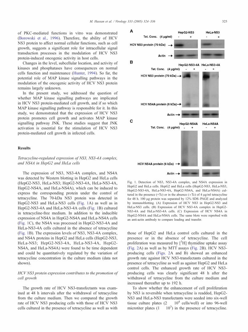
Fig. 1. Detection of NS3, NS3-4A complex, and NS4A expression in
HepG2 and HeLa cells. HepG2 and HeLa cells (HepG2-NS3, HeLa-NS3,
HepG2-NS3-4A, HeLa-NS3-4A, HepG2-NS4A, and HeLa-NS4A) cul-
tured in the presence (+Tc) or in the absence (�Tc) of 4 Ag/ml tetracycline
for 48 h. 100 Ag protein was separated by 12% SDS–PAGE and analyzed
by immunoblotting. (A) Expression of HCV NS3 in HepG2-NS3 and
HeLa-NS3 cells. (B) Expression of HCV NS3-4A complex in HepG2-
NS3-4A and HeLa-NS3-4A cells. (C) Expression of HCV NS4A in
HepG2-NS4A and HeLa-NS4A cells. The same blots were reprobed with
an anti-actin antibody to compare loading and transfer.
Results
Tetracycline-regulated expression of NS3, NS3-4A complex,
and NS4A in HepG2 and HeLa cells
The expression of NS3, NS3-4A complex, and NS4A
was detected by Western blotting in HepG2 and HeLa cells
(HepG2-NS3, HeLa-NS3; HepG2-NS3-4A, HeLa-NS3-4A,
HepG2-NS4A, and HeLa-NS4A), which can be induced to
express the corresponding protein under the control of
tetracycline. The 70-kDa NS3 protein was detected in
HepG2-NS3 and HeLa-NS3 cells (Fig. 1A) as well as in
HepG2-NS3-4A and HeLa-NS3-4A cells (Fig. 1B) cultured
in tetracycline-free medium. In addition to the inducible
expression of NS4A in HepG2-NS4A and HeLa-NS4A cells
(Fig. 1C), the NS4A was processed in HepG2-NS3-4A and
HeLa-NS3-4A cells cultured in the absence of tetracycline
(Fig. 1B). The expression levels of NS3, NS3-4A complex,
and NS4A proteins in HepG2 and HeLa cells (HepG2-NS3,
HeLa-NS3; HepG2-NS3-4A, HeLa-NS3-4A, HepG2-
NS4A, and HeLa-NS4A) were found to be time dependent
and could be quantitatively regulated by the variation of
tetracycline concentration in the culture medium (data not
shown).
HCV NS3 protein expression contributes to the promotion of
cell growth
The growth rate of HCV NS3-transfectants was exam-
ined at 48 h intervals after the withdrawal of tetracycline
from the culture medium. Then we compared the growth
rate of HCV NS3 producing cells with those of HCV NS3
cells cultured in the presence of tetracycline as well as with
those of HepG2 and HeLa control cells cultured in the
presence or in the absence of tetracycline. The cell
proliferation was measured by [3H] thymidine uptake assay
(Fig. 2A) as well as by MTT assays (Fig. 2B). HCV NS3-
producing cells (Figs. 2A and B) showed an enhanced
growth rate against HCV NS3-transfectants cultured in the
presence of tetracycline as well as against HepG2 and HeLa
control cells. The enhanced growth rate of HCV NS3-
producing cells was clearly significant 48 h after the
withdrawal of tetracycline from the culture medium and
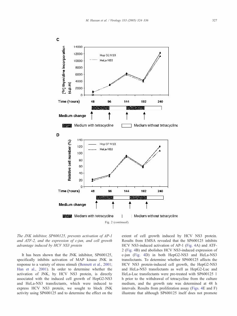
increased thereafter up to 192 h.
To show whether the enhancement of cell proliferation
by NS3 is reversible when tetracycline is readded, HepG2-
NS3 and HeLa-NS3 transfectants were seeded into six-well
tissue culture plates (2 � 104 cells/well) or into 96-well
microtiter plates (1 � 103) in the presence of tetracycline.
M. Hassan et al. / Virology 333 (2005) 324–336326
The medium was changed at 48 h intervals up to 192 h.
Subsequently, the growth rate was determined using [3H]
uptake assay (Fig. 2C) and MTT assay (Fig. 2D) at 48 h
intervals, when tetracycline was removed or readded to the
medium. Based on the obtained results we could demon-
strate that the enhancement of cell proliferation by NS3 is
reversible when tetracycline is readded to the culture
medium.
HCV NS3 protein induces the activation of JNK, but not p38
or ERK pathways
The ability of HCV NS3 protein to activate intracellular
MAP kinase pathways was examined in both HepG2-NS3
and HeLa-NS3 transfectants, which were induced to express
HCV NS3 protein for 48 h. Western blot analysis (Fig. 3)
Fig. 2. Effect of HCV NS3 protein on cell growth. HepG2-NS3 and HeLa-NS3 tra
the indicated time points. The proliferation rate was assessed by [3H] thymidine up
NS3 is reversible up on re-addition of tetracycline to the culture medium. The grow
indicated time points using [3H] thymidine uptake assay (C) or MTT assay (D).
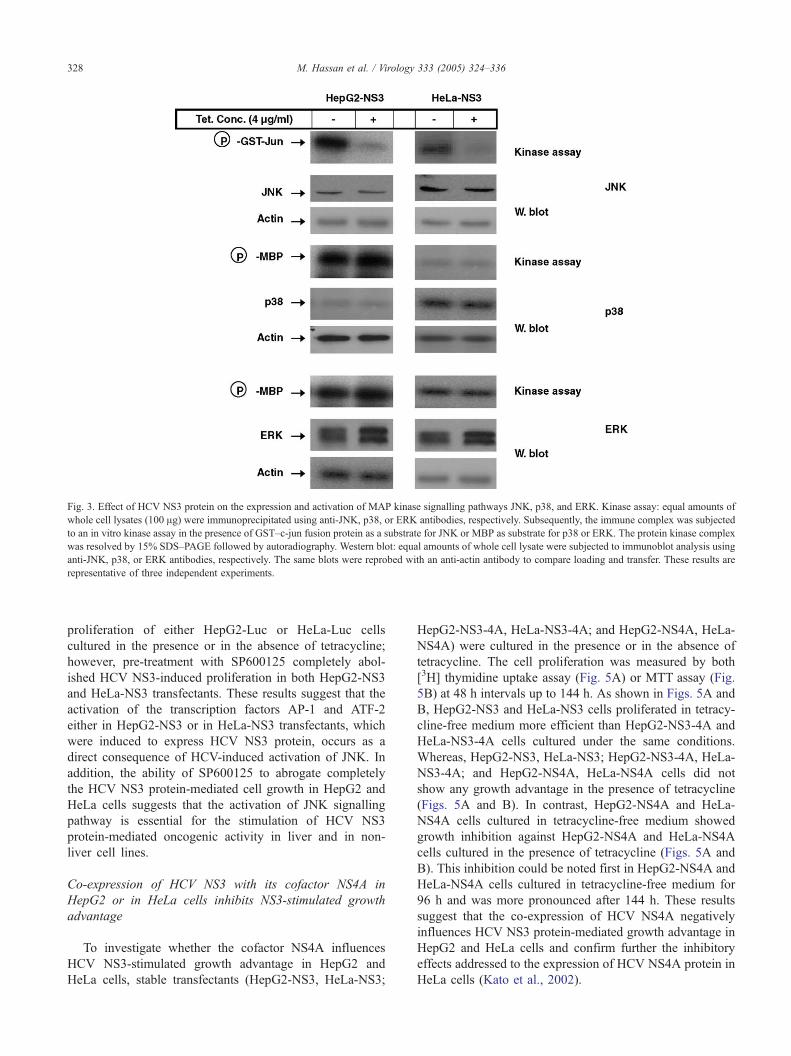
demonstrated that the expression of HCV NS3 protein does
not alter the expression of JNK, p38, or ERK either in
HepG2-NS3 or in HeLa-NS3 transfectants. In contrast, in
vitro kinase assay (Fig. 3) demonstrated that the expression
of HCV NS3 protein either in HepG2 or in HeLa cells
enhances the activation of JNK, but not those of p38 or
ERK. Although the basal expression level of p38 in HeLa-
NS3 cells was apparently higher than those in HepG2-NS3
cells, however, the basal level of p38 activity was more
pronounced in HepG2-NS3 cells. These observations
suggest that the variation at the basal expression level has
no significant effect on the corresponding basal activity.
Taken together, these obtained data suggest that the HCV
NS3-mediated activation of JNK may be important for the
stimulation of HCV NS3 protein-mediated cell growth in
liver and in non-liver cell lines.
nsfectants were cultured in the presence or in the absence of tetracycline for
take assay (A) and MTT assay (B). The enhancement of cell proliferation by
th rate of both HepG2-NS3 and HeLa-NS3 transfectants was assessed at the
The results are means F SE of three independent experiments.
Fig. 2 (continued).
M. Hassan et al. / Virology 333 (2005) 324–336 327
The JNK inhibitor, SP600125, prevents activation of AP-1
and ATF-2, and the expression of c-jun, and cell growth
advantage induced by HCV NS3 protein
It has been shown that the JNK inhibitor, SP600125,
specifically inhibits activation of MAP kinase JNK in
response to a variety of stress stimuli (Bennett et al., 2001;
Han et al., 2001). In order to determine whether the
activation of JNK, by HCV NS3 protein, is directly
associated with the induced cell growth of HepG2-NS3
and HeLa-NS3 transfectants, which were induced to
express HCV NS3 protein, we sought to block JNK
activity using SP600125 and to determine the effect on the
extent of cell growth induced by HCV NS3 protein.
Results from EMSA revealed that the SP600125 inhibits
HCV NS3-induced activation of AP-1 (Fig. 4A) and ATF-
2 (Fig. 4B) and abolishes HCV NS3-induced expression of
c-jun (Fig. 4D) in both HepG2-NS3 and HeLa-NS3
transfectants. To determine whether SP600125 affects the
HCV NS3 protein-induced cell growth, the HepG2-NS3
and HeLa-NS3 transfectants as well as HepG2-Luc and
HeLa-Luc transfectants were pre-treated with SP600125 1
h prior to the withdrawal of tetracycline from the culture
medium, and the growth rate was determined at 48 h
intervals. Results from proliferation assay (Figs. 4E and F)
illustrate that although SP600125 itself does not promote
Fig. 3. Effect of HCV NS3 protein on the expression and activation of MAP kinase signalling pathways JNK, p38, and ERK. Kinase assay: equal amounts of
whole cell lysates (100 Ag) were immunoprecipitated using anti-JNK, p38, or ERK antibodies, respectively. Subsequently, the immune complex was subjected
to an in vitro kinase assay in the presence of GST–c-jun fusion protein as a substrate for JNK or MBP as substrate for p38 or ERK. The protein kinase complex
was resolved by 15% SDS–PAGE followed by autoradiography. Western blot: equal amounts of whole cell lysate were subjected to immunoblot analysis using
anti-JNK, p38, or ERK antibodies, respectively. The same blots were reprobed with an anti-actin antibody to compare loading and transfer. These results are
representative of three independent experiments.
M. Hassan et al. / Virology 333 (2005) 324–336328
proliferation of either HepG2-Luc or HeLa-Luc cells
cultured in the presence or in the absence of tetracycline;
however, pre-treatment with SP600125 completely abol-
ished HCV NS3-induced proliferation in both HepG2-NS3
and HeLa-NS3 transfectants. These results suggest that the
activation of the transcription factors AP-1 and ATF-2
either in HepG2-NS3 or in HeLa-NS3 transfectants, which
were induced to express HCV NS3 protein, occurs as a
direct consequence of HCV-induced activation of JNK. In
addition, the ability of SP600125 to abrogate completely
the HCV NS3 protein-mediated cell growth in HepG2 and
HeLa cells suggests that the activation of JNK signalling
pathway is essential for the stimulation of HCV NS3
protein-mediated oncogenic activity in liver and in non-
liver cell lines.
Co-expression of HCV NS3 with its cofactor NS4A in
HepG2 or in HeLa cells inhibits NS3-stimulated growth
advantage
To investigate whether the cofactor NS4A influences
HCV NS3-stimulated growth advantage in HepG2 and
HeLa cells, stable transfectants (HepG2-NS3, HeLa-NS3;
HepG2-NS3-4A, HeLa-NS3-4A; and HepG2-NS4A, HeLa-
NS4A) were cultured in the presence or in the absence of
tetracycline. The cell proliferation was measured by both
[3H] thymidine uptake assay (Fig. 5A) or MTT assay (Fig.
5B) at 48 h intervals up to 144 h. As shown in Figs. 5A and
B, HepG2-NS3 and HeLa-NS3 cells proliferated in tetracy-
cline-free medium more efficient than HepG2-NS3-4A and
HeLa-NS3-4A cells cultured under the same conditions.
Whereas, HepG2-NS3, HeLa-NS3; HepG2-NS3-4A, HeLa-
NS3-4A; and HepG2-NS4A, HeLa-NS4A cells did not
show any growth advantage in the presence of tetracycline
(Figs. 5A and B). In contrast, HepG2-NS4A and HeLa-
NS4A cells cultured in tetracycline-free medium showed
growth inhibition against HepG2-NS4A and HeLa-NS4A
cells cultured in the presence of tetracycline (Figs. 5A and
B). This inhibition could be noted first in HepG2-NS4A and
HeLa-NS4A cells cultured in tetracycline-free medium for
96 h and was more pronounced after 144 h. These results
suggest that the co-expression of HCV NS4A negatively
influences HCV NS3 protein-mediated growth advantage in
HepG2 and HeLa cells and confirm further the inhibitory
effects addressed to the expression of HCV NS4A protein in
HeLa cells (Kato et al., 2002).
M. Hassan et al. / Virology 333 (2005) 324–336 329
Co-expression of HCV NS3 with its cofactor NS4A in
HepG2 or in HeLa cells does not influence either
NS3-mediated JNK activation, or the basal
activity of p38 or ERK
To determine whether the co-expression of HCV NS4A
protein influences HCV NS3 protein-mediated activation
or affects the basal activities of p38 or ERK, HepG2 and
HeLa transfectants (HepG2-NS3, HeLa-NS3; HepG2-
NS3-4A, HeLa-NS3-4A; and HepG2-NS4A; HeLa-
NS4A) were cultured in the presence or in the absence
of tetracycline. 48 h later, whole cell extracts were
prepared and in vitro kinase assay was performed. Results
from in vitro kinase assay (Fig. 6) showed that the
expression of HCV NS4A protein itself does not
influence the basal activity of the MAP kinases JNK,
p38, or ERK either in HepG2 or in HeLa cells. In
contrast, the induction of JNK activation becomes induced
in both HepG2-NS3-4A and HeLa-NS3-4A cells cultured
in tetracycline-free medium and was quite similar to those
that noted in HepG2-NS3 or in HeLa-NS3 cells cultured
under the same conditions (Fig. 6). These results indicate
that the co-expression of HCV NS4A protein does not
influence HCV NS3-mediated effects on JNK activation
in HepG2 or in HeLa cells.
Discussion
The results presented herein provide an insight into the
possible mechanisms by which the HCV NS3 protein
mediates its oncogenic activity in infected cells and
support earlier findings suggesting the potential role of
HCV NS3 protein in the promotion of cell growth in
tumor and normal cells (Kwun et al., 2001; Sakamuro et
al., 1995; Zemel et al., 2001). In this work, the HCV
NS3 protein was found to induce cell growth and to
activate the JNK signalling pathway, but not ERK or p38
kinases pathways in liver or in non-liver cell lines.
Although the confirmation of the proliferative activity of
HCV NS3 protein in liver and in non-liver cell lines,
however, there are contradicting data regarding the effect
of HCV NS3 protein on cell growth. Two studies
demonstrated that tumor or non-tumor cells stably
expressing HCV NS3 protein grow faster than their
parental cell lines (Kwun et al., 2001; Zemel et al.,
2001), whereas one study (Siavoshian et al., 2004)
showed growth inhibition of cells transiently transfected
with HCV NS3 protein. In agreement with Kwun et al.
(2001), Zemel et al. (2001), we confirmed the prolifer-
ative activity of HCV NS3 protein in our cell culture
model. Thus, we could demonstrate that the controlled
expression of HCV NS3 protein, in liver or in non-liver
cell lines, stimulates cell growth. In addition, we could
show that the HCV NS3-stimulated cell growth is JNK-
dependent activation.
Although the three MAP kinase signalling pathways
share structural similarities, the outcome of the activation is
quite different. The role of MAP kinase signalling pathways
ERK, p38, and JNK in the regulation of cell proliferation is
well documented in liver and in non-liver cell lines (Auer et
al., 1998; Bost et al., 1999; Maher, 2001; Ogata et al., 2003;
Schwabe et al., 2003; Todisco et al., 1997; Yang et al.,
1999).
Since the HCV NS3 protein does not appear to affect p38
or ERK pathways, our data suggest that the HCV NS3
protein must act at a step at which the MAP kinases p38 and
ERK do not converge in signal transduction pathway
leading to JNK activation. Although current studies
suggesting that the HCV NS3 protein may be directly
involved in hepatocarcinogenesis (Ogata et al., 2003; Yang
et al., 1999) by disturbing the regulation of cell prolifer-
ation, however, the mechanisms of the carcinogenesis are
still puzzle. In fact, various oncogenic products are related
to functional abnormalities of intracellular signal trans-
duction pathways, which have been proved to be one of the
proliferative mechanisms of cancer cells (Chang et al., 2003;
Takihara et al., 2000). In several studies, the regulation of
different signal transduction processes by HCV proteins has
been demonstrated (Hassan et al., 2004; Schulze zur Wiesch
et al., 2003; Erhardt et al., 2002, Yang et al., 2002).
Constitutive activation of Ras/Raf/MAP kinase pathway
is important for the transformation of mammalian cells
(Hamad et al., 2002; Pinkas and Leder, 2002). Furthermore,
the development of HCC and its progression have been
shown be associated with the activation of Ras/Raf/MAP
kinase pathway in both human and rodents (Ostrowski et al.,
2000).
Our finding that HCV NS3 protein induces the activation
of JNK signalling pathway together with the promotion of
cell growth suggests a potential role for this pathway in the
modulation of HCV NS3 protein-mediated oncogenic
activity in host cells. Therefore, the ability of JNK inhibitor
to abrogate HCV NS3-induced cell growth suggests that
JNK activation is an essential component in the pathway by
which HCV NS3-stimulates cell growth.
The activation of several transcription factors, such as
AP-1 and ATF-2, in response to JNK activation, has been
well documented (Ahmed et al., 2003; Botteron and
Dobbelaere, 1998; Caelles et al., 1997; Leppa et al., 2001;
Zoumpourlis et al., 2000). In many studies, the phosphor-
ylation of c-jun by JNK has been reported to be required for
the activation of AP-1, induction of c-jun expression, and
modulation of cellular transformation (Angel et al., 1988;
Behrens et al., 2000; Kennedy et al., 2003; Lin et al., 2003;
van Dam et al., 1995). Therefore, the increase of the basal
activities of AP-1 and ATF-2 as well as the basal expression
of c-jun suggests that the HCV NS3 protein promotes
cellular proliferation for maintenance of replication and
survival (Guo et al., 1999; Lenczowski et al., 1997). In this
work, we demonstrated for the first time the activation of
JNK and its substrates AP-1 and ATF-2, by the expression
M. Hassan et al. / Virology 333 (2005) 324–336330
of HCV NS3 protein, in liver and in non-liver cell lines and
delineated further the importance of the JNK signalling
pathway for modulation of HCV NS3 protein-mediated cell
growth. Therefore, based on our findings and in agreement
with relevant studies (He et al., 2003; Kwun et al., 2001;
Sakamuro et al., 1995; Zemel et al., 2001), we suggest a
potential role for the HCV NS3 protein in the development
of HCV-related HCC. However, further studies with other
mammalian cell lines, especially primary hepatocytes,
should further clarify the importance of these results.
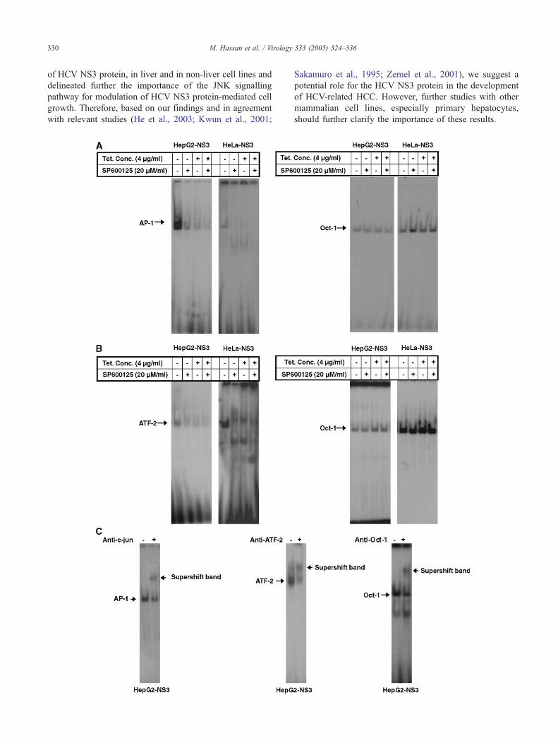
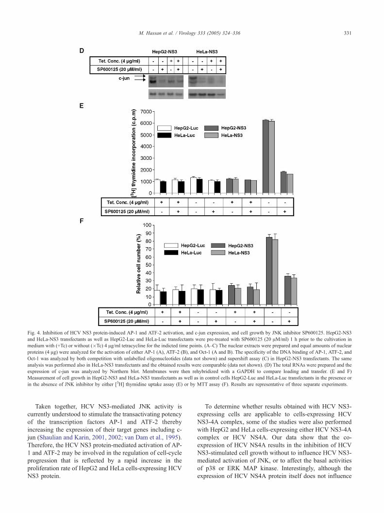
Fig. 4. Inhibition of HCV NS3 protein-induced AP-1 and ATF-2 activation, and c-jun expression, and cell growth by JNK inhibitor SP600125. HepG2-NS3
and HeLa-NS3 transfectants as well as HepG2-Luc and HeLa-Luc transfectants were pre-treated with SP600125 (20 AM/ml) 1 h prior to the cultivation in
medium with (+Tc) or without (�Tc) 4 Ag/ml tetracycline for the indicted time points. (A–C) The nuclear extracts were prepared and equal amounts of nuclear
proteins (4 Ag) were analyzed for the activation of either AP-1 (A), ATF-2 (B), and Oct-1 (A and B). The specificity of the DNA binding of AP-1, ATF-2, and
Oct-1 was analyzed by both competition with unlabelled oligonucleotides (data not shown) and supershift assay (C) in HepG2-NS3 transfectants. The same
analysis was performed also in HeLa-NS3 transfectants and the obtained results were comparable (data not shown). (D) The total RNAs were prepared and the
expression of c-jun was analyzed by Northern blot. Membranes were then rehybridized with a GAPDH to compare loading and transfer. (E and F)
Measurement of cell growth in HepG2-NS3 and HeLa-NS3 transfectants as well as in control cells HepG2-Luc and HeLa-Luc transfectants in the presence or
in the absence of JNK inhibitor by either [3H] thymidine uptake assay (E) or by MTT assay (F). Results are representative of three separate experiments.
M. Hassan et al. / Virology 333 (2005) 324–336 331
Taken together, HCV NS3-mediated JNK activity is
currently understood to stimulate the transactivating potency
of the transcription factors AP-1 and ATF-2 thereby
increasing the expression of their target genes including c-
jun (Shaulian and Karin, 2001, 2002; van Dam et al., 1995).
Therefore, the HCV NS3 protein-mediated activation of AP-
1 and ATF-2 may be involved in the regulation of cell-cycle
progression that is reflected by a rapid increase in the
proliferation rate of HepG2 and HeLa cells-expressing HCV
NS3 protein.
To determine whether results obtained with HCV NS3-
expressing cells are applicable to cells-expressing HCV
NS3-4A complex, some of the studies were also performed
with HepG2 and HeLa cells-expressing either HCV NS3-4A
complex or HCV NS4A. Our data show that the co-
expression of HCV NS4A results in the inhibition of HCV
NS3-stimulated cell growth without to influence HCV NS3-
mediated activation of JNK, or to affect the basal activities
of p38 or ERK MAP kinase. Interestingly, although the
expression of HCV NS4A protein itself does not influence
Fig. 5. Inhibition of HCV NS3-mediated cell growth by cofactor NS4A. HepG2 and HeLa cells (HepG2-NS3, HeLa-NS3, HepG2-NS3-4A, HeLa-NS3-4A,
HepG2-NS4A, and HeLa-NS4A) were cultured in the presence or in the absence of tetracycline for 48 h, and [3H] thymidine uptake assay (A) and MTT assay
(B) were performed. The results are means F SE of three independent experiments.
Fig. 6. Effect of NS4A co-expression on JNK, p38, and ERK MAP kinase activation. HepG2 and HeLa transfectants (HepG2-NS3, HeLa-NS3, HepG2-NS3-
4A, HeLa-NS3-4A, HepG2-NS4A, and HeLa-NS4A) were cultured in the presence or in the absence of tetracycline for 48 h. Cells were washed, lysed, and the
JNK, p38, and ERK were immunoprecipitated from extracts using anti-JNK, p38, or ERK antibodies, respectively. JNK activity was measured by using the
immune complex in a kinase assay with GST–c-jun as the substrate, whereas the activities of p38 or ERK were measured by using the immune complex in a
kinase assay with MBP as substrate. These results are representative of three independent experiments.
M. Hassan et al. / Virology 333 (2005) 324–336332
M. Hassan et al. / Virology 333 (2005) 324–336 333
the basal activity of the MAP kinases JNK, p38, or ERK,
the expression of HCV NS4A was found to inhibit cell
growth. Inhibition of cell proliferation by the expression of
NS4A has been also reported (Kato et al., 2002); however,
the molecular mechanisms regulating such inhibition still
remain to be determined.
In summary, our data demonstrate for the first time the
activation of JNK signalling pathway by HCV NS3 protein
and confirm further the oncogenic activity of HCV NS3
protein in liver and in non-liver cell lines. In an attempt to
further delineate part of the mechanisms whereby HCV NS3
protein mediates cell growth, we conclude that the HCV
NS3 protein may be involved in hepatocarcinogenesis
through the activation of JNK/AP-1 signalling pathway.