ACTA UNIVERSITATIS UPSALIENSIS UPPSALA 2006 Digital Comprehensive Summaries of Uppsala Dissertations from the Faculty of Medicine 174 Acute Abdominal Pain HELENA LAURELL ISSN 1651-6206 ISBN 91-554-6664-8 urn:nbn:se:uu:diva-7161
Transcript
ACTAUNIVERSITATISUPSALIENSISUPPSALA2006
Digital Comprehensive Summaries of Uppsala Dissertationsfrom the Faculty of Medicine 174
The diagnostic problem of today Has greatly changed – the change has come to stay; We all have to confess, though with a sigh, On complicated tests we much rely And use too little hand and ear and eye.
Sir Zachary Cope (1881-1974) Abdomen in Rhyme, 1947
List of papers
This thesis is based on the following papers, which are referred to in the text by the Roman numerals given below (I-IV):
I. Acute abdominal pain in a defined population – the impact of the formal competence of the physician and the use of a structured schedule for investigation. Laurell H, Hansson L-E & Gunnarsson U, submitted
II. Diagnostic pitfalls and accuracy of diagnosis in acute ab-dominal pain. Laurell H, Hansson L-E & Gunnarsson U, Scan J of Gastro-enterology 2006;41:1-6
III. Acute abdominal pain among elderly patients. Laurell H, Hansson L-E & Gunnarsson U,
Gerontology 2006;52(6):339-344
IV. Manifestations of acute appendicitis – a prospective study on acute abdominal pain. Laurell H, Hansson L-E & Gunnarsson U, manuscript
Reprints were made with the permission of the publishers.
Contents
Introduction...................................................................................................11Definitions ...........................................................................................11Historical remarks................................................................................11Modern research on AAP ....................................................................16The physiology of abdominal pain ......................................................18Epidemiology.......................................................................................19The process of decision-making ..........................................................20Diagnostic aids ....................................................................................21
Aims of the study ..........................................................................................23
Patients and methods.....................................................................................24Physicians ............................................................................................24Inclusion criteria ..................................................................................24Computer registration and validation of data ......................................24Baseline study......................................................................................25Diagnoses.............................................................................................26Follow-up.............................................................................................26Patients included..................................................................................26Flow charts ..........................................................................................28Statistics...............................................................................................30Ethics ...................................................................................................30The structured schedule for investigation............................................31
Results...........................................................................................................35Paper I..................................................................................................35Paper II ................................................................................................38Paper III ...............................................................................................42Paper IV...............................................................................................46
General discussion ........................................................................................51Measures of diagnostic performance of patients with AAP ................52Duration of hospital stay......................................................................54Use of resources...................................................................................54Computer-aided diagnosis and scoring systems ..................................55Gender perspectives.............................................................................56
Clinical symptoms, signs and laboratory examinations.......................57Shortcomings and strengths.................................................................57Future perspectives ..............................................................................58
AAP Acute abdominal pain CI Confidence interval CRP C-reactive protein CT Computed tomography ED Emergency department LR+ Positive likelihood ratio LR- Negative likelihood ratio MRI Magnetic resonance imaging NPV Negative predictive value NSAP Non-specific abdominal pain OMGE Organisation Mondiale de Gastroenterologie OR Odds ratio PPV Positive predictive value UK United Kingdom of England VAS Visual Analogue Scale
11
Introduction
Acute abdominal pain (AAP) accounts for a substantial proportion of pa-tients arriving at a surgical emergency department (de Dombal 1979, Irvin 1989, Powers 1995). As AAP may be caused by both life threatening dis-eases and conditions that are spontaneously resolved, a correct diagnosis is of importance for the prognosis of the patient. AAP also effects health econ-omy as many patients are hospitalised for what eventually turns out to be self-resolving pains, i.e. non-specific abdominal pain (NSAP).
Definitions Acute abdominal pain (AAP) is in this context defined as abdominal pain of up to seven days duration and not caused by trauma. In the literature the duration of pain-criterion is sometimes defined to be ten days (de Dombal 1972). According to the OMGE survey AAP is stated as pain with less than one week’s duration (de Dombal 1979). The latter definition was used for the present study.
Non-specific abdominal pain (NSAP) includes all causes of self-limiting pain for which no surgical, gynaecological or other medical diagnosis is made. Usually no further investigation and no treatment is required. In the literature sometimes constipation, mesenteric lymphadenitis, dysmenor-rhoea, gastroenteritis or dyspepsia, as well as patients undergoing negative laparotomy (i.e. no other surgical or other medical disease is determined and no other surgical procedure is carried out), are included in this category (de Dombal 1979). In the present papers constipation, gastroenteritis, dyspepsia and NSAP are considered to be separate entities.
Historical remarks It is likely that AAP has affected man ever since pre-historical times. How-ever, it is only within the last two hundred years that we have had more or less accurate knowledge of the intra-abdominal diseases that causes the AAP. The main reason for this comparatively late development of medical knowledge was that the only method of obtaining accurate information, by post-mortem examination of the intra-abdominal organs, was either forbid-den or disliked by the medical authorities. Moreover, surgical operations
12
upon the abdomen were not performed until the beginning of the 19th cen-tury.
Hippocrates (460-370 BC), Galen (129-199 AD) and their successors ob-served the symptoms of their patients very carefully, but they were seriously handicapped by their ignorance of the exact state, within the abdomen, cor-responding to those symptoms. They also lacked the ability to ascertain if their treatment affected the disease. We now know that many cases of at-tacks of abdominal pain are self-limiting and resolves spontaneously, and thus also ineffective treatments could seem successful during these ancient times. They recognised that acute colicky might get well or might pass on to more serious obstruction of the bowel (ileus); they also knew that deep in-flammation could result in the formation of an abscess that might burst spon-taneously or could be opened by the knife. However, every other serious abdominal condition, which was fatal, was thought to be due to an obstruc-tion of the bowels, and was called ileus. The symptoms of ileus are well described by Hippocrates: “In ileus the belly becomes hard, there are no motions; the whole abdomen is painful, there are fever and thirst and some-times the patient is so tormented that he vomits bile …. It is an acute and dangerous disease.” (Cope 1965).
It is in the writings of Celsus (42 BC – 37 AD) we find the first descrip-tion of a notable clinical sign in acute abdominal disease; resistance on pres-sure indicating underlying inflammation or peritonitis. For more than a thousand years after the time of Galen no advances in the diagnosis or treatment of acute abdominal conditions took place. However, many monasteries included also education and hospital care. Notes from monks and nuns reveal that women mostly were seeking medical care for mental disorders or infertility, men for wounds and accidents and children for fever diseases and diarrhoea. During the 13th century the first universities were established. To achieve the medical authorization from Salerno also one year of practise with an experienced physician was demanded. The first official medical authorization was granted in 1140 in Sicily.
Thomas Sydenham (1624-1689) found little practical value of the scien-tific knowledge of medicine for the treatment of the sick, he focussed on clinical observations of patients and his greatest contribution was the many descriptions of the natural history of diseases (Nilsson 1998). It was first after the publication of Bonetus’ Sepulchretum in 1679 and the remarkable wealth of pathological material described in Giovanni Morgagni’s (1682-1771) monumental work De sedibus et causis morborum (published in 1761) that a decisive change in medical views on the causation of intra-abdominal disorders took place. William Cullen (1710-1790), in his work First Lines of the Practice of Physic (1776), used the name “peritonitis” for a condition involving the lining membrane of the abdominal cavity and its extensions to the viscera.
13
In 1806, Chistopher Pemberton (1765-1822) published the book A Practi-cal Treatise on Various Diseases of the Abdominal Viscera, which is consid-ered a milestone in the clinical diagnosis of acute abdominal diseases. Pemberton emphasised the importance of meticulous clinical observations and the importance of careful first-hand observations, recorded at the time. He also described the differential diagnostics between peritonitis and colic pain (Cope 1965). Leopold Auenbrügger (1722-1809) in Vienna described the percussion technique: he might have got the idea from a brewery where they knocked on the barrels to judge how much beer was inside (Nilsson 1998). Shaking the patient (succussion), to prove fluid in the chest, was an ancient technique that was revived in the early 1800s, but was a quite violent method and unpleasant for the patient. In the 19th century, ”topographical diagnostics” was developed for determining the size of heart and liver by palpation and percussion (Johannisson 1997). When René Laënnec (1781-1826) invented the stethoscope in 1816, the technique of auscultation was immediately accepted. During this time post-mortem examinations began to be made more frequently, and there was a rapid improvement in diagnosis. From the middle of the 19th to the middle of the 20th century, most of the pathological conditions, which give rise to acute abdominal symptoms, were recognised and their symptoms gradually differentiated.
Josef Lister (1827-1912) in London realized the importance of Louis Pas-teur’s (1822-1895) work on bacteria and introduced the “antiseptic surgery” in 1867, by spraying carbolic acid in the surgery room. The reduction of postoperative infections and sepsis was remarkable, and the aseptic method was soon practised in all the countries in Europe and the USA. On 16 Octo-ber 1846 (”Ether Day”) at Massachusetts General Hospital, the dentist Wil-liam Morton (1819-1868) administrated ether to a patient of the surgeon, John Collins Warren (1778-1856) and the operation was painless. The possi-bility of performing painless surgery and the increased postoperative sur-vival, due to the aseptic method, markedly increased the possibilities of sur-gical treatment of acute abdominal conditions during the last decades of the 19th century (Rhodes 1985).
Wilhelm Conrad Röntgen’s (1845-1923) detection of a “new kind of rays” in 1895 provided the possibility of imaging bones inside the body. The technique spread immediately and in 1896 the first radiographs were per-formed in Stockholm and Uppsala. Bismuth and barium were soon shown to be impermeable to the x-rays and were used as contrast in examinations of the stomach, small and large intestines: gastric ulcers were diagnosed by this method. In 1929 the professor of radiology in Uppsala, Hugo Laurell (1884-1959) performed the first reposition of invaginated intestine by barium en-ema, which is probably the first radiological therapeutic method. His scien-tific work became the basis of radiological diagnostic methods in acute ab-dominal pain (Laurell 1930). The Penicillium was discovered in 1928 by Sir Alexander Fleming (1881-1955), but it was not in clinical use until 1941.
14
One clinical consequence of the introduction of antibiotics was the possibil-ity to wait and observe the patient and not perform an appendectomy as soon appendicitis was suspected (Ekelund 2005). In 1970, computed tomography (CT) were introduced into clinical practice, along with magnetic resonance imaging (MRI) in 1980, both made important contributions to the diagnostic performance of abdominal diseases.
The mean hospitalisation time on surgical wards in Sweden was 14.8 days in 1950 compared to 5.9 days in 1990. The number of beds in surgical care was 8774 at 63 surgical departments in 2000 (The Swedish Federation of County Councils 2003). The total number of beds for institutional care (ex-cept psychiatry and geriatrics) in Sweden was 35,726 in 1950, 49,600 in 1970, 35,503 in 1990 (SBU 1995) and 26,076 in 2000 (The Swedish Federa-tion of County Council 2003). That means 5.1, 6.1 and 3.4 beds per 100,000 inhabitants, 1950-1990 respectively (SBU 1995).
The growth of knowledge of appendicitis Lorenz Heister (1683-1758) was the first to recognise that the vermiform appendix might be the site of acute primary inflammation (Ekelund 2005). Claudius Amyand (1681-1740), physician at the English court, is said to have performed the first recorded successful appendectomy in 1736, on an 11-year old boy with a perforated appendix within an inguinal hernia sac. (Amyand 1736). During the 19th century pain in the right iliac fossa was recognized as a clinical entity. However, it was first in 1886 that Reginald Fitz (1843-1913) proved that the great majority of acute inflammatory condi-tions around the caecum had their origin in the vermiform appendix. As a physician, he gave a description of the symptoms whereby the correct diag-nosis could be made, and as a pathologist he described the varying degrees of inflammation in the appendix, the degrees of local and general peritonitis, and of abscess formation. In 1889, Charles Mac Burney (1845-1913) in New York described how an inflamed appendix could be reached and removed with small risk via a short incision. The appendectomies were lifesaving and were soon widely spread (Rhodes 1985). The mortality in appendicitis has been estimated to over 40% before the introduction of appendectomy; then decreased to around 10% in the 1910s; a few per cent in the 1940s (after the introduction of antibiotics) and a few per thousand today (Blomqvist 2001, Lally 2004). In Sweden, the first appendectomy was performed in 1889 by Karl Gustaf Lennander, who also introduced “aseptic surgery” to the country (Petrén 1932). Lennander’s successor as Head of the Surgical Department in Uppsala, Gunnar Nyström (1877-1964), wrote his thesis in 1907 on 312 patients surgically treated for non-perforated appendicitis in Uppsala during 1891-1905. Only one patient died postoperatively (of intestinal obstruction; Nyström 1907). There were 681 appendectomies performed in Sweden in 1901, increasing to 14,000 performed in 1921 (Nyström 1932).
15
Lorenz Heister (1683-1758) was the first to recognise that the vermiform appendix might be the site of acute primary inflammation. (Wikipedia: GNU Free Document.)
16
Modern research on AAP AAP can involve different medical specialities, including surgery, gynaecol-ogy, medicine and oncology. Although AAP is a common problem, early diagnosis is often difficult and this may lead to unnecessary investigation and hospitalisations. In the late 1950s, systems appeared that aided the phy-sician in making medical decisions and were based on methods that used decision trees or truth tables. Later, systems based on statistical methods appeared for different domains of clinical care, for example interpretations of electrocardiograms or diagnoses in internal medicine.
In 1970, Professor F Tim de Dombal introduced a computer-aided data system in Leeds, UK, which dealt with 13 different diagnoses for AAP. Based on patient data and the results of laboratory tests, this system pro-posed a diagnosis. In 1972, de Dombal reported a real-time comparison of physician and computer-aided diagnosis in a series of 304 prospectively collected patients suffering from AAP during 1971. The overall diagnostic accuracy of the computer system was 91.8%, and was significantly higher than the most senior clinicians, who presented an accuracy of 79.6% (de Dombal 1972). Such high diagnostic accuracy of AAP have, however, never been reproduced.
In 1986, a study was published (Adams 1986) describing computer-aided diagnosis for patients with AAP in eight UK hospitals with over 250 partici-pating doctors and 12,662 patients. Diagnostic accuracy rose from 45.6% during the baseline period to 65.3% during the period when the computer system to aid diagnosis based on a Bayesian analysis was tested. Other cen-tres outside England, that have followed the idea of prospective investigation of patients admitted with AAP include; Rispebjerg Hospital in Copenhagen, Denmark (Bjerregaard 1976); Rogaland Central Hospital, Stavanger, Nor-way (Körner 1998); Akershus Central Hospital, University of Oslo, Norway (Staniland 1980); University Central Hospital, Tampere, Finland (Ikonen 1983); Nacka Hospital, Sweden (Fenyö 2000); University Hospital, Zürich, Switzerland (Simmen 1991); Heinrich-Heine-University, Düsseldorf, Ger-many (Ohmann 1995); Tucson Veteran Administration Medical Centre, Ari-zona, USA (Orient 1986); University of Virginia Hospital, USA (Powers 1995); and Mora Hospital, Sweden (present study).
The availability of an increased number of more specified blood tests and the possibility of detailed imaging by CT and ultrasonography have had an important impact on clinical work in surgical departments. Controlled clini-cal trials have revealed that this technology improves diagnostic accuracy (John 1993, Rosen 2003). However, other studies (Flum 2005, Lee 2006) have not shown a decrease in misdiagnosis among women of reproductive age, patients older than 65 years or negative appendectomies during the last decades, despite the dramatically increased use of advanced diagnostic tech-nology. Andersson et al. (1999) found that the surgeon’s attitude to explora-
17
tion can influence the incidence of non-perforated appendicitis and conserva-tive management decreases the number of negative appendectomies (Andersson 1999). In the light of these contradictory findings, it is important to clarify the diagnostic accuracy of AAP in a population-based study, in-cluding a careful clinical examination at the emergency department. To achieve congruence in those clinical assessments, a detailed schedule of patient history and clinical symptoms and signs is required for each patient. Determining ways to define patient-categories that are more difficult to di-agnose safely, as well as identifying those diagnoses that are difficult to de-termine at an early stage, can be helpful to select patients for high-technology diagnostic tools. This is important not only for economical rea-sons, but also to reduce unnecessary exposure of the population to radiation.
Competence of the physician In the early 1990s, there were influences in Sweden claiming that the health care providers should not be allowed to use trainee physicians as on-calls at the emergency departments. The Swedish National Board of Health and Welfare (Socialstyrelsen) initiated a mapping of formal competence at the emergency departments in Sweden (Spri 1990). They determined that al-though the number of specialists in Sweden had increased by 30%, the por-tion of specialists attending at the emergency departments was the same from 1980 until 1990. The conclusion was that there are differences in the quality of medical assessment among the emergency departments in Sweden (Andrén-Sandberg 1991). A lively debate on medical competence took place in Läkartidningen (Journal for Swedish Physicians) and in 1991 a quality registration at the Department of General Surgery at Karlskrona Hospital was performed on all patients admitted to the emergency department during 3 months. No quality differences were revealed in the assessment, whether the attending physician was a specialist or a trainee physician of 1-3 years of experience (Sevonius 1991 and 1994). With the use of a detailed schedule, each physician would have the same amount of information when making the diagnosis after history taking and examining the patient. Thus, one ques-tion that can be posed is whether improved diagnostic accuracy is due to medical experience or to the amount of information achieved by a careful examination.
Another important factor, for which there is insufficient scientific know-ledge, is the precision of the preliminary diagnosis in relation to the patient’s time of arrival at the emergency department. The gender perspective is of special relevance, as females of reproductive ages are considered more diffi-cult to diagnose safely. These are all important issues in the planning of emergency care. All data on symptoms and clinical findings in patients with AAP, collected in a local database such as in the present study, is also a source for epidemiological considerations.
18
The physiology of abdominal pain Pain has been defined by The International Association for the Study of Pain as “an unpleasant sensory and emotional experience associated with oc-curred or potential tissue damage or described in terms of such a damage”. This definition emphasises the multiple aspects of pain. Pain stimulus may imply a physiological damage but is also a question of perception, sensibility and a psychological reaction (Mersky 1994).
Visceral pain Visceral pain differs from surface pain in several aspects. In general the vis-cera have sensory receptors for no other modalities of sensation except pain. Highly localised types of damage to the viscera, such as a sharp cut, rarely cause severe pain. On the other hand, diffuse stimulation of pain nerve-endings in a viscus, such as distension or spasm of a hollow viscus, stretch-ing of the ligaments or ischemia of visceral tissue, causes pain that can be severe. In cases of ischemia in visceral tissue, the formation of acidic meta-bolic products or tissue degenerative products, such as bradykinin and prote-olytic enzymes, causes pain by stimulating the pain nerve-endings. The leak-age of proteolytic acidic gastric juice through a ruptured gastric or duodenal ulcer causes widespread chemical stimulus to the visceral peritoneum and usually results in a severe pain.
Visceral pain originating in the thoracic and abdominal cavity is transmit-ted through type C-fibres in sensory nerves running through the sympathetic nervous system. These fibres only transmit a chronic-burning-aching type of pain. Interval pain is the result of rhythmic contraction of smooth muscle each time a peristaltic wave travels along the spastic gut, the obstructed ureter or the gallbladder. Mechanical stimulation of nerve endings or ische-mia due to diminished blood flow during cramp might cause this type of pain. Over-distension of a hollow viscus can collapse the blood vessels en-circling the viscus or those in the intestinal wall, thus promoting an ischemic pain. The liver parenchyma is almost insensitive to pain, however the liver capsule is extremely sensitive to both trauma and stretch (such as in hepati-tis). The bile ducts are sensitive to distension, which causes severe pain (Guyton 1986). Secondary effects of visceral pain include perspiration, nau-sea, vomiting, anxiety and paleness.
Parietal pain The parietal peritoneum is supplied with extensive innervation through fast conducting “delta” fibres from the spinal nerves. The brain has an ability to localise pain that is transmitted by spinal nerves; consequently, pain from the parietal peritoneum is often sharp and localised. When a disease affects a viscus, the pain is diffuse, but when the inflammation or infection spreads from the visceral peritoneum to the parietal peritoneum the sensation of pain
19
is sharper and easier to locate. Impulses from an inflamed appendix are re-ferred to an area around the umbilicus and are of the aching, cramping type. When the inflamed appendix touches the abdominal wall, impulses from the parietal peritoneum make it possible for the patient to localise the pain di-rectly over the irritated peritoneum in the right lower quadrant of the abdo-men and this pain is of the sharp type. This is why the pain migrates to the right iliac fossa in the progression of appendicitis. Retro-peritoneal organs (i.e. kidneys and ureters) and portions of the mesentery are supplied by both visceral and parietal fibres (Guyton 1986). Parietal pain is often aggravated when the patient coughs or moves.
Referred pain Pain from the viscera may be frequently localized to two areas of the body surface at the same time. Impulses from the viscera are transmitted via sym-pathetic afferent fibres entering the spinal cord at a certain segment. The same dermatome from which the visceral organ was originally derived in the embryo, also innervates certain areas of the skin. These dual pathways of pain transmission explains the referred pain of the gallbladder. Biliary duct and gallbladder pain, in addition to causing pain on the abdominal surface, frequently refers pain to a small area at the tip of the right scapula on the back (Guyton 1986).
EpidemiologyIn the diagnosis of AAP it is important to have a rough knowledge of how common a certain diagnosis is among patients at the emergency department. Different population-based studies on AAP have reported a distribution of diseases that are remarkably similar (Ikonen 1983, Staniland 1980).
Distribution of diagnoses reported from different studies.
The numbers in the table are percentages of the total number of participating patients in each study as given above. The world wide results are from a multicenter study on the database in Leeds (Ikonen 1983). The results reported from Stockholm are a retrospective analysis on patients admitted to several hospitals in the Stockholm area (Fenyö 2000). The results from Mora represents the present study.
The mean length of life in Sweden has almost doubled since the Middle Ages. In the 19th century the mean length of life was 50 years. In 2001, ex-pected lifetime for a newborn male in Sweden was 77.6 years and for a fe-male 82.1 years (The Swedish Federation of County Councils 2003). To-day’s population of “younger elderly”, aged 65-76 years, are more vital than the corresponding age-group living 40 years ago and their biological func-tions are relatively uninfluenced until the age of 75 years; however, there are important differences on an individual level. The mortality of cardio-vascular diseases in Sweden today is 51%, cancer diseases is 22% and infec-tious diseases only 0.7%. The total population in Sweden has increased from 7 million (1950) to 9 million inhabitants (2000) and the proportion of those aged over 65 years has increased from 10.2% to 17.2%, and the proportion aged over 80 years has increased from 1.5% to 5.1% during the same period (The Swedish Federation of County Councils 2003). Thus, a growing num-ber of patients admitted for AAP at emergency departments are of older ages. Elderly patients are considered having less specific symptoms in cases of abdominal pain and if the diagnosis is incorrect or delayed, the conse-quences are usually more severe than for younger patients. Thus, there is a need to outline secure pathways for the process of diagnosis and treatment of such patients.
The process of decision-making The physician at a surgical emergency department is confronted with a pa-tient who has a collection of symptoms and signs. Not only knowledge of the characteristics for each particular disease, but also knowledge of the preva-lence of the diseases is important. Making a correct diagnosis at an early stage is favourable, but often not possible and a tentative diagnosis is made that guides the physician into the proper ordering of investigations and deci-sions about hospitalisation. As a delay in the assessment of the patient may be life threatening or cause severe morbidity, the first judgement based on the clinical examination is crucial. The preliminary diagnosis has to be re-evaluated after further investigations are completed. The result from an in-vestigation can be either false negative (sensitivity) or false positive (speci-ficity). Making an early diagnosis is a matter of weighing the information collected against the probability of a suggested diagnosis; this process can be aided by decision-trees, scoring systems or computer-aided systems (de Dombal 1997). The physician will produce several diagnostic hypotheses of possible diagnoses, which he/she rejects or confirms during the interview
21
with the patient about the pain and earlier disease history, and during the physical examination. Often an erroneous diagnosis is due to lack of careful examination and not lack of medical skills.
Diagnostic aids
Scoring systems and computer-aided diagnosis Improvement of diagnostic accuracy, is achieved with scoring systems or computed-aided diagnostic systems (Adams 1986, Körner 1998, Lamparelli 2000). Scoring systems, such as the Alvarado score, (Alvarado 1986) Eske-linen score (Eskelinen 1992) and Ohmann score (Ohmann 1995), are mainly used on patients with suspected appendicitis. Large population-based studies on AAP have been made using a database in Leeds, UK (de Dombal 1997). Results from a multi-centre (eight hospitals) study on 12,662 patients in 1986 (Adams 1986), reported an increase of diagnostic accuracy from 45.6% to 65.3%, comparing the baseline period to the test period with computer-aided diagnosis. When data collection forms were used (4075 patients) the initial diagnostic accuracy improved from a baseline of 45.7% to 56.7%. That the data collection form alone increased diagnostic accuracy by 10% indicates the importance of careful history taking and clinical examination.
Radiological investigations During the last decade, the use of advanced diagnostic tools, such as CT or ultrasonography, has increased (Rosen 2003, Terasawa 2004). Radiological imaging as a screening instrument, in the differential diagnostics of AAP, is not only a waste of resources, but also carries the risk of producing irrelevant findings that may be misleading. Improved diagnostic tools do not diminish the need for high quality clinical assessment to select suitable patients for radiological imaging and to be able to ask the adequate questions to the radi-ologist.
Laboratory tests and clinical examination The most commonly discussed laboratory tests in the literature about AAP are C-reactive protein (CRP) and white blood cell count (Andersson 1999, 2000 and 2004, Göransson 1991, Nordback 1988, Tepel 2003). CRP is an acute phase protein, which increases in the presence of inflammation under various clinical conditions. The combination of an elevated CRP and leuco-cytosis, i.e. higher levels of white blood cell count, can detect a serious in-flammatory condition with a sensitivity of 90%, specificity of 89% and a positive predictive value (PPV) of 88% (Chi 1996).
22
Acute appendicitis is one of the most common differential diagnoses in a surgical emergency department. Nevertheless, this diagnosis is still a chal-lenge because of the variation in its manifestations. Many symptoms tradi-tionally associated with appendicitis, such as nausea, loss of appetite and right-sided rectal tenderness, have only been ascribed low or no diagnostic value (Andersson 1999). When Andersson et al. (1999) evaluated the diag-nostic value of 21 elements of disease history, clinical findings and labora-tory examinations in patients with suspected appendicitis, six of these vari-ables were found to be especially important discriminators for appendicitis. The inflammatory variables (body temperature, white blood cell counts and differential white blood cell counts, CRP concentration) had predicting pow-ers similar to those of the clinical findings (direct and rebound abdominal tenderness and abdominal guarding; Andersson 1999). This study was based on 502 patients admitted for suspected appendicitis. The present study, how-ever, was based on patient history and clinical findings from all patients admitted to the emergency department with AAP.
23
Aims of the study
The specific aims of the study were:
To evaluate the importance of the formal competence of the emergency de-partment physician, the patient’s time of arrival at the emergency department and the use of a structured schedule for investigation of patients with acute abdominal pain. (Paper I)
To identify differential diagnostic difficulties in patients with acute abdomi-nal pain at the emergency department and during hospital stay. (Paper II)
To characterise differences in clinical presentation and diagnostic accuracy between younger and more elderly patients with acute abdominal pain. (Pa-per III)
To identify the most important clinical symptoms and signs for predicting acute appendicitis among patients with acute abdominal pain. (Paper IV)
24
Patients and methods
Mora Hospital, Sweden, is a rural district hospital with a catchment popula-tion of about 87,000. This population is demographically stable and almost 100% of the emergency care is provided by Mora Hospital. This hospital also provides care for a substantial number of tourists as the population tem-porarily increases to about 160,000 during the summer and wintertime. The hospital provides full 24-hour emergency service with surgery, X-ray, inten-sive care and on-call consultants also including gynaecology, anaesthesio-logy and internal medicine.
PhysiciansAt the time of the study, there was one physician on call at the department of surgery, most often undergoing continuing education but with an experi-enced consultant available at a few minutes’ notice. The most junior physi-cians were the locums with 0 to 2 years of medical experience, pre-registrar house officers with 0.5 to 1 year experience of clinical practice after univer-sity medical qualification, followed by the senior house officers with 1-5 years experience of surgery. During the nighttime, some general physicians from the primary health care services also acted as on-call physicians at the emergency department. The specialists and consultants at the hospital and the general physicians at the primary health care centres concerned in this study were generally well experienced with many years in the profession.
Inclusion criteria Patients aged one year and over with AAP (not caused by trauma) of up to seven days duration.
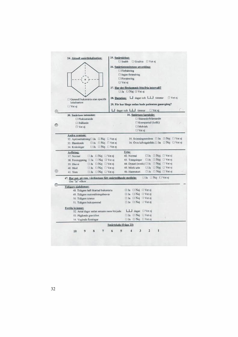
Computer registration and validation of dataOn arrival at the emergency department, all patients fulfilling the inclusion criteria had the study formulary included in their medical record. The attend-ing physician registered data for history, symptoms, clinical signs and pre-liminary diagnosis before the patient left for admission to a ward or was discharged. Localisation of pain at onset and at the time of examination was marked on a figure on the study protocol. The physician was then asked to
25
provide a primary and a secondary most likely diagnosis; the first diagnosis was then compared with the diagnosis at discharge and at follow-up. In addi-tion, results of laboratory investigations, surgery if any, duration of hospi-talisation and final diagnosis at discharge were registered by the physician responsible for that decision. All schedules were then checked by a specially trained secretary and entered into a Microsoft Access database. At this time, any obviously erroneous information detected was corrected and on computer registration logical filters detecting impossible or inconsistent combinations of data were applied. As a check of the validity of registration, 300 cases (10%) were randomised for validation. Data from the computer-ised register for those cases were checked against the medical records and if necessary against the hospital computer system for time of arrival, time of surgery etc. All erroneous data detected in these two steps were corrected in the register. The completeness of the registration was first checked by the secretaries, who indicated cases discharged with a history of AAP and with-out register formularies included in their medical records. In such cases, the physicians responsible for the present study checked to see if the case should have been included in the study, and if so, the diagnosis at discharge, any surgery and laboratory parameters were registered. During this procedure another 523 patients who should have been included in the database register were found.
At the emergency department, all patients admitted are routinely regis-tered by the nurses according to type of symptoms. Those registrations were scanned on randomized days of admission for symptoms possibly related to AAP. Patients with a symptom related to abdominal pain were checked against the study register and if not present, the medical records were checked for possible non-participation. With this method, the overall com-pleteness was calculated to be 79%. Before any calculations were performed, all stochastic or continuous variables were cleared of evidently erroneous information, that is, all data outside of the 75th percentile were checked against the record data.
Baseline study A baseline registration of logistic data such as time of admission, the level of formal competence of the attending physician and the preliminary diagnosis was created during the period 1st February 1996 to 31st January 1997. During the baseline registration period, only the most likely diagnosis was registered for each patient. During the subsequent study period, 1st February 1997 to 1st
June 2000, the initial formulary was supplemented with a detailed schedule for investigation and the physician provided a primary and a secondary diag-nosis.
26
DiagnosesThere were three diagnoses used for comparison. The preliminary diagnosis was made by the attending physician at the emergency department or the primary health care centre after the clinical examination. The discharge di-agnosis was made by the physician at the surgical ward when the patient was discharged after hospitalisation. The follow-up diagnosis was defined at the review of the case records one to three years after the initial visit to the emergency department or the primary health care centre. Thus, outpatients at the emergency department and primary health care patients had only pre-liminary diagnoses and follow-up diagnoses.
Follow-upRecords of all patients residing within the hospital catchment area were checked at least one year (mean 2.7 years) after admission. Follow-up was through checking the patients’ record at the surgical department and the pri-mary health care centre, and if necessary records from other departments at the hospital. Further investigations were registered and the discharge diagno-sis was re-evaluated according to the criteria of the World Organisation of Gastroenterology (OMGE; de Dombal 1979 and 1982, University of Leeds). This re-evaluated, final diagnosis then served as the basis for further statisti-cal calculations of the reliability of the preliminary diagnosis registered on admission to the emergency department and the diagnosis at discharge or on admission to the primary health care centres.
Patients included Patients older than one year of age and with abdominal pain of up to seven days duration who were admitted to the Mora Hospital or three primary health care centres in the hospital catchment area were prospectively regis-tered in the local database during the period February 1st 1997 to June 1st
2000. A total number of 3349 patients were registered (see the flow charts be-
low). Of these, the primary health care centres contributed with 238 patients. At follow-up 12 did not fulfil the inclusion criteria and were excluded (ab-dominal pain caused by trauma, chronic pain caused by an known diagnosis, and insufficiently filled-in investigation schedule or missing medical record), thus 3337 patients were included in the study. Of these 3337, 2979 (89%) were living within the hospital catchment area. Tourists who received surgi-cal treatment or had a diagnosis verified by radiology or endoscopy were included in the analyses, as they were considered as having an accurate di-agnosis (n=94), whereas other tourists were excluded (n=264) due to diffi-culties in acquiring medical records from a large number of hospitals. Ac-
27
cording to these criteria, 3073 records out of 3349 (92%) were included in the analyses and among them 222 out of 238 (93%) were primary health care patients. The corresponding number for the baseline registration year was 790 out of 881 (90%).
Patients included in paper I The calculations were based on 3073 patients divided in two groups: 2851 patients admitted to the hospitals emergency department and 222 provided by the primary health care centres.
Patients included in paper II A number of 2851 patients who were admitted to the hospitals emergency department were included in the study. Of these patients, 2052 were referred to a surgical ward and 799 were treated as outpatients. Calculations of sensi-tivity and specificity for the preliminary and discharge diagnoses, are based on the 2052 hospitalised patients.
Patients included in paper III Patients living outside the hospital catchment area (n=358) and patients who were admitted to the primary health care centres (n=220) were excluded, leaving 2759 patients who were admitted to the hospitals emergency de-partment in this study. The three study groups comprised 2289 patients above 20 years of age: 557 patients 65 to 79 years old, 274 patients aged 80 years or older and 1458 patients of ages 20-64 years who served as a refer-ence group.
Patients included in paper IV Patients admitted to the hospitals emergency department were included in this study (n=3099). Tourists who received surgical treatment were included, as they were considered as having a certain diagnosis (n=78), whereas other tourists were excluded (n=248) as they were not eligible for follow-up. For patients admitted more than once to the emergency department, and there-fore having two or more study protocols (n=65), only the protocol if they had surgery or the protocol registered at the first visit was analysed. There-fore, 373 protocols were excluded, leaving a total number of 2478 patients eligible for statistical analyses in this study. Another 28 patients registered as non-participating were included in the calculation of incidence rate.
28
Flow charts
Paper I:
Paper II:
3073 eligible for follow-up
222 from Pri-mary Health Care Centres
12 excluded 358 tourists 94 tourists with a safe diagnosis
264 tourists excluded
3349 reg-istered
3337 in-cluded
2052 hospital-ised
799 out-patients
2851 admitted to ED
3073 eligible for follow-up
222 from Primary Health Care Centres
12 excluded 358 tourists94 tourists with a safe diagnosis
264 tourists excluded
2851 admitted to ED
3349registered
3337included
29
Paper III:
Paper IV:
2979 from hospital catchment area
470 younger than 20 years excluded
12 excluded358 touristsexcluded
220 from Primary Health Care Centres excluded
2289 above 20 years of age
3349registered
3337included
274 aged 80 or more 1458 aged
20-64 557 aged 65-79
3099 eligible for study
248 tourists excluded
12 excluded238 from Primary Health Care Centres excluded
78 tourists with a safe diagnosis
3349registered
3337included
373 duplicates excluded
2478 study-patients
2851 admitted to ED
2204 with AAP
274appendicites
30
StatisticsStatistica software (Statsoft, Tulsa, USA) was used for the statistical calcu-lations. Distribution fit of the data was checked initially and most parameters appeared to be normally distributed when there were many patients in each group, as judged by the Kolmogorow-Smirnow test; however, groups of variable size did not always fit into that distribution model. The non-parametric Mann-Whitney U-test was generally used to calculate the signifi-cance of differences in continuous variables, and the Chi-squared test was used in cases of dichotomous response parameters and to test differences in proportions between groups (Paper I). Correlation was calculated by the Spearman test (Paper III). Calculations of sensitivity, specificity, positive predictive value (PPV), negative predictive value (NPV), positive likelihood ratio (LR+) and negative likelihood ratio (LR-) were used as measures of the diagnostic value of the preliminary and discharge diagnoses, clinical find-ings and laboratory tests (Paper II and IV).
The sensitivity of a symptom represents the probability that a patient who is suffering from for example appendicitis will have a certain symptom, and PPV is the probability that the patient is suffering from appendicitis, if a symptom or sign is present. Calculations of positive likelihood ratio for the preliminary diagnosis were used as an alternative measurement of diagnostic accuracy, independent of the prevalence of that diagnosis in this population. The LR+ indicates how many times greater the probability of a clinical find-ing is among patients with a specific diagnosis than in the baseline study population. An excellent diagnostic test has an LR+ of about 10, a fair LR+ is at least 2 and if the LR+ is less than 1 it indicates that the diagnostic test is invalid. The kappa value was calculated to measure the congruence between the preliminary and discharge diagnoses (Paper II). The kappa statistic pro-vides a measure that varies from +1, indicating perfect congruence, to 0 (zero), an indication of no greater congruence than expected by chance (Boyd 1979). To ascertain the most important diagnostic markers for appen-dicitis (Paper IV), each diagnostic variable was tested first in a univariate logistic regression analysis, expressed as odds ratio (OR) with 95% confi-dence interval (CI), and then in a multivariate analysis with adjustments for age and gender.
EthicsThe study project was approved, in advance, by the Committee of Ethics at Uppsala University, Sweden. The database also obtained approval from the Swedish authority concerning registry data. All patients were provided in-formation about the study at the emergency department and a separate in-formation sheet for the patients was included in each investigation protocol.
31
The structured schedule for investigation
32
33
34
35
Results
Paper I
During the study period of 40 months, 3073 patients with AAP were regis-tered in the local database. Of 2851 patients admitted to the emergency de-partment, 2062 patients (72%) were admitted to a surgical ward, whereas 789 (28%) were treated as outpatients. The mean age was 46 years. The male/female ratio was 0.82 (n=1382/1691). For age groups 15-45 years and over 90 years, the majority of patients were females. The ten most common final diagnoses at the emergency department were NSAP (37%), gallbladder disease (10.5%), appendicitis (9.8%), diverticulitis (4.7%), constipation (4.6%), ureteric stone (4%), gynaecological complaints (3.5%), acute pan-creatitis (3.2%), acute intestinal obstruction (3.2%) and urinary tract infec-tion (2.6%, Table 1). Detected abdominal malignancies constituted 2.8% (n=86). The sensitivities for the preliminary diagnoses at the emergency department were as follows: appendicitis 0.80, cholecystitis 0.51, gallstones 0.68, diverticulitis 0.64 and ureteric stone 0.78.
A majority of the patients attending the emergency department (88%, n=2699) were examined by a non-specialist physician with 0.5 to 5 years of experience. Most patients, 46%, were seen by pre-registrar house officers (Table 2). There was no significant difference in diagnostic performance according to category of physician (pre-registrar house officers, locums, senior house officers, general physicians and consultants). During the base-line period the pre-registrar house officers were in contact with the consult-ant in 34% of the cases, whereas the senior house officers had such contact in only 12% of the cases (p<0.001). Overall the rate of correct diagnoses was 54% and the corresponding value for baseline registration period was 58% (p=0.07).
36
Table 1 Diagnoses on admission (preliminary diagnoses) and after at least one year of follow-up (final diagnoses)
The diagnostic performance was higher for outpatients both during the baseline (65%) and during the study period (63%). Accuracy rate for the preliminary diagnosis at the emergency department was lower (P<0.001) for women (52%, n=1691) than for men (58%, n=1382). The diagnostic per-formance at the emergency department was independent of the patient’s time of arrival. The rate of correct diagnoses from midnight to 6 a.m. was 52% (n=434), from 3 a.m. to 6 a.m. 52% (n=190), and during the daytime from 6 a.m. to midnight 55% (n=2639). During the study period, the precision of the preliminary diagnoses increased over time, 52% (n=968) in 1997, 55% (n=1090) in 1998, 56% (n=764) in 1999 and 59% (n=251) during 2000. When the second preliminary diagnoses was also included in the calculation of correct diagnoses, the overall accuracy rate increased to 59% 1997, 64% 1998, 63% 1999 and 62% during 2000. The admission frequency was lower for locums (65%, p<0.001), and for general physicians at hospital, (66%, p=0.03), than for pre-registrar house officers (70%, p=0.03), senior house officers (73%, p=0.68) and consultants (70%, p=0.74).
Table 2 Category of attending physician at the emergency department and the pro-portion of correct diagnoses
Proportion correct diagnoses between preliminary diagnosis as decided by the physician responsible on admission and final diagnosis after at least one year of follow-up.
To evaluate the diagnostic process for possible pitfalls in the management of AAP, the preliminary diagnosis from the emergency department was com-pared to the discharge diagnosis. Out of 2851 patients arriving at the emer-gency department 2052 were admitted to a surgical ward and 799 were treated as outpatients. The frequency of X-ray investigations decided at the emergency department was relatively low, and CT was ordered for only 89 (3%) of the 2851 patients. The corresponding figures were 440 (15%) for plain abdominal X-ray, 526 (18%) for ultrasonography and 139 (5%) for intravenous urograhpy.
Table 3 Accuracy of diagnoses.
The calculations of sensitivity and specificity for the preliminary diagnoses are based on the 2851 patients who attended the emergency department. The kappavalue is calculated between the preliminary and discharge diagnoses and is based only on the 2052 hospitalised patients. Positive likelihood ratio (LR+) is calculated on the preliminary diagnosis. Other diagnoses includes for example mesenteric lymphadenitis, inflammatory bowel disease, intra abdominal abscess, septicemia, pneumonia, pleuritis, myocardial or renal infarction, debut of diabetes mellitus, torsion and necrosis of the omentum majus, muscular back pain and addiction to analgesics.
On the basis of table 3, the five most characteristic diagnoses were se-lected for each of the groups described below.
Group 1 – low predictive value of preliminary diagnosis Belonging to this group were diagnoses with low predictive value of the primary diagnosis at the emergency department, but with improved predic-tive value at discharge; these included NSAP, appendicitis, gallstones and constipation. Among the 208 patients initially diagnosed with constipation, 17 (8%) were later found to have an abdominal malignancy and 20 (10%) a diagnosis probably necessitating surgery (one patient with aortic aneurysm, one with incarcerated inguinal hernia, five patients with colonic obstruction, four with obstruction of the small intestine and nine with appendicitis). Fur-thermore, peptic ulcer, in spite of having a high kappa value and a sensitivity of 0.87 at discharge, indicated low sensitivity of the primary diagnosis. This low sensitivity partly emanated from cases treated as outpatients among whom no true positive cases were determined (none of those cases were confirmed at endoscopy). Those diagnoses together with the frequency of incorrect primary diagnoses are listed in Table 4. Calculation of LR+ re-vealed a low value for NSAP and moderate value appendicitis; all other di-agnoses had high LR+.
Patients considered as suffering from NSAP at follow-up (n= 1058) had a median hospital stay of 1 day, and a total number of in-hospital days during the observation period (40 months) of 1338. Calculated on 87,000 inhabi-tants and a completeness of the register of 90%, this meant 43 in-hospital days per month between 100,000 inhabitants.
Table 4 Distribution of final diagnoses correlated to the preliminary diagnosis at the emergency department.
Numbers within parentheses are percentages of cases with each final diagnosis.
Group 2 – predictive value still low at discharge The final diagnoses of malignancy, gynaecological complaints, dyspepsia, urinary tract infection and diverticulitis had good concordance between the preliminary and discharge diagnosis, but the predictive value at discharge was low compared to the final diagnosis at follow-up. In addition to malig-nancies identified during the hospital stay, abdominal malignancies were detected in another 27 patients at follow-up. Furthermore, despite consider-able improvement in validity during hospital-stay, the judgement that NSAP was still the reason for the patients’ complaints at discharge had low sensi-tivity.
SurgeryOn evaluation of patients judged as needing surgery during the hospital stay, 104 patients had an initial diagnosis usually not necessitating surgery (Table 5): these patients constituted 22% of the total 479 (23% of patients admitted to a ward) undergoing operations. In six of these operations no cause was determined for the abdominal pain and the complaint was thus classified as NSAP. As a comparison, 12 (3%) of the 375 patients operated on who had a preliminary diagnosis necessitating surgery were finally diagnosed with NSAP; whereas, a gynaecological reason for the pain was determind during 16 (4%) of these operations. Patients given a preliminary diagnosis of NSAP (n=42), constipation (n=19), other diagnoses (n=13), pancreatitis (n=6) or ureteric stone (n=6) had significantly longer time before surgery, compared with the surgically-treated patients in whom the final diagnosis was the same as the preliminary diagnosis. Patients with an initial diagnosis not necessitat-ing surgery (n=104) had a median delay of 22 hours before operation (mean was 40 hours, 95% confidence interval 30-50 hours), compared to 8 hours (mean was 15 hours, 95% confidence interval 12-18 hours) for patients with the same final diagnosis at follow-up as for the preliminary diagnosis (n=266).
41
Table 5 Diagnosis at surgery and diagnosis at discharge for 104 surgically-treated patients, who were given a preliminary diagnosis usually not necessitating surgery.
Of the patients admitted to the emergency department, with AAP, 2289 were above 20 years of age. Among these, 831 were 65 years of age or older and constituted the total study group, which was divided into one group of 65-79 years old (n=557) and one group aged 80 years or older (n=274). Patients aged 20 to 64 years (n=1458) served as a reference group. There was a higher proportion of women (Table 6) among patients 20-64 years and 80 years of age than in the group aged 65-79 years. The frequency of surgery among all patients was independent of age group. NSAP was the most com-mon preliminary diagnosis with no significant difference between the refer-ence group and the two study groups (p=0.3). The proportion of NSAP in each group was similar at discharge (Table 6), whereas at follow-up (Table 7) 64% (n=934) of the patients in the reference group were assigned a spe-cific diagnosis, compared with 76% (n=421) of the 65-79 years group and 78% (n=214) of the 80 years group (p<0.001).
Duration of pain before admission was related to age (p<0.003; reference versus the total study group; Table 6) and there was no difference in pain duration between the two study groups (p=0.9). The frequency of hospitali-sation was higher in the both study groups ( 65 years); the group aged 65-79 years (459 of 557 patients, 82%; p<0.0001) and the group of oldest patients (241 of 274 patients, 88%; p<0.0001), than in the reference group (996 of 1458 patients, 68%; Table 6). The calculated duration of hospital stay was 170 days per 100 emergency admissions for the reference group; whereas in the two study groups the duration was 320 days for the 65-79 years group and 458 days for the 80 years group. The postoperative stay was signifi-cantly longer for the study groups (7.6 days for 65-79 years and 10.7 days for 80 years) than for the reference group (3.7 days, p<0.0001; Table 8).
Older patients (the total study group) were more often misdiagnosed (429 of 831 patients, 52%) at the emergency department than reference patients were (656 of 1458 patients, 45%; p=0.002), but there was no difference be-tween the two study groups (p=0.06; Table 6). At discharge, the diagnosis was more accurate (853 of 996 hospitalised patients, 86%) in the reference group (20-64 years) than in the older patients (539 of 700 hospitalised pa-tients, 77%; p<0.0001). There was no difference between the two study groups aged 65 years (78% for 65-79 years and 75% for 80 years, p=0.3; Table 6). Hospital mortality increased with age (p<0.0001; Tables 6) when the reference group (2 deaths) was compared with the two study groups (23 deaths), and there was a difference in mortality between the two older study
43
groups (10 patients aged 65-75 years and 13 patients aged 80 years; p=0.02). However, among patients older than 65 there was no difference in mortality between those diagnosed correctly at the emergency department and those misdiagnosed in that department (p=0.9). Furthermore, there was no relation between pre-admission duration of pain and mortality (p=0.9).
Table 6 Basic demographic data and characteristics
NSAP – Non-specific abdominal pain. Preliminary diagnosis is the diagnosis at the emer-gency department. Numbers within parentheses are 95% confidence intervals unless otherwise stated.
There was no difference in duration of abdominal pain before admission between the two study groups or between patients being operated and those not being operated. There was no difference between the two separate study groups in the interval between admission and surgery (1.7 for 65-79 years and 2.1 days for 80 years; p=0.5). However, the interval was considerably longer for the total study group (1.8 days) than for the reference group (0.9 days, p<0.0001; Table 8).
A CT scan was ordered for 34 patients (2%) in the reference group and for 33 (6%) in the 65-79 years group and 20 (7%) in the 80 years group: the corresponding figures for ultrasonography were 263 (18%) for the reference group, 162 (29%) for the 65-79 years group and 77 (28%) for the 80 years group.
Table 7 Distribution of common final diagnoses at follow-up in the two study groups ( 65 years) and in the reference group (20-64 years)
NSAP – Non-specific abdominal pain.
Symptoms, signs and laboratory examinations Patients who were under 65 years of age and submitted to surgery generally had higher levels of CRP on admission than those who were not submitted to surgery did (44 for operated and 27 for non-operated; p<0.0001). In contrast, among patients older than 65, there was no significant difference in CRP between patients undergoing surgery (CRP 60) and those who did not (CRP 45; p=0.12). However, there was a difference in leucocyte counts, irrespec-tive of age group, between patients treated surgically and those not treated surgically. Patients aged 20-64 years who were surgically treated had a leu-cocyte count of 13 versus 10 for patients who were not surgically treated and for patients aged over 65 the leucocyte count was 13 versus 11 if surgically-treated or not (p<0.001). Body temperature among surgically treated patients was independent of age, although it was higher among surgically treated patients in the reference group (37.6 vs 37.2°C; p<0.0001) than in the study groups (37.4 vs 37.2°C; p=0.06).
In a logistic regression analysis on diagnoses usually known to induce an intra-abdominal inflammatory response, clinical signs such as rebound ten-derness (p<0.0001), local rigidity (p=0.003) and rectal tenderness (p=0.004) were less common with increasing age. Nevertheless, there was no age-related difference in the distribution between general and localised abdomi-nal tenderness on clinical investigation in the emergency department. Age in itself did not correlate to symptoms such as vomiting (p=0.4), constipation (p=0.7) or diarrhoea (p=0.7, Table 8).
Table 8 Characteristics of patients requiring surgery
CRP – C-reactive protein. VAS – Visual Analogue Scale, used here as an instrument for the patient to estimate the severity of pain. Numbers within parentheses are 95% confidence intervals unless otherwise stated.
Age 20-64 years 65-79 years 80 years n 243 77 38 Pre-admission pain duration (days) 1.2 (1.0-1.4) 1.7 (1.2-2.1) 1.5 (1.0-1.9) Time from admission to surgery (days) 0.9 (0.7-1.0) 1.7 (1.2-2.1) 2.1 (1.2-3.1) Body temperature ( C) 37.6 (37.5-37.7) 37.4 (37.2-37.6) 37.6 (37.3-38.0) Leucocytes 13.5 (12.9-14.1) 13.1 (12.0-14.3) 12.4 (9.6-15.3) CRP 44 (37-50) 62 (40-83) 57 (34-81) General abdominal tenderness 36 (15%) 19 (25%) 15 (39%) Rebound tenderness 150 (62%) 27 (35%) 11 (29%) Rectal tenderness 78 (32%) 13 (17%) 7 (18%) Abdominal rigidity 104 (43%) 27 (35%) 13 (34%) Vomiting 97 (40%) 37 (48%) 22 (58%) Constipation 27 (11%) 20 (26%) 10 (26%) Diarrhoea 40 (16%) 10 (13%) 8 (21%) VAS 6.4 (6.1-6.8) 6 (5.2-6.9) 5 (3.4-6.6) Hospital stay (days) 3.7 (3.3-4.1) 7.6 (6.4-8.8) 10.7 (8.4-13.0) Correct preliminary diagnosis 149 (61%) 36 (47%) 19 (50%)
46
Paper IV
Differential diagnostics and diagnostic outcome Acute appendicitis was suspected in 432 patients out of the 2478 patients admitted with AAP. Of these, 221 patients were submitted to surgery and appendicitis was confirmed. In 53 patients eventually diagnosed as having appendicitis, a different preliminary diagnosis was suggested at the emer-gency department. Consequently, 274 patients had confirmed appendicitis; 271 had their appendix surgically removed and three had an appendiceal abscess diagnosed by CT or ultrasonography and were treated with only antibiotics. None of the patients with an abscess had an appendectomy within the follow-up period (at least one year). Advanced appendicitis (gan-grenous or perforated) was identified in 165 patients (60%) and the perfora-tion rate was 25% (n=68).
Of 432 patients with suspected appendicitis, 423 (98%) were admitted to the surgical ward, two were referred to the gynaecological department (one had diverticulitis and one had torsion of the right ovary) and seven patients were sent home but agreed to return to the emergency department the next morning (none of these patients were eventually diagnosed as having appen-dicitis). One hundred and sixty patients with a preliminary diagnosis of ap-pendicitis were not surgically treated, as the pain had disappeared (111 pa-tients) or another diagnosis was confirmed (49 patients). At follow-up, 211 of the 432 patients with appendicitis as a preliminary diagnosis had a differ-ent final diagnosis, these were: 122 patients suffering from NSAP; 24 with gynaecological complaints; 13 with diverticulitis (2 with perforation); 10 with mesenteric lymphadenitis; 8 with urinary infection; 7 with biliary stone disease; 5 with gastroenteritis; 4 with ureteric stone; 3 with constipation; 3 with upper respiratory tract infection; 2 with intestinal obstruction; 2 ab-dominal malignancies; 2 torsion of colonic epiploicae; 1 with aortic dissec-tion; 1 with perforated gastric ulcer; 1 dyspepsia; 1 with Meckel’s diverticu-litis; 1 with Crohn’s disease, and 1 with pneumonia. Of these 211 patients, 53 underwent surgery and among 316 appendectomies the result was nega-tive in 45 cases (14%). Of the 432 patients with suspected appendicitis, pre-operative CT scan was performed in only six cases and ultrasound in 36 cases.
The preoperative clinical diagnosis of appendicitis had a sensitivity of 0.81, specificity of 0.90, PPV of 0.51, LR+ of 8.1, diagnostic accuracy of 0.89 and a kappa value of 0.78. The incidence rate of patients treated surgi-cally for appendicitis was 102 per year and 100,000 inhabitants.
47
Gender and age differences Males predominantly suffered from appendicitis (57%), whereas, females (57%; p<0.001) were predominant among patients with AAP but without appendicitis. Patients with appendicitis were generally younger (mean age of 32) than patients without appendicitis (mean ages of 47; p<0.001).
Clinical symptoms Upper gastrointestinal complaints, such as loss of appetite (anorexia), nausea and vomiting, were more common in the appendicitis group (p<0.001), com-pared to other patients with AAP. There was no difference in duration of pain between the two groups (p=0.95). In appendicitis patients, the pain of-ten had a gradual onset and improvement of the pain was rare. Furthermore, continuous pain and dull pain were common, and pain-free intervals, fluctu-ating pain and colic pain were not. Migration of pain to the right iliac fossa was recorded in 16% of appendicitis patients and previous episodes of simi-lar pain in 15%. Pain migration to the right iliac fossa was uncommon among patients not diagnosed with appendicitis (3%; p<0.001), whereas previous episodes of pain were more common in this group (37%; p<0.001, Table 9).
Table 9 Differences in characteristics of abdominal pain between appendicitis pa-tients and other patients with AAP.
App – appendicitis. AAP – acute abdominal pain; this group includes all patients except those with appendicitis. Sens – sensitivity, Spec – specificity, Acc – accuracy, PPV – positive pre-dictive value, NPV – negative predictive value, LR+ – positive likelihood ratio, LR- – nega-tive likelihood ratio. int. –intensity. Aggr. of pain; moving – aggravation of pain when mov-ing. Aggr. of pain; coughing – aggravation of pain when coughing. Allev. of pain; not mov. – alleviation of pain when not moving. Migr. of pain to right IF – migration of pain to right iliac fossa. VAS – Visual Analogue Scale; patients´ estimation of severity of pain graded from 0 to 10. Prev. ep. of similar pain – previous episodes of similar pain. Numbers with ranges in parentheses are 95% confidence limits of the mean.
Clinical findings Clinical signs of peritonitis such as aggravation of pain when moving or coughing and alleviation when not moving were more common (p<0.001) in appendicitis patients. There was no difference in the severity of the pain, between the appendicitis patients and other patients with AAP (Table 9). Isolated tenderness in the right iliac fossa and rebound tenderness were more frequent (p<0.001, Table 10) among appendicitis patients, as was right-sided rectal tenderness (p<0.001, Table 10). No patient with appendicitis had ten-derness located only in the left iliac fossa. On abdominal examination, pa-tients younger than 20 years had lower LR+ values for signs of appendicitis on physical examination, with the exception of the muscle response parame-ters (local guarding and general rigidity), than patients over 50 years of age had. When testing the same symptoms and signs according to gender, there were no differences in LR+, except for right-sided rectal tenderness and pain migration: males had a higher LR+ (6.5 and 5.3) than in females did (4.7 and 3.7). There were no differences in the degree of inflammation, except a higher LR+ for general rigidity in perforated (7.5) compared to phlegmonous (1.5) and gangrenous appendicitis (1.0).
Table10 Differences in clinical findings from the physical examination on admission between patients with appendicitis and other patients with acute abdominal pain.
App – appendicitis. AAP – acute abdominal pain; this group includes all patients except those with appendicitis. Sens – sensitivity, Spec – specificity, Acc – accuracy, PPV – positive pre-dictive value, NPV – negative predictive value, LR+ – positive likelihood ratio, LR- – nega-tive likelihood ratio. Tend. – tenderness, IF – iliac fossa, abdom. – abdominal.
Symptoms n (%) App
n (%) AAP
SensApp
Spec App
Acc App
PPV App
NPVApp
LR+ App
LR- App
n 274 2204 Tend. in right IF only 194 (71) 403 (18) 0.70 0.82 0.80 0.32 0.96 3.89 0.37 Tend. in left IF only 0 (0) 220 (10) General abdom. tend. 36 (13) 422 (19) 0.13 0.81 0.73 0.08 0.88 0.68 1.07 Rebound tenderness 201 (73) 516 (23) 0.73 0.77 0.76 0.28 0.96 3.17 0.35 Local guarding 107 (39) 277 (13) 0.39 0.87 0.82 0.28 0.92 3.00 0.70 General rigidity 15 (5) 39 (2) 0.05 0.98 0.88 0.28 0.89 2.50 0.97 Right-sided rectal tend. 74 (27) 120 (5) 0.27 0.95 0.87 0.38 0.91 5.40 0.77
49
Diagnostic markers for appendicitis The highest OR (univariate) was found for isolated tenderness in the right iliac fossa (3.29), followed by rebound tenderness (3.00), right-sided rectal tenderness (2.53), migration of pain to the right iliac fossa (2.18), local guarding (2.11), aggravation of pain on movement (1.99), and general rigid-ity (1.79). In the multivariate analysis these variables still had the highest ORs (Table 11).
Table 11 Multivariate analysis of the diagnostic value of symptoms and signs for diagnosis of appendicitis.
In the multivariate analyses the factors were adjusted for age and gender. Each group was processed in separate models, except for the last group, which was split into three groups: isolated tenderness in right iliac fossa and rebound tenderness; general rigidity; local guarding and right-sided tenderness.
Factors Univariate Odds Ratio
analyses ± 95% CI
MultivariateOdds Ratio
analyses ± 95% CI
Upper gastro-intestinal symptoms: Anorexia 1.51 1.31-1.73 1.37 1.16-1.63 Nausea 1.56 1.35-1.80 1.41 1.19-1.67 Pain characteristics: Gradual onset of pain 1.20 1.06-1.38 1.02 0.86-1.21 Impairment of pain 1.70 1.49-1.94 1.59 1.34-1.89 Continuous pain 1.54 1.35-1.77 1.45 1.22-1.72 Signs of localised inflammation: Aggravation of pain on movement 1.99 1.74-2.26 1.70 1.35-2.16 Migration of pain to right iliac fossa 2.18 1.82-2.62 2.02 1.61-2.56 Findings at abdominal examination: Isolated tenderness in right iliac fossa 3.29 2.86-3.79 2.96 2.50-3.51 Rebound tenderness 3.00 2.60-3.46 2.45 2.07-2.91 General rigidity 1.79 1.32-2.43 1.92 1.30-2.83 Local guarding 2.11 1.84-2.42 1.81 1.49-2.20 Right-sided rectal tenderness 2.53 2.16-2.98 2.19 1.80-2.66
50
A combination of the three most frequently recorded clinical findings with high LR+, namely isolated tenderness in the right iliac fossa, rebound tenderness and aggravation of pain when moving, gave an LR+ of 9.5 (Table 12).
Table 12 Clinical symptoms and signs diagnostically important for appendicitis, in combination with the two most common clinical findings: isolated tenderness in the right iliac fossa (=1) and rebound tenderness (=2).
App – appendicitis. AAP – acute abdominal pain; this group includes all patients except those with appendicitis. Sens – sensitivity, Spec – specificity, Acc – accuracy, PPV – positive pre-dictive value, NPV – negative predictive value, LR+ – positive likelihood ratio, LR- – nega-tive likelihood ratio. 1 – isolated tenderness in the right iliac fossa, 2 – rebound tenderness. Aggr. of pain; moving – aggravation of pain when moving.
Studies on patients with AAP need to include many patients, as there are a large number of diagnoses to be considered. Due to the many different eti-ologies of abdominal pain and varying symptoms and signs for the same diagnosis in different patients, clinical diagnosis is often difficult. Making an early and correct diagnosis is crucial because a good prognosis is dependent on urgent treatment in some cases (Silen 2005). The finding that the level of formal competence did not make any difference in diagnostic performance was unexpected. The hypothesis was that the more experienced a physician was, the better the diagnostic results would be. One interpretation is that the even inexperienced physicians have good medical knowledge and can see the patterns of symptoms and signs characteristic for different diseases. Hence, the information collected and the careful examination of the patient are more important than formal competence (Hancock 1987). The introduc-tion of the structured schedule encouraged this behaviour in all physician categories.
Many other studies (Eskelinen 1992, Körner 1998, Lamprelli 2000) have focused on a certain diagnosis, most commonly appendicitis and patients with suspected appendicitis. Making a comparison between studies is diffi-cult due to different manners of follow-up (or no follow-up), study design and diagnostic criteria. In some studies (Bjerregaard 1976, de Dombal 1972, Ikonen 1983, Irvin 1989, Simmen 1991, Staniland 1980) many diagnoses, in this study defined as specific entities, are grouped together with NSAP, such as gastroenteritis, constipation, mesenterial lymphadenitis, dyspepsia; thus, diagnostic accuracy can reach higher levels due to fewer possible choices of diagnoses. In the present study, the physicians had 30 defined diagnoses to choose from, according to the schedule. Furthermore, patients who did not fit in any of these categories were classified as “other diagnosis”. In several reports, fewer different diagnoses, ranging from 8 to 13, were used (Bjerre-gaard 1976, de Dombal 1972, Ikonen 1983, Simmen 1991, Staniland 1980). One of these reports, (Bjerregaard 1976) used 10 diagnoses, and the diagnos-tic performance of the attending physicians was 55.1%. With the same set of diagnoses on this group of patients, the overall diagnostic performance would have been 54%. This comparison indicates that the predictive value of the preliminary diagnosis obtained in the present study was in parity with that of other studies.
52
Measures of diagnostic performance of patients with AAP An obviously poor overall diagnostic performance of about 50%, observed in several studies (Adams 1986, Bjerregaard 1976, Gunn 1976), should not necessarily be considered as bad management, as in many cases the pain is self-limiting and there is no need for surgical intervention. Instead specific outcome-measures should be introduced in this field of clinical research. With the exception of NSAP, appendicitis and gallstones, the specificity of most diagnostic entities in the present study was fairly high. Although these are only three diagnoses with low specificity, this was a quantitative prob-lem, as those patients constituted more than half of the cases in the present study. NASP was considered as the main diagnostic problem both quantita-tively and qualitatively.
With the exception of colonic obstruction and peptic ulcer, NSAP was the most frequent misjudgement: 9% of the patients finally receiving surgery were initially diagnosed as suffering from NSAP, resulting in a significant delay of surgery. However, those patients constituted only 6% of the 641 patients considered as having NSAP at the emergency department.
Overall, the diagnostic performance was independent of the time of the patient’s arrival at the emergency department. The diagnostic performance for older patients was lower than for younger patients, as has previously been reported (Kizer 1998, Geloven 2000). The consequences of this are outlined below, and the effect on handling was more pronounced for older patients as a larger proportion suffered from specific conditions often neces-sitating treatment.
Mortality rate as a measure of diagnostic outcome Mortality rate is of general interest and not difficult to measure. In this mate-rial the in-hospital mortality was 1.2% (25 of 2052 hospitalised patients) of whom only two patients were under 65 years of age. Delay in surgery is one crucial factor for morbidity and mortality in older age groups. High age as an independent factor is not necessarily associated with an increased operative risk (Arenal 2003, Hosking 1989), but here are possibilities for improve-ment.
Mortality is uncommon and risk factors for mortality may therefore be difficult to evaluate (Gunnarsson 2003). Grading by a system such as the American Society of Anaesthesiologists (ASA), which was not done in the present study, is probably the best predictor of mortality (Arenal 2003). However, making such grading is difficult for an on-call physician and is not appropriate for all patients arriving at the emergency department. Hence, the ASA-grading would have low validity in a study with the present design.
53
Bad surgical management as a measure Another parameter is bad surgical management (de Dombal 1997). One way of measuring this is to compare patients requiring surgical treatment and the proportion of those with an initial diagnosis of not requiring surgery. If there is a delay of more than 24 hours from the patient’s arrival at the emergency department to the operating theatre, it could be considered as a serious man-agement error. In the present study, 104 out of 274 surgically treated patients initially received such a diagnosis, 46 of which waited more than 24 hours before surgery. However, in some cases such as intestinal obstruction or cholecystitis, the true diagnosis may be revealed during the first 24 hours, but for medical reasons it is appropriate to choose conservative management and observation instead of surgery. Hence, these cases are not to be consid-ered as management errors.
Patients with an initial diagnosis not necessitating surgery had a median delay before operation of 22 hours (mean was 40 hours, with 98% CI of 30-50 hours), compared to 8 hours (mean 15 hours, 95% CI of 12-28 hours) for patients with the same final diagnosis at follow-up as the preliminary diag-nosis. However, the specificity of diagnoses requiring surgery was high, as in the study by Rozycki et al. (Rozycki 2002) indicating that the risk for surgery in false positive cases is low.
A diagnosis of constipation at the emergency department was associated with a considerable risk for mistakes, where 9% of the patients in fact re-quired surgery for potentially life threatening reasons and 8% were later found to have an abdominal malignancy. An active search for the cause of constipation is mandatory, especially in older patients.
Elderly patients surgically treated for AAP had a mean time of 2.1 days from admission at the emergency department to surgery, patients aged 65-79 years waited 1.7 days and patients under 65 years of age waited 0.9 days. More severe disease among older patients with AAP, as also reported by Watters et al. (Watters 1996), may be an effect of increased occurrence of concurrent diseases at higher ages and because of a delay before admission to hospital and a longer interval between admission and the decision to oper-ate due to less specific symptoms.
Morbidity and negative appendectomies as a measure One example of morbidity could be if the patient had a stoma, which could have been avoided, or was unnecessarily operated. The rate of negative ap-pendectomies is one such measure. A negative appendectomy is an operation for suspected appendicitis, where a normal appendix is found and removed but no other surgical disease is detected and no other surgical procedure is performed. The negative appendectomy rate is expressed by the number of normal appendices removed in relation to all appendices removed for sus-pected appendicitis (de Dombal 1997). The perforation rate among patients
54
with appendicitis might not be a good measurement, as data from this study indicate that the appendix in many cases was already perforated on arrival at the emergency department (Andersson 2002, Hansson 2006).
Duration of hospital stay Patients above 80 years of age stayed in hospital almost three times longer postoperatively than patients younger than 80 years. Using duration of hospi-tal stay as a measure of diagnostic performance can be misleading, as many elderly patients remain on the surgical ward for days, waiting to receive a place in the Community Care.
Considering a short hospital stay as an indicator of good management is not desirable, as patients are then often referred for further investigations on an outpatient basis: this can lead to diagnostic delays or even missed diagno-ses. In the present study, the following diagnoses were identified as having low diagnostic accuracy even at discharge: malignancy, gynaecological complaints, dyspepsia, urinary tract infection and diverticulitis. Changes in discharge diagnoses at follow-up often resulted from information obtained from outpatient investigations that were already ordered at discharge.
Use of resourcesIn AAP, CT is often done for patients whose diagnosis is strongly suspected on clinical grounds. For determining the additive value of CT (Gwynn 2001), information on the sensitivity and specificity of the diagnosis made by clinical examination is important, and whether the result of such a test will change the suspected diagnosis and surgical management needs to be considered. In some cases, however, a disease is strongly suspected but there is a need for performing a radiological examination that can increase the diagnostic certainty to a point where a decision can be taken. The use of CT in undetermined cases of AAP has increased (Rosen 2003, Terasawa 2004), and it is becoming commonly used as a diagnostic tool for patients with sus-pected appendicitis (Flum 2005). The most common differential diagnostic entities for appendicitis in this study were NSAP, gynaecological com-plaints, diverticulitis, mesenteric lymphadenitis, urinary infection and gas-troenteritis. These diagnoses, except some cases of gynaecological com-plaints, did not require surgery, but in some cases required medical treat-ment, observation and repeated clinical examination of the abdomen to con-firm the diagnosis.
This study on patients with appendicitis, confirmed that the assessment in cases of suspected appendicitis could still be based on clinical judgements in combination with laboratory tests: the CT scan can thus be saved for the more equivocal cases of AAP (Tobati 2003). One group of patients that should have extra attention at the emergency department are the elderly, as
55
the proportion of patients considered as suffering from NSAP at follow-up was lower in the older age groups. A possible explanation for this may be a larger proportion of patients with specific organ pathology and this justifies a more liberal use of CT among older patients presenting with AAP. Another explanation of the lower proportion of NSAP among elderly is that older patients arrive after a longer history of AAP than younger patients do, and some self-limiting pain has already been resolved.
Although in the present study the majority of aged patients were not in-vestigated radiologically, an increase in the proportion of patients submitted for radiological examinations by the emergency department during the early period after the onset of AAP may considerably enhance the diagnostic accu-racy at that stage (Geloven 2000). An increase in cost for radiology may also be warranted by the possibility of diminishing the consumption of in-hospital resources through an early correct diagnosis. A more precise identi-fication of patients at the emergency department who would benefit from CT could lead to a shorter average hospital stay. With a growing proportion of patients reaching high ages, there may be increasing demands on clinical resources for this group of patients (Arenal 2003, Reiss 1992); although, the need for surgery after admission to the emergency department was not in-creased among elderly patients in this study, compared to patients of younger ages.
Computer-aided diagnosis and scoring systems The computer-aided system in Leeds, UK, is mathematically based on prob-ability analyses and uses Bayes’ theorem, where each patient’s symptoms are compared with cases collected in the database (Adams 1986, Hancock 1987, Horrocks 1972). As the probability of a single symptom associated with a certain disease varies with the prevalence of that disease, the database must comprise a sufficient number of patients collected from the same study population (Staniland 1980, Fenyö 1983). This might explain why the results of the following studies did not reach the initial diagnostic performance of 92%, as seen in Leeds in 1972 (de Dombal 1972), and why it has not become a widely used tool for diagnostic performance. The present database in Mora, Sweden, could serve as a base for a diagnostic computer-aided system for the Swedish population.
Scoring systems are manual recording of symptoms and signs, each giv-ing a certain point, which is added up to produce a score. Scoring systems are mainly used to improve diagnostic accuracy in appendicitis, although the outcome of these scoring systems has not always been convincing (Al-Hashemy 2004, Denizbasi 2003, Franke 1998, Körner 1998, Lintula 2005). The best results reported (Enochsson 2004, Körner 1998) are among women of childbearing age, which is a group of patients known to have low diagnos-tic accuracy and where the use of CT is limited because of radiation. A retro-
56
spective comparison of the present results with a scoring system for appen-dicitis might prove beneficial.
The problem with scoring systems for diagnosing appendicitis is the high incidence of false positive diagnoses resulting in negative appendectomies: the Modified Alvarado Score appears the most reliable score, compared to Ohmann and Eskelinen (Franke 1998, Horzic 2005, Lamparelli 2000). How-ever, the scoring systems are an aid to diagnosis and do not substitute for the clinicians’ judgement of further diagnostic investigations or treatment. Scores are not commonly used in clinical routine (Enochsson 2004, Eske-linen 1992).
Gender perspectives Women constituted the largest proportion of patients in this study, with a male/female ratio of 0.82 (n=1382:1691). Gynaecological complaints in-crease the number of diagnoses for females, and may be a plausible explana-tion why the diagnostic performance at the emergency department was lower for women (52%) than for men (58%; p<0.001). A lower specificity of the preliminary diagnosis for women suffering from appendicitis has also been presented in other studies (Al-Hashemy 2004, Andersson 1992 and 2000, Denizbasi 2003, Flum 2002).
There were a higher proportion of women among patients aged 20-64 years and patients older than 80 years. Gynaecological complaints and self-resolving pain, judged as NSAP, constituted the major part of admittance to the emergency department in the younger age group: this is also reported in a previous study (Sheridan 1992). That women have longer life expectancy than men could also explain the dominance of women among patients aged over 80 years presenting with AAP.
Several gynaecological diseases cause local inflammation that can mimic appendicitis; however, there was no difference according to gender in the presentation of appendicitis symptoms. When testing symptoms that were common for appendicitis, there were no gender differences in LR+ for symp-toms typical for appendicitis, except for right-sided rectal tenderness and pain migration, which had a higher LR+ in males (6.5 and 5.3) than in fe-males (4.7 and 3.7).
Gynaecological diseases in this study had low sensitivity for both the pre-liminary diagnosis and the discharge diagnosis. The change in diagnosis at follow-up could be due to logistic assessment, as a patient referred for a gy-naecological examination and presenting no symptoms of a gynaecological complaint was sent back to the emergency department. However, if the pa-tient suffered from a gynaecological disease, she was discharged from the surgical department and the true diagnosis was later identified at the follow-up. Many of these patients were considered as suffering from NSAP as a preliminary diagnosis.
57
Clinical symptoms, signs and laboratory examinations Clinical signs, such as rebound tenderness, local rigidity and rectal tender-ness, in diagnoses usually known to induce an intra-abdominal inflammatory response, were less common in patients of higher ages. Patients above 65 years also generally had a longer duration of pain before arriving at the emergency department. The explanations for these findings could be differ-ences in the habits of seeking medical advice or physiological causes of changes in the sensation of pain intensity among people of higher ages. Laboratory parameters and body temperature were of limited value among older patients, which was in concordance with earlier studies, (Bugliosi 1990, Parker 1996) and justified a more generous strategy regarding the use of CT for elderly patients.
Classic clinical symptoms for appendicitis, such as isolated pain in the right iliac fossa, rebound tenderness, aggravation of pain when moving, pain migration to the right iliac fossa, local guarding and right-sided rectal ten-derness proved to be reliable in predicting acute appendicitis. Previous stud-ies on appendicitis suggesting that right-sided rectal tenderness is invalid as a diagnostic test, with even a reverse relationship to appendicitis (Andersson 1999 and 2000, John 1993), was not supported by the present data where rectal tenderness proved to have high power in predicting appendicitis. A combination of the three most frequently recorded clinical findings with high LR+ for appendicitis, i.e. isolated tenderness in the right iliac fossa, rebound tenderness and aggravation of pain by movement, gave an LR+ of 9.5 indi-cating that it was a competent diagnostic test for appendicitis.
Shortcomings and strengths A majority of the physicians participating in the present study were non-specialist physicians with a short time of experience of general surgery, the specialists were on the contrary very experienced with many years in the profession. This is the actual situation in many hospitals in Scandinavia and unfortunately, the proportion of patients assessed at the emergency depart-ment by a surgical specialist was very small (only 1.3%). In a study focusing on the level of formal competence of the physician as a determining factor, this could be considered as a disadvantage. However, the greatest improve-ment in diagnostic performance may be expected during the first years of medical practice.
Previous studies (Paterson-Brown 1990, Gunn 1976) have stated that structured data-sheets for history taking and physical examination improve diagnostic accuracy and should become routine. However, the introduction of a schedule for investigation in this study did not improve diagnostic accu-racy compared to that in the baseline registration year. The awareness of being studied can improve the results (the Hawthorne effect; Adams 1986);
58
thus, even the introduction of the baseline registration schedule may have increased the diagnostic performance: the design of the study did not allow a study of this effect. The proportion of correct diagnoses increased each year during the study period, although no individual feedback on the results of the diagnostic accuracy was given the physicians.
The strength of the present study was the follow-up, with a pertinent re-viewing of each medical record. In most other studies on patients with AAP (Adams 1986, Bowrey 1997, Powers 1995, Wilson 1977), the final diagnosis that serves as the correct diagnosis for comparison is the discharge diagno-sis. Another study (Staniland 1980) simply entered each patient’s symptoms and signs into the computer-aided diagnostic system in Leeds and compared the proportion of diagnoses with the Leeds’ patients.
The preliminary diagnosis was not blinded to the reviewer in this study, as the entire medical file needed to be available for making a final diagnosis.
The prospective study design, with a high inclusion rate of 79% of all pa-tients admitted to the surgical emergency department and including patients with gynaecological complaints, makes it very likely that there is no system-atic bias in the selection of the patients to this study, and might also explain why the frequency of appendicitis was lower in this population compared to other studies in Europe (Bjerregaard 1976, Staniland 1980, Simmen 1991).
Future perspectives The emergency departments in many countries are currently staffed by emergency physicians. Trainee physicians are not allowed to be on-call without on-site supervision. This will probably also be the scenario within 10 years in many Swedish hospitals, and if so, surgeons will not be attending as the first-line physician at the emergency department (Karlsson 2004). Therefore, smaller surgery departments may have difficulties in staffing the emergency departments with emergency physicians. The data presented here supports the possibility of organising emergency departments in different ways, depending on local circumstances.
Patients with AAP will continue to be a challenge, and radiological imag-ing will be needed, albeit more selectively used than presently. The Guide-lines for referral to Clinical Radiology, stated by the European Community (Guidelines 2001), claims that exposure to ionising radiation in the total population has to be diminished, thus, the use of CT will probably decrease in favour of MRI. One advantage of MRI is the potential of visualising in-flammatory processes in the abdominal cavity, such as diverticulitis and appendicitis (Nitta 2005).
Analyses of health economy, including case-costing data will probably di-rect the decisions made by the clinical physicians at the emergency depart-ments in a much more concrete way and awareness of the actual costs of different investigative methods will become common knowledge to the phy-
59
sicians. Determining an appropriate way of identifying patients suffering from NSAP will be the most cost effective course of action concerning pa-tients admitted to the emergency department with AAP.
60
Conclusions
A structured schedule for investigation did not improve diagnostic precision in the emergency department for patients with acute abdominal pain.
The diagnostic performance was independent of the formal competence of the emergency department physician and of the patients’ time of arrival at the emergency department. The diagnostic performance was significantly lower for female patients.
Non-specific abdominal pain is the main differential diagnostic problem for the emergency department and for diagnoses requiring surgery.
Constipation is a diagnostic pitfall and when making this diagnosis a careful re-evaluation is necessary. Further research is needed to identify patients with malignancies.
Both the preliminary diagnosis at the emergency department and the dis-charge diagnosis were less reliable among elderly than among younger pa-tients. Elderly patients more often had specific organic disease and arrived at the emergency department after a longer history of abdominal pain than younger patients did. A more generous strategy regarding CT scans among elderly patients with acute abdominal pain, even in the absence of pro-nounced signs of an inflammatory intra-abdominal process, is recommended.
Classical clinical findings indicating localised inflammation, such as isolated pain in the right iliac fossa, rebound tenderness, right-sided rectal tenderness, pain migration to the right iliac fossa, local guarding and aggravation of pain when moving, were reliable for predicting acute appendicitis. This confirms the opinion that the patient’s history of pain combined with careful clinical examination still has an important role in detecting appendicitis among pa-tients with acute abdominal pain.
61
Summary in Swedish(sammanfattning på svenska)
Akut buksmärta är en av de vanligaste orsakerna till att patienter uppsöker kirurgisk akutmottagning. Att handha patienter med akut buksmärta är en utmaning då vissa allvarliga tillstånd kan förlöpa med vaga symtom medan andra tillstånd där smärtan kan försvinna spontant kan uppträda med svår smärta. Under lång tid historiskt sett behandlades akut buksmärta endast symtomatiskt, dvs med smärtstillande medel, dieter, kräkningsframkallande medel, laxantia och lavemang. Det är först då utvecklingen av patofysiologi och laboratorieteknik tog fart, i mitten av 1800-talet, som kunskap om buk-sjukdomar och diagnostik ökade och kunde tillämpas kliniskt. Operationer under narkos och utvecklingen av röntgendiagnostik i slutet av 1800-talet ökade också möjligheterna till utredning och behandling av bukorganens sjukdomstillstånd. Att snabbt ställa en riktig diagnos eller åtminstone förstå vad som behöver göras kan vara avgörande för sjukdomsförloppet. Den kli-niska färdigheten hos läkaren kan alltså vara den begränsande faktorn. På 1950-talet kom de första systemen för insamling av patientdata och dator-stödd diagnostik. År 1972 presenterade F T de Dombal i Leeds, England, en jämförelse mellan läkares förmåga att ställa en korrekt diagnos på patienter med akut buksmärta och ett datorprogram som tagits fram för diagnostik av akut buksmärta (de Dombal 1972). Andelen rätt ställda diagnoser för läkarna blev 45% mot datorns 98%, dessa resultat har dock inte gått att upprepa och i de flesta studier ligger resultaten kring 50% för en kliniskt ställd diagnos och 65% vid datorstöd (Adams 1986, Bjerregaard 1976).
I syfte att studera patienter med akut buksmärta vid en akutmottagning i Sverige och bygga upp en lokal databas med information om anamnestiska och kliniska fynd, startades ett pilotprojekt i Mora 1996. Under ett år använ-des en förenklad blankett vid varje fall av akut buksmärta på akutmottag-ningen, där läkarens kliniska kompetensgrad registrerades samt vilken pre-liminärdiagnos denne ansåg troligast. En betydligt mer omfattande blankett på 4 sidor, med 111 parametrar, användes sedan under 40 månader 1997-2000.
I det första delarbetet har vi tittat på panoramat av sjukdomar som är or-sak till patienternas akuta buksmärta. De vanligaste diagnoserna var: ospeci-fik buksmärta, dvs smärta som släpper spontant och där man ej funnit någon
62
diagnos (37%), gallbesvär (10,5%), blindtarmsinflammation (9,8%), in-flammation av tjocktarmsfickor (4,7%), förstoppning (4,6%), njurstenssmär-ta (4%), gynekologiska besvär (3,5%), akut bukspottkörtelinflammation (3,2%), tarmvred (3,2%) och urinvägsinfektion (2,6%). Diagnostiken för kvinnor med akut buksmärta var sämre jämfört med manliga patienter. Vi har även studerat andelen korrekta diagnoser i förhållande till utbildningsni-vå och fann att det inte var någon signifikant skillnad mellan olika läkarka-tegorier. Detta är intressant då diskussioner har förts om att läkare under utbildning, dvs icke-legitimerade läkare inte självständigt skall få handlägga patienter på akutmottagningen. En Spri-studie gjord 1990 (Spri 1990) gjorde gällande att andelen specialister i primärjouren ej hade ökat trots att andelen specialister ökade med 30% under åren 1980 till 1990. Av detta faktum drogs slutsatsen att det förelåg stora skillnader i omhändertagandet av pati-enter beroende på om primärjouren var specialist eller inte (Andrén-Sandberg 1991). Dessa påståenden, som ej var grundade på några kvalitativa studier, mötte genast motstånd i debattartiklar i Läkartidningen (Sevonius 1991). Redan 1991 redovisades ett kvalitetssäkringsprojekt från Karlskrona, där man under 3 månader registrerade samtliga allmänkirurgiska och urolo-giska patienter som sökte på akutmottagningen. Man kunde inte notera några avgörande skillnader i omhändertagandet av dessa patienter, oavsett om pri-märjouren var specialist eller läkare under utbildning med <1 till 3 års kirur-gisk tjänstgöring (Sevonius 1991 och 1994). Våra resultat stämmer väl med den studie som gjordes i Karlskrona. Man kan tycka att utbildning och erfa-renhet borde ge duktigare läkare, men vår tolkning är att det är den totala kompetensen som är avgörande, det vill säga möjligheten för primärjour att konsultera sin bakjour (överläkare) gör att även yngre läkares bedömning av patienter är tillförlitlig. I denna studie ringde AT-läkarna sin bakjour i betyd-ligt större utsträckning (34% mot 12%) än ST-läkarna (som kommit längre och påbörjat sin specialistutbildning inom kirurgi eller ortopedi). Vidare kunde vi fastställa att den diagnostiska säkerheten inte var sämre nattetid, trots att vissa AT-läkare fick vara nattjour under studieperioden. Svensk Kirurgisk Förening har engagerat sig i jourfrågan och gjort klart att ST-läkare, dvs legitimerade läkare som ej är färdiga specialister, kan bemanna jouren (Ekelund 2005). Specialist i Sverige blir man först efter 12 års utbild-ning och tjänstgöring. Efter grundutbildning på Läkarlinjen i 5,5 år, erhålles läkarexamen, därefter göres allmäntjänstgöring (AT) i 2 år som avslutas med prov för att få läkarlegitimation. Därefter följer 5 års specialisttjänstgöring, (ST) som ger specialistkompetens inom ett visst område.
I delarbete 2 har vi studerat fallgropar vid diagnostik av akut buksmärta. Vi har tittat på tillförlitligheten av den preliminärdiagnos som sattes i sam-band med undersökningen på akutmottagningen och jämfört den med den diagnos som patienten fick vid utskrivning. Slutligen gjordes en jämförelse av utskrivningsdiagnosen med en slutlig diagnos som bestämdes vid en
63
genomgång av samtliga journaler tidigast ett år efter besöket på akutmottag-ningen. Vår bedömning var att en uppföljningstid på 1-3 år, skulle räcka för att kunna bedöma om utskrivningsdiagnosen var korrekt. De vanligaste dia-gnoserna delades in i grupper beroende på diagnosens tillförlitlighet. Det visade sig att diagnoser med låg tillförlitlighet på akutmottagningen, men med betydligt ökad tillförlitlighet av utskrivningsdiagnosen, dvs tillstånd där den inneliggande utredningen i hög grad hade bidragit till att klarlägga orsa-ken till buksmärtan, var ospecifik buksmärta, blindtarmsinflammation, gall-stenssmärta, förstoppning och magsår. Diagnoser med låg tillförlitlighet både på akutmottagningen och vid utskrivningen var bukmaligniteter, gyne-kologiska sjukdomar, dyspeptiska besvär, urinvägsinfektioner och inflamma-tion av fickor på tjocktarmen. Överensstämmelsen mellan preliminärdiagnos och utskrivningsdiagnos var hög i denna grupp, det var bara det att de inte korrelerade med den slutliga diagnos som bestämdes vid journalgenomgång-en. Exempel på orsaker till detta var bland annat svar på biopsier eller senare operation som ej bekräftade diagnosen, uppföljande undersökningar som gjordes polikliniskt, tex gynekologisk undersökning, gastroskopier, tjocktarmsröntgen eller datortomografi, som visade annan genes till smärta eller normalt fynd samt urinodlingssvar som inte visade bakterieväxt. Av 479 patienter som genomgick operation under vårdtiden, fick 104 patienter en preliminärdiagnos som vanligtvis inte behandlas med operation och som under vårdtiden visade sig vara felaktig. Detta bidrog sannolikt till att ge-nomsnittstiden från undersökningen på akutmottagningen till operation var betydligt längre i denna grupp, medeltid 22 timmar, med 95% konfidensin-tervall på 30 till 50 timmar, jämfört med patienter som fick en korrekt dia-gnos på akutmottagningen, där medeltiden till operation var 8 timmar, med 95% konfidensintervall på 12-18 timmar. En diagnos med speciella diagnos-tiska svårigheter är ospecifik buksmärta, där smärtan viker spontant och ingen specifik orsaksdiagnos kan fastställas. Smärtan kan dock väcka oro hos patienten som vill att utredningar ska göras. Ospecifik buksmärta är van-ligt (i denna studie 37%) och ska egentligen inte föranleda någon extensiv utredning, men det kan i vissa fall vara svårt för läkaren att avstå från fortsatt utredning. Den vanligaste alternativa diagnosen för patienter som lades in på misstanke om blindtarmsinflammation, var ospecifik buksmärta. Här före-ligger ytterligheter gällande behandling, där inflammation av blindtarmen ska opereras och den ospecifika buksmärtan inte behandlas alls. Detta brukar dock visa sig under observationstiden på sjukhuset. En annan diagnos som uppmärksammades var förstoppning, där 9% av patienter som opererades för ett potentiellt livshotande tillstånd (bland annat tarmvred och blindtarmsin-flammation) hade fått förstoppning som första diagnos. Bland patienter som fått preliminärdiagnos förstoppning visade sig 8% har en tumör. Vår re-kommendation är att beställa en utredning av tjocktarmen hos äldre patienter som fått diagnosen förstoppning.
64
Det tredje delarbetet handlar om diagnostik av akut buksmärta hos äldre. Vi jämförde patienter som var 65-79 år och patienter över 80 år med patien-ter i åldern 20-64 år. Patienter över 65 år hade generellt längre smärtduration innan de sökte vård. Orsaken till detta kan vara andra sökmönster i de äldre generationerna, långa avstånd där vissa patienter i sjukhusets nordvästra upptagningsområde har uppåt 25 mil till sjukhuset samt fysiologiska orsaker som kan bero en förändrad smärtupplevelse i högre åldrar. Andelen felaktig preliminärdiagnos var högre bland äldre patienter, 49% för 65-79 åringar, 56% för patienter över 80 år jämfört med 45% för 20-64 åringar. Även ut-skrivningsdiagnosen var oftare inkorrekt bland patienter över 65 år; 22% respektive 25% jämfört med 16%. Statistiskt signifikant skillnad föreligger för dessa siffror. Diagnosen ospecifik buksmärta var efter journalgenomgång signifikant ovanligare bland äldre patienter, tydande på att äldre oftare har en organspecifik orsak till akut buksmärta. Patienter yngre än 65 år som opere-rades hade förhöjda inflammationsprover jämfört med jämnåriga som ej opererades. Detta mönster sågs ej bland patienter över 65 år. Bland patienter som hade en diagnos som ger upphov till retning av bukhinnan, var det ovanligare hos de äldre att vid palpation av buken finna de för detta tillstånd typiska fynden: släppömhet, lokalt muskelförsvar och ömhet vid undersök-ning av ändtarmen.
Avhandlingens fjärde och sista delarbete behandlar en av de vanligaste orsakerna till akut buksmärta; akut blindtarmsinflammation. I denna studie hade 274 (11%) av 2478 patienter som sökte på akutmottagningen blind-tarmsinflammation. Av dessa hade 165 patienter (60%) avancerad inflamma-tion, dvs gangränös eller brusten blindtarm. Av de 432 patienter som på akutmottagningen hade misstänkt blindtarmsinflammation, var de 160 som aldrig blev opererade beroende på att smärtan försvann spontant (111 patien-ter; 26%) eller för att en annan orsak till smärta upptäcktes (49 patienter; 11%). Totalt 211 patienter av 432 med misstänkt blindtarmsinflammation fick en annan slutdiagnos. De vanligaste diagnoserna bland dessa var: 122 patienter hade ospecifik buksmärta, 24 hade gynekologisk orsak, 13 hade inflammerade fickor på tjocktarmen (varav i 2 fall hade en tarmficka perfo-rerat), 10 hade virusorsakad körtelbuk, 8 hade urinvägsinfektion och 7 hade gallstens smärta. Av dessa 211 patienter opererades 53. Det utfördes 316 blindtarmsoperationer och i 45 fall (14%) fann man ingen orsak till buksmär-tan. Bedömningarna av dessa 432 patienter baseras huvudsakligen på klinis-ka undersökningar: datortomografi gjordes endast på 6 patienter och ultra-ljudsundersökning på 36 patienter. Den diagnostiska tillförlitligheten för blindtarmsinflammation var trots det hög: 0,89 diagnostisk ”accuracy”. Inci-densen av blindtarmsinflammation för befolkningen i Mora lasaretts upptag-ningsområde var 102 per år och 100 000 invånare. Blindtarmsinflammation var vanligare bland män (57%). Medelåldern var 32 år. Symtom som var vanligare bland patienter med blindtarmsinflammation jämfört med de pati-
65
enter som hade en annan diagnos var: nedsatt aptit, illamående, kräkning, gradvis försämring av smärtan, kontinuerlig, molande smärta, försämring av smärta vid hosta eller rörelse, isolerad smärta i bukens högre nedre del samt släppömhet vid bukpalpation och ömhet uppåt höger vid undersökning av ändtarmen. Det var ingen av patienterna med blindtarmsinflammation som hade isolerad ömhet i bukens vänstra nedre del. Smärtvandring, dvs smärta flyttar sig från centrala delen av buken och lokaliserar sig efter hand nertill på höger sida av buken, förekom i 16% av blindtarmspatienterna medan detta beskrevs endast hos 3% bland övriga patienter med buksmärta. I en multivariat regressions analys visade sig följande fynd ha störst värde vid diagnostik av akut blindtarmsinflammation; isolerad ömhet i bukens högra, nedre del (Odds Ratio 3,29), släppömhet (3,00), ömhet uppåt höger vid un-dersökning av ändtarmen (2,53), smärtvandring (2,18) och lokalt muskelför-svar (2,11). Kombinationen av isolerad ömhet i bukens högra, nedre del, släppömhet och försämring av smärtan vid rörelse sågs hos 38% av blind-tarmspatienterna och gav ett positivt likelihood ratio på 9,5.
Sammanfattning Akut buksmärta kan orsakas av en mängd olika tillstånd. Den första bedöm-ningen på akutmottagningen är viktig, inte minst för patientsäkerheten. I en del fall krävs omedelbara åtgärder och i denna studie blev 17% av patienter-na opererade under vårdtiden. Det vanligaste är dock att smärtan är ofarlig och släpper spontant. Detta ställer stora krav på diagnostiken. Ur detta popu-lationsbaserade material med 3349 registrerade patienter som sökt pga akut buksmärta, har vi identifierat dels de diagnoser som är svåra att ställa på akutmottagningen, och dels de diagnoser som är osäkra även vid utskrivning och som kan behöva följas upp. Vi har även identifierat två patientgrupper som är svårare att bedöma; kvinnor och patienter över 65 år. Våra resultat visade att efter en noggrann klinisk undersökning på akutmottagningen stämmer diagnosen i 54% av fallen totalt. Patienter med blindtarmsinflam-mation är en stor grupp, där behandlingen är kirurgi. Vi har identifierat de viktigaste symtomen i en jämförelse med alla andra patienter som söker pga akut buksmärta, dvs inte bara med dem som har misstänkt blindtarmsin-flammation. Såväl sensitiviteten (att peka ut den som är sjuk) som specifici-teten (att inte felaktigt peka ut någon) för blindtarmsinflammation var hög, 0,81 respektive 0,90. Vi menar att den kliniska undersökningen är viktig och räcker oftast som underlag för att selektera de patienter som behöver blind-tarmsoperation, utan onödig fördröjning i väntan på radiologisk undersök-ning som kan bekräfta diagnosen. Vi får inte glömma den kunskap som finns om klinisk undersökning och bara förlita oss på röntgenfynd. Utifrån denna kartläggning har vi förhoppning om att kunna bidra med kunskap om de fallgropar som förekommer i den akuta handläggningen av en i akutsjukvård stor och mycket viktig patientgrupp.
66
Acknowledgements
I wish to express my sincere gratitude to all those who have made it possible for me to accomplish this work. My special thanks are due to:
Ulf Gunnarsson, my tutor, for inspiring enthusiasm, incredible working-capacity and unfailing support, for sharing your great knowledge of statistics and scientific work, for skilful database design, and for leading our research group in a very wise way of both scientific atmosphere and friendship.
Lars-Erik Hansson, my co-tutor, for encouragement and for valuable help in designing the studies and skilful choice of statistical analyses, and for valuable comments.
Lars Påhlman, my co-tutor, for giving me the opportunity to work in the colorectal team, and for sharing your deep knowledge in the scientific field of Surgery.
Ewa Lundgren and Ulf Haglund, present and former Heads of the Depart-ment of Surgery, and Lars Wiklund, Head of the Department of Surgical Sciences, for providing conditions that made this work possible.
Friends and colleagues in the research group, for sharing your knowledge of statistical analyses at our seminars, reading my papers and giving con-structive criticism, and afterwards sharing time over a little meal and a beer, always at the same restaurant, and especially Pia Jestin for being supportive in both science and life wisdom.
Maud Marsden and Sue Pajulouma, for skilful linguistic revision and in-valuable comments.
Hans Garmo, for supporting me in understanding the statistical analyses whenever I needed help.
Rolf Heuman, my tutor in surgical skills, former Head of the Department of Surgery in Mora, for encouraging me in both clinical practice and research.
67
Friends and colleagues at the Department of Surgery in Mora, especially those who helped to fill in the investigation schedules, and also the secretar-ies, for their competent work in completing the database. My special thanks are also due to Viviann Pers, who entered the information into the database.
Friends and colleagues at the Department of Surgery in Uppsala, espe-cially my dear colleagues at the colorectal team and my colorectal room-mates Ulla-Maria Gustafsson and Lars Österlund, and former roommates Erik Lundin and Johan Hansson.
The personnel at the wards 70 B1 and 70 B2, at the outpatient clinic, at the operating unit and at the emergency department, Ulla and Lena at the endoscopy unit, and personnel at Samariterhemmets Hospital, for their support and professionalism in daily (and nighttime) work.
Agneta Gustafson and Elisabeth Bergqvist, for valuable help in practical issues.
Malin Degerman-Gunnarsson, Thyra and August, family of Ulf Gunnars-son, for letting us share his time during those busy years.
My mother, Marianne and my late father, Åke, for always believing in me and encouraging me to do the things I wanted to do, and for taking good care of our children during times of work. Ingela Collin, my sister, and MatsLaurell, my brother, and their families, for encouragement.
Fredrik Clason, my father-in-law and Stina Söderlundh, my mother-in-law, and my sisters-in-law, Gertrud Tinnis and Hannah Carlshamre, for encouragement and for taking good care of our children during times of work.
Yvonne Sundin, for support, skilful training and care of my horse Gaiego.
Most of all, Isaac, my husband, for love and friendship, for your endless patience, your expert database support, for supplying me with computers and all kinds of gadgets and for sharing your great knowledge of medicine and data computing, and for being a wonderful father to our children, Martinand Lisa, who always have been cheerful and supportive, so that I could complete this work.
This work has been supported financially by the Centre for Clinical Research (CKF) in Dalarna, the Cancer Foundation, Uppsala University Hospital, the Bengt Ihre Foundation and the Department of Surgery in Mora.
68
References
Adams ID, Chan M, Clifford PC et al. Computer aided diagnosis of acute abdominal pain: a multicentre study. Br Med J (Clin Res Ed) 1986;293:800-804.
Al-Hashemy AM, Seleem MI. Appraisal of the modified Alvarado Score for acute appendicits in adults. Saudi Med J 2004;25(9):1229-1231.
Alvarado A. A practical score for the early diagnosis of acute appendicitis. Ann Emerg Med 1986;15(5):557-564.
Amyand C. Of an inguinal rupture, with a pin in the appendix coeci, incrusted with stone; and some observaions on wounds in the guts. Phil Trans Royal Soc1736;39:329.
Andersson RE, Hugander A, Thulin AJ. Diagnostic accuracy and perforation rate in appendicitis: association with age and sex of the patient and with appendectomy rate. Eur J Surg 1992;158(1):37-41.
Andersson RE, Hugander AP, Ghazi SH, Ravn H, Offenbartl SK, Nyström PO et al. Diagnostic value of disease history, clinical presentation, and inflammatory parameters of appendicitis. World J Surg 1999;23(2):133-140.
Andersson RE, Hugander A, Ravn H, Offenbartl K, Ghazi SH, Nyström PO et al. Repeated clinical and laboratory examinations in patients with an equivocal diagnosis of appendicitis. World J Surg 2000;24(4):479-485 DISCUSSION 48.
Andersson RE, Hugander AP, Ghazi SH, Ravn H, Offenbartl SK, Nyström PO, Olaison GP. Why does the clinical diagnosis fail in suspected appendicitis? Eur J Surg 2000;166(10):796-802.
Andersson RE. [Time to re-evaluate management of suspected appendicitis. Patients might be spared “unnecessary” surgery.] Läkartidningen 2002;99(41):4034-4038.
Andersson REB. Meta-analysis of the clinical and laboratory diagnosis of appendicitis. Br J Surg 2004;91:28-37.
Andrén-Sandberg Å. Läkare har moralisk skyldighet att arbeta jour. Läkartidningen1991;88:196-199.
Arenal JJ, Bengoechea-Beeby M. Mortality associated with emergency abdominal surgery in the elderly. Can J Surg 2003;46:111-116.
Bjerregaard B, Brynitz S, Holst-Christensen J, Kajala E, Lund-Kristensen J, Hilden J et al. Computer-aided diagnosis of the acute abdomen: a systemfrom Leeds used on Copenhagen patients. Decision-making and Medical Care 1976;165-170.
Blomqvist PG, Andersson RE, Granath F, Lambe M, Ekbom AR. Mortality after appendectomy in Sweden, 1987-1996. Ann Surg 2001;233(4):455-460.
Bowrey DJ, Wheeler JMD, Evans RON, Fligelstone LJ, Vellcott KD. Can emer-gency surgical referrals be reduced? A prospective study. J R Coll Surg Edinb.1997;42:381-382.
Boyd NF, Pater JF, Ginsburg AD. Observer variation in the classification of information from medical records. J Chronic Dis 1979;32:327-332.
Bugliosi TF, Meloy TD, Vukov LF: Acute abdominal pain in the elderly. AnnEmerg Med 1990;19:1383-1386.
69
Chi CH, Shiesh SC, Chen KW, Wu MH, Lin XZ. C-reactive protein for the evaluation of acute abdominal pain. Am J Emerg Med 1996;14(3):254-256.
Cope, Zachary. A History of the Acute Abdomen. London, Oxford University Press 1965: pp 123-355.
de Dombal FT, Leaper DJ, Staniland JR, McCann AP, Horrocks JC. Computer-aided diagnosis of acute abdominal pain. Br Med J 1972;2:9-13.
de Dombal FT. Acute abdominal pain. An O.M.G.E survey. acute abdominal pain survey. Scand J Gastroenterol 1979;14:29-43.
de Dombal FT. Acute abdominal pain. The O.M.G.E. Acute abdominal pain survey- progress report, 1982.
de Dombal FT. Computer aided diagnosis of Acute Abdominal Pain from the Clini-cal Information Science Unit, University of Leeds and Media Innovations Ltd.
de Dombal FT, Clamp SE, Wardle KS. Measuring surgical performance in acute abdominal pain: some reflections from international studies. Eur J Surg1997;163:323-329.
Denizbasi A, Unluer EE. The role of the emergency medicine resident using the Alvarado score in the diagnosis of acute appendicitis compared with the general surgery resident. Eur J Emerg Med 2003;10(4):296-301.
Ekelund G (red), Engarås B, Gunnarsson U, Sjödahl R, Wallin G: Svensk Kirurgisk Förening 100 år 1905-2005. S 16-29, 340.
Enochsson L, Gudbjartsson T, Hellberg A, Rudberg C, Wenner J, Ringqvist I et al. The Fenyö-Lindberg scoring system for appendicitis increases positive predictive value in fertile women - a prospective study in 455 patients randomized to either laparoscopic or open appendectomy. Surg Endosc2004;18(10):1509-1513.
Eskelinen M, Ikonen J, Lipponen P. Clinical diagnosis of acute appendicitis. A prospective study of patients with acute abdominal pain. Theor Surg 1992;7:81-85.
Eskelinen M, Ikonen J, Lipponen P. A computer-based diagnostic score to aid in diagnosis of acute appendicitis. A prospective study of 1333 patients with acute abdominal pain. Theor Surg 1992;7:86-90.
Fenyö G, de Dombal FT, Clamp SE et al. [Clinical and computer-assisted diagnosis of 233 patients with acute abdomen]. Läkartidningen 1983;80:1607-1612.
Fenyö G, Boijsen M, Enochsson L et al. [Acute abdomen calls for considerable care resources. Analysis of 3727 in-patients in the county of Stockholm during the first quarter of 1995]. Läkartidningen 2000;97:4008-4012.
Flum DR, Morris A, Koepsell T, Dellinger EP. Has misdiagnosis of appendicitis decreased over time? A population-based analysis. JAMA 2001;286(14):1748-1753.
Flum DR, McClure TD, Morris A, Koepsell T. Misdiagnosis of appendicitis and the use of diagnostic imaging. J Am Coll Surg 2005;201(6):933-939.
Franke C, Ohmann C, Yang Q. [Clinical value of diagnostic score for appendicitis: results of a prospective intervention study.] Langenbecks Arch Chir Suppl Kongressbd. 1998;155(Suppl I):511-515.
van Geloven AA, Biesheuvel TH, Luitse JS, Hoitsma HF, Obertop H. Hospital admissions of patients aged over 80 with acute abdominal complaints. Eur J Surg 2000;166:866-871.
Guidelines: Riktlinjer för remittering till bilddiagnostik. Europeiska Kommisionen, Luxemburg 2001.
Gunn AA. The diagnosis of acute abdominal pain with computer analysis. J R Coll Surg Edinb 1976;21:170-172.
70
Gunnarsson U, Seligsohn E, Jestin P, Påhlman L. Registration and validity of surgical complications in colorectal cancer surgery. Br J Surg 2003;90(4):454-459.
Guyton AC. Textbook of Medical Physiology. W.B. Saunders Company, Philadelphia 1986; 7th edition: pp 598-601.
Gwynn LK. The diagnosis of acute appendicitis: clinical assessment versus computed tomography evaluation. J Emerg Med 2001;21(2):119-123.
Göransson J, Larsson Å. Plasma levels of C-reactive protein in the diagnosis of acute abdominal disease. Surg Res Comm 1991;11:107-118.
Hancock DM, Heptinstall M, Old JM, Lobo FX, Contractor BR, Chaturvedi S et al. Computer aided diagnosis of acute abdominal pain. The practical impact of a “theoretical” exercise. Theor Surg 19872:99-105.
Hansson L-E, Laurell H, Gunnarsson U. Impact of time in the development of acute appendicitis. Submitted 2006.
Horrocks JC, McCann AP, Staniland JR, Leaper DJ, de Dombal FT. Computer-aided diagnosis: description of an adaptable system and operational experience with 2034 cases. Br Med J 1972;2:5-9.
Horzic M, Salamon A, Kopljar M, Skupnjak M, Cupurdija K, Vanjak D. Analysis of scores in diagnosis of acute appendicitis in women. Coll Antopol2005;29(1):133-138.
Hosking MP, Warner MA, Lobdel CM, Offord KP, Melton LJ. Outcomes of surgery in patients 90 years of age and older. JAMA 1989;261:1909-1915.
Humphpreys T, Shofer FS, Jacobson S, Coutifaris C, Stemhagen A. Preformatted charts improve documentation in the emergency department. Ann of Emerg Me-dicine 1992;21(5):534-540.
Ikonen JK, Rokkanen PU, Grönroos P et al. Presentation and diagnosis of acute abdominal pain in Finland: a computer aided study. Ann Chir Gynaecol1983;72:332-336.
Irvin TT. Abdominal pain: a surgical audit of 1190 emergency admissions. Br J Surg 1989;76(11):1121-1125.
Johannisson K. Tecknen. Norstedts Förlag AB, Stockholm 1997; 1:a upplagan: s 130-147.
John H, Neff U, Kelemen M. Appendicitis diagnosis today: clinical and ultrasonic deductions. World J Surg 1993;17(2):243-249.
Karlsson H, Tjernström E. Om 5 år ska akutläkare sköta samtliga våra akutmottagningar. Sjukhusläkaren 2004;5.
Kizer KW, Vassar MJ. Emergency department diagnosis of abdominal disorders in the elderly. Am J Emerg Med 1998;16:357-362.
Körner H, Söndenaa K, Söreide JA, Andersen E, Nysted A, Lende TH. Structured data collection improves the diagnosis of acute appendicitis. Br J Surg1998;85:341-344.
Lally KP, Cox CS, Andrassy RJ. Appendix. Sabiston Textbook of Surgery. Townsend CM, Beauchamp RD, Evers BM, Mattox KL (eds). Elsevier Saunders, Philadelphia 2004.
Lamparelli MJ, Hoque HM, Pogson CJ, Ball AB. A prospective evaluation of the combined use of the modified Alvarado score with selective laparoscopy in adult females in the management of suspected appendicitis. Ann R Coll Surg Engl 2000;82(3):192-195.
Landstingsförbundet (The Swedish Federation of County Counsils). Facts on the County Councils and Regions, 2003.
Laurell, H. Om akuta bukfall, framför allt ur röntgenologisk synpunkt. Läkartid-ningen 1930;27(3):65-77,27(4):101-112.
71
Lee SL, Walsh AJ, Ho HS. Computed tomography and ultrasonography do not improve and may delay the diagnosis and treatment of acute appendicitis. Arch Surg 2001;136(5):556-562.
Lintula H, Pesonen E, Kokki H, Vanamo K, Eskelinen M. A diagnostic score for children with suspected appendicitis. Langenbecks Arch Surg 2005;390(2):164-170.
Mersky H, Bogduk N (eds). Classification of chronic pain. Descriptions of chronic pain syndromes and definitions of pain terms. International Association for the Study of Pain. Elsevier, New York 1994.
Nilsson I, Peterson H-I. Medicinens Idéhistoria. SNS Förlag (Studie-förbundet Näringsliv och Samhälle) 1:a upplagan 1998.
Nitta N, Takahashi M, Furukawa A, Murata K, Mori M, Fukushima M. MR imaging of the normal appendix and acute appendicitis. J Magn Reson Imaging2005;21(2):156-165.
Nordback I, Harju E. Inflammation parameters in the diagnosis of acute appendicitis. Acta Chir Scand 1988;154:43-48.
Nyström G. Studier öfver resultaten af behandling för appendicit vid Akademiska sjukhuset i Uppsala. Opererade fall av appendicit utan varig peritonit. Akademisk afhandling af Gunnar Nyström 1907.
Nyström G. Wachsende appendizitismortalität. Acta Chir Scan 1932;72:237-260. Ohmann C, Yang Q, Franke C. Diagnostic scores for acute appendicitis. Abdominal
Pain Study Group. Eur J Surg 1995;161(4):273-281. Orient JM. Evaluation of abdominal pain: clinicians' performance compared with
three protocols. South Med J 1986;79:793-799. Parker JS, Vukov LF, Wollan PC. Abdominal pain in the elderly: use of temperature
and laboratory testing to screen for surgical disease. Fam Med 1996;28:193-197. Paterson-Brown S, Vipond MN. Modern aids to clinical decision-making in the
acute abdomen. Br J Surg 1990;77:13-18. Petrén G. Kirurgens historia. Kort översikt i dess samband med den allmän-
medicinska utvecklingen. Nordisk Lärobok i Kirurgi 1932. Powers R, Guertler A. Abdominal pain in the ED: stability and change over 20
years. Am J Emerg Med 1995;1:301-303. Reiss R, Deutsch A, Nudelman I. Surgical problems in octogenarians:
epidemiological analysis of 1,083 consecutive admissions. World J Surg1992;16:1017-1020 discussion 10.
Rhodes P. An Outline History of Medicine. University Press, Cambridge, 1st edition 1985.
Rosen MP, Siewert B, Sands DZ, Bromberg R, Edlow J, Raptopoulos V. Value of abdominal CT in the emergency department for patients with abdominal pain. Eur Radiol 2003;13:418-424.
Rozycki GS, Tremblay L, Feliciano DV, Joseph R, DeDelva P, Salomone JP et al. Three hundred consecutive emergent celiotomies in general surgery patients: influence of advanced diagnostic imaging techniques and procedures on diagnosis. Ann Surg 2002;235(5):681-688 DISCUSSION 688.
SBU: Statens beredning för utvärdering av medicinsk metodik. [The Swedish Coun-cil on Technology Assessment in Health Care.] Den medicinska utvecklingen i Sverige 1960-1992. SBU-rapport nr 124.
Sevonius D, Sjöblom U, Forssell H. [An emergency surgery department can be staffed with non-specialists without obvious impairment of the quality of care.] Läkartidningen 1991;88:2577-2578.
72
Sevonius D, Sjöblom U, Forssell H. [A quality study at a department of emergency surgery. The level of competence doesn't effect the quality.] Läkartidningen1994;91:4110-4114.
Sheridan WG, White AT, Havard T, Crosby DL. Non-specific abdominal pain: the resource implications. Ann R Coll Surg Engl 1992;74(3):181-185.
Silen W. Cope’s Early Diagnosis of the Acute Abdomen. Oxford university press, New York 2005; 21st edition, revised by W Silen.
Simmen HP, Decurtins M, Rotzer A, Duff C, Brütsch HP, Largiadér F. Emergency room patients with abdominal pain unrelated to trauma: prospective analysis in a surgical university hospital. Hepato-Gastroenterol. 1991;38:279-282.
Spri. Kartläggning av läkarkompetens vid landets akutmottagningar. Stockhom. Spri 1990. (Projekt PMI.)
Staniland JR, Clamp SE, de Dombal FT et al. Presentation and diagnosis of patients with acute abdominal pain: comparisons between Leeds, U.K. and Akershus county, Norway. Ann Chir Gynaecol 1980;69:245-250.
Tepel J, Sommerfeld A, Klomp HJ, Kapischke M, Eggert A, Kremer B. Prospective evaluation of diagnostic modalities in suspected acute appendicitis. Langenbecks Arch Surg 2004:389(3):219-224.
Terasawa T, Blackmore CC, Bent S, Kohlwes RJ. Systematic review: computed tomography and ultrasonography to detect acute appendicitis in adults and adolescents. Ann Intern Med 2004;141(7):537-546.
Torbati SS, Guss DA. Impact of helical computed tomography on the outcomes of emergency department patients with suspected appendicitis. Acad Emerg Med2003;10(8):823-829.
Watters JM, Blakslee JM, March RJ, Redmond ML. The influence of age on the severity of peritonitis. Can J Surg 1996;39:142-146.
Wilson DH, Wilson PD, Walmsley RG, Horrocks JC, De Dombal FT. Diagnosis of acute abdominal pain in the accident and emergency department. Br J Surg1977;64:250-254.
Acta Universitatis UpsaliensisDigital Comprehensive Summaries of Uppsala Dissertationsfrom the Faculty of Medicine 174
Editor: The Dean of the Faculty of Medicine
A doctoral dissertation from the Faculty of Medicine, UppsalaUniversity, is usually a summary of a number of papers. A fewcopies of the complete dissertation are kept at major Swedishresearch libraries, while the summary alone is distributedinternationally through the series Digital ComprehensiveSummaries of Uppsala Dissertations from the Faculty ofMedicine. (Prior to January, 2005, the series was publishedunder the title “Comprehensive Summaries of UppsalaDissertations from the Faculty of Medicine”.)