ORIGINAL ARTICLE Acute Biliary Pancreatitis: An Experience in a Tertiary Level Hospital of Nepal Sujan Manandhar & Smith Giri & Prakash Poudel & Ramesh Singh Bhandari & Paleswan Joshi Lakhey & Pradeep Vaidya Received: 6 February 2012 / Accepted: 28 May 2012 # Association of Surgeons of India 2012 Abstract Acute Biliary Pancreatitis is one of the common- est forms of pancreatitis in Nepal. Controversies exist as to the most appropriate way of management of these cases. The present study was carried out to evaluate the manage- ment and outcome of Acute Biliary Pancreatitis cases in a tertiary level hospital of Nepal. A retrospective analysis of the patients managed with Acute Biliary Pancreatits was done. All patients admitted with the diagnosis of Acute Biliary Pancreatitis over a period of 2 years were included in the study. The variables measured were age, sex, clinical presentation, laboratory investigations, mode of treatment and outcome. A total of 45 cases had Acute Biliary Pancre- atitis suggesting a prevalence of 28 %. The mean age was 45±10 year. 39 patients (86.6 %) were treated with conser- vative management. 23 patients (54 %) had an uneventful recovery without any complications. 21 patients (46 %) developed some form of complications but recovered suc- cessfully. Mortality was seen in only one patient in the conservatively treated group. In a resource poor setting such as Nepal, definitive management is not always possible. Conservative management of Acute Biliary Pancreatitis has a favorable outcome in the majority of our patients with acceptable morbidity and mortality. Definitive management can be safely performed during index admission where possible. Keywords Acute pancreatitis . Biliary . Conservative management . Outcome Introduction Acute pancreatitis (AP), as suggested by its name, means sudden inflammation of the pancreas. It is clinically charac- terized by sudden onset of abdominal pain and elevated levels of pancreatic enzymes in the blood [1]. Its incidence ranges from 10 to 50/100,000 per annum [2]. This disease has an overall mortality of approximately 4–6 %, and the mortality increases to 17–39 % in severe cases [3]. Gallstones are the commonest cause of pancreatitis in the developed countries accounting for about 60 % of all cases [4, 5]. In Nepal, the exact data are unavailable, but one study has suggested it to be as common as alcoholic pancreatitis, together accounting for about 66 % of all causes [6]. The diagnosis of gallstone (biliary) pancreatitis can be made by ultrasonography and by liver enzymes [7, 8]. The sensitivity of the ultrasonography in the detection of gallstones is more than 95 % in uncomplicated cases, but this reduces to 67– 78 % during an attack of pancreatitis due to the ileus [7]. A threefold elevation of alanine aminotransferase (ALT) has a positive predictive value of 95 % in detecting gallstones as a cause of pancreatitis [8]. MRCP and endoluminal ultraso- nography could be additional diagnostic tools, but their exact role in biliary pancreatitis is yet to be evaluated [3]. Majority of patients with biliary pancreatitis recover without significant sequelae. However, about 15–30 % cases have severe episodes requiring a multidisciplinary care [9]. The common complications are local (necrosis, pseudocyst formation, abscesses, hemorrhage) and systemic (pleural effusion, adult respiratory distress syndrome, renal insufficiency, multiorgan failure) [9, 10]. S. Manandhar : S. Giri (*) : P. Poudel : R. S. Bhandari : P. J. Lakhey : P. Vaidya Department of Surgery, Tribhuvan University Teaching Hospital, Kathmandu, Nepal e-mail: [email protected]Indian J Surg DOI 10.1007/s12262-012-0533-5
Transcript
ORIGINAL ARTICLE
Acute Biliary Pancreatitis: An Experience in a Tertiary LevelHospital of Nepal
Received: 6 February 2012 /Accepted: 28 May 2012# Association of Surgeons of India 2012
Abstract Acute Biliary Pancreatitis is one of the common-est forms of pancreatitis in Nepal. Controversies exist as tothe most appropriate way of management of these cases.The present study was carried out to evaluate the manage-ment and outcome of Acute Biliary Pancreatitis cases in atertiary level hospital of Nepal. A retrospective analysis ofthe patients managed with Acute Biliary Pancreatits wasdone. All patients admitted with the diagnosis of AcuteBiliary Pancreatitis over a period of 2 years were includedin the study. The variables measured were age, sex, clinicalpresentation, laboratory investigations, mode of treatmentand outcome. A total of 45 cases had Acute Biliary Pancre-atitis suggesting a prevalence of 28 %. The mean age was45±10 year. 39 patients (86.6 %) were treated with conser-vative management. 23 patients (54 %) had an uneventfulrecovery without any complications. 21 patients (46 %)developed some form of complications but recovered suc-cessfully. Mortality was seen in only one patient in theconservatively treated group. In a resource poor setting suchas Nepal, definitive management is not always possible.Conservative management of Acute Biliary Pancreatitishas a favorable outcome in the majority of our patients withacceptable morbidity and mortality. Definitive managementcan be safely performed during index admission wherepossible.
Acute pancreatitis (AP), as suggested by its name, meanssudden inflammation of the pancreas. It is clinically charac-terized by sudden onset of abdominal pain and elevatedlevels of pancreatic enzymes in the blood [1]. Its incidenceranges from 10 to 50/100,000 per annum [2]. This diseasehas an overall mortality of approximately 4–6 %, and themortality increases to 17–39 % in severe cases [3].
Gallstones are the commonest cause of pancreatitis in thedeveloped countries accounting for about 60 % of all cases[4, 5]. In Nepal, the exact data are unavailable, but one studyhas suggested it to be as common as alcoholic pancreatitis,together accounting for about 66 % of all causes [6]. Thediagnosis of gallstone (biliary) pancreatitis can be made byultrasonography and by liver enzymes [7, 8]. The sensitivityof the ultrasonography in the detection of gallstones is morethan 95 % in uncomplicated cases, but this reduces to 67–78 % during an attack of pancreatitis due to the ileus [7]. Athreefold elevation of alanine aminotransferase (ALT) has apositive predictive value of 95 % in detecting gallstones as acause of pancreatitis [8]. MRCP and endoluminal ultraso-nography could be additional diagnostic tools, but theirexact role in biliary pancreatitis is yet to be evaluated [3].
Majority of patients with biliary pancreatitis recoverwithout significant sequelae. However, about 15–30 %cases have severe episodes requiring a multidisciplinarycare [9]. The common complications are local (necrosis,pseudocyst formation, abscesses, hemorrhage) and systemic(pleural effusion, adult respiratory distress syndrome, renalinsufficiency, multiorgan failure) [9, 10].
S. Manandhar : S. Giri (*) : P. Poudel :R. S. Bhandari :P. J. Lakhey : P. VaidyaDepartment of Surgery, Tribhuvan University Teaching Hospital,Kathmandu, Nepale-mail: [email protected]
Indian J SurgDOI 10.1007/s12262-012-0533-5
This study was carried out to evaluate the management andoutcome of acute biliary pancreatitis cases at Tribhuvan Uni-versity Teaching Hospital, a tertiary-level hospital, in Nepal.
Methods
This was a retrospective, descriptive study carried out atTribhuvan University Teaching Hospital, Kathmandu,Nepal. Hospital records of all the patients admitted to thesurgery department from August 2009 to July 2010 with thediagnosis of acute biliary pancreatitis were obtained.
The relevant data were collected in a standard pro forma.The variables used for analysis were the mode of clinicalpresentation, supportive laboratory parameters, findings ofimaging studies, and methods of treatment. A diagnosis ofacute pancreatitis was made based on clinical presentation,laboratory, and radiological findings. The biliary cause ofpancreatitis was ascertained based on ultrasonographic de-tection of gallstones, a threefold elevation of ALT and byruling out other causes by history and clinical examination.
Ethical review was taken from Institutional ReviewBoard of Institute of Medicine before commencing thestudy. The data analysis was done using StatisticalPackage for Social Sciences (SPSS) for Windows™version 17.0 software (SPSS Inc., Chicago, USA). De-scriptive statistics computed were frequencies, mean,standard deviation, and range.
Results
During the study period, a total of 162 diagnosed cases ofacute pancreatitis were admitted to the department of sur-gery at Tribhuvan University Teaching Hospital. Out these,45 cases (28 %) had been diagnosed as acute biliarypancreatitis.
The mean age of the study population was 45±10 years.The most frequent age group was 40–50 years which com-prised 46.7 % of the cases (Table 1). Out the 45 cases, 18(40 %) were male patients, with a male-to-female ratio of1:1.5. The mean duration of hospital stay was about 6 days,but it ranged from 2 to 22 days. The mean duration ofpresentation to the hospital after the onset of abdominal painwas 5 days.
The most common symptom at presentation was pain(100 %), followed by vomiting (93 %), fever (24 %), andjaundice (22 %) (Fig. 1). Twenty-four patients (53.3 %) hada history of being diagnosed to have gallstone byultrasonography.
Diagnosis in our patients was made on the basis ofclinical presentations, serum amylase, lipase, ultrasonogra-phy, ALT, and computerized tomography (CT) scan (Fig. 2).Serum amylase was raised in 51 % (more than threefold in11 %), and lipase was raised in 73 % (more than threefold in26.7 %). ALT was raised in 46 % patients (more thanthreefold in 24.4 %). Ultrasonography was suggestive ofacute pancreatitis in 17 patients (37.7 %), and a CT scansuggested acute pancreatitis in 17 patients (37.7 %). How-ever, eight patients (17 %) were diagnosed on the basis oftypical clinical presentation supported by somewhat sugges-tive investigations (amylase/lipase raised between two andthree times of the normal level, raised liver enzymes, andultrasonographic evidence of cholelithiasis) and absence ofan alternative diagnosis. Ultrasonography revealed choleli-thiasis in 40 patients (89.9 %) and cholelithiasis along withcholedocholithiasis in 5 patients (11.1 %).
Severity assessment of the patients was done using theAtlanta classification and Ranson’s score where 23 (51 %)and 27 (60 %) patients, respectively, were identified to have
Table 1 Distribution ofpatients according toage group (N045)
Age group(years)
Number ofpatients (N)
Percentage(%)
<20 year 2 4.4
20–30 year 1 2.2
30–40 year 9 20
40–50 year 21 46.7
50–60 year 9 20
60–70 year 2 4.4
More than 70 year 1 2.2
Total 45 100 %
4542
12 10 11
25
15
05
101520253035404550
Pain Vomiting Tachycardia Jaundice Fever Peritonism TachycardiaNumber of patients
Fig. 1 Distribution of thepatients according to symptomsand signs (N045)
Indian J Surg
severe pancreatitis (Table 2). CT scan was not done in allcases but in affording patients with severe presentation torule out complications. In our study, CT scan was done in 17patients (37.7 %) in which 16 (94 %) had a Balthazard’s CTseverity index of four or more.
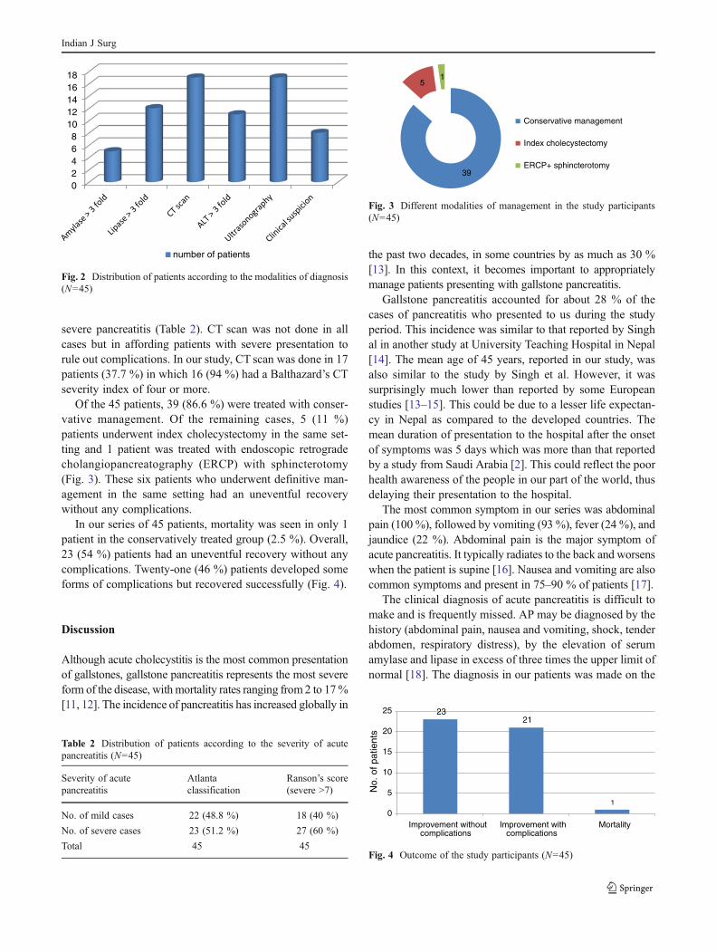
Of the 45 patients, 39 (86.6 %) were treated with conser-vative management. Of the remaining cases, 5 (11 %)patients underwent index cholecystectomy in the same set-ting and 1 patient was treated with endoscopic retrogradecholangiopancreatography (ERCP) with sphincterotomy(Fig. 3). These six patients who underwent definitive man-agement in the same setting had an uneventful recoverywithout any complications.
In our series of 45 patients, mortality was seen in only 1patient in the conservatively treated group (2.5 %). Overall,23 (54 %) patients had an uneventful recovery without anycomplications. Twenty-one (46 %) patients developed someforms of complications but recovered successfully (Fig. 4).
Discussion
Although acute cholecystitis is the most common presentationof gallstones, gallstone pancreatitis represents the most severeform of the disease, withmortality rates ranging from2 to 17%[11, 12]. The incidence of pancreatitis has increased globally in
the past two decades, in some countries by as much as 30 %[13]. In this context, it becomes important to appropriatelymanage patients presenting with gallstone pancreatitis.
Gallstone pancreatitis accounted for about 28 % of thecases of pancreatitis who presented to us during the studyperiod. This incidence was similar to that reported by Singhal in another study at University Teaching Hospital in Nepal[14]. The mean age of 45 years, reported in our study, wasalso similar to the study by Singh et al. However, it wassurprisingly much lower than reported by some Europeanstudies [13–15]. This could be due to a lesser life expectan-cy in Nepal as compared to the developed countries. Themean duration of presentation to the hospital after the onsetof symptoms was 5 days which was more than that reportedby a study from Saudi Arabia [2]. This could reflect the poorhealth awareness of the people in our part of the world, thusdelaying their presentation to the hospital.
The most common symptom in our series was abdominalpain (100 %), followed by vomiting (93 %), fever (24 %), andjaundice (22 %). Abdominal pain is the major symptom ofacute pancreatitis. It typically radiates to the back and worsenswhen the patient is supine [16]. Nausea and vomiting are alsocommon symptoms and present in 75–90 % of patients [17].
The clinical diagnosis of acute pancreatitis is difficult tomake and is frequently missed. AP may be diagnosed by thehistory (abdominal pain, nausea and vomiting, shock, tenderabdomen, respiratory distress), by the elevation of serumamylase and lipase in excess of three times the upper limit ofnormal [18]. The diagnosis in our patients was made on the
02468
1012141618
number of patients
Fig. 2 Distribution of patients according to the modalities of diagnosis(N045)
Table 2 Distribution of patients according to the severity of acutepancreatitis (N045)
Severity of acutepancreatitis
Atlantaclassification
Ranson’s score(severe >7)
No. of mild cases 22 (48.8 %) 18 (40 %)
No. of severe cases 23 (51.2 %) 27 (60 %)
Total 45 45
39
51
Conservative management
Index cholecystectomy
ERCP+ sphincterotomy
Fig. 3 Different modalities of management in the study participants(N045)
2321
1
0
5
10
15
20
25
Improvement without complications
Improvement with complications
Mortality
No.
of p
atie
nts
Fig. 4 Outcome of the study participants (N045)
Indian J Surg
basis of clinical presentation, a raised serum amylase(51 %), lipase (73 %), and ALT (80 %) and ultrasonography(37.7 %). In our study, 82.3 % had either of these parameterssuggestive of AP. The remaining eight cases (17.7 %) had aborderline elevation of amylase or lipase between two andthree times the upper limit of normal, typical clinical fea-tures of pancreatitis, raised liver enzymes, and ultrasono-graphic evidence of cholelithiasis without visualization ofthe pancreas. A CT scan for the confirmation of the diagno-sis, however, could not be done as these patients could notafford the cost of the investigation. None of these patientshad a history of alcohol intake. In light of these cumulativefindings, we made a diagnosis of acute biliary pancreatitis.
Ultrasonography may show pancreatic swelling, but thepancreas is visualized in only 25–50 % of patients withacute pancreatitis. However, it is helpful in establishingbiliary etiology [19]. In our series, gallbladder stones weredetected in all of our patients and common bile duct stonewas visualized in 11.1 %.
In our series, 51 % of patients had severe pancreatitisaccording to Atlanta’s classification and 60 % according toRanson’s scoring. Studies have suggested that about 25 % ofcases will present with severe pancreatitis, the rest present-ing with the mild form of the disease [1]. The high propor-tion of severe cases in our study population might bebecause our patients presented very late to the hospital, thusdelaying timely diagnosis and treatment.
Many authors recommend definitive management in thesame hospital stay in acute biliary pancreatitis. The benefitsinclude less cost of treatment, shorter hospital stay, lesscomplications, and prevention of recurrence [20, 21]. Inour series, six (13 %) patients underwent definitive manage-ment in the same setting (five cholecystectomy and oneERCP and sphincterotomy). These patients had an unevent-ful recovery without any complications.
Overall in our series, 54 % had an uneventful recoveryand 46 % had complications that required other interven-tions. The mortality rate in the conservatively treated groupwas 2.5 % (n01). Other studies have reported complicationrates from 15 to 40 % [9, 22].
The UK guidelines for the management of acute biliarypancreatitis have suggested that all patients with severe acutebiliary pancreatitis should undergo ERCP and sphincterotomywithin 72 h. In mild disease, all fit patients must undergolaparoscopic cholecystectomywith intraoperative cholangiog-raphy, or if not fit for surgery then endoscopic sphincterotomyduring the same admission to prevent further attacks [19].However, definitive management is not always possible in aresource-poor setting such as in our country. Our findingshave suggested that even conservative management of acutebiliary pancreatitis can lead to excellent outcomes. The mor-tality rates that we have reported are comparable to thoseshown by studies from developed countries [21–24].
The main limitation of our study was its retrospectivedesign. As such, we are limited in both the extent and thetype of information available for each patient’s hospitaliza-tion. For example, specific information regarding the type ofpancreatitis (i.e., acute vs. acute on chronic), patient’s bodymass index (BMI), the presence of pancreatic necrosis, and/or the different types of inpatient procedures performedwould have been more helpful in analyzing the specificfactors involved in determining morbidity and mortality.We acknowledge our limitation of not being able to do adetailed imaging in all of our patients as some of ourpatients could not afford more expensive investigations suchas a CT scan. As a result, the diagnosis of acute biliarypancreatitis had to be made based on the clinical presenta-tion and suggestive investigations in some cases. Hence, inorder to validate our findings, further appropriately designedresearches are recommended.
References
1. Fogel EL, Sherman S (2003) Acute biliary peritonitis: when shouldthe endoscopist intervene? Gastroenterology 125:229–235
2. Ramjan M, Hameed F, Ahmad B (2009) Incidence of SIRS inacute biliary pancreatitis. APMC 3:59–62
3. Alexakis N, Neopolemos JP (2005) Algorithm of the diagnosis andtreatment of acute biliary pancreatitis. Scand J Surg 84:124–129
5. Mitchell R, Byrne M, Baillie J (2003) Pancreatitis. Lancet361:1447–1451
6. Sharma SK, Thapa PB, Maharjan DA, Baral N (2009) Influence ofduration of symptoms over perioperative outcomes during emer-gency laparoscopic cholecystectomy. Kathmandu Univ Med J7:120–124
7. Neoptolemos J, Hall A, Finlay D, Berry J, Carr-Locke D, FossardD (1984) The urgent diagnosis of gallstones in acute pancreatitis: aprospective study of three methods. Br J Surg 71:230–233
8. Tenner S, Dubner H, Steinberg W (1994) Predicting gallstonepancreatitis with laboratory parameters: a meta-analysis. Am JGastroenterol 89:1863–1866
10. McFadden DW (1991) Organ failure and multiple organ systemfailure in pancreatitis. Pancreas 6:37–43
11. Abu-Zidan F (2001) Predicting severe pancreatitis. Arch Surg136:1210
12. Eland IA, Sturkenboom MJ, Wilson JH, Stricker BH (2000) Inci-dence and mortality of acute pancreatitis between 1985 and 1995.Scand J Gastroenterol 35:1110–1116
13. Sanjay P, Yeeting S,WhighamC, JudsonHK, Christoph K, PolignanoFM, Tait IS (2009) Management guidelins for gallstone pancreatitis.Are the targets achievable. J Pancreas (Online) 10:43–47
14. Singh DR, Mehta A, Dangol UM (2004) Controversies in themanagement of acute pancreatitis. Kathmandu Univ Med J2:203–207
15. Dube MG, Lobo DN, Rowlands BJ, Beckingham IJ (2001) Auditof acute pancreatitis management: a tale of two hospitals. J R CollSurg Edinb 46:292–299
Indian J Surg
16. Ojetti V, Migneco A, Manno A, Verbo A, Rizzo G, Gentiloni-Silveri N (2005) Management of acute pancreatitis in emergency.Eur Rev Med Pharmacol Sci 9:133–140
17. Webster PD, Spainhour JB (1974) Pathophysiology and manage-ment of acute pancreatitis. Hosp Pract 9:59–66
18. Steinberg W, Goldstein S, Davis N et al (1985) Diagnostic assaysin acute pancreatitis. Ann Intern Med 102:576–580
19. UK Working Party on Acute Pancreatitis (2005) UK guidelines forthe management of acute pancreatitis. Gut 54:1–9
20. Moreau JA, Zinsmeister AR, Melton LJ III, DiMagno EP (1988)Gallstone pancreatitis and the effect of cholecystectomy: a popu-lation based cohort study. Mayo Clin Proc 63:466–473
21. Fan ST, Lai EC, Mok FP, Lo CM, Zheng SS, Wong J (1993) Earlytreatment of acute biliary pancreatitis by endoscopic papillotomy.N Engl J Med 328:228–232
22. Papavramidis TS, Zandes N, Hatzimisios K, Koutsimani Th,Kehagia F, Agorastou P, Doulgerakisand M, Patoulidis I (2008)Acute gallstone pancreatitis: a constant challenge for the surgeon.Indian J Surg 70:224–226
23. Uhl W, Muller CA, Krahenbuhl L, Schmid SW, Scholzel ST, BuchlerMW (1999) Acute gallstone pancreatitis: timing of laparoscopic cho-lecystectomy in mild and severe disease. Surg Endosc 13:1070–1076
24. Liu CL, Lo CM, Fan ST (1997) Acute biliary pancreatitis: diag-nosis and management. World J Surg 21:149–154