145
Acute Rehabilitation Services Orientation Manual

Acute
Rehabilitation
Services
Orientation
Manual

Table of Contents
1. General Information
a. Orientation Checklist (paper copy only)
b. Mission/Vision
c. Organization Chart
d. Kronos
e. Paycodes
f. Productivity Explanation
g. Floors
h. Door Codes/Phone Numbers (paper copy only)
i. Pager Protocol
j. Rehabilitation Phone/Pager List (paper copy only)
k. Star Voicemail Instructions
l. Ordering Equipment
m. Discharge Planning Criteria
n. Occupational Therapy Prioritization Guidelines
o. Physical Therapy Prioritization Guidelines
p. OT and PT Recommended Treatment Frequencies
q. OT Six Clicks (AM-PAC)
r. PT Six Clicks (AM-PAC)
s. Lab Values Interpretation
t. Billing/CPT Codes
u. Continuing Education Approval
v. Functional Outcomes Measures
w. Orientation Quiz
x. Acute Care Physical Therapy: Tips and Tricks
2. Area Specific
a. Hematology/Oncology/Stem Cell (11n, 10t, outpatient)
b. Burn (11s, outpatient)
c. Trauma (10n)
d. Ortho Trauma (10s)
e. SICU (9t)
f. Surgical Stepdown (9n, 9s)
g. MICU (8t)
h. General Medicine (8n) and Cardiac Stepdown (8s)
i. Transplant and Surgical Care Unit (7t)
j. Cardiac (7s, 7n, 5s, COBS)
k. Spine (6s)
l. Neurology/Neurosurgery/NICU (6n,6t)
m. CVICU (5n)
n. Gynecology / Oncology (4East)
o. TOBS
p. ED
q. Recovery Room/Medical Center East
r. MCN (3, 4, 5, 6, 7)
s. VPH

Mission/Vision

Kronos

Paycodes
Bi- Weekly (Non- Exempt) Pay Codes
PAY CODES DESCRIPTION
BRV Bereavement
D01 $1 Premium Pay
D02 $2 Premium Pay
D03 $3 Premium Pay
D04 $2.50 Premium Pay
D05 $5 Premium Pay
D10D $10 Premium Pay
D20D $20 Premium Pay
D25 $25 Premium Pay
D30D $30 Premium Pay
D4A $4 Premium Pay
FMLA Record Purposes Only
JRY1 Jury Duty (shift 1)
JRY2 Jury Duty (shift 2)
JRY3 Jury Duty (shift 3)
L02D Lead Differential Pay
NonFMLA Record Purposes Only
NWK Non-Worked
OCN On-Call
OFS1 Offsite (shift 1)
OFS2 Offsite (shift 2)
OFS3 Offsite (shift 3)
OUT-Unpaid Record Purposes Only
OCV On-Call Visit (Vanderbilt Home Care
Only)
P15 Premium used by (VUMC Parking Only)
P35 Premium used by (VUMC Parking Only)
PAL 1 Paid Administrative Leave (shift 1)
PAL 2 Paid Administrative Leave (shift 2)
PAL 3 Paid Administrative Leave (shift 3)
PLN Paid Parental Leave
PNS Flex PTO - Scheduled
PNU Flex PTO - Unscheduled
SNS Grandfathered Sick - Scheduled
SNU Grandfathered Sick - Unscheduled
WORK RULE(Class Code) SPECIFIC DESCRIPTION
CB2 Call Back 2 Hour Min
CBO Call Back with Overtime
FOT Float with Overtime
INS In-Service
ORI Orientation
Meal – Unpaid Meal break Hours - Unpaid
Temp Departure – Unpaid Temporary Departure - Unpaid
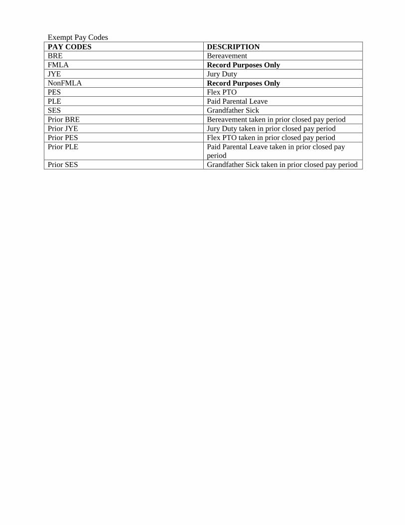
Exempt Pay Codes
PAY CODES DESCRIPTION
BRE Bereavement
FMLA Record Purposes Only
JYE Jury Duty
NonFMLA Record Purposes Only
PES Flex PTO
PLE Paid Parental Leave
SES Grandfather Sick
Prior BRE Bereavement taken in prior closed pay period
Prior JYE Jury Duty taken in prior closed pay period
Prior PES Flex PTO taken in prior closed pay period
Prior PLE Paid Parental Leave taken in prior closed pay
period
Prior SES Grandfather Sick taken in prior closed pay period
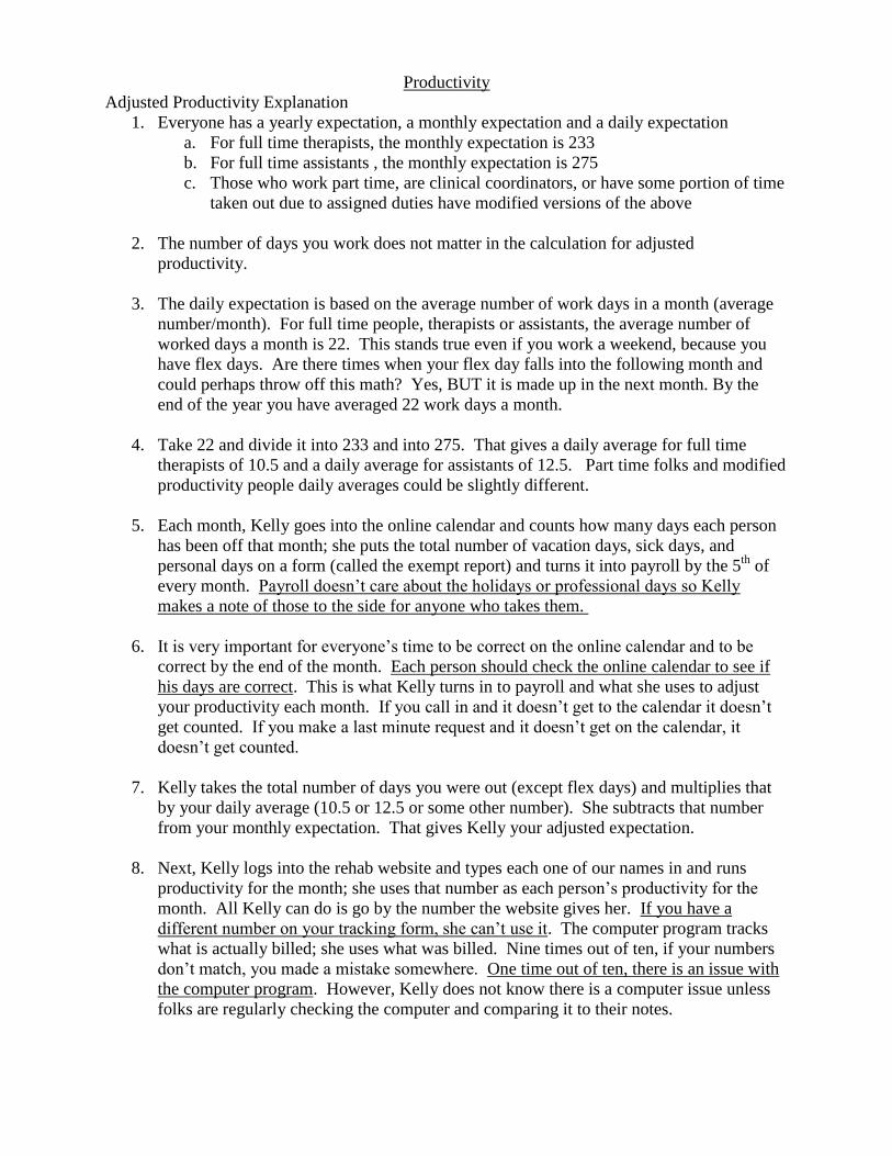
Productivity
Adjusted Productivity Explanation
1. Everyone has a yearly expectation, a monthly expectation and a daily expectation
a. For full time therapists, the monthly expectation is 233
b. For full time assistants , the monthly expectation is 275
c. Those who work part time, are clinical coordinators, or have some portion of time
taken out due to assigned duties have modified versions of the above
2. The number of days you work does not matter in the calculation for adjusted
productivity.
3. The daily expectation is based on the average number of work days in a month (average
number/month). For full time people, therapists or assistants, the average number of
worked days a month is 22. This stands true even if you work a weekend, because you
have flex days. Are there times when your flex day falls into the following month and
could perhaps throw off this math? Yes, BUT it is made up in the next month. By the
end of the year you have averaged 22 work days a month.
4. Take 22 and divide it into 233 and into 275. That gives a daily average for full time
therapists of 10.5 and a daily average for assistants of 12.5. Part time folks and modified
productivity people daily averages could be slightly different.
5. Each month, Kelly goes into the online calendar and counts how many days each person
has been off that month; she puts the total number of vacation days, sick days, and
personal days on a form (called the exempt report) and turns it into payroll by the 5th
of
every month. Payroll doesn’t care about the holidays or professional days so Kelly
makes a note of those to the side for anyone who takes them.
6. It is very important for everyone’s time to be correct on the online calendar and to be
correct by the end of the month. Each person should check the online calendar to see if
his days are correct. This is what Kelly turns in to payroll and what she uses to adjust
your productivity each month. If you call in and it doesn’t get to the calendar it doesn’t
get counted. If you make a last minute request and it doesn’t get on the calendar, it
doesn’t get counted.
7. Kelly takes the total number of days you were out (except flex days) and multiplies that
by your daily average (10.5 or 12.5 or some other number). She subtracts that number
from your monthly expectation. That gives Kelly your adjusted expectation.
8. Next, Kelly logs into the rehab website and types each one of our names in and runs
productivity for the month; she uses that number as each person’s productivity for the
month. All Kelly can do is go by the number the website gives her. If you have a
different number on your tracking form, she can’t use it. The computer program tracks
what is actually billed; she uses what was billed. Nine times out of ten, if your numbers
don’t match, you made a mistake somewhere. One time out of ten, there is an issue with
the computer program. However, Kelly does not know there is a computer issue unless
folks are regularly checking the computer and comparing it to their notes.
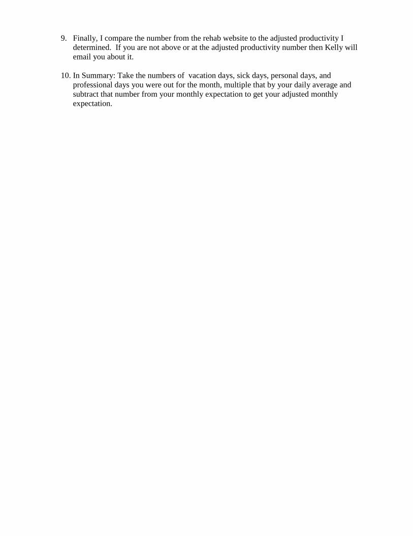
9. Finally, I compare the number from the rehab website to the adjusted productivity I
determined. If you are not above or at the adjusted productivity number then Kelly will
email you about it.
10. In Summary: Take the numbers of vacation days, sick days, personal days, and
professional days you were out for the month, multiple that by your daily average and
subtract that number from your monthly expectation to get your adjusted monthly
expectation.

Floors
Floors: VUH
11 North: Hematology/General Medicine 11 South: Burns
10 North: Trauma 10 South: Ortho Trauma
10 Tower: Oncology/Stem Cell 9 North: Surgical Stepdown
9 South: General Surgery 9 Tower: Surgical ICU (SICU)
8 North: General Medicine 8 South: Cardiac Stepdown
8 Tower: Medical ICU (MICU) 7 North: Cardiac Stepdown
7 South: Non-surgical Cardiac 7 Tower: Transplant & Surgical Care Unit
6 North: Neurology /Neurosurgery Stepdown 6 Tower: Neuro ICU
6 South: Spine Surgery 5 North: Cardiovascular ICU (CVICU)
5 South: Cardiac Surgery 4 East: Gynecology / Oncology
3 North: Main Recovery Room 3 MCE: Medical Center East Outpatient Surgery
1st Floor: Emergency Room and TOBS
Floors: MCN
S74: ACE Unit (Geriatric) S64: Orthopedics
S54: Palliative Care and General Medicine S44: Colorectal Surgery and General Medicine
S34: General Medicine
Pager
Rehab Pager Protocol
1. Requests to rehab pager can be text page, numeric page, or voicemail. To check
voicemail: Dial rehab pager: 835-1147, wait for voice message to begin, enter password:
*191919. For numeric page, call phone number provided and gather necessary
information found on Rehab Pager Tracking Form.
2. All requests to the rehab pager should be redirected to the primary OT and/or PT
assigned to the respective floor, even if the patient is assigned to an assistant. This allows
the OT and/or PT familiar with the case load to follow up with interdisciplinary team and
to determine the priority of treatments, if needed. The respective OT /PT is also
responsible for contacting the COTA/PTA if they are needed to address an updated
note/treatment request, as well as to re-prioritize treatments if necessary. Refer to the
daily list of OT/PT floor assignments for pager numbers.
3. If the necessary information (see Tracking Form) is provided in the request, simply
contact the respective OT/PT via text or page with the information.
4. If the information is not provided, return the page or voicemail, gather necessary
information, and then forward to respective OT/PT.
5. Group pages to all OTs and/or PTs will no longer be necessary when there is a primary
therapist assigned where the patient is located. If there is no therapist assigned, i.e.
TOBS, EMER, CTU, MCE3, VPH, a group page may be necessary to find a therapist to
address the request. Or, the person responding to the rehab pager may need to address the
request personally.
6. Record each request on Tracking Form and leave form in folder taped to side of Mark’s
desk.
Acceptable requests for therapy:
1. Updated note – legitimate requests are only when the following criteria are met:
a. Discharge recommendation is placement (SNF or IPR), and
b. Expected discharge day is the same day or the following day, and
c. The patient has not been treated in over 48 hrs.
2. Treatment, not an updated note – these requests are typically not a priority and may be
left on the treatment list for the following day. This should be determined by OT/PT
assigned to floor.
3. If therapy documentation does not support discharge recommendation, patient may need
to be seen for a treatment to change recommendation or for more supportive
documentation. This should be determined by OT/PT assigned to floor.
4. Requests for custom hand splints.
Unacceptable requests for therapy:
1. Evaluation request if there are no therapy orders. Advise caller to have orders entered.
Therapist does not enter order.
2. Request to change discharge recommendation from placement (SNF or IPR) to home due
to improved status is not necessary. However, the team may still request final therapy
treatment to ensure home safety/caregiver training. This should be determined by OT/PT
assigned to floor.
3. Back braces and other braces recommended by Otrho are not the responsibility of
therapist, i.e. requests for brace adjustments or initial donning/fitting of brace. Bledsoe

boots, hinged knee braces, post-op shoes, knee immobilizers, etc. that are recommended
by Ortho need to be placed by the Ortho cast tech (Refer caller to room number 936-
1196). For back brace adjustment requests, refer caller to brace company (Superior, 615-
340-0068 or Applied Orthotics aka Bulow, 615-327-9343). If a brace is ordered for
patient and is not on the patient when the therapist arrives, the evaluation should be held.
4. Ordering equipment, i.e. canes, walkers – Refer caller to Service Center, 3-9600.
5. Request to transfer patient back to bed. Advise caller that nursing staff can assist and may
use a Stedy or lift if necessary.
Repeated calls regarding same patient:
1. Inform caller that request to pager has already been made by another team member. Ask
caller if the patient was discussed in huddle. If not, advise them to discuss patient with
CM or NP who attends huddle in the future, to prevent repeat calls to pager.
Ways to send a page:
1. Open StarPager in Starpanel. Click on StarPager and enter information into labeled fields.
Then click sent.
2. Go to www.satellink.net. Then click on the link marked Send-A-Page. Enter the pagers
10 digit phone number and enter message. Then click send.
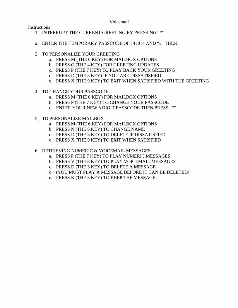
Voicemail
Instructions
1. INTERRUPT THE CURRENT GREETING BY PRESSING “*”
2. ENTER THE TEMPORARY PASSCODE OF 147014 AND “#” THEN:
3. TO PERSONALIZE YOUR GREETING
a. PRESS M (THE 6 KEY) FOR MAILBOX OPTIONS
b. PRESS G (THE 4 KEY) FOR GREETING UPDATES
c. PRESS P (THE 7 KEY) TO PLAY BACK YOUR GREETING
d. PRESS D (THE 3 KEY) IF YOU ARE DISSATISFIED
e. PRESS X (THE 9 KEY) TO EXIT WHEN SATISFIED WITH THE GREETING
4. TO CHANGE YOUR PASSCODE
a. PRESS M (THE 6 KEY) FOR MAILBOX OPTIONS
b. PRESS P (THE 7 KEY) TO CHANGE YOUR PASSCODE
c. ENTER YOUR NEW 6 DIGIT PASSCODE THEN PRESS “#”
5. TO PERSONALIZE MAILBOX
a. PRESS M (THE 6 KEY) FOR MAILBOX OPTIONS
b. PRESS N (THE 6 KEY) TO CHANGE NAME
c. PRESS D (THE 3 KEY) TO DELETE IF DISSATISFIED
d. PRESS X (THE 9 KEY) TO EXIT WHEN SATISFIED
6. RETRIEVING NUMERIC & VOICEMAIL MESSAGES
a. PRESS P (THE 7 KEY) TO PLAY NUMERIC MESSAGES
b. PRESS V (THE 8 KEY) TO PLAY VOICEMAIL MESSAGES
c. PRESS D (THE 3 KEY) TO DELETE A MESSAGE
d. (YOU MUST PLAY A MESSAGE BEFORE IT CAN BE DELETED)
e. PRESS K (THE 5 KEY) TO KEEP THE MESSAGE

Ordering Equipment
1. Call Service Center 39600 (in VUH) or 31354 (in MCN)
2. Some adaptive dressing equipment and leg lifter can be found on 10s and 6th
floor MCN
service centers
3. Typical equipment ordered from Service Center
a. Rolling walker
i. Inform them if standard or bariatric is needed
ii. Make sure you tell them to bring wheels
iii. Typically can only order walker or cane for patient not both
iv. Come out of floors “budget” and not usually billed to patient
b. Platform attachments for rolling walker
c. Cane
d. Reacher
e. Sock aid
f. Long handled sponge
g. Leg lifter (often referred to as “leash” by other disciplines
h. Long handled shoe horn
i. Walker basket
j. “Scrotal support” (ie jock strap)
k. Multipodus boots
l. Pressure relief boots
4. Therapy does not order from service center (nursing or MD must order)
a. Knee immobilizer
b. Postop shoe
c. Cam walker boot
d. Bledsoe boot
e. Ankle air cast
f. Wrist cockup prefabricated splint
i. MD typically needs to sign a DME form
ii. Form needs to be filled out and returned to Service Center

Discharge Planning Criteria
1. Inpatient Rehabilitation (IPR):
a. Patient needs to be able to tolerate 3 hours of therapy per day or 15 hours over 7
days, for certain diagnoses. This should be communicated with the team or
documented
b. Patient needs to require 2 disciplines. PT and OT, OT and SLP, or SLP and PT
c. It is required that 2 disciplines provide evaluations supporting the patient’s need
for rehabilitation placement for acceptance
d. Observation patients can be discharged directly to inpatient rehab
e. Inpatient pulmonary rehabilitation is only approved for patients with a pulmonary
diagnosis and new or increased oxygen requirements ; the patients must also meet
inpatient rehabilitation criteria
2. Skilled Nursing Facility (SNF):
a. Patients need at least PT to recommend SNF placement, but more insurance
carriers require both PT and OT
b. Patient can be skilled for other reasons besides therapy: IV antibiotics, new
feeding tube, and wound care for stage III or IV decubiti
c. Must be hospitalized 3 midnights with inpatient status
d. Observation patients cannot be discharged directly to a SNF with a few
exceptions including if a physician speaks to the medical director of the insurance
company
e. To meet criteria for SNF placement, the patient must have a therapy need and
benefit from daily therapy.
f. Many facilities have SNF and long term care in the same building, therapy is not
required for long term care placement.
3. Assisted Living Facility (ALF) - not a therapy recommendation, this is determined by
social work and case management
a. This is not a therapy recommendation, from a therapy perspective, we typically
recommend home health
b. A patient will have to pay out of pocket. Insurance does not cover ALF
placement. Average median monthly cost is $3,395
(www.tnassistedlivingfacilities.org/directory/tn/ retrieved 3/08/17).
c. To meet admission criteria, patients must be independent with transfers,
ambulation, wheelchair mobility, ADL, and medication management; otherwise,
the patient may have to pay additional fees depending upon the facility
requirements.
d. The patient cannot transfer from the hospital to an ALF, they have to meet certain
admission criteria before an ALF will accept them
e. It is a good idea to collaborate with care team for these patients to ensure facilities
can meet the patient’s needs based on information obtained in the evaluation.
4. Home with hospice:
a. Patient must have a caregiver 24/7
b. The patient can receive PT and OT while receiving hospice care, but many
hospice companies do not provide services. Often, this is because they cannot
afford to provide it. With this being said, document specifics to why therapy is
recommended.
c. If home PT and OT is recommended, the specifics regarding what therapy is
needed at home will need to be documented (i.e. family training, etc)

5. Home with Continuous Assistance:
a. This does not mean 24/7 assist, ONLY document what the patient needs
continuous assist performing in the discharge recommendation
b. If it is not clear if the patient will have continuous assist at home, the patient must
meet criteria in order for therapy to recommend SNF or inpatient rehabilitation
placement. You cannot recommend SNF or inpatient rehabilitation based solely
on social issues.
6. Home with Outpatient Therapy:
a. The patient needs to have transportation to and from therapy
b. Some specialty diagnoses are more suited for outpatient therapy rather than home
health such as: vestibular rehabilitation, some orthopedic diagnoses, higher level
neurological conditions
c. A patient cannot receive outpatient therapy if he/she is receiving home health
nursing
7. Home with Home Health Therapy:
a. The patient must be home bound, which means they can only leave for medical
reasons and church
b. In order to get home health OT, nursing, Speech Therapy, or PT must ‘open the
case.’
8. Intermediate Care Facility (ICF) – not a therapy recommendation, this is determined by
social work and case management
a. Nursing home placement without a skilled intervention
b. If the patient was admitted from a ICF and lives there, this is considered their
home
c. These facilities are sometimes confused with a SNF and an evaluation order might
be triggered
d. If the patient is receiving therapy in the ICF side, Medicare B is billed.
9. Long Term Acute Care (LTAC) - not a therapy recommendation, this is determined by
social work and case management
a. Patients meet criteria for medical reasons outside of therapy (i.e. complex wound
care, vent dependence, hemodynamically fragile).
b. No therapy evaluation is needed for admission
c. Patients can receive therapy at the LTAC

Occupational Therapy Prioritization Guidelines
General Principles
1. Patients who can’t be discharged until seen by OT must be seen as soon as possible.
2. Patients who are able to participate will be seen before those who are not (participate =
awake/alert, follows commands).
3. Extubated patients will be seen before those who are intubated (exception: intubated with
RASS score of -1 and following commands.
4. Patients who are not in restraints will be seen before those in restraints (exception are people
who have CHI or restraints per protocol)
5. Patients who are in dialysis when orders are received will be seen the next day.
6. Patients who require bracing (TLSO, Aspen, Quick Draw, Knee Brace, Bledsoe, etc.) must
have brace before OT evaluation or treatment will be completed.
7. Last minute requests (verbal or written) for either new consults or a treatment may not be
seen if it is after 3:00 p.m.; however, every attempt will be made to accommodate the need of
the patient and/or the team.
8. Post – surgical patients that the MD would like up day of surgery need to be on the floor by
2:00 p.m. for time for the nerve block to wear off. Patients must be alert.
9. OT orders that are entered to motivate a patient are not appropriate orders. Patients who are
able to mobilize but refuse to do not qualify for skilled OT.
10. Refusals: Patients who adamantly refuse therapy (i.e. “I don’t need therapy”) will be
discharged. Patients who refuse OT will be seen the following day. If patient refuses
therapy a third time, he/she will be discharged from therapy.
Prioritization (Organized by Service and ranked from Highest to Lowest)
1. Note: It is assumed that OT will start with Patient Closest to Discharge
2. The following services must be seen the day order is issued.
a. 23 hour admit/observation patients
b. POD-3 STSG
c. Hand patients
d. Splinting/Casting – All plastics and ortho splints/casts that must be completed
e. Whirlpools
3. Splinting/Casting – splinting/casting for general positioning or prevention of contractures
4. Neurosurgery – Elective Backs & Craniotomies
5. Trauma
6. Orthopedics
7. General Neurosurgery (including Neurosurgery ICU)
8. Neurology
9. General Burns
10. Medicine
11. Surgery
a. Transplant
b. General
c. Tumor
d. Colo-Rectal
e. Head & Neck Cancer
12. Non-Surgical Cardiac
13. Surgical Cardiac
14. Geriatrics
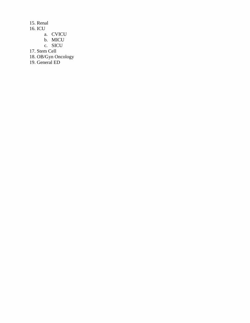
15. Renal
16. ICU
a. CVICU
b. MICU
c. SICU
17. Stem Cell
18. OB/Gyn Oncology
19. General ED

Physical Therapy Orientation Guidelines
General Principles
1. Patients who are able to participate will be seen before those who are not (participate =
awake/alert, follows commands consistently).
2. Extubated patients will be seen before those who are intubated (exception: intubated with
RASS score of -1 and following commands.
3. Patients who are not in restraints will be seen before those in restraints (exception are people
who have CHI or restraints per protocol)
4. Patients who are going to OR within 3 days of receiving orders will be seen after their
surgery (exception is trauma: delayed surgery patients).
5. Patients who are in dialysis when orders are received will be seen the next day.
6. Bed rest orders are commonly seen: These patients become lower priority; Bed rest orders
indicate that the patient must remain in bed (example of true bed rest is unstable spine). Bed
rest does not indicate that the patient may be up with assistance or up for therapy.
Recommend update activity orders when entering PT orders if true bed rest is not desired.
7. Screens should be done by MD before PT order is put in for equipment needed for patient to
mobilize: We do not fit for braces (i.e. TLSO, Aspen Quick Draw, Hinged Knee Brace,
Immobilizer, Bledsoe)
8. Last minute requests (verbal or written) for either new consults or a treatment may not be
seen if it is after 3:00 p.m.; however, every attempt will be made to accommodate the need of
the patient and/or the team.
9. Post – surgical patients that the MD would like up day of surgery need to be on the floor by
2:00 p.m. for time for the nerve block to wear off. Patients must be alert.
10. PT orders that are entered to motivate a patient are not appropriate orders. Patients who are
able to mobilize but refuse to do not qualify for skilled PT.
11. Refusals: Patients who adamantly refuse therapy the first time (i.e. “I don’t need therapy”)
will be discharged immediately. Patients who do not adamantly refuse will be seen the next
day. If they refuse again, therapy orders will be discharged.
High Priority Patients: Evaluation and Treatments
1. Total Joint Patients
2. Ortho Oncology
3. Discharge Planning
4. Patients who can be discharged as soon as PT evaluates them
5. Uninsured patients who are discharging home and can actively participate in therapy (we
may be the only therapy they get)
6. Evaluation for discharge planning with plan already in place for next day
7. Vascular surgery (amputations) – require early discharge planning
8. Plastics patients who are off bed rest and do not have sitting restrictions
9. GSWs with nerve injuries - Need DC recs quickly
10. Multi Trauma with non-op fxs - Need DC recs quickly
11. Trauma Evals for patients admitted to Receiving - Need DC recs quickly
12. Isolated CHIs that are able to participate in PT - Need DC recs quickly
13. New Paraplegics: extubated, spine issues dealt with
14. Burn
a. Burn patients on the step down side that have a burn which crosses a joint (this
places them at high risk for contractures)
b. POD 3 LE STSG (MD protocol states that ROM is re-initiated on POD 3)

c. Dorsal foot burns
15. Unique Therapies
a. Whirlpools (driven by physician order)
b. Custom splints (splints or casts that are fabricated by therapy)
c. Baclofen Trials: PT performs assessment to determine if trial is successful.
16. Short Stay
a. ED patients with “observation” orders (not being admitted to the hospital) -
Medicare guidelines driving quick PT evaluations
b. TIA patients - Very short admissions
Medium Priority Patients
17. Post Op/Discharge Planning
a. Organ transplants – POD 2 or later is better for discharge planning and gives a
clearer picture of the patient’s abilities.
b. POD 1 donor sites
c. Evaluations asking for discharge recommendations, but the patient is not
discharging the day the order is received.
d. Abdomial surgeries, Cardiothoracic surgeries, Colorectal surgeries, Gyn/Onc
surgeries, Vascular surgeries (non-amputations) and Whipple surgeries – POD 2
gives a clearer picture of abilities
e. New CVA’s - usually unavailable the day of admit and the day after admit
f. Neurosurgery spine surgeries – significant pain POD 1; pain much improved POD
2 and 3
18. Other
a. LE burns not crossing a joint
b. Geriatric Evals (those from ALF/home/independent living)
c. H&N patients - drains come out at later dates for exercise
d. CHI’s
e. ED pt’s with admit orders
f. Multi-trauma pts admitted to ICU
g. New quads: not vent dependent/or on trach collar trials
Low Priority Patients
19. No Discharge Needs
20. SJS (TENS) on BU - no immediate discharge needs
21. Nursing home residents - discharge usually established
22. Exacerbations of disease - usually recover; clearer picture later in admission.
23. “Prevent de-conditioning” patients – frequently do not require skilled PT
24. Readmits from rehab facilities: may return quickly to rehab facility.
25. Nursing Appropriate
a. Orthospine surgeries without neuro deficit - rarely have PT needs.
b. Swanns - limited mobility (physician guideline) - nursing can get OOB
c. Bariatric patients (PT tends to receive orders because nursing is not comfortable
getting the patient up)
Weekends: High Priorities
26. Total joints
27. Ortho Oncology

28. POD3 burns
29. Burns crossing joint
30. Dorsal burns
31. Pediatric burns
32. Patients discharging over the weekend and need one more treatment
33. Isolated fractures if impacting their discharge
34. Whirlpools
35. Custom splints
36. Plastics patients off bed rest and ambulation can be initiated
37. Patients who need updated notes for discharge on Monday can be seen by therapy on
Monday morning and the note documented immediately following.

OT and PT Recommended Treatment Frequencies
Orthopedic
PT OT
Total Hip's BID 7 days a week 1 Visit
Total Knees BID 7 days a week 1 Visit
Single and B Knees B Knees
Isolated Fractures 7X/wk 7X/wk
IPOP 7X/wk NA
Tibial Plateau Ex-Fix Removal BID 7 days a week NA
Burns
Deep Partial/Full Thickness that Crosses 1 or more Joints and/or Affects ROM of a Joint
PT OT
BID 7X/wk BID 7X/wk
PT Exception: If intubated and sedated 3X/Week
Partial Thickness that Crosses 1 or more Joints and/or Affects ROM of a Joint
5X/wk 5X/wk
Superficial Burns/Partial Thickness Burns that do NOT Cross a Joint or Affect ROM
1X for education 1X for education
Donor Sites
1X for education NA
Oncology
Allo Stem Cell Transplant
PT OT
3X/wk without Steroid Myopathy 2X/wk
Up to 5X/wk with Steroid Myopathy 2X/wk
Auto Stem Cell Transplant
2X/wk 2X/wk
Head and Neck Cancer
3X/wk 2 Visits
Gyn Onc
3X/wk 2 - 3X/wk
General Oncology
PT OT
3X/wk
3 Visits unless ADL issues
con't
Trauma
Multi-Trauma
PT OT
Up to 5X/wk Up to 5X/wk
Isolated CHI
RANCHO I - II 1-2X/wk 1 - 2X/wk
RANCHO III 2X/wk 2X/wk
RANCHO IV & Up 3X/wk 3-4X/wk
Internal Traumatic Injuries
3X/wk 3X/wk
Paraplegic (New Onset)
4X/wk 4X/wk
Quadraplegic: (Complete, New)
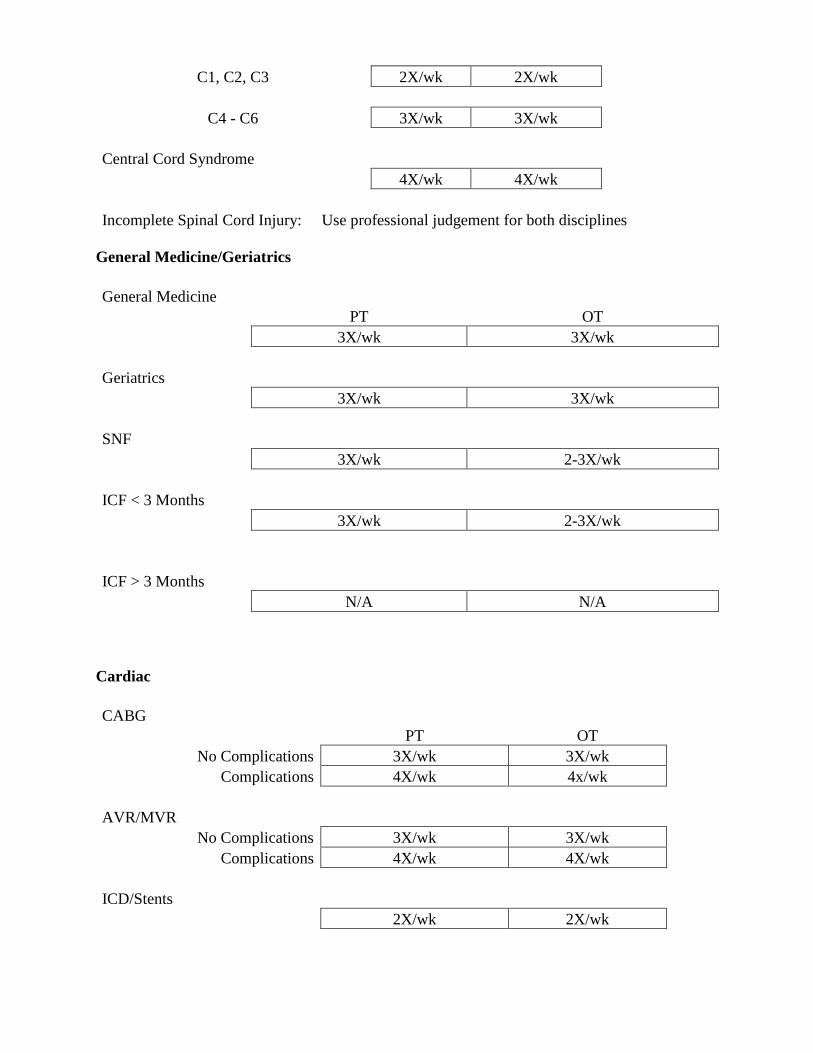
C1, C2, C3 2X/wk 2X/wk
C4 - C6 3X/wk 3X/wk
Central Cord Syndrome
4X/wk 4X/wk
Incomplete Spinal Cord Injury: Use professional judgement for both disciplines
General Medicine/Geriatrics
General Medicine
PT OT
3X/wk 3X/wk
Geriatrics
3X/wk 3X/wk
SNF
3X/wk 2-3X/wk
ICF < 3 Months
3X/wk 2-3X/wk
ICF > 3 Months
N/A N/A
Cardiac
CABG
PT OT
No Complications 3X/wk 3X/wk
Complications 4X/wk 4x/wk
AVR/MVR
No Complications 3X/wk 3X/wk
Complications 4X/wk 4X/wk
ICD/Stents
2X/wk 2X/wk
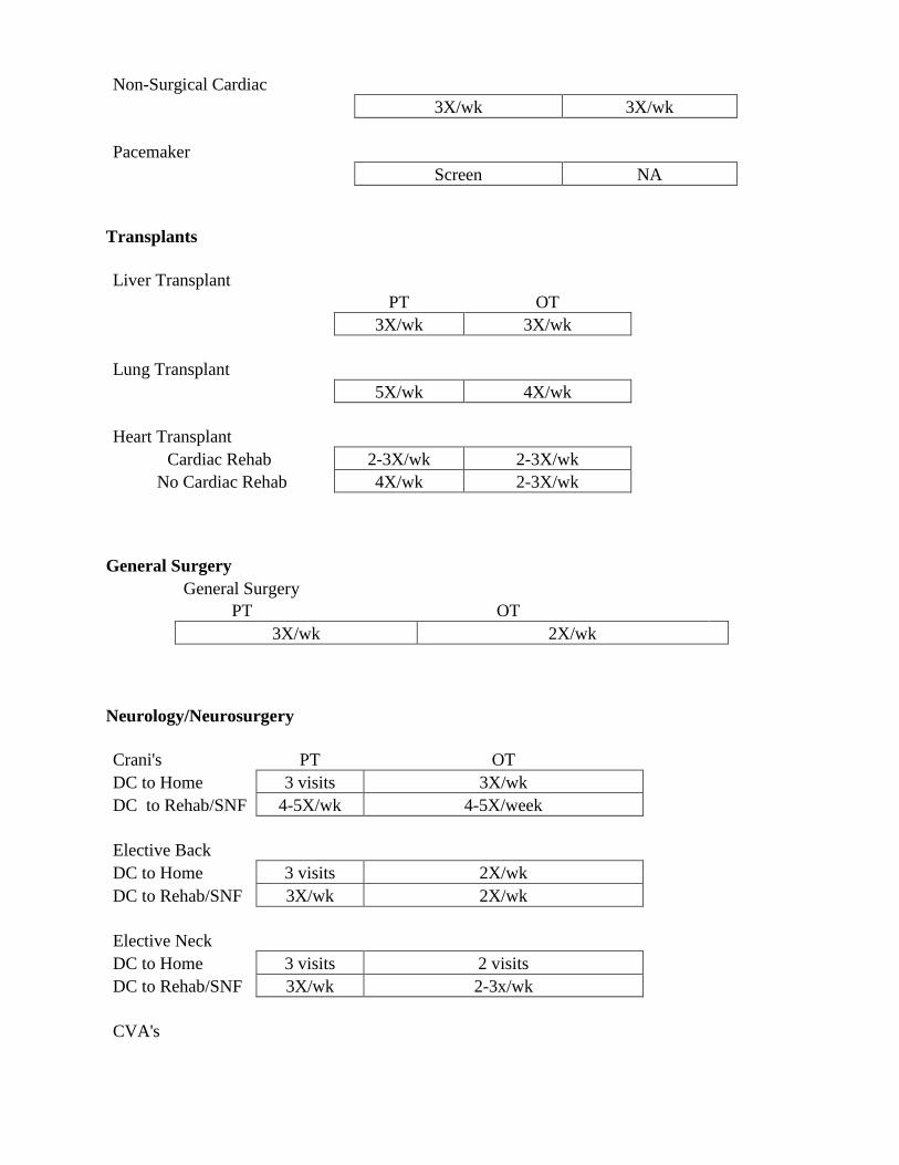
Non-Surgical Cardiac
3X/wk 3X/wk
Pacemaker
Screen NA
Transplants
Liver Transplant
PT OT
3X/wk 3X/wk
Lung Transplant
5X/wk 4X/wk
Heart Transplant
Cardiac Rehab 2-3X/wk 2-3X/wk
No Cardiac Rehab 4X/wk 2-3X/wk
General Surgery
General Surgery
PT OT
3X/wk 2X/wk
Neurology/Neurosurgery
Crani's PT OT
DC to Home 3 visits 3X/wk
DC to Rehab/SNF 4-5X/wk 4-5X/week
Elective Back
DC to Home 3 visits 2X/wk
DC to Rehab/SNF 3X/wk 2X/wk
Elective Neck
DC to Home 3 visits 2 visits
DC to Rehab/SNF 3X/wk 2-3x/wk
CVA's
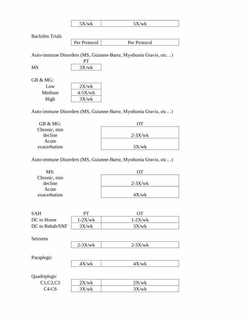
5X/wk 5X/wk
Baclofen Trials
Per Protocol Per Protocol
Auto-immune Disorders (MS, Guianne-Barre, Mysthenia Gravis, etc…)
PT
MS 3X/wk
GB & MG:
Low 2X/wk
Medium 4-5X/wk
High 3X/wk
Auto-immune Disorders (MS, Guianne-Barre, Mysthenia Gravis, etc…)
GB & MG:
OT
Chronic, min
decline
2-3X/wk
Acute
exacerbation
5X/wk
Auto-immune Disorders (MS, Guianne-Barre, Mysthenia Gravis, etc…)
MS:
OT
Chronic, min
decline
2-3X/wk
Acute
exacerbation
4X/wk
SAH PT OT
DC to Home 1-2X/wk 1-2X/wk
DC to Rehab/SNF 3X/wk 3X/wk
Seizures
2-3X/wk 2-3X/wk
Paraplegic
4X/wk 4X/wk
Quadriplegic
C1,C2,C3 2X/wk 2X/wk
C4-C6 3X/wk 3X/wk
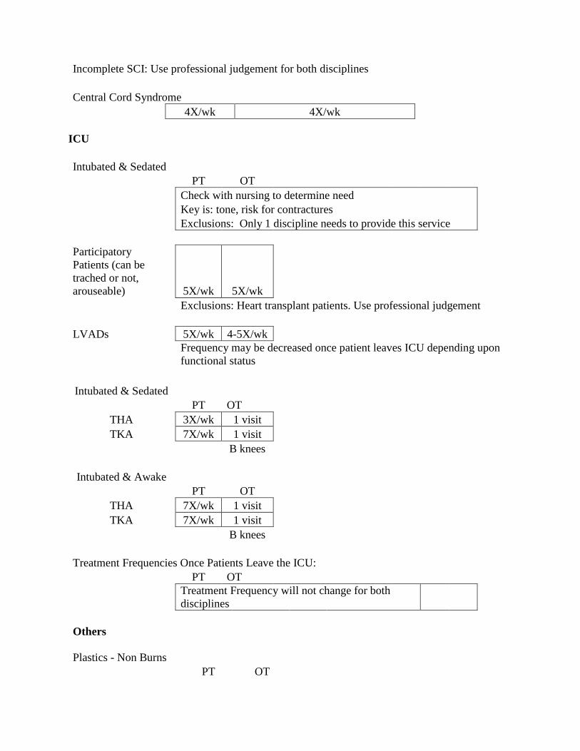
Incomplete SCI: Use professional judgement for both disciplines
Central Cord Syndrome
4X/wk 4X/wk
ICU
Intubated & Sedated
PT OT
Check with nursing to determine need
Key is: tone, risk for contractures
Exclusions: Only 1 discipline needs to provide this service
Participatory
Patients (can be
trached or not,
arouseable) 5X/wk 5X/wk
Exclusions: Heart transplant patients. Use professional judgement
LVADs 5X/wk 4-5X/wk
Frequency may be decreased once patient leaves ICU depending upon
functional status
Intubated & Sedated
PT OT
THA 3X/wk 1 visit
TKA 7X/wk 1 visit
B knees
Intubated & Awake
PT OT
THA 7X/wk 1 visit
TKA 7X/wk 1 visit
B knees
Treatment Frequencies Once Patients Leave the ICU:
PT OT
Treatment Frequency will not change for both
disciplines
Others
Plastics - Non Burns
PT OT

3X/wk 3X/wk
Not on bedrest is the key
Gyn
1 Visit N/A
Vascular Surgery
4X/wk 2-3X/wk

OT Six Clicks (AM-PAC)
General Guidelines
1. Complete for all evaluations and treatments completed
2. Is linked to a G-code if patient is an outpatient/observation status and has Medicare part
B then G code will be “dropped” for billing
3. Is based on 1 person assistance and no adaptive equipment/assistive devices
a. For example, if patient is moderate assistance x2 then they become maximal
assistance
b. For example, if patient is maximal assistance x2 they become dependent
4. Rate patient on level of difficulty with personal & instrumental/daily activity
a. Lower body dressing
b. Bathing (washing, rinsing, drying)
c. Toileting (toilet, bedpan, urinal)
d. Upper body dressing
e. Personal grooming
f. Eating Meals
5. Levels of difficulty
a. Dependent (unable)
b. Mod assist or Max assist (a lot)
c. SBA or Min assist (a little)
d. Modified independent to independent (none)
6. Produces outcome score out of 24 with a modifier
a. CH (0% impaired)
b. CI (at least 1% but less than 20% impaired)
c. CJ (at least 20% but less than 40% impaired)
d. CK (at least 40% but less than 60% impaired)
e. CL (at least 60% but less than 80% impaired)
f. CM (at least 80% but less than 100% impaired)
g. CN (100% impaired)
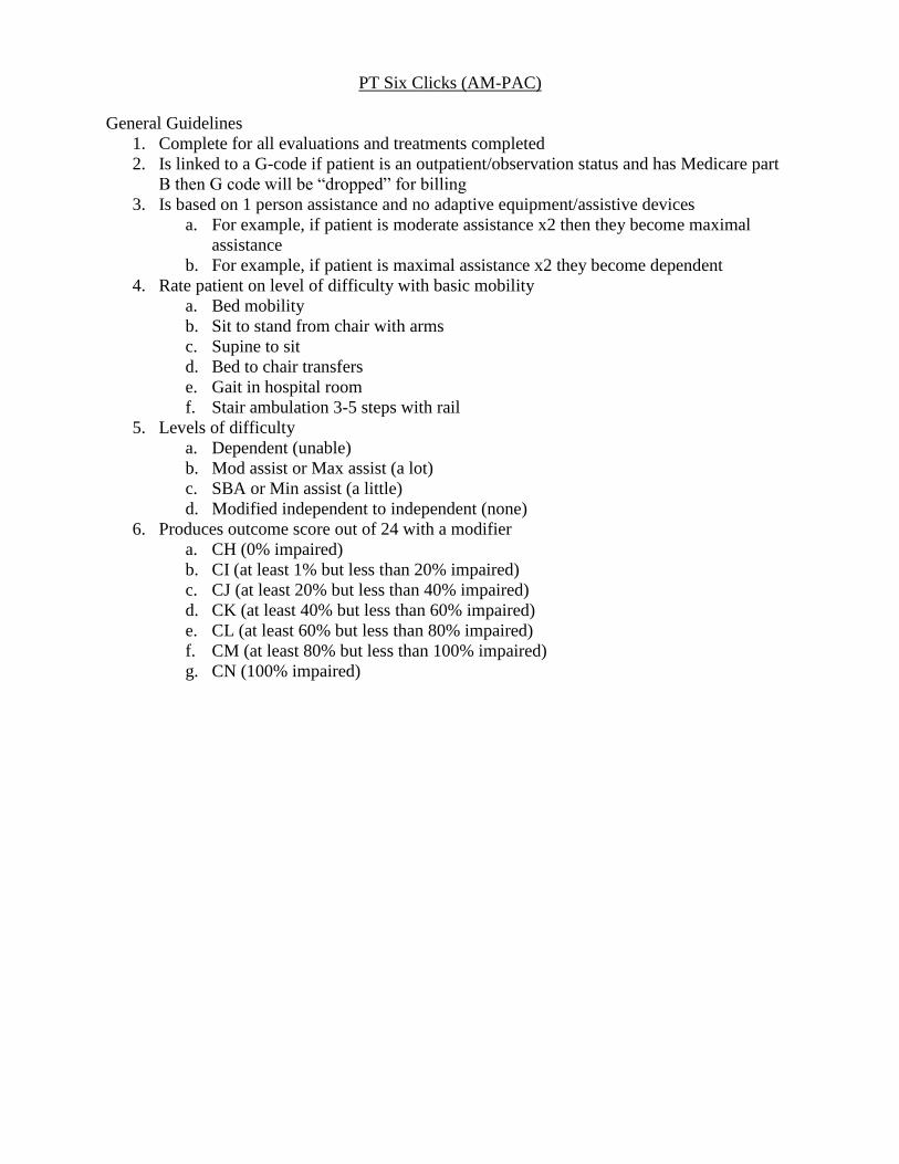
PT Six Clicks (AM-PAC)
General Guidelines
1. Complete for all evaluations and treatments completed
2. Is linked to a G-code if patient is an outpatient/observation status and has Medicare part
B then G code will be “dropped” for billing
3. Is based on 1 person assistance and no adaptive equipment/assistive devices
a. For example, if patient is moderate assistance x2 then they become maximal
assistance
b. For example, if patient is maximal assistance x2 they become dependent
4. Rate patient on level of difficulty with basic mobility
a. Bed mobility
b. Sit to stand from chair with arms
c. Supine to sit
d. Bed to chair transfers
e. Gait in hospital room
f. Stair ambulation 3-5 steps with rail
5. Levels of difficulty
a. Dependent (unable)
b. Mod assist or Max assist (a lot)
c. SBA or Min assist (a little)
d. Modified independent to independent (none)
6. Produces outcome score out of 24 with a modifier
a. CH (0% impaired)
b. CI (at least 1% but less than 20% impaired)
c. CJ (at least 20% but less than 40% impaired)
d. CK (at least 40% but less than 60% impaired)
e. CL (at least 60% but less than 80% impaired)
f. CM (at least 80% but less than 100% impaired)
g. CN (100% impaired)

Lab Values Interpretation
Billing/CPT Codes
General Guidelines
1. Patient must be under the care of and referred for therapy services by a ‘qualified
professional’:
a. Physician
b. Optometrist – for low vision services
c. Podiatrist – subject to each state’s Scope of Practice
d. Nurse Practitioner, Physician’s Assistance, or Clinical Nurse Specialist – subject
to each state’s Scope of Practice
2. Require the skills of a PT/PTA. Routine part of nursing care is not covered. (turning
patients, walking in hallway)
3. Must have written treatment plan with specific therapeutic goals and modalities/procedures
outlined in terms of type, frequency, and duration.
4. Must document functional limitations and short/long term goals which are objective and
measurable.
5. The plan of care must be certified/approved by the physician or optometrist.
6. Skills of the therapist are NOT required to maintain functional.
7. Designing a maintenance regimen/HEP required to delay or minimize muscular/functional
deterioration in chronic disease is reasonable and necessary to establish and assist the patient
with the maintenance program/HEP. No more than 2-4 visits to complete and instruct in the
program are considered medically necessary.
8. Not covered when there is a temporary loss or reduction of function that is reasonable
expected to improve spontaneously without therapy. (ie: short hospital stay for pneumonia)
9. Not covered to identify patients who might need therapy. (ie: screening)
10. Not covered if they are duplicative of other concurrent rehab services.
11. Educational component of treatment is included in the service described by the CPT code and
therefore is NOT a separate charge.
12. Documentation of services is included in the CPT charge and therefore there is not separate
coverage for time spent for documentation.
13. The ICD-9 section of this LCD is meant to include ‘functional’ diagnosis. The functional
diagnosis, not necessarily the clinical diagnosis, conveys coverage.
Evaluation Guidelines
1. There must be a change from normal functional ability to warrant an evaluation.
2. If multiple body parts or systems are involved, they should all be assessed at evaluation
with only one evaluation code used. (i.e. Neck and knee pain)
3. Evaluations from other rehab disciplines are covered as long as their not duplicative.
4. Visits for SOLE purpose of wheelchair management/assessment does not required 97001,
it’s covered under 97542 wheelchair management.
5. Routine screening, assessments, and routine re-assessments are not covered.
6. Pre-op/post-op instructions for SOLE purpose of HEP and/or assistive device instruction
are not covered.
Re-evaluation Guidelines
1. Not routinely covered for purposes of updating the plan of care. Reassessments are part
of the routine aspect of intervention.
2. Billed as a separate charge if their has been a significant change/deterioration in patients
condition OR
3. Patient exhibits a demonstrable change in functional ability/decline and a re-evaluation of
needs to re-establish appropriate treatments goals and interventions is necessary.
Therapeutic Exercise Guidelines
1. Performed on dry land.
2. To restore strength, ROM, flexibility.
3. Passive ROM only for 2-4 visits unless recent surgery. (RTC repair)
4. Therapeutic exercise component of manual lymphatic drainage is covered under 97110
and generally requires no more than 1-2 visits.
5. Not covered for: overall fitness, flexibility, endurance enhancing, aerobic conditioning,
weight reduction, maintenance exercises, and passive exercises not aimed at restoring
loss of function.
6. Generally 12-18 visits within 4-6 weeks.
7. 1-2 units per visit of code is generally covered.
Gait Training Guidelines
1. Covers FES for SCI patients. (see specific requirements in LCD)
2. Not covered if patients walking ability is not expected to improve.
3. Repetitive walk-strengthening exercise for feeble or unstable patients or to increase
endurance or gait distance does not require skilled therapist and is not covered – work on
mechanics instead.
4. Generally 12-18 visits within 4-6 weeks.
Therapeutic Activities Guidelines
1. Covers a broad range of rehab techniques that involve movement.
2. Use of functional activities (bending, lifting, carrying, reaching, catching, transfers) to
restore functional performance in a progressive manner.
3. Activities are usually directed at a loss or restriction of mobility, strength, balance, or
coordination.
4. Generally 10-12 visits.
5. One to two units per visit of code is generally covered.
Self-Care/Home Management Guidelines
o ADL’s and compensatory training, meal preparation, safety procedures, instructions in
use of assistive technology devices and adaptive equipment.
o One to two units per visit of code is generally covered.
Orthotic Management and Training Guidelines
1. Custom Orthotics
a. Compenents: Assessment, Fabrication, Fitting. These are covered under ‘L’
code.
b. Training – covered under 97760
i. To enhance performance of tasks or movements, supports weak or
ineffective joints or muscles, reduces/corrects joint
limitations/deformities, and/or protects body parts from injury.
ii. ROM prior to placing the orthotic/positioner for purpose of
maintaining ROM is not covered.
iii. Monitoring of the device is NOT covered.

iv. Reasonable to need 1-2 visits for education/instruction on use of
device.
c. 97116 Gait training and 97535 Self Care are not covered on the same day
without a modifier.
d. Wheelchair positioning for custom fabricated seating system is covered under
97760.
Check Orthotic Guidelines
1. For modification of the customized orthotic/prosthetic device.
2. Indicated when patient has loss or change of function related to the device. (pain,
decreased swelling, skin breakdown, falls)
3. Generally 1-2 visits is sufficient.
4. One to two units per visit are generally covered.
5. Re-evaluation is not covered the same day
Occupational Therapy Evaluation Codes
History Exam Decision Making
Low Complexity
(97165)
Occupational profile and medical
and therapy, which includes a
brief hx including review of
medical and/or therapy records
relating to the presenting
problems
1-3 performance
deficits(physical, cognitive or
psychosocial) that result in
activity limitations and/or
participation restrictions
low complexity using
occupational profile, analysis of
data from problem-focused
assessment and consideration of
limited number of treatment
options.
Moderate Complexity
(97166)
Occupational profile and medical
and therapy, which includes a hx
including review of medical
and/or therapy records relating to
the presenting problems and
expanded additional review of
physical, cognitive or psycho-
social review related to current
function
3-5 or more performance
deficits(physical, cognitive or
psychosocial) that result in
activity limitations and/or
participation restrictions
Moderate complexity using
occupational profile, analysis of
data from problem-focused
assessment and consideration of
limited number of treatment
options. Minimal to moderate
modification of tasks or
assistance with the assessment to
complete the evaluation
component
High Complexity
(97167)
Occupational profile and medical
and therapy, which includes a hx
including review of medical
and/or therapy records relating to
the presenting problems and
extensive additional review of
physical, cognitive or psycho-
social review related to current
function
5 or more performance
deficits(physical, cognitive or
psychosocial) that result in
activity limitations and/or
participation restrictions
High complexity using
occupational profile, analysis of
data from problem-focused
assessment and consideration of
limited number of treatment
options. Minimal to moderate
modification of tasks or
assistance with the assessment to
complete the evaluation
component
Physical Therapy Evaluation Codes
History Exam Clinical Presentation Decision Making
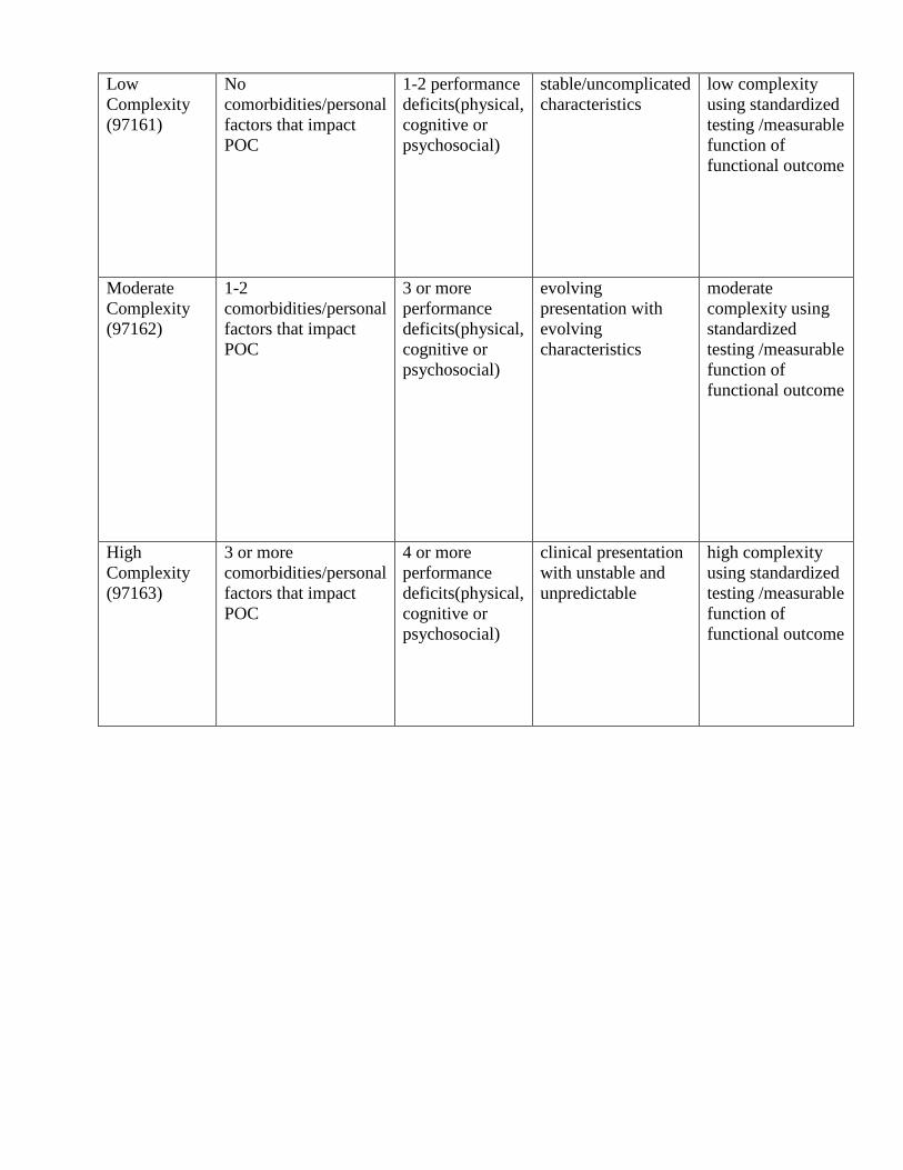
Low
Complexity
(97161)
No
comorbidities/personal
factors that impact
POC
1-2 performance
deficits(physical,
cognitive or
psychosocial)
stable/uncomplicated
characteristics
low complexity
using standardized
testing /measurable
function of
functional outcome
Moderate
Complexity
(97162)
1-2
comorbidities/personal
factors that impact
POC
3 or more
performance
deficits(physical,
cognitive or
psychosocial)
evolving
presentation with
evolving
characteristics
moderate
complexity using
standardized
testing /measurable
function of
functional outcome
High
Complexity
(97163)
3 or more
comorbidities/personal
factors that impact
POC
4 or more
performance
deficits(physical,
cognitive or
psychosocial)
clinical presentation
with unstable and
unpredictable
high complexity
using standardized
testing /measurable
function of
functional outcome
Continuing Education Approval
General Guidelines
1. Acute care rehab puts on several courses a year through department (outpatient also puts on
several courses per year)
2. Information on course can be found on Vanderbilt Acute Care Rehabilitation Services
website (https://ww2.mc.vanderbilt.edu/rehabilitationservices/26314):
3. Must fill out request form and return it to Kelly Floyd
4. If it is a course outside of Vanderbilt, typically you need approval from Kelly Floyd by
filling out form, make sure there is a PTO spot on calendar, then you must pay for course
(unless otherwise determined by Kelly Floyd), attend course, and submit for reimbursement
if Kelly Floyd approves

REHAB SERVICES CONTINUING EDUCATION
DUES / FUNDS APPLICATION
(PLEASE COMPLETE ALL ITEMS BELOW)
Therapist Name:________________________Date:_____________ SS#:________________
Course Name:_________________________________________________________________
Course Location:______________________________________________________________
Instructors:___________________________________________________________________
Course Description:____________________________________________________________
(CHECK ONE (1) BLOCK IN EACH CATEGORY BELOW)
TYPE OF COURSE: PURPOSE OF COURSE: APPLICATION TO PRESENT SERVICE
AREA/JOB ROLE:
Therapy Techniques New Program Development Strong
Research New Skill Acquisition Moderate
Supervision Existing Skill Refinement Indirect
Administration Dues-Renew/New Nominal
Documentation
Billing /Coding
*Date of Pre-Approval for pertinent time off ______________ by________________________
Course Hours Earned:__________ Course Dates:__________________________________
Number of professional leave days to be used: ________
If other than professional leave days to be used, please explain:________________________
Comments regarding course benefits:_____________________________________________
Planned in-service date: ____________ In-service scheduled on Rehab calendar
ENTER ESTIMATED COSTS:
REGISTRATION / TUITION:. . . . $______________
TRAVEL:. . . Airfare: . . . . . . . . . . … $______________
(_______miles @ .36 per mile)........ Mileage:. . . . . . . . . . … .$______________
(_____ nights @ $_______ per night) Hotel: . . . . . . . . . . . … $______________
(_____ days @ $_______ per day).... .Food:. . . . . . . . . . . . … $______________
Parking / Limo: . . . . $______________
TOTAL:. . . . . . . . . . . . . . . . . . . . . …… .$______________
________I WILL REGISTER AND PAY FOR COURSE MYSELF
(All monies will be reimbursed upon course/travel completion, with manager approval.)
________PLEASE REGISTER AND MAKE PAYMENT FOR ME TO ATTEND
(Be sure to fill out appropriate forms-see attached ‘Steps for Traveler Document’)
For Administrative Use Only: APPROVED DENIED
Total Amount Approved: $________________
Manager Signature:_________________________________ Date:_____________________
In-service Scheduled:____________________________________________________________

Functional Outcomes Measures
Physical Therapy
1. Timed Up and Go
2. Timed Walk Tests (2 and 6 minutes)
3. DGI
4. 5x Sit to Stand
5. Chelsea Critical Care Physical Assessment
6. Functional Reach Test
7. Modified Ashworth Scale
8. Gait Scale
9. Perme ICU Mobility Score
10. 10- Meter Walk Test
Occupational Therapy
1. Chelsea Critical Care Physical Assessment
2. Functional Reach Test
3. Perme ICU Mobility Score
4. Timed Up and Go
5. Modified Barthal Index
6. Orienation Log
7. Short Blessed Score
8. SLUMS Test
9. MOCA

Orientation Quiz
Ongoing Competency Review Questions:
1. An 80-year-old patient is placed in isolation when infected with methicillin-resistant
Staphylococcus aureus (MRSA) in their urine. When treating the patient, therapy staff
should:
a. Gown and glove outside the patient’s room and when exiting remove isolation
gear inside the room prior to sanitizing at the threshold of the door
b. Put gloves on
c. Do not take the patient in the hall
d. Mask
2. After seeing an acute care patient, to prevent late charting, your documentation must be
completed:
a. Within 24 hours
b. By 2 PM the next day
c. By 10 AM the next day
d. By the end of the day you saw the patient
3. When asked to complete an updated note, you should:
a. Document as soon as possible if the patient has already been treated
b. Treat other patients as usual and document the updated note at the end of the day
c. Treat the patient as soon as you can and document the note following the
treatment
d. Both A and C
4. If a patient has a Richmond Agitation and Sedation Scare (RASS) score of -2 to +1, can
they be evaluated or treated by therapy?
a. Yes
b. No
5. When you are working with a geriatric patient in the round wing, the patient starts to
report dizziness and you notice the patient is pale. What should you do:
a. Take vital signs
b. Let the patient sit to rest
c. Nothing
d. Both A and B
6. It is Thursday. A patient was evaluated Monday and has a discharge recommendation for
inpatient rehabilitation. The patient has not been seen since the evaluation. During the
evaluation, the patient walked 200 feet with minimal assist and completed most ADL
with supervision except lower body dressing, which was minimal assist. The team has
called and said the patient has progressed well, has been walking with nursing, and is
medically ready for discharge. The patient would be able to return home if cleared by
therapy. You should:
a. Put the patient on the list to be treated tomorrow
b. Evaluate/treat another patient
c. See the patient that day - Thursday
d. None of the above

7. Following a femoral cardiac catheterization, a patient will be on be bedrest:
a. 1 hour
b. Not at all
c. 6 hours
d. 30 minutes
8. You are doing a chart review on a general medicine patient and notice the patient fell
while hospitalized and currently has back pain. A thoracic x-ray shows a compression
fracture. What should you do:
a. Hold treatment until there is a plan for the patient’s compression fracture
b. Go ahead with treatment, the patient has out of bed activity orders
c. Go ahead with treatment, but only sit edge of bed to minimize back pain
d. Hold treatment because of the patient’s pain
9. You are doing a chart review and note the patient has been diagnosed with a deep vein
thrombosis in the left arm. The patient has been on anticoagulation during their hospital
stay and nursing says the patient is OK to treat. What should you do:
a. You are not sure, but you plan to ask another therapist if it is OK to see the patient
b. You can proceed with treatment
c. You could call the team to make sure it’s OK to see the patient
d. All of the above are correct
10. When mobilizing a patient who has an external ventricular drain (EVD), the nurse must
always clamp the drain prior to mobility (choose one):
a. True
b. False
11. You are doing a chart review on trauma. The patient has conflicting weight bearing
restrictions. The activity order days WBAT R LE and progress note says NWB R LE.
What is the most appropriate action to take?
a. Call the ortho team for clarification
b. Let the patient WBAT on the R LE
c. Tell the charge nurse about it and ask them to call the team
d. Put in a contact note/failed attempt
Ideas to Decrease Refusals
1. Pain or Not feeling well…..
a. Start by attempting to build rapport saying something like, “I’m sorry to hear that,
have you been hurting all day or have you had pain medication recently.…” Once
this discussion has taken place, say
b. Let me talk to your nurse to see if there is anything she can do.
c. Let me help you move around, get up, then get you in a better position to help
with pain
d. Let’s work together and give it a try for 5-10 minutes. If your pain gets really bad,
we can lie back down and that’s good information for me to take back to your
doctor.
e. It doesn’t have to be an hour, just moving is important

f. We have many patients who actually feel better once the process of moving
around is over because it loosens up the joints and muscles
2. Tired or Just Returned to bed…..
a. Start with building rapport by saying something like, “I’m so sorry, our timing is
never perfect, but….”
b. If you can work with me for 10-15 minutes, I’ll help you back to bed.
c. I’m not here to do a lot of exercises, I just want to see how well you do with
______________
3. Combined Strategies for both Pain and Tired….
a. The more you get you and move around, the better you will be able to move in
order to get home/out of the hospital faster
b. OT or PT is an important part of your recovery. We are similar to a medication.
You need to consistently work with us to see improvement
c. Therapy is a part of the care plan created with your physician
d. Your medical team needs to see consistent performance in order to feel that you
are safe to go home/leaving the hospital
e. Moving around will help prevent complications of remaining in bed such as
pneumonia, getting weaker, and blood clots
f. Now is a good time because therapy can often help and give you better tips when
you are not feeling as good. It gives us a better picture of what home might be
like.

Acute Care Physical Therapy: Tips and
Tricks
LINES
Peripheral IV
Short catheter inserted through the skin into a peripheral vein (any vein not inside the
chest or abdomen)
Arm and hand veins are used most commonly, with leg and foot veins used to a much
lesser extent
Infiltration occurs when an IV fluid or medication accidentally enters the surrounding
tissue rather than the vein.
TIPS:
o Make sure there is enough slack on the line during mobility.
o When IV is antecubital or in the wrist, patients usually find this uncomfortable,
however, elbow or wrist ROM will not harm the patient.
o The IV pump will alarm if the line becomes kinked.
Central line
Usually located in the superior vena cava or inferior vena cava, or within the right atrium
of the heart.
An experienced clinician knowing the appropriate landmarks and/or using an ultrasound
probe is needed to place this line safely.
Surrounding structures, such as the pleura and carotid artery are at risk of damage during
placement with the potential for pneumothorax or even cannulation of the artery.
This has several advantages over a peripheral IV:
o It can deliver fluids and medications that would be overly irritating to peripheral
veins because of their concentration or chemical composition. These include some
chemotherapy drugs and total parenteral nutrition (TPN).
o Medications reach the heart immediately and are quickly distributed to the rest of
the body.
o There is room for multiple parallel compartments (lumen) within the catheter, so
that multiple medications can be delivered at once even if they would not be
chemically compatible within a single tube.
o Central venous pressure and other physiological variables can be measured
through the line.
TIPS:
o Make sure there is enough slack on the lines during mobility.
o There are no contraindications to shoulder ROM on the side of the central line.
Peripherally inserted central catheter (PICC)

PICC lines are used when intravenous access is required over a prolonged period of time
or when medications to be infused would cause quick damage and early failure of a
peripheral IV and when a conventional central line may be too dangerous to attempt.
Typical uses include long chemotherapy regimens, extended antibiotic therapy, or total
parenteral nutrition (TPN).
Line is placed under ultrasound guidance, usually in the arm, and then carefully advanced
upward until the catheter is in the superior vena cava or the right atrium. An X-ray must
be used to verify that the tip is in the right place when fluoroscopy was not used during
the insertion.
A PICC can be single, double, or triple-lumen.
The chief advantage of a PICC over other types of central lines is that it is safer to insert
with a relatively low risk of uncontrollable bleeding and essentially no risks of damage to
the lungs or major blood vessels.
A PICC can be left in place for months to years if needed for patients who require
extended treatment.
TIP: Make sure there is enough slack on the lines during mobility.
Swan Ganz Catheter (Pulmonary Artery Catheter)
A thin hollow tube called a catheter is inserted through a vein in the neck or groin. It is
carefully moved up into the right atrium of the heart and threaded through two heart
valves (the tricuspid and pulmonary valve) and placed into the pulmonary artery.
There is a balloon at the tip of the catheter that is inflated only when measurements are
being taken.
Measures Central Venous Pressure (CVP), Pulmonary Capillary Wedge Pressure
(PCWP), Cardiac Index (CI) , Stroke Volume (SV), Pulmonary Artery Pressure (PAP),
and Cardiac Output (CO).
Used for the following:
o Diagnosis of shock states (cardiogenic vs septic vs hypovolemic)
o Diagnosis of idiopathic pulmonary hypertension
o Diagnosis of valvular disease, intracardiac shunts, cardiac tamponade, and
pulmonary embolus (PE)
o Monitoring and management of complicated Acute Myocardial Infarction (AMI)
o Assessing hemodynamic response to medical therapies
o Management of multiorgan system failure and/or severe burns
o Management of hemodynamic instability after cardiac surgery
o Assessment of response to treatment in patients with idiopathic pulmonary
hypertension
o Management of postoperative open heart surgical patients
o Assessment of valvular heart disease
o Assessment of cardiac tamponade/constriction
TIPS:
o There are many lines that are connected to the Swan Ganz. Make sure you have
all lines organized prior to mobility.
o At VUMC, patients are only allowed to transfer bed to chair with this line in
place.
Arterial Lines (art line)
A thin catheter is inserted into an artery, usually the wrist (radial artery), armpit (axillary
artery), groin (femoral artery), or foot (pedal artery).
Line is most commonly used to monitor the blood pressure real-time (rather than by
intermittent measurement) and to obtain samples for arterial blood gas measurements.
TIPS:
o At VUMC, PT must check with MD before mobilizing patients with femoral art
lines.
o Avoid repetitive ROM to joint that arterial line is near.
Temporary Vascular Catheter (vas cath)
Can be placed in the groin (femoral) or in the neck (internal jugular).
It is used for hemodialysis (HD).
This catheter has a red and blue port.
TIPS:
o At VUMC, patients are usually on bed rest if femoral.
o Patient may do bed exercises except for hip flexion on the side the femoral
catheter is placed.
o No precautions if placed in internal jugular (IJ).
Chest Tubes (CT)
A CT is a flexible plastic tube that is inserted through the side of the chest into the pleural
space.
It is connected to a canister which can be to suction or to water seal.
Indications:
o Pneumothorax: accumulation of air in the pleural space
o Pleural effusion: accumulation of fluid in the pleural space
o Chylothorax: a collection of lymphatic fluid in the pleural space
o Empyema: a pyogenic infection of the pleural space
o Hemothorax: accumulation of blood in the pleural space
o Hydrothorax: accumulation of serous fluid in the pleural space
TIPS: o Check with nursing to see if patient can come off of wall suction.
o Usually patients are able to come off suction for short time during activity.
o Some patients must have chest tubes remain on suction secondary to air leakage
or subcutaneous air; in these cases there is portable suction available for
ambulation. o Should a chest tube be accidentally pulled, petroleum gauze is best to cover chest tube
site to allow air to escape but not enter. If you do not have access to petroleum gauze,
use a towel or cloth. Contact nursing and return patient to bed.
o Avoid tipping canister. If canister becomes tipped, notify nursing.
Intra Arterial Balloon Pump (IABP)

The IABP is a polyethylene balloon mounted on a catheter inserted into femoral artery
then advanced to the aorta until the tip lies just below the origin of the L subclavian
artery.
Used to decrease myocardial oxygen demand while at the same time increasing cardiac
output. By increasing cardiac output it also increases coronary blood flow and therefore
myocardial oxygen delivery. It actively deflates in systole increasing forward blood flow
by reducing afterload and actively inflates in diastole increasing blood flow to the
coronary arteries. These actions have the combined result of decreasing myocardial
oxygen demand and increasing myocardial oxygen supply.
Simply, it reduces the workload of the heart.
Indications: cardiogenic shock, acute mitral regurgitation and septal perforation, unstable
angina, post cardiothoracic surgery, and bridge to heart transplant.
Complications: Since the device is placed in the femoral artery and aorta it could provoke
ischemia, and compartment syndrome. The leg is at highest risk of becoming ischemic if
femoral artery becomes obstructed. Placing the balloon too distal from the arcus aortae
may induce occlusion of the renal artery and subsequent renal failure. Other possible
complications are cerebral embolism during insertion, infection, dissection of the aorta or
iliac artery, perforation of the artery and hemorrhage in the mediastinum. Mechanical
failure of the balloon itself is also a risk which entails vascular surgery to remove under
that circumstance. After balloon removal there is also a risk of 'embolic shower' from
micro clots that have formed on the surface of the balloon, and can lead to peripheral
thrombosis, myocardial ischemia, hemodynamic decompensation, and late
pseudoaneurysm.
TIPS:
o Patient on bed rest when IABP placed in the femoral artery
o IABP can be placed axillary allowing patient is able to get OOB and ambulate.
o At VUMC, you must have RN present when ambulating patient on IABP.
Endotracheal Tube (ETT)
Insterted through the mouth into the trachea.
Most tubes have an inflatable cuff to seal the trachea and bronchial tree against air
leakage and aspiration of gastric contents, blood, secretions, and other fluids.
Primary purpose is to establish and maintain a patent airway and to ensure the adequate
exchange of oxygen and carbon dioxide.
Minor complications: sore throat, lacerations of the lips or gums or other structures
within the upper airway, chipped, fractured or dislodged teeth, and nasal injury.
More serious complications include bronchospasm, laryngospasm, perforation of the
trachea or esophagus, and pulmonary aspiration of gastric contents or other foreign
bodies resulting in aspiration pneumonitis.
Hypopharyngeal suctioning.
Tracheostomy (trach)
Placed surgically via incision on the anterior aspect of the neck and opening a direct
airway into the trachea.
Indications:
o Severe facial trauma
o Head and neck cancers
o Large congenital tumors of the head and neck
o Acute angioedema and inflammation of the head and neck
o Failed endotracheal intubation
o Need for long-term mechanical ventilation and tracheal toilet
o Severe Obstructive Sleep Apnea (OSA)
Tips for working with intubated/trached patients
When working with a patient who is intubated/trached, be careful to keep the ET
tubing/trach tubing in a neutral position as to prevent excessive pulling or pouring of
fluid within the tubing down into ETT/trach.
You should take tubing out of holder when moving patient (ie rolling, transfer to EOB, sit
to stand).
Transfer patient to same side of bed as ventilator.
Once patient is stable at EOB, then it would be safe to place tubing back in holder.
It is important to monitor mechanically ventilated patients carefully during treatments
especially RR and O2 sats. The RR on the vent is usually more accurate than RR on
monitor.
Supplemental Oxygen Delivery
Nasal Cannula (NC)
o Carries 1–6 liters of oxygen per minute.
High Flow Nasal Cannula (HFNC)
o High flows of an air/oxygen blend can be administered via a nasal cannula to
accurately deliver high volume of oxygen therapy.
o Respiratory gas humidification allows the high flows to be delivered comfortably
via the cannula.
o Can give up to 15L O2
Venturi Mask (Venti mask)
o Delivers a constant FiO2 regardless of changes in the ventilatory pattern.
o Can increased FiO2 to a maximum of 50%.
Nonrebreather (NRB)
o Permits inhalation of pure O2 (100%).
o Be careful with COPD patients.
Noninvasive Ventilation
CPAP
o Continuous positive airway pressure (CPAP) machine was initially used mainly
by patients for the treatment of sleep apnea at home, but now is in widespread use
across intensive care units as a form of ventilation.
o The CPAP machine delivers a stream of compressed air via a hose to a nasal
pillow, nose mask or full-face mask, splinting the airway (keeping it open under
air pressure) so that unobstructed breathing becomes possible, reducing and/or
preventing apnea
BIPAP
o Provides two levels of pressure: Inspiratory Positive Airway Pressure (IPAP) and
a lower Expiratory Positive Airway Pressure (EPAP) for easier exhalation.
o Typically uses to treat COPD and acute respiratory failure.
Ventilator Settings
CPAP (Continuous Positive Airway Pressure)
o Patient is spontaneously breathing
o Lowest vent setting sometimes called wean settings
SIMV (Synchronous Intermittent Mandatory Ventilation)
o The ventilator delivers preset breaths in coordination with the respiratory effort of
the patient.
o Spontaneous breathing is allowed between breaths.
PRVC (Pressure Regulated Volume Control)
o The ventilator delivers preset breaths in coordination with the respiratory effort of
the patient. With each inspiratory effort, the ventilator delivers a full assisted tidal
volume.
o Spontaneous breathing independent of the ventilator is not allowed.
TIPS:
o Closely monitor your patient and their vitals, esp. anxiety, need for suction, O2
sats, and RR.
o Watch patient closely when reaching for mouth.
o Your patient may begin to cough more when upright due to mucus in lungs
mobilizing or gag reflex.
o You may want RN nearby during mobility for suctioning.
o At VUMC, PT is allowed to give patient 100% O2 breaths.
Sedation
When patients are intubated, sedatives are usually initiated.
Medications usually used for sedation are propofol, precedex, and fentanyl.
Level of sedation determined by RASS (Richmond Agitation Sedation Scale). The scale
ranges from -5 to +4. The more negative, then the more sedated. The more positive, then
the more agitated. Zero is normal.
It is safe to work with patients that are intubated and RASS -1 to +1 as long as patient is
hemodynamically stable.
Richmond Agitation–Sedation Scale
+4 Combative Overtly combative or violent; immediate danger to staff
+3 Very agitation Pulls on or removes tube(s) or catheter(s) or has aggressive behavior
toward staff

+2 Agitated Frequent nonpurposeful movement or patient–ventilator dyssynchrony
+1 Restless Anxious or apprehensive but movements not aggressive or vigorous
0 Alert and calm
-1 Drowsy
Not fully alert, but has sustained (more than 10 seconds) awakening,
with eye contact, to voice
-2 Light sedation Briefly (less than 10 seconds) awakens with eye contact to voice
-3 Moderate sedation Any movement (but no eye contact) to voice
-4 Deep sedation No response to voice, but any movement to physical stimulation
-5 Unarousable No response to voice or physical stimulation
Vasopressors
This group of drugs is useful for treatment of hypotension.
Drugs cause vasoconstriction resulting in increase in peripheral vascular resistance
At high doses, and especially when it is combined with other vasopressors, it can lead
to limb ischemia and limb death.
Ex: Levophed (Norepinephrine) and Vasopressin
TIPS:
o Watch BP carefully either by peripheral BP cuff or art line.
o If patient has had recent increase in pressor dose or recently started on
pressors, may want to HOLD PT at that time.
Hemodialysis (HD)
Process for removing waste and excess water from the blood, and is primarily used to
provide an artificial replacement for lost kidney function in people with renal failure.
May be used for those with an acute disturbance in kidney function (acute kidney injury,
previously acute renal failure) or for those with progressive but chronically worsening
kidney function–a state known as chronic kidney disease stage 5 (previously chronic
renal failure or end-stage kidney disease).
TIP: PT unable to work with patient on HD at VUMC.
Continuous dialysis
Continuous veno-venous hemodialysis (CVVHD) or Continuous Renal Replacement
Therapy (CRRT)
Indications: ARF, ARDS, after surgery with cardiopulmonary bypass, refractory
congestive heart failure.
Provides slow and balanced fluid removal and/or hemodialysis
Usually patient will be on some type of vasopressor.
TIPS:
o Ask RN if patient’s BP has remained stable over the last 12-24 hours. If so, PT
may be warranted.
o Have someone watch this line exclusively during mobility.

Peritoneal dialysis
Performed through port in peritoneal cavity.
Provides more independence.
Dialysis usually performed at home 4-5 times daily.
Patients are at risk for peritonitis.
TIP: Cannot perform PT while patient is draining or filling.
Sepsis
Potentially deadly medical condition that is characterized by a whole-body inflammatory
state and the presence of a known or suspected infection.
The body may develop this inflammatory response by the immune system to microbes in
the blood, urine, lungs, skin, or other tissues.
Common infections are peritonitis, urinary tract infection or pyelonephritis, meningitis,
bacterial pneumonia or cellulitis.
In hospitalized patients, common sites of infection include intravenous lines, surgical
wounds, surgical drains, and decubitus ulcers.
Septicemia or bacteremia refers to the presence of pathogenic organisms in the
bloodstream, leading to sepsis.
Septic shock is defined as sepsis with refractory arterial hypotension or hypoperfusion
abnormalities in spite of adequate fluid resuscitation.
When severe, may also include end organ dysfunction. Examples are acute lung injury
(ALI), acute respiratory distress syndrome (ARDS), encephalopathy, liver dysfunction,
acute kidney injury (AKI) or acute renal failure (ARF), and systolic and diastolic heart
failure.
Symptoms of sepsis can include:
o Confusion or delirium
o Hypotension
o Tachycardia
o Hypothermia
o Hyperventilation
o Fever
Gastrointestinal Bleed (GI bleed)
Loss of blood in the gastrointestinal tract, from the pharynx to the rectum.
Upper GI bleed - An upper source is characterized by hematemesis (vomiting up blood)
and melena (tarry stool containing altered blood).
Lower GI bleed - indicated by bright red blood per rectum (BRBPR).
Causes:
o Anal fissure
o Hemorrhoids

o Cancer
o Intestinal polyps (a pre-cancerous condition)
o Abnormal blood vessels in the lining of the intestines (also called
angiodysplasias)
o Diverticulosis
o Crohn's disease or ulcerative colitis
o Esophageal varices
o Esophagitis
o Gastric (stomach) ulcer
o Mallory-Weiss tear
o Radiation injury to the bowel
Testing
o EGD
o Colonoscopy
o PCV (Hematocrit)
Adult males: 42%-54%
Adult women: 38%-46%
TIPS:
o Not indicated if patient is actively bleeding.
o Check Packed Cell Volume (PCV) or hematocrit (HCT).
Percentage of blood volume that is occupied by red blood cells.
o If getting blood products, check with nursing if okay to mobilize.
Diabetes Mellitus (DM)
Lifelong (chronic) disease in which there are high levels of sugar in the blood.
Insulin is a hormone produced by the pancreas to control blood sugar. Diabetes can be
caused by too little insulin, resistance to insulin, or both.
Normal process by which food is broken down and used by the body for energy. Several
things happen when food is digested:
o A sugar called glucose enters the bloodstream. Glucose is a source of fuel for the
body.
o An organ called the pancreas makes insulin. The role of insulin is to move
glucose from the bloodstream into muscle, fat, and liver cells, where it can be
used as fuel
People with diabetes have high blood sugar because their body cannot move sugar into
fat, liver, and muscle cells to be stored for energy. This is because either:
o Their pancreas does not make enough insulin
o Their cells do not respond to insulin normally
o Both of the above
High blood sugar (hyperglycemia) symptoms:
o Blurry vision
o Excess thirst
o Fatigue
o Frequent urination
o Hunger
o Weight loss
Low blood sugar (hypoglycemia) symptoms:
o Double vision or blurry vision
o Fast or pounding heartbeat
o Feeling cranky or acting aggressive
o Feeling nervous
o Headache
o Hunger
o Shaking or trembling
o Sleeping trouble
o Sweating
o Tingling or numbness of the skin
o Tiredness or weakness
o Unclear thinking
Fasting blood glucose level -- diabetes is diagnosed if it is higher than 126 mg/dL twice.
Levels between 100 and 126 mg/dL are called impaired fasting glucose or pre-diabetes.
These levels are risk factors for type 2 diabetes.
Patient with DM: controlled BG usually 80-150 mg/dL before meals.
Emergency situations:
o Diabetic hyperglycemic hyperosmolar coma - condition of extremely high blood
sugar levels, dehydration, and decreased consciousness
o Diabetic ketoacidosis (DKA)
Usually trigger by an illness or infection
Occurs when the body cannot use sugar (glucose) as a fuel source because
there is no insulin or not enough insulin. Fat is used for fuel instead.
Byproducts of fat breakdown, called ketones, build up in the body.
In high levels, ketones are poisonous.
Blood glucose levels rise (usually higher than 300 mg/dL) because the
liver makes glucose to try to combat the problem. However the cells
cannot pull in that glucose without insulin.
Symptoms can include: deep, rapid breathing, dry skin and mouth,
flushed face, fruity smelling breath, nausea and vomiting, and stomach
pain
Can cause secondary problems:
o Diabetic retinopathy
o Diabetic nephropathy
o Diabetic neuropathy

o Delayed healing
o Diabetic ulcers
Lung Disease
Restrictive
Respiratory diseases that restrict lung expansion resulting in a decreased lung volume, an
increased work of breathing, and inadequate ventilation and/or oxygenation.
Pulmonary function test (PFT) demonstrates a decrease in the forced vital capacity
(FVC).
Causes:
o Asbestosis caused by long-term exposure to asbestos dust.
o Radiation fibrosis, usually from the radiation given for cancer treatment.
o Certain drugs such as amiodarone, bleomycin and methotrexate.
o As a consequence of another disease such as rheumatoid arthritis.
o Hypersensitivity pneumonitis due to an allergic reaction to inhaled particles.
o Acute respiratory distress syndrome (ARDS), a severe lung condition occurring in
response to a critical illness or injury.
o Idiopathic pulmonary fibrosis (IPF).
o Idiopathic interstitial pneumonia, of which there are several types.
o Sarcoidosis.
o Eosinophilic pneumonia.
o Lymphangioleiomyomatosis.
o Pulmonary Langerhans' cell histiocytosis.
o Pulmonary alveolar proteinosis.
o Neuromuscular diseases, including Myasthenia gravis and Guillain Barre.
o Nonmuscular diseases of the chest wall, e.g. kyphosis and obesity.
Obstructive
Respiratory disease characterized by airway obstruction.
It is generally characterized by inflamed and easily collapsible airways, obstruction to
airflow, and frequent office visits and hospitalizations.
Types of obstructive lung disease include:
o Asthma - Airways are hyper responsive and become inflamed producing excess
mucus. The muscles around the airways tighten making the airways narrower.
Asthma is usually triggered by breathing in things in the air such as dust or pollen
that produce an allergic reaction. It may be triggered by other things such as an
upper respiratory tract infection, cold air, exercise or smoke. Asthma causes
recurring episodes of wheezing, breathlessness, chest tightness, and coughing.
o Bronchiectasis - Abnormal, irreversible dilatation of the bronchi caused by
destructive and inflammatory changes in the airway walls.
o Bronchitis - Hyperplasia and hyper secretion of mucus glands.
o Chronic obstructive pulmonary disease (COPD) – Progressive condition that is
not fully reversible. The term COPD includes the conditions emphysema and
chronic bronchitis although most patients with COPD have characteristics of both
conditions to varying degrees. The residual volume, the volume of air left in the
lungs following full expiration, is often increased in COPD, as is the total lung
capacity, while the vital capacity remains relatively normal. The increased total
lung capacity (hyperinflation) can result in the clinical feature of a "barrel chest" -
a chest with a large front-to-back diameter that occurs in some individuals with
COPD. The most common cause of COPD is cigarette smoking. COPD is a
gradually progressive condition and usually only develops after about 20 pack-
years of smoking. COPD may also be caused by breathing in other particles and
gases. Severe emphysema has been treated with lung volume reduction surgery,
with some success in carefully chosen cases. Lung transplantation is also
performed for severe COPD in carefully chosen cases. Alpha 1-antitrypsin
deficiency is a fairly rare genetic condition that results in COPD (particularly
emphysema) due to a lack of the antitrypsin protein which protects the fragile
alveolar walls from protease enzymes released by inflammatory processes.
TIPS:
o Patients with COPD are CO2 retainers, therefore, their O2 sats are usually mid to
high 80s with activity. Be careful increasing supplemental O2 as increased O2
results in increased CO2 resulting in decreased respiratory drive.
o Monitor O2 sats at rest and with activity.
o Educated COPD patients on pursed lip breathing.
o Educated patients with lung disease on energy conservation (i.e. sitting to perform
tasks, increased rest breaks, etc).
o Educated patients who use supplemental O2 on smoking cessation or to at least
remove supplemental O2 when smoking!
Cardiac
How the Heart Works
A healthy heart beats about 60 to 80 times per minute to pump blood throughout the
body. The right and left sides of the heart work together. Blood that is low in oxygen first
enters the right upper chamber (right atrium) of the heart. The blood flows from the right
atrium to the lower chamber (right ventricle) through the open tricuspid valve. The blood
then travels through the pulmonary artery to the lungs where oxygen is added. Oxygen-
rich blood then returns to the left side of the heart. The blood flows from the left upper
chamber (left atrium) to the lower chamber (left ventricle) through the open mitral valve.
From the left ventricle, the blood is pumped into a network of arteries (blood vessels) that
carry the blood throughout the body.
Blood passes through a valve before leaving each chamber of the heart. There are four
valves in your heart; valves make sure blood flows in only one direction through your
heart.
Congestive Heart Failure (CHF)
Systolic heart failure (systolic dysfunction)
o Occurs when the heart muscle does not contract with enough force, so there is not
enough oxygen-rich blood pumped throughout the body.
Diastolic heart failure (diastolic dysfunction)

o Occurs when the heart contracts normally (a normal ejection fraction) but the
ventricle does not relax or fill properly, so less blood enters the heart.
Ejection fraction (EF) o Measurement of the amount of blood pumped out of the left ventricle with each
heartbeat.
o In most cases, the term “ejection fraction” refers to left ventricular ejection
fraction.
50-70% Normal
36-49% Below Normal
35-40% May confirm diagnosis of systolic heart failure
<35% May be at risk for life threatening irregular heartbeats
o Ejection fraction is usually expressed as a percentage. A normal heart pumps a
little more than half the heart’s blood volume with each beat.
o A LVEF of 65%, for example, means that 65% of the total amount of blood in the
left ventricle is pumped out with each heartbeat.
o The LVEF may be lower when the heart muscle has become damaged due to a
heart attack, heart muscle disease (cardiomyopathy), or other causes.
o An EF of 35 to 40% may confirm a diagnosis of systolic heart failure. Someone
with diastolic failure can have a normal EF.
o An EF of less than 35% increases the risk of life- threatening irregular heartbeats
that can cause sudden cardiac arrest (loss of heart function) and sudden cardiac
death. An implantable cardioverter defibrillator (ICD) may be recommended for
these patients.
o Your EF can go up and down based on your heart condition and the therapies that
have been prescribed.
Signs of heart failure:
Shortness of Breath (Dyspnea)
o When the heart begins to fail, blood backs up in the veins attempting to carry
oxygenated blood from the lungs to the heart. As fluid pools in the lungs, it
interferes with normal breathing. In turn, you may experience breathlessness
during exercise or other activities. As the condition worsens, shortness of breath
may occur when at rest or asleep. These periods of breathlessness may leave you
feeling exhausted and anxious.
Fatigue
o As heart failure becomes more severe, the heart is unable to pump the amount of
blood required to meet all of the body's needs. To compensate, blood is diverted
away from less-crucial areas, including the arms and legs, to supply the heart and
brain. As a result, people with heart failure often feel weak (especially in their
arms and legs), tired and have difficulty performing ordinary activities such as
walking, climbing stairs or carrying groceries.
Chronic Cough or Wheezing
o The fluid buildup in the lungs may result in a persistent cough or wheezing, that
may produce phlegm (a thick, mucous-like substance) that may be tinged with
blood.
Rapid or Irregular Heartbeat

o The heart may speed up to compensate for its failing ability to adequately pump
blood throughout the body. Patients may feel a fluttering in the heart
(palpitations) or a heartbeat that seems irregular or out of rhythm. This often is
described as a pounding or racing sensation in the chest.
Lack of Appetite or Nausea
o When the liver and digestive system become congested they fail to receive a
normal supply of blood. This can make you feel nauseous or full, even if you
haven't eaten.
Mental Confusion or Impaired Thinking
o Abnormal levels of certain substances, such as sodium, in the blood and reduced
blood flow to the brain can cause memory loss or disorientation, which you may
or may not be aware of.
Fluid Buildup and Swelling
o Because blood flow to the kidneys is restricted, the kidneys produce hormones
that lead to salt and water retention. This causes swelling, also called edema, that
occurs most often in the feet, ankles and legs.
Rapid Weight Gain
o The fluid build-up throughout the body, may cause you to gain weight quickly.
The Stages of Heart Failure – NYHA Classification
In order to determine the best course of of therapy, physicians often assess the stage of heart failure
according to the New York Heart Association (NYHA) functional classification system. This system
relates symptoms to everyday activities and the patient's quality of life.
Class Patient Symptoms
Class I (Mild) No limitation of physical activity. Ordinary physical
activity does not cause undue fatigue, palpitation, or
dyspnea (shortness of breath).
Class II (Mild) Slight limitation of physical activity. Comfortable at rest,
but ordinary physical activity results in fatigue, palpitation,
or dyspnea.
Class III (Moderate) Marked limitation of physical activity. Comfortable at rest,
but less than ordinary activity causes fatigue, palpitation,
or dyspnea.
Class IV (Severe) Unable to carry out any physical activity without
discomfort. Symptoms of cardiac insufficiency at rest. If
any physical activity is undertaken, discomfort is
increased.
Testing you may see in charts:
Blood tests
o Creatinine: normal range 0.6 – 1.5 mg/dL
o B-type Natriuretic Peptide (BNP)

BNP is a substance secreted from the ventricles (lower chambers of the
heart) in response to changes in pressure that occur when heart failure
develops and worsens.
The level of BNP in the blood increases when heart failure symptoms
worsen, and decreases when the heart failure condition is stable.
Females with no CHF (55-64) 32
Females with no CHF (75+) 78
Males with no CHF (55-64) 20
Males with no CHF (75+) 48
Mild heart failure 186
Moderate heart failure 791
Severe heart failure 2013
Chest x-ray (CXR)
o Shows the size of your heart and whether there is fluid build-up around the heart
and lungs.
Echocardiogram (ECHO)
o Graphic outline of the heart’s movement.
o During an echo, a probe is placed on the surface of your chest. This wand sends
ultrasound waves that provide pictures of the heart's valves and chambers so the
pumping action of the heart can be studied.
o Echo is often combined with an ultrasound to find changes in the blood flow
across the heart's valves and the pressure within the heart’s chambers.
Electrocardiogram (EKG or ECG)
o Records the electrical impulses in the heart. During the test, small, flat, sticky
patches called electrodes are placed on your chest. The electrodes are attached to
an electrocardiograph monitor (EKG) that charts your heart’s electrical activity on
graph paper.
Stress Test
o An exercise stress test is used to provide information about how the heart
responds to stress. It usually involves walking on a treadmill or pedaling a
stationary bike at increasing levels of difficulty, while the electrocardiogram,
heart rate, and blood pressure are monitored. When one is not able to do activity,
medications may be used to "stress" the heart. This is called a pharmacological
stress test.

Coronary Artery Disease
Narrowing of the small blood vessels that supply blood and oxygen to the heart.
Leading cause of death in the United States for men and women.
Caused by the buildup of plaque in the arteries to your heart.
Risk factors for heart disease:
o Age. The risk of heart disease increases with age.
o Gender. Men have a higher risk of getting heart disease than women who are still
getting their menstrual period. After menopause, the risk for women is closer to
the risk for men.
o Genes. If your parents or other close relatives had heart disease, you are at higher
risk.
o Race. African Americans, Mexican Americans, American Indians, Hawaiians,
and some Asian Americans also have a higher risk for heart problems.
o Diabetes is a strong risk factor for heart disease.
o High blood pressure increases your risks of heart disease and heart failure.
o Extra cholesterol in your blood builds up inside the walls of your heart's arteries
(blood vessels).
o Smokers have a much higher risk of heart disease.
o Chronic kidney disease can increase your risk.
o People with narrowed arteries in another part of the body (examples are stroke
and poor blood flow to the legs) are more likely to have heart disease.
o Substance abuse (such as cocaine).
o Obesity.
o Not getting enough exercise and feeling depressed or having excess stress are
other risk factors.
Myocardial Infarction (MI or “heart attack”)
The interruption of blood supply to a part of the heart, causing heart cells to die. This is
most commonly due to occlusion (blockage) of a coronary artery following the rupture of
a vulnerable atherosclerotic plaque, which is an unstable collection of lipids (fatty acids)
and white blood cells (especially macrophages) in the wall of an artery. The resulting
ischemia (restriction in blood supply) and oxygen shortage, if left untreated for a
sufficient period of time, can cause damage or death (infarction) of heart muscle tissue
(myocardium).
Classical symptoms of acute myocardial infarction include sudden chest pain (typically
radiating to the left arm or left side of the neck), shortness of breath, nausea, vomiting,
palpitations, sweating, and anxiety (often described as a sense of impending doom).
Women may experience fewer typical symptoms than men, most commonly shortness of
breath, weakness, a feeling of indigestion, and fatigue.
Approximately one quarter of all myocardial infarctions are "silent", without chest pain
or other symptoms.
Check Troponin levels in chart.

o Very sensitive and specific indicators of damage to the heart muscle
o A patient who had suffered from a myocardial infarction would have an area of
damaged heart muscle and so would have elevated cardiac troponin levels in the
blood.
o Normal troponin is < 0.05.
o PT is indicated 24 hours after troponins have begun to trend down.
Non ST Elevation Myocardial Infarction (NSTEMI)
Does not cause changes on an electrocardiogram (ECG), however, chemical markers in
the blood indicate that damage has occurred to the heart muscle.
In NSTEMI, the blockage may be partial or temporary, and so the extent of the damage
relatively minimal.
ST segment elevation myocardial infarction (STEMI)
Is caused by a prolonged period of blocked blood supply.
It affects a large area of the heart muscle, and so causes changes on the ECG as well as in
blood levels of key chemical markers.
Things to consider when treating a patient post MI:
During interview, ask patient what their s/s were prior to their MI.
Ask patient to tell you if they have any recurrence of these s/s during activity.
If s/s occur, notify nursing immediately.
PT is contraindicated if patient actively having MI or troponin level still trending up.
Activity should be held until 24 hours after first downtrending troponin.
Cardiac Catheterization (AKA: heart cath, coronary angiography)
An invasive imaging procedure that involves putting a catheter in the femoral or radial
artery and guiding it to your heart with the aid of a special x-ray machine.
There are two types of cardiac catheterization, left and right. In a left heart
catheterization, contrast dye may be injected through the catheter and x-ray movies of
your valves, coronary arteries, and heart chambers are taken. Right heart catheterization
does not require contrast dye and is used to measure heart function.
Once the catheter is in place, it can be used to perform a number of procedures including
angioplasty, PCI (percutaneous coronary intervention) angiography.
Percutaneous coronary intervention (PCI) is a therapeutic procedure to treat the stenotic
(narrowed) coronary arteries of the heart found in coronary heart disease. These stenotic
segments are due to the build up of cholesterol-laden plaques that form due to
atherosclerosis.
Bare-metal stents (BMS) are vascular stents without a coating. They are mesh-like tubes
of thin wire.
Drug-eluting stents (DES) are coronary stents placed into narrowed, diseased peripheral
or coronary arteries that slowly releases a drug to block cell proliferation.
TIP: Check with MD team/RN regarding hours of bedrest post procedure.

Cardiomyopathy
This disease changes the heart muscle and weakens it. The weakened heart muscle may
become thin and get larger. The entire heart muscle is weak and pumps less effectively.
Over time, the heart goes into a state of pump failure. It cannot supply the body with
enough blood flow. Often, it is a long term (chronic) disease and you will have to adjust
your lifestyle. Treatment rarely cures cardiomyopathy, but may reduce the symptoms.
Types of Cardiomyopathy
o Dilated Cardiomyopathy - heart muscle is “too thin”.
o Hypertrophic Cardiomyopathy - heart muscle is “too thick”.
o Restrictive Cardiomyopathy - heart muscle cannot relax and fill well with blood.
Heart Valve Disease
There are four valves in the normal heart.
o Mitral valve: allows the blood to flow from the left atrium into the left ventricle.
o Tricuspid valve: between the right atrium and the right ventricle which stops the
backflow of blood between the two.
o Aortic valve: between the left ventricle and the aorta.
o Pulmonary valve: between the right ventricle and the pulmonary artery,
These valves control the flow of blood through the chambers of the heart. Valves open
wide enough for blood to flow through, then close tightly so the blood does not leak
backward.
When one or more of these valves is damaged, the flow of blood changes. Damaged valves
may be due to a birth defect, infections or rheumatic heart disease that scars the valves.
Valve damage can occur with aging.
Valve disease often results from a narrowing of the valve opening (stenosis) and/or leaking
of a valve (regurgitation). A narrow valve restricts blood flow through the heart. A
leaking valve results in less blood being pumped through the heart as blood leaks
backward. The valve problems cause strain on the heart as it must work harder. The heart
muscle becomes stretched.
Irregular beats, shortness of breath, swelling, and the formation of blood clots can result.
In the early stages of valve disease, medicines, diet and exercise can control signs.
When signs worsen, surgery is often needed to repair or replace the diseased valve.
Many diseases can impair the pumping action of the ventricles. For example, the muscles
of the ventricles can be weakened by heart attacks, infections (myocarditis) or toxins
(alcohol, some chemotherapy agents).
Diseases such as hemochromatosis (iron overload) or amyloidosis can cause stiffening of
the heart muscle
and impair the ventricles' capacity to relax and fill; this is referred to as diastolic
dysfunction. The most common cause of this is longstanding high blood pressure
resulting in a thickened (hypertrophied) heart.
Many disease processes can impair the pumping efficiency of the heart to cause congestive
heart failure. In the United States, the most common causes of congestive heart failure
are:
o coronary artery disease
o high blood pressure (hypertension)
o longstanding alcohol abuse
o disorders of the heart valves
o unknown (idiopathic) causes
Heart Failure Medicines
Angiotensin Converting Enzyme (ACE) Inhibitors
Used for the treatment of hypertension.
These medications block the formation of angiotensin II, a hormone with many
potentially adverse effects on the heart and circulation in patients with heart failure.
These drugs have demonstrated a remarkable improvement of symptoms in patients,
prevention of clinical deterioration, and prolongation of survival.
They have been recently been shown to prevent the development of heart failure and
heart attacks.
Examples of ACE inhibitors include:
o captopril (Capoten)
o enalapril (Vasotec)
o lisinopril (Zestril, Prinivil)
o benazepril (Lotensin)
o ramipril (Altace)
Beta-blockers
Certain hormones, such as epinephrine (adrenaline), norepinephrine, and other similar
hormones, act on the beta receptors of various body tissues and produce a stimulative
effect. The effect of these hormones on the beta receptors of the heart is a more forceful
contraction of the heart muscle.
Beta-blockers are agents that block the action of these stimulating hormones on the beta
receptors of the body's tissues.
In congestive heart failure, however, the stimulating effect of these hormones, while
initially useful in maintaining heart function, appears to have detrimental effects on the
heart muscle over time.
Studies have demonstrated an impressive clinical benefit of beta-blockers in improving
heart function and survival in individuals with congestive heart failure who are already
taking ACE inhibitors.
TIP: Beta blockers suppress the HR, therefore, with activity their HR will not increase
appropriately. Use their BP to determine activity tolerance.
Digoxin
Stimulates the heart muscle to contract more forcefully.
Diuretics
Important component of the treatment of congestive heart failure to prevent or alleviate
the symptoms of fluid retention.
These drugs help keep fluid from building up in the lungs and other tissues by promoting

the flow of fluid through the kidneys.
Prevents deterioration of the patient's condition thereby avoiding hospitalization.
Potential side effects of diuretics include: dehydration, electrolyte abnormalities, low
potassium levels, and low blood pressure.
It is important to prevent low potassium levels by taking supplements. Such electrolyte
disturbances may make patients susceptible to serious heart rhythm disturbances.
Examples of various classes of diuretics include:
o furosemide (Lasix)
o hydrochlorothiazide (Hydrodiuril)
o bumetanide (Bumex)
o torsemide (Demadex)
o spironolactone (Aldactone)
o metolazone (Zaroxolyn)
TIP: Patients call this their “water pill”. Diuretics increase the frequency of urination.
Therefore, during a treatment session, be mindful of this. You may want to stick near the
restroom or place an undergarment on patient.
Things to consider when treating a patient with heart failure
Heart failure patients are often admitted with volume overload and have a daily fluid
restriction; check with nursing before getting them anything else to drink besides what is
already in the room.
If patients are on high doses of diuretics, nursing has to take strict measurements of
liquids going in and out. Therefore, if your patient does not have a foley, make sure there
is a “hat” in the commode to measure their urine.
Watch for arrhythmias. For example, atrial fibrillation, PVC, V tach.
When taking history, patient may tell you they sleep in recliner which is due to
paroxysmal nocturnal dyspnea. They have too much fluid in their lungs and are unable
to lie flat.
Treat to patient’s tolerance.
If patient with lower EF, you should expect to see fatigue and shortness of breath with
minimal activity requiring frequent rest breaks.
Patient may benefit from education on energy conservation (i.e. sitting to perform
dressing, cooking, etc). May also benefit from tub bench and having strategically placed
chairs within the home for rest breaks.
Atrial fibrillation (AF or A-fib)
Most common cardiac arrhythmia (abnormal heart rhythm).
The upper chambers of your heart (atria) experience chaotic electrical signals. As a result,
they quiver. The AV node — the electrical connection between the atria and the
ventricles — is overloaded with impulses trying to get through to the ventricles. The
ventricles also beat rapidly, but not as rapidly as the atria.
Its name comes from the fibrillating (i.e., quivering) of the heart muscles of the atria,
instead of a coordinated contraction.
Characteristic findings are the absence of P waves, with unorganized electrical activity in
their place, and irregular R-R intervals due to irregular conduction of impulses to the
ventricles

Can be associated with a rapid ventricular response (RVR) which is any ventricular rate
greater than 100 beats per minute (bpm).
Some people are asymptomatic despite having frequent episodes, while others experience
symptoms that are troubling or incapacitating.
May result in palpitations, fainting, chest pain, or congestive heart failure.
People with AF usually have a significantly increased risk of stroke (up to 7 times that of
the general population).
Stroke risk increases during AF because blood may pool and form clots in the poorly
contracting atria and especially in the left atrial appendage (LAA).
Atrial fibrillation may be treated with medications which either slow the heart rate or
revert the heart rhythm back to normal.
Cardioversion is a noninvasive conversion of an irregular heartbeat to a normal heartbeat
using electrical or chemical means: o Electrical cardioversion involves the restoration of normal heart rhythm through the
application of a DC electrical shock.
o Chemical cardioversion is performed with drugs, such as amiodarone, dronedarone,
procainamide, dofetilide, ibutilide, propafenone or flecainide.
Surgical and catheter-based therapies (ablation) may also be used to prevent recurrence of
AF in certain individuals.
People with AF are often given anticoagulants such as warfarin (Coumadin) to protect
them from stroke.
Things to consider when treating a patient with atrial fibrillation
If patient has atrial fibrillation with RVR, PT is contraindicated.
Check with nursing to make sure patient is rate controlled.
When patient is on Coumadin, INR must be in appropriate range. INR for patient on
Coumadin is 2-3.
Monitor patient, as some patients are asymptomatic.
Check with MD team regarding HR ranges acceptable for therapy. At Vanderbilt, PT is
stopped when patient sustains HR over 130.
If patient becomes symptomatic, take a rest break and can attempt further activity as
patient tolerates.
Some patients may require surgery to correct their heart disease. May include the following:
Pacemaker
o Medical device that uses electrical impulses, delivered by electrodes contacting
the heart muscles, to regulate the beating of the heart.
o The primary purpose of a pacemaker is to maintain an adequate heart rate, either
because the heart's native pacemaker is not fast enough, or there is a block in the
heart's electrical conduction system.
o Modern pacemakers are externally programmable and allow the cardiologist to
select the optimum pacing modes for individual patients.
o Some combine a pacemaker and defibrillator in a single implantable device.
Others have multiple electrodes stimulating differing positions within the heart to
improve synchronisation of the lower chambers of the heart.

ICD (defibrillator)
o Electrical impulse generator which is implanted in patients who are at risk of
sudden cardiac death due to ventricular fibrillation and ventricular tachycardia.
o The device is programmed to detect cardiac arrhythmia and correct it by
delivering a jolt of electricity.
For the above, the generator is inserted in the left shoulder area where an incision is made
below the collar bone creating a small pocket. The lead or leads (the number of leads
varies depending on the type of pacemaker) are fed into the heart through a large vein
using a fluoroscope to monitor the progress of lead insertion.
TIPS:
o Vanderbilt precautions are no lifting/pushing/pulling > 10 lbs with the affected
extremity and no shoulder flexion > 90 degrees for 6 weeks.
o Consider modifications for dressing tasks.
Coronary Artery Bypass Graft (CABG)
o Used to treat severe CAD.
o Arteries or veins from elsewhere in the patient's body (usually from the leg) are
grafted to the coronary arteries to bypass atherosclerotic narrowings and improve
the blood supply to the coronary circulation supplying the myocardium.
o Usually performed through median sternotomy, however, can be done through
minimally invasive techniques.
o TIPS:
Vanderbilt precautions for median sternotomy are no
lifting/pushing/pulling > 10 lbs for 4 weeks.
Must consider modifications for mobility in order to maintain patient’s
precautions.
Only able to use assistive device (AD) for balance.
Valve Replacements
o Indicated for regurgitation (leaky) or stenosis.
o Valve replaced through median sternotomy or minimally invasive technique.
o Recently, have begun seeing valves replaced via heart cath.
Heart Transplant
o Performed through median sternotomy.
o Patient will begin immunosupression drugs post op.
o Will need to wear mask when out of room.
Left Ventricular Assist Device (LVAD)
o It is a battery-operated, mechanical pump-type device that is surgically implanted
through median sternotomy. It helps maintain the pumping ability of a heart that
can't effectively work on its own.
o 3 devices used at VUMC:
HeartMate II
Is a continuous flow rotary LVAD.
Has only moving part which is a small spinning rotor that receives
power from the Systems Controller.
The rotor moves blood thru the pump which is capable of
providing flow from 3 to 10 L per minute.
Ruby and ceramic barons (low thermal conductivity).
It is VALVELESS.
PVAD (Paracorpereal Ventricular Assist Device)
Easily implanted and removed as the pump lies outside the body
allowing support of a wide range of patient sizes.
Appropriate for short or long term support and FDA approved for
bridge to transplant and post cardiotomy recovery.
Inside are 2 mechanical tilting disc valves that maintain
unidirectional flow.
IVAD (Implantable Ventricular Assist Device)
Only implantable device that offers left, right and biventricular
support.
Appropriate for short or long term support and FDA approved for
bridge to transplant and post cardiotomy recovery.
Inside are 2 mechanical tilting disc valves that maintain
unidirectional flow.
Hematology/Oncology [11n/10t/outpatient]
Common Abbreviations
APML Acute Promyelocytic Leukemia
AML Acute Myeloid Leukemia
ALL Acute Lymphoid Leukemia
CML Chronic Myeloid Leukemia
CLL Chronic Lymphoid Leukemia
MDS Myelodysplastic Syndrome
NHL Non-Hodgkin Lymphoma
HL Hodgkin Lymphoma
DLBCL Diffuse Large B-Cell Lymphoma
BMT Bone Marrow Transplant
RIC Reduced Intensity Conditioning
MUD Matched Unrelated Donor
MRD Matched Related Donor
PBSCT Peripheral Blood Stem Cell Transplant
DCSCT Double Cord Stem Cell Transplant
UCBT Umbilical Cord Stem Cell Transplant
GVHD Graft Versus Host Disease
Overview of Cancers and Blood Disorders
1. Leukemia: cancer of blood cells (starts in bone marrow and blood)
a. Blood Consists of
i. Blood stem cells develop into 1 of 3 main blood cell components:
1. RBC
2. Platelets
3. WBC
ii. Lymphocytes - protect against germs; make up lymphoid tissue
iii. B cells: protect by maturing into plasma cells, then making antibodies
iv. T cells: destroy directly or boost/slow activity of other immune system
cells
v. Granulocytes - contain enzymes that destroy germs
vi. Neutrophil
vii. Basophil
viii. Eosinophil
ix. Monocytes - become macrophages that destroy germs by surrounding and
digesting them
b. Classification:
i. Acute vs Chronic
1. Acute:
a. Cells not able to mature properly
b. Immature cells continue to reproduce and build up
2. Chronic:
a. Cells mature partly, but not completely
b. Cells appear normal but do not fight infection as well as
normal cells

c. These cells usually live longer, build up, and crowd our
normal cells
ii. Lymphocytic vs Myeloid
1. Lymphocytic: start in immature forms of lymphocytes
2. Myeloid: start in early forms of myeloid cells; the cells that make
WBC (other than lymphocytes), RBC, or platelet-making cells
2. Lymphoma: cancer of lymphocytes (starts in lymph system)
a. Classification:
i. Non-Hodgkin’s: Absence of Reed-Sternberg cell
ii. Hodgkin’s disease: Presence of Reed-Sternberg cell
3. Sarcoma:
a. Soft tissue - develop from fat, muscle, nerves, fibrous tissues, blood vessels, deep
skin tissue
4. Osteosarcomas
a. develop in bone
5. Carcinoma: cancer arising in epithelial tissue of the skin or of the lining of the internal
organs
a. Types:
i. Basal cell
ii. Squamous cell
iii. Renal cell
iv. Adenocarcinoma
v. Invasive ductal carcinoma
vi. Ductal carcinoma in situ (DCIS)
6. MDS: a group of blood and bone marrow disorders
a. Stem cells do not mature as expected
b. The number of immature cells (blasts) and abnormally developed cells (dysplastic
cells) increase
c. Can progress to acute myeloid leukemia (AML)
Chemotherapy Treatments
1. R-CHOP: Rituximab + Cyclophosphamide + Doxorubicin Hydrochloride
(Hydroxydaunomycin) + Vincristine Sulfate (Oncovin) + Prednisone
a. Treats: Non-Hodgkin’s Lymphoma
2. Hyper-CVAD: Cyclophosphamide + Vincristine Sulfate + Doxorubicin Hydrochloride
(Adriamycin) + Dexamethasone
a. Treats: ALL and some Non-Hodgkin’s Lymphoma
3. FOL-F-IRI: Folinic acid (Leucovorin, calcium folinate, or FA) + Fluorouracil (5FU) +
Irinotecan (Campto)
a. Treats: colorectal cancer
4. DA-EPOCH - R: Dose Adjusted + Etoposide + Prednisone + Oncovin (Vincristine) +
Cyclophosphamide + Hydroxydaunorubicin (Doxorubicin) + Rituximab
a. Treats: B-Cell Lymphoma
5. FLAG-IDA: Fludara + Cytarabine + Idarubicin
a. Treats: AML
6. 7&3: Cytosar + Daunorubicin + Idarubicin + Mitoxantrone
a. Treats: AML
7. Clag-M: Cladribine + Cytarabine + Mitoxantrone

a. Treats: AML
8. HiDAC: High Dose Cytarabine
a. Treats: AML
9. RICE: Rituximab + Ifosfamide + Carboplatin + Etoposide + Mesna
a. Treats: Non-Hodgkin’s Lymphoma
10. CBV: Cetuximab + BCNU (Carmustine) + VP-16 (Etoposide)
a. Treats: Lymphoma with stem cell transplant
11. FLU/BU/Thymo: Fludarabine + Busulfan + Thymoglobulin
a. Treats: Graft vs Host disease
Oncology/Chemotherapy Guidelines
1. Common side effects of chemotherapy include fatigue, nausea/vomiting, diarrhea, and
mucositis, peripheral neuropathy, and edema.
2. Be mindful of patients with metastatic disease/lytic lesions; resistive exercise and/or
manual muscle testing is typically contraindicated due to risk of fracture
3. Many patients spend prolonged time in the hospital during their treatment, sometimes
upwards of 30+ days (specifically those patients with blood cancers who are receiving
initial chemotherapy treatment)
4. Neutropenic Precautions- Check with the nurse to determine if the patient needs to wear a
mask once they exit the room. The patient’s WBC will be listed under their labs in Star
Panel. It is best not to cover on 11N if you are ill or suspect you are coming down with an
illness. If in doubt wear a mask. Avoid covering 11N if you have received the nasal flu
mist for ~ 1 week since you have received a live strain of the flu.
5. 10T Gym-There is a gym located in the back hallway on 10T that is open for patient and
therapy treatment use. Equipment includes stationary bike, Nu-Step, Total Gym and mat
table. Feel free to utilize the gym as needed, but be sure to thoroughly clean equipment
once finished. Of note, patients on contact precautions are not allowed to use the gym.
6. “Walk Nashville” Is a map on 11N in which patients can track distance walked while in-
patient. The map highlights local landmarks and their respective distances from VUMC.
The number of laps the patient walks on the unit can then be converted to miles. The
patient can chart their progress daily on an activity log. This serves to provide a walking
goal for patients to minimize risk for immobility/inactivity.
Stem Cell/Bone Marrow Transplant Guidelines
1. A stem cell transplant is a procedure that delivers healthy bone marrow stem cells into a
patient whose marrow is not working properly or has been ablated by chemo/radiation.
2. There are 3 types of transplants:
a. Autologous/autograft- Healthy bone marrow is taken from the patient prior to
chemotherapy or radiation treatment.
b. Allogeneic/allograft- Stem cells come from another individual. Matched related
donor (MRD) or matched unrelated donor (MUD).
i. Patients receiving an allograft transplant will be under strict watch, living
close to the hospital for the first 100 days following transplant. Patients
typically live in a local apartment during this time.
ii. Patients are initially neutropenic and are required to wear a mask in public
during this time to reduce risk of infection.

iii. Anti-rejection drugs are needed to suppress the immune system after
transplant, thus placing patients at increased risk for infection.
iv. A major complication with allograft transplants is graft vs host disease
(GVHD). The transplanted stem cells can attack the patient's own body.
Common symptoms related to acute GVHD are diarrhea, skin rash, and/or
abnormal liver function. Chronic GVHD symptoms may include dry eyes
and/or mouth, scleroderma, scarring of the lungs, and chronic diarrhea.
v. High-dose steroids are the most effective treatment for acute GVHD.
Unfortunately steroid induced myopathy is a common side effect of this
medication.
vi. Proximal muscle groups are most affected by steroid myopathy (hip
flexors, quads, shoulder girdle muscles).
vii. A comprehensive rehabilitation approach is critical to minimize fatigue,
weakness, deconditioning, and physical impairment in the SCT
population.
c. Umbilical cord blood transplant
3. Prior to transplant the patient undergoes ablative/myeloablative treatment. The patient
receives high doses of chemotherapy and/or radiation (conditioning). This process
destroys the patient's abnormal blood cells or cancer. It also minimizes graft rejection.
4. In some cases, the patient may receive lower levels of radiation or chemotherapy prior to
transplant. This is referred to as a reduced intensity, nonmyeloablative, or mini transplant
i.e. RIC MUD (reduced intensity conditioning /matched unrelated donor).
5. Following transplant, it takes approximately 10 - 20 days for the new stem cells to
establish themselves this is referred to as engraftment.
Stem Cell Pre/Post Transplant (outpatient clinic)
1. This program has been developed for the allogeneic stem cell transplant patients. These
patients are likely to encounter complications related to GVHD and have an increased risk
for functional decline.
2. Out-patient physical therapy takes place within the Stem Cell Clinic on the 2nd
floor of the
TVC. Physical therapist is available in the clinic Monday, Wednesday, and Friday from
1:00 until 4:00 pm.
3. Pre and Post Transplant PT evaluation
a. Pre-transplant evaluation is performed to:
i. Determine physical readiness for transplant
ii. Establish a baseline
iii. Identify musculoskeletal problems early
iv. Initiate early patient participation in exercise or skilled PT services
b. Post transplant the patient may return to the clinic for a subsequent PT evaluation
(i.e. if steroids are initiated, there is a change in function, fall, etc). This data can
then be compared to the baseline evaluation and specific goals can be formulated
to address deficits present.
c. There is also a re-evaluation template for patients who have had a significant
change in their status after a hospitalization. Patients do not require an outpatient
re-evaluation every time they are admitted to the hospital; rather, a re-evaluation
only needs to be done if the patient has had a prolonged hospital admit or a
significant decline in status.
d. Common Assessments completed during evaluation:

i. 50 ft Walk Test – Patients are timed as they walk a distance of 25 ft, turn
around, and walk back to the starting point. The patient is instructed to
walk as fast as possible.
ii. 6 Minute Walk Test – Patients are asked to walk as far and as fast as
possible for 6 minutes.
iii. Forward Reach – Patients should stand next to a wall with his/her shoulder
forward flexed to 90*, elbow extended, wrist at neutral, and fingers
extended. Keeping the heels on the floor, the patient should reach forward
along the ruler as far as possible. This distance reached should be
measured and recorded in centimeters.
iv. Repeated Sit-to-Stand – Ask the patient to sit in a standard chair with
armrests. Then ask the patient to rise to a standing position and then to
return to a sitting position, as quickly as possible 2 times. Measure how
long it takes him/her to do this. Then, give the patients a few seconds to
relax and then repeat the test only this time allow the patient to use UE to
assist.
v. Unilateral Stance – Have the patient stand on either foot holding the
opposite foot off the ground without touching the floor. Next, have the
patient repeat on the other side. The goal is for the patient to achieve 20
seconds each leg.

Burns [11s/outpatient]
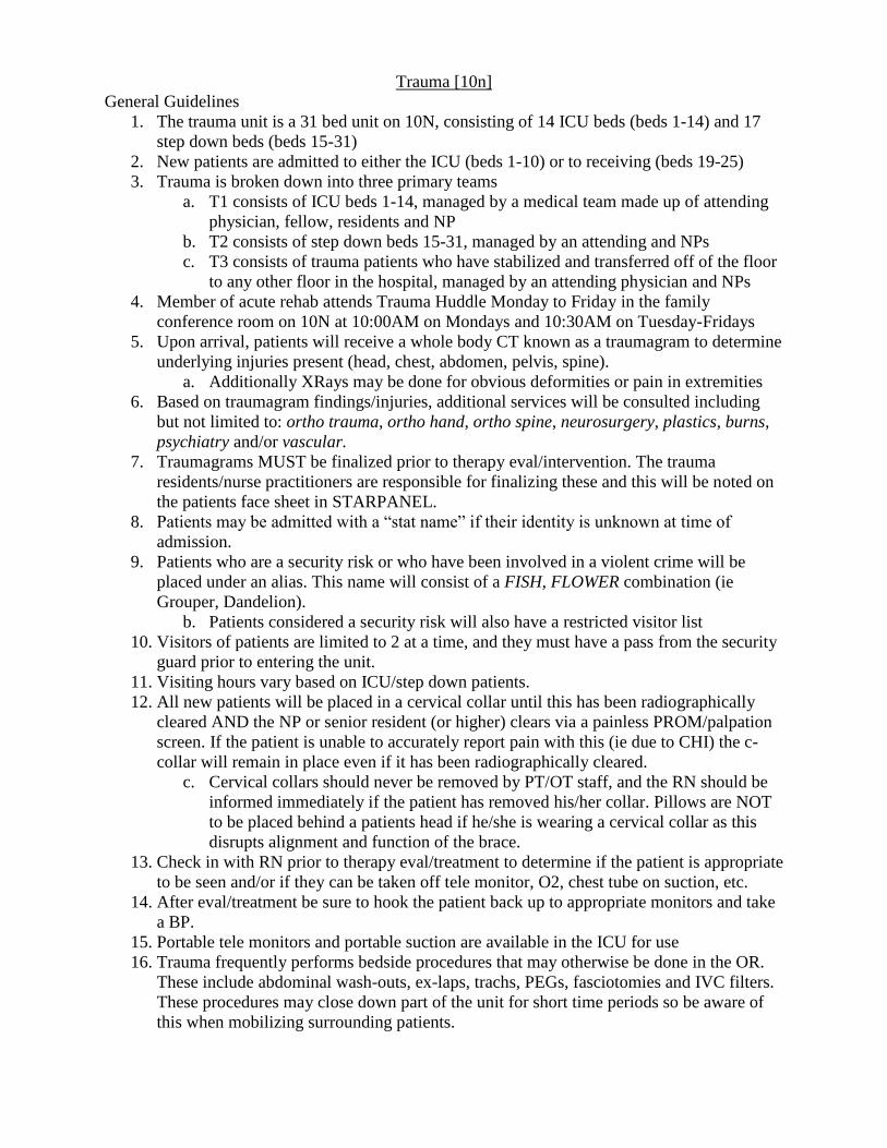
Trauma [10n]
General Guidelines
1. The trauma unit is a 31 bed unit on 10N, consisting of 14 ICU beds (beds 1-14) and 17
step down beds (beds 15-31)
2. New patients are admitted to either the ICU (beds 1-10) or to receiving (beds 19-25)
3. Trauma is broken down into three primary teams
a. T1 consists of ICU beds 1-14, managed by a medical team made up of attending
physician, fellow, residents and NP
b. T2 consists of step down beds 15-31, managed by an attending and NPs
c. T3 consists of trauma patients who have stabilized and transferred off of the floor
to any other floor in the hospital, managed by an attending physician and NPs
4. Member of acute rehab attends Trauma Huddle Monday to Friday in the family
conference room on 10N at 10:00AM on Mondays and 10:30AM on Tuesday-Fridays
5. Upon arrival, patients will receive a whole body CT known as a traumagram to determine
underlying injuries present (head, chest, abdomen, pelvis, spine).
a. Additionally XRays may be done for obvious deformities or pain in extremities
6. Based on traumagram findings/injuries, additional services will be consulted including
but not limited to: ortho trauma, ortho hand, ortho spine, neurosurgery, plastics, burns,
psychiatry and/or vascular.
7. Traumagrams MUST be finalized prior to therapy eval/intervention. The trauma
residents/nurse practitioners are responsible for finalizing these and this will be noted on
the patients face sheet in STARPANEL.
8. Patients may be admitted with a “stat name” if their identity is unknown at time of
admission.
9. Patients who are a security risk or who have been involved in a violent crime will be
placed under an alias. This name will consist of a FISH, FLOWER combination (ie
Grouper, Dandelion).
b. Patients considered a security risk will also have a restricted visitor list
10. Visitors of patients are limited to 2 at a time, and they must have a pass from the security
guard prior to entering the unit.
11. Visiting hours vary based on ICU/step down patients.
12. All new patients will be placed in a cervical collar until this has been radiographically
cleared AND the NP or senior resident (or higher) clears via a painless PROM/palpation
screen. If the patient is unable to accurately report pain with this (ie due to CHI) the c-
collar will remain in place even if it has been radiographically cleared.
c. Cervical collars should never be removed by PT/OT staff, and the RN should be
informed immediately if the patient has removed his/her collar. Pillows are NOT
to be placed behind a patients head if he/she is wearing a cervical collar as this
disrupts alignment and function of the brace.
13. Check in with RN prior to therapy eval/treatment to determine if the patient is appropriate
to be seen and/or if they can be taken off tele monitor, O2, chest tube on suction, etc.
14. After eval/treatment be sure to hook the patient back up to appropriate monitors and take
a BP.
15. Portable tele monitors and portable suction are available in the ICU for use
16. Trauma frequently performs bedside procedures that may otherwise be done in the OR.
These include abdominal wash-outs, ex-laps, trachs, PEGs, fasciotomies and IVC filters.
These procedures may close down part of the unit for short time periods so be aware of
this when mobilizing surrounding patients.

Spine Clearance
1. Once the traumagram is finalized spine injuries will be considered first. Patients will
remain flat in bed in reverse trendelenburg position until their spine has been cleared.
2. If the spine is cleared on the traumagram then OOB activity orders will come from the
trauma service
3. Transverse process and spinous process fractures do not typically require a spine team
consult unless multiple levels are affected.
4. All other spine injuries will require a spine consult for either ortho spine or neurosurgery
(depending on which team is on call the day of the patients admission).
5. If a spine consult is placed, the plan must be finalized and completed with OOB activity
clearance prior to initiation of therapy services.
6. OOB activity orders/need for brace will be found in STARPANEL under inpatient
orders, but it is encouraged to double check this with the operative note/latest progress
note written in the chart.
7. Surgical fixation required:
a. Surgery must be completed with upright films done with or without appropriate
brace as ordered by the consulting spine service
b. Upright films must be done, read and cleared with OOB orders placed by spine
service prior to intervention (or verbal order from NP after they have spoken to
spine)
8. Non-operative management:
a. A brace may or may not be indicated for non-op patients, and this should be
specifically listed in the activity order and progress note
b. If a brace is or is not recommended, the patient will still need to have upright
films done prior to OOB activity
c. Uprights must be done, read and cleared with OOB orders placed by the
consulting spine service prior to intervention by therapy (or as above a verbal
order from NP is ok if they have spoken to the spine team)
9. Common braces include: TLSO, TLSO with hip spica, CTLSO, CTO, LSO, Jewitt brace,
Minerva brace, Aspen TLSO, and quickdraw. Most all braces must be donned/doffed in
supine via log roll unless specified (ie quickdraw for comfort may be donned EOB).
Orthopedic Injuries
1. All orthopedic injuries are handled by ortho trauma, ortho hand and/or plastics.
2. WB activity orders will be entered by appropriate consult service
3. Activity orders should be placed in StarPanel under in-patient orders
4. Operative notes, and/or progress notes should also specify WB status
5. If conflicting WB status is found, page NP or consulting service for clarification
6. PELVIC, HIP and FEMUR fractures need an operative plan and definitive fixation if
determined to require surgical intervention prior to therapy intervention with documented
WB status by ortho.
7. Fractures in traction contradict therapy intervention
8. Pelvic fractures may initially be non-op with orders for ‘post-mobilization films’ in
which patients will have films taken after they have worked with therapy. Pending films
patients may or may not require surgical intervention.
9. FOOT, ANKLE, TIB/FIB, TIBIAL PLATEAU, PATELLA and any UE fractures are ok to
mobilize even if the patient will eventually require surgical intervention. All of these
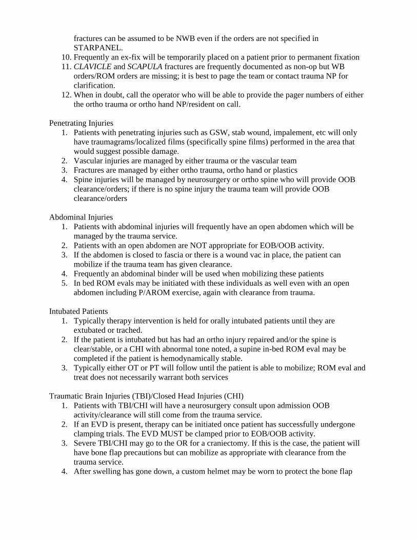
fractures can be assumed to be NWB even if the orders are not specified in
STARPANEL.
10. Frequently an ex-fix will be temporarily placed on a patient prior to permanent fixation
11. CLAVICLE and SCAPULA fractures are frequently documented as non-op but WB
orders/ROM orders are missing; it is best to page the team or contact trauma NP for
clarification.
12. When in doubt, call the operator who will be able to provide the pager numbers of either
the ortho trauma or ortho hand NP/resident on call.
Penetrating Injuries
1. Patients with penetrating injuries such as GSW, stab wound, impalement, etc will only
have traumagrams/localized films (specifically spine films) performed in the area that
would suggest possible damage.
2. Vascular injuries are managed by either trauma or the vascular team
3. Fractures are managed by either ortho trauma, ortho hand or plastics
4. Spine injuries will be managed by neurosurgery or ortho spine who will provide OOB
clearance/orders; if there is no spine injury the trauma team will provide OOB
clearance/orders
Abdominal Injuries
1. Patients with abdominal injuries will frequently have an open abdomen which will be
managed by the trauma service.
2. Patients with an open abdomen are NOT appropriate for EOB/OOB activity.
3. If the abdomen is closed to fascia or there is a wound vac in place, the patient can
mobilize if the trauma team has given clearance.
4. Frequently an abdominal binder will be used when mobilizing these patients
5. In bed ROM evals may be initiated with these individuals as well even with an open
abdomen including P/AROM exercise, again with clearance from trauma.
Intubated Patients
1. Typically therapy intervention is held for orally intubated patients until they are
extubated or trached.
2. If the patient is intubated but has had an ortho injury repaired and/or the spine is
clear/stable, or a CHI with abnormal tone noted, a supine in-bed ROM eval may be
completed if the patient is hemodynamically stable.
3. Typically either OT or PT will follow until the patient is able to mobilize; ROM eval and
treat does not necessarily warrant both services
Traumatic Brain Injuries (TBI)/Closed Head Injuries (CHI)
1. Patients with TBI/CHI will have a neurosurgery consult upon admission OOB
activity/clearance will still come from the trauma service.
2. If an EVD is present, therapy can be initiated once patient has successfully undergone
clamping trials. The EVD MUST be clamped prior to EOB/OOB activity.
3. Severe TBI/CHI may go to the OR for a craniectomy. If this is the case, the patient will
have bone flap precautions but can mobilize as appropriate with clearance from the
trauma service.
4. After swelling has gone down, a custom helmet may be worn to protect the bone flap
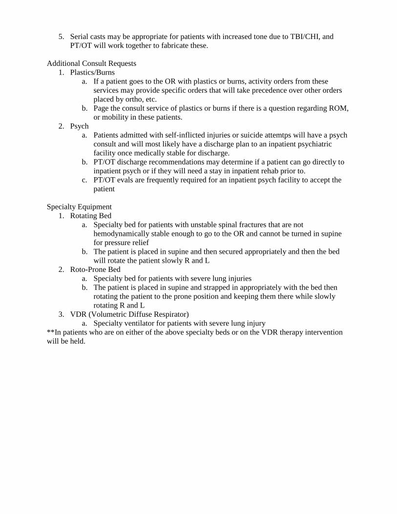
5. Serial casts may be appropriate for patients with increased tone due to TBI/CHI, and
PT/OT will work together to fabricate these.
Additional Consult Requests
1. Plastics/Burns
a. If a patient goes to the OR with plastics or burns, activity orders from these
services may provide specific orders that will take precedence over other orders
placed by ortho, etc.
b. Page the consult service of plastics or burns if there is a question regarding ROM,
or mobility in these patients.
2. Psych
a. Patients admitted with self-inflicted injuries or suicide attemtps will have a psych
consult and will most likely have a discharge plan to an inpatient psychiatric
facility once medically stable for discharge.
b. PT/OT discharge recommendations may determine if a patient can go directly to
inpatient psych or if they will need a stay in inpatient rehab prior to.
c. PT/OT evals are frequently required for an inpatient psych facility to accept the
patient
Specialty Equipment
1. Rotating Bed
a. Specialty bed for patients with unstable spinal fractures that are not
hemodynamically stable enough to go to the OR and cannot be turned in supine
for pressure relief
b. The patient is placed in supine and then secured appropriately and then the bed
will rotate the patient slowly R and L
2. Roto-Prone Bed
a. Specialty bed for patients with severe lung injuries
b. The patient is placed in supine and strapped in appropriately with the bed then
rotating the patient to the prone position and keeping them there while slowly
rotating R and L
3. VDR (Volumetric Diffuse Respirator)
a. Specialty ventilator for patients with severe lung injury
**In patients who are on either of the above specialty beds or on the VDR therapy intervention
will be held.
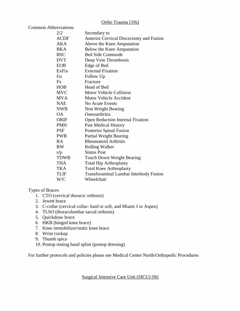
Ortho Trauma [10s]
Common Abbreviations
2/2 Secondary to
ACDF Anterior Cervical Discectomy and Fusion
AKA Above the Knee Amputation
BKA Below the Knee Amputation
BSC Bed Side Commode
DVT Deep Vein Thrombosis
EOB Edge of Bed
ExFix External Fixation
f/u Follow Up
Fx Fracture
HOB Head of Bed
MVC Motor Vehicle Collision
MVA Motor Vehicle Accident
NAE No Acute Events
NWB Non Weight Bearing
OA Osteoarthritis
ORIF Open Reduction Internal Fixation
PMH Past Medical History
PSF Posterior Spinal Fusion
PWB Partial Weight Bearing
RA Rheumatoid Arthritis
RW Rolling Walker
s/p Status Post
TDWB Touch Down Weight Bearing
THA Total Hip Arthroplasty
TKA Total Knee Arthroplasty
TLIF Transforaminal Lumbar Interbody Fusion
W/C Wheelchair
Types of Braces
1. CTO (cervical thoracic orthosis)
2. Jewett brace
3. C-collar (cervical collar- hard or soft, and Miami J or Aspen)
4. TLSO (thoracolumbar sacral orthosis)
5. Quickdraw brace
6. HKB (hinged knee brace)
7. Knee immobilizer/static knee brace
8. Wrist cockup
9. Thumb spica
10. Postop resting hand splint (postop dressing)
For further protocols and policies please see Medical Center North/Orthopedic Procedures
Surgical Intensive Care Unit (SICU) [9t]

General Guidelines
1. Patients on the surgical ICU include primarily the following: general surgery, liver
transplant, head and neck cancer surgery, trauma overflow, orthopedic surgery patients
requiring ICU care.
2. Rooms 27- 61 are SICU rooms, and SICU MD’s and NP’s round on these patients.
PT/OT orders can be obtained from these MD’s/NP’s. (MD/NP work room is to the left
of patient room 9625, and they can often be found there and are willing to answer
questions.)
3. Rooms 1-25 are surgical stepdown rooms. The patient’s primary team is responsible for
these patients, and questions and orders go through them. If the SICU has open beds,
patients from other areas may be transferred there. They are considered “boarders” and
are followed by their primary team (There is a green capital “B” on their doors.).
4. Talk to the patient’s nurse before and after evaluating or treating them. They know the
most about the patient’s condition and precautions. Ask them if the patient has recently
received any medications (e.g. BP meds, pain meds) that might affect their safety with
mobility/standing activities in order to more safely monitor the PT/OT session.
5. Determine if the patient has the bed alarm or a chair alarm activated prior to mobilizing
the patient. Reactivate these alarms at the end of the PT/OT session. Keep the room
curtain open at the end of the PT/OT session if the patient has been determined to be a
falls risk.
6. These patients need to stay on their monitor or a portable monitor during treatment unless
they have transfer orders to a non-monitored floor or their nurse says they are stable
enough to come off the monitor for PT. Use your clinical judgment regarding using or
not using portable telemetry with stepdown patients especially for patients with a cardiac
history. Documenting vitals is helpful in monitoring a patient’s response to treatment and
for rehab facilities to note when screening patients for admission. The portable monitors
are located on the back (ICU) hall on the counter and next to the large red travel
monitors, to the right if you are facing the charge nurse. It is recommended that you
change out the battery pack for a fully charged one before using. Please clean and return
monitors here after use. Place patients back on their monitor in their room and run their
blood pressure before leaving.
7. Before treatment, be aware of all lines and drains on the patient and around the bed.
Examine a post-op abdomen before mobilizing to check for drains, indwelling pain
catheters, chest tubes, dressings, wound vacs. Pin or secure drains so they do not hang
and ideally toward the front so the patient will not sit or lie on them.
8. Dressings, safety pins, finger monitors, extension tubing and other supplies can be found
in the carts in the rooms. The access code to the carts is 1234 “Enter.”
9. There is a dedicated wheelchair for PT/OT use in the equipment room for use when
ambulating patients in the interest of fall prevention. The combination on the lock is F-
A-L-L.
10. The service center on 9T has inflatable seat cushions for patients who are prone to
pressure areas when sitting. Do not place a pillow under these seat cushions when the
patient is sitting in a bedside chair. You can place folded blankets under the blue
cushions to raise the height of the chair seat. The service center also has abdominal
binders which can be trimmed to the appropriate size. Items taken from the service center
need to be scanned before being taken for inventory purposes. You can access the
scanner by scanning the bar code taped to the inventory monitors, identifying the patient
for whom you are getting supplies, then scanning the item’s bar code on the cart with the
scanning gun.
11. Respiratory supplies are also found in the 9T service center to the right of the door as you
enter. Extension oxygen tubing, adaptors, venti tubing, nasal cannulas can be found
there. They do not need to be scanned for inventory since Respiratory Therapy stocks
their supplies.
12. Oxygen tanks are located both in the clean utility room and in the 9T service center.
Place used tanks in the dirty utility room. Wheels for the tanks are in the clean and dirty
utility room.
13. 9T has Smooth Moves patient handling equipment in the equipment room. Some total
lift slings are in the 9T service center in the cabinet under the counter. Sara slings can be
ordered through the hospital service center. Slippery sheets are with the linen in the clean
utility room.
14. Warm blankets are located in the clean utility room.
15. Neuro chairs are stored in the middle hallway where x-ray equipment is located (door is
marked “radiology”).
16. Walkers are stored in the equipment room. You may find walkers in the dirty utility
room as well. Clean walkers after using and return to them to the equipment room.
17. Thoracic walkers can be found in the middle hallway across from the clean/dirty utility
rooms.
Liver Transplants
1. Donor is matched to a potential recipient on the basis of several factors: ABO blood type,
body size, degree of medical urgency and MELD score (Model for End-Stage Liver
Disease)
2. Model for End-Stage Liver Disease (MELD): a reliable measure of mortality risk in
patients with end stage liver disease. MELD is a numerical scale used for adult liver
transplant candidates and the range is from 6 (less ill) to 40 (gravely ill). The individual
score determines how urgently a patient needs a liver transplant within the next three
months. The number is calculated using the most recent laboratory tests.
3. Cannot go to SNF due to cost of anti-rejection medications
4. Have to stay local for 30 days and have 24 hour/day caregiver x30 days to be listed for
transplant
5. Typically have 2 right sided JP drains after surgery with large crescent moon scar on
stomach
6. 3 Month Mortality based on MELD score
a. 40 or more -- 71.3% mortality
b. 30-39 -- 52.6% mortality
c. 20-29 -- 19.6% mortality
d. 10-19 -- 6.0% mortality
e. <9 -- 1.9% mortality
Partial Hepatectomy
1. Surgical procedure to remove tumors in the liver. Removing the cancerous portion
of the liver may help to prevent the disease from spreading more.
Dialysis

1. If patient has a femoral vas cath for dialysis, PT/OT need clarification on
clearance to mobilize.
2. Hemodialysis (HD)
a. Process for removing waste and excess water from the blood, and is
primarily used to provide an artificial replacement for lost kidney function
in people with renal failure.
b. May be used for those with an acute disturbance in kidney function (acute
kidney injury, previously acute renal failure) or for those with progressive
but chronically worsening kidney function-a state known as chronic
kidney disease stage 5 (previously chronic renal failure or end-stage
kidney disease).
c. PT/OT unable to work with patient on HD at VUMC.
3. Continuous veno-venous hemodialysis (CVVHD) or Continuous Renal
Replacement Therapy (CRRT)
a. Indications: ARF, ARDS, after surgery with cardiopulmonary bypass,
refractory congestive heart failure.
b. Provides slow and balanced fluid removal and/or hemodialysis
c. Usually patient will be on some type of vasopressor.
d. Ask RN if patient's BP has remained stable over the last 12-24 hours. If
so, PT/OT may be warranted. One person watches this line exclusively
during mobility.
4. Peritoneal Dialysis
a. Performed through port in peritoneal cavity.
b. Provides more independence.
c. Typically unable to see patient while they are “filling” and “emptying”
d. Patients are at risk for peritonitis.
Head and Neck Surgeries
1. Precautions:
a. Fistula or infection—Hold AROM until approved by MD. Usually these patients
have to wait until they are discharged to begin AROM exercises.
b. Chyle leak—Hold AROM exercises until cleared by MD. Chyle is a milky lymph
liquid that my leak into the operative area and can be seen in the JP drains. It is
normally managed by diet or further surgery.
2. Glossectomy: surgery to remove all or part of the tongue. This surgery is used to
treat tongue cancer.
a. Partial – removal of part of the tongue
b. Hemi – one side of the tongue is removed
c. Total – total removal of the tongue
3. Laryngoscopy: an examination of the back of the throat, including the voice box
(larynx). Direct laryngoscopy uses a tube called laryngoscope, which is placed in
the back of your throat. The tube may be flexible or stiff. This procedure allows
the doctor to see deeper in the throat and to remove a foreign object or sample of
tissue for biopsy.
4. Tracheostomy: surgical procedure to create an opening through the neck into the
trachea (windpipe). A tube is usually placed through this opening to provide an
airway and to remove secretions from the lungs. This tube is called a
tracheostomy tube or trach tube.
5. Mandibulectomy: procedure that is used to eradicate disease that involves the
lower jaw or mandible. This procedure can be used in various settings, including
infectious etiologies (e.g. osteomylelitis), osteoradionecrosis, or a benign (e.g.
ameloblastoma) or malignant neoplastic process (e.g. invasive squamous cell
carcinoma) that involves the jaw. The area removed is then replaced with either: a
muscle and skin ‘flap’, or a combination of metal pate, skin, muscle, and/or bone
‘flap’
6. Selective neck dissection: This is the least invasive of the head and neck
surgeries. Semi-reclining is often the most comfortable position for performing
their exercises.
7. Bilateral selective neck dissection: Same as for selective neck dissection yet on
bilateral neck.
8. Modified and radical neck dissection: These surgeries are more invasive. You
can emphasize supporting the involved upper extremity(s) by placing thumb in a
belt loop, crossing arms, placing hands in pockets, etc.
Radial forearm free flap (RFFF)
1. These patients are to be PFWB for two weeks.
2. Ambulate with a platform walker initially, as needed, progressing to a standard or rolling
walker at two weeks post-op if walker is still needed.
3. The goal is to avoid wrist extension for the first two weeks to protect the skin graft on the
donor arm.
4. You may see a wound vac on the donor arm.
5. Begin gentle wrist AROM exercises POD 14 (splint should be off sooner). Tell the
patient to avoid extreme wrist extension and remind them to avoid lifting heavy objects.
6. If the plastics team has done the RFFF, protocol for starting wrist ROM is as follows:
When the dressing comes down on POD 5, if the skin graft has taken, the patient may
begin ROM of the hand and wrist. The plastics team normally enters this information in
Wiz or in their daily note.
7. If these flaps include a bony component (osteocutaneous radial forearm free
flap/OCRFFF) they are harvested by a hand surgeon and undergo ORIF of the radius. In
these cases, these patients are PFWB on the donor UE and have wrist movement
precautions until they see the hand surgeon for splint removal.
8. The hand service drives the onset of movement and will assess the patient’s need for
hand therapy.
Fibular free flap (FFF)
1. Begin gentle heel cord stretching POD 6.
2. The patient is to be NWB initially in order to protect the skin graft on the donor leg, and
it is important that they be out of bed and mobilizing early.
3. You may see a wound vac on the donor leg.
4. Drs. Sinard, Mannion, and Rhode often allow their patients to be WBAT around POD 5.
5. If this is not clear in the orders, ask the OTO team.
6. If the plastics team has done the FFF, they will indicate when they want WB to start, and
you may see an indwelling Doppler monitor.
7. Assess for the most appropriate assistive device as needed.
Scapular free flap (SFF)

1. Begin AAROM exercises to donor arm POD 7 (unless otherwise ordered) in semi-
reclining or supine to comfort.
2. Emphasize to the patient that they should avoid resistive use of the donor arm for the first
six weeks due to donor muscle being detached and reattached to the scapula.
3. Instruct the patient in pendulum exercises to be performed when they are comfortable
bending at the waist to perform them.
4. The patient can be provided a sling for comfort until they can tolerate ambulating without
the sling.
5. Remind the patient to elevate the donor arm on pillows when resting.
Pec flap
1. Begin gentle AAROM to donor side shoulder POD 3.
2. The extent of the incision into the axilla will often determine tolerance for ROM.
3. Emphasize the importance of ROM to the donor side shoulder and instruct in self-assisted
ROM.
Cervocofacial advancement flap
1. Sometimes seen in conjunction with other head and neck surgical reconstruction.
2. Ask the team about head/neck movement restrictions.
3. Often these flaps have tight closures and need a week or more for primary healing.
4. Patients can be instructed to start performing their head and neck exercises on a
designated date
Lateral thigh free flap
1. No restrictions, only if neck JP drains are present.
Iliac crest free flap
1. Bed to chair only usually until POD 3 then WBAT for ambulation with/without assistive
device depending on comfort.
Lateral arm free flap
1. Triceps are divided and will be most affected.
2. Begin gentle UE AROM exercises POD 5.
Rectus free flap
1. No restrictions, only if neck JP drains are present.
Carotid replacement
1. Does not affect the course of PT.
2. AROM exercises may be initiated according to guidelines related to drain removal.
Necrotizing Fasciitis
1. An infection caused by bacteria.
2. It can destroy skin, fat, and the tissue covering the muscles within a very short time.
3. The disease sometimes is called flesh-eating bacteria.
4. When it occurs on the genitals, it is called Fournier’s gangrene.
Gastrointestinal Perforation (Perforated Bowel)

1. A hole that develops through the whole wall of the esophagus, stomach, small
intestine, large bowel, rectum, or gallbladder. This condition is a medical
emergency.
2. Gastrointestinal perforation can be caused by a variety of illnesses. These include:
Appendicitis, Cancer, Crohn's disease, Diverticulitis, Gallbladder disease, Peptic ulcer
disease, Ulcerative colitis
Gastrointestinal Bleed (GI bleed)
1. Loss of blood in the gastrointestinal tract, from the pharynx to the rectum.
2. Upper GI bleed - An upper source is characterized by hematemesis (vomiting up
blood) and melena (tarry stool containing altered blood).
3. Lower GI bleed - indicated by bright red blood per rectum (BRBPR).
4. Causes: Anal fissure, Hemorrhoids, Cancer, Intestinal polyps (a pre-cancerous
condition), abnormal blood vessels in the lining of the intestines (also called
angiodysplasias), Diverticulosis, Crohn's disease or ulcerative colitis, Esophageal
varices, Esophagitis, Gastric (stomach) ulcer, Mallory-Weiss tear, Radiation injury to
the bowel
5. Testing: EGD, Colonoscopy, Labs
6. Tips:
a. Check labs
b. PT/OT not indicated if patient is actively bleeding.
c. If getting blood products, check with nursing if okay to mobilize.
Small Bowel Resection (SBR)
1. Surgery to remove part or all of the small bowel. It is done when part of the small bowel
is blocked or diseased.
2. Most digestion (breaking down and absorbing nutrients) of the food you eat takes place in
the small intestine.
Whipple (PANCREATICODUODENECTOMY)
1. The most commonly performed surgery to remove tumors in the pancreas.
2. In a standard Whipple procedure, the surgeon removes the head of the pancreas, the
gallbladder, part of the duodenum which is the uppermost portion of the small intestine, a
small portion of the stomach called the pylorus, and the lymph nodes near the head of the
pancreas. The surgeon then reconnects the remaining pancreas and digestive organs so
that pancreatic digestive enzymes, bile, and stomach contents will flow into the small
intestine during digestion. In another type of Whipple procedure known as pylorus
preserving Whipple, the bottom portion of the stomach, or pylorus, is not removed.
3. In both cases, the surgery usually lasts between 5-8 hours.
Gastrojejunostomy
1. Surgical anastomosis between the stomach and the small intestine.
Colostomy
1. Surgical resection of the healthy end of the large intestine or colon through an incision
in the anterior abdominal wall brought through to the surface of the skin (stoma).
2. In a colostomy takedown, the two ends of the colon are reconnected and the area
where the stoma was created and the abdomen are closed.
Ileostomy
1. Surgical resection of the small intestine (the ileum) out onto the surface of the skin to
form a "stoma".
2. With a temporary ileostomy, all or part of the colon is removed, but at least part of the
rectum is left intact.
Pancreaticojejunostomy
1. Surgical connection of the pancreatic duct with the small intestine.
Roux-En-Y (ROUX-EN-Y HEPATICOJEJUNOSTOMY OR
CHOLEDOCHOJEJUNOSTOMY)
1. Surgical reconstruction to reestablish bile flow involving the small intestine to provide
drainage without reflux, used as a treatment for sclerosing cholangitis, injured bile
ducts, and bile duct tumors.
Exploratory Laparotomy (EX-LAP)
1. Operation where a cut is made into the abdomen. It is a method used to explore the
abdomen, a diagnostic tool that allows physicians to examine the abdominal organs.
Cholecystectomy
1. Surgical procedure to remove the gallbladder
Esophageal Perforation
1. A hole in the esophagus. The esophagus is the tube food passes through as it goes from the
mouth to the stomach.
2. CAUSES: The contents of the esophagus can pass into the mediastinum, the surrounding
area in the chest, when there is a hole in the esophagus.
3. Often results in infection of the mediastinum (mediastinitis). The most common cause of
an esophageal perforation is injury during a medical procedure. However, the use of
flexible instruments has made this problem rare. The esophagus may also become
perforated as the result of: A tumor, Gastric reflux with ulceration, Previous surgery on
the esophagus, Swallowing a foreign object or caustic chemicals such as household
cleaners, disk batteries, and battery acid, Trauma or injury to the chest and esophagus
Enterocutaneous Fistula (ECF)
1. An abnormal connection that develops between the intestinal tract or stomach and the
skin. As a result, contents of the stomach or intestines leak through to the skin.
2. Often occur after bowel surgery. Other causes include: infection, perforated peptic
ulcer, inflammatory bowel disease, Crohn's disease or ulcerative colitis. An ECF may also
develop from an abdominal injury or trauma, such as a stabbing or gunshot.
Hernia Repair
1. A hernia occurs when inner layers of abdominal muscle become weakened. The lining of
the abdomen then bulges out into a small sac, and part of the intestine or abdominal tissue
may enter the sac. Hernias occur most commonly in the groin (inguinal hernia), the navel
(umbilical hernia), and at the incision site of a previous surgery.

Thoracentesis
1. Procedure to remove fluid from the space between the lining of the outside of the lungs
(pleura) and the wall of the chest.
2. Used to find the cause of a pleural effusion. The procedure also might be done to help
you breathe easier.
Video-Assisted Thoracscopic Surgery (VATS)
1. A minimally invasive surgical technique used to diagnose and treat problems in the chest.
2. During a video-assisted thoracoscopic surgery procedure, a tiny camera (thoracoscope)
and surgical instruments are inserted into your chest through several small incisions. The
thoracoscope transmits images of the inside of your chest onto a video monitor, guiding
the surgeon in performing the procedure.
3. Surgeons use the video-assisted thoracoscopic surgery technique to perform a variety of
operations.
Bronchoscopy
1. Procedure that allows doctors to look at your airway through a thin viewing
instrument called a bronchoscope. During a bronchoscopy camera.gif, your doctor
will examine your throat, larynx, trachea, and lower airways.
2. Bronchoscopy may be done to diagnose problems with the airway, the lungs, or with
the lymph nodes in the chest, or to treat problems such as an object or growth in the
airway.
Lung Surgery
1. Biopsy of an unknown growth
2. Lobectomy: to remove one or more lobes of a lung
3. Lung transplant
4. Pneumonectomy: to remove a lung
5. Pleurodesis: surgery to prevent the buildup or return of fluid to the chest
6. Surgery to remove an infection or blood in the chest cavity (empyema)
7. Surgery to remove small balloon-like tissues (blebs) that cause pneumothorax
8. Wedge Resection: to remove part of a lobe in a lung
Thoracotomy
1. A surgical cut that a surgeon makes to open the chest wall.
Nephrectomy
1. Kidney removal, or nephrectomy, is surgery to remove all or part of a kidney.
a. Partial Nephrectomy: part of one kidney removed
b. Simple Nephrectomy: all of one kidney removed
c. Radical nephrectomy: removal of one entire kidney, surrounding lymph nodes,
and the adrenal gland
Esophagectomy
1. Surgery to remove part or all of the esophagus. After it is removed, the esophagus is
rebuilt from part of your stomach or part of your large intestine.
2. Often have epidural, chest tube, Dobhoff tube, NG tube, foley, etc
3. Good to have two sets of hands for initial evaluation.

Hyperthermic Intraperitoneal Chemotherapy (HIPEC)
1. Cancer that has spread to the lining surfaces of the peritoneal (abdominal) cavity from
primary colorectal cancer, ovarian cancer, gastric cancer, appendiceal cancer or from
mesothelioma and pseudomyxoma peritonei--known as peritoneal carcinomatosis, are
often difficult to treat.
2. The term "Intra peritoneal" means that the treatment is delivered to the abdominal cavity.
The term "Hyperthermic Chemotherapy" means that the solution containing
chemotherapy is heated to a temperature greater than normal body temperature.
3. Before HIPEC is administered, cytoreductive surgery is performed. This surgery is the
removal of all visible tumors throughout the peritoneal cavity.
4. During the HIPEC procedure, the surgeon will continuously circulate a heated sterile
solution--containing a chemotherapeutic agent--throughout the peritoneal cavity to kill
any remaining cancer cells.
a. HIPEC has several benefits as compared to standard chemotherapy:
b. HIPEC is a single treatment done in the operating room, while standard
chemotherapy requires multiple treatments
c. 90% of the chemotherapy drug stays within the abdominal cavity, making it less
toxic for the patient
d. It allows for a more intense dose of chemotherapy
Sepsis
1. Potentially deadly medical condition that is characterized by a whole-body
inflammatory state and the presence of a known or suspected infection.
2. The body may develop this inflammatory response by the immune system to
microbes in the blood, urine, lungs, skin, or other tissues.
3. Common infections are peritonitis, urinary tract infection or pyelonephritis,
meningitis, bacterial pneumonia or cellulitis.
4. In hospitalized patients, common sites of infection include intravenous lines, surgical
wounds, surgical drains, and decubitus ulcers.
5. Septicemia or bacteremia refers to the presence of pathogenic organisms in the
bloodstream, leading to sepsis.
6. Septic shock is defined as sepsis with refractory arterial hypotension or
hypoperfusion abnormalities in spite of adequate fluid resuscitation.
7. When severe, may also include end organ dysfunction. Examples are acute lung injury
(ALI), acute respiratory distress syndrome (ARDS), encephalopathy, liver
dysfunction, acute kidney injury (AKI) or acute renal failure (ARF), and systolic and
diastolic heart failure.
8. Symptoms of sepsis can include:
a. Confusion or delirium
b. Hypotension
c. Tachycardia
d. Hypothermia
e. Hyperventilation
f. Fever
Atrial Fibrillation (AF or A-FIB)
1. Most common cardiac arrhythmia (abnormal heart rhythm)

2. The upper chambers of the heart (atria) experience chaotic electrical signals. The AV
node-the electrical connection between the atria and the ventricles is overloaded with
impulses trying to get through to the ventricles. The ventricles also beat rapidly but not as
rapidly as the atria.
3. Its name comes from the fibrillating (IE quivering) of the heart muscles of the atria,
instead of a coordinated contraction.
4. Can be associated with a rapid ventricular response (RVR) which is any ventricular rate
greater than 100 beats per min (bpm).
5. Some individuals are asymptomatic despite having frequent episodes, while others
experience symptoms that are troubling or incapacitating.
6. May result in palpitations, fainting, chest pain or congestive heart failure.
7. Some individuals with A-fib have a significantly increased risk of stroke.
8. Stroke risk increases during A-fib because blood may pool and form clots in the poorly
contracting atria and especially in the left atrial appendage (LAA).
9. A-fib may be treated with medications which either slow the heart rate or revert the heart
rhythm back to normal.
10. Cardioversion is a noninvasive conversion of an irregular heartbeat to a normal heartbeat
using electrical or chemical means:
11. Electrical cardioversion involves the restoration of normal heart rhythm through the
application of DC electrical shock.
12. Chemical cardioversion is performed with drugs, such as amiodarone, dronedarone,
procainamide, dofetilide, propafenone or flecainide.
13. Surgical and catheter-based therapies (ablation) may also be used to prevent recurrence
of A-fib in certain individuals.
14. People with A-fib are often given anticoagulants such as warfarin (Coumadin) to reduce
risk of stroke.
15. If a patient has atrial fibrillation with RVR, PT/OT is contraindicated.
16. Check with nursing to make sure patient is rate controlled
17. When a patient is on Coumadin, INR must be in an appropriate range.
18. Monitor patients, as some patients are asymptomatic
19. If patient becomes symptomatic, take a rest break and can attempt further activity as
patient tolerates.
Lung Disease
1. Restrictive:
a. Respiratory diseases that restrict lung expansion resulting in a decreased lung
volume, an increased work of breathing, and inadequate ventilation and/or
oxygenation
b. Causes:
i. Asbestosis
ii. Radiation fibrosis
iii. Acute respiratory distress syndrome (ARDS), a severe lung condition
occurring in response to a critical illness or injury
iv. Idiopathic pulmonary fibrosis (IPF)
v. Sarcoidosis
2. Obstructive:
a. Respiratory disease characterized by airway obstruction
b. Generally characterized by inflamed and easily collapsible airways, obstruction to
air flow, and frequent office visits and hospitalizations
c. Types include:
i. Asthma
ii. Bronchiectasis
iii. Bronchitis
iv. Chronic obstructive pulmonary disease (COPD)
d. Tips:
i. Patients with COPD are CO2 retainers, therefore, their O2 sats are usually
mid to high 80’s with activity. Be careful increasing supplemental O2 as
increased O2 results in increased CO2 resulting in decreased respiratory
drive.
ii. Monitor O2 sats at rest and with activity.
iii. Educate COPD patients on pursed lip breathing.
iv. Educate patients with lung disease on energy conservation (e.g. sitting to
perform tasks, increasing rest breaks, etc.)
Common Drains/Lines
1. Jackson Pratt Drain (JP)
a. Used to remove fluids that build up in an area of the body after surgery.
b. Removes fluids by creating suction in the tube.
2. Hemovac Drain
a. Placed into a vascular cavity where blood drainage is expected after surgery, such
as with abdominal and orthopedic surgery.
b. Suction is maintained by compressing a spring-like device in the collection unit.
As the drainage accumulates in the collection unit, it expands and suction is lost.
3. Accordion Drain
a. Small, compact and allows for patient complete ambulation
b. One hand operation to activate suction
c. Delivers initial vacuum of approximately 4.5 psi (230 mm Hg)
d. Reverts to gravity drainage after bellows re-expansion preventing back-up
e. Closed system for infection control
4. Chest Tube (CT)
a. Drains blood, fluid, or air from around the lungs allowing the lungs to fully
expand. A flexible plastic tube is inserted through the side of the chest into the
pleural space and is connected to a canister which can be to suction or to water
seal.
b. Indications:
i. Pneumothorax: accumulation of air in the pleural space
ii. Pleural effusion: accumulation of fluid in the pleural space
iii. Chylothorax: a collection of lymphatic fluid in the pleural space
iv. Empyema: a pyogenic infection of the pleural space
v. Hemothorax: accumulation of blood in the pleural space
vi. Hydrothorax: accumulation of serous fluid in the pleural space
c. Tips:
i. Check with nursing to see if patient can come off of wall suction
ii. Usually patients are able to come off suction for short time during activity
iii. Some patient must have chest tubes remain on suction secondary to air
leakage or subcutaneous air; in these cases there is portable suction
available for ambulation.
iv. Should a chest tube be accidentally pulled, petroleum guaze is best to
cover chest tube site to allow air to escape but not enter. If you do not
have access to petroleum gauze, use a towel or cloth. Contact nursing and
return patient to bed.
v. If canister tipped, clamp tubing and notify nursing
5. Wound Vac
a. Conducts negative pressure wound therapy (NPWT). The device consists of a
dressing which is fitted with a tube and attached to the wound VAC.
b. Negative pressure wound therapy is most commonly used with chronic wounds
which are not responding to other forms of treatment and sometimes with surgical
wounds which have reopened.
6. Tunneled Epidural Catheter (TEC)
a. Very thin, flexible tube that is implanted into the spine (specifically, the epidural
space) and tunneled under your skin. Through it, the patient can receive ongoing
b. Doses of medication that stops nerves in the spinal cord from sensing pain.
7. Patient Controlled Analgesia (PCA)
a. Method of pain control that gives patients the power to control their pain. In PCA,
a computerized pump called the patient-controlled analgesia pump, which
contains a syringe of pain medication as prescribed by a doctor, is connected
directly to a patient's intravenous (IV) line
8. Total Parenteral Nutrition (TPN)
a. Method of feeding that bypasses the gastrointestinal tract. Fluids are given into a
vein, which provides most of the nutrients the body needs. This method is used
when a patient cannot or should not receive feedings or fluids by mouth.
9. Bowel Management System (BMS)
a. Temporary containment device, indicated for bedridden or immobilized,
incontinent patients with liquid or semi-liquid stool.
b. It is designed to safely and effectively contain and divert fecal matter, protect
patients' wounds from fecal contamination, and reduce both the risk of skin
breakdown and spread of infection.
10. Percutaneous Endoscopic Gastrostomy (PEG)
a. Flexible feeding tube is placed through the abdominal wall and into the stomach.
PEG allows nutrition, fluids and/or medications to be put directly into the
stomach, bypassing the
11. Dobhoff Tube (DHT)
a. Small-bore, flexible, nasogastric (NG) feeding tube that is used to administer
nourishment and medicine to people who cannot ingest anything by mouth.
b. The tube is inserted into the stomach by way of the nasal passage.
c. Unlike a tube used for gastrointestinal drainage ,there is no suction attached to a
Dobhoff tube.
12. Nasograstric Tube (NGT)
a. A flexible tube that is passed through the nose and down through the nasopharynx
and esophagus into the stomach.
b. It can be used to remove the contents of the stomach, including air, to decompress
the stomach, or to remove small solid objects such as poison, from the stomach.
c. If you can ambulate the patient and interrupt suction, clamp the tube to prevent
stomach contents from draining out of the tube.
d. Reconnect after treatment.
13. Central Line
a. Usually located in the superior vena cava or inferior vena cava, or within the right
atrium of the heart.
b. This has several advantages over a peripheral IV:
i. It can deliver fluids and medications that would be overly irritating to
peripheral veins because of their concentration or chemical composition.
These include some chemotherapy drugs and total parenteral nutrition
(TPN).
ii. Medications reach the heart immediately and are quickly distributed to the
rest of the body.
iii. There is room for multiple parallel compartments (lumen) within the
catheter, so that multiple medications can be delivered at once even if they
would not be chemically compatible within a single tube.
iv. Central venous pressure (CVP) can be measured through the line.
v. CVP: venous BP (pressure of blood returning to the heart)
c. Tip:
i. Make sure there is enough slack on the lines during mobility.
ii. There are no contraindications to shoulder ROM on the side of the central
line.
14. Peripherally Inserted Central Catheter (PICC)
a. PICC lines are used when intravenous access is required over a prolonged period
of time, or when medications to be infused would cause quick damage and early
failure of a peripheral IV, and when a conventional central line may be too
dangerous to attempt.
b. Typical uses include long chemotherapy regimens, extended antibiotic therapy, or
total parenteral nutrition (TPN).
c. Line is placed under ultrasound guidance, usually in the arm, and then carefully
advanced upward until the catheter is in the superior vena cava or the right atrium.
An X-ray must be used to verify that the tip is in the right place when fluoroscopy
was not used during the insertion.
d. A PICC can be single, double, or triple-lumen.
e. The chief advantage of a PICC over other types of central lines is that it is safer to
insert with a relatively low risk of uncontrollable bleeding and essentially no risks
of damage to the lungs or major blood vessels.
f. A PICC can be left in place for months to years if needed for patients who require
extended treatment.
15. Arterial Line (ART LINE)
a. Monitors BP real-time (rather than by intermittent measurement)
b. A thin catheter is inserted into an artery, usually the wrist (radial artery), armpit
(axillary artery), groin (femoral artery), or foot (pedal artery).
c. Intra-arterial blood pressure measurement is more accurate than measurement of
BP by noninvasive means, especially in the critically ill.
d. Permits the rapid recognition of BP changes that is vital for patients on
continuous infusions of vasoactive drugs
e. Tip: Avoid repetitive ROM to joint that arterial line is near.

Respiratory in the SICU
1. Nasal cannula
a. When a patient requires greater than 6L supplemental O2, they may be put on a
high flow nasal cannula which is easily identified by its darker green tubing. Up
to 15L O2 can be given this way.
b. Another type of nasal cannula used on the SICU is Optiflow which is attached to
ventilator-type tubing which leads to a humidification and warming chamber.
2. Trach Collar
a. Used when a patient has been trached normally due failure to wean from the
ventilator or oral/airway edema following head and neck cancer surgery or trauma
to the airway
b. Supplemental oxygen is also referred to as FiO2 or fractional index of O2. Room
air is 21% FiO2. SICU patients may be on 28-50% trach collar. The percentage
of O2 can be determined by reading the dial over the water bottle at the wall O2
outlet. It is documented as % FiO2 and not by liters of O2.
c. When converting a trach collar to portable O2 for ambulation, it is necessary to
obtain a venti tube kit from the SICU service center. The corresponding adaptor
is identified by matching the percentage of O2 written on the end and identifying
how many liters of O2 are needed which is also on the end of the adaptor. Many
tanks go directly from 6L to 10L, so if an adaptor indicates 8L is required, set the
tank to 10L.
3. Face Mask
a. Delivers a constant FiO2 to both the nose and the mouth to a maximum of 50%
4. Nonrebreather
a. Allows inhalation of 100% O2, is a face mask with a bag attached
5. BIPAP:
a. Provides two levels of pressure, one during inspiration and a lower pressure
during expiration
b. Typically used to treat acute respiratory failure
6. CPAP
a. CPAP machine delivers a stream of compressed air via a hose to a nasal pillow,
nose mask, or full face mask, splinting the airway (keeping it open under air
pressure) so that unobstructed breathing becomes possible reducing and/or
preventing apnea.
7. Ventilator
a. CPAP (Continuous Positive Airway Pressure)
i. Patient is spontaneously breathing.
ii. Lowest vent setting sometimes called wean settings.
b. SIMV (Synchronous Intermittent Mandatory Ventilation)
i. The ventilator delivers preset breaths in coordination with the respiratory
effort of the patient.
ii. Spontaneous breathing is allowed between breaths.
c. PRVC (Pressure Regulated Volume Control)
i. The ventilator delivers preset breaths in coordination with the respiratory
effort of the patient. With each inspiratory effort, the ventilator delivers a
full assisted tidal volume.
ii. Spontaneous breathing independent of the ventilator is not allowed.
8. Tips:

a. Closely monitor your patient and their vital signs—SpO2, RR, need for
suctioning.
b. Watch patient closely when they reach for their mouth.
c. Patient may begin coughing more when upright due to mobilization of mucus or
gagging.
d. You may want the nurse nearby during mobility for suctioning or helping with
tubing.
e. Therapist is allowed to give patient 100% O2 breaths (on vent panel) during
treatment if patient needs a boost.
Early Mobility in the SICU
1. The SICU early mobility policy is attached.
2. Nurses do an excellent job of mobilizing their patients out of bed accordingly. They have
been told not to wait for PT/OT before mobilizing their patients unless they have
questions for us and concerns regarding patient safety they would like PT/OT to address.
3. The nurses provide PROM unless there is a concern for contractures or positioning
requiring a PT or OT consult.
4. Intubated patients and early mobility
a. If an intubated patient is medically stable and able to actively participate in
PT/OT, with the approval of their nurse they can be mobilized into dangling,
transferred to a bedside chair, or even ambulated on the vent. Ambulating a
vented patient requires their nurse, respiratory therapy, PT or OT, and possibly
more hands to help with equipment.
5. The generic upper and lower extremity exercises can be printed off from the SICU site.
Go to www.medschool.vanderbilt.edu/trauma-and-scc/MDSCC-Manual. Click on
Patient Management Guidelines” and click on “Mobility Exercises.”
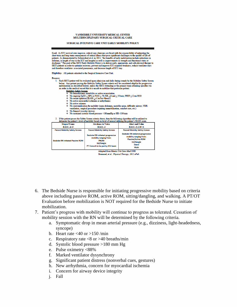
6. The Bedside Nurse is responsible for initiating progressive mobility based on criteria
above including passive ROM, active ROM, sitting/dangling, and walking. A PT/OT
Evaluation before mobilization is NOT required for the Bedside Nurse to initiate
mobilization.
7. Patient’s progress with mobility will continue to progress as tolerated. Cessation of
mobility session with the RN will be determined by the following criteria.
a. Symptomatic drop in mean arterial pressure (e.g., dizziness, light-headedness,
syncope)
b. Heart rate <40 or >150 /min
c. Respiratory rate <8 or >40 breaths/min
d. Systolic blood pressure >180 mm Hg
e. Pulse oximetry <88%
f. Marked ventilator dysynchrony
g. Significant patient distress (nonverbal cues, gestures)
h. New arrhythmia, concern for myocardial ischemia
i. Concern for airway device integrity
j. Fall
k. Endotracheal tube removal
8. Passive range of motion (PROM)
a. Passive ROM should be performed on appropriate SICU patients every two hours,
alternating bilateral upper extremities and bilateral lower extremities in
coordination with turning.
b. The bedside nurse is ultimately responsible for ensuring passive ROM is
performed. This can be performed by the family after education from the
physical/occupational therapist, nurse, or care partner.
c. Pamphlets will be distributed to family including diagrams of PROM maneuvers
and instructions on its performance. T
d. The bedside nurse will be responsible for assuring safety with lines and tubes
during family assisted passive range of motion.


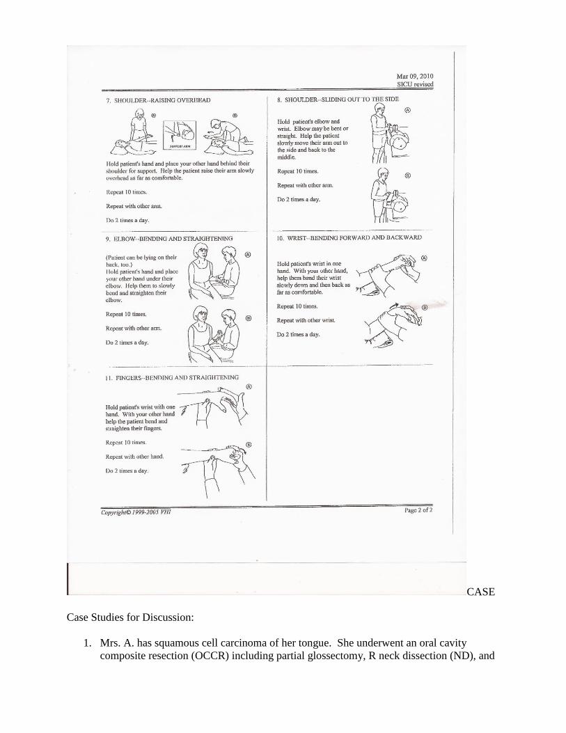
CASE
Case Studies for Discussion:
1. Mrs. A. has squamous cell carcinoma of her tongue. She underwent an oral cavity
composite resection (OCCR) including partial glossectomy, R neck dissection (ND), and

L radial forearm free flap (RFFF) for reconstruction of her tongue and is on the SICU for
flap monitoring. She has a trach and is on 35% FiO2 via trach collar. She has 2 JP drains
on the R side of her neck and 1 at the antecubital fossa of her L elbow above her splinted
forearm. She has an IV in her R UE and a PCA pump. She is 75 years old and uses a
walker at baseline. You have PT/OT orders to mobilize her/teach ADL and teach her the
appropriate exercises, discharge recs. She is in bed when you arrive, and the nurse has
agreed with you getting the patient up and ambulating with her.
a) What other information do you need from the nurse?
b) What are her precautions?
c) What will you do to get Mrs. A. and the room organized?
d) What equipment do you need?
e) What exercises will you teach her?
f) Where can you find head and neck guidelines and copies of the exercises?
a. Does Mrs. A. need to be on oxygen for her out of bed activities? Has she recently
taken any meds that might make her dizzy or otherwise need extra precaution to
prevent falls? Does she need a chair alarm?
b. PWB L UE x 2 wks. so no pushing through L hand till then, start active exercises L
wrist in 2 wks., falls, JP drains, supplemental O2
c. Plan to get Mrs. A. out of the R side of the bed to prevent use of her L UE for her
transfer. Place bedside chair on R side of bed and make sure it is locked. Check to
see if bed alarm needs to be disarmed. Find all JP’s and pin them to her gown where
you can see them. Unplug IV pole and keep it to the R of the patient during transfer
and ambulation. Organize telemetry wires to prevent tangling. Check for
presence/location of foley cath and place it where it won’t get caught under patient
during transfer. Ask Mrs. A. if she needs to press her PCA pump.
d. Potentially an O2 tank and wheels and venti tubing to adapt her trach collar O2
requirements, L platform rolling walker, assume you will need a portable monitor
since patient is on the SICU, safety pins for JP’s
e. Head and neck exercises, L wrist exercises
f. Guidelines on cabinet door in MCE office where printed exercises are located, also
check rehab shared drive, exercises in MCE office cabinet, also in folders in 7N room
2. Mr. B. has lung cancer. He underwent a R thoracotomy for R lower lobe resection and
has a R CT which is on wall suction. He is on the SICU due to a cardiac history, and his
recovery has been complicated by A-fib which is rate controlled by meds. He is on 2L
supplemental O2 via nasal cannula. He has an IV in his L UE and a PCA pump. You
have PT/OT orders for mobilization/ADL and discharge recs. He is in bed when you
arrive, and the nurse has agreed with you getting the patient up and mobilizing him to his
tolerance.
a) What other information do you need from the nurse?
b) What are his precautions?
c) What do you need to do get Mr. B. and the room organized?
d) What equipment do you need?
e) What will be you be monitoring in particular while ambulating?
f) What exercises and adaptations for bathing/dressing are especially important for
this patient?

a. Has Mr. B. recently taken any meds that might make him dizzy or otherwise need extra
precaution to prevent falls? What is the maximum heart rate recommended for this
patient? Does he need a chair alarm? You do not necessarily need to ask about
supplemental O2 since Mr. B. is on 2L perhaps for maintenance reasons while in bed.
You can monitor his SpO2 during the initial transfer to the edge of the bed and determine
his O2 needs yourself. Get a tank and wheels and bring his nasal cannula if in doubt or
if he drops below 92% with the transfer.
b. He has a chest tube and his chest tube is to wall suction. Check the activity orders to see
if the patient can come off wall suction to ambulate AND check the chest tube orders
under treatment orders to see if there is a suction requirement. If so, you will need
portable suction to move away from the bed.
c. Examine Mr. B.’s surgical site for location and chest tube. Ask Mr. B. if he wants to
press his PCA pump before getting up. Mr. B. may do better getting out of the L side of
the bed in order to avoid discomfort from rolling onto his chest tube. Decide where you
want to place his bedside chair in relation to CT with suction tube, O2 tubing length, and
telemetry lines. Check to see if bed alarm needs to be disarmed. Unplug IV pole and
keep it to the L of the patient during transfer and ambulation. Organize telemetry wires.
Check for presence of foley cath.
d. Thoracic walker might be best for hanging CT, holding portable suction, O2 tank,
portable monitor (needed due to patient being SICU patient and needing close
monitoring of heart rate as well as SpO2 while mobilizing). Portable suction is in
equipment room or on crash carts (ask charge nurse before using one from crash cart
and replace suction canister after use—place used one in patient’s room for future use).
Have a chair nearby or resting place in mind in case patient needs to sit and rest.
e. Heart rate (remember, patient is rate controlled for A-fib) and SpO2
f. R shoulder exercises due to presence of chest tube—work toward full flexion, abduction,
also deep breathing exercises to ensure full excursion of ribs. Encourage the use of a tub
seat when showering. Teach him to dress his R arm first and to bring his foot up to his
opposite knee to assist with donning socks
3. Which of the following lines/pieces of equipment can be safely be disconnected for treatment?
When would you want to keep them during mobilization?
Heart monitor
o Keep on SICU patients, patients with cardiac histories, arrhythmias, disconnect
only when you are assured patient has not had previous heart rate issues per
nurse
Pulse oximeter
o Keep on SICU patients, patients already wearing supplemental O2 or who tend to
desat with activity per nurse, disconnect for patients who have been satting well
even with exertion
Arterial line
o Cannot disconnect the actual line but can disconnect from the monitor via the
large connection on the wire, keep connected if nurse indicates, most SICU
patients can be disconnected for mobility away from bed (be sure to have nurse
zero the art line upon reconnection)
Central line

o Normally attached to IV, nurse may offer to pause and disconnect for mobility,
otherwise keep connected and watch for kinking or pulling on patient
Feeding tube
o Dobhoff and PEG—ask nurse before pausing tube feeds and be sure to resume
them before leaving patient
Swan Ganz catheter
o Never disconnected, okay to transfer patients out of bed to chair, have nurse
watch Swan while moving patient
Nasogastric tube
o Ask nurse if NG tube can be clamped for mobility. Patient may need NG to
remain to suction to prevent accumulation of gastric fluids and vomiting.
Epidural
o This small catheter carrying medication into the epidural space of the spinal cord
is for pain management and cannot be disconnected. It originates from a yellow
box usually hanging near the head of the bed. Carry the box with you during
mobility, and be sure not pull excessively on this line or get it caught under the
patient.
PCA pump
o This pain pump is connected to the patient’s IV line. It can be paused by the
nurse and disconnected for patient mobilization.
4. Mr. J. is a 56 y/o male admitted through the ED to the general medicine floor (8N) with AMS
and SOB. His medical team concluded that his symptoms were related a progression of his
diseases. His MELD score is 30. His past medical history is HTN, anemia, DMII, syncope,
hepatopulmonary syndrome and end stage liver disease. PT/OT completed an initial evaluation
two days after his admission date on 4/1/16. The rehab team’s discharge recommendations were
Home with 24 hr. care/assistance and HHPT/OT.
During his admission he underwent a liver transplant on 4/5/16. He has now transferred to an
SICU bed on 9T. He was extubated on 4/6/16.
On this date (4/7/16) his lab values for the day are glucose 250, hematocrit 42%, hemoglobin 9.9,
potassium 3.6, and his sodium is 138. His vital signs for the last three hours are blood pressure
145/72, 160/78, 179/75 and now it is 168/98. His heart rate has ranged from 90 to 110 and his
oxygen on 3 liters is 87-94%.
a. Based on these lab values and vital signs, do you think this patient is appropriate for PT/OT
intervention?
Sometimes in clinical cases we need more information on the patient. However, if we are purely
looking at the lab values and vital signs right now, I would say that the patient is appropriate to
be seen because their vitals are stable. Even though the blood pressure is high, I would ask the
RN if any BP medications have been given recently. The glucose is within functional normal
limits, but it is also on the high side. His O2 has dropped below 90%, however, with his medical

h/o hepatopulmonary syndrome, his O2 reserve may be lower than an average patient. Again, in
my opinion, I would see this patient, but I would monitor blood pressure and monitor vitals.
b. Would you complete a treatment note or a re-evaluation on this patient?
As a therapist you would complete a re-evaluation due to this patient having undergone a
significant medical change (liver transplant). If you were going to see this patient as an
assistant, following his medical record review, you would need to notify your supervising
therapist and let him/her know that a therapist would need to see this patient to update
POC/goals. The medical team may have already placed a new rehab consult, and then it would
show up as a PRN order on the list.
c. Based on the patient’s medical history and admitting diagnosis/medical course, what are some
appropriate questions to ask related to PLOF?
I think it is important to have a detailed explanation of his activity tolerance (both long
term/short term). Ask if he checks his O2 levels at home or if he utilizes supplemental O2 at
home. If so, what does he normally measure and how much supplemental O2 does he utilize? If
the patient is confused, ask the patient’s family member/primary caregiver. It will also be
important to ask where the patient lives (city). This will be important information when
determining discharge recommendations due to the requirement that patients reside locally to be
near VUMC (less an hour’s drive) following liver transplant.
Upon sitting up, Mr. J. reports that he feels dizzy and as if he might faint.
a. What issues might be causing these symptoms?
Here are some issues I came up with that could be reasons why he feels dizzy or might faint and
has shortness of breath. One might be orthostatic hypotension. Maybe he was lying down when
they took the blood pressure at first and now that we have him sitting up his blood pressure has
dropped. Maybe the blood pressure medication has started to kick in and dropped his blood
pressure. Maybe he has increased or decreased glucose. That 250 level for him may be on the
high side and he is feeling the effects of that. Possibly because he is NPO and receiving insulin
his glucose could have decreased and he is feeling dizzy.
b. What would you monitor in this case and what would you do in this situation?
I would monitor the heart rate, blood pressure (check BP in supine, sitting, and, if able,
standing), and oxygen level and see if the symptoms resolve within one to two minutes. If not, I
would lie the patient down and recheck the vitals and communicate with the RN or the MD.
5. Ms. S. is a 40 y/o female admitted from an OSH with necrotizing fasciitis. Patient has a
medical h/o IV drug use and colitis. Patient is s/p irrigation and debridement of necrotizing
fasciitis of the right flank from abdomen extending to left thigh. Patient with prolonged
intubation (1 week) requiring trach placement. Patient admitted to the SICU for airway
monitoring. PT/OT has been consulted.

a. What precautions would we consider while in room and what would we need to include in our
documentation?
Important precautions to consider would be the % of supplemental O2 (documented as __%fiO2
via trach collar; can also include amount in liters), any drain placement, monitoring of vitals,
and that patient is a falls risk.
b. What would you need to do to get patient/room organized before mobilization?
Consider the need for a portable O2 tank and converter for the trach collar (located on right
side in 9T service center). Keep patient monitored with portable monitor. Pin any drains to gown
before mobilization.

Surgical Stepdown [9n/s]
9 North
1. 9N is a monitored floor. The patients on this floor include the same as on the SICU/9T,
trauma/10N overflow, and medicine/8N/8S overflow
2. 9N supplies and linens are accessed behind the nurses station as well as from the side and
back hallways.
3. The break room is located to the right of the nurses station on the left side of the hall. The
code is ‘234’
4. Guidelines:
a. Talk to the nurse before treating the patient.
b. On 9N, patients must stay on their monitor unless they have orders in the medical
record to be off their monitors. Portable monitors are located in every room on 9N
c. Be aware of all lines and drains including chest tubes to wall suction. Portable
suction machines will be ordered by the patient’s nurse for ambulation outside of
the room. The location of the portable suction machine varies. Start by checking
in the storage areas between rooms 10 & 11
d. Lung transplant patient’s need to wear masks when ambulating outside their
rooms
e. Oxygen tanks are available in the storage areas between rooms 10 & 11
f. 9N Neuro chairs, large, stretchy footies for patients with edematous and bariatric
feet are located in the supply room
g. Reachers, long-handled sponges, and leg lifters are located in the supply room
h. Thoracic walkers aid in carrying oxygen, chest tubes, and foley catheters
i. Walkers and a few crutches are located in the storage areas between rooms 10/11
9 South
1. 9S is a non-monitored floor. The patients on this floor include the same as 9N.
2. Head and neck cancer surgery patients, liver transplants patients, bariatric surgery
patients, urology and endo/oncology, gyno/oncology, and thoracic surgery patients are
often transferred to this floor.
3. Talk to nursing prior to treating the patient
4. Neuro chairs are shared between 9N and 9S
5. 9S supplies and linens are accessed from behind the 9S nursing station with access doors
from side and back hall.
6. The 9S break room is located to the right before you enter 9S and before patient rooms.
The code is ‘241’
Free Flap Dangle Protocol
1. Begin dangling the surgical extremity on post operative day (POD) 6.
2. Dangle the surgical extremity for 10 minutes 4times daily while patient is awake waiting
at least 1 hour between each dangle session being POD 6.
3. Increase dangle time by 10 minutes every 24 hours (example POD 7 dangle 20 minutes
4times daily and POD 8 dangle 30 minutes 4timers daily).
4. Once up to 2hours 4times daily, may dangle the surgical extremity ad lib or all day.
5. When surgical extremity is not in the dangle position elevate on 2 pillows.
6. Precautions: if flap becomes swollen, blue, congested, or other visible clinical signs of
not tolerating the dangle stop advancement.
7. If there are no swollen, blue, or congestion events typically full ad lib dangle occurs
about POD 16.
8. To access dangle protocol: go to wiz order, click on web button to left of the sign off
button, click on e-docs, type in word dangle, click on search, you will then be able to
view protocol and print it
Post-surgical stepdown
1. Noncritical patients after surgery(excluding cardiac and neuro)
2. Often come from SICU, trauma, or directly from surgery
3. Postop patients often need PT/OT for retraining and compensatory training given new
precautions, functional impairments, and safety concerns prior to discharge
4. Precautions and considerations: often have numerous/various drains, chest tubes on
suction or may be taken off suction, NGT occasionally must remain to suction,
abdominal binders for abdominal surgeries (not in activity orders rather in nursing
orders/treatments, lung transplants have sternal precautions x6 weeks, wound vac often
with plastics patient
General Guidelines for Head/Neck Surgery Patients
1. Many of these patients will have both PT and OT orders entered. Sometimes they do not
require both services to see them. PT can screen the order and may see the patient first to
determine if OT is required. OT input is valuable for geriatric patients as well as for
patients with upper extremity limitations due to neck surgery or upper extremity donor
sites for free flaps. OT consultation is also needed for patients who have undergone
orbital exenteration as part of their head and neck cancer surgery.
2. These patients may perform active head and neck exercises with drains in. Many of these
protocols are physician-driven, and they may be instructed by their physician to perform
activity different from the protocol.
3. Sometimes these patients leave the day after surgery and may even leave with their drains
still in. Please attempt to see these patients early in the day and teach them their
exercises before they are discharged.
4. When you evaluate the patient, it is all right to assess cervical and shoulder AROM for
evaluation purposes. The patient can perform cervical/shoulder motions.
5. Advise these patients not to drive until it is okay with their physician. Also, they should
not lift anything heavier than a gallon of milk for the first five weeks to allow for healing.
6. Please inform the patient that when they return to the oto clinic post-operatively they may
be referred to outpatient PT at their MD’s discretion. If you note deficits during the
evaluation, encourage them to ask their MD about them if they do not improve by their
follow up visit.

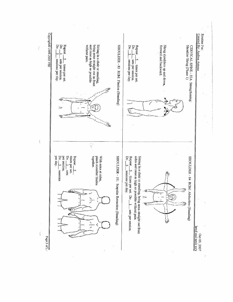

Medical Intensive Care (MICU) [8t]
General Information
1. Located on the 8th
floor of the critical care tower
2. Medical receptionist’s number – 615-322-0938
3. The MICU has 3 teams (MICU 1, MICU 2 and MICU NP). Each team consists of an
attending, a fellow, residents, and interns. Each team has a pager number in WIZ. They
round each morning for several hours. They are always on the floors unless they are
covering a stat. PT/OT round with fellows between 11:00am-12:00pm to discuss
caseload and appropriateness of orders for next day.
4. You can look at the sign on each door to determine which team the patient is on along
with the pager number for that team.
5. The case manager that covers MICU is Kirsten Vascik. Stacy Jones is the social worker.
They are available when needs arise however we do not communicate with them on a
daily basis.
6. Patients with respiratory impairments are usually admitted to this floor along with
patients admitted with medicine overdose, liver failure awaiting transplant, and anoxic
head injuries.
7. Ask nurse FIRST if it is appropriate to take patients off monitors. If nursing requests
constant monitoring or PT prefers to monitor patient, the portable monitors are located in
the closest behind the charge nurse’s desk. Batteries are located on the counter across
from room 8609.
8. Swipe your badge to get into supply room and clean and dirty utility rooms
9. Femoral Vas Cath – if there is a concern discuss with team
10. CVVHD (CRRT) – may work with patient if BP is stable
11. Vasopressors
a. This group of drugs is useful for treatment of hypotension.
b. Pressors usually seen in the MICU are levophed and dopamine
12. Tips for working with intubated patients
a. When working with a patient who is intubated, be careful to keep the ETT in a
neutral position as to prevent excessive pulling or pouring of fluid within the
tubing down into ETT.
b. You should take ETT out of holder when moving patient (ie rolling, transfer to
EOB, sit to stand).
c. Once patient is stable at EOB, then it would be safe to place ETT back in holder.
d. It is important to monitor intubated patients carefully during treatments especially
RR and O2 sats. The RR on the vent is usually more accurate than RR on
monitor.
13. Sedation
a. Medications usually used for sedation are propofol, precedex, and fentanyl
b. Level of sedation determined by RASS (Richmond Agitation Sedation Scale).
The scale ranges from -5 to +4. The more negative, then the more sedated. The
more positive, then the more agitated. Zero is normal.
c. OT/PT works with patients that are intubated and mildly sedated. PT/OT usually
becomes involved when a patient is a RASS -1.
d. Richmond Agitation–Sedation Scale
Score Term Description
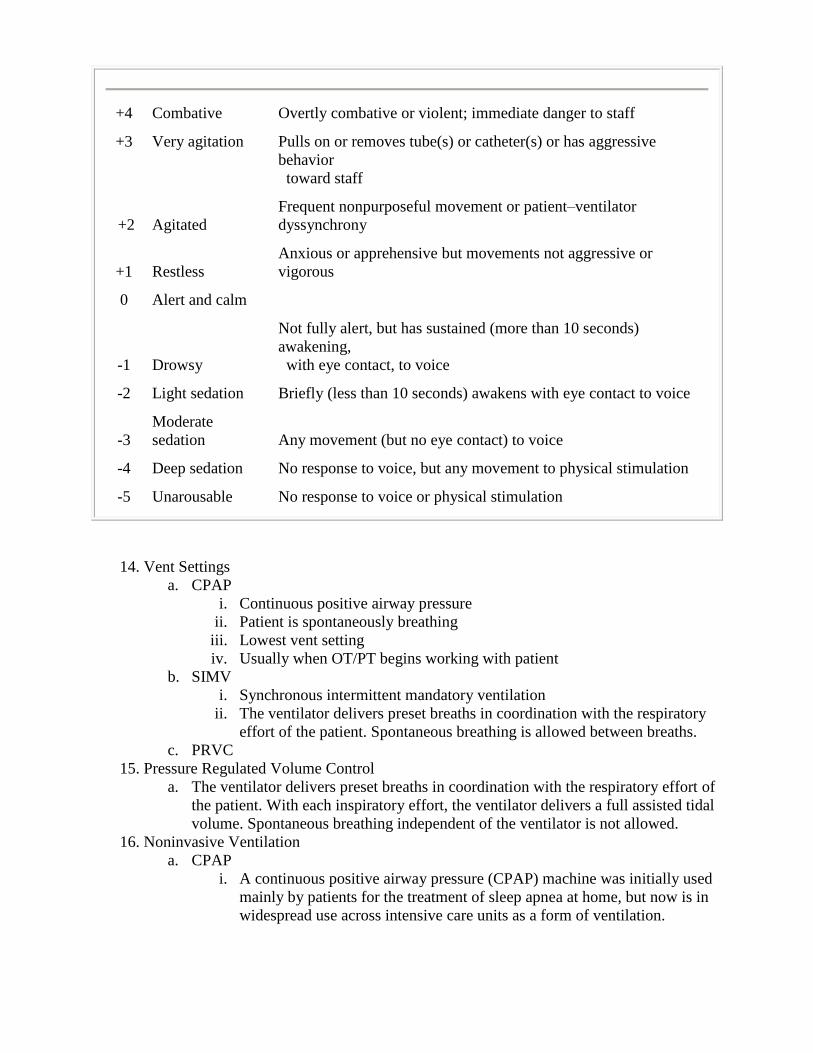
+4 Combative Overtly combative or violent; immediate danger to staff
+3 Very agitation Pulls on or removes tube(s) or catheter(s) or has aggressive
behavior
toward staff
+2 Agitated
Frequent nonpurposeful movement or patient–ventilator
dyssynchrony
+1 Restless
Anxious or apprehensive but movements not aggressive or
vigorous
0 Alert and calm
-1 Drowsy
Not fully alert, but has sustained (more than 10 seconds)
awakening,
with eye contact, to voice
-2 Light sedation Briefly (less than 10 seconds) awakens with eye contact to voice
-3
Moderate
sedation Any movement (but no eye contact) to voice
-4 Deep sedation No response to voice, but any movement to physical stimulation
-5 Unarousable No response to voice or physical stimulation
14. Vent Settings
a. CPAP
i. Continuous positive airway pressure
ii. Patient is spontaneously breathing
iii. Lowest vent setting
iv. Usually when OT/PT begins working with patient
b. SIMV
i. Synchronous intermittent mandatory ventilation
ii. The ventilator delivers preset breaths in coordination with the respiratory
effort of the patient. Spontaneous breathing is allowed between breaths.
c. PRVC
15. Pressure Regulated Volume Control
a. The ventilator delivers preset breaths in coordination with the respiratory effort of
the patient. With each inspiratory effort, the ventilator delivers a full assisted tidal
volume. Spontaneous breathing independent of the ventilator is not allowed.
16. Noninvasive Ventilation
a. CPAP
i. A continuous positive airway pressure (CPAP) machine was initially used
mainly by patients for the treatment of sleep apnea at home, but now is in
widespread use across intensive care units as a form of ventilation.

ii. The CPAP machine delivers a stream of compressed air via a hose to a
nasal pillow, nose mask or full-face mask, splinting the airway (keeping it
open under air pressure) so that unobstructed breathing becomes possible,
reducing and/or preventing apnea
b. BIPAP
i. Provides two levels of pressure: Inspiratory Positive Airway Pressure
(IPAP) and a lower Expiratory Positive Airway Pressure (EPAP) for easier
exhalation.

General Medicine [8n] and Cardiac Stepdown [8s]
1. 8north is a general medicine floor (including Riven teams 1-6, Riven 11, Nonteaching
private practice, Morgan teams 1-4, Rogers hepatology, Rogers pulmonary, Rogers ID,
Rogers GI, Rogers renal)
2. Riven team and private practice are attending only (no residents or medical students)
3. Rogers and Morgan teams have medical students, residents, fellows, and attendings
4. 8south is a cardiac stepdown floor and has 6 general medicine “stepdown” beds as well
5. Common diagnoses on unit are: cancer, cellulitis, spine compression fractures, COPD,
decubitis ulcers, electrolyte abnormalities, general debilitation/failure to thrive, hepatic
encephalopathy, HIV/AIDS, pneumonia, CF, CHF, diabetes, falls, rhabdomyolysis,
dementia, TB, and altered mental status.
6. Physical therapy primary focus: discharge recommendations, assess for assistive
devices/braces and AFOs/mobility devices, gait training, balance, home safety, caregiver
training, home safety, determine if OT/SLP are needed if not ordered, and 6minute walk
test for patients on lung transplant list (at least weekly).
7. Occupational therapy primary focus: discharge recommendations, medication
management, assess need for adaptive equipment, caregiver training, home safety,
cognition for I/ADLs, sustained activity tolerance, ROM, splinting, and energy
conservation.
8. "Bedrest" orders- frequently means patient is not allowed to get up without assistance. If
therapy orders states OOB/therapy orders status ambulation/recent daily notes say OOB
or ambulation usually patient not on bedrest. Page patient's primary team to request
clarification and order changed.
9. Patients with must have troponins downtrending (or clearance from primary medical
team if not clearing due to kidney issues).
10. Patients with DVT or PE must be on anticogulation x24 hours prior to therapy evaluation.
11. SNF placement factors:
a. Therapy (PT and OT or PT alone)
b. IV antibiotics
c. Wound care with Stage III or IV ulcer or new PEG tube.
d. Patient does not need therapy evaluations if going for IV antibiotics or wound
care.
12. If there is a possibility of transition to nursing home placement/intermediate care facility
once therapy is completed then recommend SNF. It is typically difficult to admit
someone to an intermediate care facility from any other setting besides SNF. Patients
from an intermediate care facility typically do not require a PT/OT evaluation to return
unless they had a decline in function.
13. Effects of bedrest for geriatric patients: patients 65 years and older lose 30% of there
strength and endurance for every day they are in bed. This is why a geriatric patient that
is on bedrest x3 days will not likely be able to return directly home at discharge.
14. HIV/AIDS patients: In general it takes 2 months for HAART therapy to produce results
for patient. If you evaluate HIV/AIDS patient who has not been on HAART therapy
previously the true rehab potential will not be known for 2 months. If functional status is
limited it is recommended that patient discharge to SNF. If the patient improves after two
months patient can transfer to inpatient rehab facility. If the patient does not significantly
improve then they can transition to the intermediate care facility.
15. Respiratory isolation: There are 2 negative pressure rooms on 8 north (other rooms can
become negative pressure if air filter is placed in room). Used for patients who are on

respiratory isolation. Students cannot work with airborne patients since require N95
masks. Patients must stay in room unless medically necessary tests/procedures only
approved by team. HIV/AIDS patients are on respiratory isolation until TB has been
ruled out if patient comes in with respiratory symptoms or fever.
16. Cystic fibrosis (CF): Patient will often be on contact isolation due to infection or contact
for reverse isolation to protect the patient from outside infection. Often the team will ask
for a 6 minute walk test to be performed in order for the patient to qualify for the lung
transplant list (if medically appropriate). Nursing staff can also perform this test. Kelly
agreed to minimum physical therapist completing 6minute walk test once per week if
active on transplant list.
17. Geriatric team consults: there may be geriatric patients that are being followed by other
services instead of geriatric primary team. Geriatric team will do a geriatric consult for
patients with physicians request one. You can suggest to the primary team that the
geriatric team be consulted if you feel expertise could benefit from patient. When
speaking to primary team, make sure primary team understands why.
18. Neutropenic precautions: patient has love white blood cell count thus making it harder to
fight off infection. There will be a sign on patient's door. Patient must wear mask when
out of room and if you are sick you wear a mask into room.
19. General medicine conference: Monday to Friday 10:30-11:30am is the general medicine
conference. The secretary for general medicine keeps all physicians pagers while in
conference, so you will not be able to reach. You can send text message with your
question and ask for contact when out of conference.
20. Member of acute rehab attends General Medicine Huddle Monday, Wednesday, and
Friday in the Brittingham Conference Room (backside of 7t) 1355 Morgan 3, 1410
Morgan 1, 1425 Morgan 2, 1440 Morgan 4, 1455 pulmonary, 1510 ID
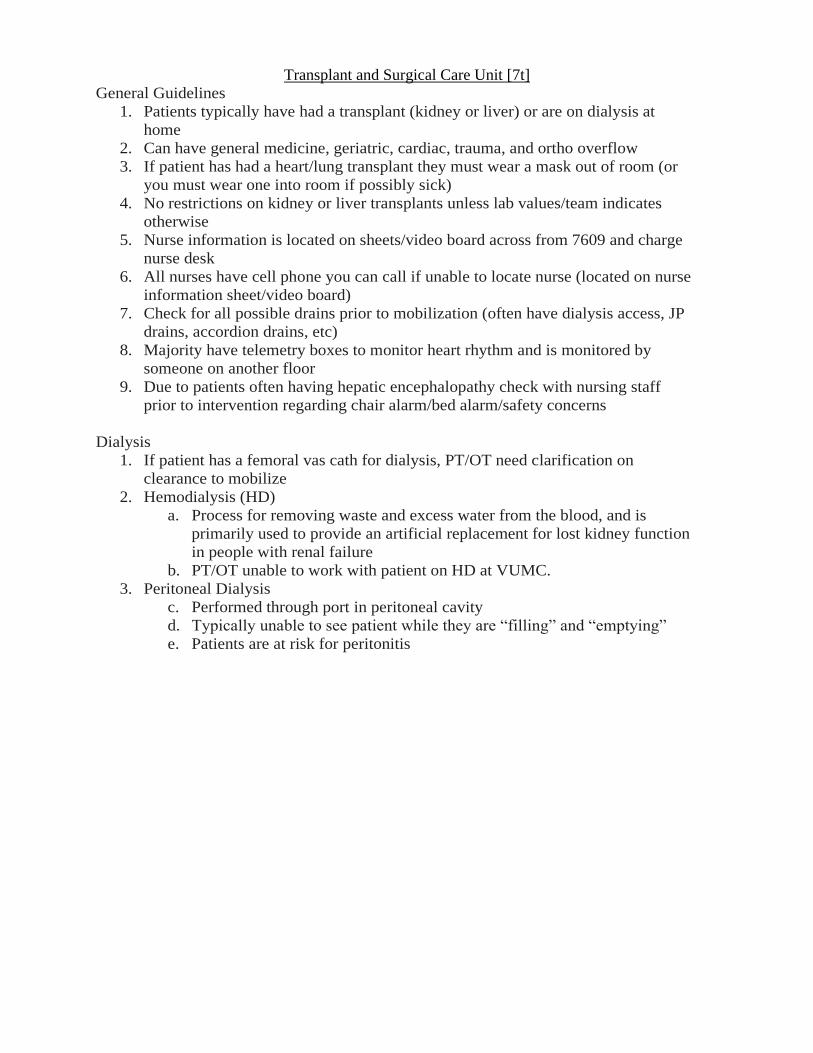
Transplant and Surgical Care Unit [7t]
General Guidelines
1. Patients typically have had a transplant (kidney or liver) or are on dialysis at
home
2. Can have general medicine, geriatric, cardiac, trauma, and ortho overflow
3. If patient has had a heart/lung transplant they must wear a mask out of room (or
you must wear one into room if possibly sick)
4. No restrictions on kidney or liver transplants unless lab values/team indicates
otherwise
5. Nurse information is located on sheets/video board across from 7609 and charge
nurse desk
6. All nurses have cell phone you can call if unable to locate nurse (located on nurse
information sheet/video board)
7. Check for all possible drains prior to mobilization (often have dialysis access, JP
drains, accordion drains, etc)
8. Majority have telemetry boxes to monitor heart rhythm and is monitored by
someone on another floor
9. Due to patients often having hepatic encephalopathy check with nursing staff
prior to intervention regarding chair alarm/bed alarm/safety concerns
Dialysis
1. If patient has a femoral vas cath for dialysis, PT/OT need clarification on
clearance to mobilize
2. Hemodialysis (HD)
a. Process for removing waste and excess water from the blood, and is
primarily used to provide an artificial replacement for lost kidney function
in people with renal failure
b. PT/OT unable to work with patient on HD at VUMC.
3. Peritoneal Dialysis
c. Performed through port in peritoneal cavity
d. Typically unable to see patient while they are “filling” and “emptying”
e. Patients are at risk for peritonitis

Cardiac/Vascular Stepdown [7n/7s/5s] and Cardiac Observation (COBS)
1. There are four categories of cardiac teams that are separated as follows: cardiac surgery,
heart failure, arrhythmia, and general/other. All major lines (Swan, arterial, mechanical
ventilation etc.) are removed before a patient transfers to the step down unit. Typically,
catheters, pacer wires, chest tubes, JP drains, O2 lines, and telemetry leads are the main
lines that require organization prior to mobility.
2. 5S can have has patients with a Swan. They allow up to 2 at one time
3. All patients on these floors are cardiac monitored, which means they MUST be
monitored at all times.
4. Check with nursing prior to seeing all patients; heart rhythms and rates can change
quickly and they are the best source of information to determine if a patient is stable and
ready for therapy.
5. All patients need EOB/OOB activity orders; if they are on bedrest you will need to call
the team pager to obtain them.
6. Vascular patients seen on these floors include LE amputations, LE bypass grafts (fem-
fem, fem-pop, axillary-fem, etc.), aortobifem bypass, AAA repairs.
7. For vascular patients with LE amputations (i.e. toes amps, transmet amps, ray amps), they
are NWB on the operative extremity following surgery unless otherwise specified by the
vascular team. This is important to remember as vascular does not enter a weight bearing
status in the activity order set.
8. Patients with BKA’s should be in a knee immobilizer when they come out of the
operating room; you may remove this for ROM, but must replace it once treatment is
complete. If no KI is present, call vascular
9. Patients with BKA’s and AKA’s come out of the operating room with sciatic and femoral
nerve blocks; these are placed/removed by the acute pain service. Patient may mobilize
with these blocks; the lines terminate into small pouches and are time released (i.e. the
patient does not have to push a button like the PCA pumps).
10. Cardiac Obeservation (COBS)
a. Located on 5t. Enter it from the left/back hall entrance to the tower building.
Cardiac observation patients and cardiac cath patients.
11. Occupational Therapy’s Role with Cardiac Patient’s in the Acute Care Setting
a. Cardiac patients are often limited in endurance, which impacts performance of
ADL and IADL tasks.
b. Patient’s who have had a sternotomy have sternal precautions to adhere to while
performing ADL tasks.
c. OT intervention focuses on compensatory strategies for self care tasks, education
on energy conservation techniques, relaxation techniques, and home or work
modifications.
d. Compensatory Strategies:
i. One handed dressing techniques
ii. Adaptive equipment
e. Energy Conservation Techniques:
i. Provide patient with energy conservation techniques
f. Deep Breathing Techniques:
i. Dyspnea Control Postures
1. When sitting, bend slightly forward at waist and prop forearms on
table or thighs
2. When standing, lean forward and prop on countertop, shopping

cart, etc.
ii. Pursed-Lip Breathing
1. Purse lips
2. Exhale through pursed lips
3. Inhale deeply through nose
4. Exhalation should take twice as long as inhalation
iii. Diaphragmatic Breathing
1. Patient lies supine, inhales slowly and exhale through pursed lips
2. Book placed below ziphoid process helps as visual cue (rise with
inhalation, fall with exhalation)
g. Basal Metabolic Equivalent (MET)
i. Energy cost of an activity
Basal Metabolic Equivalent Table of Self-Care and Homemaking Tasks
MET Level Self Care Ambulation and Household
Tasks
1.2 Eating, Seated Hand Sewing
1.5 Sweeping Floor
Machine sewing
1.65 Transfers, bed to chair
2 Washing face and hands,
brushing hair
2.5 Seated sponge bath
Standing sponge bath
Dressing and Undressing
Dusting
Kneading Dough
Hand washing small items
2.6 Vacuuming
3.0 Seated warm shower Preparing a meal
3.5 Standing shower, warm Climbing stairs at 24 feet/min
3.6 Bowel movement on toilet
3.9 Making a bed
4.2 Hot Shower
4.4 Changing bed linens
4.7 Bowel movement on bedpan
5.0 Sexual intercourse Walking up stairs at 30
feet/min
Spine Surgery (Ortho Spine/Neurosurgery) [6s]
Neck Surgeries
1. Always check the activity orders for these patients and check with the nursing staff prior to
mobilization
2. These patients may be required to wear a cervical collar or cervical thoracic orthosis (CTO).
3. Cervical collars must remain on the patient at all times unless otherwise specified in the
orders. These patients will receive a second set of cervical pads for showering. These are
typically seen with cervical fusions.
4. The CTO is to be worn when the patient is upright. The patient will wear a cervical collar
when in the bed. In elective surgeries/fusions, these braces may be donned sitting EOB
unless otherwise specified in the orders. These braces are typically utilized for lower
cervical/upper thoracic surgeries/fusions.
5. If a patient has had removal of hardware (ROH), it is not assumed that the pt can don their
brace on the edge of the bed. Clarify with the neurosurgery NP and/or MD. Don the brace in
supine until clarification can be obtained.
6. These patients have restrictions that include no bending, twisting or lifting >5lbs.
7. The patient may have a JP. Safety pins may be found in the supply room and the drain
should be attached to the gown during mobility so that gravity is not pulling down on it. Be
careful not to roll over or put weight on these drains.
8. Some patients may not require a brace. Avoid extreme neck flexion and extension with
these patients. Ask the NP or team to clarify any questions regarding ROM activities.
9. Some patients may receive a soft cervical collar to wear when riding in a car to avoid
unexpected or sudden movements.
10. Full manual muscle testing of the shoulders and elbows should not be completed with these
patients, only mild testing is permitted. Full AROM is permissible
Back Surgeries
1. Always check the activity orders for these patients and check with nursing staff prior to
mobilization. The activity order will provide information on the type of brace needed.
2. These patients may develop a CSF leak in surgery and have 24 hour bedrest orders. Check
the operative note for the time OR contact the NP/MD for details.
3. The most common sign of a CSF leak is a severe headache when upright. If this occurs,
return the patient to supine and alert the team of your findings.
4. All back surgery patients will need to abide by strict spine precautions. These include no
bending, twisting or lifting more than 10 pounds. We often use the synonym BLT to help
patients remember these precautions.
5. There are different braces your patients may have for mobilization. Check the activity
orders. Some patients do not require the use of a brace. Common braces are TLSO and
Aspen quickdraw.
6. Some patients have fusions starting in the lower thoracic to upper lumbar region through the
sacrum. These patients maybe needs adaptive equipment for home use and lower body
dressing.
7. Aspen quick draws are simple and use Velcro. They do not provide stability but serve as
reminders for back precautions.
8. The neurosurgery service allows all elective surgery patients who have been stabilized to
don their braces EOB. ALL braces may be donned sitting EOB unless otherwise specified.

9. If a patient has had removal of hardware (ROH), it is not assumed that the pt can don their
brace on the edge of the bed. Clarify with the neurosurgery NP and/or MD. Don the brace in
supine until clarification can be obtained.
10. Please order adaptive equipment or assistive devices POD #1 (if possible). This makes it
easier for the assistants when they return to see the patient on POD #2.
Neurology/Neurosurgery/Neurology Intensive Care Unit (NICU) [6N/6T]
General Guidelines
1. 6T3 is the neuro ICU with both neurosurgery and neurology patients.
2. 6N is the neurosurgery step down unit and contains both neurosurgery and neurology
patients.
3. 6T3 includes beds 6601 to 6661 with four even numbered beds (6440-6446) in the middle
hallway and one on the end; 6656 is across from 6659 and 6661.
4. 6601-6646 are ICU beds, the rest of the beds are ICU stepdown, which is not the same as a
floor bed.
5. The team pager numbers can be found on the white dry erase boards throughout the
intensive care unit.
6. The ICU is run by the neurocare team – anesthesia. This team manages the patient’s from a
medical/critical illness standpoint and rounds on each patient in ICU status every morning.
However, the primary team (neurosurgery or neurology) is in charge of the patient’s care
and activity orders.
7. Neurology and neurosurgery participate in a morning discharge planning huddle at 8:15 AM
along with nursing, therapy, case management, and social work. The neurology residents
and neurosurgery nurse practitioners facilitate huddle.
8. There are 3 case managers and 3 social workers as well as a discharge planner that cover the
patients on the neurology and neurosurgery caseloads.
9. There are 7 nurse practitioners for neurosurgery. They cover according to service. There is
one for the tumor service; 3 for spine, including ortho-spine; 1 for the movement disorder
team; and 2 for the endovascular service.
10. Always check with the nursing staff on the ICU prior to mobilizing a patient or removing
the monitors. Portable monitors are available in the equipment room (code 432*).
11. Handouts of home safety information have been created for craniotomies and back
surgeries. It is important patients receive this information prior to discharge. It is preferred
these are provided on POD #1. These are located near the MR desks on 6T3 and 6N.
12. Patients who have had a lumbar puncture are on bedrest for 2 hours following the procedure.
13. The neurology service has a stroke and a general neurology team. The stroke team follows
all stroke patients and the general team follows all other neuro dxs ie: multiple sclerosis
(MS), guillian barre syndrome, etc.
14. Vanderbilt is a Comprehensive Stroke Center. As a result there are benchmarks that must
be met. As part of these requirements, a skilled OT, PT, and/or ST assessment is required
for every stroke patient unless the doctors can document the patient is back to his/her
baseline. If you visualize/observe a patient to be independent and a formal assessment is not
needed, a contact note should be placed in the medical record so the coding person can
qualify this and we meet this benchmark with every stroke patient.
Craniotomies
1. These patients have typically undergone resections in various areas of the brain.
2. We see these patients for home safety and to determine if functional limitations exist. We
have a home safety handout.
3. These patients should avoid bending over, lifting, or straining due to increased pressure,
dizziness, and pain.
4. The HOB should be kept >30* at all times for swelling purposes.
5. Offer these patients assistive devices and AE as needed - no restrictions.
6. Though uncommon, these patients may develop a cerebrospinal fluid leak (CSF). Signs
may include a severe headache when upright that is worse than surgical pain OR a clearish
fluid leaking from the nasal cavity. Immediately return the patient to supine and alert the
nursing staff if this occurs with your patient.
7. There is also the potential for post-op swelling following a crani. The effects are most
pronounced by post-op day 2. This may translate into a decline in mental/physical
performance.
Craniectomies
1. These patients have had a bone flap from the skull removed usually for swelling purposes
and to allow decompression of the brain.
2. The flap may be stored in the abdomen or frozen; check the stomach for incisions. Most
recently, they have consistently been frozen.
3. Some of these patients will have helmets for OOB activities. However, most get their
helmets upon discharge. The neurosurgery NP is responsible for obtaining the helmet.
Rarely, a bicycle helmet is used for this purpose.
4. Be careful not to touch the area where the flap was removed and not to bump it against
anything.
5. Those with an occipital flap removal will generally have a foam donut for the back of their
heads.
6. If the patient has no bone flap, the side where the bone flap is missing should be listed as a
precaution on the evaluation.
External ventricular drains (EVDs)
1. External ventricular drains (EVDs) are placed in patients with hydrocephalus or with
increased intracranial pressures (ICPs). These drains collect excess cerebrospinal fluid
(CSF). These are typically placed surgically. They are open when the patient is in bed.
2. EVDs are gravity dependent. They are lined up with the patient’s external auditory meatus
when draining. If the drain is below the ear, the patient will drain too much; if too high, not
enough.
3. The neurosurgeons prefer the patient’s ICP be below 20. When monitoring, this is a purple
line on the monitor. Monitoring is ineffective when the EVD is open or the drain is no
longer in line with the ear. If there is a question regarding ICP, ask the nursing staff.
4. Lumbar drains are placed for an increase in CSF as well. These drains are typically opened
for a period of time and then clamped off, controlling the amount of CSF that is drained.
5. THESE DRAINS MUST BE CLAMPED PRIOR TO MOBILIZING THE PATIENT OR
EVEN MOVING/RAISING THE HEAD OF THE BED!!!!!!!!!
6. Patients who have a subdural JP drain or Subdural Evacuating Port System (SEPS) drain
placed for SDH are not to be mobilized until the drain is removed.
Stroke:
1. Do not mobilize patients with “BEDREST” activity orders unless you receive a verbal
order. Either put in a failed attempt or call/text page the team.
2. Some stroke patients will have permissive hypertension goals. These are noted at the end of
the neurology notes. Check the latest progress note or with nursing for the most up to date
parameters.
3. Hemorrhagic stroke patients will typically have a lower BP parameter to avoid a larger
bleed. Ischemic stroke patients will typically have a higher parameter to perfuse the

damaged brain tissue. Always ask the nurses what the blood pressure parameters are as they
may have changed.
4. Always monitor a stroke patient unless otherwise told. Monitoring blood pressure and
staying within parameters is extremely important in these patients.
5. If a patient is noted to be pressure dependent, perform your assessment in supine first to
establish a baseline. Monitor the pt’s blood pressure throughout activity so any neuro
changes can be co-related with the patient’s BP. Return the pt to supine and report your
finding to the nurse and team if a change in the patient’s neuro exam is observed.
6. Patients who have received tPA are on bedrest x 12 hours after administration of tPA and
must be put back to bed after therapy evaluations until they reach the 24 hour mark.
Subarachnoid Hemorrhages
1. Subarachnoid hemorrhages (SAH) can be caused traumatically or atraumatically. Typically
we see atraumatic SAH on the neuroscience floor.
2. Atraumatic SAHs have several causes. If the cause was an aneurysm or arteriovenous
malformation (AVM), the aneurysm or AVM must be secured either by coiling,
embolization, gluing, or removal via craniotomy prior to mobilization. Mobilizing an
unsecured aneurysm or AVM could lead to further bleeding. Usually the surgeons do a CT
angiography or diagnostic cerebral angiogram to determine the cause of the SAH.
3. If a patient comes in with a new SAH and PT/OT are consulted, make sure to find the cause
prior to mobilization and put a hold on these patients if an aneurysm or AVM is present
and/or has not been secured. Also, if an aneurysm/AVM is suspected and not diagnosed, the
patient should be placed on hold until definitive diagnosis or reason for the SAH is found.
4. SAH have precautions that include: dimmed lighting, quiet environment, avoiding extremes
such as hot and cold, and avoiding straws. A sign is placed on these patient’s doors. We
will not usually have orders on a patient under precautions.
5. SAH are at risk for vasospasms for up to 3 weeks post bleed. Pt’s will be monitored via
transcranial Doppler ultrasound (TCD) on M, W, F of each week. Check these reports or
the neurology progress reports to find out if a patient is in vasospasm.
6. When a patient develops vasospasms, the patient can typically be mobilized unless they are
symptomatic from a neurological standpoint. It’s always a good idea to check with the nurse
practitioner prior to mobility.
7. SAH blood pressure goals may vary depending on whether the patient is in vasospasm so
always confirm with the nursing staff or doctors.
Baclofen Trials
1. PT participates in intrathecal baclofen trials (ITB). These trials are for patients with
spasticity and assesses for the need for an intrathecal baclofen pump. Patients are assessed
3-4 times during their hospital stay.
2. The baclofen trial forms are housed in the MR’s drawer on the 6N floor. Use this for the
modified ashworth scale and spasm scale. This will make the trial much smoother for you.
3. Patients are typically admitted on Sunday and the trial is performed on Monday. Get to the
patient’s room no later than 8:30 for a pre-trial assessment.
4. Patients are typically NPO prior to the injection. It may be difficult for the doctors to do the
LP bedside and the patient may need to undergo anesthesia for the injection.
5. You will perform a pre-trial assessment prior to the initial injection of baclofen. Always
give the nurse your pager number and let him/her know you are finished with your pre-
assessment. Have the nurse page you with the time your patient was injected.
6. The patient must remain on bedrest for 2 hours following the injection and will typically be
lying in a supine position.
7. You will re-assess the patient every 2 hours from the initial injection time up until a 6 hour
mark. If the patient was injected later, a 6 hour assessment is not necessary.
8. The baclofen is most effective at the 4 hour assessment. It begins to wear off after 4 hours.
9. Let the nurse or team know when you are finished as the patient will discharge shortly after
your last assessment.
10. The trial is deemed successful if the patient’s spasticity dropped 2 grades on the Modified
Ashworth Scale in one muscle group. It is also the therapist’s discretion to deem a trial
successful based on the subjective findings of the patient (ease of transfers, toileting,
hygiene, dressing, etc).
11. It takes approximately 24 hours for the full effects of the baclofen to wear off if it was
effective. This is important for those patients who rely on spasticity for mobility to be
aware of for safety.
Epilepsy Monitoring Unit
1. 6N has an epilepsy monitoring unit (EMU). Patients will undergo continuous
electroencephalography (EEG) to monitor electrical activity to pinpoint seizures in the
brain.
2. Patients undergoing continuous EEG are not usually seen by therapy. However, you may
get the occasional consult. The patient can mobilize as far as the cables will allow.
General Neurology
1. Conversion and malingering disorders are seen in forms of strokes or spinal cord injuries on
the neuro floors. Treat these patients as you would any patient with the present deficits. A
technique that helps with these patients is to distract them during functional tasks and you
may see inconsistencies in the presentation of their symptoms.
2. Patients with Guillian Barre Syndrome or any form of demyelinating disorder may under go
IVIG therapy for several days while in the hospital. Often, patients are admitted for this
therapy.
3. Some patients will undergo plasmaphoresis exchange (PLEX). This is done in most
autoimmune disorders. These patients are typically off the floors in the morning but are
okay for therapy in the afternoon. They go to dialysis daily for their treatment.
4. General neurology houses a number of patients with different disorders. Contact the team
with any questions.
Cardiovascular Intensive Care Unit (CVICU) [5N]
1. Patients are to be monitored continuously unless specified by nursing staff. There are
portable monitors available when mobilizing patients in the CVICU.
2. It is best to communicate with nurse prior to working with patients.
3. There are 3 teams on this unit.
a. The cardiac surgery team (CSX) consists of a critical care intensivist, cardiac
surgery fellow, and nurse practitioners
b. The cardiac critical care team (CCU) consists of an attending, cardiac fellow and
residents.
c. The vascular team consists of an attending, residents and nurse practitioner.
4. For cardiac surgery, there are nurse practitioners present daily and each one is assigned
patients to follow for the day. If you have questions about your patient, it is best to
communicate with the nurse practitioner assigned to that patient. The patient’s RN
knows the nurse practitioner or sometimes the nurse practitioner’s name and number is
written on the patient’s white board.
5. If your patient is on the cardiac critical care team, then you should communicate with the
residents. There are usually on the floor and sitting at the middle nurse’s station across
from room 12.
6. If your patient is on the vascular team, the cardiac surgery nurse practitioners follow
these patients until moved to step down floors. The vascular patients housed on this unit
are usually complicated AAA repairs or aortofemoral bypasses. When these patients
leave the CVICU, they transfer to the stepdown cardiac floors, usually 6S.
7. Cardiac surgeries usually seen in the CVICU include CABG (Coronary Artery Bypass
Graft), valve replacements or repairs (for example: aortic valve replacement abbreviated
AVR or aortic valve repair abbreviated AVr), heart transplants, LVAD (Left Ventricular
Assist Device) and lung transplants.
8. If CABG or AVR/AVr is done emergently, then it would be performed via median
sternotomy. If elective, then could be performed minimally invasive through an
anterior/lateral thoracotomy or partial sternotomy. You must look at the operative note to
determine which approach was taken is order to determine post op precautions.
9. A CABG can be performed on pump (OPCABG) or off pump (CABG) which will be
stated in the operative note. On pump means that the surgery was performed with the
patient on the heart lung bypass pump.
10. Double lung transplants have sternal precautions x 8 weeks. Try to see as often as
possible.
11. Patients awaiting heart transplant will also be on this unit. They usually stay for several
weeks. Some of these patients will have a Swan Ganz catheter intact or an IABP
(intraaortic balloon pump) which usually stay in place until the heart transplant is
performed.
a. If your patient has a Swan Ganz intact, then they are not allowed to walk. The
CCU team usually orders PT for strength and sustained activity tolerance.
Exercise options for these patients include an arm bike that can be used as a leg
bike that can be left in the patient’s room and a standing exercise program.
Depending on the patient’s compliance, PT can provide an exercise program and
follow up once per week to make any changes.
b. If your patient has an IABP that is placed femorally, then they are on bedrest. If
their IABP is axillary, then this patient is able to walk in the halls with PT, but
you must have an RN present.
12. Heart failure patients are often admitted with volume overload and have a daily fluid
restriction; check with nursing before getting them anything else to drink besides what is
already in the room. There should be a sign on the door.
13. Atrial fibrillation/flutter - check with nursing to make sure they are rate controlled. DO
NOT mobilize any patient in an UNCOTROLLED a-fib/flutter rate.
14. Flolan is a medicine used to treat pulmonary HTN; It can be administered via a face
mask, intravenously or small canister on vent; when flolan is located on canister on vent,
the canister must remain upright.
15. Sternal Precautions (patients with median sternotomy)
a. Patients are not to lift, push, or pull more than 10 pounds resistance (about the
weight of a gallon of milk) for four weeks.
b. Patients cannot get in shower until pacer wires and/or chest tubes are pulled.
c. Patients are not to drive for 2-4 weeks. Physician will advise.
d. Patients are not to use vacuum cleaner for four weeks.
e. Patients are allowed to use an assistive device provided that they need for balance
rather than support; they are NOT allowed to hold themselves up with an assistive
device
16. Pacemaker/ICD precautions
a. Patients are not to reach above 90 degrees shoulder flexion/abduction with
pacemaker or ICD involved extremity for 6 weeks.
b. Pacemakers are generally placed on the L side, but can be on the R.
c. Patients are given a sling to wear for 24 hours after pacemaker placement, but can
be removed and discarded after this time.
d. Patients are not to lift more than 10 pounds for 6 weeks.
e. Exercise can be performed with UE 90 degrees or below.
f. Do not get incision wet for 14 days.
g. TENS is contraindicated.
h. A patient my become dizzy or collapse prior to ICD firing.
i. If ICD has recently fired, check with nursing staff or physician prior to treating
patient.
j. If an ICD fires while you are working with a patient, you may feel an electrical
current. Inform nursing if ICD fires.
17. External Temporary Pacemaker and Pacer Wires
a. Pacer wires are blue with a brown and white cord to the temporary pacemaker
box.
b. Patient may not get out of bed for two hours following removal of pacer wires.
c. Do not get pacer wires wet
d. All post op patients with have pacer wires.
e. Watch pacer box closely with activity. Recommend holding box personally with
activity.
18. Transvenous Pacer (TVP)
a. The pacer wires are blue with a gray cord to the temporary pacemaker box.
b. Patients are on bed rest with this type of temporary pacer.
c. It is usually located in the subclavian or femoral vein
19. Chest Tubes
a. Check with nursing to see if patient can come off of wall suction.
b. Usually patients are able to come off suction for short time during activity.
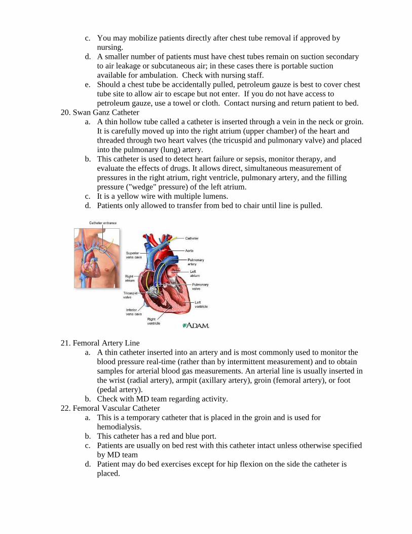
c. You may mobilize patients directly after chest tube removal if approved by
nursing.
d. A smaller number of patients must have chest tubes remain on suction secondary
to air leakage or subcutaneous air; in these cases there is portable suction
available for ambulation. Check with nursing staff.
e. Should a chest tube be accidentally pulled, petroleum gauze is best to cover chest
tube site to allow air to escape but not enter. If you do not have access to
petroleum gauze, use a towel or cloth. Contact nursing and return patient to bed.
20. Swan Ganz Catheter
a. A thin hollow tube called a catheter is inserted through a vein in the neck or groin.
It is carefully moved up into the right atrium (upper chamber) of the heart and
threaded through two heart valves (the tricuspid and pulmonary valve) and placed
into the pulmonary (lung) artery.
b. This catheter is used to detect heart failure or sepsis, monitor therapy, and
evaluate the effects of drugs. It allows direct, simultaneous measurement of
pressures in the right atrium, right ventricle, pulmonary artery, and the filling
pressure ("wedge" pressure) of the left atrium.
c. It is a yellow wire with multiple lumens.
d. Patients only allowed to transfer from bed to chair until line is pulled.
21. Femoral Artery Line
a. A thin catheter inserted into an artery and is most commonly used to monitor the
blood pressure real-time (rather than by intermittent measurement) and to obtain
samples for arterial blood gas measurements. An arterial line is usually inserted in
the wrist (radial artery), armpit (axillary artery), groin (femoral artery), or foot
(pedal artery).
b. Check with MD team regarding activity.
22. Femoral Vascular Catheter
a. This is a temporary catheter that is placed in the groin and is used for
hemodialysis.
b. This catheter has a red and blue port.
c. Patients are usually on bed rest with this catheter intact unless otherwise specified
by MD team
d. Patient may do bed exercises except for hip flexion on the side the catheter is
placed.
23. Intra Arterial Balloon Pump (IABP)
a. Patient on bed rest when IABP placed in the femoral artery
b. When placed in axillary location, then patient is able to get OOB and ambulate.
c. Must have RN present when ambulating patient on IABP.
d. Once RN staff determine patient is stable, then therapy may ambulate patient
without RN present.
e. The IABP is a polyethylene balloon mounted on a catheter, which is generally
inserted into the aorta through the femoral artery in the leg. The balloon is guided
into the descending aorta, approximately 2 cm from the left subclavian artery.
f. Used to decrease myocardial oxygen demand while at the same time increasing
cardiac output. By increasing cardiac output it also increases coronary blood flow
and therefore myocardial oxygen delivery. It actively deflates in systole
increasing forward blood flow by reducing afterload thus, and actively inflates in
diastole increasing blood flow to the coronary arteries. These actions have the
combined result of decreasing myocardial oxygen demand and increasing
myocardial oxygen supply
g. Inflation of the balloon can be triggered according to the patient's
electrocardiogram, their blood pressure, a pacemaker (if they have one), or by a
pre-set internal rate.
h. Indications: cardiogenic shock, acute mitral regurgitation and septal perforation,
unstable angina, post cardiothoracic surgery, and bridge to heart transplant
24. Left Ventricular Assist Device (LVAD)
a. The left ventricle is the large, muscular chamber of the heart that pumps blood out
to the body. A left ventricular assist device (LVAD) is a battery-operated,
mechanical pump-type device that's surgically implanted. It helps maintain the
pumping ability of a heart that can't effectively work on its own
b. HeartWare and HeartMate II is the device used at VUMC currently
i. Is a continuous flow rotary LVAD
ii. Has one moving part which is a small spinning rotor that receives power
from the Systems Controller (the brain).
iii. The rotor moves blood thru the pump which is capable of providing flow
from 3 to 10 L per minute
iv. Ruby and ceramic barons (low thermal conductivity)
v. It is VALVELESS!!
c. Since LVAD is valveless, you will be unable to detect an O2 sat or BP like usual.
d. Patients will come out of OR with art line. You need to monitor MAP (mean
arterial pressure) from art line. Usually, MD team likes it around 60.
e. As far as HR, patient may have an underlying HR. However, if no underlying HR,
then you will be unable to get accurate HR.
f. Monitor these patients by signs and symtoms (ie diaphoresis, SOB, cyanosis, etc).
g. Patient with be on AC power in bed. If you would like to go further than that line
allows, you will need to place patient on batteries.
h. Make sure controller (the brain) is securely attached to abdominal binder or has
special binder for LVAD patients.
i. Do not let controller fall off patient/binder. This is connected to the power cord
which is exiting the patient’s abdomen. This site needs to heal adequately to
avoid infection.
j. These patients have sternal precautions.

k. How to switch from A/C (wall power) battery
i. Obtain charged battery from charger
1. Press button on front of battery, if all 5 lights are green, then
battery is fully charged
ii. Place charged battery in holder.
1. Make sure battery clicks in!!
iii. Place holster on patient and place batteries in holster securing with velcro
strap.
iv. Starting with black cord, unscrew cord from AC power and attach to
closest battery ensuring plenty of slack on cord.
1. Cords requiring unscrewing and pulling from power source then
pushing and screwing to the battery to achieve proper connection.
2. Once cord is pulled from a power source, there will be a loud
beeping until attached to another power source.
v. Then move to white cord, unscrewing from AC power and attaching to
available battery with the same steps as above.
vi. Make sure brain is securely attached to patient before mobility!!
vii. Perform these steps in reverse when going back to AC power.

Gynecology / Oncology [4east]
General Guidelines
1. Located on the 4th
floor of MCE and 4th
floor VUH
2. Typically PT gets more evaluations due to bedrest patients requiring therex program
3. Can be pregnancy, post-partum, gynecology surgery, gynecology, or oncology patients
4. Check with nurse prior to entering patient’s room for any acute medical status change
5. Typically need to bring any equipment with you

TOBS
1. Located on the 1st floor of The Vanderbilt Clinic (TVC). It is down the hall from the ED.
2. Rooms begin with “15”.
3. All patients admitted to this unit are considered observation status with an expectation
that they will be hospitalized for 72 hours or less. If the patient converts over from
observation to Inpatient status than they are transferred to a floor in the hospital.
4. The patient population is strictly observation patients for all medical teams (with
Riven/general medicine observation patients being largest patient population)
5. All beds have portable telemetry.
6. Supplies are located in the back right of the unit. Linens are located in a room on the back
hallway.
Emergency Room (ED)
1. Emergency Room consists of 3 “pods”- “A” pod is located on the 1st floor of VUH to
right of central hallway door as you enter. “B” pod is located on the 1st floor of VUH to
left of central hallway door as you enter. “C” pod is located across the hall on the 1st floor
of VUH.
2. “A” pod typically houses trauma, orthopedic injuries, TIA/CVA, PE/DVT, and critically
ill patients. “B” and “C” pod typically house medicine patients, smaller orthopedic
injuries, and more stable patients.
3. Utilization Management reviews all of the emergency room patients daily to determine if
they meet criteria for hospital admission or if they should be in observation status.
Patients may be listed as being admitted in the AM but then listed as observation status in
the PM or even after discharge.
4. To check if a patient is in observation status you can print the Medilinks list and look for
any patients that have any red writing in the MRN column (OP, E, A - medicare).
Another way to check is by going into Medilinks select Reports >TVUH>Orders> Acute
Medicare OP, E, A with Active Orders. This will show everyone in the hospital that is in
observation status.
5. If the patient has an observation status order and an admit order, the patient is in
observation status. The admit order does not negate the observation status one.
6. Management of Observation Patients
a. All emergency room patients should be looked at in Star Panel in the AM and the
PM to determine if they are in observation status or not.
b. All patients in observation status should be evaluated in a timely manner pending
medical work-up or clearance (ie. ruling out a fracture, determining a weight
bearing status, ruling out a PE/DVT, ect…)
c. If the word observation is anywhere in the orders then that patient is in
observation status.
d. If you are unable to evaluate an observation patient you should either document a
failed attempt note or add a comment to that patient’s order in MediLinks.
e. Use the CBOR templates found in charting category when documenting
observation patients.
7. Discharge recommendations for Observation Patients
a. In general, insurance will only pay for home with home health therapy or
inpatient rehab
b. Insurance will not pay for an observation patient to discharge to a SNF however
there are a few exceptions to this rule thus you will need to speak to the patient’s
SW or CM.
c. If SNF is the appropriate discharge recommendation and the patient’s insurance
will not cover SNF, contact the team to inform them you are recommending a
SNF and why.
Recovery Room/ Medical Center East (MCE) Outpatient Surgery
1. Recovery room (VHR) is
a. Located on the 3rd
floor of VUH. When leaving the office, turn left and head for
VUH elevator bank. Go down hallway right before elevator bank and it is on the
left hand side with double wooden doors. The code to enter is the hospital wide
code.
2. MCE (HR with a letter and number such as HRD4) is located in Medical Center East
a. Is the surgical observation area or the same day surgery area
b. The letter indicates the pod they are in and number corresponds to the patient
room number.
c. Located on 3rd
floor MCE. When leaving the office, turn right into day surgery
waiting room. Turn left at the reception desk and enter through double doors.
3. Perform PT/OT evaluations for orthopedic patients and day surgery patients.
4. You will have to take all necessary equipment with you to these areas as there is not
storage for PT/OT equipment. It is best to call area prior to going to ensure your patient
has not discharged or moved room.
5. Nurse or medical receptionist can call for any needed equipment for discharge (such as
reacher, sockaid, long handled sponge, cane, crutches, walker, etc)
Medical Center North (MCN)
General Information
1. Floors: 3rd
General Medicine, 4th
Colorectal, 5th
Palliative Care and General Medicine, 6th
Orthopedic, 7th
Geriatric/ ACE Unit (Acute Care of the Elderly)
2. Loaner rehabilitation equipment for patient usage is kept in 6413 (rehab gym). Loaner
equipment is labeled with MCN Rehab or VUH Rehab. If patient requires equipment for
discharge, therapist or case manager should order prior to discharge. When patient is
discharged the equipment should be picked up by staff and returned to 6413. Dirty/used
equipment is placed on right side of room. Clean equipment is on the wall at back of
room. Wheelchairs are locked with a chain (keys are located in the drawer on your right
as you enter 6413).
3. Theraband/Theraputty is located in the drawers on the right as you enter 6413.
4. Rolling walker (standard and bariatric), platform attachments, axillary crutches, and
straight canes can be obtained for home usages from Materials Management/Service
Center. Adolescent rolling walker can be ordered from Children’s Hospital or obtained
by case manager.
5. Specialty equipment (such as AFO, spinal orthosis, hip orthosis) can be ordered by
MD/PA/NP from outside vender.
6. Smooth Moves equipment is available on all units equipment rooms. If you borrow other
Smooth Moves equipment from another unit, you are responsible for speaking to CSL
prior to taking it, cleaning it, and returning equipment.
Floors
1. 3rd
floor
a. General medicine patients.
b. Room numbers begin with “34” and encircle nurses station except for 2 rooms in
hallway near nurses station.
c. Member of acute rehab attends Riven Huddle 3 days per week (Monday,
Wednesday, and Thursday) on 8th
floor ICU conference room at 11am.
d. Patient information board is located on wall across from nurse station .
e. Supplies and linens are located in the supply room across from the stairwell
behind the nurse station.
2. 4th
floor
a. Colorectal patients, general medicine, and overflow orthopedic/trauma patients.
b. Room numbers begin with “44”.
c. Colorectal patients are usually very mobile after surgery and PT evaluations are
entered for difficulty mobilizing after surgery.
d. Patient information board is located on the wall behind the nurse station.
e. Supply room is located across from the hallway stairwell and linens are located
across from the back stairwell.
3. 5th
floor
a. General medicine and palliative care patients.
b. Room numbers begin with “54”.
c. Patient information board is located on the wall behind the medical receptionist at
the nurses desk.
d. Supplies are located across from the hallway stairwell. Cold nutrition items are in
refrigerator in locked room off pantry across from nurses desk. Linens are kept in
2 cabinets on back side of nurses desk.

e. Therapy is often asked to evaluate and treat palliative care patients who may or
may not be at end of life. Communicate with nurse as well as case manager
regarding teams specific needs from therapy.
f. If picture of butterfly is on the patients door they are actively dying and therapy is
not appropriate.
4. 6th
floor
a. Orthopedic patients.
b. Room numbers begin with “64”.
c. Member of acute rehab attends Orthopedic Huddle Monday to Friday at 9:15am
(at nurses station)
d. If further information is needed it is typically quicker to page team NP/PA and
then resident. Only at last resort do you page resident.
e. Patient information board is located behind the medical receptionist at the nurse
desk.
f. Supplies are kept in locked room across from the hallway stairwell. Linens are
kept in 2 cabinets on back side of nurses desks. Cold nutrition items are in
refrigerator in locked room off pantry across from nurses desk.
g. Rehab staff are advised to read operative report for any orthopedic surgery
specific information related to post-op care of patient. If any discrepancy in orders
look to attending MD operative report and then contact NP/PA for team for order
clarification. NP/PA will contact attending MD and relay information to therapy.
Do not guess which order is correct. If no NP/PA page the junior resident on the
team.
h. PA/NP/resident pager numbers are posted on the cabinets over the desk area in
6413.
i. Braces/specialty splints are routinely orders after certain procedures/injuries.
Must be ordered by NP/PA/reside. Therapy does not obtain and do not complete
initial fittings (should be done by nurse/cast tech/NP/PA/resident).
j. Most orthopedic patients will require home exercise program. Individualized can
be printed off via the software system. Diagnosis specific home exercise programs
have been preprinted and are located in top drawer of small file cabinet near the
door in 6413.
5. 7th
floor
a. ACE unit (acute care of the elderly) houses geriatric patients.
b. Room numbers begin with “74”. Rooms encircle nurses station and down entire
hallway.
c. Member of acute rehab attends Geriatric Huddle Monday to Friday at 11:00 am
(across from 7432).
d. ACE unit is a locked unit.
e. Supplies and linens are located in locked rooms in the unit.
f. Patient information board is located behind the nurses desk.
THA/TKA Overview
1. The following treatment guidelines are for the standard joint arthroplasty patient. Any
specific restrictions/precautions would be present in the PT post-op orders and would
supercede these treatment guidelines.
2. THA:
a. Day of Surgery/Initial Evaluation
i. Instruct/review THA dislocation precautions
ii. Initiate APs & BLE isometrics in supine – encourage patient to perform
these exercises q 30 minutes while awake
iii. May begin supine AAROM to operative LE as tolerated
iv. Instruct in bed mobility using leg lifter on operative limb if needed
v. Transfer training using appropriate assistive device, WBAT on operative
limb
vi. Gait training with assistive device, WBAT operative LE
vii. Ice packs to operative hip for pain & edema management as patient
tolerates – must use 20 minutes for benefit; try 1 ice pack in groin/inguinal
area of surgical hip & another at lateral or posterior aspect of surgical hip
b. POD 1 – Discharge
i. Review THA dislocation precautions each therapy visit
ii. Supine exercises – APs, LE isometrics, AAROM to operative limb with
no restriction to single plane movements
iii. Gait training – progress from rolling walker/crutches to cane at PTs
discretion
iv. Stair training prior to discharge home
v. BID treatment sessions
3. TKA/Bilateral TKA/UKA:
a. General Information:
i. Pillow under the operative knee is not allowed! If sidelying, patients may
have a pillow between their knees. A pillow or folded towel placed under
the foot/ankle of the operative leg with the patient lying supine in bed
(knee & toes facing ceiling) is encouraged for a passive hamstring stretch
– goal is to maintain this stretch for 30 minutes TID
ii. Cooling machine or ice packs are used for pain & edema management &
should be worn prn & definitely after PT sessions unless the patient does
not tolerate the cool; the cold therapy must be used for 20 minutes for
benefit; patients are instructed to keep cooling machine/ice off the
operative knee twice as long as duration of cold on knee (if used ice for 20
minutes, leave off for 40 minutes before reapplying)
b. Day of Surgery/Initial Evaluation
i. Initiate APs & isometrics (quad sets, hamstring sets) to BLE
ii. Begin range of motion exercises to operative LE in supine &/or sitting
with no restriction to motion
iii. Bed mobility training using leg lifter on operative LE only if needed
iv. Transfer training with assistive device, WBAT on operative limb
v. Gait training with appropriate assistive device, WBAT
c. POD 1 – Discharge
i. P/A/AAROM operative LE – any exercise you like working on knee
flexion & extension as well as hip abduction; closed chain exercises (mini-
squats, SLS, weight shifting in standing, etc)
ii. Soft tissue work on operative LE hamstrings (quads are typically too
edematous to initiate this in the immediate post-op phase)

iii. Femoral/tibial joint mobilization (Grades I & II) to increase joint motion –
the only TKA you would not mob would be a hinged TKA (most often
used with tumor patients)
iv. Quad sets, SAQ/TKE, SLR, any quad strengthening exercises you wish
v. Hamstring stretching: any you like. Drs. Shinar & Polkowski encourage
patients to place folded pillow under their operative foot/heel when lying
supine (keep & toes facing ceiling) to accomplish passive HS stretch –
goal for this stretch position is 30 minutes TID (ask family & nursing staff
to assist with this stretch)
vi. Gait training, WBAT on operative LE
vii. Stair training prior to discharge home
viii. BID treatment sessions
Orthopedic Rehabilitation Guidelines
1. Pelvic Fractures
a. Trauma to the pelvis may result in a variety of fractures – Open Book, Saddle,
Lateral Compression, Vertical Shear, Pubic Rami, Ilium, etc. Fractures may or
may not be surgically repaired based upon the fracture pattern, location, bone
quality and surgeon preference.
b. Precautions / Restrictions:
i. Weight bearing may be limited based upon the fracture type, location, etc.
ii. Hip range of motion and strengthening exercises to the patients tolerance.
iii. Patients, in general, may sit upright but may prefer to “slouch” sit.
c. Treatment:
i. Daily treatment consisting of mobility/gait training on level surfaces &
stairs,
ii. APs/isometrics BLE prn, AAROM LE with motion to patient unless
otherwise
iii. ordered, HEP (Early LE Rehab usually appropriate)
2. ORIF Acetabulum Fracture
a. Internal fixation performed following traumatic displaced fracture of the
acetabulum. Traumatic posterior hip dislocation (TPHD) is frequently associated
with acetabulum fractures; closed reduction of the hip is performed ASAP
followed by operative fixation.
b. Precautions / Restrictions:
i. Protective weight bearing, typically TDWB, on the operative leg.
ii. Use of an assistive device for ambulation ~ 12 weeks.
iii. Lower extremity range of motion and strengthening exercises as tolerated;
no restrictions to motion unless specifically stated by the attending
surgeon.
c. Treatment:
i. Daily to BID treatment (dependent upon patient progress); bed mobility
training using leg lifter on involved LE, gait training on level surfaces &
stairs (NWB/TDWB – non-op, TDWB – after ORIF), APs/isometrics
BLEs prn, AAROM involved LE with no motion restrictions unless
specific motion (flexion) restrictions are ordered; home exercise program
with f/u PT services as indicated.
3. Hip Fractures
a. Most hip fractures require operative fixation. The type of fixation is dependent
upon the fracture location (femoral head, femoral neck, Intertrochanteric,
Subtrochanteric), fracture pattern, patient’s age, bone quality, open
wound/extensive soft tissue damage and surgeon preference.
b. Femoral head and neck fractures may be repaired using Percutaneous Pins
(CRPP), Dynamic Hip Screw (DHS), Trochanteric Femoral Nail (TFN), Bipolar
Hemiarthroplasty or Total Hip Arthroplasty (THA).
c. Intertrochanteric fractures may be fixated with CRPP, DHS, TFN, Bipolar or
THA. Subtrochanteric fractures may be repaired using an Intermedullary Nail
(IMN), or TFN.
4. Bipolar Hemiarthroplasty
a. A fixation device used for femoral head, femoral neck & IT hip fracture repair.
The prosthesis is one piece, a femoral component with an acetabular cup fixated
to the femoral stem; the femoral head of the prosthesis articulates within the
acetabular cup, which articulates within the patient’s acetabulum. No surgical
intervention is performed in the acetabulum.
b. Precautions / Restrictions:
i. Weight bearing as tolerated ambulation using an assistive device.
ii. Avoid extreme Simultaneous / Combination movements of the operative
hip. Patients are allowed to fully flex, extend, abduct, adduct, or rotate
their operative hip in cardinal planes of motion. Movement restrictions are
observed ~ 6 weeks post-operatively.
5. ORIF Hip Fractures
a. Precautions / Restrictions:
i. Weight bearing status per attending orthopedic surgeon; varies from NWB
to WBAT; generally, WBAT is allowed unless the surgeon is concerned
over fracture hardware purchase, the presence of osteoporetic bone or
extensive soft tissue damage/open wounds.
ii. No hip dislocation precautions following ORIF of hip fractures with No
restrictions to hip range of motion; all motions are to patient tolerance.
b. Hip Fracture Treatment:
i. BID treatments; APs/isometrics q 2 hours while awake; AAROM
operative LE with no restrictions to hip motion for ORIF, avoid
‘combination’ movements of operative hip with bipolar hemiarthroplasty;
bed mobility training using leg lifter on operative LE; gait training using
appropriate assistive device with WB as ordered by the attending
orthopedic surgeon; stair training prior to discharge home; home exercise
program & f/u PT services as indicated.
6. Total Hip Arthroplasty (THA)
a. An elective procedure for patients with hip pain secondary to OA, RA, AVN,
traumatic arthritis, or tumor. The prosthesis consists of an acetabular component
(metal cup and liner which may be metal, ceramic or polyethylene) and a femoral
component. The prosthesis may by implanted by cement or cementless fixation;
cementless fixation is the choice for Vanderbilt surgeons. Revision THA
components at Vandy are cementless. Rehab following Revision THA follows the
same guidelines as Primary THA.
b. Precautions / Restrictions:

i. Avoid Simultaneous / Combination movements of the operative hip with
posterior & posteriolateral approach. Patients are allowed to flex, extend,
abduct, adduct, or rotate their hip in cardinal planes of motion with NO
restriction or limitation of motion. These hip precautions are to be
observed for 6 weeks post-op for standard THA, 12 weeks for patients
having megaprosthesis implant (tumor). Anterior approaches require NO
hip precautions and will be WBAT.
ii. Return to sports: we follow the AAOS guidelines for return to sports after
THA (these are provided to patients in the pre-op booklet) which allow
specific sports for anyone, others with experience & do not recommend
high-impact sports which involve repetitive jumping as this tends to
decrease the life of the implant – hip resurfacing patients with a metal-on-
metal implant are allowed to participate in high-level sporting activities,
timing of return to sports is dictated by the attending surgeon.
iii. Weight bearing status for primary & revision THA is WBAT; intra-
operative/periprosthetic fractures are treated with protective weight
bearing, usually TDWB.
c. Treatment:
i. BID sessions; PT initiated day of surgery when possible; APs/isometrics
encouraged every 30 minutes while awake; bed mobility training using leg
lifter on operative LE; DOS/POD 1 to discharge – transfer training, gait
training using appropriate assistive device, WBAT on operative LE, begin
AAROM operative LE in single, cardinal planes of movement with no
restriction to single motions – movement to the patient’s tolerance; sitting
&/or standing exercises using walker or furniture for balance/support
with emphasis on hip abduction & hip extension when able; progression
from walker/crutches to cane allowed by surgeon and at therapist’s
discretion; ice packs to operative hip for pain control if desired; stair
training prior to discharge; home exercise program & f/u PT services.
7. Birmingham Hip Resurfacing (BHR)
a. An elective procedure; an alternative procedure for early stages of hip arthritis to
prolong the need for THA. The implant consists of a metal acetabular component
and a metal femoral component. Both the acetabulum and femoral head are
“resurfaced” but the femoral head is not resected as with THA. This procedure is
for active, “young”, male patients with early stages of arthritis and excellent bone
quality.
b. Precautions/Restrictions:
i. Same as for THA
c. Treatment:
i. Same as for THA; typically these patients are discharged POD 1 following
rehab treatments and progress very quickly in their rehab program and are
encouraged to attend OPPT at discharge.
8. THA Resection
a. Surgical procedure to resect infected THA prosthesis. After prosthesis removal,
an antibiotic impregnated spacer or antibiotic-coated hip femoral component may
be implanted. Patients receive long-term antibiotic therapy and then re-
implantation/revision THA.
b. Precautions / Restrictions:
i. Dislocation precautions – avoid combination movements of the operative
hip for as long as the spacer is in place.
ii. NWB-TDWB on the operative lower extremity.
iii. Hip range of motion, strengthening and positioning (avoidance of
rotational extremes) to the patient’s tolerance.
c. Treatment:
i. Daily sessions, depending upon patient progression with mobility;
APs/isometrics q 30 minutes during waking hours; bed mobility training
using leg lifter on operative LE; supine AAROM to operative limb; gait
training, WB per the surgeon on level surfaces & stairs; ice packs to
operative hip prn for pain control as patient desires; home exercise
program & f/u PT as needed.
9. Hip Core Decompression
a. A surgical procedure performed in the early stages of avascular necrosis (AVN)
of the femoral head to promote revascularization of the femoral head. In this
procedure, a core of bone is removed from the femoral head utilizing a lateral
approach. This procedure is an attempt to postpone the need for THA.
b. Precautions / Restrictions:
i. Strict NWB on the operative extremity for 12 weeks.
ii. No limitations on hip range of motion or strengthening exercises.
c. Treatment:
i. Daily – BID sessions; many of these patients are discharged home POD 1
after PT session; APs/isometrics q 30 minutes while awake; bed mobility
training using leg lifter on operative LE; NWB operative LE gait training
with assistive device on level surfaces & stairs; AAROM operative LE
with NO restrictions to motion; home exercise program with f/u PT as
indicated.
10. Total Knee Arthroplasty (TKA)
a. An elective surgery for patients with debilitating knee pain secondary to arthritic
conditions or tumor. The prosthesis consists of tibial, femoral, and patellar
components. Implants at VUMC are cemented. Patients undergoing revision or
bilateral TKA follow the same rehab program as unilateral TKA.
b. Precautions / Restrictions:
i. Weight bearing as tolerated ambulation with an assistive device.
ii. Per AAOS guidelines for return to sports - avoid running and jumping in
contact sports (lifelong) unless a mobile-bearing implant is used.
11. Uni-Condylar Knee Arthroplasty (UKA)
a. An elective surgical procedure for knee pain due to OA, RA, or traumatic arthritis
with uni-compartmental joint disease. One compartment of the knee is resurfaced,
either medially or laterally, with the prosthesis. Knee rehab is as for TKA. The
goal if for discharge home POD 1 with this patient population.
12. Patella-Femoral Arthroplasty(PFA)
a. An elective surgical procedure for knee pain due to cartilage degeneration Over
the distal femur. A metal implant is placed in the trochlear groove and a plastic
patellar implant on the resurfaced patella. Knee rehab is the same as for TKA with
goal of discharge home on POD 1.
b. Treatment:

i. Same for TKA/UKA/PFA; BID sessions until discharge; all adult
reconstruction surgeons are treating their patients uniformly except Dr.
Holt’s
ii. patients are to sleep in a knee immobilizer on the operative knee for
iii. 2 weeks after their surgery for a passive hamstring stretch; attempt to
initiate PT DOS, if unable begin POD 1 a.m. with APs/BLE isometrics
iv. q 30 minutes while awake, bed mobility training using leg lifter on the
v. operative LE, WBAT transfer/gait training with appropriate assistive
device on level surfaces, supine &/or sitting AAROM operative knee with
emphasis on knee flexion/extension & hip abduction, hamstring stretching
exercises, quad strengthening, cooling machine to operative knee while in
bed; stair training; instruction in home exercise program (handouts given
by PT) with f/u PT services arranged prior to discharge home by case
manager.
13. TKA Resection
a. Performed for infected TKA. The knee prosthesis is surgically removed and the
alignment/ space between the femur and tibia is maintained by an articulating
antibiotic spacer. Long-term antibiotic therapy is instituted following TKA
resection. When the infection is cleared, TKA Revision is performed.
b. Precautions / Restrictions:
i. WB per surgeon orders with articulating spacers, typically TDWB but
may be WBAT.
ii. No motion restrictions with articulating spacer; patients are typically
placed in a protective hinged knee brace until TKA revision.
iii. Avoid any varus/valgus strain to knee joint as the spacer can dislocate.
c. Treatment:
i. Daily sessions, dependent upon patient progression with exercises and
mobility; POD 1- Discharge – APs/isometrics every 30 minutes while
awake, bed mobility training using leg lifter on operative LE, transfer/gait
training on level surfaces and stairs using assistive device with weight
bearing status as ordered by surgeon; if an articulating spacer is present,
gentle A/AAROM exercises to operative extremity (hip, knee, ankle, foot)
in hinged knee brace (perform exercises & mobilization wearing knee
brace), general muscle strengthening to operative extremity (no weights or
theraband to knee joint musculature), home exercise program & f/u PT
services as necessary.
14. Foot
a. Precautions / Restrictions:
i. Weight bearing status is dependent upon the fracture location and/or
surgical reconstruction. Ankle arthrodesis, calcaneal and talar fractures are
NWB; ankle fractures WB may vary from NWB to WBAT.
ii. Metatarsal Fractures should be protected with a hard sole shoe or boot
when ambulating.
b. Treatment:
i. Bed mobility/ gait training using assistive devices on level surfaces and
stairs; “knee walkers” are especially beneficial in this patient population if
the patient has difficulty maintaining correct weight bearing status; “knee
walkers” are currently covered by most commercial insurance plans and

several local DME companies have rental plans – contact the orthopedic
SW or case manager if patients wish to pursue rental of this device.
15. Spine
a. Precautions / Restrictions:
i. Current orthopedic spine surgeons typically do not use bracing following
elective thoracic &/or lumbar surgical procedures; cervical collars are
used following elective c. spine procedures. Bracing is used for traumatic
spinal fractures by orthopedic surgeons and are typically worn for 12
weeks. If the surgeon orders thoracic &/or lumbar bracing, patients must
wear their spinal orthosis any time they are in an upright position. The
brace should be applied while the patient is lying supine, unless otherwise
stated by the surgeon. This is accomplished by “log-rolling” the patient
side-to-side (avoiding trunk rotation).
b. General orthopedic post-operative spinal restrictions are avoid any spinal rotation
or “twisting” activities, avoid excessive forward spinal flexion (bend/flex at hips
instead of spine)and do not lift any object weighing more than 1 gallon of milk (~
9 lbs) for 6 -12 weeks.
c. No spine specific exercise for 6-12 weeks until healing has occurred. Patients may
perform active or active-assisted exercises with the upper or lower extremities in
the presence of general muscle weakness.
16. Total Shoulder Arthroplasty (TSA)
a. An elective surgical procedure for severe shoulder pain secondary to OA, RA,
AVN, traumatic arthritis, or tumor of the shoulder. A surgical procedure also
performed following traumatic 3- or 4-part Humeral Head Fracture.
b. Initiation of rehab varies by surgeon from post-operative Day 1 to 2 weeks post-
op.
c. Precautions / Restrictions:
i. Shoulder range of motion is performed in the plane of the scapula; ~ 40
degrees of shoulder abduction.
ii. Timing of beginning rehab varies between surgeons (Dr. Kuhn - no
motion to operative shoulder for 2 weeks) as well as does treatment
guidelines with specifics communicated to rehab staff by specific surgeon.
iii. NWB on the operative upper extremity for 6 weeks. Most surgeons use a
specialty arm sling which is placed following surgery during this time
frame.
17. External Fixators
a. A closed reduction method of fracture repair using percutaneous pins/wires
allowing easy access to soft tissues in the event of needed wound care.
b. Precautions / Restrictions:
i. Extremities may be moved, supported, and/or suspended by lifting the
external fixator; this will not displace the fixator.
ii. No restriction to range of motion to joints proximal and/or distal to the
fixation device.
iii. Weight bearing status per surgeon.
18. Intermedullary Nail
a. An internal fixation device used to treat traumatic fractures in the long bones.
There are proximal and distal incisions, which may limit range of motion at 2
joints and contribute to significant edema of the entire extremity.
b. Precautions / Restrictions:
i. Primarily a weight bearing implant, however, fracture pattern as well as
the presence of any soft tissue injuries dictates specific weight bearing
status; open fractures are usually NWB.
ii. Exercise program should be aggressive to the entire operative extremity,
focusing on joint range of motion and muscle strengthening.
19. Allograft
a. A complex, intricate surgical procedure involving the removal of part or all of a
damaged bone which is then replaced / reconstructed with a cadaveric human
bone. Most often performed on patients with skeletal tumors or necessary because
of bone deficiency / loss due to previous surgical procedures or massive skeletal
trauma.
b. Precautions / Restrictions:
i. Weight bearing status per orthopedic surgeon; generally, protective /
limited weight bearing.
ii. With tumor patients, the specific rehab program will be outlined by the
attending orthopedic surgeon. DO NOT initiate any exercise program with
Dr. Schwartz’s tumor resection patients unless specific exercises are
ordered by the surgeon; if in doubt, contact the resident on the orthopedic
oncology service regarding specific rehab interventions.
c. Treatment:
i. Daily treatment sessions focusing on bed mobility training using leg lifter
on operative LE as needed, transfer training, gait training with appropriate
assistive device and WB status per surgeon on level surfaces & stairs;
patients with WBAT status are not required to use an assistive device
unless the patient &/or therapist deem it necessary for safe mobilization.
General Orthopedic Guidelines
1. When moving the patient, always support the injured extremity at the joints.
2. Use the palm of your hands, not fingertips, to move or support the patient.
3. The patient will experience less discomfort if you support the injured extremity and allow
the patient to do the actual moving.
4. When assisting the patient out of bed, exit the bed on the side of the unaffected extremity.
5. Take your time. Moving patients too quickly leads to pain and can result in injury.
6. Pillows should be used to elevate an injured extremity, giving support to the entire
extremity, unless contraindicated by the injury/surgical procedure. When the patient is
sidelying, pillows to the back and hips and between the knees provide support and proper
body alignment.
7. Most orthopedic patients can and should be turned side-to-side. As a general rule, the
patient should be turned toward their unaffected side whenever possible; tilting to the
affected side is also possible.
8. Patients with orthopedic injuries, especially spinal injury/surgery, should be turned as a
unit avoiding spinal rotation. Beds should be made with a “turning sheet” placed on top
of the bottom sheet. This can be used to support the patient’s shoulders and hips as they
are slowly turned sideward.
9. Most spinal and lower extremity braces are to be applied with the patient lying supine in
bed. Most braces may be removed for skin checks/assessment. If in doubt, ask the
physician for skin check and brace wear clarification.
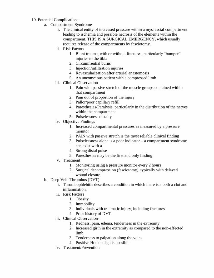
10. Potential Complications
a. Compartment Syndrome
i. The clinical entity of increased pressure within a myofascial compartment
leading to ischemia and possible necrosis of the elements within the
compartment. THIS IS A SURGICAL EMERGENCY, which usually
requires release of the compartments by fasciotomy.
ii. Risk Factors
1. Blunt trauma, with or without fractures, particularly “bumper”
injuries to the tibia
2. Circumfrential burns
3. Injection/infiltration injuries
4. Revascularization after arterial anastomosis
5. An unconscious patient with a compressed limb
iii. Clinical Observation
1. Pain with passive stretch of the muscle groups contained within
that compartment
2. Pain out of proportion of the injury
3. Pallor/poor capillary refill
4. Paresthesias/Paralysis, particularly in the distribution of the nerves
within the compartment
5. Pulselessness distally
iv. Objective Findings
1. Increased compartmental pressures as measured by a pressure
monitor
2. PAIN with passive stretch is the most reliable clinical finding
3. Pulselessness alone is a poor indicator – a compartment syndrome
can exist with a
4. Strong distal pulse
5. Paresthesias may be the first and only finding
v. Treatment
1. Monitoring using a pressure monitor every 2 hours
2. Surgical decompression (fasciotomy), typically with delayed
wound closure
b. Deep Vein Thrombus (DVT)
i. Thrombophlebitis describes a condition in which there is a both a clot and
inflammation.
ii. Risk Factors
1. Obesity
2. Immobility
3. Individuals with traumatic injury, including fractures
4. Prior history of DVT
iii. Clinical Observation-
1. Redness, pain, edema, tenderness in the extremity
2. Increased girth in the extremity as compared to the non-affected
limb
3. Tenderness to palpation along the veins
4. Positive Homan sign is possible
iv. Treatment/Prevention

1. TED stockings
2. Sequential compression device use
3. Ankle pumps and isometric exercises
4. Mobilization
5. Anticoagulation therapy
c. Fat Embolism Syndrome
i. Fat embolism is the presence of fat globules in the bloodstream that
become deposited in the pulmonary circulation. Fat embolism syndrome is
the pulmonary insufficiency commonly associated with fat emboli.
ii. Risk Factors
1. Total joint replacement
2. Multiple trauma injuries
3. Crush injuries
4. Fractures in bones containing marrow fat
a. Femur
b. Tibia
c. Humerus
d. Pelvis
e. Ribs
iii. Clinical Observation-
1. Chest pain may be present
2. Apprehension
3. Behavior changes (may be subtle initially)
4. Restlessness
5. Agitation
6. Irritability
7. Confusion
8. Lethargy
9. Respiratory changes
10. Dyspnea
11. Tachypnea
12. Wheezes
13. Tachycardia
iv. Treatment
1. Oxygen
2. Crystalloid fluid replacement
3. Heparin therapy
d. Hemorrhage
i. Hemorrhage is the abnormal loss of blood from the body.
ii. Risk Factors
1. Dislodged blood clot due to stress at an operative site
2. Infection
3. Erosion of a blood vessel by a fracture
4. Fractures
5. Surgery
6. Abnormal clotting mechanisms
7. Anticoagulants
8. Ulcers

iii. Clinical Observation-
1. Anxiety, dizziness, weakness
2. Restlessness
3. Rapid, shallow respiration’s
4. Cold, moist skin
5. Decrease in blood pressure
6. Excessive drainage from wounds or drain output
7. Decreased urine output
iv. Treatment
1. Apply direct pressure/pressure dressing at bleeding site
2. Volume replacement
3. Vitamin K or factor replacement
4. Iron
5. Surgery to locate bleeding site or suturing/resuturing wound or
incision
ORTHOPEDIDC ABBREVIATIONS, PROCEDURES AND DEFINITIONS:
AAROM Active, Assisted Range of Motion
AE Above Elbow
AEA Above Elbow Amputation
AFO Ankle Foot Orthosis
AKA Above Knee Amputation
Allograft A cadaveric human bone transplant
Amputation Surgical removal of part or all of a body tissue or extremity
AROM Active Range of Motion
Arthrodesis Fusion of a joint to relieve pain by eliminating motion
ASF Anterior Spinal Fusion
AVN Avascular Necrosis
Avulsion Ligaments torn loose from their attachment to the bone
BB Both bones
BBFA Both bones of the forearm
BE Below Elbow
BEA Below Elbow Amputation
Bipolar Hemiarthroplasty Surgical implant for treatment of hip fractures
BK Below Knee
BKA Below Knee Amputation
BLE Bilateral Lower Extremity
C sp Cervical Spine
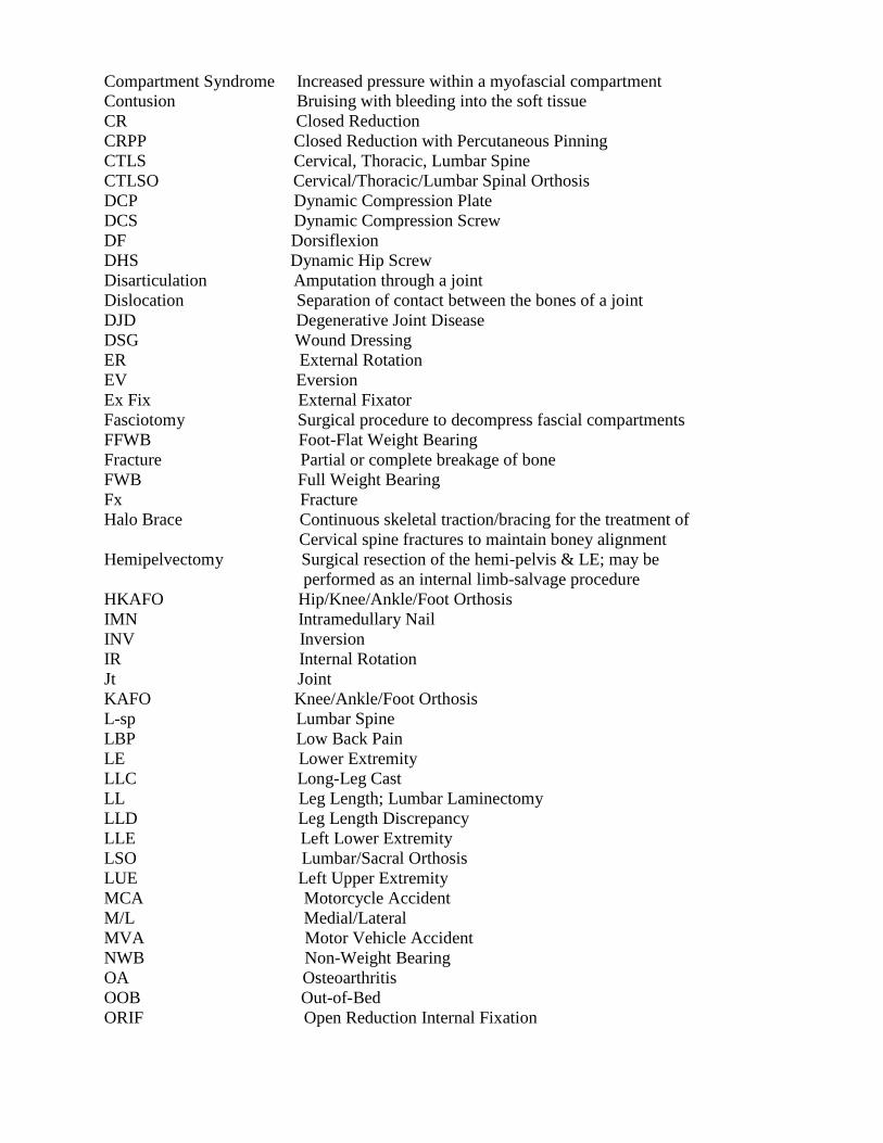
Compartment Syndrome Increased pressure within a myofascial compartment
Contusion Bruising with bleeding into the soft tissue
CR Closed Reduction
CRPP Closed Reduction with Percutaneous Pinning
CTLS Cervical, Thoracic, Lumbar Spine
CTLSO Cervical/Thoracic/Lumbar Spinal Orthosis
DCP Dynamic Compression Plate
DCS Dynamic Compression Screw
DF Dorsiflexion
DHS Dynamic Hip Screw
Disarticulation Amputation through a joint
Dislocation Separation of contact between the bones of a joint
DJD Degenerative Joint Disease
DSG Wound Dressing
ER External Rotation
EV Eversion
Ex Fix External Fixator
Fasciotomy Surgical procedure to decompress fascial compartments
FFWB Foot-Flat Weight Bearing
Fracture Partial or complete breakage of bone
FWB Full Weight Bearing
Fx Fracture
Halo Brace Continuous skeletal traction/bracing for the treatment of
Cervical spine fractures to maintain boney alignment
Hemipelvectomy Surgical resection of the hemi-pelvis & LE; may be
performed as an internal limb-salvage procedure
HKAFO Hip/Knee/Ankle/Foot Orthosis
IMN Intramedullary Nail
INV Inversion
IR Internal Rotation
Jt Joint
KAFO Knee/Ankle/Foot Orthosis
L-sp Lumbar Spine
LBP Low Back Pain
LE Lower Extremity
LLC Long-Leg Cast
LL Leg Length; Lumbar Laminectomy
LLD Leg Length Discrepancy
LLE Left Lower Extremity
LSO Lumbar/Sacral Orthosis
LUE Left Upper Extremity
MCA Motorcycle Accident
M/L Medial/Lateral
MVA Motor Vehicle Accident
NWB Non-Weight Bearing
OA Osteoarthritis
OOB Out-of-Bed
ORIF Open Reduction Internal Fixation
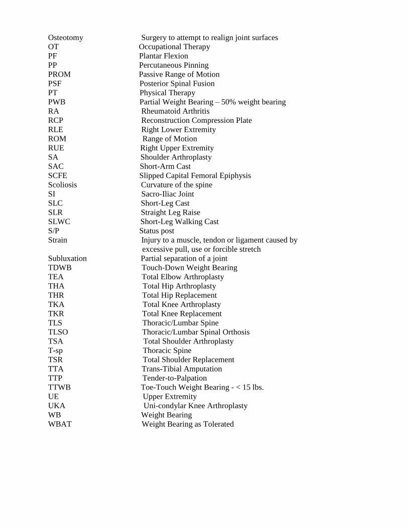
Osteotomy Surgery to attempt to realign joint surfaces
OT Occupational Therapy
PF Plantar Flexion
PP Percutaneous Pinning
PROM Passive Range of Motion
PSF Posterior Spinal Fusion
PT Physical Therapy
PWB Partial Weight Bearing – 50% weight bearing
RA Rheumatoid Arthritis
RCP Reconstruction Compression Plate
RLE Right Lower Extremity
ROM Range of Motion
RUE Right Upper Extremity
SA Shoulder Arthroplasty
SAC Short-Arm Cast
SCFE Slipped Capital Femoral Epiphysis
Scoliosis Curvature of the spine
SI Sacro-Iliac Joint
SLC Short-Leg Cast
SLR Straight Leg Raise
SLWC Short-Leg Walking Cast
S/P Status post
Strain Injury to a muscle, tendon or ligament caused by
excessive pull, use or forcible stretch
Subluxation Partial separation of a joint
TDWB Touch-Down Weight Bearing
TEA Total Elbow Arthroplasty
THA Total Hip Arthroplasty
THR Total Hip Replacement
TKA Total Knee Arthroplasty
TKR Total Knee Replacement
TLS Thoracic/Lumbar Spine
TLSO Thoracic/Lumbar Spinal Orthosis
TSA Total Shoulder Arthroplasty
T-sp Thoracic Spine
TSR Total Shoulder Replacement
TTA Trans-Tibial Amputation
TTP Tender-to-Palpation
TTWB Toe-Touch Weight Bearing - < 15 lbs.
UE Upper Extremity
UKA Uni-condylar Knee Arthroplasty
WB Weight Bearing
WBAT Weight Bearing as Tolerated
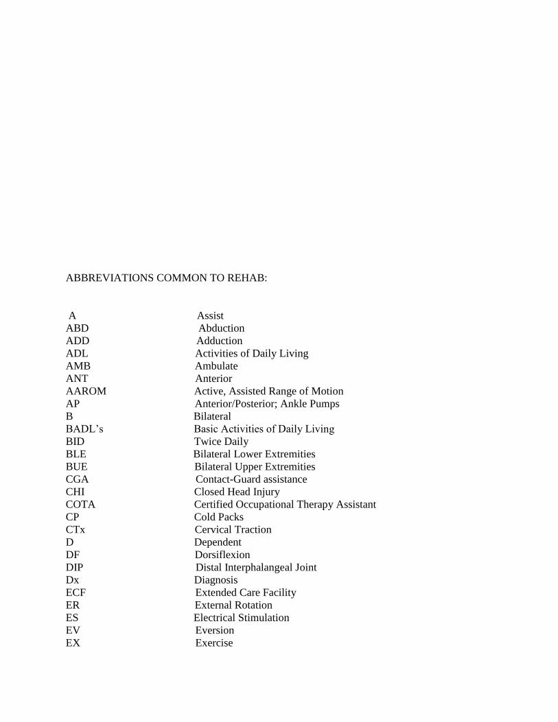
ABBREVIATIONS COMMON TO REHAB:
A Assist
ABD Abduction
ADD Adduction
ADL Activities of Daily Living
AMB Ambulate
ANT Anterior
AAROM Active, Assisted Range of Motion
AP Anterior/Posterior; Ankle Pumps
B Bilateral
BADL’s Basic Activities of Daily Living
BID Twice Daily
BLE Bilateral Lower Extremities
BUE Bilateral Upper Extremities
CGA Contact-Guard assistance
CHI Closed Head Injury
COTA Certified Occupational Therapy Assistant
CP Cold Packs
CTx Cervical Traction
D Dependent
DF Dorsiflexion
DIP Distal Interphalangeal Joint
Dx Diagnosis
ECF Extended Care Facility
ER External Rotation
ES Electrical Stimulation
EV Eversion
EX Exercise

/, EXT Extension
EVAL Evaluation
FES Functional Electrical Stimulation
, FL Flexion
FFWB Foot-Flat Weight Bearing
FWB Full Weight Bearing
Fx Fracture
GT Gait
HEP Home Exercise Program
HH Home Health
HHA Home Health Agency; Hand-Held Assistance
HI Home Instruction
HP Hot Pack
HS Hamstrings
HOB Head-of-Bed
I Independent
IADL’s Intermediate Activities of Daily Living
INV Inversion
IP Interphalangeal Joint
IR Internal Rotation
Jt Joint
LBQC Large-Based Quad Cane
LLE Left Lower Extremity
LTG Long-Term Goal
LUE Left Upper Extremity
Ms Muscle
MAX Maximal
MCP Metacarpal Phalangeal Joint
MH Moist Heat
MIN Minimal
MMT Manual Muscle Test
Mob Mobilization
MOD Moderate
MVT Movement
NH Nursing Home
NWB Non-Weight Bearing
OHF Overhead Frame
OOB Out-of-Bed
OT Occupational Therapy/Therapist
PIP Proximal Interphalangeal Joint
PF Plantar Flexion
PNF Proprioceptive Neuromuscular Facilitation
POST Posterior
PRE Progressive Resistive Exercises
PROM Passive Range of Motion
PROX Proximal
PT Physical Therapy/Therapist
PT Posterior Tibialis

PTA Physical Therapy Assistant
PTx Pelvic Traction
PWB Partial Weight Bearing
QD Every Day
QS Quadriceps; Quad Sets
R Right
REPS Repetitions
RLE Right Lower Extremity
ROM Range of Motion
RUE Right Upper Extremity
Rx Treatment; Prescription
SAQ Short-Arc Quads
SBA Stand-By Assistance
SBQC Small-Based Quad Cane
SCU Sub-Acute Unit
SLR Straight Leg Raise
SNF Skilled Nursing Facility
SP Speech Pathology
STG Short Term Goal
SW Social Work
TDWB Touch-Down Weight Bearing
THER EX Therapeutic Exercise
TENS Transcutaneous Electrical Nerve Stimulation
TID Three Times Daily
TT Transfer Training
TTWB Toe-Touch Weight Bearing
TX Traction; Treatment; Trauma
US Ultrasound
UV Ultraviolet
VMO Vastus Medialis Oblique
WB Weight Bearing
WBAT Weight Bearing as Tolerated
W/C, WC Wheelchair
WFL Within Functional Limits
WNL Within Normal Limits
WP Whirlpool
Wt Weight

Vanderbilt Psychiatric Hospital (VPH)
General Information
1. Call to VPH prior to walking to VPH patient in order to ensure patient availability
2. Chart review at VUMC
3. Know patient’s full name prior to visiting VPH
4. Wear your name badge
5. Sign in and out at VPH desk
6. You will be escorted to room and out after visit
7. If patient requires reacher/sockaid/cane/walker therapist can order, however, typically
requires equipment to be kept by nursing staff. If patient requires equipment after the MD
will write a script and outpatient
8. OT/PT not appropriate for patients who simply refuse to participate in activities or to get
out of bed. The patient needs to have specific skilled therapy acute care needs
9. Patient’s with developmental delays or diagnoses such as CP would be better served by a
pediatric clinician; the pediatric therapy pager is 835-1161
10. It is up to VPH to accept a patient for admission that has IPR/SNF recommendation
Coverage:
1. If an adult VPH patient has continued OT/PT needs, acute rehab OT/PT is responsible for
continued care of that patient
2. If a VPH patient comes in to the VOI and needs a splint, one of the acute care OTs needs
to make the splint; VPH is an inpatient facility; the OT will need to get a written order
from the MD; to document on the patient, the OT will have to search under “find a
patient” and add charting


