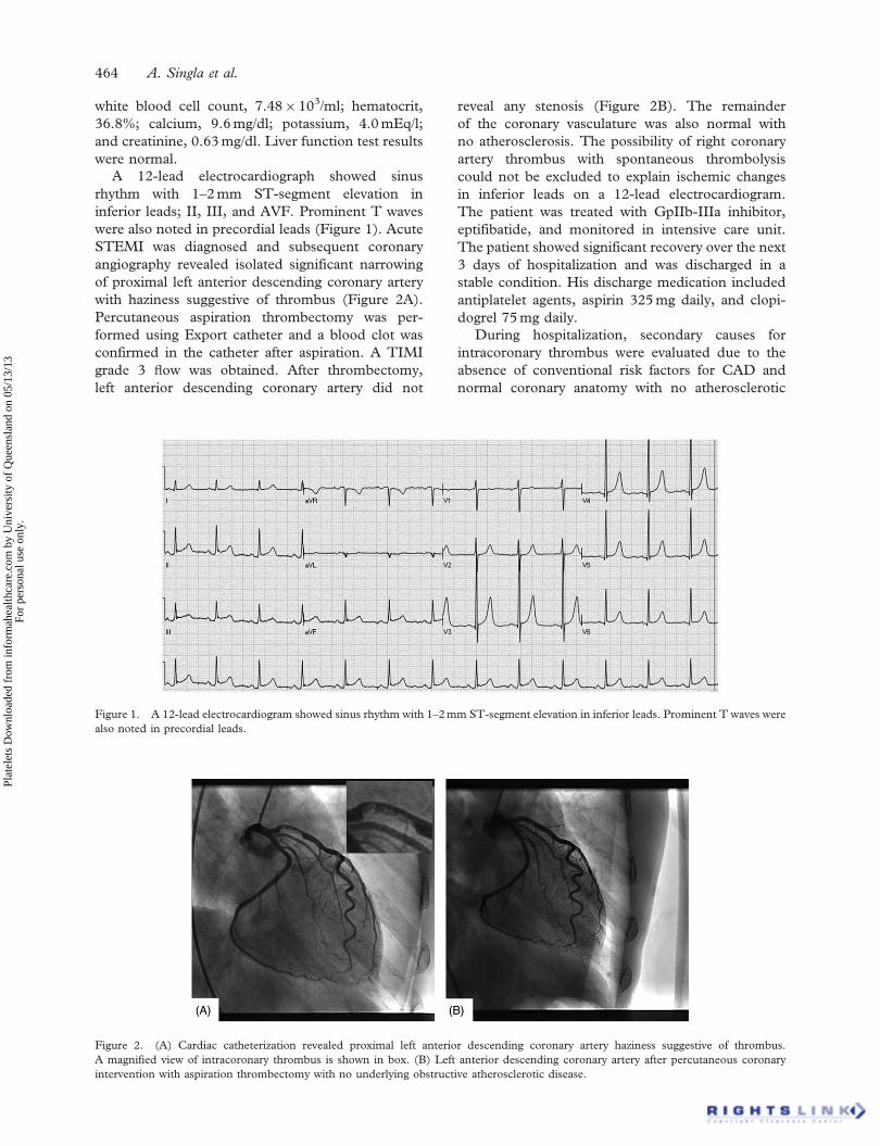
Platelets, September 2012; 23(6): 463–466
Copyright � 2012 Informa UK Ltd.
ISSN: 0953-7104 print/1369-1635 online
DOI: 10.3109/09537104.2011.640966
ORIGINAL ARTICLE
Acute ST-segment elevation myocardial infarction: A rare initialpresentation of previously undiagnosed essential thrombocythemia
ANAND SINGLA, DINESH JAGASIA, MUKESH GARG, PHILIP A LOWRY,
& DWIGHT STAPLETON
Guthrie Clinic, One Guthrie Square, Sayre, PA, USA
AbstractEssential thrombocythemia (ET) is a myeloproliferative disorder characterized by hemorrhagic and thromboticcomplications. We describe a rare case of ST-segment elevation myocardial infarction (STEMI) in a patient with previouslyundiagnosed ET, confirmed by gene mutation. A 68-year-old man presented with severe acute chest pain and was diagnosedwith STEMI. Primary coronary angiography showed severe stenosis with thrombus in the proximal left anterior descendingcoronary artery. Percutaneous aspiration thrombectomy was performed with no residual stenosis. The patient wasdischarged on antiplatelet agents, aspirin, and clopidogrel. Further investigations for intracoronary thrombus with nounderlying atherosclerotic disease revealed positive Janus kinase 2 (JAK2) V617F gene mutation, and this was consistentwith a diagnosis of ET with elevated platelet count. This case describes a rare initial presentation of previously undiagnosedET with acute STEMI and highlights the potential importance of secondary workup for non-atherosclerotic causes ofSTEMI with isolated intracoronary thrombus otherwise normal coronary vasculature with no focal atherosclerosis.
Keywords: Essential thrombocythemia, ST-segment elevation myocardial infarction
Introduction
Essential thrombocythemia (ET) is a myeloprolifera-
tive disorder characterized by pathological clonal
proliferation of megakaryocytes with persistently
elevated platelet count [1, 2]. The clinical manifesta-
tions of ET include hemorrhagic and thrombotic
complications. Recent studies have demonstrated
that thrombotic complications, especially thrombus
in the cerebral, coronary, and peripheral arteries,
are more frequent than hemorrhages in patients
with ET [3]. The incidence of acute coronary
events in patients with ET has been reported as
high as 9.4% [4].
In the medical literature, few cases of acute
myocardial infraction (AMI) have been reported in
association with ET. Here, we report a rare case of
ST-segment elevation myocardial infarction
(STEMI) as the first clinical presentation of pre-
viously undiagnosed ET, with positive Janus kinase 2
(JAK2) V617F gene mutation.
Case discussion
A 68-year-old Caucasian man with no significant
past medical history presented to the emergency
room reporting chest pain. His symptoms of severe,
crushing, non-radiating, and retrosternal chest pain
started 2 hours before he came to the emergency
room. He reported no history of cardiac problems,
was not taking any cardiac medications, and had no
family history suggesting any cardiac diseases.
Physical examination revealed moderate distress
and diaphoresis. His pulse was 88 beats per minute,
his blood pressure was 128/84 mm Hg, and his
respiratory rate was 18 breaths per minute with 98%
oxygen saturation on room air. Auscultation revealed
normal S1 and S2 heart sounds with no murmur or
gallop. His chest was clear to auscultation on both
sides. Cardiac markers suggested cardiac ischemia;
troponin I peaked at 3.13 ng/ml. He had thrombo-
cytosis at the time of admission (platelet
count¼ 539� 103/l). Other laboratory values were
Correspondence: Anand Singla, Fellow, Cardiovascular Diseases, Department of Cardiology, Guthrie Clinic/Robert Packer Hospital, One Guthrie
Square, Sayre, PA 18840, USA. Tel: 570-882-2331. Fax: 570-887-2290. E-mail: [email protected]
(received 5 October 2011; revised 28 October 2011; accepted 9 November 2011)
Plat
elet
s D
ownl
oade
d fr
om in
form
ahea
lthca
re.c
om b
y U
nive
rsity
of
Que
ensl
and
on 0
5/13
/13
For
pers
onal
use
onl
y.
white blood cell count, 7.48� 103/ml; hematocrit,
36.8%; calcium, 9.6 mg/dl; potassium, 4.0 mEq/l;
and creatinine, 0.63 mg/dl. Liver function test results
were normal.
A 12-lead electrocardiograph showed sinus
rhythm with 1–2 mm ST-segment elevation in
inferior leads; II, III, and AVF. Prominent T waves
were also noted in precordial leads (Figure 1). Acute
STEMI was diagnosed and subsequent coronary
angiography revealed isolated significant narrowing
of proximal left anterior descending coronary artery
with haziness suggestive of thrombus (Figure 2A).
Percutaneous aspiration thrombectomy was per-
formed using Export catheter and a blood clot was
confirmed in the catheter after aspiration. A TIMI
grade 3 Fow was obtained. After thrombectomy,
left anterior descending coronary artery did not
reveal any stenosis (Figure 2B). The remainder
of the coronary vasculature was also normal with
no atherosclerosis. The possibility of right coronary
artery thrombus with spontaneous thrombolysis
could not be excluded to explain ischemic changes
in inferior leads on a 12-lead electrocardiogram.
The patient was treated with GpIIb-IIIa inhibitor,
eptifibatide, and monitored in intensive care unit.
The patient showed significant recovery over the next
3 days of hospitalization and was discharged in a
stable condition. His discharge medication included
antiplatelet agents, aspirin 325 mg daily, and clopi-
dogrel 75 mg daily.
During hospitalization, secondary causes for
intracoronary thrombus were evaluated due to the
absence of conventional risk factors for CAD and
normal coronary anatomy with no atherosclerotic
Figure 2. (A) Cardiac catheterization revealed proximal left anterior descending coronary artery haziness suggestive of thrombus.
A magnified view of intracoronary thrombus is shown in box. (B) Left anterior descending coronary artery after percutaneous coronary
intervention with aspiration thrombectomy with no underlying obstructive atherosclerotic disease.
Figure 1. A 12-lead electrocardiogram showed sinus rhythm with 1–2 mm ST-segment elevation in inferior leads. Prominent T waves were
also noted in precordial leads.
464 A. Singla et al.
Plat
elet
s D
ownl
oade
d fr
om in
form
ahea
lthca
re.c
om b
y U
nive
rsity
of
Que
ensl
and
on 0
5/13
/13
For
pers
onal
use
onl
y.
lesions after the thrombus was removed.
Transthoracic echocardiogram did not reveal any
evidence of intracardiac thrombus. Left ventricular
systolic function was within normal limits with no
wall motion abnormalities. The possibility of para-
doxical embolization with patent foramen ovale was
ruled out with negative bubble study on transthoracic
echocardiogram. Investigations for hypercoagulabil-
ity demonstrated a heterozygous mutation for
Factor V Leiden, otherwise a normal profile includ-
ing Anti thrombin III, lupus anticoagulant, antic-
ardiolipin antibody, protein C, protein S, and
prothrombin mutation. The platelet counts remained
high while in hospital.
During follow-up visit as outpatient at 1 month,
our patient showed persistent elevation of platelet
count with one episode of platelet count more than
600� 103/l. A possible diagnosis of ET was con-
sidered. Genetic studies were positive for the JAK2
V617F gene mutation. As our patient had high risk
factors for thrombotic complications, treatment
with cytoreductive therapy using hydroxyurea was
instituted to lower the platelet count though our
patient was asymptomatic. Six months after his index
hospitalization, patient continues to do well with
no further thrombotic or hemorrhagic complications.
Discussion
Normal coronary angiogram in patients with AMI is
not a rare presentation, and accounts for 3% of the
total cases [5]. Multiple etiological factors have been
reported including coronary spasm, coagulation
disorders, collagen vascular diseases, embolization,
and oral contraceptive use [6]. In our case, ET with
JAK2 V617F mutation initially presented with
acute STEMI secondary to intracoronary thrombus
formation, otherwise normal coronary arteries, and
required percutaneous thrombectomy with success-
ful revascularization.
ET is a myeloproliferative clonal disorder result-
ing from the transformation of pluripotent hemato-
poietic cells [1, 2]. The incidence rate for ET is
2.5 cases per 100� 103 populations per year [7].
Approximately, half of the patients diagnosed with
ET are asymptomatic and others commonly have
vasomotor symptoms. However, ET is associated
with increased risk for thrombotic and hemorrhagic
complications causing significant morbidity and
mortality. Both thrombosis and hemorrhage result
from qualitative and quantitative defects of the
platelets [8]. After the initial diagnosis of ET,
a total of 7–17% thrombotic event rates have been
reported over a follow-up period of 3–7 years [9, 10].
Thrombotic complications included both arterial and
venous events; stroke, transient ischemic attacks,
retinal artery or venous occlusions, coronary artery
ischemia, pulmonary embolism, hepatic or portal
vein thrombosis, deep vein thrombosis, and digital
ischemia.
The association of coronary thrombotic events
with ET is well established [11–14]. In the last two
decades, multiple risk factors have been identified
to predict ET-related thrombotic complications.
Age more than 60, previous history of thrombotic
complications, and platelet count41500� 109/l were
reported as high risk factors for thrombotic compli-
cations [15]. Hydroxyurea is the treatment of choice
to prevent thrombotic events in ET for high-risk
individuals with age460 [16]. De Stefano et al. [17]
demonstrated a significant reduction of 70% in
recurrent thrombotic events in patients with poly-
cythemia vera or ET who had acute coronary
syndrome as the index thrombosis. Furthermore,
the contemporary use of an antiplatelet agent with
cytoreductive therapy showed enhanced efficacy
in preventing re-thrombosis. Though in this case,
patient had platelet counts less than 600� 109/l,
treatment with hydroxyurea was instituted in addi-
tion to an antiplatelet agent (aspirin 81 mg daily) with
history of recent coronary thrombotic event and age
factor. There are several reports of complications at
platelet counts lower than 600� 109/l in patients
diagnosed with ET. In a report by Regev et al. [18],
severe thrombotic complications were reported at
platelet counts lower than 600� 109/l in sympto-
matic patients with ET. It was recommended that
symptomatic patients with ET and relatively low
platelet counts should be treated with target platelet
counts well into the lower normal range.
The JAK2 somatic mutation is closely related to
chronic myeloproliferative disorders and more than
half of individuals with ET exhibit JAK2 V617F
mutation [19]. However, JAK2 V617F-mutated
patients are not predisposed to increased incidence
of thrombotic events relative to JAK2 wild type [15].
Furthermore, increased sensitivity to cytoreductive
therapy using hydroxyurea has been reported in
this group [19]. In our case, positive JAK2 V617F
mutation confirmed the diagnosis for ET after the
initial presentation with acute STEMI. Our patient
was also reported to have Factor V heterozygous
mutation, known to be associated with a 3–6-fold
increase in the risk of venous thrombosis [20]. There
is no association of Factor V heterozygous mutation
with arterial thrombosis. Multiple studies have
concluded that patients with this mutation alone do
not have increased risk of AMI or stroke [21].
Factor V heterozygous mutation is not an uncom-
mon pathology with prevalence rate of 4–6% in
general population [20, 22]. We believe that this
finding in our case is incidental in nature and
unrelated to current presentation.
Acute ST-segment elevation myocardial infarction 465
Plat
elet
s D
ownl
oade
d fr
om in
form
ahea
lthca
re.c
om b
y U
nive
rsity
of
Que
ensl
and
on 0
5/13
/13
For
pers
onal
use
onl
y.
Conclusions
Our case describes a rare initial presentation of
previously undiagnosed ET with acute STEMI.
It highlights the importance of secondary workup
for STEMI with isolated intracoronary thrombus
otherwise normal coronary vasculature with no focal
atherosclerosis. Though a rare cause, ET should
be considered as a differential for a patient
presenting with persistently elevated platelet count
and a thrombus in a patent coronary artery.
Declaration of interest: There are no potential
conflicts of interest to be disclosed.
References
1. Fialkow PJ, Faguet GB, Jacobson RJ, Vaidya K, Murphy S.
Evidence that essential thrombocythemia is a clonal disorder
with origin in a multipotent stem cell. Blood 1981;58:
916–919.
2. Campbell PJ, Green AR. The myeloproliferative disorders.
N Engl J Med 2006;355:2452–2466.
3. Mizuta E, Takeda S, Sasaki N, Miake J, Hamada T,
Shimoyama M, Tajima F, Igawa O, Shigemasa C,
Hisatome I. Acute myocardial infarction in a patient with
essential thrombocythemia: Successful treatment with percuta-
neous transluminal coronary recanalization. Circ J 2005;69:
1000–1002.
4. Rossi C, Randi ML, Zerbinati P, Rinaldi V, Girolami A. Acute
coronary disease in essential thrombocythemia and polycythe-
mia vera. J Intern Med 1998;244:49–53.
5. Raymond R, Lynch J, Underwood D, Leatherman J, Razavi M.
Myocardial infarction and normal coronary arteriography: A 10
year clinical and risk analysis of 74 patients. J Am Coll Cardiol
1988;11:471–477.
6. Da Costa A, Isaaz K, Faure E, Mourot S, Cerisier A,
Lamaud M. Clinical characteristics, aetiological factors and
long-term prognosis of myocardial infarction with an absolutely
normal coronary angiogram; a 3-year follow-up study of 91
patients. Eur Heart J 2001;22:1459–1465.
7. Mesa RA, Silverstein MN, Jacobsen SJ, Wollan PC, Tefferi A.
Population-based incidence and survival figures in essential
thrombocythemia and agnogenic myeloid metaplasia:
An Olmsted County Study, 1976–1995. J Hematol 1999;
61:10–15.
8. Schafer AI. Molecular basis of the diagnosis and treatment of
polycythemia Vera and essential thrombocythemia. Blood
2006;107:4214–4222.
9. Cortelazzo S, Viero P, Finazzi G, D’Emilio A, Rodeghiero F,
Barbui T. Incidence and risk factors for thrombotic
complications in a historical cohort of 100 patients with
essential thrombocythemia. J Clin Oncol 1990;8:556–562.
10. Fenaux P, Simon M, Caulier MT, Lai JL, Goudemand J,
Bauters F. Clinical course of essential thrombocythemia in
147 cases. Cancer 1990;66:549–556.
11. Gul C, Kurum T, Demir M, Ozbay G, Vural O, Iqbal O,
Fareed J. Acute myocardial infarction in a patient with
essential thrombocythemia treated with glycoprotein IIb/IIIa
inhibitor. Clin Appl Thromb Hemost 2004;10:77–79.
12. Daya SK, Gowda RM, Landis WA, Khan IA. Essential
thrombocythemia-related acute ST-segment elevation
myocardial infarction. A case report and literature review.
Angiology 2004;55:319–323.
13. Watanabe T, Fujinaga H, Ikeda Y, Higashi T, Murakami M,
Kawahara K, Hayashi I, Niki T, Shigekiyo T, Wakatsuki T.
Acute myocardial infarction in a patient with essential
thrombocythemia who underwent successful stenting: A case
report. Angiology 2005;56:771–774.
14. Bildirici U, Celikyurt U, Ural E. Essential thrombocythemia:
A case of acute ST-segment elevation myocardial infarction in
a young female. Clin Cardiol 2009;32:104–105.
15. Griesshammer M. Risk factors for thrombosis and bleeding
and their influence on therapeutic decisions in patients with
essential thrombocythemia. Semin Thromb Hemost 2006;32:
372–380.
16. Cortelazzo S, Finazzi G, Ruggeri M, Vestri O, Galli M,
Rodeghiero F, Barbui T. Hydroxyurea for patients with
essential thrombocythemia and high risk of thrombosis.
N Engl J Med 1995;332:1132–1136.
17. De Stefano V, Za T, Rossi E, Vannucchi AM, Ruggeri M,
Elli E, Mico C, Tieghi A, Cacciola RR, Santoro C, et al.
Recurrent thrombosis in patients with polycythemia vera and
essential thrombocythemia: Incidence, risk factors, and effect
of treatments. Haematologica 2008;93:372–380.
18. Regev A, Stark P, Blickstein D, Lahav M. Thrombotic
complications in essential thrombocythemia with relatively
low platelet counts. Am J Hematol 1997;56:168–172.
19. Campbell PJ, Scott LM, Buck G, Wheatley K, East CL,
Marsden JT, Duffy A, Boyd EM, Bench AJ, Scott MA, et al.
DeEnition of subtypes of essential thrombocythaemia and
relation to polycythaemia vera based on JAK2 V617F
mutation status: A prospective study. Lancet 2005;366:
1945–1953.
20. Price DT, Ridker PM. Factor V Leiden mutation and the risks
for thromboembolic disease: A clinical perspective. Ann
Intern Med 1997;127:895–903.
21. Ridker PM, Hennekens CH, Lindpaintner K, Stampfer MJ,
Eisenberg PR, Miletich JP. Mutation in the gene coding for
coagulation factor V and the risk of myocardial infraction,
stroke, and venous thrombosis in apparently healthy men.
N Engl J Med 1995;332:912–917.
22. Ridker PM, Miletich JP, Hennekens CH, Buring JE. Ethnic
distribution of factor V Leiden in 4047 men and women.
Implication for venous thromboembolism screening. JAMA
1997;277:1305–1307.
466 A. Singla et al.
Plat
elet
s D
ownl
oade
d fr
om in
form
ahea
lthca
re.c
om b
y U
nive
rsity
of
Que
ensl
and
on 0
5/13
/13
For
pers
onal
use
onl
y.