Acute upper gastrointestinal haemorrhage Kelvin Palmer* GI Unit, Western General Hospital, Edinburgh, UK Acute gastrointestinal haemorrhage is a common medical emergency that has a hospital mortality of approximately 10%. Peptic ulcer bleeding, complicating non-steroidal anti-inflammatory drugs, aspirin or Helicobacter pylori infection is the most common cause of major bleeding. Gastro-oesophageal varices are less common but managing the underlying liver disease and the severity of bleeding may be demanding. The prognosis of patients presenting with acute bleeding is dictated by the presence of medical co-morbidities and by the severity of liver disease in patients with varices. Validated prognostic scoring systems, based upon the severity of bleeding, diagnosis, endoscopic findings and extent of co-morbidities, predict mortality and have clinical utility. The treatment of non-variceal bleeding is based upon cardiovascular resuscitation followed by endoscopic therapy in patients with active bleeding or major stigmata of recent haemorrhage. Proton pump inhibitor drugs reduce the risk of re-bleeding but have little effect on mortality. Emergency surgery is undertaken for uncontrolled bleeding or re-bleeding that cannot be controlled by further endoscopic therapy. Oesophageal varices are managed by fluid resuscitation, antibiotics and endoscopic band ligation. Vasoactive drugs may stop active bleeding but have no effect upon mortality. Management of the complications of the underlying liver disease and complete variceal ablation in a banding programme are essential. Gastric varices are treated by injection with tissue adhesives or transjugular intrahepatic porto-systemic shunt (TIPSS) insertion. Surgical intervention has little role in the management of varices and patients who do not respond to endoscopic therapies are best treated by TIPSS. Keywords: gastrointestinal bleeding/endoscopy/bleeding ulcer/varices/proton pump inhibitor drugs Introduction The incidence of acute upper gastrointestinal haemorrhage in the UK ranges 50 –190/10 000/year and is highest in areas of social depri- vation. In Hong Kong, the incidence has decreased by 30% over the last 10 years. In contrast, the number of admissions for bleeding is stable or slightly increasing in elderly patients in the UK. The preva- lence of Helicobacter pylori, use of non-steroidal anti-inflammatory drugs (NSAIDs) and prevalence of liver disease are important factors. 1 British Medical Bulletin 2007; 83: 307–324 & The Author 2007. Published by Oxford University Press. DOI: 10.1093/bmb/ldm023 All rights reserved. For permissions, please e-mail: [email protected]Accepted: August 23, 2007 *Correspondence to: K. Palmer, GI Unit, Western General Hospital, Edinburgh, UK. E-mail: Kelvin.Palmer@ luht.scot.nhs.uk Downloaded from https://academic.oup.com/bmb/article/83/1/307/386560 by guest on 03 December 2021
Transcript
Acute upper gastrointestinal haemorrhage
Kelvin Palmer*
GI Unit, Western General Hospital, Edinburgh, UK
Acute gastrointestinal haemorrhage is a common medical emergency that has a
hospital mortality of approximately 10%. Peptic ulcer bleeding, complicating
non-steroidal anti-inflammatory drugs, aspirin or Helicobacter pylori infection is
the most common cause of major bleeding. Gastro-oesophageal varices are less
common but managing the underlying liver disease and the severity of bleeding
may be demanding. The prognosis of patients presenting with acute bleeding is
dictated by the presence of medical co-morbidities and by the severity of liver
disease in patients with varices. Validated prognostic scoring systems, based
upon the severity of bleeding, diagnosis, endoscopic findings and extent of
co-morbidities, predict mortality and have clinical utility. The treatment of
non-variceal bleeding is based upon cardiovascular resuscitation followed by
endoscopic therapy in patients with active bleeding or major stigmata of recent
haemorrhage. Proton pump inhibitor drugs reduce the risk of re-bleeding but
have little effect on mortality. Emergency surgery is undertaken for uncontrolled
bleeding or re-bleeding that cannot be controlled by further endoscopic
therapy. Oesophageal varices are managed by fluid resuscitation, antibiotics and
endoscopic band ligation. Vasoactive drugs may stop active bleeding but have
no effect upon mortality. Management of the complications of the underlying
liver disease and complete variceal ablation in a banding programme are
essential. Gastric varices are treated by injection with tissue adhesives or
The incidence of acute upper gastrointestinal haemorrhage in the UKranges 50–190/10 000/year and is highest in areas of social depri-vation. In Hong Kong, the incidence has decreased by 30% over thelast 10 years. In contrast, the number of admissions for bleeding isstable or slightly increasing in elderly patients in the UK. The preva-lence of Helicobacter pylori, use of non-steroidal anti-inflammatorydrugs (NSAIDs) and prevalence of liver disease are important factors.1
British Medical Bulletin 2007; 83: 307–324 & The Author 2007. Published by Oxford University Press.
DOI: 10.1093/bmb/ldm023 All rights reserved. For permissions, please e-mail: [email protected]
Accepted: August 23,
2007
*Correspondence to:
K. Palmer,
GI Unit, Western General
Hospital, Edinburgh, UK.
E-mail: Kelvin.Palmer@
luht.scot.nhs.uk
Dow
nloaded from https://academ
ic.oup.com/bm
b/article/83/1/307/386560 by guest on 03 Decem
ber 2021
The mortality of patients admitted to hospital for acute gastrointesti-nal bleeding is about 10%, rising to more than 30% in patients whobleed as inpatients. In the UK, crude mortality has not changed inmore than half a century although the case mix has changed greatlyover this time, and patients are now older and have greater medical dis-ability than was the case 50 years ago.1
Diagnosis and aetiology
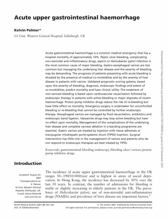
Peptic ulcer is the most frequent cause of major, life-threatening acutegastrointestinal bleeding (Fig. 1) (Table 1). Significant haemorrhageresults from erosion of an underlying artery (Fig. 2), and the magnitudeof bleeding is related to the size of the arterial defect and the diameterof the artery. Consequently, bleeding may be particularly severe fromlarge, posterior duodenal ulcers which erode the gastroduodenal arteryand high, lesser curve gastric ulcers involving branches of the leftgastric artery. Most patients present with little or no history of dyspep-sia. A history of aspirin or NSAID use is common.
Fig. 1 Endoscopic view of an actively bleeding posterior duodenal artery.
K. Palmer
308 British Medical Bulletin 2007;83
Dow
nloaded from https://academ
ic.oup.com/bm
b/article/83/1/307/386560 by guest on 03 Decem
ber 2021
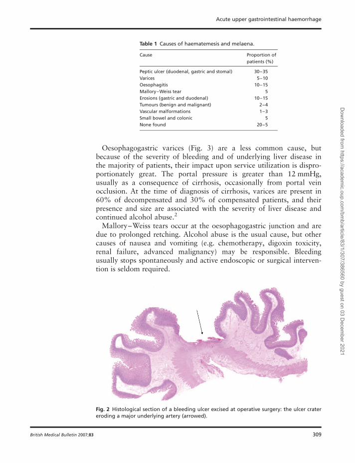
Oesophagogastric varices (Fig. 3) are a less common cause, butbecause of the severity of bleeding and of underlying liver disease inthe majority of patients, their impact upon service utilization is dispro-portionately great. The portal pressure is greater than 12 mmHg,usually as a consequence of cirrhosis, occasionally from portal veinocclusion. At the time of diagnosis of cirrhosis, varices are present in60% of decompensated and 30% of compensated patients, and theirpresence and size are associated with the severity of liver disease andcontinued alcohol abuse.2
Mallory–Weiss tears occur at the oesophagogastric junction and aredue to prolonged retching. Alcohol abuse is the usual cause, but othercauses of nausea and vomiting (e.g. chemotherapy, digoxin toxicity,renal failure, advanced malignancy) may be responsible. Bleedingusually stops spontaneously and active endoscopic or surgical interven-tion is seldom required.
Table 1 Causes of haematemesis and melaena.
Cause Proportion of
patients (%)
Peptic ulcer (duodenal, gastric and stomal) 30–35
Varices 5–10
Oesophagitis 10–15
Mallory–Weiss tear 5
Erosions (gastric and duodenal) 10–15
Tumours (benign and malignant) 2–4
Vascular malformations 1–3
Small bowel and colonic 5
None found 20–5
Fig. 2 Histological section of a bleeding ulcer excised at operative surgery: the ulcer cratereroding a major underlying artery (arrowed).
Acute upper gastrointestinal haemorrhage
British Medical Bulletin 2007;83 309
Dow
nloaded from https://academ
ic.oup.com/bm
b/article/83/1/307/386560 by guest on 03 Decem
ber 2021
Oesophagitis is common in elderly patients presenting with ‘coffee-ground’ haematemesis. Bleeding is seldom life-threatening; in mostcases, conservative supportive therapy combined with proton pumpinhibitor drugs is all that is necessary. However, it is important to beaware that, in this group of patients, coffee-ground vomiting may haveanother cause (drug toxicity, underlying renal or cardiac failure, pan-creatitis or colon cancer), even when oesophagitis is proved at endo-scopy. Although the natural history of the bleeding event may bebenign, the prognosis is dependent upon underlying causes.
Gastritis, duodenitis and gastroduodenal erosions are associated withNSAIDs and H. pylori infection. In most patients, supportive therapyand cessation of NSAID use or H. pylori eradication therapy achieve afavourable outcome.
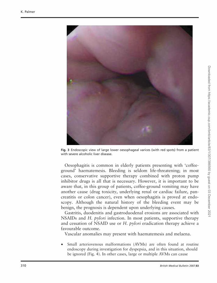
Vascular anomalies may present with haematemesis and melaena.
† Small arteriovenous malformations (AVMs) are often found at routineendoscopy during investigation for dyspepsia, and in this situation, shouldbe ignored (Fig. 4). In other cases, large or multiple AVMs can cause
Fig. 3 Endoscopic view of large lower oesophageal varices (with red spots) from a patientwith severe alcoholic liver disease.
K. Palmer
310 British Medical Bulletin 2007;83
Dow
nloaded from https://academ
ic.oup.com/bm
b/article/83/1/307/386560 by guest on 03 Decem
ber 2021
significant bleeding. This usually leads to insidious development of iron-deficiency anaemia, but occasionally, major acute haemorrhage occurs.Most AVMs have no obvious cause and present in elderly patients; inyounger patients, they are sometimes caused by hereditary haemorrhagictelangiectasia. Other patients have valvular heart disease or an artificialheart valve, and bleeding may be exacerbated by anticoagulant drugs.
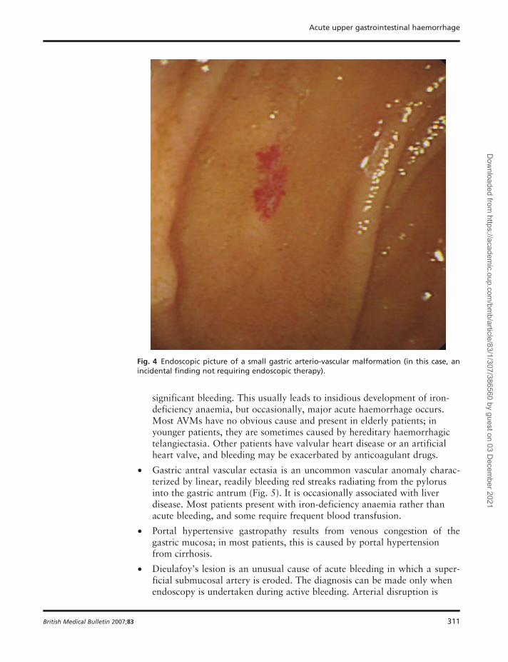
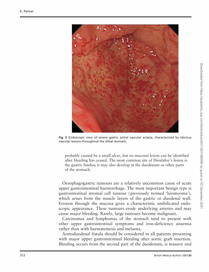
† Gastric antral vascular ectasia is an uncommon vascular anomaly charac-terized by linear, readily bleeding red streaks radiating from the pylorusinto the gastric antrum (Fig. 5). It is occasionally associated with liverdisease. Most patients present with iron-deficiency anaemia rather thanacute bleeding, and some require frequent blood transfusion.
† Portal hypertensive gastropathy results from venous congestion of thegastric mucosa; in most patients, this is caused by portal hypertensionfrom cirrhosis.
† Dieulafoy’s lesion is an unusual cause of acute bleeding in which a super-ficial submucosal artery is eroded. The diagnosis can be made only whenendoscopy is undertaken during active bleeding. Arterial disruption is
Fig. 4 Endoscopic picture of a small gastric arterio-vascular malformation (in this case, anincidental finding not requiring endoscopic therapy).
Acute upper gastrointestinal haemorrhage
British Medical Bulletin 2007;83 311
Dow
nloaded from https://academ
ic.oup.com/bm
b/article/83/1/307/386560 by guest on 03 Decem
ber 2021
probably caused by a small ulcer, but no mucosal lesion can be identifiedafter bleeding has ceased. The most common site of Dieulafoy’s lesion isthe gastric fundus; it may also develop in the duodenum or other partsof the stomach.
Oesophagogastric tumours are a relatively uncommon cause of acuteupper gastrointestinal haemorrhage. The most important benign type isgastrointestinal stromal cell tumour (previously termed ‘leiomyoma’),which arises from the muscle layers of the gastric or duodenal wall.Erosion through the mucosa gives a characteristic umbilicated endo-scopic appearance. These tumours erode underlying arteries and maycause major bleeding. Rarely, large tumours become malignant.
Carcinomas and lymphomas of the stomach tend to present withother upper gastrointestinal symptoms and iron-deficiency anaemiarather than with haematemesis and melaena.
Aortoduodenal fistula should be considered in all patients presentingwith major upper gastrointestinal bleeding after aortic graft insertion.Bleeding occurs from the second part of the duodenum, is massive and
Fig. 5 Endoscopic view of severe gastric antral vascular ectasia, characterized by obviousvascular lesions throughout the distal stomach.
K. Palmer
312 British Medical Bulletin 2007;83
Dow
nloaded from https://academ
ic.oup.com/bm
b/article/83/1/307/386560 by guest on 03 Decem
ber 2021
may recur over hours or days. All such patients should be referred to avascular unit immediately after initial resuscitation.
Small bowel or right-sided colonic disease sometimes presents withmelaena and rarely with haematemesis. Colonoscopy, barium radi-ology, capsule endoscopy and enteroscopy are used to identify theunderlying tumour or vascular anomaly when upper gastrointestinalendoscopy fails to identify a bleeding source. In young patients, ableeding Meckel’s diverticulum should be considered.
Risk assessment
At the time of first assessment, it is important to identify patients whohave significant liver disease. Patients with liver disease are bestmanaged by gastroenterologists (or hepatologists) at presentation. Mostpatients will have a history of alcohol abuse or exposure to hepatotoxicviruses and have clinical evidence of liver disease and abnormal serumliver function tests.
Patients without liver disease
Death following admission to hospital for gastrointestinal bleeding isalmost invariably a consequence of decompensated co-morbidity; it isseldom caused by ex-sanguination. Sudden blood loss and circulatorycollapse may result in fatal cardiac or cerebrovascular events in patientswith underlying vascular disease, and postoperative complications fol-lowing emergency surgery are more likely in the presence of medicalco-morbidity. Therefore, risk assessment is based on the severity of thehaemorrhage and medical co-morbidity.
When patients present with acute upper gastrointestinal haemor-rhage, it is crucial to define factors with prognostic value. Those athigh risk of continuing bleeding or re-bleeding need intensive monitor-ing and early endoscopic intervention, whereas low-risk patientsshould be ‘fast-tracked’ towards early hospital discharge.
Re-bleeding is associated with a 10-fold increase in hospital mor-tality. In clinical trials, it is often used as an end-point for definingsuccess or failure of putative treatments. Mortality is particularly highin patients who bleed during a hospital stay for another serious disease(about 40% in published series, compared with 10–12% in patientsadmitted for gastrointestinal bleeding).
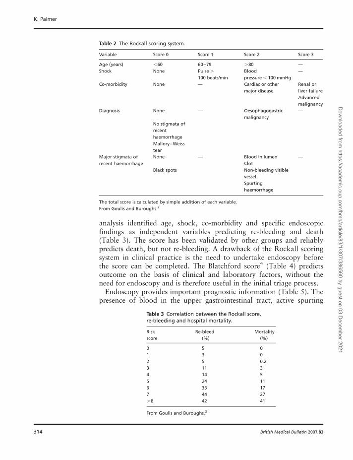
The Rockall score3 (Table 2) is a useful risk assessment tool. It wasdeveloped from a large audit of patients admitted to hospitals inEngland for acute upper gastrointestinal bleeding. Multi-variant
Acute upper gastrointestinal haemorrhage
British Medical Bulletin 2007;83 313
Dow
nloaded from https://academ
ic.oup.com/bm
b/article/83/1/307/386560 by guest on 03 Decem
ber 2021
analysis identified age, shock, co-morbidity and specific endoscopicfindings as independent variables predicting re-bleeding and death(Table 3). The score has been validated by other groups and reliablypredicts death, but not re-bleeding. A drawback of the Rockall scoringsystem in clinical practice is the need to undertake endoscopy beforethe score can be completed. The Blatchford score4 (Table 4) predictsoutcome on the basis of clinical and laboratory factors, without theneed for endoscopy and is therefore useful in the initial triage process.
Endoscopy provides important prognostic information (Table 5). Thepresence of blood in the upper gastrointestinal tract, active spurting
Table 2 The Rockall scoring system.
Variable Score 0 Score 1 Score 2 Score 3
Age (years) ,60 60–79 .80 —
Shock None Pulse .
100 beats/min
Blood
pressure , 100 mmHg
—
Co-morbidity None — Cardiac or other
major disease
Renal or
liver failure
Advanced
malignancy
Diagnosis None — Oesophagogastric
malignancy
—
No stigmata of
recent
haemorrhage
Mallory–Weiss
tear
Major stigmata of
recent haemorrhage
None — Blood in lumen —
Clot
Black spots Non-bleeding visible
vessel
Spurting
haemorrhage
The total score is calculated by simple addition of each variable.
From Goulis and Buroughs.2
Table 3 Correlation between the Rockall score,re-bleeding and hospital mortality.
Risk
score
Re-bleed
(%)
Mortality
(%)
0 5 0
1 3 0
2 5 0.2
3 11 3
4 14 5
5 24 11
6 33 17
7 44 27
.8 42 41
From Goulis and Buroughs.2
K. Palmer
314 British Medical Bulletin 2007;83
Dow
nloaded from https://academ
ic.oup.com/bm
b/article/83/1/307/386560 by guest on 03 Decem
ber 2021
haemorrhage and a ‘non-bleeding visible vessel’ are signs of poor prog-nosis. Active ulcer bleeding implies an 80–90% risk of continuinghaemorrhage or re-bleeding. A visible vessel (representing adherentblood clot or a pseudo-aneurysm over the arterial defect) is associatedwith a 50% risk of re-bleeding during that hospital stay (Fig. 5).5
Therapeutic endoscopists attempt to wash the bleeding point vigor-ously to display these major endoscopic stigmata of recent haemor-rhage, using washing catheters and snares to remove blood clot. Thesemanoeuvres risk provoking further bleeding, but this can usually bemanaged by one of the techniques described in what follows.Sometimes, the clot cannot be removed, and the presence of non-adherent blood clot carries an intermediate risk of further bleeding.
Table 4 The Blatchford scoring system.
Variable Score
Urea (mmol/l)
. 6.5, 8.0 2
. 8 , 10 3
. 10 , 25 4
. 25 6
Haemoglobin (g/dl)
Men
. 12 , 13 1
. 10 , 12 3
, 10 6
Women
. 12 , 13 1
, 10 6
Systolic BP (mmHg)
100–109 1
90–99 2
, 90 3
Pulse . 100 1
Melaena 1
Syncope 2
Liver disease 2
Cardiac failure 2
From Rockall et al.3
Table 5 Endoscopic stigmata and the risk of re-bleeding.
Endoscopic finding Risk of
re-bleeding (%)
Clean base 3
Flat spots 7
Oozing only 10
Adherent clot 33
Non-bleeding visible
vessel
50
Active bleeding 90
Acute upper gastrointestinal haemorrhage
British Medical Bulletin 2007;83 315
Dow
nloaded from https://academ
ic.oup.com/bm
b/article/83/1/307/386560 by guest on 03 Decem
ber 2021
Patients with liver disease
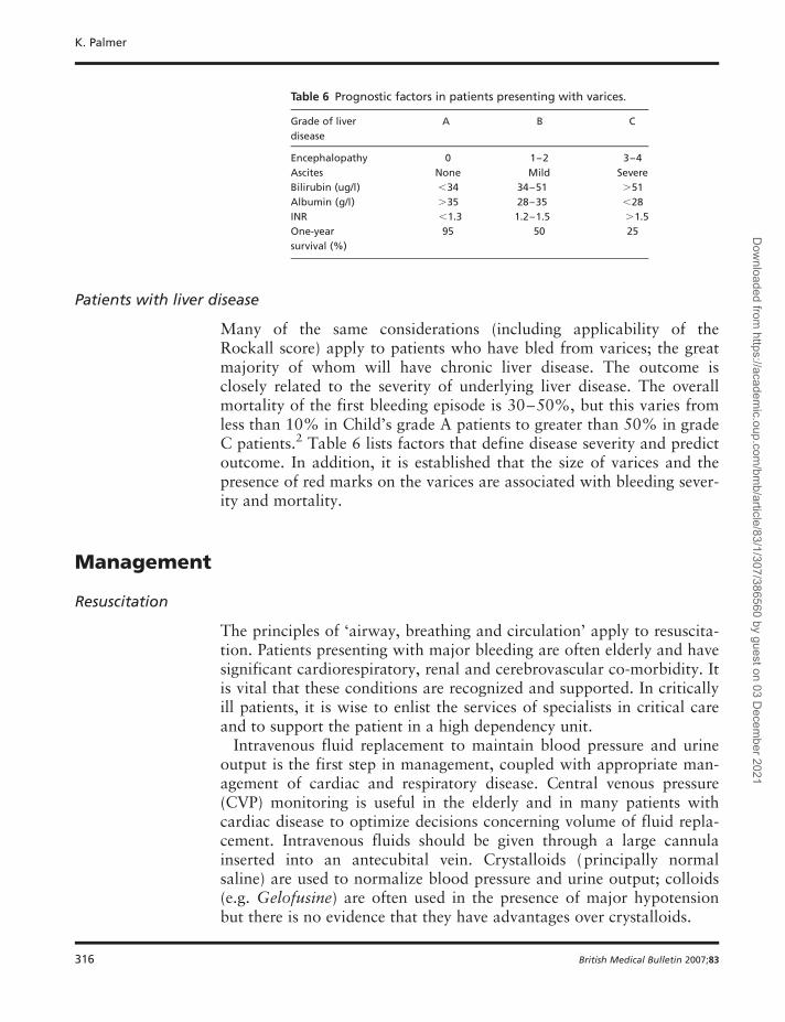
Many of the same considerations (including applicability of theRockall score) apply to patients who have bled from varices; the greatmajority of whom will have chronic liver disease. The outcome isclosely related to the severity of underlying liver disease. The overallmortality of the first bleeding episode is 30–50%, but this varies fromless than 10% in Child’s grade A patients to greater than 50% in gradeC patients.2 Table 6 lists factors that define disease severity and predictoutcome. In addition, it is established that the size of varices and thepresence of red marks on the varices are associated with bleeding sever-ity and mortality.
Management
Resuscitation
The principles of ‘airway, breathing and circulation’ apply to resuscita-tion. Patients presenting with major bleeding are often elderly and havesignificant cardiorespiratory, renal and cerebrovascular co-morbidity. Itis vital that these conditions are recognized and supported. In criticallyill patients, it is wise to enlist the services of specialists in critical careand to support the patient in a high dependency unit.
Intravenous fluid replacement to maintain blood pressure and urineoutput is the first step in management, coupled with appropriate man-agement of cardiac and respiratory disease. Central venous pressure(CVP) monitoring is useful in the elderly and in many patients withcardiac disease to optimize decisions concerning volume of fluid repla-cement. Intravenous fluids should be given through a large cannulainserted into an antecubital vein. Crystalloids (principally normalsaline) are used to normalize blood pressure and urine output; colloids(e.g. Gelofusine) are often used in the presence of major hypotensionbut there is no evidence that they have advantages over crystalloids.
Table 6 Prognostic factors in patients presenting with varices.
Grade of liver
disease
A B C
Encephalopathy 0 1–2 3–4
Ascites None Mild Severe
Bilirubin (ug/l) ,34 34–51 .51
Albumin (g/l) .35 28–35 ,28
INR ,1.3 1.2–1.5 .1.5
One-year
survival (%)
95 50 25
K. Palmer
316 British Medical Bulletin 2007;83
Dow
nloaded from https://academ
ic.oup.com/bm
b/article/83/1/307/386560 by guest on 03 Decem
ber 2021
Blood transfusion is administered to patients who are shocked andbleeding actively. Blood is also transfused when the haemoglobin con-centration is less than 10 g/dl. The evidence for this transfusionthreshold is relatively poor, but it is known that a haemoglobin concen-tration of less than 7 g/dl has significant adverse cardiac effects in theintensive care setting, and it is reasonable to pre-empt this by using alevel of 10 g/dl in bleeding patients.
Patients with liver disease present specific problems. Hepatic ence-phalopathy, renal failure and ascites may all develop or worsen as aconsequence of bleeding and warrant specific management. All liverdisease patients who develop bleeding should receive antibiotics toprevent life-threatening sepsis.
Monitoring
Monitoring includes measurement of pulse, blood pressure, urineoutput (through an indwelling catheter) and (in selected patients)CVP. Actively bleeding patients with evidence of shock (defined aspulse . 100 beats/min and/or systolic blood pressure , 100 mmHg) arebest managed in a high-dependency environment.
Endoscopy
Endoscopy is the primary diagnostic investigation and is undertakenafter optimum resuscitation has been achieved. In most cases, it is bestperformed within 24 h of admission, on the first available elective list.Out-of-hours emergency endoscopy is occasionally required in activelybleeding, shocked patients.
Endoscopy has three purposes.
1. To provide an accurate diagnosis. Certain diagnoses greatly influencemanagement; for example, oesophageal varices and active bleeding frompeptic ulcers require specific endoscopic and pharmacologicalinterventions.
2. Prognostic information. Stigmata of recent haemorrhage (Table 4) andvariceal appearances—or their absence—help direct the patient to thehigh-dependency unit, the general ward or, in some very low-risk cases, toimmediate hospital discharge. Most importantly, endoscopy facilitatesapplication of specific therapies to high-risk bleeding lesions.
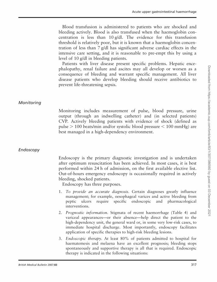
3. Endoscopic therapy. At least 80% of patients admitted to hospital forhaematemesis and melaena have an excellent prognosis; bleeding stopsspontaneously and supportive therapy is all that is required. Endoscopictherapy is indicated in the following situations:
Acute upper gastrointestinal haemorrhage
British Medical Bulletin 2007;83 317
Dow
nloaded from https://academ
ic.oup.com/bm
b/article/83/1/307/386560 by guest on 03 Decem
ber 2021
† bleeding oesophageal varices;
† peptic ulcer with major stigmata of recent haemorrhage (active spurtingbleeding, a non-bleeding visible vessel and non-adherent clot);
† vascular malformations, including actively bleeding AVMs, gastric antralvascular ectasia and the Dieulafoy’s lesion;
† active bleeding from a Mallory–Weiss tear (rarely).
Non-variceal bleeding
The evidence supporting endoscopy for non-variceal therapy is princi-pally based on clinical trials for peptic ulcer haemorrhage. Three cat-egories of direct endoscopic treatment have been evaluated; eachattempts to seal the arterial defect created by the ulcer.
Injection
Direct injection of fluids into the bleeding ulcer using disposableneedles is technically straightforward. Its efficacy is proved by random-ized prospective clinical trials, although the mechanism of benefitremains speculative; tamponade by compressing the artery within thefibrous confines of the chronic ulcer, vasoconstriction induced by adre-naline, endarteritis caused by sclerosants or alcohol and a direct effecton blood clot formation from fibrin glue or thrombin may all berelevant.
The most widely used injection fluid is 1:10 000 adrenaline. Thisstops active bleeding in more than 90% of patients, but 15–20%re-bleed.6 Adrenaline injection is extremely safe and has no significantcomplications. Addition of sclerosants (polidocanol, sodium tetradecylsulphate, ethanolamine) or alcohol does not reduce the risk ofre-bleeding and carries a risk of life-threatening necrosis of the injectedarea; for these reasons, they are not recommended. Fibrin glue (amixture of thrombin and fibrinogen injected through separate channelsof a sophisticated needle) and human thrombin are probably the mosteffective injection materials and have a low complication rate but arenot freely available.
Heat energy
In this method, devices are applied directly to the bleeding point atendoscopy to cause coagulation and thrombosis. The heater probe ispushed firmly onto the bleeding lesion to apply tamponade, anddefined pulses of heat energy are then given to coagulate the vessel.Clinical trials have shown the device to be as effective and as safe asinjection therapy. Multi-polar coagulation, in which electrical energy isconducted between multiple probes on the tip of an endoscopicallypositioned catheter, is as effective as the heater probe. The argon
K. Palmer
318 British Medical Bulletin 2007;83
Dow
nloaded from https://academ
ic.oup.com/bm
b/article/83/1/307/386560 by guest on 03 Decem
ber 2021
plasma coagulator also appears to be effective in arresting bleeding inlimited clinical trials. Thermal treatments can cause perforations butthis risk is very low.
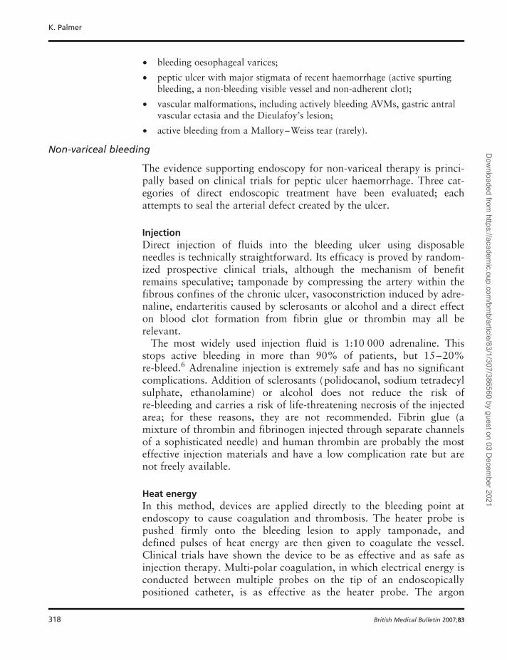
Mechanical devices
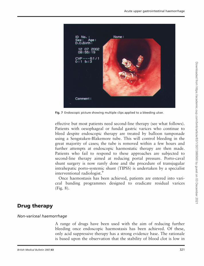
‘Endoclips’ can be applied to visible vessels (Fig. 6). They can be diffi-cult to deploy on awkwardly positioned ulcers, but may be the bestoption for the treatment of major bleeding ulcers and for the Deulafoylesion. Arterial defects of more than 1 mm diameter do not usuallyrespond to injection therapy, but an adequately positioned clip canstop bleeding from relatively large arteries. The major hazard is exacer-bation of bleeding should application prove unsuccessful.
Combinations of endoscopic therapy
Although the exact modes of action of these endoscopic therapies arelargely speculative, it is clear that each achieves haemostasis by a
Fig. 6 Endoscopic view of a visible vessel (in this case, a branch of the gastroduodenalartery) within a deep posterior duodenal ulcer.
Acute upper gastrointestinal haemorrhage
British Medical Bulletin 2007;83 319
Dow
nloaded from https://academ
ic.oup.com/bm
b/article/83/1/307/386560 by guest on 03 Decem
ber 2021
different mechanism. A meta-analysis of published trials shows thatcombination of injection and thermal treatments is superior to singlemodality treatment.7
Endoscopy should be repeated within 12–36 h in patients undergoingendoscopic haemostasis. Those with residual bleeding or persistingmajor stigmata are subjected to further therapy.
Re-bleeding after endoscopic therapy
Endoscopic therapy can achieve primary haemostasis in most bleedingulcer patients. However, re-bleeding occurs in 15–20% of cases,usually within the first 24 h. It is most common when the initial bleed-ing episode was severe; thus, shocked patients presenting with active,spurting haemorrrhage from large, posterior duodenal ulcers are thegroup most likely to re-bleed.
Management following re-bleeding is often difficult and is largelybased on clinical judgement and local expertise. Discussion betweenendoscopist and gastrointestinal surgeon is vital. In most patients, it isappropriate to repeat the endoscopy and re-treat the bleeding lesion. Atrial in Hong Kong showed that the mortality and blood transfusionrequirements of patients who re-bled after initially successful endo-scopic therapy were similar whether they were treated with urgentsurgery or repeat endoscopic therapy.8
Once adequate haemostasis is achieved by endoscopic re-treatment,an expectant policy is reasonable. Further bleeding is an absolute indi-cation for operative intervention.
Variceal bleeding
Patients with a high probability of variceal bleeding should undergourgent endoscopy following resuscitation. Other complications of liverdisease including hepatic encephalopathy, renal failure and sepsis mustbe recognized and managed. All patients should receive prophylacticantibiotics prior to endoscopy. Patients may be very sensitive to benzo-diazepine sedation, whereas others will have alcohol withdrawal syn-dromes. For these reasons, and because bleeding may be profuse,general anaesthetic with endotracheal intubation is often necessary forendoscopy.
Patients with active oesophageal variceal bleeding are treated byband ligation (Fig. 7) or endoscopic sclerotherapy (injecting polidocha-nol, ethanolamine or other sclerosants into the bleeding varix). Gastricvarices are difficult to treat; intravariceal fibrin glue injection may be
K. Palmer
320 British Medical Bulletin 2007;83
Dow
nloaded from https://academ
ic.oup.com/bm
b/article/83/1/307/386560 by guest on 03 Decem
ber 2021
effective but most patients need second-line therapy (see what follows).Patients with oesophageal or fundal gastric varices who continue tobleed despite endoscopic therapy are treated by balloon tamponadeusing a Sengstaken-Blakemore tube. This will control bleeding in thegreat majority of cases; the tube is removed within a few hours andfurther attempts at endoscopic haemostatic therapy are then made.Patients who fail to respond to these approaches are subjected tosecond-line therapy aimed at reducing portal pressure. Porto-cavalshunt surgery is now rarely done and the procedure of transjugularintrahepatic porto-systemic shunt (TIPSS) is undertaken by a specialistinterventional radiologist.9
Once haemostasis has been achieved, patients are entered into vari-ceal banding programmes designed to eradicate residual varices(Fig. 8).
Drug therapy
Non-variceal haemorrhage
A range of drugs have been used with the aim of reducing furtherbleeding once endoscopic haemostasis has been achieved. Of these,only acid suppressive therapy has a strong evidence base. The rationaleis based upon the observation that the stability of blood clot is low in
Fig. 7 Endoscopic picture showing multiple clips applied to a bleeding ulcer.
Acute upper gastrointestinal haemorrhage
British Medical Bulletin 2007;83 321
Dow
nloaded from https://academ
ic.oup.com/bm
b/article/83/1/307/386560 by guest on 03 Decem
ber 2021
an acid environment. It is crucial that gastric pH does not fall below 6,and the only practical means of achieving this is constant infusionof a proton pump inhibitor. This can be achieved most readily byintravenous infusion of a proton pump inhibitor drug (e.g.Omeprazole, 80 mg bolus followed by an 8 mg/h infusion for 72 h).Meta-analyses have demonstrated that this significantly reduces the riskof re-bleeding and the need for emergency surgery, although it has notbeen shown to significantly reduce mortality.10
Somatostatin and tranexamic acid are also sometimes used to reduceulcer re-bleeding although the evidence base for their use is less secure.
Variceal haemorrhage
Long-acting analogues of vasopressin (e.g. Terlipressin or Glypressin)reduce portal venous pressure and may stop active variceal bleeding.2
They also increase renal blood flow, thereby decreasing the risk ofrenal failure. Complications include coronary artery spasm, abdominal
Fig. 8 Endoscopic view of bands applied to an oesophageal varix.
K. Palmer
322 British Medical Bulletin 2007;83
Dow
nloaded from https://academ
ic.oup.com/bm
b/article/83/1/307/386560 by guest on 03 Decem
ber 2021
colic and limb ischaemia. In clinical practice, most variceal bleedingpatients are treated both endoscopically and by a 72 h infusion of thesedrugs.
Surgical intervention
Surgery now has very little place in the management of variceal bleed-ing and is limited to porto-caval shunt in patients with very well com-pensated stable liver disease.
For patients with ulcer bleeding, emergency surgery is undertakenwhen endoscopic therapy combined with pharmacological interventionfails to secure permanent haemostasis as follows:
† active bleeding that cannot be controlled by endoscopic therapy becausetorrential haemorrhage obscures the bleeding point, or active bleeding con-tinues despite successful application of endoscopic therapy;
† re-bleeding following initially successful endoscopic treatment (it is reason-able to repeat endoscopic therapy on one occasion after re-bleeding, pro-viding local expertise is available after discussion between endoscopist andsurgeon in the case of selected patients).
The type of operation depends on the site of the ulcer. Bleeding duode-nal ulcers are treated by under-running the ulcer, sometimes withpyloroplasty. Gastric ulcers are treated with partial gastrectomy orsimple ulcer excision. Vagotomy is no longer undertaken becauseproton pump inhibitor drugs abolish acid secretion.
Secondary prophylaxis
After haemostasis has been achieved, it is important to prevent recur-rent haemorrhage. For ulcer patients, eradication of H. pylori effec-tively abolishes the risk of late re-bleeding. In patients who need, forgood reason, to continue NSAID therapy, the following should beconsidered.
† Use the least toxic NSAID (usually Ibuprofen) that controls the arthriticsymptoms.
† Co-prescribe a proton pump inhibitor with the NSAID.
† Consider the use of a COX-2-specific anti-inflammatory drug, rather thana conventional NSAID. These are associated with significantly fewer recur-rent ulcer-related adverse events (both haemorrhage and perforation)although concerns regarding increased vascular events have largely pre-cluded their use.
Acute upper gastrointestinal haemorrhage
British Medical Bulletin 2007;83 323
Dow
nloaded from https://academ
ic.oup.com/bm
b/article/83/1/307/386560 by guest on 03 Decem
ber 2021
The management of patients with H. pylori who need to continuetaking an NSAID remains controversial. Gastritis (an inevitable conse-quence of H. pylori infection) induces mucosal prosta-glandin pro-duction, and this may protect the gastroduodenal mucosa fromthe harmful effects of NSAIDs. However, current studies suggest thatthe magnitude of prostaglandin production is unlikely to outweigh thedeleterious effects of H. pylori, and that eradication therapy isindicated in patients with a bleeding ulcer who are H. pylori-positiveand require NSAID therapy.
Although b-blockers have an established role as primary prophylaxisfollowing variceal haemorrhage (i.e. reducing the risk of variceal bleed-ing in patients who have never bled), their use as secondary prophy-laxis (preventing variceal re-bleeding) is not established, andendoscopic variceal ablation by a programme of repeated varicealbanding is the treatment of choice.
References
1 Church NC, Palmer KR (2004) Non-variceal gastrointestinal haemorrhage. In McDonaldJWD, Burroughs AK, Feagan BG (eds) Evidence-based Gastroenterology and Hepatology,
2nd ed. Blackwell Publications, 139–159.2 Goulis J, Buroughs AK (2004) Portal hypertensive bleeding. In McDonald JWD, Burroughs
4 Blatchford O, Murray WR, Blatchford M (2000) A risk score to predict need for treatmentfor upper gastrointestinal haemorrhage. Lancet, 356, 1319.
5 Bornman PC, Theodorou N, Shuttleworth RD et al. (1985) Importance of hypovolaemicshock and endoscopic signs in predicting recurrent haemorrhage from peptic ulceration: aprospective evaluation. Brit Med J, 291, 245–247.
6 Chung SCS, Leung JWC, Steele RJC (1988) Endoscopic injection of adrenaline for activelybleeding ulcers: a randomised trial. Brit Med J, 296, 1631–1633.
7 Calvet X, Vergara M, Brullet E et al. (2004) Addition of a second endoscopic treatment fol-lowing epinephrine injection improves outcome in high-risk bleeding ulcers.Gastroenterology, 126, 441–450.
8 Lau JY, Sung JJ, Lam YH et al. (1999) Endoscopic retreatment compared with surgery inpatients with recurrent bleeding after initial endoscopic control of bleeding ulcers. N Eng JMed, 340, 751–754.
9 Sauer P, Thielmann L, Stremmel W et al. (1997) Transjugular intrahepatic portosystemicshunt versus sclerotherapy plus propanolol for variceal rebleeding. Gastroenterology, 113,