AD_________________ Award Number: W81XWH-08-1-0062 TITLE: Merlin, the Hippo Pathway, and Tumor Suppression in Meningiomas PRINCIPAL INVESTIGATOR: Anita Lal, Ph.D. CONTRACTING ORGANIZATION: Regents of the University of California San Francisco, CA 94143-0962 REPORT DATE: February 2009 TYPE OF REPORT: Final PREPARED FOR: U.S. Army Medical Research and Materiel Command Fort Detrick, Maryland 21702-5012 DISTRIBUTION STATEMENT: Approved for public release; distribution unlimited The views, opinions and/or findings contained in this report are those of the author(s) and should not be construed as an official Department of the Army position, policy or decision unless so designated by other documentation.
Transcript
AD_________________ Award Number: W81XWH-08-1-0062 TITLE: Merlin, the Hippo Pathway, and Tumor Suppression in Meningiomas PRINCIPAL INVESTIGATOR: Anita Lal, Ph.D. CONTRACTING ORGANIZATION: Regents of the University of California San Francisco, CA 94143-0962 REPORT DATE: February 2009 TYPE OF REPORT: Final PREPARED FOR: U.S. Army Medical Research and Materiel Command Fort Detrick, Maryland 21702-5012 DISTRIBUTION STATEMENT: Approved for public release; distribution unlimited The views, opinions and/or findings contained in this report are those of the author(s) and should not be construed as an official Department of the Army position, policy or decision unless so designated by other documentation.
REPORT DOCUMENTATION PAGE Form Approved
OMB No. 0704-0188 Public reporting burden for this collection of information is estimated to average 1 hour per response, including the time for reviewing instructions, searching existing data sources, gathering and maintaining the data needed, and completing and reviewing this collection of information. Send comments regarding this burden estimate or any other aspect of this collection of information, including suggestions for reducing this burden to Department of Defense, Washington Headquarters Services, Directorate for Information Operations and Reports (0704-0188), 1215 Jefferson Davis Highway, Suite 1204, Arlington, VA 22202-4302. Respondents should be aware that notwithstanding any other provision of law, no person shall be subject to any penalty for failing to comply with a collection of information if it does not display a currently valid OMB control number. PLEASE DO NOT RETURN YOUR FORM TO THE ABOVE ADDRESS. 1. REPORT DATE (DD-MM-YYYY) 07-02-2009
2. REPORT TYPE Final
3. DATES COVERED (From - To) 08 JAN 2008 - 7 JAN 2009
4. TITLE AND SUBTITLE Merlin, the Hippo Pathway, and Tumor Suppression in
5a. CONTRACT NUMBER
Meningiomas 5b. GRANT NUMBER W81XWH-08-1-0062
5c. PROGRAM ELEMENT NUMBER
6. AUTHOR(S) Anita Lal; Gilson S Baia
5d. PROJECT NUMBER
5e. TASK NUMBER
5f. WORK UNIT NUMBER
7. PERFORMING ORGANIZATION NAME(S) AND ADDRESS(ES)
8. PERFORMING ORGANIZATION REPORT NUMBER
Regents of the University of California c/o Office of Sponsored Research 3333 California Street, Ste. 315 San Francisco, CA 94143-0962
9. SPONSORING / MONITORING AGENCY NAME(S) AND ADDRESS(ES) 10. SPONSOR/MONITOR’S ACRONYM(S) U.S. Army Medical Research and Materiel Command Fort Detrick, Maryland 21702-5012 11. SPONSOR/MONITOR’S REPORT NUMBER(S) 12. DISTRIBUTION / AVAILABILITY STATEMENT Approved for public release; distribution unlimited
13. SUPPLEMENTARY NOTES
14. ABSTRACT The goal of this proposal was to determine whether merlin exerts its tumor suppressive effects in meningiomas by signaling through the Hippo pathway. Using paired meningioma cell lines, where the only difference is expression of merlin, we have shown that loss of merlin is associated with a clear increase in the protein levels of YAP, a transcriptional co-activator and downstream effector of the Hippo pathway. In addition to this increase, merlin loss was associated with nuclear localization of YAP. Using RNA interference and flow cytometry, we have shown that depletion of YAP results in a reversal of the enhanced proliferation phenotype caused by the loss of merlin. Thus, Merlin signals through YAP, a core component of the Hippo pathway, to regulate cell proliferation in meningiomas.
INTRODUCTION Intracranial meningiomas, often multicentric, occur in ~50% of NF2 patients and are associated with an increased risk of mortality. Mutations in the NF2 gene are detected in up to 60% of sporadic meningiomas and mice with a conditional knockout of NF2 in the leptomeninges develop meningiomas. Thus, it is clear that meningiomas are a serious clinical problem for NF2 patients and that loss of NF2 is critical for the development of meningiomas. However, the mechanism(s) by which the protein product of the NF2 gene, merlin, functions as a tumor suppressor in meningiomas is unclear. In Drosophila, it has been shown that merlin signals via the Hippo pathway to regulate growth and apoptosis. Components and signaling mechanism(s) of the Hippo pathway are conserved in vertebrates and several orthologues of this pathway have been implicated in the genesis of mammalian cancers. The aim of this proposal was to link the hippo signaling pathway to loss of merlin and its tumor suppressive functions in human meningiomas. BODY
Using funding from a New Investigator Award and either RNA interference or retroviral mediated gene transfer, we have generated three paired meningioma and arachnoidal cell lines where the only difference is expression of merlin. These cell lines and the phenotypes generated by merlin loss in these cell lines are described in greater detail in the manuscript (recently published in Neoplasia) included in Appendix 1, and in the annual and final reports associated with proposal NF050061. In this proposal, these cell lines have been used to show a link between merlin loss and signaling through the hippo signaling pathway. Experimental details and the key findings from these experiments are also described in detail in the manuscript in Appendix 1.
Outlined below is a summary of the research accomplishments associated with each task outlined in the approved Statement of Work. Tables and Figures related to the text are included in the Supporting Data Section. We refer to Appendix 1 when referring to a figure in the included manuscript. For example, (Figure 1 in Appendix 1) would imply Figure 1 in the first attached manuscript in Appendix 1, while (Figure 1) refers to the Supporting Data Section. Task 1. Characterize the effect of merlin expression on activation of components of the hippo pathway in meningioma cell lines (Months 1-5)
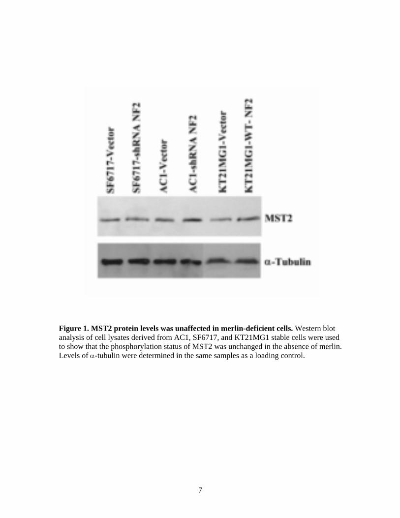
We firstly assessed protein levels and phosphorylation status of Mst1, Mst2, Lats1, Lats2 and YAP in meningioma cell lines. Protein levels of Mst2 (antibody from cell signaling #3952) were unaffected by merlin loss in meningioma cells (Figure 1). Similar results were obtained for Lats1 (antibody from cell signaling #9351S), Phospho-Lats1 (antibody from cell signaling #9157), while several attempts at optimizing western blot conditions for Mst1 (antibody from cell signaling), Phospho-Mst1/2 (antibody from cell signaling #3681S), Lats2 (antibody from Novus, #NB 200-199) did not result in clean, definite western blot results. In contrast, YAP protein levels were clearly induced by merlin loss (Figure 5A in Appendix 1) in meningioma cell lines. Phospho-YAP levels were also induced by merlin loss (Figure 2). However, given that total YAP levels are altered, the extent to which phosphorylation of YAP contributes to YAP function in meningiomas in unclear. Previous studies have reported that YAP translocates between the nucleus and the cytoplasm (1) and that it would need to be localized in the nucleus in order to perform its transcriptional coactivator functions. Therefore, we also performed immunocytochemistry for YAP in the paired cell lines and found that YAP was translocated to the nucleus in merlin-deficient cells (Figure 5B in Appendix 1).
We used quantitative PCR to assess the transcript levels of downstream target genes and found that, among the genes analyzed, Cyclin E1 transcript levels were induced in merlin-deficient cells (Figure 6 in Appendix 1). In addition, we showed by western blot that Cyclin E1 protein levels were also induced (Figure 6 in Appendix 1). Cyclin E1 is a downstream target of the Hippo pathway in Drosophila (2, 3) and our results suggest that merlin signals through YAP to Cyclin E1 to effect cell proliferation.
LAL, ANITA
5
Task 2. Evaluate the functional effects of depleting and overexpressing YAP (orthologue of Yorkie) in meningioma cell lines (Months 5-12) We generated a YAP siRNA construct in the pSUPERneo retroviral vector instead of the pSUPERpuro retroviral construct since we had serious toxicity issues while using puromycin to determine kill curves in our meningioma cell lines and would be unable to use puromycin to generate stables in meningioma cell lines. Since our stable cell lines were already resistant to neomycin owing to the expression of the NF2 siRNA, we performed transient transfections with the YAPsiRNA construct. We optimized transfection conditions such as DNA concentration, time post-tranfection to harvest, cell densities to obtain optimal depletion of YAP levels. Merlin loss induces cell proliferation, and this can be measured as an increase in the percentage of cells in the S-phase of the cell cycle using BrdU incorporation and flow cytometry. We used a similar methodology to assess the functional effects of depletion of YAP on the paired merlin-positive and merlin-negative cell lines. We found that depletion of YAP reversed the growth phenotype associated with merlin loss. While merlin loss enhanced S-phase entry, reduction of YAP caused ~50% decrease in the percentage of cells in the S-phase (Figure 7 in Appendix 1). These experiments were repeated three times with the same result and transfections of the YAP siRNA in AC1 cells also resulted in a similar trend. Thus, these results clearly demonstrate that YAP was We subcloned the coding region of YAP in pBABEhygro and generated stable cell lines in merlin positive cell lines, AC1 and MENII-1/SF6717 using retroviral mediated gene transfer. However, several attempts at confirming YAP over-expression by western blot analysis were negative (Figure 3), indicating a problem with either the construct or the retroviral infection. Given that the experiments using RNA interference gave us clear results and confirmed a functional link between merlin and YAP, the hippo signaling pathway effector, we did not pursue this line of investigation any further. KEY RESEARCH ACCOMPLISHMENTS
1) We have shown that protein levels of YAP are upregulated in response to merlin loss using three paired meningioma and arachnoidal cell lines where the only difference is expression of merlin
2) We have also shown that loss of merlin is associated with nuclear localization of YAP 3) We have shown that merlin loss is associated with an increase in transcript and protein levels of cyclin
E1. 4) We have used RNA interference to deplete YAP levels and reverse the enhanced proliferation phenotype
caused by merlin loss. 5) We have evidence that links the tumor suppressive mechanism of merlin to the hippo signaling pathway
in human meningiomas. REPORTABLE OUTCOMES
1) We are using the results of this research as preliminary data to apply for an investigator initiated neurofibromatosis grant in 2009.
2) We have successfully published a research publication in Neoplasia using the data described in this proposal.
CONCLUSION
The aim of this proposal was to link the hippo signaling pathway to loss of merlin and its tumor suppressive functions in human meningiomas. We have shown that in three independent paired meningioma and arachnoidal cell lines, where the only difference is merlin expression, loss of merlin is associated with an
LAL, ANITA
6
increase in protein levels of YAP and a concurrent nuclear localization of the induced YAP. In addition, we have shown that merlin loss is associated with an increase in transcript and protein levels of Cyclin E1 while levels of Cyclin D1 were unaffected. These results suggest that merlin signals through YAP which in turn effects Cyclin E1 transcription and cell proliferation. Using a shRNA specific for YAP, we have shown that depletion of YAP eliminates the enhanced proliferation induced in merlin-deficient cells, suggesting that merlin regulates cell proliferation by signaling through YAP. Thus, we have linked merlin to YAP, the downstream effector of the Hippo pathway, and have shown that merlin function is dependent on YAP induction. REFERENCES 1. Hao Y, Chun A, Cheung K, Rashidi B, Yang X. Tumor suppressor LATS1 is a negative regulator of oncogene YAP. J Biol Chem 2008;283(9):5496-509. 2. Saucedo LJ, Edgar BA. Filling out the Hippo pathway. Nat Rev Mol Cell Biol 2007;8(8):613-21. 3. Pan D. Hippo signaling in organ size control. Genes Dev 2007;21(8):886-97. BIBLIOGRAPHY PUBLICATIONS Striedinger KS, VandenBerg SR, Baia GS, McDermott MW, Gutmann DH and Lal A: The neurofibromatosis 2 tumor suppressor gene product, merlin, regulates meningioma cell growth by signaling through YAP. Neoplasia 10:1204-12, 2008 PERSONNEL RECEIVING PAY FROM THIS RESEARCH EFFORT Anita Lal, Ph.D. Gilson S Baia, MS
7
Figure 1. MST2 protein levels was unaffected in merlin-deficient cells. Western blot analysis of cell lysates derived from AC1, SF6717, and KT21MG1 stable cells were used to show that the phosphorylation status of MST2 was unchanged in the absence of merlin. Levels of α-tubulin were determined in the same samples as a loading control.
8
Figure 2. Total YAP and Phospho YAP expression was increased in merlin-deficient cells. Total YAP and Phospho YAP levels were measured by western blot analysis in cell lysates from SF6717 and KT21MG1 stable cells. Total YAP and Phospho YAP levels were induced by merlin loss.
9
Figure 3. Total YAP expression was negative in SF6717 stable clones. Western blot analysis of cell lysates derived from SF6717 stable cell lines did not confirm expression of YAP. Given that our experiments using shRNA specific for YAP were reproducible, definitive and conclusive, we did not pursue the overexpressing YAP line of investigation.
www.neoplasia.com
Volume 10 Number 11 November 2008 pp. 1204–1212 1204
The Neurofibromatosis 2 TumorSuppressor Gene Product,Merlin, Regulates HumanMeningioma Cell Growth bySignaling through YAP1
Katherine Striedinger*, Scott R. VandenBerg*,Gilson S. Baia*, Michael W. McDermott*,David H. Gutmann† and Anita Lal*
*Brain Tumor Research Center, Department of NeurologicalSurgery, University of California, San Francisco, CA 94143,USA; †Department of Neurology, Washington UniversitySchool of Medicine, St. Louis, MO 63110, USA
AbstractNeurofibromatosis type 2 (NF2) is an autosomal dominant disorder characterized by the occurrence of schwanno-mas and meningiomas. Several studies have examined the ability of the NF2 gene product, merlin, to function as atumor suppressor in diverse cell types; however, little is known about merlin growth regulation in meningiomas. InDrosophila, merlin controls cell proliferation and apoptosis by signaling through the Hippo pathway to inhibit thefunction of the transcriptional coactivator Yorkie. The Hippo pathway is conserved in mammals. On the basis ofthese observations, we developed human meningioma cell lines matched for merlin expression to evaluate merlingrowth regulation and investigate the relationship between NF2 status and Yes-associated protein (YAP), the mam-malian homolog of Yorkie. NF2 loss in meningioma cells was associated with loss of contact-dependent growthinhibition, enhanced anchorage-independent growth and increased cell proliferation due to increased S-phase en-try. In addition, merlin loss in both meningioma cell lines and primary tumors resulted in increased YAP expressionand nuclear localization. Finally, siRNA-mediated reduction of YAP in NF2-deficient meningioma cells rescued theeffects of merlin loss on cell proliferation and S-phase entry. Collectively, these results represent the first demon-stration that merlin regulates cell growth in human cancer cells by suppressing YAP.
Neoplasia (2008) 10, 1204–1212
Address all correspondence to: Anita Lal, Brain Tumor Research Center, Departmentof Neurological Surgery, Box 0520, University of California, San Francisco, CA-94143. E-mail: [email protected] support: Department of Defense New Investigator Award (W81XWH-06-1-0221) and Concept Award (NF073066) to A. Lal and Department of Defense Awardto D. Gutmann.Received 29 May 2008; Revised 4 August 2008; Accepted 5 August 2008
IntroductionNeurofibromatosis type 2 (NF2) is a cancer predisposition syndromephenotypically characterized by the occurrence of multiple nervoussystem tumors. The two most common tumors in this inherited syn-drome are schwannomas and meningiomas [1]. Whereas meningio-mas from individuals with NF2 exhibit biallelic inactivation of theNF2 gene, loss of NF2 expression is also detected in as many as60% of sporadic meningiomas [2]. Similarly, genetically engineeredmice with leptomeningeal NF2 inactivation also develop meningio-mas [3,4]. These findings strongly implicate the NF2 gene in thepathogenesis of meningiomas; however, the molecular mechanismby which NF2 regulates cell growth relevant to meningioma tumor-igenesis remains unsolved.
Merlin (or schwannomin), the product of the NF2 gene, is amember of the protein 4.1 family that links the actin cytoskeletonto plasma membrane proteins [5]. Although few studies have exam-ined merlin loss in meningioma cells, loss of merlin in fibroblasts andSchwann cells results in loss of contact-dependent inhibition of pro-liferation, enhanced growth in soft agar and tumor formation in mice
[6,7]. In these cell types, merlin has been implicated in epidermalgrowth factor receptor [8], β1-integrin [9], and CD44 [7] functionas well as Ras [10], Rac1 [11,12], phosphatidylinositol 3-kinase [13],mitogen-activated protein kinase [14], and signal transducer and ac-tivator of transcription [15] intracellular signaling. It is not knownwhether any of these growth control pathways are deregulated inNF2-deficient meningioma tumors.
Negative regulation of growth by merlin is conserved inDrosophila,where it acts upstream of the Hippo signaling pathway to coordinately
Neoplasia Vol. 10, No. 11, 2008 Merlin Signals Through YAP in Meningiomas Striedinger et al. 1205
regulate cell proliferation and apoptosis [16,17]. Mutations in mer-lin or other components of the Hippo pathway such as the serine/threonine kinases, Hippo and Warts, the adaptor molecule, Salvador,or Mats, results in activation of the transcriptional coactivator, Yorkie.Yorkie regulates expression of downstream target genes includingcyclin E andDIAP1 (Drosophila inhibitor of apoptosis protein 1) caus-ing increased growth, delayed cell cycle exit, inhibition of apoptosis,and enhanced cell survival [16,17].Individual components of the Hippo pathway are highly con-
served in mammals, where they also regulate cell proliferation andapoptosis (Figure 1) [18,19]. Mice and humans have two Wartsorthologs, Lats1 and Lats2. Mice deficient for Lats1 develop soft-tissue sarcomas and ovarian tumors [20]. The human ortholog ofSalvador, hWW45, is mutated in cancer cell lines [21]. The twomammalian Hippo homologues, Mst1 and Mst2, promote apoptosisand regulate cell cycle exit [22]. Vertebrate Mst2 can rescue the lethal-ity and overgrowth phenotypes of Hippo mutants in Drosophila [23].Similar to their Drosophila counterparts, human Mst2 phosphory-lates and activates both Lats1 and Lats2 [24]. The Yes-associatedprotein (YAP), the mammalian ortholog of Yorkie, is the primary ef-fector of the mammalian Hippo pathway. Similar to the function ofYorkie in Drosophila, YAP causes aberrant tissue expansion in miceand induces epithelial transformation in mammary cells [25,26].Given the conservation of components and mechanisms that op-
erate downstream of merlin between Drosophila and mammals, wetested the functional relationship among merlin, Hippo pathway reg-ulation, and growth suppression in human meningioma tumors. Wedeveloped nonneoplastic and neoplastic meningeal cell lines thatmimic gain or loss of NF2 expression and used these matched lines
to examine merlin regulation of YAP. We found that absence ofmerlin results in loss of contact-dependent inhibition of growthand promotes anchorage-independent growth. Merlin loss enhancescell proliferation by increasing entry into the S-phase and cyclin E1expression. Inactivation of merlin results in increased YAP expressionand nuclear accumulation of YAP in these meningioma cell lines andin primary human meningioma tumors. Finally, we show that YAPsuppression reverses the proliferation effects associated with merlinloss in meningiomas. Collectively, these data demonstrate that merlinregulates cell growth in a YAP-dependent manner in meningiomasand suggests that YAP is a compelling target for therapeutic inhibi-tion of human meningioma tumor growth.
Materials and Methods
Tumor Samples, Cell Lines, and CultureAll human meningioma tumor samples were collected by the
Neurological Surgery Tissue Bank using protocols approved by theUniversity of California, San Francisco Committee on Human Re-search. Human meningioma cell lines used were KT21MG1 [27]and MENII-1. MENII-1 cells were isolated from a surgically resectedgrade II meningioma and were immortalized by the expression oftelomerase and the human papillomavirus E6/E7 genes as describedearlier [28]. Human arachnoidal cells (AC1) were cultured by plat-ing small fragments of surgically resected spinal arachnoid tissueon scores scratched on the bottom of six-well tissue culture plates.Within 7 days, cells with characteristic arachnoidal morphologygrowing as a monolayer of polygonal cells with large cytoplasmicarcs migrate out of these scores. The arachnoidal origins of thesecells were verified by positive staining for vimentin and desmoplakin.Primary cultures of arachnoidal cells were immortalized by stabletransfection with the human papillomavirus E6/E7 oncogenes andtelomerase as described earlier [28]. All cell lines were maintainedin Dulbecco’s modified Eagle’s medium supplemented with 10%fetal bovine serum and appropriate antibiotic selection markers.
Expression Constructs and AntibodiesThe pSUPER.retro.neo-NF2-siRNA construct was generated by
digesting the pSUPER.retro.neo vector (Oligoengine) with BglIIand Hind III and ligating the annealed oligos (5′-gatccccGCAG-CAAGCACAATACCATttcaagagaATGGTATTGTGCTTGCTGC-ttttta and 5′-agcttaaaaaGCAGCAAGCACAATACCATtctctt-gaaATGGTATTGTGCTTGCTGCggg) that contain a 19-nucleo-tide NF2 target sequence (in caps) using the strategy describedearlier [29]. The pSUPER.retro.neo-YAP-siRNA construct was alsogenerated in pSUPER.retro.neo using the strategy described aboveand a previously described YAP target sequence (CCAGAGAATCA-GTCAGAGA) [30]. A nonspecific mammalian scramble sequence(Oligoengine, Seattle, WA) in pSUPER.retro.neo (pSUPER.retro.neo-Control) was used as a control for the development of stablecell lines. Wild type NF2, S518A NF2, and S518D NF2 mutantconstructs in pUHD10.3 have been previously described [7,31].These three constructs were subcloned into pBABE-Hygro usingstandard techniques. Merlin polyclonal (A19, #sc-331) and monoclo-nal (B12, #sc-55575) antibodies and the cyclin E1 monoclonalantibody (13A3, #sc-56310) were from Santa Cruz Biotechnology,Santa Cruz, CA. The YAP monoclonal antibody (#4912) was fromCell Signaling (Danvers, MA), the cyclin D1 monoclonal antibody
Figure 1. Schematic of the mammalian Hippo signaling pathway.The Hippo pathway is an evolutionary conserved cellular pathwaythat coordinately regulates cell proliferation and apoptosis. Merlinhas been proposed to interact with unknown membrane proteinsand transduce a signal that stimulates the phosphorylation ofLATS1/2 by the serine/threonine kinases MST1/2 that interact withhWW45. LATS1/2 inhibits the transcriptional coactivator YAP re-sulting in suppressed expression of downstream target genessuch as cyclins that are involved in cell growth and proliferation.
1206 Merlin Signals Through YAP in Meningiomas Striedinger et al. Neoplasia Vol. 10, No. 11, 2008
(clone DCS-6) was from BD Pharmingen (Franklin Lakes, NJ),and the α-tubulin (#CP06) antibody was from Calbiochem (SanDiego, CA).
Retroviral Infection and Selection of Stable Cell PopulationsTo stably suppress NF2 in MENII-1 and AC1 cell lines, retroviral
supernatants were generated by transfecting Phoenix A packag-ing cells with pSUPER.retro.neo-NF2-siRNA or pSUPER.retro.neo-Control using Lipofectamine 2000 Plus Reagent (Invitrogen,Carlsbad, CA). The 48-hour posttransfection supernatant was har-vested, filtered, and used to infect MENII-1 and AC1 cell lines inthe presence of 8 μg/ml polybrene. Stable cell populations were se-lected using 500 μg/ml G418. In parallel, stable cell populations ex-pressing wild type or mutant NF2 were generated by transfectingPhoenix A cells with the particular pBABE-Hygro construct and in-fecting KT21MG1 cells with the 48-hour posttransfection supernatant.Stable cell populations were selected using 200 μg/ml hygromycin.Empty pBABE-Hygro vector was used as a negative control.
Transient Suppression of YAPMerlin-positive and -negative MENII-1 stable cell populations
generated above were plated at 80% confluency in 100-mm dishesand transfected with 10 μg of pSUPER.retro.neo-YAP siRNA usingLipofectamine 2000 Plus Reagent (Invitrogen). Empty pSUPER.retro.neo vector was used as a negative control. The 72-hour post-
transfection cells were subjected to flow cytometry to quantify bro-modeoxyuridine (BrdU) uptake (described below).
Quantitative Polymerase Chain ReactionQuantitative polymerase chain reaction (PCR) was performed in
three independent experiments using cDNA templates with the I-cycler machine (Bio-Rad, Hercules, CA) and SYBR Green I (Molec-ular Probes, Eugene, OR) using PCR conditions and data analysis asdescribed earlier [32]. Primers specific for GAPDH and actin wereused to verify the integrity of the cDNA and to normalize cDNAyields. The primers used were: GAPDH, 5′-GGAAGCTTGTCA-TCAATGGAA and 5′-AAATGAGCCCCAGCCTTCTC; Actin,5 ′ -CAGGAGGAGCAATGATCTTG and 5 ′ -ACTCTTC-CAGCCTTCCTTCC; NF2, 5′-ACCGTTGCCTCCTGACATACand 5′-TCGGAGTTCTCATTGTGCAG; YAP, 5′-GCAGTTGG-GAGCTGTTTCTC and 5′-GCCATGTTGTTGTCTGATCG;cyclin E1, 5′-CCATCCTTCTCCACCAAAGA and 5′-TTTGA-TGCCATCCACAGAAA; cyclin D1, 5′-TGTTTGCAAGCAG-GACTTTG and 5′-CCTTCCGGTGTGAAACATCT.
Western Blot AnalysisTotal cell lysates were prepared either in buffer A (50 mM Tris-
HCl, pH 7.5; 1 mM EDTA pH 8.0, 1% Triton) for merlin detec-tion or in 1× SDS buffer following manufacturer’s instructions (Cell
Figure 2. In vitro model system of merlin expression in human meningeal cells. Endogenous merlin was silenced in AC1 and MENII-1cells using NF2 specific siRNA. In parallel, merlin isoform 1 was exogenously expressed in KT21MG1 cells using retroviral mediatedgene transfer. (A) NF2 transcript levels were measured by quantitative PCR and showed a 5.6-fold reduction in MENII-1-NF2-siRNA cellscompared with MENII-1-Control cells. Asterisk denotes statistical significance (P < .05). (B) Western blot analysis of cell lysates derivedfrom AC1, MENII-1, and KT21MG1 stable cell populations was used to confirm loss or gain of merlin. Whereas merlin expression was ob-served in AC1-Control and MENII-1-Control cells, NF2 siRNA abolished expression of merlin in AC1-NF2-siRNA and MENII-1-NF2-siRNAcells. In parallel, KT21MG1-Control cells lacked merlin, whereas KT21MG1-NF2 cells expressed wild type merlin. Levels of α-tubulin weredetermined in the same samples as a loading control. Immunoblot of one representative experiment of three with similar results isshown. (C) Immunofluorescence using the A19 polyclonal antibody against merlin revealed the presence of cytoplasmic staining inAC1-Control and MENII-1-Control cells and its absence in AC1-NF2-siRNA and MENII-1-NF2-siRNA cells. In contrast, KT21MG1-Controlcells had no staining, whereas KT21MG1-NF2 cells had cytoplasmic staining. Merlin immunolabeling is shown in green, and nuclearDAPI counterstaining is shown in blue.
Neoplasia Vol. 10, No. 11, 2008 Merlin Signals Through YAP in Meningiomas Striedinger et al. 1207
Signaling) for YAP and cyclin protein detection. Protein (50-200 μg)was resolved by electrophoresis for each sample and was transferredto a polyvinylidenefluoride membrane. Membranes were blocked in5% low-fat dry milk in Tris-buffered saline–Tween 20 and incubatedovernight at 4°C with either merlin A19 or B12 antibodies, or theYAP, cyclin E1, or cyclin D1 antibody, or α-tubulin. Incubation withhorseradish peroxidase–conjugated goat antirabbit or antimouse im-munoglobulin (Jackson Immunoresearch, West Grove, PA) was per-formed for 1 hour at room temperature. Bound antibody wasvisualized by chemiluminescence using the SuperSignal West Picosubstrate (Pierce Chemical Co., Rockford, IL). The molecularweights were determined with the use of prestained protein ladders(BioRad, Hercules, CA, and Invitrogen). Films were scanned and ex-ported as TIFF files.
Immunofluorescence MicroscopyIndirect immunofluorescence for merlin (A19 antibody or B12 an-
tibody) and YAP was performed as described earlier [28]. Briefly, cellswere fixed, permeabilized, blocked, and sequentially incubated withprimary and secondary (Alexa 488 goat antirabbit IgG or Alexa 546goat antimouse IgG) antibodies. Cells were mounted in DAPImount-ing media, examined, and photographed with a microscope (Zeiss,Thornwood, NY).
ImmunohistochemistryImmunohistochemical staining was performed on 5-μm formalin-
fixed, paraffin-embedded sections from meningioma tissue (8 pri-mary tumors) and from a meningioma tissue microarray (29 primarytumors) for merlin (B12 antibody) and YAP as described earlier [33].Slides were reviewed in consultation with a neuropathologist (S.R.V.)and included 33 WHO grade I and 4 WHO grade III meningiomas.
Growth Curves and Soft Agar AssayMerlin-positive and -negative stable cell populations (20,000 cells)
were plated in 24-well plates, and cells from three wells were countedat 3, 6, 12, 18, 25, and 30 days. To assess colony growth in soft agar,50,000 cells were plated in Dulbecco’s modified Eagle’s medium in0.4% low melting temperature agarose upon a layer of 0.8% agarose.After 8 weeks, colonies were stained with 0.005% crystal violet, andcolonies larger than 100 μm in diameter were scored by countingunder a microscope.
Flow CytometryCells (70-80% confluent) were incubated with 1 mM BrdU for
3 hours at 37°C and processed using the fluorescein isothiocyanateBrdU Flow Kit (BD Biosciences, San Jose, CA) following manufac-turer’s instructions. Briefly, 1 × 10⁶ trypsinized cells were fixed,permeabilized, and digested with DNAse. Cells were then stainedwith fluorescein isothiocyanate–conjugated anti-BrdU and 7-amino-actinomycin (7-AAD). Flow cytometry was performed on a BectonDickinson FACSCalibur machine. For each experiment, 10,000events were counted. Data acquisition was performed with the Cell-Quest software (BD Biosciences), and data were analyzed using FlowJo v8.5.3.
StatisticsAll data are expressed as mean ± SEM. GraphPad Prism version 4
was used for statistical analysis, consisting of unpaired t test and sig-nificant differences with a P < .05.
Results
Establishment of Matched NF2-Expressing and NF2-DeficientHuman Arachnoidal and Meningioma Cell Lines
To develop a meningioma-specific NF2 in vitro model system,we determined the expression levels of endogenous merlin in normalhuman arachnoidal and meningioma cell lines (data not shown). Onthe basis of these results, we selected one arachnoidal cell line (AC1),one merlin-positive grade II meningioma cell line (MENII-1), andone merlin-negative grade III meningioma cell line (KT21MG1)for further analysis. Paired cell lines were generated by either sup-pressing NF2 expression in AC1 and MENII-1 cells using RNA in-terference (siRNA) or by overexpressing the full-length human NF2cDNA (isoform 1, lacking exon 16 sequences) in KT21MG1 cells afterretroviral-mediated gene transfer. Stable cell populations expressingeither NF2 siRNA (AC1-NF2-siRNA or MENII-1-NF2-siRNA) ora nonspecific target siRNA (AC1-Control or MENII-1-Control) were
Figure 3. Suppression of merlin causes loss of contact-dependentinhibition of growth and promotes anchorage-independentgrowth. (A) Growth curves in the presence and absence of merlinexpression. Cultures were subconfluent during the first 6 days.MENII-1-NF2-siRNA cells (dotted lines) continued growing afterconfluent conditions and have less contact-dependent inhibitionof growth compared with MENII-1-Control cells (solid lines). Eachline corresponds to representative cultures. (B) NF2 suppressionpromotes anchorage-independent growth. Marked increased incolony formation in soft agar was observed in cells without merlinexpression as MENII-1-NF2-siRNA and KT21MG1-Control cellscompared with MENII-1-Control and KT21MG1-NF2, respectively.Representative images of the colonies formed (upper panel) andthe mean number of colonies per well (lower panel) are shown.Error bars equal ±SE of three independent experiments. Asterisksdenote statistical significance (P < .05).
1208 Merlin Signals Through YAP in Meningiomas Striedinger et al. Neoplasia Vol. 10, No. 11, 2008
selected. NF2 transcript levels were 5.6-fold lower in MENII-1-NF2-siRNA cells when compared with MENII-1-Control cells using quan-titative PCR (Figure 2A). Similarly, merlin protein expression wasundetectable in AC1-NF2-siRNA and MENII-1-NF2-siRNA cellsby Western blot analysis and immunofluorescence (Figure 2, B andC ). In parallel, stable cell populations expressing either exogenousmerlin (KT21MG1-NF2) or empty vector (KT21MG1-Control)were selected. Expression of merlin in KT21MG1-NF2 cells was con-firmed byWestern blot and immunofluorescence (Figure 2, B and C ).In total, we have generated three separate human meningeal and me-ningioma cell lines that differ only in their expression of merlin forsubsequent study.
Merlin Loss Increases S-Phase Entry, Cell Proliferation, andPromotes Anchorage-Independent Growth
Next, we assessed the effect of merlin loss on the growth proper-ties of these human meningioma cell lines. MENII-1-NF2-siRNAcells exhibited a more pronounced loss of contact-dependent inhibi-tion of growth compared with MENII-1-Control cells (Figure 3A).Merlin loss also promoted colony formation in soft agar. In theseexperiments, MENII-1-NF2-siRNA cells formed a greater numberof colonies (10.6 ± 3.2) larger than 100 μm in diameter comparedwith MENII-1-Control cells (0.4 ± 0.4; P = .01; Figure 3B). Con-versely, merlin expression in KT21MG1 cells significantly decreasedthe formation of colonies (32 ± 6.3) compared with merlin-negativeKT21MG1 cells (361 ± 4.9; P ≤ .0001; Figure 3B).
To determine the effect of merlin on cell cycle progression, wemeasured BrdU incorporation and total DNA content by flow cy-tometry. Loss of merlin in AC1 and MENII-1 cells resulted in asignificant increase in the percentage of BrdU-positive cells, indicatedby an increase in S-phase entry (Figure 4). This is consistent with theobserved increase in proliferation in NF2-deficient cells. Conversely,expression of exogenous merlin in KT21MG1 cells induced G0/G1
arrest and a concomitant decrease in the S-phase cell population (Fig-ure 4). These results demonstrate that merlin functions as a negativegrowth regulator for both nonneoplastic leptomeningeal cells andmeningioma cells and establishes this system as a tractable experi-mental platform for examining growth regulatory pathways.
Merlin Loss Is Associated with an Increase inYAP Protein Expression
In Drosophila, merlin controls cell proliferation and apoptosis bysignaling through the Hippo pathway and its effector protein Yorkie,the ortholog of YAP [16,18]. To determine whether merlin mightregulate meningioma cell growth by modulating Hippo pathway sig-naling, we investigated whether changes in merlin expression wereassociated with altered levels of YAP using our human NF2 meningi-oma model system. Transcript levels of YAP were unaffected by merlinloss in MENII-1 cells (data not shown); however, YAP protein ex-pression was elevated in arachnoidal and meningioma cells lackingmerlin expression and decreased in KT21MG1 cells expressing wildtype merlin (Figure 5A). These results suggest that merlin regulatesYAP expression at the translational or posttranslational level.
Figure 4. Merlin loss enhances S-phase entry. AC1 andmeningioma cell lines (MENII-1 and KT21MG1) were labeled with BrdU and 7-AADto assess the cell cycle distribution of individual cells by flow cytometry. (A) Representative flow cytometric histograms indicate an in-crease in the percent of BrdU-positive cells in AC1-NF2-siRNA, MENII-1-NF2-siRNA, and KT21MG1-Control cells compared with AC1-Control, MENII-1-Control, and KT21MG1-NF2 cells, respectively. (B) Bar graphs depict the percentage of cells in the S-phase of the cellcycle (BrdU-positive cells) averaged from three independent experiments. Error bars correspond to ±SE. Asterisks denote statistical sig-nificance using unpaired t test (P< .05). (C) Table shows themean of the percentage of cells ±SE in each phase of the cell cycle from threeindependent experiments.
Neoplasia Vol. 10, No. 11, 2008 Merlin Signals Through YAP in Meningiomas Striedinger et al. 1209
Merlin becomes active after dephosphorylation of the conservedC-terminal serine 518 (S518) residue [31,34]. Previous studies haveshown that merlin mutants in which this phosphorylatable residueis changed to alanine (S518A) are constitutively nonphosphorylatedand active, whereas those containing aspartic acid (S518D) arepseudophosphorylated and nonfunctional as negative growth reg-ulators [31,34]. Using these mutants, we assessed the effect ofmerlin phosphorylation status on YAP protein levels in KT21MG1human meningioma cells. YAP protein levels were markedly up-regulated in KT21MG1 cells expressing the S518D inactive mutantcompared with those with active merlin S518A expression (Fig-ure 5A), suggesting that active and functional merlin was requiredto inhibit YAP.
YAP has been reported to shuttle between the cytoplasm andthe nucleus where it can function as a transcriptional coactivator[35]. To determine whether merlin regulates the subcellular localiza-tion of YAP, we used fluorescence immunocytochemistry to examineNF2-deficient and NF2-expressing AC1 and MENII-1 cells. YAPwas preferentially expressed in the cytoplasm of AC1-Control andMENII-1-Control cells. In contrast, YAP was localized in the nu-cleus in AC1-NF2-siRNA and MENII-1-NF2-siRNA cells (Fig-ure 5B). Collectively, these data indicate that merlin regulates YAPprotein expression and YAP nuclear localization.
To assess whether a similar association exists between merlin andYAP in human meningioma tumors, we surveyed 37 sporadic primarymeningioma tumors by immunohistochemistry. After immunostain-ing with a merlin-specific monoclonal antibody, meningiomas wereclassified as either merlin-positive, if tumors exhibited any positiveimmunoreactivity, or merlin-negative, if tumors had no immunore-activity. We then assessed protein levels of YAP in adjacent serialsections. 13 (92%) of 14 merlin-negative meningiomas exhibitedstrong nuclear YAP immunoreactivity. In contrast, 22 (95%) of23 merlin-positive meningiomas had weak to no YAP immunore-activity (Figure 5C ). These results further support the in vitro re-sults demonstrating that merlin regulates YAP protein levels in vivo.
Merlin Loss Is Associated with an Increase in Protein Levels ofCyclin E1
Previous studies have identified cyclin E1 as a transcriptional targetof the Hippo pathway in Drosophila [36–38] and an essential reg-ulator of progression from G1 to S cell cycle progression in mam-malian cells [39,40]. Also, merlin has been reported to inhibit cellproliferation by repressing cyclin D1 in human mesothelioma cells[41]. To determine whether merlin regulates cyclin E1 and/or cy-clin D1 levels in human meningioma cells, we measured cyclin E1and D1 RNA and protein expression by quantitative PCR andWestern blot analysis, respectively. Cyclin E1 transcript levels wereat least 2.5-fold higher in MENII-1-NF2-siRNA cells comparedwith MENII-1-Control cells, whereas cyclin D1 transcript levelswere the same in MENII-1-NF2-siRNA and MENII-1-Controlcells (Figure 6A). In addition, cyclin E1 protein levels were ele-vated in MENII-1-NF2-siRNA and AC1-NF2-siRNA cells com-pared with MENII-1-Control and AC1-Control cells, respectively(Figure 6B). Conversely, exogenous expression of merlin in NF2-deficient KT21MG1 cells resulted in decreased cyclin E1 proteinlevels. In contrast, cyclin D1 protein levels were unaffected by theabsence or presence of merlin in both MENII-1 and KT21MG1cells (Figure 6C ). These data suggest that merlin likely regulates cell
Figure 5. Protein levels of YAP are up-regulated and localized tothe nucleus in NF2-deficient cells. (A) Total cell lysates were sub-jected to Western blot using a YAP- or merlin-specific antibody.Increased YAP protein expression was observed when NF2 wassuppressed in AC1 and MENII-1 cells compared with controls.Conversely, exogenous expression of merlin decreased YAP inKT21MG1 cells compared with controls. Expression of a nonphos-phorylated, active merlin (S518A NF2) was also associated withlower levels of YAP compared with the expression of pseudophos-phorylated inactivemerlin (S518DNF2). Levels of α-tubulin were de-termined in the same samples as loading control. Results werereproduced in three independent experiments. (B) YAPwas translo-cated to the nucleus in merlin-deficient cells. Immunofluorescencestaining was used to show that YAP was localized to the nucleus inAC1-NF2-siRNA, MENII-1-NF2-siRNA, and KT21MG1-Control cells.In contrast, YAPwas primarily cytoplasmic in AC1-Control, MENII-1-Control, and KT21MG1-NF2 cells. Merlin immunolabeling is shownin red; YAP staining is shown in green; and nuclear DAPI counter-staining is shown in blue. (C) In situ immunostaining of merlin andYAP in serial sections of primary human meningioma tumors. Wesurveyed 37 primary meningiomas by immunohistochemistry. YAPexpression was minimal to absent in 95% of merlin-positive menin-giomas (a representative tumor is shown here asMeningioma 1). Incontrast, YAPwas expressed and localized to the nucleus in 92%ofmerlin-negative meningiomas (a representative tumor is shownhere as Meningioma 2). Arrow depicts an example of YAP nuclearlocalization. Insets show images at higher magnification.
1210 Merlin Signals Through YAP in Meningiomas Striedinger et al. Neoplasia Vol. 10, No. 11, 2008
growth by modulating cyclin E1 expression at the transcriptional levelin meningiomas.
YAP Reduction Reverses the Growth Phenotype Associated withMerlin Loss
Lastly, to determine whether YAP expression is necessary for theenhanced S-phase entry induced by merlin loss in meningioma cells,we transiently depleted YAP using RNA interference in MENII-1-Control and MENII-1-NF2-siRNA cells (Figure 7). Reduced YAPexpression in NF2-deficient MENII-1 meningioma cells caused a∼50% decrease in the percentage of cells in S-phase (9 ± 1.2) com-pared with mock-transfected cells (20 ± 0.2; P = .001; Figure 7, Band C ). In contrast, YAP siRNA treatment had a minor effect onMENII-1-Control cells (∼20% reduction; P = .08; Figure 7, B andC ). Collectively, these experiments demonstrate that YAP transducesthe merlin growth regulatory signal in meningiomas and that theHippo growth control pathway is responsible for merlin tumor sup-pressor function in this tumor type.
DiscussionThe Hippo signaling pathway is emerging as an evolutionarily
conserved mechanism that controls organ size and growth relevantto tumorigenesis [18,37]. In this study, we investigated the functionalassociation between NF2 gene expression and the downstream ef-fector of the Hippo pathway in human meningiomas. We provideseveral lines of converging and complementary evidence that merlinfunctions through YAP in meningiomas. First, using paired menin-gioma cell lines differing only in NF2 expression, we show that
YAP expression is increased in a dose-response manner upon merlinloss. This regulation occurs at the translational or posttranslationallevel and results in YAP nuclear localization. Second, the relation-ship between merlin expression and YAP nuclear localization is alsoobserved in human surgical meningioma specimens in vivo. Theseobservations are consistent with findings made in other solid can-cers in which Hippo signaling is deregulated [42]. Third, sup-pressed YAP expression in merlin-deficient meningioma cellsattenuates the cell growth and S-phase cell cycle progression asso-ciated with merlin loss. To the best of our knowledge, these resultsrepresent the first demonstration that merlin regulates cell growthin human meningioma cells by suppressing YAP, the main Hippopathway effector protein.
YAP has previously been implicated in other human cancers, in-cluding pancreatic ductal adenocarcinoma, and has been shown tofunction as an oncogene that induces epithelial transformation ofmouse mammary cells [26,43]. Nuclear localization of YAP is depen-dent on the phosphorylation status of a conserved Ser residue and isnecessary for its cotranscriptional activator function [35,44]. YAP as-sociates with multiple transcription factors in the nucleus such as p73and TEAD/TEF [44,45]. ErbB4 receptors have been reported to re-cruit YAP and relocate to the nucleus to regulate transcription[46,47]. In contrast, LATS1 inactivates YAP oncogenic function bysequestering YAP in the cytoplasm and, consequently, suppressing itstranscriptional regulation of cellular genes [35]. Our findings are inaccordance with these and suggest that the tumorigenic behavior ofmeningioma cells is driven in part by YAP nuclear localization andthe transcription of genes involved in increased proliferation.
Figure 6. Cyclin E1 and cyclin D1 expression was increased in merlin-deficient cells. (A) Transcript levels of cyclin E1 and cyclin D1 weremeasured in MENII-1 cells using quantitative PCR. At least a 2.5-fold increase in the transcript levels of cyclin E1 was seen in MENII-1-NF2-siRNA cells compared with MENII-1-Control cells, whereas transcript levels of cyclin D1 were unchanged. Asterisk denotes statis-tical significance (P < .05). (B) Western blot analysis of cell lysates derived from AC1, MENII-1, and KT21MG1 stable cells was used toshow that protein levels of cyclin E1 were increased in the absence of merlin. Levels of α-tubulin were determined in the same samplesas a loading control. (C) Western blot analysis of cell lysates derived from MENII-1 and KT21MG1 stable cells was used to show thatprotein levels of cyclin D1 were unchanged in the absence of merlin. Levels of α-tubulin were determined in the same samples as aloading control.
Neoplasia Vol. 10, No. 11, 2008 Merlin Signals Through YAP in Meningiomas Striedinger et al. 1211
We demonstrate that merlin controls the cell cycle in meningiomas.Merlin suppression results in increased S-phase entry, and merlinexpression resulted in G0/G1 arrest. Similar results were obtainedin mesotheliomas [41] and schwannomas [48]. These results pro-vide strong evidence that merlin functions as a tumor suppressorby controlling the G0/G1– to S-phase checkpoint of the cell cycle.Cyclin E1 is thought to be essential for this cell cycle transition inhumans [39,40] and has been identified as a downstream target ofYorkie in Drosophila [36–38]. In contrast, cyclin E1 was not in-
duced after overexpression of YAP in mammary epithelial cells[26]. Instead, it has been shown that cyclin D1 is regulated byYAP in mouse intestine [25] and by merlin in human mesothelio-mas [41]. Our experiments show that merlin regulates cyclin E1but not cyclin D1 in human meningiomas. These differences mostlikely represent cell type–specific differences in gene expression.Further studies are needed to determine whether merlin regulates cy-clin E1 by signaling through YAP.
The fact that NF2 patients develop only certain CNS tumors alsoemphasizes the cell type–specific tumorigenic effects of merlin.Merlin loss results specifically in aberrant growth of Schwann andmeningeal cell types. However, not all meningiomas have NF2 loss,suggesting that other mechanisms that do not involve the Hippopathway result in meningioma formation. Alternatively, it remainsto be elucidated whether other components of the Hippo pathway,downstream of merlin, are possibly deregulated in these tumors re-sulting in the same phenotype as merlin loss.
Treatment strategies for NF2 and sporadic meningioma patientsare restricted to traditional forms of cancer therapy such as surgeryand radiation therapy. These options are sometimes insufficient be-cause of the location of these tumors, the recurrence despite therapiesand the occurrence of multiple tumors. Although receptor tyrosinekinases inhibitors are emerging as pathway targets in other CNS tu-mors such as glioblastomas [49], targeted therapies have not yet beenproposed for meningioma patients. This is mainly because of a lackof knowledge regarding relevant signaling pathways. Our resultsargue that YAP is an attractive candidate as a key mediator of NF2growth regulation and tumorigenesis in meningioma, making it a po-tential target for the development of therapies for NF2 and menin-gioma patients.
AcknowledgmentsThe authors thank the Neurological Surgery Tissue Bank at the Uni-versity of California, San Francisco, CA, for the primary meningiomatumor samples. The authors also thank Jason Pomerantz for fruitfuldiscussions and critical review of the manuscript.
References[1] Ferner RE (2007). Neurofibromatosis 1 and neurofibromatosis 2: a twenty first
century perspective. Lancet Neurol 6, 340–351.[2] Ruttledge MH, Sarrazin J, Rangaratnam S, Phelan CM, Twist E, Merel P,
Delattre O, Thomas G, Nordenskjold M, Collins VP, et al. (1994). Evidencefor the complete inactivation of the NF2 gene in the majority of sporadic me-ningiomas. Nat Genet 6, 180–184.
[3] Kalamarides M, Niwa-Kawakita M, Leblois H, Abramowski V, Perricaudet M,Janin A, Thomas G, Gutmann DH, and Giovannini M (2002). Nf2 gene inac-tivation in arachnoidal cells is rate-limiting for meningioma development in themouse. Genes Dev 16, 1060–1065.
[4] Kalamarides M, Stemmer-Rachamimov AO, Takahashi M, Han ZY, Chareyre F,Niwa-Kawakita M, Black PM, Carroll RS, and Giovannini M (2008). Naturalhistory of meningioma development in mice reveals: a synergy of Nf2 and p16(Ink4a) mutations. Brain Pathol 18, 62–70.
[5] Gautreau A, Louvard D, and Arpin M (2002). ERM proteins and NF2 tumorsuppressor: the yin and yang of cortical actin organization and cell growth sig-naling. Curr Opin Cell Biol 14, 104–109.
[6] Johnson KC, Kissil JL, Fry JL, and Jacks T (2002). Cellular transformationby a FERM domain mutant of the Nf2 tumor suppressor gene. Oncogene 21,5990–5997.
[7] Morrison H, Sherman LS, Legg J, Banine F, Isacke C, Haipek CA, GutmannDH, Ponta H, and Herrlich P (2001). The NF2 tumor suppressor gene product,merlin, mediates contact inhibition of growth through interactions with CD44.Genes Dev 15, 968–980.
Figure 7. Down-regulation of YAP decreased proliferation inmerlin-deficient cells. MENII-1-Control and MENII-1-NF2-siRNA cells weretransiently transfectedwith YAP-specific siRNA or empty vector. (A)YAP transcript levels were measured in MENII-1-Control cells usingquantitative PCR. The YAP-specific siRNA caused a 50% reductionin YAP transcript levels compared with controls inMENII-1 cells. (B,C) BrdU incorporation and 7-AAD staining were measured by flowcytometry. (B) One representative experiment shows the distributionof cells in G0-G1, S, and G2 phases of the cell cycle. The table belowshows themean of the percentage of cells ±SE in each phase of thecell cycle from three independent experiments. (C) Bar graphs depictthe percentage of cells in the S-phase of the cell cycle (BrdU-positivecells) averaged from three independent experiments. Error bars cor-respond to ±SE. Suppression of YAP decreased the percentage ofcells in S-phase inMENII-1-NF2-siRNAcells to levels similar toMENII-1-Control cells.
1212 Merlin Signals Through YAP in Meningiomas Striedinger et al. Neoplasia Vol. 10, No. 11, 2008
[8] Curto M, Cole BK, Lallemand D, Liu CH, and McClatchey AI (2007).Contact-dependent inhibition of EGFR signaling by Nf2/Merlin. J Cell Biol177, 893–903.
[9] Fernandez-Valle C, Tang Y, Ricard J, Rodenas-Ruano A, Taylor A, Hackler E,Biggerstaff J, and Iacovelli J (2002). Paxillin binds schwannomin and regulatesits density-dependent localization and effect on cell morphology. Nat Genet 31,354–362.
[10] Tikoo A, Varga M, Ramesh V, Gusella J, and Maruta H (1994). An anti-Rasfunction of neurofibromatosis type 2 gene product (NF2/Merlin). J Biol Chem269, 23387–23390.
[11] Shaw RJ, Paez JG, Curto M, Yaktine A, Pruitt WM, Saotome I, O’Bryan JP,Gupta V, Ratner N, Der CJ, et al. (2001). The Nf2 tumor suppressor, merlin,functions in Rac-dependent signaling. Dev Cell 1, 63–72.
[12] Morrison H, Sperka T, Manent J, Giovannini M, Ponta H, and Herrlich P(2007). Merlin/neurofibromatosis type 2 suppresses growth by inhibiting theactivation of Ras and Rac. Cancer Res 67, 520–527.
[13] Rong R, Tang X, Gutmann DH, and Ye K (2004). Neurofibromatosis 2 (NF2)tumor suppressor merlin inhibits phosphatidylinositol 3-kinase through bindingto PIKE-L. Proc Natl Acad Sci USA 101, 18200–18205.
[14] Chadee DN, Xu D, Hung G, Andalibi A, Lim DJ, Luo Z, Gutmann DH, andKyriakis JM (2006). Mixed-lineage kinase 3 regulates B-Raf through mainte-nance of the B-Raf/Raf-1 complex and inhibition by the NF2 tumor suppressorprotein. Proc Natl Acad Sci USA 103, 4463–4468.
[15] Scoles DR, Nguyen VD, Qin Y, Sun CX, Morrison H, Gutmann DH, and PulstSM (2002). Neurofibromatosis 2 (NF2) tumor suppressor schwannomin and itsinteracting protein HRS regulate STATsignaling.HumMol Genet 11, 3179–3189.
[16] Hamaratoglu F, Willecke M, Kango-Singh M, Nolo R, Hyun E, Tao C, Jafar-Nejad H, and Halder G (2006). The tumour-suppressor genes NF2/Merlin andExpanded act through Hippo signalling to regulate cell proliferation and apopto-sis. Nat Cell Biol 8, 27–36.
[17] Harvey K and Tapon N (2007). The Salvador-Warts-Hippo pathway—anemerging tumour-suppressor network. Nat Rev Cancer 7, 182–191.
[18] Dong J, Feldmann G, Huang J, Wu S, Zhang N, Comerford SA, Gayyed MF,Anders RA, Maitra A, and Pan D (2007). Elucidation of a universal size-controlmechanism in Drosophila and mammals. Cell 130, 1120–1133.
[19] Zeng Q and Hong W (2008). The emerging role of the hippo pathway in cellcontact inhibition, organ size control, and cancer development in mammals.Cancer Cell 13, 188–192.
[20] Turenchalk GS, St John MA, Tao W, and Xu T (1999). The role of lats in cellcycle regulation and tumorigenesis. Biochim Biophys Acta 1424, M9–M16.
[21] Tapon N, Harvey KF, Bell DW, Wahrer DC, Schiripo TA, Haber DA, andHariharan IK (2002). Salvador promotes both cell cycle exit and apoptosis inDrosophila and is mutated in human cancer cell lines. Cell 110, 467–478.
[22] Dan I, Watanabe NM, and Kusumi A (2001). The Ste20 group kinases as reg-ulators of MAP kinase cascades. Trends Cell Biol 11, 220–230.
[23] Wu S, Huang J, Dong J, and Pan D (2003). Hippo encodes a Ste-20 familyprotein kinase that restricts cell proliferation and promotes apoptosis in con-junction with salvador and warts. Cell 114, 445–456.
[24] Chan EH, Nousiainen M, Chalamalasetty RB, Schafer A, Nigg EA, and SilljeHH (2005). The Ste20-like kinase Mst2 activates the human large tumor sup-pressor kinase Lats1. Oncogene 24, 2076–2086.
[25] Camargo FD, Gokhale S, Johnnidis JB, Fu D, Bell GW, Jaenisch R, andBrummelkamp TR (2007). YAP1 increases organ size and expands undifferen-tiated progenitor cells. Curr Biol 17, 2054–2060.
[26] Overholtzer M, Zhang J, Smolen GA, Muir B, Li W, Sgroi DC, Deng CX,Brugge JS, and Haber DA (2006). Transforming properties of YAP, a candidateoncogene on the chromosome 11q22 amplicon. Proc Natl Acad Sci USA 103,12405–12410.
[27] Tanaka K, Sato C, Maeda Y, Koike M, Matsutani M, Yamada K, and Miyaki M(1989). Establishment of a human malignant meningioma cell line with ampli-fied c-myc oncogene. Cancer 64, 2243–2249.
[28] Baia GS, Slocum AL, Hyer JD, Misra A, Sehati N, Vandenberg SR, Feuerstein BG,
Deen DF, McDermott MW, and Lal A (2006). A genetic strategy to overcome thesenescence of primary meningioma cell cultures. J Neurooncol 78, 113–121.
[29] Brummelkamp TR, Bernards R, and Agami R (2002). Stable suppression oftumorigenicity by virus-mediated RNA interference. Cancer Cell 2, 243–247.
[30] Levy D, Adamovich Y, Reuven N, and Shaul Y (2007). The Yes-associated pro-tein 1 stabilizes p73 by preventing Itch-mediated ubiquitination of p73. CellDeath Differ 14, 743–751.
[31] Surace EI, Haipek CA, and Gutmann DH (2004). Effect of merlin phosphor-ylation on neurofibromatosis 2 (NF2) gene function. Oncogene 23, 580–587.
[32] Cuevas IC, Slocum AL, Jun P, Costello JF, Bollen AW, Riggins GJ, McDermottMW, and Lal A (2005). Meningioma transcript profiles reveal deregulated notchsignaling pathway. Cancer Res 65, 5070–5075.
[33] Yoo H, Baia GS, Smith JS, McDermott MW, Bollen AW, Vandenberg SR,Lamborn KR, and Lal A (2007). Expression of the hypoxia marker carbonicanhydrase 9 is associated with anaplastic phenotypes in meningiomas. Clin Can-cer Res 13, 68–75.
[34] Rong R, Surace EI, Haipek CA, GutmannDH, and Ye K (2004). Serine 518 phos-phorylation modulates merlin intramolecular association and binding to criticaleffectors important for NF2 growth suppression. Oncogene 23, 8447–8454.
[35] Hao Y, Chun A, Cheung K, Rashidi B, and Yang X (2008). Tumor suppressorLATS1 is a negative regulator of oncogene YAP. J Biol Chem 283, 5496–5509.
[36] Pan D (2007). Hippo signaling in organ size control. Genes Dev 21, 886–897.[37] Edgar BA (2006). From cell structure to transcription: Hippo forges a new path.
Cell 124, 267–273.[38] Saucedo LJ and Edgar BA (2007). Filling out the Hippo pathway. Nat Rev Mol
Cell Biol 8, 613–621.[39] Ohtsubo M, Theodoras AM, Schumacher J, Roberts JM, and Pagano M
(1995). Human cyclin E, a nuclear protein essential for the G1-to-S phase tran-sition. Mol Cell Biol 15, 2612–2624.
[40] Ohtsubo M and Roberts JM (1993). Cyclin-dependent regulation of G1 inmammalian fibroblasts. Science 259, 1908–1912.
[41] Xiao GH, Gallagher R, Shetler J, Skele K, Altomare DA, Pestell RG, Jhanwar S,and Testa JR (2005). The NF2 tumor suppressor gene product, merlin, inhibitscell proliferation and cell cycle progression by repressing cyclin D1 expression.Mol Cell Biol 25, 2384–2394.
[42] Zhao B, Wei X, Li W, Udan RS, Yang Q, Kim J, Xie J, Ikenoue T, Yu J, Li L,et al. (2007). Inactivation of YAP oncoprotein by the Hippo pathway is involvedin cell contact inhibition and tissue growth control. Genes Dev 21, 2747–2761.
[43] Guo J, Kleeff J, Zhao Y, Li J, Giese T, Esposito I, Buchler MW, Korc M, andFriess H (2006). Yes-associated protein (YAP65) in relation to Smad7 expressionin human pancreatic ductal adenocarcinoma. Int J Mol Med 17, 761–767.
[44] Basu S, Totty NF, Irwin MS, Sudol M, and Downward J (2003). Akt phosphor-ylates the Yes-associated protein, YAP, to induce interaction with 14-3-3 andattenuation of p73-mediated apoptosis. Mol Cell 11, 11–23.
[45] Vassilev A, Kaneko KJ, Shu H, Zhao Y, and DePamphilis ML (2001). TEAD/TEF transcription factors utilize the activation domain of YAP65, a Src/Yes–associated protein localized in the cytoplasm. Genes Dev 15, 1229–1241.
[46] Omerovic J, Puggioni EM, Napoletano S, Visco V, Fraioli R, Frati L, Gulino A,and Alimandi M (2004). Ligand-regulated association of ErbB-4 to the tran-scriptional co-activator YAP65 controls transcription at the nuclear level. ExpCell Res 294, 469–479.
[47] Komuro A, Nagai M, Navin NE, and Sudol M (2003). WWdomain-containingprotein YAP associates with ErbB-4 and acts as a co-transcriptional activator forthe carboxyl-terminal fragment of ErbB-4 that translocates to the nucleus. J BiolChem 278, 33334–33341.
[48] Schulze KM, Hanemann CO, Muller HW, and Hanenberg H (2002). Transduc-tion of wild-type merlin into human schwannoma cells decreases schwannomacell growth and induces apoptosis. Hum Mol Genet 11, 69–76.
[49] Stommel JM, Kimmelman AC, Ying H, Nabioullin R, Ponugoti AH,WiedemeyerR, Stegh AH, Bradner JE, Ligon KL, Brennan C, et al. (2007). Coactivation ofreceptor tyrosine kinases affects the response of tumor cells to targeted therapies.Science 318, 287–290.