. INTRODUCTIONlaucoma is described clinically as a multifactorial dis-ase that ultimately leads to the death of retinal ganglionells (RGCs). RGC axons pass through a sieve-like struc-ure, the lamina cribrosa, in the optic nerve head, forminghe optic nerve that conveys visual information to therain. RGC axonal health is intimately dependent on theormal structure and function of the lamina cribrosa.1–3
revious in vitro and postmortem histological studies onuman and monkey glaucomatous eyes have shown earlyathological alterations in the lamina cribrosa morphol-gy, connective tissue support structure, and cellularrchitecture.1,4–17 Changes in the lamina cribrosa mor-hology have been shown to disrupt ganglion cell axonsraversing the structure, causing blockage of axoplasmicow or even mechanical nipping of the axons.1,2,5,18–25
hese prior studies of laminar morphology in glaucoma-ous eyes of humans and macaques with experimentallaucoma have provided important information about theathophysiology of glaucoma. However, studying laminarorphology in vivo would have an additional benefit of
racking changes over time. Prior in vivo studies ofamina cribrosa in normal and glaucomatous eyes haveeen conducted using optic disc photography26 and confo-al scanning laser ophthalmoscopy.27–32 Bhandari et al.31
nd Fontana et al.,32 in particular, modified the confocalLO in an effort to better visualize laminar pores.29–32
or these studies, they selected subjects whose laminaas visible with biomicroscopy to facilitate imaging, pos-
ibly because the presence of ocular aberrations33 limitedhe confocality of their SLO. To study the benefits of in-reased image quality the main aim of our study was tomage the lamina cribrosa after correcting ocular aberra-ions using an adaptive optics confocal scanning laserphthalmoscope (AOSLO).
In the AOSLO, ocular aberrations33 are corrected on-ine during retinal imaging by a deformable mirror,hereby improving image quality. The specific instrument
as been described in detail elsewhere.34 The use of adap-ive optics to correct ocular aberrations greatly improvesesolution, increases throughput or amount of light reach-ng the detector, and increases contrast by limiting theight in the image to that which comes from the plane ofocus. The AOSLO has been used previously for imaginghotoreceptors, retinal vasculature, and blood flow veloc-ty in foveal capillaries.35–38 Adaptive optics also improvesonfocal optical sectioning ability by focusing the lightrom the surface of interest to a smaller spot. The confocalperture can therefore be smaller in an AOSLO than in aegular confocal SLO, thereby improving optical section-ng. Improved optical sectioning is important in imagingf the lamina cribrosa because the optic nerve head is aomplex structure that contains connective tissue, astro-ytes, and incoming axons overlying lamina cribrosa.
Rhesus monkeys with experimental glaucoma inducedn one eye, leaving the fellow eye as a control, were useds subjects in our study. The monkey model of experimen-al glaucoma has been used extensively, and previoustudies have clearly established its validity and advan-ages.5,8,9,16–21,24,39–41 To our knowledge, this is the firsttudy of high-resolution imaging of the primate opticerve head using the AOSLO; our aim was to visualizend quantify laminar morphological changes associatedith glaucoma.
ween 5 and 7 years were subjects in this study. All of thexperimental and animal care procedures were reviewednd approved by the Institutional Animal Care and Useommittee (IACUC) of the University of Houston, and
hey adhered to the ARVO statement for Use of Animalsn Ophthalmic and Vision Research. Unilateral laser-nduced ocular hypertension was the experimental modelf glaucoma.39–42 The treatment procedures for induction
007 Optical Society of America
oetrut
ix�mwwmattltbilolvao5tce
oselsemoacflvpo1tfbbcppaitfenvt
Ff
1418 J. Opt. Soc. Am. A/Vol. 24, No. 5 /May 2007 Vilupuru et al.
f ocular hypertension have been described in detaillsewhere42 and will not be presented here. Ocular hyper-ension was induced in the right eyes of three of the fourhesus monkeys, while both eyes of one monkey remainedntreated as a bilateral control for AOSLO imaging. All ofhese animals were subjects in other studies as well.
All imaging sessions were conducted after anesthetiz-ng the monkey with ketamine �20–25 mg kg−1,IM�,ylazine �0.8–0.9 mg kg−1,IM� and atropine sulfate0.04 mg/kg,SC�.43 The anesthetic mixture is effective ininimizing eye movements.43 The pupils were dilatedith 2.5% phenylephrine, 1% tropicamide, and imagingas conducted after maximum dilation was achieved. Theonkey was placed in a head holder that was attached to5-degree-of-motion (X-Y-Z tip tilt) goniometer stage
hat allowed for translation of the pupil as well as tip andilt of the monkey’s eye about the pupil center. This al-owed us to align the head in order to center and visualizehe optic nerve head since eye movements were arrestedy anesthesia.43 The eye to be imaged was held open us-ng a lid speculum; a contact lens, with 1% methyl cellu-ose in its sag, was placed on the cornea to prevent loss ofptical clarity due to corneal dehydration. The wave-ength of the scanning laser was 660 nm, and the field ofiew of the instrument was 2.5 deg (512�480 pixels) forll the imaging sessions. The laser power at the eye wasf the order of 20 �W, and the beam diameter was.9 mm. The subjects’ refractive errors were corrected tohe nearest 0.25 diopter (dB) with spherical and cylindri-al trial lenses placed at the spectacle plane. Ocular ab-rrations were measured at the retinal eccentricity of the
ig. 1. Axial sections at the optic nerve head traveling in the anocal plane for laminar analysis was identified by locating the pl
ptic nerve head using the Shack–Hartmann wavefrontensor33 in the AOSLO. If the optic nerve head had, forxample, a large cupping causing weak backscattering ofight from the lamina cribrosa, aberrations were mea-ured at a retinal location beside the optic nerve head. Ab-rrations were corrected using a 37-channel deformableirror (Xinetics, Inc., Devens, Massachusetts). Correction
f the aberrations (adaptive optics compensation) waschieved at a frequency of 5 Hz so that compensation oc-urred well within 1 s. The same light �660 nm� was usedor both measurement of aberrations and imaging theamina cribrosa. The adaptive optics compensation pro-ided �2 �m lateral resolution and the ability to locatelanes in the retina to better than 10 �m accuracy (�1/10f the axial resolution36), but each optical section included00 �m of tissue. Confocal optics in the AOSLO were usedo generate en-face axial sections of the optic nerve headrom the surface of the optic nerve head to the lamina cri-rosa. In this way the best plane to image the lamina cri-rosa was identified, and video sequences of the laminaribrosa were acquired at that plane and stored to a com-uter disk at a rate of 30 fps to be analyzed off line. Su-erior, central, and inferior laminar pores were imaged byppropriate alignment of the animal’s head. After imag-ng of the lamina cribrosa of the first eye was completed,he second eye was prepared for imaging in the sameashion. Each imaging session lasted about 3 h for bothyes. The right eye of the monkey in which both eyes wereormal was imaged on two different occasions to assessariability in our measurement or analysis. Both eyes ofhe other three monkeys were imaged once.
o-posterior direction from the top left to bottom right frame. Thewhich the lamina was in best focus.
terior-tane at
ATapofivttqfibtaivlAoL
pemtic
poitw[S
anM
iece
Fscd
Fiet clearly
Vilupuru et al. Vol. 24, No. 5 /May 2007/J. Opt. Soc. Am. A 1419
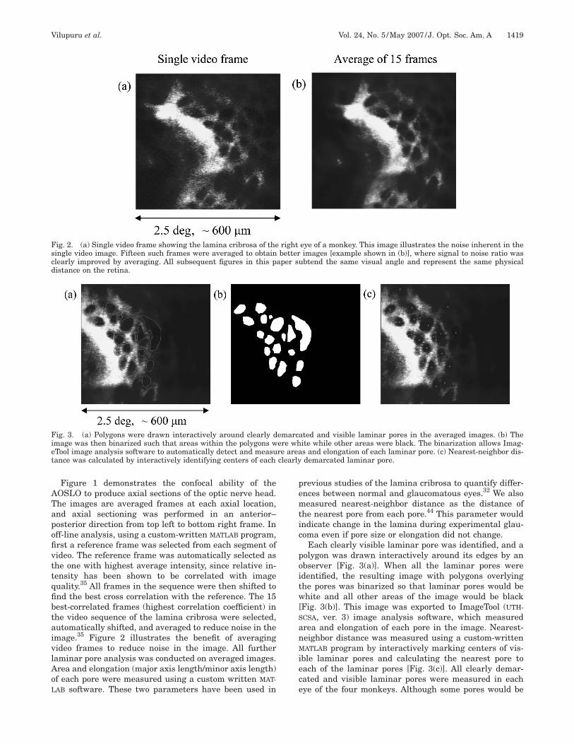
Figure 1 demonstrates the confocal ability of theOSLO to produce axial sections of the optic nerve head.he images are averaged frames at each axial location,nd axial sectioning was performed in an anterior–osterior direction from top left to bottom right frame. Inff-line analysis, using a custom-written MATLAB program,rst a reference frame was selected from each segment ofideo. The reference frame was automatically selected ashe one with highest average intensity, since relative in-ensity has been shown to be correlated with imageuality.35 All frames in the sequence were then shifted tond the best cross correlation with the reference. The 15est-correlated frames (highest correlation coefficient) inhe video sequence of the lamina cribrosa were selected,utomatically shifted, and averaged to reduce noise in themage.35 Figure 2 illustrates the benefit of averagingideo frames to reduce noise in the image. All furtheraminar pore analysis was conducted on averaged images.rea and elongation (major axis length/minor axis length)f each pore were measured using a custom written MAT-
AB software. These two parameters have been used in
ig. 2. (a) Single video frame showing the lamina cribrosa of theingle video image. Fifteen such frames were averaged to obtainlearly improved by averaging. All subsequent figures in this pistance on the retina.
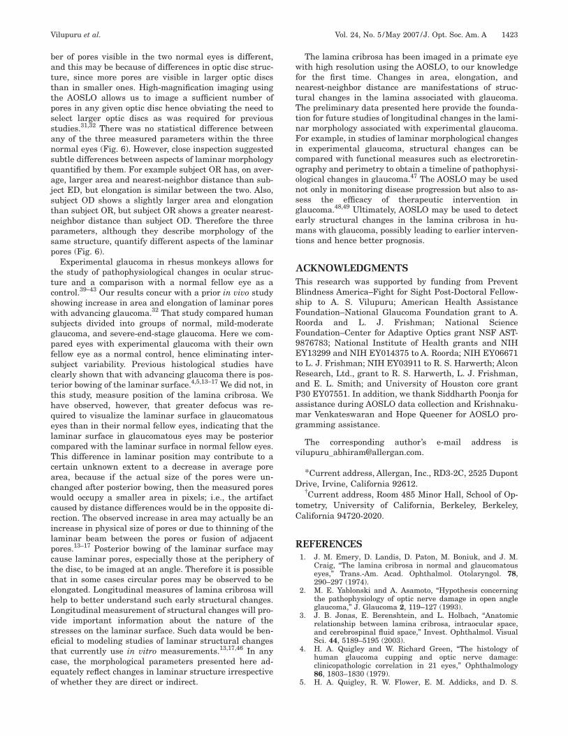
ig. 3. (a) Polygons were drawn interactively around clearly dmage was then binarized such that areas within the polygons wTool image analysis software to automatically detect and measuance was calculated by interactively identifying centers of each
revious studies of the lamina cribrosa to quantify differ-nces between normal and glaucomatous eyes.32 We alsoeasured nearest-neighbor distance as the distance of
he nearest pore from each pore.44 This parameter wouldndicate change in the lamina during experimental glau-oma even if pore size or elongation did not change.
Each clearly visible laminar pore was identified, and aolygon was drawn interactively around its edges by anbserver [Fig. 3(a)]. When all the laminar pores weredentified, the resulting image with polygons overlyinghe pores was binarized so that laminar pores would behite and all other areas of the image would be black
Fig. 3(b)]. This image was exported to ImageTool (UTH-
CSA, ver. 3) image analysis software, which measuredrea and elongation of each pore in the image. Nearest-eighbor distance was measured using a custom-writtenATLAB program by interactively marking centers of vis-
ble laminar pores and calculating the nearest pore toach of the laminar pores [Fig. 3(c)]. All clearly demar-ated and visible laminar pores were measured in eachye of the four monkeys. Although some pores would be
eye of a monkey. This image illustrates the noise inherent in theimages [example shown in (b)], where signal to noise ratio was
ubtend the same visual angle and represent the same physical
ated and visible laminar pores in the averaged images. (b) Theite while other areas were black. The binarization allows Imag-s and elongation of each laminar pore. (c) Nearest-neighbor dis-demarcated laminar pore.
rightbetter
aper s
emarcere whre area
isAfgaf
me
vBd
of tw
FiDsrPme
Fatbes
1420 J. Opt. Soc. Am. A/Vol. 24, No. 5 /May 2007 Vilupuru et al.
maged in two different videos during any given imagingession, care was taken not to reanalyze the same pore.verage and standard errors of the mean were calculated
or the three measured morphological parameters. Eachlaucomatous right and normal left eye was imaged once;verages and standard errors of mean represent datarom one imaging session.
A-scan ultrasonography was performed in all eyes of allonkeys to measure axial length. Morphological param-
ters obtained in measures of visual field angle were con-
Fig. 4. Lamina cribrosa images from two normal (left) eyes
ig. 5. (a) Altman–Bland analysis of inter-observer variabilityn the measurement of laminar pore area for the same image.ifference between observers is plotted against mean of mea-
urements. The solid line is mean of differences; dashed linesepresent two standard deviations above and below the mean. (b)lot of inter-session reproducibility of laminar area measure-ents. The vertical bars represent average laminar pore area;
rror bars show the standard error of the mean.
erted to true retinal size using the method described byennett et al.45 using the measured axial lengths. Stan-ard automated perimetry (SAP) was performed behav-
o monkeys. Laminar pores are clearly visible in both cases.
ig. 6. The three morphological parameters (area, elongation;nd nearest-neighbor distance) measured in all control eyes ofhe four monkeys ED, OR, OD, and JA. In each graph the grayars represent individual eyes and the black bar is the group av-rage of all control eyes. The bars are means and the error barshow the standard error of the mean.
iomtidmb
3Fetrsspi
psotdtwmfoaeue
Twma6nmnaac
alhidtaimeam62gtggsen
Fhg
Vilupuru et al. Vol. 24, No. 5 /May 2007/J. Opt. Soc. Am. A 1421
orally on both eyes of each of the monkeys as part ofther experiments conducted on these subjects. A Gold-ann size III target was used with white-on-white con-
rast to estimate the mean deviation or average sensitiv-ty difference from normal across the visual field. Meaneviation was used as a descriptor of the stage of experi-ental glaucoma in each of the eyes. The procedure has
een described in detail elsewhere.39–41
. RESULTSigure 4 shows the lamina cribrosa images of two normalyes. The laminar pores are clearly visible and in focus athis axial location. These images have been averaged toeduce noise as described above (see Fig. 2). Before the re-ults are reported for glaucomatous eyes, data will be pre-ented on inter-observer variability and inter-session re-eatability to validate the image analysis approach takenn this study.
Figure 5(a) presents an Altman–Bland plot for laminarore area to assess degree of agreement between two ob-ervers. The figure shows difference in area between twobservers versus mean of the two measurements from thewo observers. The dashed line represents the mean of theifferences, and the solid lines are two standard devia-ions from the mean. The finding that the mean differenceas less than zero indicates that observer B, on average,easured larger areas than observer A. However, the dif-
erence was not significant compared with the magnitudef laminar pore areas �p�0.05�. In addition, there is nopparent trend in the differences between the two observ-rs when greater magnitude of pores was measured. Fig-re 5(b) plots average laminar pore area for one normal
ye measured at two different times separated by 60 days. O
he error bars represent standard errors of mean. Thereas no statistically significant difference between the twoeasurements (t-test; p=0.95), indicating good repeat-
bility in both image acquisition and the analysis. Figurepresents data for average area, elongation, and nearest-eighbor distance for all four normal left eyes in fouronkeys. In each plot, gray bars show the average for theormal left eye of each monkey and black bars show theverage of all normal eyes. Although there was variabilitymong the four left eyes, the difference was not statisti-ally significant (ANOVA, p=0.3).
Figure 7 shows examples of the lamina cribrosa of (a)n eye (right) with experimental glaucoma and (b) its fel-ow control (left). The eye with experimental glaucomaas, on average, larger and more elongated holes, signify-
ng altered laminar morphology. The difference in meaneviation between the two eyes of this monkey accordingo behavioral SAP was 6.5 dB, which represents a moder-te loss of sensitivity across the visual field. Differencesn laminar morphology between the two eyes of all four
onkeys are plotted in Fig. 8 for (a) average area, (b)longation, and (c) nearest-neighbor distance. To reiter-te, both eyes of monkey ED are normal, the difference inean deviation between the two eyes of monkey OR is
.88 dB, of monkey OD is 6.5 dB, and of monkey JA is7.5 dB. The area of the laminar pores was significantlyreater in the three experimental glaucoma eyes than inheir respective normal fellow eyes (t-test; p�0.05). Elon-ation was also systematically greater in experimentallaucoma eyes than in their counterparts, but statisticalignificance was reached only for monkey JA, which hadnd-stage experimental glaucoma. Similarly, nearest-eighbor distance was significantly greater for monkeys
D and JA but not for monkey OR. Elongation and
ig. 7. (Color online) (a) Experimental glaucoma and (b) normal eyes of the same monkey (OD). In the two panels the central optic nerveead is imaged in the two eyes to compare the lamina cribrosa. The images illustrate that, on average, pores in the eye with experimentallaucoma have greater area and elongation than its normal fellow eye, indicating altered morphology in the glaucomatous eye.
ngO
4Tpatlbstapmlc
ncpwntrnt
pplsdtlfwatssf
vwpvpoasmcbmbwsfd
amitestmotaanbr(
mw
FdbBsi
1422 J. Opt. Soc. Am. A/Vol. 24, No. 5 /May 2007 Vilupuru et al.
earest-neighbor distance were greater in the severelylaucomatous eye of monkey JA than in monkeys OR andD with only moderate damage.
. DISCUSSIONhe AOSLO, a recently proposed imaging technology,34
rovided high-resolution, high-contrast, magnified im-ges of lamina cribrosa in both normal and experimen-ally induced glaucomatous eyes. Prior in vivo studies ofaminar morphological changes had measured differencesetween glaucomatous stages.31,32. However, the priortudies used a 20 deg field of view and were limited in ob-aining higher-magnification and higher-resolution im-ges because of ocular aberrations. In our study, laminarores were imaged using a 2.5 deg window at a higheragnification, which provided details of the anterior
aminar surface. In most eyes, ocular aberrations wereorrected without much difficulty at the level of the optic
ig. 8. (a) Average area, (b) elongation, and (c) nearest-neighboristance comparisons between experimental glaucoma eyes (grayars) and their contralateral (black bars) in all four monkeys.ars plot average laminar morphology, error bars represent thetandard error of the mean. Asterisks indicate that the differences statistically significant.
erve head, but in some cases appropriate correctionould not be achieved when the field of view used encom-assed different depth planes. In such eyes, aberrationsere measured at a retinal location adjacent to the opticerve head and the same correction was applied to imagehe lamina cribrosa. AOSLO imaging of the lamina crib-osa developed in this study provides interesting prelimi-ary data and justification for such studies of early struc-ural changes in glaucoma in the future.
Imaging the lamina cribrosa may have important ap-lication in glaucoma, as it has been postulated to be therimary site of injury to ganglion cell axons, ultimatelyeading to death of ganglion cells and blindness.1–24Serialectioning of the lamina cribrosa is illustrated in Fig. 1,emonstrating the ability of the AOSLO to optically sec-ion from the surface of the optic nerve head down to theamina cribrosa. Confocal optical sectioning identified forurther analysis the focal plane where the lamina cribrosaas at best focus. We presume that at this focal plane were imaging the anterior laminar surface, because at-empts to focus posterior to this surface reveal no newtructure. Using the current technology we are unable toection the lamina cribrosa itself to measure pores at dif-erent depths.
The 2.5 deg window employed encompassed most of theisible laminar pores at the center of the disc. In addition,e moved the eye to image laminar pores at extreme su-erior and inferior aspects of the disc to capture all of theisible laminar surface. The averaged images were post-rocessed to quantify laminar pores, ensuring that nonef the pores was included more than once in the finalnalysis. Area and elongation of laminar pores were de-cribed in a previous study as differentiating betweenild and severe glaucomatous stages.32 In addition, we
hose to measure nearest-neighbor distance, which haseen used previously as a measure of randomness in aeasurement sample.44 Nearest-neighbor distance may
e an effective identifier of change in laminar morphologyhen lamina cribrosa is affected, for example, by shear
tress causing distortion of laminar surface.4–7,14–17 In ef-ect, we sought to provide analysis parameters that wouldetect the earliest alterations in laminar morphology.Automated image analysis methodology was attempted
s suggested in prior studies31 to quantify the laminarorphology. However, applying similar analyses to our
mages failed to capture the details of the lamina visibleo the observer because of the intensity variability withinach image and between images. During each imagingession our aim was to obtain the best possible images ofhe lamina notwithstanding standardization of input lu-inance levels. Hence, even within the same eyes imaged
n different dates, average intensity levels varied, andherefore interactive identification of laminar pores waslways more reliable than automatic computerized imagenalysis to identify and measure area, elongation, andearest-neighbor distance. This approach was validatedy insignificant inter-observer variability and significantepeatability in laminar morphological measurementsFig. 5).
Images of the lamina cribrosa in two normal eyes of twoonkeys (Fig. 4) demonstrate inter-subject variability,hich is reflected in the quantification in Fig. 6. The num-
batttpssansqajstnpsp
ttcswsgpfsctthqelcTcacwcrilpcttehLvsetceo
wfntTtnFicoonsgemt
ATBsFRF9EtRaPamg
v
D
tC
R
Vilupuru et al. Vol. 24, No. 5 /May 2007/J. Opt. Soc. Am. A 1423
er of pores visible in the two normal eyes is different,nd this may be because of differences in optic disc struc-ure, since more pores are visible in larger optic discshan in smaller ones. High-magnification imaging usinghe AOSLO allows us to image a sufficient number ofores in any given optic disc hence obviating the need toelect larger optic discs as was required for previoustudies.31,32 There was no statistical difference betweenny of the three measured parameters within the threeormal eyes (Fig. 6). However, close inspection suggestedubtle differences between aspects of laminar morphologyuantified by them. For example subject OR has, on aver-ge, larger area and nearest-neighbor distance than sub-ect ED, but elongation is similar between the two. Also,ubject OD shows a slightly larger area and elongationhan subject OR, but subject OR shows a greater nearest-eighbor distance than subject OD. Therefore the threearameters, although they describe morphology of theame structure, quantify different aspects of the laminarores (Fig. 6).Experimental glaucoma in rhesus monkeys allows for
he study of pathophysiological changes in ocular struc-ure and a comparison with a normal fellow eye as aontrol.39–43 Our results concur with a prior in vivo studyhowing increase in area and elongation of laminar poresith advancing glaucoma.32 That study compared human
ubjects divided into groups of normal, mild-moderatelaucoma, and severe-end-stage glaucoma. Here we com-ared eyes with experimental glaucoma with their ownellow eye as a normal control, hence eliminating inter-ubject variability. Previous histological studies havelearly shown that with advancing glaucoma there is pos-erior bowing of the laminar surface.4,5,13–17 We did not, inhis study, measure position of the lamina cribrosa. Weave observed, however, that greater defocus was re-uired to visualize the laminar surface in glaucomatousyes than in their normal fellow eyes, indicating that theaminar surface in glaucomatous eyes may be posteriorompared with the laminar surface in normal fellow eyes.his difference in laminar position may contribute to aertain unknown extent to a decrease in average porerea, because if the actual size of the pores were un-hanged after posterior bowing, then the measured poresould occupy a smaller area in pixels; i.e., the artifact
aused by distance differences would be in the opposite di-ection. The observed increase in area may actually be anncrease in physical size of pores or due to thinning of theaminar beam between the pores or fusion of adjacentores.13–17 Posterior bowing of the laminar surface mayause laminar pores, especially those at the periphery ofhe disc, to be imaged at an angle. Therefore it is possiblehat in some cases circular pores may be observed to belongated. Longitudinal measures of lamina cribrosa willelp to better understand such early structural changes.ongitudinal measurement of structural changes will pro-ide important information about the nature of thetresses on the laminar surface. Such data would be ben-ficial to modeling studies of laminar structural changeshat currently use in vitro measurements.13,17,46 In anyase, the morphological parameters presented here ad-quately reflect changes in laminar structure irrespectivef whether they are direct or indirect.
The lamina cribrosa has been imaged in a primate eyeith high resolution using the AOSLO, to our knowledge
or the first time. Changes in area, elongation, andearest-neighbor distance are manifestations of struc-ural changes in the lamina associated with glaucoma.he preliminary data presented here provide the founda-
ion for future studies of longitudinal changes in the lami-ar morphology associated with experimental glaucoma.or example, in studies of laminar morphological changes
n experimental glaucoma, structural changes can beompared with functional measures such as electroretin-graphy and perimetry to obtain a timeline of pathophysi-logical changes in glaucoma.47 The AOSLO may be usedot only in monitoring disease progression but also to as-ess the efficacy of therapeutic intervention inlaucoma.48,49 Ultimately, AOSLO may be used to detectarly structural changes in the lamina cribrosa in hu-ans with glaucoma, possibly leading to earlier interven-
ions and hence better prognosis.
CKNOWLEDGMENTShis research was supported by funding from Preventlindness America–Fight for Sight Post-Doctoral Fellow-hip to A. S. Vilupuru; American Health Assistanceoundation–National Glaucoma Foundation grant to A.oorda and L. J. Frishman; National Scienceoundation–Center for Adaptive Optics grant NSF AST-876783; National Institute of Health grants and NIHY13299 and NIH EY014375 to A. Roorda; NIH EY06671
o L. J. Frishman; NIH EY03911 to R. S. Harwerth; Alconesearch, Ltd., grant to R. S. Harwerth, L. J. Frishman,nd E. L. Smith; and University of Houston core grant30 EY07551. In addition, we thank Siddharth Poonja forssistance during AOSLO data collection and Krishnaku-ar Venkateswaran and Hope Queener for AOSLO pro-
*Current address, Allergan, Inc., RD3-2C, 2525 Dupontrive, Irvine, California 92612.†Current address, Room 485 Minor Hall, School of Op-
ometry, University of California, Berkeley, Berkeley,alifornia 94720-2020.
EFERENCES1. J. M. Emery, D. Landis, D. Paton, M. Boniuk, and J. M.
Craig, “The lamina cribrosa in normal and glaucomatouseyes,” Trans.-Am. Acad. Ophthalmol. Otolaryngol. 78,290–297 (1974).
2. M. E. Yablonski and A. Asamoto, “Hypothesis concerningthe pathophysiology of optic nerve damage in open angleglaucoma,” J. Glaucoma 2, 119–127 (1993).
3. J. B. Jonas, E. Berenshtein, and L. Holbach, “Anatomicrelationship between lamina cribrosa, intraocular space,and cerebrospinal fluid space,” Invest. Ophthalmol. VisualSci. 44, 5189–5195 (2003).
4. H. A. Quigley and W. Richard Green, “The histology ofhuman glaucoma cupping and optic nerve damage:clinicopathologic correlation in 21 eyes,” Ophthalmology86, 1803–1830 (1979).
5. H. A. Quigley, R. W. Flower, E. M. Addicks, and D. S.
1
1
1
1
1
1
1
1
1
1
2
2
2
2
2
2
2
2
2
2
3
3
3
3
3
3
3
3
3
3
4
4
4
4
4
4
1424 J. Opt. Soc. Am. A/Vol. 24, No. 5 /May 2007 Vilupuru et al.
McLeod, “The mechanism of optic nerve damage inexperimental acute intraocular pressure elevation,” Invest.Ophthalmol. Visual Sci. 19, 505–517 (1980).
6. H. A. Quigley, E. M. Addicks, W. Richard Green, and A. E.Maumenee, “Optic nerve damage in human glaucoma II.The site of injury and susceptibility to damage,” Arch.Ophthalmol. (Chicago) 99, 635–649 (1981).
7. H. A. Quigley, R. M. Hohman, E. M. Addicks, R. W. Massof,and W. Richard Green, “Morphologic changes in the laminacribrosa correlated with neural loss in open-angleglaucoma,” Am. J. Ophthalmol. 95, 673–691 (1983).
8. H. A. Quigley, A. Brown, and M. E. Dorman-Pease,“Alterations in elastin of the optic nerve head in humanand experimental glaucoma,” Br. J. Ophthamol. 75,552–557 (1991a).
9. H. A. Quigley, M. E. Dorman-Pease, and A. E. Brown,“Quantitative study of collagen and elastin of the opticnerve head and sclera in human and experimental monkeyglaucoma,” Curr. Eye Res. 10, 877–888 (1991b).
0. M. R. Hernandez, W. M. Andrzejewska, and A. H. Neufeld,“Changes in the extracellular matrix of the human opticnerve head in primary open-angle glaucoma,” Am. J.Ophthalmol. 109, 180–188 (1990).
1. M. R. Hernandez, “The optic nerve head in glaucoma: roleof astrocytes in tissue remodeling,” Prog. Ret. Eye Res. 19,297–321 (2000).
2. J. D. O. Pena, P. A. Netland, I. Vidal, D. A. Dorr, A. Rasky,and M. R. Hernandez, “Elastosis of the lamina cribrosa inglaucomatous optic neuropathy,” Exp. Eye Res. 67,517–524 (1998).
3. A. J. Bellezza, R. T. Hart, and C. F. Burgoyne, “The opticnerve head as a biomechanical structure: initial finiteelement modeling,” Invest. Ophthalmol. Visual Sci. 41,2991–3000 (2000).
4. C. F. Burgoyne and J. C. Morrison, “The anatomy andpathophysiology of the optic nerve head in glaucoma,” J.Glaucoma 10 (Suppl. 1), S16–S18 (2001).
5. A. J. Bellezza, C. J. Rintalan, H. W. Thompson, J. C.Downs, R. T. Hart, and C. F. Burgoyne, “Deformation of thelamina cribrosa and anterior scleral canal wall in earlyexperimental glaucoma,” Invest. Ophthalmol. Visual Sci.44, 623–637 (2003).
6. J. C. Downs, J.-K. F. Suh, K. A. Thomas, A. J. Bellezza, R.T. Hart, and C. F. Burgoyne, “Viscoelastic materialproperties of the peripapillary sclera in normal and early-glaucomatous monkey eyes,” Invest. Ophthalmol. VisualSci. 46, 540–546 (2005).
7. C. F. Burgoyne, J. C. Downs, A. J. Bellezza, J.-K. F. Suh,and R. T. Hart, “The optic nerve head as a biomechanicalstructure: a new paradigm for understanding the role ofIOP-related stress and strain in the pathophysiology ofglaucomatous optic nerve head damage,” Prog. Ret. EyeRes. 24, 39–73 (2005).
8. D. R. Anderson and A. Hendrickson, “Effect of intraocularpressure on rapid axoplasmic transport in monkey opticnerve,” Invest. Ophthalmol. 13, 771–783 (1974).
9. H. A. Quigley and R. Anderson “The dynamics and locationof axonal transport blockade by acute intraocular pressureelevation in primate optic nerve,” Invest. Ophthalmol. 15,606–616 (1976).
0. D. S. Minckler, A. H. Bunt, and G. W. Johanson,“Orthograde and retrograde axoplasmic transport duringacute ocular hypertension in the monkey,” Invest.Ophthalmol. Visual Sci. 16, 426–441 (1977).
1. R. L. Radius, “Distribution of pressure-induced fast axonaltransport abnormalities in primate optic nerve: anautoradiographic study,” Arch. Ophthalmol. (Chicago) 99,1253–1257 (1981a).
2. R. L. Radius, “Regional specificity in anatomy at thelamina cribrosa,” Arch. Ophthalmol. (Chicago) 99, 478–480(1981b).
3. R. L. Radius and B. Bade, “Axonal transport interruptionand anatomy at the lamina cribrosa,” Arch. Ophthalmol.(Chicago) 100, 1661–1664 (1982).
4. D. S. Minckler, “Correlations between anatomic features
and axonal transport in primate optic nerve head,” 84,429–452 (1986).
5. J. E. Morgan, G. Jeffery, and A. J. E. Foss, “Axon deviationin the human lamina cribrosa,” Br. J. Ophthamol. 82,680–683 (1998).
6. G. Tezel, K. Trinkaus, and M. B. Wax, “Alterations in themorphology of lamina cribrosa pores in glaucomatouseyes,” Br. J. Ophthamol. 88, 251–256 (2004).
7. J. Morgan-Davies, N. Taylor, A. R. Hill, P. Aspinall, C. J.O’Brien, and A. Azuara-Blanco, “Three dimensionalanalysis of the lamina cribrosa in glaucoma,” Br. J.Ophthamol. 88, 1299–1304 (2004).
8. H. Maeda, M. Nakamura, and M. Yamamoto,“Morphometric features of laminar pores in lamina cribrosaobserved by scanning laser ophthalmoscopy,” Jpn. J.Ophthalmol. 43, 415–421 (1999).
9. F. W. Fitzke, H. Woon, G. T. Timberlake, L. Robinson, J.Marshal, and A. C. Bird, “Optical modifications to ascanning laser ophthalmoscope for high magnification,narrow optical section imaging,” Lasers LightOphthalmology 4, 7–14 (1991).
0. W. H. Woon, F. W. Fitzke, A. C. Bird, and J. Marshall,“Confocal imaging of the fundus using a scanning laserophthalmoscope,” Br. J. Ophthamol. 76, 470–474 (1992).
1. A. Bhandari, L. Fontana, F. W. Fitzke, and R. A. Hitchings,“Quantitative analysis of the lamina cribrosa in vivo usinga scanning laser opthlamoscope,” Curr. Eye Res. 16, 1–8(1997).
2. L. Fontana, A. Bhandari, F. W. Fitzke, and R. A. Hitchings,“In vivo morphometry of the lamina cribrosa and itsrelation to visual field loss in glaucoma,” Curr. Eye Res. 17,363–369 (1998).
3. J. Liang and D. R. Williams, “Aberrations and retinalimage quality of the normal human eye,” J. Opt. Soc. Am. A14, 2873–2883 (1997).
4. A. Roorda, F. Romero-Borja, W. J. Donnelly, H. Queener, T.Hebert, and M. Campbell, “Adaptive optics scanning laserophthalmoscopy,” Opt. Express 10, 405–412 (2002).
5. K. Venkateswaran, A. Roorda, and F. Romero-Borja,“Theoretical modeling and evaluation of the axialresolution of the adaptive optics scanning laserophthalmoscope,” J. Biomed. Opt. 9, 132–138 (2004).
6. F. Romero-Borja, K. Venkateswaran, A. Roorda, and T.Hebert, “Optical slicing of human retinal tissue in vivowith the adaptive optics scanning laser ophthalmoscope,”Appl. Opt. 44, 4032–4040 (2005).
7. S. Poonja, S. Patel, L. Henry, and A. Roorda, “Dynamicvisual stimulus presentation in an adaptive optics scanninglaser ophthalmoscope,” J. Refract. Surg. 21, S575–580(2005).
8. J. Martin and A. Roorda, “Direct and noninvasiveassessment of parafoveal capillary leukocyte velocity,”Ophthalmology 112, 2219–2224 (2005).
9. R. S. Harwerth, E. L. Smith III, and L. DeSantis,“Experimental glaucoma: perimetric field defects andintraocular pressure,” J. Glaucoma 6, 390–401 (1997).
0. R. S. Harwerth, L. Carter-Dawson, F. Shen, E. L. Smith III,and M. L. J. Crawford, “Ganglion cell losses underlyingvisual field defects from experimental glaucoma,” Invest.Ophthalmol. Visual Sci. 40, 2242–2250 (1999).
1. R. S. Harwerth, M. L. J. Crawford, L. J. Frishman, S.Viswanathan, E. L. Smith III, and L. Carter-Dawson,“Visual field defects and neural losses from experimentalglaucoma,” Prog. Ret. Eye Res. 21, 91–125 (2002).
2. D. Gaasterland and C. Kupfer, “Experimental glaucoma inthe rhesus monkey,” Invest. Ophthalmol. 13, 455–457(1974).
3. L. J. Frishman, F. Shen, L. Du, J. G. Robson, R. S.Harwerth, E. L. Smith III, L. Carter-Dawson, and M. L. J.Crawford, “The scotopic electroretinogram of macaque afterretinal ganglion cell loss from experimental glaucoma,”Invest. Ophthalmol. Visual Sci. 37, 125–141 (1996).
4. P. J. Diggle, Statistical Analysis of Spatial Point Patterns(Academic, 1983).
5. A. G. Bennett, A. R. Rudnicka, and D. F. Edgar,“Improvements on Littmann’s method of determining the
4
4
4
4
Vilupuru et al. Vol. 24, No. 5 /May 2007/J. Opt. Soc. Am. A 1425
size of retinal features by fundus photography,” Graefe’sArch. Clin. Exp. Ophthalmol. 232, 361–367 (1994).
6. I. A. Sigal, J. G. Flanagan, and C. Ross Ethier, “Factorsinfluencing optic nerve head biomechanics,” Invest.Ophthalmol. Visual Sci. 46, 4189–4199 (2005).
7. S. Viswanathan, L. J. Frishman, J. G. Robson, R. S.Harwerth, and E. L. Smith III, “The photopic negativeresponse in the macaque electroretinogram is reduced byexperimental glaucoma,” Invest. Ophthalmol. Visual Sci.
40, 1124–1136 (1999).
8. E. WoldeMussie, G. Ruiz, M. Wijono, and L. A. Wheeler,“Neuroprotection of retinal ganglion cells by brimonidine inrats with laser-induced chronic ocular hypertension,”Invest. Ophthalmol. Visual Sci. 42, 2849–2855 (2001).
9. W. A. Hare, E. WoldeMussie, R. N. Weinreb, H. Ton, G.Ruiz, M. Wijono, B. Feldmann, L. Zangwill, and L.Wheeler, “Efficacy and safety of memantine treatment forreduction of changes associated with experimentalglaucoma in monkey, II: structural measures,” Invest.