minerals Article Adhesion to Mineral Surfaces by Cells of Leptospirillum, Acidithiobacillus and Sulfobacillus from Armenian Sulfide Ores Arevik Vardanyan 1 , Narine Vardanyan 1 , Anna Khachatryan 1 , Ruiyong Zhang 2,3, * and Wolfgang Sand 2,4,5 1 Institute of Microbiology of SPC “Armbiotechnology” of NAS of Armenia, Gyurjyan 14, Yerevan 0056, Armenia; [email protected] (A.V.); [email protected] (N.V.); [email protected] (A.K.) 2 Aquatic Biotechnology, Biofilm Centre, University of Duisburg-Essen, Universitätsstraße 5, 45141 Essen, Germany; [email protected]3 Federal Institute for Geosciences and Natural Resources (BGR), Stilleweg 2, 30655 Hannover, Germany 4 Institute of Bioscience, Environmental Microbiology, TU Bergakademie Freiberg, Leipziger Straße 29, 09599 Freiberg, Germany 5 College of Environmental Science and Engineering, Donghua University, Shanghai 201600, China * Correspondence: [email protected] or [email protected]Received: 6 December 2018; Accepted: 20 January 2019; Published: 24 January 2019 Abstract: Bioleaching of metal sulfides is an interfacial process where adhesion and subsequent biofilm formation are considered to be crucial for this process. In this study, adhesion and biofilm formation by several acidophiles (Acidithiobacillus, Leptospirillum and Sulfobacillus) isolated from different biotopes with sulfide ores in Armenia were studied. Results showed that: (1) these bacteria adhere to pyrite surfaces to various extents. A correlation between pyrite biooxidation and adhesion of S. thermosulfidooxidans 6, L. ferriphilum CC, L. ferrooxidans ZC on pyrite surfaces is shown. It is supposed that bioleaching of pyrite by S. thermosulfidooxidans 6, L. ferriphilum CC, L. ferrooxidans ZC occurs by means of indirect leaching: by ferric iron of bacterial origin; (2) cells of At. ferrooxidans 61, L. ferrooxidans ZC and St. thermosulfidooxidans 6 form a monolayer biofilm on pyrite surfaces. The coverage of pyrite surfaces varies among these species. The order of the biofilm coverage is: L. ferrooxidans ZC ≥ At. ferrooxidans 61 > St. thermosulfidooxidans 6; (3) the extracellular polymeric substances (EPS) analysis indicates that the tested strains produce EPS, if grown either on soluble ferrous iron or solid pyrite. EPS are mainly composed of proteins and carbohydrates. Cells excrete higher amounts of capsular EPS than of colloidal EPS. In addition, cells grown on pyrite produce more EPS than ones grown on ferrous iron. Keywords: adhesion; biofilm; extracellular polymeric substances (EPS); atomic force microscopy (AFM); epifluorescence microscopy (EFM) 1. Introduction Biomining is the term used to describe the technology that uses microbes to achieve metal extraction from minerals or waste materials. Biomining of gold or other precious metals normally involves a pre-treatment process using iron-oxidizing and/or sulfur-oxidizing bacteria or archaea prior to cyanide extraction [1]. This process is known as biooxidation. Compared to the traditional mineral processing technology like ore smelting/toasting, biomining is more attractive regarding to its lower energy costs and also is more environmentally friendly. In addition, due to the autotrophic lifestyle of most bioleaching microbes, bioleaching involves less carbon footprints (CO 2 fixation by Minerals 2019, 9, 69; doi:10.3390/min9020069 www.mdpi.com/journal/minerals
Transcript
minerals
Article
Adhesion to Mineral Surfaces by Cells ofLeptospirillum, Acidithiobacillus and Sulfobacillusfrom Armenian Sulfide Ores
Arevik Vardanyan 1 , Narine Vardanyan 1, Anna Khachatryan 1, Ruiyong Zhang 2,3,*and Wolfgang Sand 2,4,5
2 Aquatic Biotechnology, Biofilm Centre, University of Duisburg-Essen, Universitätsstraße 5,45141 Essen, Germany; [email protected]
3 Federal Institute for Geosciences and Natural Resources (BGR), Stilleweg 2, 30655 Hannover, Germany4 Institute of Bioscience, Environmental Microbiology, TU Bergakademie Freiberg, Leipziger Straße 29,
09599 Freiberg, Germany5 College of Environmental Science and Engineering, Donghua University, Shanghai 201600, China* Correspondence: [email protected] or [email protected]
Received: 6 December 2018; Accepted: 20 January 2019; Published: 24 January 2019�����������������
Abstract: Bioleaching of metal sulfides is an interfacial process where adhesion and subsequentbiofilm formation are considered to be crucial for this process. In this study, adhesion and biofilmformation by several acidophiles (Acidithiobacillus, Leptospirillum and Sulfobacillus) isolated fromdifferent biotopes with sulfide ores in Armenia were studied. Results showed that: (1) these bacteriaadhere to pyrite surfaces to various extents. A correlation between pyrite biooxidation and adhesionof S. thermosulfidooxidans 6, L. ferriphilum CC, L. ferrooxidans ZC on pyrite surfaces is shown. It issupposed that bioleaching of pyrite by S. thermosulfidooxidans 6, L. ferriphilum CC, L. ferrooxidans ZCoccurs by means of indirect leaching: by ferric iron of bacterial origin; (2) cells of At. ferrooxidans61, L. ferrooxidans ZC and St. thermosulfidooxidans 6 form a monolayer biofilm on pyrite surfaces.The coverage of pyrite surfaces varies among these species. The order of the biofilm coverage is:L. ferrooxidans ZC ≥ At. ferrooxidans 61 > St. thermosulfidooxidans 6; (3) the extracellular polymericsubstances (EPS) analysis indicates that the tested strains produce EPS, if grown either on solubleferrous iron or solid pyrite. EPS are mainly composed of proteins and carbohydrates. Cells excretehigher amounts of capsular EPS than of colloidal EPS. In addition, cells grown on pyrite producemore EPS than ones grown on ferrous iron.
Biomining is the term used to describe the technology that uses microbes to achieve metalextraction from minerals or waste materials. Biomining of gold or other precious metals normallyinvolves a pre-treatment process using iron-oxidizing and/or sulfur-oxidizing bacteria or archaeaprior to cyanide extraction [1]. This process is known as biooxidation. Compared to the traditionalmineral processing technology like ore smelting/toasting, biomining is more attractive regarding toits lower energy costs and also is more environmentally friendly. In addition, due to the autotrophiclifestyle of most bioleaching microbes, bioleaching involves less carbon footprints (CO2 fixation by
autotrophs) compared to smelting operations (CO2 emission) [2]. Biomining is an increasingly appliedbiotechnological procedure for processing of ores in the mining industry (biohydrometallurgy) [3].This technology converts an insoluble valuable metal sulfide into a soluble form by means ofmicroorganisms [4]. Iron-oxidizing bacteria destroy the lattice of the sulfide minerals to make thegold available for further extraction by cyanidation [1]. Studies have shown that Leptospirillumsp. (mainly Leptospirillum ferriphilum) are the dominating iron-oxidizing bacteria in gold-bearingarsenopyrite (FeAsS) and pyrite (FeS2) in biooxidation reactors functioning at or over 40 ◦C [5–8].A high Fe3+/Fe2+ ratio, elevated temperatures (40 ◦C), as well as extremely low pH values (pH 1.0)are the most favorable conditions for the growth of the bacteria of the genus Leptospirillum [7,9].At present, this genus includes: L. ferrooxidans (Group I), L. ferriphilum, “L. rubarum” (GroupII), and “L. ferrodiazotrophum” (Group III). Bacteria of the genus Leptospirillum are Gram-negative,acidophilic, motile vibrios, which fix carbon dioxide using the energy of Fe (II) ion oxidation [5,10–13].
Recently, scientists emphasized the importance of bacteria of the genus Leptospirillum and theirmixed cultures with other bacteria for biooxidation and bioleaching processes at temperatures above40 ◦C [12,14–16].
The bacterial leaching of sulfide minerals is primarily an indirect process involving the reductionof ferric ions (Fe (III)):
MS + 2Fe3+ →M2+ + S0+ 2Fe2+
According to the indirect “contact” mechanism the oxidation of minerals is caused by Fe(III) ions complexed in extracellular polymeric substances (EPS). Thus, adhesion and biofilmformation by iron-oxidizing bacteria is essential for an efficient biooxidation or bioleaching of sulfideminerals [16–20]. The EPS mediate cell attachment to minerals as well as electrochemical reactionstaking place at mineral surfaces during bioleaching processes [16,19,21]. However, there are few dataconcerning the adhesion of leaching bacteria like Leptospirillum spp. on pyrite, as well as evidencefor a relation between their adhesion and intensity of iron-oxidizing activities in mineral bioleachingprocesses [22,23].
Most bacteria and archaea use the biofilm lifestyle in natural and artificial systems. A biofilm isconsidered to be a dynamic structure of a microbial population enclosed in a matrix [24–26]. The biofilmmatrix is a complex mixture of EPS comprised of polysaccharides, proteins, lipids, and nucleic acidsplus detritus [27]. Filling the intercellular space between cells, the EPS generate the structure of thebiofilm matrix. The primary function of the EPS is to mediate the attachment of cells onto a substrateas well as to protect the cells from unfavorable environmental influences [28]. In addition, EPS havethe function of aggregation formation and stabilization of biofilms. Depending on the mechanismsrequired for removal of EPS and their association with the cells, EPS can be subdivided into capsular(tightly bound) EPS and colloidal (loosely bound) EPS [29,30].
Biofilm formation is a prerequisite for efficient bioleaching of minerals [16,21]. Thus, in order toimprove bioleaching and better control of biocorrosion the study of EPS structure and function of EPSfrom bioleaching bacteria is of crucial importance [31].
Despite the environmental and economic importance of these bacterial activities, very littlefundamental information concerning chemical and biochemical events at the mineral surface duringdissolution due to the presence of bioleaching microorganisms is available. The aim of the work isto study the main biological properties of iron-oxidizing and sulfur-oxidizing bacteria isolated fromsulfide ores in Armenia. These include their ability to adhere on pyrite and to form biofilm and EPSduring the bioleaching process.
2. Materials and Methods
2.1. Bacterial Strains and Growth Conditions
The iron-oxidizing strains Leptospirillum ferrooxidans ZC, L. ferriphilum CC, and the ironand sulfur-oxidizing bacteria Acidithiobacillus ferrooxidans 61, At. ferrooxidans 13Zn, Sulfobacillus
Minerals 2019, 9, 69 3 of 13
thermosulfidooxidans 6 were isolated from several natural biotopes with sulfide ores in Armenia(Figure S1). The pure cultures of these bacteria were obtained by streak plate method [32]. The bacterialcharacteristics are shown in Table 1. Iron-oxidizing bacteria were cultivated in Mackintosh medium [33]containing 4.9 g/L ferrous iron at optimal temperature. In case of Sb. thermosulfidooxidans 0.02% yeastextract was added. Cells were harvested at 8000 rpm for 10 min at 20 ◦C, washed once and resuspendedin 10 mL Mackintosh medium without ferrous iron for attachment and bioleaching tests.
Table 1. Description of the bioleaching strains *.
Isolates Original Sites EnergySubstrates
OptimumpH
OptimumT (◦C)
At. ferrooxidans 61Tandzut (Polymetallic,
gold containing) ore acid drainagewater, Lori Province, Armenia
Fe2+, S0,metal sulfides
1.8–2.0 30
At. ferrooxidans 13Zn An industrial leaching pulp ofzinc concentrate, Armenia
Fe2+, S0,metal sulfides
1.8–2.0 30–35
L. ferrooxidans ZC An industrial leaching pulp ofzinc concentrate, Armenia
Fe2+,metal sulfides
1.8–2.0 30–37
L. ferriphilum CC An industrial leaching pulp ofcopper concentrate, Armenia
Fe2+,metal sulfides
1.8–2.0 37
Sb. thermosulfidooxidans 6Drmbon ore dumps (gold, copper)
in Martakert Province(Nagorno-Karabakh), Armenia
Fe2+, S0,metal sulfides
1.7–1.8 37–50
* Modified according to Reference [32].
2.2. Bacterial Adhesion Test
Cells of At. ferrooxidans 61, At. ferrooxidans 13Zn, L. ferrooxidans ZC, L. ferriphilum CC andSb. thermosulfidooxidans 6 were tested for bacterial adhesion onto the surface of pyrite grains for0 min, 30 min, 60 min, and 120 min. The number of planktonic cells was determined by direct countingunder the microscope by means of a Thoma chamber or by the most probable number (MPN).
For enumeration of iron-oxidizing bacteria, the method of ten-fold serial dilution was used.Dilutions (10−1–10−9) of samples were used for inoculation. Cultures were incubated in Mackintoshmedium. After 7–10 days of incubation the growth of bacteria was checked by light microscope(Leica, Wetzlar, Germany). Obtained results of bacterial cells were calculated using McCrady’stable [34].
The number of adhered cells was determined from the difference between the initial titre ofbacterial cells and the cells observed in the liquid phase. The adhesion was determined as thepercentage of adhered cells versus the initial cell number.
2.3. Bioleaching of Pyrite
For pyrite bioleaching experiments, strains were grown in Mackintosh medium containing Fe2+
as an energy source. In the logarithmic growth phase, the cells were collected by centrifugation at6000 rpm for 10 min. The collected biomass was washed with acidified Mackintosh medium (pH 1.8)and resuspended in the same medium. Pyrite (~95% FeS2) from the Shamlugh ore deposit in Armeniaground to 43–63 µm was used for leaching experiments. Leaching experiments were carried out in250 mL conical flasks. 50 mL Mackintosh medium without Fe2+, adjusted to pH 2.0 by H2SO4 anda bacterial suspension with 5 g pyrite grains were added to the flasks. The initial cell number was1–2 × 10⁸ cells/mL. The bioleaching experiments were carried out at 37 ◦C with shaking (180 rpm).The intensity of pyrite oxidation was estimated by the quantity of the dissolved Fe3+ and Fe2+ ionsin the medium. Sampling was performed over a period of 25–30 days. Fe2+ and Fe3+ ions weredetermined by complexometric titration by using ethylenediaminetetraacetic acid (EDTA). Total ironions were determined by atomic-absorption spectrophotometry AAS 1N (Carl Zeiss, Jena, Germany)using an air-propane-butane flame.
Minerals 2019, 9, 69 4 of 13
2.4. Pyrite Coupon Preparation
Pyrite coupons were cut from pyrite cubes from Navajun (Spain) with a diamond saw.These coupons were cleaned and sterilized according to a previous report [35]. Biofilm formationexperiments were performed with 5 × 10⁸ cells/mL. Experiments were carried out in 100-mLErlenmeyer flasks containing 50 mL Mackintosh medium (pH 1.8) at 30, 37 and 45 ◦C with shaking at130 rpm.
2.5. Visualization of Attached Cells on Pyrite Coupons
Pyrite slices were rinsed with sterile Mackintosh medium and deionized water. Cells attached topyrite coupons and their EPS were stained by Syto 9 (Invitrogen) and fluorescently labeled with Con Aaccording to a previous report [36]. Stained samples were dried at room temperature and visualizedby epifluorescence microscopy (EFM) (Zeiss, Jena, Germany) combined with atomic force microscopy(AFM) (BioMaterial™ Workstation, JPK Instruments, Berlin, Germany) for the investigation of cellmorphology and distribution on the surfaces of pyrite coupons. At least three spots of each samplewere documented.
2.6. Image Processing
Digital image analysis and a rough calculation of surface coverage were done by using theAxiovision LE 4.8 software (Zeiss, Jena, Germany) and an extended version of the software ImageJ(v1.52g, National Institutes of Health, Bethesda, USA) [37], respectively. AFM files were processedusing JPKSPM_Data_Processing_v5.0.81 (JPK Instruments, Berlin, Germany).
2.7. EPS Extraction
EPS extraction strains were grown in 10 L Mackintosh medium at 30, 37 and 45 ◦C with shakingat 130 rpm. In the stationary growth phase, cells were harvested by centrifugation for 10 min at10,000 rpm at 4 ◦C. The colloidal and capsular EPS were obtained according to a previous report [29].
Extraction of EPS of pyrite-grown cells: Bacterial strains were grown on pyrite in Mackintoshmedium. To obtain sessile cells, 10 g of pyrite grains were washed with basal salt solution. Detachmentof sessile cells from pyrite grains was performed mechanically by vortexing in 20 mL of basal saltsolution at maximum speed for 5 min [38]. Solutions were used for analyses of the EPS composition ofthe strains. The content of carbohydrates, protein, and uronic acids were determined in 1 mL aliquots.
2.8. Determination of the Chemical Composition of the EPS
Carbohydrates were determined by the Dubois method [39] using glucose as standard andmeasured at 490 nm. The protein content was determined by the Bradford method using bovine serumalbumin (BSA) as standard and measured at 595 nm [40]. The presence of uronic acids was determinedby the Blumenkrantz and Asboe-Hansen method [41]. Cell lysis was evaluated by determinationof 3-Deoxy-D-manno-oct-2-ulosonic acid (KDO) [42]. The concentration of carbohydrates, protein,and uronic acids were determined in µg per mL of culture medium.
2.9. Statistical Analysis
Experiments were done in triplicate; the results are given in mean values. Standard deviations (SD)generally amount to≤10% for adhesion,≤15% for EPS analysis, and≤6% for surface coverage analysis.
3. Results and Discussion
3.1. Adhesion of Newly Isolated Strains and Bioleaching of Pyrite
The planktonic cells of L. ferriphilum CC grown on pyrite culture decreased by 1–2 × 10⁸ cells/mLto 1 × 10⁵ cells/mL and to 1 × 10⁴ cells/mL, in 30 and 120 min (respectively) after inoculation.
Minerals 2019, 9, 69 5 of 13
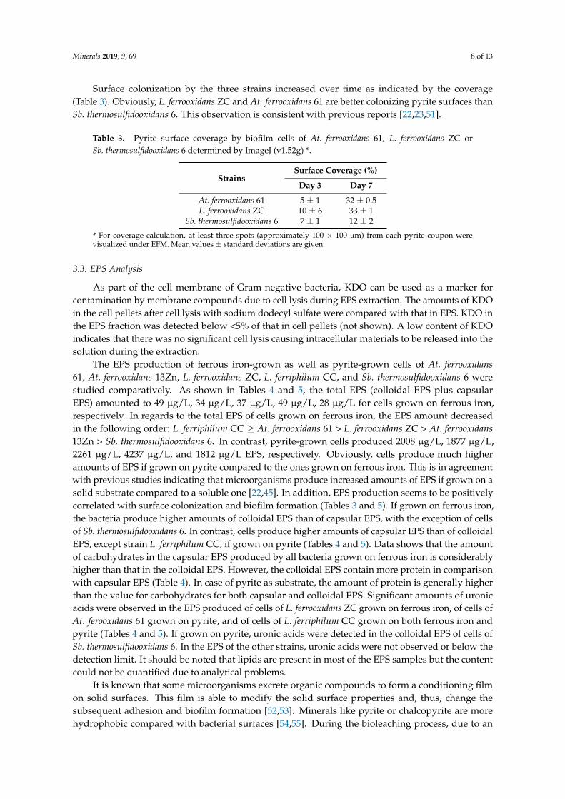
The adhesion was 99% and 100% respectively (Table 2). The initial adhesion of L. ferriphilum CCon pyrite mostly occurred within the first 30 min, whereas for cells of Sb. thermosulfidooxidans 6 andL. ferrooxidans ZC the adhesion reached only 88% and 93% after 120 min (Table 2).
Table 2. Adhesion * of four leaching isolates to pyrite determined by direct counting or most probablenumber (MPN).
DurationStrains
L. ferrooxidans ZC Sb. thermosulfidooxidans 6 At. ferrooxidans 61 L. ferriphilum CC
0 0 0 0 030 min 50 50 62 9960 min 65 61 83 99
120 min 93 88 93 100
* Values given in % of initial inoculum.
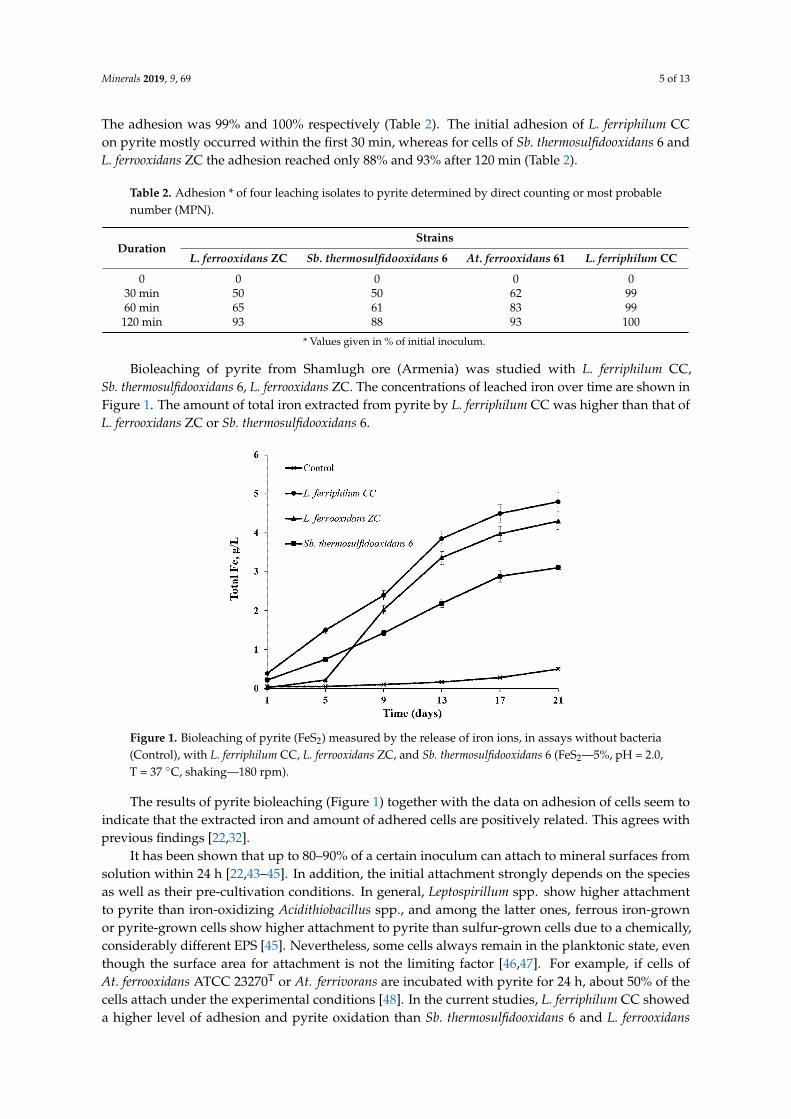
Bioleaching of pyrite from Shamlugh ore (Armenia) was studied with L. ferriphilum CC,Sb. thermosulfidooxidans 6, L. ferrooxidans ZC. The concentrations of leached iron over time are shown inFigure 1. The amount of total iron extracted from pyrite by L. ferriphilum CC was higher than that ofL. ferrooxidans ZC or Sb. thermosulfidooxidans 6.
Figure 1. Bioleaching of pyrite (FeS2) measured by the release of iron ions, in assays without bacteria(Control), with L. ferriphilum CC, L. ferrooxidans ZC, and Sb. thermosulfidooxidans 6 (FeS2—5%, pH = 2.0,T = 37 ◦C, shaking—180 rpm).
The results of pyrite bioleaching (Figure 1) together with the data on adhesion of cells seem toindicate that the extracted iron and amount of adhered cells are positively related. This agrees withprevious findings [22,32].
It has been shown that up to 80–90% of a certain inoculum can attach to mineral surfaces fromsolution within 24 h [22,43–45]. In addition, the initial attachment strongly depends on the speciesas well as their pre-cultivation conditions. In general, Leptospirillum spp. show higher attachmentto pyrite than iron-oxidizing Acidithiobacillus spp., and among the latter ones, ferrous iron-grownor pyrite-grown cells show higher attachment to pyrite than sulfur-grown cells due to a chemically,considerably different EPS [45]. Nevertheless, some cells always remain in the planktonic state, eventhough the surface area for attachment is not the limiting factor [46,47]. For example, if cells ofAt. ferrooxidans ATCC 23270T or At. ferrivorans are incubated with pyrite for 24 h, about 50% of thecells attach under the experimental conditions [48]. In the current studies, L. ferriphilum CC showeda higher level of adhesion and pyrite oxidation than Sb. thermosulfidooxidans 6 and L. ferrooxidans
Minerals 2019, 9, 69 6 of 13
ZC. The quantity of cell occurred in the following order: L. ferriphilum CC > L. ferrooxidans ZC >At. ferrooxidans 61. Sb. thermosulfidooxidans 6 showed the lowest adhesion to pyrite.
3.2. Biofilm Formation
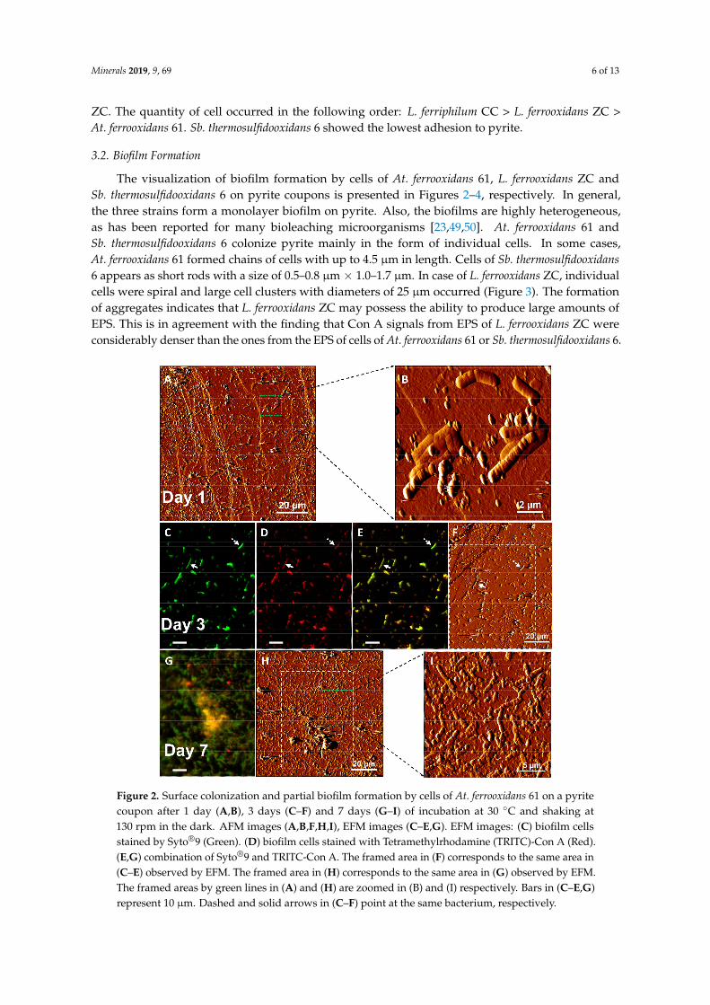
The visualization of biofilm formation by cells of At. ferrooxidans 61, L. ferrooxidans ZC andSb. thermosulfidooxidans 6 on pyrite coupons is presented in Figures 2–4, respectively. In general,the three strains form a monolayer biofilm on pyrite. Also, the biofilms are highly heterogeneous,as has been reported for many bioleaching microorganisms [23,49,50]. At. ferrooxidans 61 andSb. thermosulfidooxidans 6 colonize pyrite mainly in the form of individual cells. In some cases,At. ferrooxidans 61 formed chains of cells with up to 4.5 µm in length. Cells of Sb. thermosulfidooxidans6 appears as short rods with a size of 0.5–0.8 µm × 1.0–1.7 µm. In case of L. ferrooxidans ZC, individualcells were spiral and large cell clusters with diameters of 25 µm occurred (Figure 3). The formationof aggregates indicates that L. ferrooxidans ZC may possess the ability to produce large amounts ofEPS. This is in agreement with the finding that Con A signals from EPS of L. ferrooxidans ZC wereconsiderably denser than the ones from the EPS of cells of At. ferrooxidans 61 or Sb. thermosulfidooxidans 6.
Figure 2. Surface colonization and partial biofilm formation by cells of At. ferrooxidans 61 on a pyritecoupon after 1 day (A,B), 3 days (C–F) and 7 days (G–I) of incubation at 30 ◦C and shaking at130 rpm in the dark. AFM images (A,B,F,H,I), EFM images (C–E,G). EFM images: (C) biofilm cellsstained by Syto®9 (Green). (D) biofilm cells stained with Tetramethylrhodamine (TRITC)-Con A (Red).(E,G) combination of Syto®9 and TRITC-Con A. The framed area in (F) corresponds to the same area in(C–E) observed by EFM. The framed area in (H) corresponds to the same area in (G) observed by EFM.The framed areas by green lines in (A) and (H) are zoomed in (B) and (I) respectively. Bars in (C–E,G)represent 10 µm. Dashed and solid arrows in (C–F) point at the same bacterium, respectively.
Minerals 2019, 9, 69 7 of 13
Figure 3. Surface colonization and partial biofilm formation by L. ferrooxidans ZC on a pyrite couponafter 3 days (A–E) and 7 days (F) of incubation at 30 ◦C and shaking at 130 rpm in the dark. AFM images(D,E), EFM images (A–C,F). EFM images: (A) biofilm cells stained by Syto®9 (Green). (B) biofilm cellsstained by TRITC-Con A (Red). (C,F) combination of Syto®9 and TRITC-Con A. The framed area in(D) corresponds to the same area in (A–C) observed by EFM. The framed area in (D) is zoomed in (E).Typical morphology of spiral is shown in (E). Bars in (A–C,F) represent 10 µm.
Figure 4. Surface colonization and partial biofilm formation by Sb. thermosulfidooxidans 6 on a pyritecoupon after 3 days (A–D) and 7 days (E–H) of incubation at 30 ◦C and shaking at 130 rpm in the dark.AFM images (D,H), EFM images (A–C,E–G). EFM images: (A,E) biofilm cells stained by Syto®9 (Green).(B,F), biofilm cells stained by TRITC-Con A (Red). (C,G) combination of Syto®9 and TRITC-Con A.The framed area in (D) corresponds to the same area in (A–C) observed by EFM. Bars in (A–C,E–G)represent 10 µm. Solid arrows in (A–D) point at the same bacterium.
Minerals 2019, 9, 69 8 of 13
Surface colonization by the three strains increased over time as indicated by the coverage(Table 3). Obviously, L. ferrooxidans ZC and At. ferrooxidans 61 are better colonizing pyrite surfaces thanSb. thermosulfidooxidans 6. This observation is consistent with previous reports [22,23,51].
Table 3. Pyrite surface coverage by biofilm cells of At. ferrooxidans 61, L. ferrooxidans ZC orSb. thermosulfidooxidans 6 determined by ImageJ (v1.52g) *.
* For coverage calculation, at least three spots (approximately 100 × 100 µm) from each pyrite coupon werevisualized under EFM. Mean values ± standard deviations are given.
3.3. EPS Analysis
As part of the cell membrane of Gram-negative bacteria, KDO can be used as a marker forcontamination by membrane compounds due to cell lysis during EPS extraction. The amounts of KDOin the cell pellets after cell lysis with sodium dodecyl sulfate were compared with that in EPS. KDO inthe EPS fraction was detected below <5% of that in cell pellets (not shown). A low content of KDOindicates that there was no significant cell lysis causing intracellular materials to be released into thesolution during the extraction.
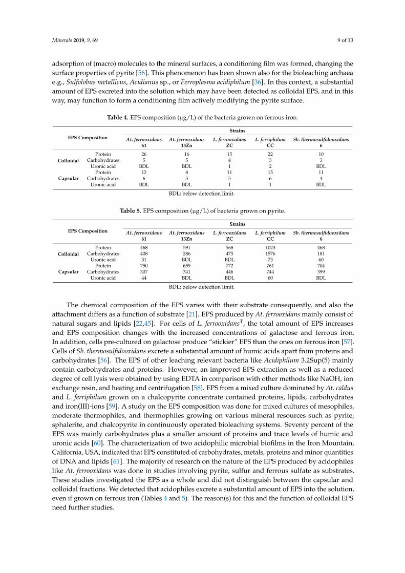
The EPS production of ferrous iron-grown as well as pyrite-grown cells of At. ferrooxidans61, At. ferrooxidans 13Zn, L. ferrooxidans ZC, L. ferriphilum CC, and Sb. thermosulfidooxidans 6 werestudied comparatively. As shown in Tables 4 and 5, the total EPS (colloidal EPS plus capsularEPS) amounted to 49 µg/L, 34 µg/L, 37 µg/L, 49 µg/L, 28 µg/L for cells grown on ferrous iron,respectively. In regards to the total EPS of cells grown on ferrous iron, the EPS amount decreasedin the following order: L. ferriphilum CC ≥ At. ferrooxidans 61 > L. ferrooxidans ZC > At. ferrooxidans13Zn > Sb. thermosulfidooxidans 6. In contrast, pyrite-grown cells produced 2008 µg/L, 1877 µg/L,2261 µg/L, 4237 µg/L, and 1812 µg/L EPS, respectively. Obviously, cells produce much higheramounts of EPS if grown on pyrite compared to the ones grown on ferrous iron. This is in agreementwith previous studies indicating that microorganisms produce increased amounts of EPS if grown on asolid substrate compared to a soluble one [22,45]. In addition, EPS production seems to be positivelycorrelated with surface colonization and biofilm formation (Tables 3 and 5). If grown on ferrous iron,the bacteria produce higher amounts of colloidal EPS than of capsular EPS, with the exception of cellsof Sb. thermosulfidooxidans 6. In contrast, cells produce higher amounts of capsular EPS than of colloidalEPS, except strain L. ferriphilum CC, if grown on pyrite (Tables 4 and 5). Data shows that the amountof carbohydrates in the capsular EPS produced by all bacteria grown on ferrous iron is considerablyhigher than that in the colloidal EPS. However, the colloidal EPS contain more protein in comparisonwith capsular EPS (Table 4). In case of pyrite as substrate, the amount of protein is generally higherthan the value for carbohydrates for both capsular and colloidal EPS. Significant amounts of uronicacids were observed in the EPS produced of cells of L. ferrooxidans ZC grown on ferrous iron, of cells ofAt. ferooxidans 61 grown on pyrite, and of cells of L. ferriphilum CC grown on both ferrous iron andpyrite (Tables 4 and 5). If grown on pyrite, uronic acids were detected in the colloidal EPS of cells ofSb. thermosulfidooxidans 6. In the EPS of the other strains, uronic acids were not observed or below thedetection limit. It should be noted that lipids are present in most of the EPS samples but the contentcould not be quantified due to analytical problems.
It is known that some microorganisms excrete organic compounds to form a conditioning filmon solid surfaces. This film is able to modify the solid surface properties and, thus, change thesubsequent adhesion and biofilm formation [52,53]. Minerals like pyrite or chalcopyrite are morehydrophobic compared with bacterial surfaces [54,55]. During the bioleaching process, due to an
Minerals 2019, 9, 69 9 of 13
adsorption of (macro) molecules to the mineral surfaces, a conditioning film was formed, changing thesurface properties of pyrite [56]. This phenomenon has been shown also for the bioleaching archaeae.g., Sulfolobus metallicus, Acidianus sp., or Ferroplasma acidiphilum [36]. In this context, a substantialamount of EPS excreted into the solution which may have been detected as colloidal EPS, and in thisway, may function to form a conditioning film actively modifying the pyrite surface.
Table 4. EPS composition (µg/L) of the bacteria grown on ferrous iron.
The chemical composition of the EPS varies with their substrate consequently, and also theattachment differs as a function of substrate [21]. EPS produced by At. ferrooxidans mainly consist ofnatural sugars and lipids [22,45]. For cells of L. ferrooxidansT, the total amount of EPS increasesand EPS composition changes with the increased concentrations of galactose and ferrous iron.In addition, cells pre-cultured on galactose produce “stickier” EPS than the ones on ferrous iron [57].Cells of Sb. thermosulfidooxidans excrete a substantial amount of humic acids apart from proteins andcarbohydrates [56]. The EPS of other leaching relevant bacteria like Acidiphilum 3.2Sup(5) mainlycontain carbohydrates and proteins. However, an improved EPS extraction as well as a reduceddegree of cell lysis were obtained by using EDTA in comparison with other methods like NaOH, ionexchange resin, and heating and centrifugation [58]. EPS from a mixed culture dominated by At. caldusand L. ferriphilum grown on a chalcopyrite concentrate contained proteins, lipids, carbohydratesand iron(III)-ions [59]. A study on the EPS composition was done for mixed cultures of mesophiles,moderate thermophiles, and thermophiles growing on various mineral resources such as pyrite,sphalerite, and chalcopyrite in continuously operated bioleaching systems. Seventy percent of theEPS was mainly carbohydrates plus a smaller amount of proteins and trace levels of humic anduronic acids [60]. The characterization of two acidophilic microbial biofilms in the Iron Mountain,California, USA, indicated that EPS constituted of carbohydrates, metals, proteins and minor quantitiesof DNA and lipids [61]. The majority of research on the nature of the EPS produced by acidophileslike At. ferrooxidans was done in studies involving pyrite, sulfur and ferrous sulfate as substrates.These studies investigated the EPS as a whole and did not distinguish between the capsular andcolloidal fractions. We detected that acidophiles excrete a substantial amount of EPS into the solution,even if grown on ferrous iron (Tables 4 and 5). The reason(s) for this and the function of colloidal EPSneed further studies.
Minerals 2019, 9, 69 10 of 13
4. Conclusions
Several isolates from biotopes of sulfide ores in Armenia show the capability to form biofilms andproduce EPS, while grown on either soluble ferrous iron or solid pyrite surfaces. The dissolution ofpyrite is correlated with the quantity of adhering/attached cells. A monolayer biofilm developed onpyrite by At. ferrooxidans 61, Sb. thermosulfidooxidans 6 and L. ferrooxidans ZC. Large cell clusters formedby L. ferrooxidans ZC indicate their ability to produce high amount of EPS on pyrite. Biochemicalanalyses of EPS of the planktonic and attached cells on pyrite grains show that the main componentsof EPS are proteins, carbohydrates and uronic acids.
Supplementary Materials: The following are available online at http://www.mdpi.com/2075-163X/9/2/69/s1,Figure S1: Sampling areas in different biotopes of sulfide ores in Armenia.
Author Contributions: A.V. and R.Z. conceived and designed the experiments; A.V., A.K., and R.Z. performedthe experiments; A.V. and N.V. analyzed the data; N.V. and W.S. contributed reagents/materials/analysis tools;A.V. and R.Z. wrote the manuscript. WS corrected the manuscript.
Funding: This work was supported by the MES-BMBF STC Grant 12GE 005 of the Ministry of Education andScience of Armenia and the German Federal Ministry of Education and Research.
Conflicts of Interest: The authors declare no conflict of interest. The funders had no role in the design of thestudy; in the collection, analyses, or interpretation of data; in the writing of the manuscript, and in the decision topublish the results.
References
1. Dew, D.W.; Lawson, E.N.; Broadhurst, J.L. The biox® process for biooxidation of gold-bearing ores orconcentrates. In Biomining; Springer: Berlin/Heidelberg, Germany, 1997; pp. 45–80.
2. Johnson, D.B. The Evolution, current status, and future prospects of using biotechnologies in the mineralextraction and metal recovery sectors. Minerals 2018, 8, 343. [CrossRef]
4. Schippers, A.; Hedrich, S.; Vasters, J.; Drobe, M.; Sand, W.; Willscher, S. Biomining: Metal recovery from oreswith microorganisms. In Geobiotechnology I: Metal-Related Issues; Schippers, A., Glombitza, F., Sand, W., Eds.;Springer: Berlin/Heidelberg, Germany, 2014; pp. 1–47.
5. Coram, N.J.; Rawlings, D.E. Molecular relationship between two groups of the genus Leptospirillum and thefinding that Leptospirillum ferriphilum sp. Nov. Dominates South African commercial biooxidation tanks thatoperate at 40 ◦C. Appl. Environ. Microbiol. 2002, 68, 838–845. [CrossRef] [PubMed]
6. Lawson, E. The composition of mixed populations of leaching bacteria active in gold and nickel recoveryfrom sulphide ores. In Proceedings of the International Biohydrometallurgy Symposium, IBS97, Sydney,Australia, 4–6 August 1997; Australian Mineral Foundation: Glenside, South Australia, 1997; p. 4.
7. Rawlings, D.; Coram, N.; Gardner, M.; Deane, S. Thiobacillus caldus and Leptospirillum ferrooxidans arewidely distributed in continuous flow biooxidation tanks used to treat a variety of metal containing ores andconcentrates. In Process Metallurgy; Elsevier: New York, NY, USA, 1999; Volume 9, pp. 777–786.
8. Schippers, A. Microorganisms involved in bioleaching and nucleic acid-based molecular methods for theiridentification and quantification. In Microbial Processing of Metal Sulfides; Donati, E., Sand, W., Eds.; Springer:Dordrecht, The Netherlands, 2007; pp. 3–33.
9. Foucher, S.; Battaglia-Brunet, F.; d’Hugues, P.; Clarens, M.; Godon, J.; Morin, D. Evolution of the bacterialpopulation during the batch bioleaching of a cobaltiferous pyrite in a suspended-solids bubble column andcomparison with a mechanically agitated reactor. Hydrometallurgy 2003, 71, 5–12. [CrossRef]
10. Goltsman, D.S.A.; Denef, V.J.; Singer, S.W.; VerBerkmoes, N.C.; Lefsrud, M.; Mueller, R.S.; Dick, G.J.; Sun, C.L.;Wheeler, K.E.; Zemla, A. Community genomic and proteomic analyses of chemoautotrophic iron-oxidizing“Leptospirillum rubarum” (Group II) and “Leptospirillum ferrodiazotrophum” (Group III) bacteria in acid minedrainage biofilms. Appl. Environ. Microbiol. 2009, 75, 4599–4615. [CrossRef] [PubMed]
11. Markosyan, G. A new iron-oxidizing bacterium, Leptospirillum ferrooxidans gen. Et sp. Nov. Biol. Zh. Arm.1972, 25, 26.
12. Vardanyan, N.; Akopyan, V. Leptospirillum-like bacteria and evaluation of their role in pyrite oxidation.Microbiology 2003, 72, 438–442. [CrossRef]
13. Sand, W.; Rohde, K.; Sobotke, B.; Zenneck, C. Evaluation of Leptospirillum ferrooxidans for leaching.Appl. Environ. Microbiol. 1992, 58, 85–92.
14. Fu, B.; Zhou, H.; Zhang, R.; Qiu, G. Bioleaching of chalcopyrite by pure and mixed cultures of Acidithiobacillusspp. and Leptospirillum ferriphilum. Int. Biodeterior. Biodegrad. 2008, 62, 109–115. [CrossRef]
15. Okibe, N.; Gericke, M.; Hallberg, K.B.; Johnson, D.B. Enumeration and characterization of acidophilicmicroorganisms isolated from a pilot plant stirred-tank bioleaching operation. Appl. Environ. Microbiol. 2003,69, 1936–1943. [CrossRef]
16. Vera, M.; Schippers, A.; Sand, W. Progress in bioleaching: Fundamentals and mechanisms of bacterial metalsulfide oxidation-part A. Appl. Microbiol. Biotechnol. 2013, 97, 7529–7541. [PubMed]
17. Sand, W.; Gehrke, T.; Jozsa, P.G.; Schippers, A. (bio)chemistry of bacterial leaching—Direct vs. Indirectbioleaching. Hydrometallurgy 2001, 59, 159–175. [CrossRef]
18. Sand, W.; Gerke, T.; Hallmann, R.; Schippers, A. Sulfur chemistry, biofilm, and the (in)direct attackmechanism—A critical evaluation of bacterial leaching. Appl. Microbiol. Biotechnol. 1995, 43, 961–966.[CrossRef]
19. Li, Q.; Wang, Q.; Zhu, J.; Zhou, S.; Gan, M.; Jiang, H.; Sand, W. Effect of extracellular polymeric substances onsurface properties and attachment behavior of Acidithiobacillus ferrooxidans. Minerals 2016, 6, 100. [CrossRef]
20. Li, Q.; Sand, W.; Zhang, R. Enhancement of biofilm formation on pyrite by Sulfobacillus thermosulfidooxidans.Minerals 2016, 6, 71. [CrossRef]
21. Zhang, R.; Bellenberg, S.; Neu, T.R.; Sand, W.; Vera, M. The biofilm lifestyle of acidophilicmetal/sulfur-oxidizing microorganisms. In Biotechnology of Extremophiles: Advances and Challenges;Rampelotto, P.H., Ed.; Springer: Basel, Switzerland, 2016; pp. 177–213.
22. Harneit, K.; Göksel, A.; Kock, D.; Klock, J.-H.; Gehrke, T.; Sand, W. Adhesion to metal sulfide surfaces by cellsof Acidithiobacillus ferrooxidans, Acidithiobacillus thiooxidans and Leptospirillum ferrooxidans. Hydrometallurgy2006, 83, 245–254. [CrossRef]
23. Li, Q.; Yang, B.; Zhu, J.; Jiang, H.; Li, J.; Zhang, R.; Sand, W. Comparative analysis of attachment tochalcopyrite of three mesophilic iron and/or sulfur-oxidizing acidophiles. Minerals 2018, 8, 406. [CrossRef]
24. Flemming, H.-C.; Neu, T.R.; Wingender, J. The Perfect Slime: Microbial Extracellular Polymeric Substances (EPS);IWA Publishing: London, UK, 2016.
25. Sutherland, I.W. Biofilm exopolysaccharides: A strong and sticky framework. Microbiology 2001, 147, 3–9.[CrossRef] [PubMed]
26. Van Wolferen, M.; Orell, A.; Albers, S.V. Archaeal biofilm formation. Nat. Rev. Microbiol. 2018, 16, 699–713.[CrossRef]
27. Flemming, H.-C.; Wingender, J.; Szewzyk, U.; Steinberg, P.; Rice, S.A.; Kjelleberg, S. Biofilms: An emergentform of bacterial life. Nat. Rev. Microbiol. 2016, 14, 563–575. [CrossRef]
28. Flemming, H.C.; Neu, T.R.; Wozniak, D.J. The EPS matrix: The “house of biofilm cells”. J. Bacteriol. 2007,189, 7945–7947. [CrossRef] [PubMed]
29. Castro, L.; Zhang, R.; Muñoz, J.A.; González, F.; Blázquez, M.L.; Sand, W.; Ballester, A. Characterization ofexopolymeric substances (EPS) produced by Aeromonas hydrophila under reducing conditions. Biofouling2014, 30, 501–511. [CrossRef] [PubMed]
30. Sheng, G.-P.; Yu, H.-Q.; Li, X.-Y. Extracellular polymeric substances (EPS) of microbial aggregates in biologicalwastewater treatment systems: A review. Biotechnol. Adv. 2010, 28, 882–894. [CrossRef] [PubMed]
31. Sand, W.; Gehrke, T. Extracellular polymeric substances mediate bioleaching/biocorrosion via interfacialprocesses involving iron(III) ions and acidophilic bacteria. Res. Microbiol. 2006, 157, 49–56. [CrossRef][PubMed]
32. Vardanyan, A.; Stepanyan, S.; Vardanyan, N.; Markosyan, L.; Sand, W.; Vera, M.; Zhang, R. Study andassessment of microbial communities in natural and commercial bioleaching systems. Miner. Eng. 2015,81, 167–172. [CrossRef]
35. Zhang, R.; Bellenberg, S.; Castro, L.; Neu, T.R.; Sand, W.; Vera, M. Colonization and biofilm formation of theextremely acidophilic archaeon Ferroplasma acidiphilum. Hydrometallurgy 2014, 150, 245–252. [CrossRef]
36. Zhang, R.Y.; Neu, T.R.; Bellenberg, S.; Kuhlicke, U.; Sand, W.; Vera, M. Use of lectins to in situ visualizeglycoconjugates of extracellular polymeric substances in acidophilic archaeal biofilms. Microb. Biotechnol.2015, 8, 448–461. [CrossRef]
37. Abramoff, M.D.; Magalhaes, P.J.; Ram, S.J. Image processing with imagej. Biophotonics Intern. 2004, 11, 36–42.38. Bellenberg, S.; Florian, B.M.; Vera, M.A.; Rohwerder, T.; Sand, W. Comparative study of planktonic and
sessile cells from pure and mixed cultures of Acidithiobacillus ferrooxidans and Acidiphilium cryptum growingon pyrite. Adv. Mater. Res. 2009, 71–73, 333–336. [CrossRef]
39. Dubois, M.; Gilles, K.A.; Hamilton, J.K.; Rebers, P.; Smith, F. Colorimetric method for determination of sugarsand related substances. Anal. Chem. 1956, 28, 350–356. [CrossRef]
40. Bradford, M.M. A rapid and sensitive method for the quantitation of microgram quantities of proteinutilizing the principle of protein-dye binding. Anal. Biochem. 1976, 72, 248–254. [CrossRef]
41. Blumenkrantz, N.; Asboe-Hansen, G. New method for quantitative determination of uronic acids.Anal. Biochem. 1973, 54, 484–489. [CrossRef]
42. Karkhanis, Y.D.; Zeltner, J.Y.; Jackson, J.J.; Carlo, D.J. A new and improved microassay to determine2-keto-3-deoxyoctonate in lipopolysaccharide of gram-negative bacteria. Anal. Biochem. 1978, 85, 595–601.[CrossRef]
43. DiSpirito, A.A.; Dugan, P.R.; Tuovinen, O.H. Sorption of Thiobacillus ferrooxidans to particulate material.Biotechnol. Bioeng. 1983, 25, 1163–1168. [CrossRef] [PubMed]
44. Baldensperger, J.; Guarraia, L.; Humphreys, W. Scanning electron microscopy of thiobacilli grown oncolloidal sulfur. Arch. Microbiol. 1974, 99, 323–329. [CrossRef] [PubMed]
45. Gehrke, T.; Telegdi, J.; Thierry, D.; Sand, W. Importance of extracellular polymeric substances fromThiobacillus ferrooxidans for bioleaching. Appl. Environ. Microbiol. 1998, 64, 2743–2747.
46. Sand, W.; Gehrke, T.; Hallmann, R.; Schippers, A. Towards a novel bioleaching mechanism. Miner. Process.Extr. Metall. Rev. 1998, 19, 97–106. [CrossRef]
47. Liu, J.; Li, Q.; Sand, W.; Zhang, R. Influence of Sulfobacillus thermosulfidooxidans on initial attachment andpyrite leaching by thermoacidophilic archaeon Acidianus sp. DSM 29099. Minerals 2016, 6, 76. [CrossRef]
48. Bellenberg, S.; Barthen, R.; Boretska, M.; Zhang, R.; Sand, W.; Vera, M. Manipulation of pyrite colonizationand leaching by iron-oxidizing Acidithiobacillus species. Appl. Microbiol. Biotechnol. 2015, 99, 1435–1449.
49. Noël, N.; Florian, B.; Sand, W. AFM & EFM study on attachment of acidophilic leaching organisms.Hydrometallurgy 2010, 104, 370–375.
50. Florian, B.; Noël, N.; Thyssen, C.; Felschau, I.; Sand, W. Some quantitative data on bacterial attachment topyrite. Miner. Eng. 2011, 24, 1132–1138. [CrossRef]
51. Bellenberg, S.; Buetti-Dinh, A.; Galli, V.; Ilie, O.; Herold, M.; Christel, S.; Boretska, M.; Pivkin, I.V.; Wilmes, P.;Sand, W. Automated microscopic analysis of metal sulfide colonization by acidophilic microorganisms.Appl. Environ. Microbiol. 2018, 84, e01835–e01818. [CrossRef] [PubMed]
52. Bakker, D.P.; Busscher, H.J.; van Zanten, J.; de Vries, J.; Klijnstra, J.W.; van der Mei, H.C. Multiple linearregression analysis of bacterial deposition to polyurethane coatings after conditioning film formation in themarine environment. Microbiology 2004, 150, 1779–1784. [CrossRef] [PubMed]
53. Garg, A.; Jain, A.; Bhosle, N.B. Chemical characterization of a marine conditioning film. Int. Biodeterior.Biodegrad. 2009, 63, 7–11. [CrossRef]
54. Diao, M.; Taran, E.; Mahler, S.; Nguyen, T.A.; Nguyen, A.V. Quantifying adhesion of acidophilic bioleachingbacteria to silica and pyrite by atomic force microscopy with a bacterial probe. Colloids Surf. B Biointerfaces2014, 115, 229–236. [CrossRef] [PubMed]
55. Zhu, J.; Li, Q.; Jiao, W.; Jiang, H.; Sand, W.; Xia, J.; Liu, X.; Qin, W.; Qiu, G.; Hu, Y. Adhesion forces betweencells of Acidithiobacillus ferrooxidans, Acidithiobacillus thiooxidans or Leptospirillum ferrooxidans and chalcopyrite.Colloids Surf. B Biointerfaces 2012, 94, 95–100. [CrossRef]
56. Li, Q.; Sand, W. Mechanical and chemical studies on EPS from Sulfobacillus thermosulfidooxidans: Fromplanktonic to biofilm cells. Colloids Surf. B Biointerfaces 2017, 153, 34–40. [CrossRef]
57. Aguirre, P.; Guerrero, K.; Sanchez-Rodriguez, A.; Gentina, J.C.; Schippers, A. Making sticky cells: Effect ofgalactose and ferrous iron on the attachment of Leptospirillum ferrooxidans to mineral surfaces. Res. Microbiol.2018, 169, 569–575. [CrossRef]
58. Tapia, J.; Munoz, J.; Gonzalez, F.; Blázquez, M.; Malki, M.; Ballester, A. Extraction of extracellular polymericsubstances from the acidophilic bacterium Acidiphilium 3.2 sup (5). Water Sci. Technol. 2009, 59, 1959–1967.[CrossRef]
59. Zeng, W.; Qiu, G.; Zhou, H.; Liu, X.; Chen, M.; Chao, W.; Zhang, C.; Peng, J. Characterization of extracellularpolymeric substances extracted during the bioleaching of chalcopyrite concentrate. Hydrometallurgy 2010,100, 177–180. [CrossRef]
60. Govender, Y.; Gericke, M. Extracellular polymeric substances (EPS) from bioleaching systems and itsapplication in bioflotation. Miner. Eng. 2011, 24, 1122–1127. [CrossRef]