Adipose-derived mesenchymal stem cellsand platelet-rich plasma synergisticallyameliorate the surgical-inducedosteoarthritis in Beagle dogsSungho Yun1, Sae-Kwang Ku2 and Young-Sam Kwon1,3*
Abstract
Background: The purpose of this study is to investigate the clinical effects of platelet-rich plasma (PRP) andadipose-derived mesenchymal stem cell (MSC) as the fundamental treatment of osteoarthritis (OA).
Methods: Twenty four Beagle dogs were used as cranial cruciate ligament transection models. The dogs were dividedinto four groups (n = 6) according to the intra-articular injection materials: the control group with phosphate-bufferedsaline (PBS), the PRP group with PRP, the MSC group with MSCs emerged in PBS, and the MSC and PRP co-treatment(MP) group with MSCs and PRP.
Results: Lameness score, focal compression strength, articular extracellular matrix (ECM) compositions, histopathology,and real-time PCR were used to evaluate the effects of PRP and MSCs on canine OA. In the order of MP, PRP, and MSCgroup, these all showed positive effects on the evaluated categories. The lameness scores were lower, and the focalcompression strengths of the affected femoral articular surface cartilages were higher than those in the OA controlgroup. Also, the inflammatory changes, when evaluated with Mankin scoring and histomorphologic examination, weresignificantly ameliorated with the treatment of PRP and/or MSCs. The glycosaminoglycan and collagen composition ofextracellular matrix was more favorable in the test groups. The ECM-related genes significantly increased through theup-regulation, while the protein expressions of inflammatory cytokines were decreased through the inhibitory effectsof PRP and MSCs on chondrocyte apoptosis and inflammatory cytokines.
Conclusions: Taken together, this study suggests that PRP and MSCs treatments have a beneficial effect on OA via thestimulation of ECM synthesis and chondrocyte proliferation and via the inhibition of inflammatory reaction.
BackgroundOsteoarthritis (OA) is the most common clinical syn-drome of joint pain and dysfunction, accompanied byvarying degrees of functional limitation and the re-duced quality of life. Because it is mostly irreversibleand progressive, OA joint consequently loses theircartilage layer [1].
The most ideal treatment of OA is focused on block-ing the catabolic activity of cartilage and enhancing re-generation of normal cartilage. Until now, therapies forthe OA commonly focused on the palliation of pain anddiscomfort, improvement on functional movement, andprevention of further degeneration. Thus, the primaryapproach in the clinical treatment of OA involves theextensive use of NSAIDs, analgesics, and hyaluronanwhich allows the brief symptomatic relief but providesno apparent disease-modifying effect [2–4]. Therefore,there is a critical need for the development of the alter-native agents that can fundamentally prevent the de-struction of cartilages or stimulate its proper repair. In
* Correspondence: [email protected] of Veterinary Surgery, College of Veterinary Medicine,Kyungpook National University, Daegu 702-701, Republic of Korea3Stem Cell Therapeutic Research Institute, Kyungpook National University,Daegu 702-701, Republic of KoreaFull list of author information is available at the end of the article
these aspects, various efforts have been tried to searchfor the effective cartilage-preserving methods with cellsources.To sum up, the isolated chondrocyte expansion and
implantation method is regarded as the fundamental so-lution. However, the main concern of the most culturedchondrocytes is losing the characteristics of producinghyaline-like cartilage [5, 6]. In a canine model, it hasbeen reported that cultured autologous chondrocytesfailed to return to the normal hyaline cartilage [7].In human, the intra-articular injection of 1.0 × 10⁸ cells
of adipose-derived mesenchymal stem cell (MSC) couldimprove the function, reduce the pain, and regeneratethe hyaline-like cartilage [8]. MSCs are typically not ob-served in synovial fluid. However, MSCs appear in syn-ovial fluid in the OA condition, and they are thought toplay an important role in the regeneration of damagedtissue and have anti-inflammatory effect [9]. As the pa-tient ages, the quality and quantity of MSCs decreaseand there is a reduction of their ability to proliferate anddifferentiate. Also, the depletion of healthy MSCs isregarded as one of the reasons worsening OA [10]. Forthese reasons, it is believed that the direct administra-tion of MSCs could promote the positive role of themby preventing their depletion and improving cartilageregeneration [11].The platelet-rich plasma (PRP) is defined as the plasma
with the platelet population of more than 1.0 × 10⁶
cells/μl, and it has typically four- to eightfold moreplatelets than the normal plasma [12]. PRP has vari-ous growth factors such as platelet-derived growthfactor and transforming growth factor beta [13]. Ithas been well known that PRP has angiogenic, anti-inflammatory, and anti-catabolic effects [13, 14]. Ithas also been reported that transforming growth fac-tor beta and fibroblast growth factor from PRP havean anabolic effect on cartilage metabolism [15]. Thesefactors not only regulate the cell migration and prolif-eration but also enhance the wound healing andextracellular matrix (ECM) remodeling via the stimu-lation of angiogenesis [16, 17].Based on the previous studies, it is hypothesized that
the PRP could have a synergistic effect on the cartilageregeneration with the combination of MSCs. Therefore,the purpose of this study is to examine the effect of PRPand MSCs on the morphologic change and regenerationof articular cartilage in the inflammation process usingcanine OA model.
MethodsPRP preparationAutologous PRP was prepared in each dog using doublespin method. Fifty milliliters of fresh blood was collectedwith 7 ml of acid citrate dextrose formula A. Then, the
blood was centrifuged at 1200 rpm for 10 min into threelayers: plasma, buffy coat, and red blood cell. After theplasma and buffy coat were separated into a new tube,the mixture was centrifuged at 2500 rpm for 10 min.Discarding the supernatant, only the lower 20 % of theplasma was harvested. The collected plasma (PRP) wastested using complete blood cell count test (Cell-Dyne,Abbott Lab., USA) to make sure it had 1.0 × 10⁶ plate-lets/μl or more. All prepared PRP in this study wereused within 6 h.
Adipose-derived mesenchymal stem cell isolation andcultureApproximately 15 g of fat tissue was aseptically collectedfrom the flank of a dog. It was rinsed with phosphate-buffered saline (PBS) several times to remove anyremaining anesthetic agent and blood. Rinsed fat tissuewas then digested using 0.075 % collagenase type I (colla-genase type 1A, Sigma-Aldrich, USA) in 37 °C water-chamber for 2 h with shaking or inverting every 30 min.After adding the equal volumes of Dulbecco modifiedEagle medium and 10 % fetal bovine serum, it was centri-fuged at 1200 rpm for 10 min. The supernatant anddigested lipids were discarded. The cell pellet was washedwith PBS, and it was filtered through 100-μm nylon mesh.After centrifuging at the same condition, the cells weresuspended into a 100 × 20 mm cell culture dish with low-glucose Dulbecco modified Eagle medium and 10 % fetalbovine serum. After 24 h, non-adherent cells and debriswere washed with PBS, and cell culture media was re-placed twice per week. MSCs were collected and used be-tween passage 1 and 2 in all the experiments of this paper.The flow cytometry analysis with established MSCs wasperformed, and obtained MSCs were negative for clusterof differentiation (CD) 34 and CD45 and strongly positivefor CD29 and CD44 (Additional file 1: Fig. S6).
The canine model of cranial cruciate ligament transectionThe procedures were approved by Institutional AnimalCare and Use Committees of Kyungpook National Uni-versity. Twenty four physically healthy Beagle dogs wereused in this experiment. The weights of dogs were 7.7 ±1.1 kg (mean ± standard deviation), and ages werebetween 2–3 years old. Under the anesthetic state, thecranial cruciate ligament of a right hind limb was ex-cised with a no. 11 scalpel blade. The connective tissuesand skin were sutured with routine procedure. Analge-sics (tramadol 8 mg/kg BID, subcutaneous) and antibi-otics (enrofloxacin 5 mg/kg SID, subcutaneous) wereadministrated for 3 days after the surgery. After a weekfor soft tissue healing, each dog regularly walked for10 min per day for 2 months. Then, treatment was givenevery week for 1 month. Another 2 months later, thedogs were sacrificed and stifle samples were collected.
Yun et al. Journal of Orthopaedic Surgery and Research (2016) 11:9 Page 2 of 12
MSC and PRP applicationAfter the canine OA model, the subjects were treatedevery week for 1 month with an intra-articular injectionwith each material according to the groups: the controlgroup with 1 ml of PBS, the PRP group with 1 ml ofPRP, the MSC group with 1.0 × 10⁷ MSCs in 1 ml ofPBS, and the MSC and PRP co-treatment (MP) groupwith 1.0 × 10⁷ MSCs in 1 ml of PRP. The contralateralstifle joint of the dogs in control group were used as asham group in a histopathological examination, and notreatment material was given.
EvaluationsLameness scoreThe lameness score was measured before the surgeryand by every month after the surgery. All dogs had nor-mal gait and no lameness before surgery. Previously de-scribed scoring system was used [18], and is as followed:0, no detectable lameness; 1, minor lateral weight shiftbut no lameness at walk and trot; 2, no lameness at awalk but mild lameness at a trot; 3, mild lameness at awalk and significant lameness at trot; 4, non-weightbearing at a trot; and 5, non-weight bearing at walk andstanding. As a blind test, three veterinarians assessed thegrade of lameness.
The measurement of focal compressive strengthsAfter sacrifice and sampling, the ex vivo compressionstrengths of the femoral and tibial articular surfaces(0.2 mm) of each sample were detected with a comput-erized testing machine (SV-H1000, Japan Instrumenta-tion System Co., Tokyo, Japan) as N (Newton). Themeasured points were central region of the medial fem-oral and tibial condyle.
Histological processThe articular cartilages of central region of the lateralfemoral and tibial condyle were taken from kneejoints of each group, and they were separately fixedin 10 % neutral buffered formalin (NBF), and thendecalcified in decalcifying solution (24.4 % formic acidand 0.5 N sodium hydroxide) for 14 days (mixed de-calcifying solution was exchanged once a day for14 days). Each femoral and tibial articular surface car-tilage was longitudinally trimmed, then embedded inparaffin, sectioned (3–4 μm) using tungsten bladderequipped automated polycut microtome (ModelRM2255, Leica, Wetzlar, Germany), and stained withSirius red stain for cartilaginous tissues. In each pre-pared histological samples, the histological profileswere interpreted under a light microscope (ModelEclipse 80i, Nikkon, Tokyo, Japan) as blinds to groupdistribution when this analysis was made.
Analysis of ECM compositionsSome parts of the cartilage on the femoral and tibial ar-ticular surface taken from the knee joints of each groupwere separately lyophilized to obtain the dry weight.Then, the piece was digested and used for glycosamino-glycan (GAG) and collagen (COL) analyses [19, 20]. Theconcentration of GAG was determined through the di-methyl-methylene blue sulfated GAG assay using a UV/VIS spectrophotometer (Optizen Pop, Mecasys, Daejeon,Korea). Collagen content was determined by Erlich’s hy-droxyproline assay [21]. The hydroxyproline content wasconverted to the collagen content using the followingequation: (μg hydroxyproline × dilution factor)/0.13 = μgcollagen, based on the fact that hydroxyproline repre-senting approximately 13 % of the amino acid content ofcollagen in the human meniscus [22]. The concentra-tions of GAG and collagen were standardized to tissuedry weight and expressed as microgram per milligram toallow comparison among the experimental groups.
ECM-related chondrogenic gene mRNA expressionsThe SOX9 and aggrecan messenger RNA (mRNA) ex-pressions on the femoral and tibial articular surface car-tilages were detected using real-time PCR. Briefly, RNAwas extracted using Trizol reagent (Invitrogen, Carlsbad,CA, USA). The RNA concentrations and quality weredetermined by CFX96™ Real-Time System (Bio-Rad,Hercules, CA, USA). To remove contaminating DNA,samples were treated with recombinant DNase I (DNA-free; Ambion, Austin, TX, USA). RNA was reverse tran-scribed using the reagent High-Capacity cDNA ReverseTranscription Kit (Applied Biosystems, Foster City, CA,USA) according to the manufacturer’s instructions. ThecDNA strand was synthesized from the total RNA andthen the mixture of primers and the cDNA productswas amplified by PCR, and the conditions of PCR ampli-fication were 58 °C for 30 min, 94 °C for 2 min, 35 cyclesof 94 °C for 15 s, 60 °C for 30 s, 68 °C for 1 min, andthen 72 °C for 5 min. Analysis was carried out using ABIStep One Plus Sequence Detection System (Applied Bio-systems, Foster City, CA, USA), and their expressionlevels were calculated as relative to sham group. Theexpression of glyceraldehyde 3-phosphate dehydrogenase(G3PDH) mRNA was used as a control for tissue integ-rity in all of the samples. The sequences of the PCRoligonucleotide primers were as listed in Table 1.
BrdU uptake measurementTo assess the effects of MSC and PRP or their co-treatment (MP) on the proliferation of cells within thedog knee joints, proliferating cells were labeled by anintraperitoneal injection of 5-bromo-2′-deoxyuridine(BrdU). Dogs were given intraperitoneal injections ofBrdU (Sigma-Aldrich, St. Louise, MO, USA) 50 mg/kg,
Yun et al. Journal of Orthopaedic Surgery and Research (2016) 11:9 Page 3 of 12
in a volume of 1 ml/kg and dissolved in saline, and theanimals were sacrificed 72 h later. BrdU uptakes weredetected with immunohistochemistry using an anti-BrdU antibody, as shown in histomorphometry sections.
ImmunohistochemistryImmunoreactivity for BrdU as cell proliferating markerwas considered using purified primary antibody withavidin-biotin-peroxidase complex (ABC). Immunoreactiv-ities in the prepared femoral and tibial surface cartilagetissues against caspase-3, cleaved poly(ADP-ribose) poly-merase (PARP), tumor necrosis factor (TNF)-α, cyclooxy-genase (COX)-2, interleukin (IL)-1β, interferon (IFN)-γ,and inducible nitric oxide synthase (iNOS) were also add-itionally observed after treatment MSCs, PRP, or theircombination. Briefly, endogenous peroxidase activity wasblocked by incubated in methanol and 0.3 % H2O2 for30 min, and non-specific binding of immunoglobulin wasblocked with normal horse serum blocking solution for1 h in humidity chamber after epitope retrievals by pre-treatment of trypsin (Sigma-Aldrich, St. Louise, MO,USA) and 2 N HCl, on the prepared unstained sections.The primary antisera were treated for overnight at 4 °C inhumidity chamber and then incubated with biotinylateduniversal secondary antibody and ABC reagents for 1 h atroom temperature in humidity chamber. Finally, sectionswere reacted with peroxidase substrate kit for 3 min atroom temperature. All sections were rinse in 0.01 M PBSfor three times, between each step. The primary antiseraand detection kits for immunohistochemistry used in thisstudy were described in Additional file 1: Table S1.
HistomorphometryTo observe more detailed histopathological changes, thearticular cartilage injuries stained with Safranin O stain-ing were evaluated and recorded using the Mankin scor-ing systems referred by the other studies [23]. With thissystem, the higher the score, the higher the level of OA(semiquantative scores; max = 12). The thicknesses offemur and tibia articular cartilages (micrometer per car-tilage) were measured with the histomorphometricalanalyses at prepared longitudinally trimmed samples,
using a computer based automated image analyzer(iSolution FL ver 9.1, IMT i-solution Inc., Vancouver,Quebec, Canada). A total of six histological regions offemoral and tibial articular surface regions of eachgroup were considered for further analysis. The cellsoccupied by over 20 % of immunoreactivities, thedensity, of each antibody for caspase-3, PARP, TNF-α,COX-2, IL-1β, IFN-γ, and iNOS were regarded aspositive, and the numbers of each immunoreactivecells were counted separately in each of the femoraland tibial articular surface regions as cells per squaremillimeter, under blinds condition.
Statistical analysisThe values were expressed as mean ± standard deviation(SD). A multiple comparison tests were conducted fordifferent groups. According to the result of variancehomogeneity by the Levene test, one-way ANOVA testand the least-significant differences (LSD) multi-comparison test were used for parametric comparison,and Kruskal-Wallis H test was used for non-parametriccomparisons, followed by Mann-Whitney (MW) U testwith Bonferroni correction. Statistical analyses were con-ducted using SPSS for Windows (Release 14.0K, IBMSPSS Inc., USA).
ResultsAlthough the lameness score in MP group was de-creased compared to those in the other groups, therewere no significant changes between groups. The lame-ness score was significantly decreased at 2 months andat 3 months after treatment in the PRP group and MPgroup when compared with the previous treatment,respectively (Fig. 1).The focal compressive strength of the femoral and
tibial articular surface cartilages were significantly de-creased in the control group as compared to that ofthe sham group. However, the focal compressivestrength significantly increased by the treatment of allthree test materials. In MP group, the focal compres-sive strength was higher than those in any othertreated groups (Fig. 2).The Mankin scores of control group were significantly
increased as compared with those of shame group.When compared with control group, it was found thatthe Mankin score significantly reduced with the treat-ment of all three test materials on the femoral and tibialarticular cartilages. Especially, the Mankin score of MPtreated dogs was lowest compared to those of MSC orPRP treated dogs (Table 2).The thickness of articular cartilages was higher in
the MSC and PRP than control group. The more fa-vorable effect on the articular surface was examined
Table 1 Oligonucleotides for real-time PCR used in this study
Target 5′–3′ Sequence NCBI accessionno.
SOX9 Sense AAGCTCTGGAGGCTGCTGAA NM_001002978.1
Antisense ACTTGTAATCCGGGTGGTCTTTC
Aggrecan Sense CTATGAGGACGGCTTTCACC U65989.2
Antisense AGACCTCACCCTCCATCTCC
G3PDH Sense TATTGTCGCCATCAATGACC NM_01003142
Antisense TACTCAGCACCAGCATCACC
PCR polymerase chain reaction, NCBI National Center forBiotechnology Information
Yun et al. Journal of Orthopaedic Surgery and Research (2016) 11:9 Page 4 of 12
Fig. 1 The lameness score of the OA dogs. Control, treated with 1 ml of PBS; PRP, treated with 1 ml of PRP; MSC, treated with 1.0 × 10⁷ MSC in1 ml of PBS; MP, treated with 1.0 × 10⁷ MSC cell in 1 ml of PRP. Asterisk indicates significantly different with day 0 within PRP group by MW test;Number sign significantly different with day 0 within MP group by MW test
Fig. 2 Focal compressive strengths on the femoral and tibial articular cartilages. Values are expressed as mean ± SD of six dogs, N (Newton). ap < 0.05as compared with sham control by LSD test; bp < 0.05 as compared with control by LSD test; cp < 0.05 as compared with MSC treated group by LSDtest; dp < 0.01 and ep < 0.05 as compared with PRP treated group by LSD test
Yun et al. Journal of Orthopaedic Surgery and Research (2016) 11:9 Page 5 of 12
Table 2 Mankin scores on the femur and tibia articular cartilages
Values are expressed as mean ± SD of six dogsMSC mesenchymal stem cells, PRP platelet-rich plasma, MP MSC and PRP co-treatmentap < 0.01 as compared with sham control by LSD testbp < 0.05 as compared with sham control by LSD testcp < 0.01 as compared with control by LSD testdp < 0.05 as compared with control by LSD testep < 0.01 as compared with MSC treated group by LSD testfp < 0.05 as compared with MSC treated group by LSD testgp < 0.01 as compared with PRP treated group by LSD testhp < 0.05 as compared with PRP treated group by LSD test
Yunet
al.JournalofOrthopaedic
Surgeryand
Research (2016) 11:9
Page6of
12
in MP group, compared to those of MSC and PRPgroups (Fig. 3).The contents of COL and GAG as the main compo-
nent of ECM were significantly decreased in controlgroup compared with those of sham group, but it wassignificantly increased in all treated groups comparedwith control group (Fig. 4).When the real-time PCR performed to determine the
ECM-related genes, the expression of cartilage aggrecanand SOX9 was decreased as compared with those ofsham group. However, those down-regulated gene ex-pressions were significantly increased by the treatment
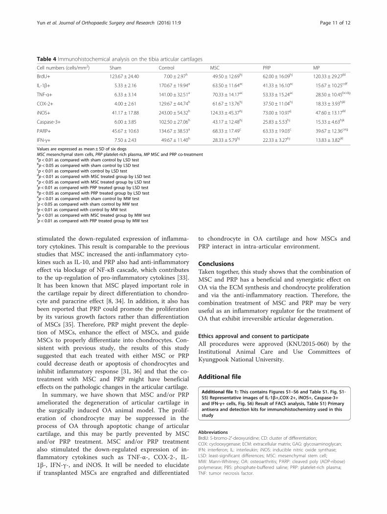
of all three test materials, and it was most potent in MPgroup (Fig. 5).The BrdU-positive cells in the femoral and tibial ar-
ticular cartilages significantly decreased in control groupcomparing to that of sham group. The values were sig-nificantly increased in all treated groups compared withcontrol group. The increase of cell proliferation on thecartilage was most significant in MP group than MSC orPRP group (Fig. 6, Tables 3 and 4).Immunopositive cells to caspase-3 and PARP were sig-
nificantly increased in the femoral and tibial cartilages ofcontrol group when compared with those of the sham
Fig. 3 Representative general histopathological images and the thicknesses of the femoral and tibial articular cartilages. Of sham (a, b), control(c, d), MSC (e, f), PRP (g, h), and MP (i, j). H&E and Safranin O stain. Scale bars = 90 μm. ap < 0.01 as compared with sham control by LSD test;bp < 0.01 and cp < 0.05 as compared with control by LSD test; dp < 0.01 as compared with MSC treated group by LSD test; ep < 0.01 and fp < 0.05as compared with PRP treated group by LSD test
Yun et al. Journal of Orthopaedic Surgery and Research (2016) 11:9 Page 7 of 12
group. However, these increased cells were significantlyreduced by treatment of all three test materials, and thereduction was most significant in MP group (Fig. 6,Tables 3 and 4).TNF-α, COX-2, IL-1β, iNOS, and IFN-γ staining cells
were increased significantly in the femoral and tibial car-tilages of control group compared with sham group.However, these increases of pro-inflammatory cytokineson the cartilages were diminished in treated groups(Fig. 6 and Additional file 1: Fig. S1-S5, Tables 3 and 4).
DiscussionOsteoarthritis (OA) is characterized by the loss of articu-lar cartilage components with inflammation, eventuallyresulting in impaired joint function [24, 25]. For this rea-son, this study mainly focused on the evaluation of theclinical signs, the change of ECM component and articu-lar cartilage, the gene expression related to chondrogen-esis, and the articular pathologic processes such as
inflammation and apoptosis with the treatment of PRPand/or MSC.Previously, it has been reported that the intra-articular
injection of PRP reduced the lameness score [26]. Simi-larly, there were a meaningful decrease of lamenessscore with the treatment of PRP and MSC mixed withPRP in this canine OA model. Based on this result, weanticipate that PRP would relieve pain and improvearticular function in an arthritic condition.It is well known that the structure of articular cartilage
is altered by the disorganization of collagen network, de-crease of proteoglycan contents, and disruption of theintegrity of the ECM in the process of OA [27]. There-fore, we tested the compressive strength of the articularcartilage to observe whether PRP or MSCs has a protect-ive effect on the articular damage by OA. As previousstudies have reported, Mankin score is a good index ofosteoarthritis [28] and it is highly associated with com-pressive strength of articular cartilage [27]. We evaluated
Fig. 4 COL and GAG contents on the femoral articular cartilages. ap < 0.01 as compared with sham control by LSD test; bp < 0.01 and cp < 0.05 ascompared with control by LSD test; dp < 0.01 as compared with MSC treated group by LSD test; ep < 0.01 and fp < 0.05 as compared with PRPtreated group by LSD test
Yun et al. Journal of Orthopaedic Surgery and Research (2016) 11:9 Page 8 of 12
Mankin score of the affected articular cartilage. Inaddition, the thickness of articular cartilage and the con-tent of COL and GAG were measured to confirm theOA condition. As a result, there were decreases inMankin score as well as increases in the thickness ofcartilage and the content of COL and GAG in thePRP and/or MSC treatment. These results suggestedthat PRP and MSC may have protective and regenera-tive effects on the degenerative cartilage of OA.Aggrecan is known as a core protein of cartilage-
specific proteoglycan, and it was reported that themRNA expression of aggrecan was down-regulated ac-cording to the severity of OA condition [29, 30]. The ex-pression of SOX9, the essential transcription factor forthe chondrogenic differentiation, was also inhibited byOA model [30]. In this study, the down-regulations ofSOX-9 and aggrecan in the control group were increasedby the MSC and PRP treatment. Based on these results,we can suppose that the increase of ECM-related factors
such as aggrecan, SOX9, COL, and GAG may be closelyassociated with the recovery of damaged articular cartil-age. From this aspect, it could be explained by the factthat PRP and/or MSC treatment was associated with theincrease of focal compressive strengths and the decreaseof Mankin score. These findings are compatible with aprevious report indicated that the inhibition of SOX9gene expression and the content of GAG might result incartilage degeneration [30].We next performed BrdU staining to examine whether
the MSC and/or PRP treatment promoted the prolifera-tion of chondrocytes. It was shown that BrdU-positivecells decreased in OA condition and increased with atreatment of MSC and/or PRP. We also performed im-munostaining for the caspase-3 and PARP expressionexamine whether apoptotic change during OA could beaffected by PRP and/or MSC treatment. Results showedthat the number of caspase-3- and PARP-positive cellswas increased in the OA condition and was reduced
Fig. 5 mRNA expressions of aggrecan and SOX9 on the femoral and tibial articular cartilages. Values are expressed as mean ± SD of six dogs,relative to control/G3PDH mRNA. ap < 0.01 and bp < 0.05 as compared with sham control by LSD test; cp < 0.01 and dp < 0.05 as compared withcontrol by LSD test; ep < 0.01 as compared with MSC treated group by LSD test; fp < 0.01 and gp < 0.05 as compared with PRP treated group byLSD test
Yun et al. Journal of Orthopaedic Surgery and Research (2016) 11:9 Page 9 of 12
with a treatment of MSC and/or PARP. These resultswere consistent with the findings of previous study thatcaspase-3 led to PARP to cleavage and consequentlycaused the progression of OA with cell apoptosis anddeath [31]. Therefore, we propose that the proliferationof chondrocyte may be suppressed in the process of OAthrough apoptotic change of articular cartilage, and thismay be partly prevented by MSC and/or PRP treatment.
We examined the protein expression of cytokines rela-tive to inflammatory reaction in the cartilage tissue. Ithas been known that pro-inflammatory cytokines medi-ated the activation of various inflammatory pathwaysand played a role in the progression of OA [32]. In thepresent study, the numbers of TNF-α-, COX-2-, IL-1β-,IFN-γ-, and iNOS-positive cells were increased in theOA condition, and MSC and/or PRP treatment
Fig. 6 Representative immunohistochemical findings of femoral and tibial articular cartilage (BrdU, TNF-α, and PARP). Of sham (a, b), control(c, d), MSC (e, f), PRP (g, h), and MP (i, j). Scale bars = 90 μm
Table 3 Immunohistochemical analysis on the femur articular cartilages
Values are expressed as mean ± SD of six dogsMSC mesenchymal stem cells, PRP platelet-rich plasma, MP MSC and PRP co-treatmentap < 0.01 as compared with sham control by LSD testbp < 0.01 as compared with control by LSD testcp < 0.01 as compared with MSC treated group by LSD testdp < 0.01 as compared with PRP treated group by LSD testep < 0.05 as compared with PRP treated group by LSD testfp < 0.01 as compared with sham control by MW testgp < 0.01 as compared with control by MW testhp < 0.01 as compared with MSC treated group by MW testip < 0.01 as compared with PRP treated group by MW test
Yun et al. Journal of Orthopaedic Surgery and Research (2016) 11:9 Page 10 of 12
stimulated the down-regulated expression of inflamma-tory cytokines. This result is comparable to the previousstudies that MSC increased the anti-inflammatory cyto-kines such as IL-10, and PRP also had anti-inflammatoryeffect via blockage of NF-κB cascade, which contributesto the up-regulation of pro-inflammatory cytokines [33].It has been known that MSC played important role inthe cartilage repair by direct differentiation to chondro-cyte and paracrine effect [8, 34]. In addition, it also hasbeen reported that PRP could promote the proliferationby its various growth factors rather than differentiationof MSCs [35]. Therefore, PRP might prevent the deple-tion of MSCs, enhance the effect of MSCs, and guideMSCs to properly differentiate into chondrocytes. Con-sistent with previous study, the results of this studysuggested that each treated with either MSC or PRPcould decrease death or apoptosis of chondrocytes andinhibit inflammatory response [31, 36] and that the co-treatment with MSC and PRP might have beneficialeffects on the pathologic changes in the articular cartilage.In summary, we have shown that MSC and/or PRP
ameliorated the degeneration of articular cartilage inthe surgically induced OA animal model. The prolif-eration of chondrocyte may be suppressed in theprocess of OA through apoptotic change of articularcartilage, and this may be partly prevented by MSCand/or PRP treatment. MSC and/or PRP treatmentalso stimulated the down-regulated expression of in-flammatory cytokines such as TNF-α-, COX-2-, IL-1β-, IFN-γ-, and iNOS. It will be needed to elucidateif transplanted MSCs are engrafted and differentiated
to chondrocyte in OA cartilage and how MSCs andPRP interact in intra-articular environment.
ConclusionsTaken together, this study shows that the combination ofMSC and PRP has a beneficial and synergistic effect onOA via the ECM synthesis and chondrocyte proliferationand via the anti-inflammatory reaction. Therefore, thecombination treatment of MSC and PRP may be veryuseful as an inflammatory regulator for the treatment ofOA that exhibit irreversible articular degeneration.
Ethics approval and consent to participateAll procedures were approved (KNU2015-060) by theInstitutional Animal Care and Use Committees ofKyungpook National University.
Additional file
Additional file 1: This contains Figures S1–S6 and Table S1. Fig. S1-S5) Representative images of IL-1β+,COX-2+, iNOS+, Caspase-3+and IFN-γ+ cells, Fig. S6) Result of FACS analysis, Table S1) Primaryantisera and detection kits for immunohistochemistry used in thisstudy
Values are expressed as mean ± SD of six dogsMSC mesenchymal stem cells, PRP platelet-rich plasma, MP MSC and PRP co-treatmentap < 0.01 as compared with sham control by LSD testbp < 0.05 as compared with sham control by LSD testcp < 0.01 as compared with control by LSD testdp < 0.01 as compared with MSC treated group by LSD testep < 0.05 as compared with MSC treated group by LSD testfp < 0.01 as compared with PRP treated group by LSD testgp < 0.05 as compared with PRP treated group by LSD testhp < 0.01 as compared with sham control by MW testip < 0.05 as compared with sham control by MW testjp < 0.01 as compared with control by MW testkp < 0.01 as compared with MSC treated group by MW testlp < 0.01 as compared with PRP treated group by MW test
Yun et al. Journal of Orthopaedic Surgery and Research (2016) 11:9 Page 11 of 12
Competing interestsThe authors declare that they have no competing interests.
Authors’ contributionsSY and YSK built the idea and protocols of this paper and wrote themanuscript. SKK participated in data collection and analysis. All authorsapproved this manuscript.
Author details1Department of Veterinary Surgery, College of Veterinary Medicine,Kyungpook National University, Daegu 702-701, Republic of Korea.2Department of Anatomy and Histology, College of Korean Medicine, DaeguHaany University, Gyeongsan 712-715, Republic of Korea. 3Stem CellTherapeutic Research Institute, Kyungpook National University, Daegu702-701, Republic of Korea.
Received: 6 November 2015 Accepted: 6 January 2016
References1. Goldring MB, Goldring SR. Osteoarthritis. J Cell Physiol. 2007;213:626–34.2. Lo GH, LaValley M, McAlindon T, Felson DT. Intra-articular hyaluronic acid in
treatment of knee osteoarthritis: a meta-analysis. JAMA. 2003;290:3115–21.3. Arrich J, Piribauer F, Mad P, Schmid D, Klaushofer K, Mullner M. Intra-
articular hyaluronic acid for the treatment of osteoarthritis of the knee:systematic review and meta-analysis. CMAJ. 2005;172:1039–43.
4. Tamura T, Ohmori K. Rhein, an active metabolite of diacerein, suppressesthe interleukin-1 alpha-induced proteoglycan degradation in cultured rabbitarticular chondrocytes. Jpn J Pharmacol. 2001;85:101–4.
5. Lin L, Zhou C, Wei X, Hou Y, Zhao L, Fu X, et al. Articular cartilage repairusing dedifferentiated articular chondrocytes and bone morphogeneticprotein 4 in a rabbit model of articular cartilage defects. Arthritis Rheum.2008;58:1067–75.
6. Marlovits S, Hombauer M, Truppe M, Vecsei V, Schlegel W. Changes in theratio of type-I and type-II collagen expression during monolayer culture ofhuman chondrocytes. J Bone Joint Surg (Br). 2004;86:286–95.
7. Nganvongpanit K, Pothacharoen P, Chaochird P, Klunklin K, Warrit K,Settakorn J, et al. Prospective evaluation of serum biomarker levels andcartilage repair by autologous chondrocyte transplantation and subchondraldrilling in a canine model. Arthritis Res Ther. 2009;11:R78–8.
8. Jo CH, Lee YG, Shin WH, Kim H, Chai JW, Jeong EC, et al. Intra-articularinjection of mesenchymal stem cells for the treatment of osteoarthritis ofthe knee: a proof-of-concept clinical trial. Stem Cells. 2014;32:1254–66.
9. Sekiya I, Ojima M, Suzuki S, Yamaga M, Horie M, Koga H, et al. Humanmesenchymal stem cells in synovial fluid increase in the knee withdegenerated cartilage and osteoarthritis. J Orthop Res. 2011;30:943–9.
10. Murphy JM, Dixon K, Beck S, Fabian D, Feldman A, Barry F. Reducedchondrogenic and adipogenic activity of mesenchymal stem cells frompatients with advanced osteoarthritis. Arthritis Rheum. 2002;46:704–13.
11. Barry F, Murphy M. Mesenchymal stem cells in joint disease and repair.Nat Rev Rheumatol. 2013;9:584–94.
12. Metcalf KB, Mandelbaum BR, McIlwraith CW. Application of platelet-richplasma to disorders of the knee joint. Cartil. 2013;4:295–312.
13. Bendinelli P, Matteucci E, Dogliotti G, Corsi MM, Banfi G, Maroni P, et al.Molecular basis of anti-inflammatory action of platelet-rich plasma onhuman chondrocytes: mechanisms of NF-kappaB inhibition via HGF. J CellPhysiol. 2010;225:757–66.
14. El-Sharkawy H, Kantarci A, Deady J, Hasturk H, Liu H, Alshahat M, et al.Platelet-rich plasma: growth factors and pro- and anti-inflammatoryproperties. J Periodontol. 2007;78:661–9.
15. Danisovic L, Varga I, Polak S. Growth factors and chondrogenicdifferentiation of mesenchymal stem cells. Tissue Cell. 2011;44:69–73.
16. Demidova-Rice TN, Wolf L, Deckenback J, Hamblin MR, Herman IM. Humanplatelet-rich plasma- and extracellular matrix-derived peptides promoteimpaired cutaneous wound healing in vivo. PLoS ONE. 2012;7:e32146.
17. Zhang N, Wu YP, Qian SJ, Teng C, Chen S, Li H. Research progress in themechanism of effect of PRP in bone deficiency healing. Sci World J. 2013.
18. Bennett D, Eckersall PD, Waterston M, Marchetti V, Rota A, McCulloch E, etal. The effect of robenacoxib on the concentration of C-reactive protein insynovial fluid from dogs with osteoarthritis. BMC Vet Res. 2013;9:42.
19. Warnock JJ, Baltzer WI, Duesterdieck-Zellmer K, Ott J. Minimally invasivesynovium harvest for potential use in meniscal tissue engineering. Res VetSci. 2012;93:1472–80.
20. Warnock JJ, Spina J, Bobe G, Duesterdieck-Zellmer KF, Ott J, Baltzer WI, et al.Culture of canine synoviocytes on porcine intestinal submucosa scaffolds asa strategy for meniscal tissue engineering for treatment of meniscal injuryin dogs. Vet J. 2014;199:49–56.
21. Reddy GK, Enwemeka CS. A simplified method for the analysis ofhydroxyproline in biological tissues. Clin Biochem. 1996;29:225–9.
22. Ignat’eva NY, Danilov NA, Averkiev SV, Obrezkova MV, Lunin VV, Sobol’ EN.Determination of hydroxyproline in tissues and the evaluation of thecollagen content of the tissues. J Anal Chem. 2007;62:51–7.
23. van der Sluijs JA, Geesink RG, van der Linden AJ, Bulstra SK, Kuyer R,Drukker J. The reliability of the Mankin score for osteoarthritis. J OrthopRes. 1992;10:58–61.
24. Qin J, Liu YS, Liu J, Li J, Tan Y, Li XJ, et al. Effect of angelica sinensispolysaccharides on osteoarthritis in vivo and in vitro: a possible mechanism topromote proteoglycans synthesis. Evid Based Complement Alternat Med. 2013.
25. Na JY, Song KB, Kim S, Kwon YB, Kim DG, Lee JK, et al. Effects of HPL-04 ondegenerative osteoarthritis. J Korean Soc Food Sci Nutr. 2014;43:30–9.
26. Kon E, Buda R, Filardo G, Di Martino A, Timoncini A, Cenacchi A, et al.Platelet-rich plasma: intra-articular knee injections produced favorableresults on degenerative cartilage lesions. Knee Surg Sports TraumatolArthrosc. 2010;18:472–9.
27. Franz T, Hasler EM, Hagg R, Weiler C, Jakob RP, Mainil-Varlet P. In situcompressive stiffness, biochemical composition, and structural integrity ofarticular cartilage of the human knee joint. Osteoarthr Cartil. 2001;9:582–92.
28. Pauli C, Whiteside R, Heras FL, Nesic D, Koziol J, Grogan SP, et al. Comparison ofcartilage histopathology assessment systems on human knee joints at all stagesof osteoarthritis development. Osteoarthr Cartil. 2012;20:476–85.
29. Lorenz H, Wenz W, Ivancic M, Steck E, Richter W. Early and stableupregulation of collagen type II, collagen type I and YKL40 expression levelsin cartilage during early experimental osteoarthritis occurs independent ofjoint location and histological grading. Arthritis Res Ther. 2005;7:R156–65.
30. Yagi R, McBurney D, Laverty D, Weiner S, Horton Jr WE. Intrajointcomparisons of gene expression patterns in human osteoarthritis suggest achange in chondrocyte phenotype. J Orthop Res. 2005;23:1128–38.
31. Shakibaei M, John T, Seifarth C, Mobasheri A. Resveratrol inhibits IL-1 beta-induced stimulation of caspase-3 and cleavage of PARP in human articularchondrocytes in vitro. Ann N Y Acad Sci. 2007;1095:554–63.
32. Goldring MB. The role of the chondrocyte in osteoarthritis. Arthritis Rheum.2000;43:1916–26.
33. Andia I, Maffulli N. Platelet-rich plasma for managing pain and inflammationin osteoarthritis. Nat Rev Rheumatol. 2013;9:721–30.
34. Chung JY, Song M, Ha CW, Kim JA, Lee CH, Park YB. Comparison of articularcartilage repair with different hydrogel-human umbilical cord blood-derivedmesenchymal stem cell composites in a rat model. Stem Cell Res Ther. 2014;5:39.
35. Mishra A, Tummala P, King A, Lee B, Kraus M, Tse V, et al. Buffered platelet-rich plasma enhances mesenchymal stem cell proliferation andchondrogenic differentiation. Tissue Eng Part C Methods. 2009;15:431–5.
36 Pereira RC, Scaranari M, Benelli R, Strada P, Reis RL, Cancedda R, et al. Dualeffect of platelet lysate on human articular cartilage: a maintenance ofchondrogenic potential and a transient proinflammatory activity followedby an inflammation resolution. Tissue Eng Part A. 2013;19:1476–88.
• We accept pre-submission inquiries
• Our selector tool helps you to find the most relevant journal
• We provide round the clock customer support
• Convenient online submission
• Thorough peer review
• Inclusion in PubMed and all major indexing services
• Maximum visibility for your research
Submit your manuscript atwww.biomedcentral.com/submit
Submit your next manuscript to BioMed Central and we will help you at every step:
Yun et al. Journal of Orthopaedic Surgery and Research (2016) 11:9 Page 12 of 12


![A bibliometric analysis of two decades of aromatherapy …...by Eugene Garfield in 1964 [9]. With the wide availability of bibliometric analytical software, there is a rapid prolif-eration](https://static.documents.pub/doc/80x56/60e838bdf047ae7347388d0f/a-bibliometric-analysis-of-two-decades-of-aromatherapy-by-eugene-garfield-in.jpg)


