Start CPR Give oxygen Attach monitor/defibrillator VF/pulseless VT If no signs of return of spontaneous circu- lation (ROSC), go to or If ROSC, go to Post-Cardiac Arrest Care ProACLS.com Adult Cardiac Arrest Algorithm VF, Pulselss VT, Asystole, PEA Push hard (at least 2 inches (5cm)) and fast (100-120/min) and allow complete chest recoil. Minimize interruptions in compres- sions. Avoid excessive ventilation Rotate compressor every 2 minutes, or sooner if fatigued. If no advanced airway, 30:2 com- pression-ventilation ratio. Quantitative waveform capnogra- phy - If PetCO2 <10 mmHg, attempt to improve CPR quality. Intra-arterial pressure - If relaxation phase (diastolic) pressure <20 mmHg, attempt to improve CPR quality. Dose/Details Yes Asystole/PEA No CPR 2 min IV/IO access Yes No CPR 2 min Epinephrine every 3-5 min Consider advanced airway, capnography Yes No CPR 2 min Amiodarone Treat reversible causes CPR 2 min IV/IO access Epinephrine every 3-5 min Consider advanced airway, capnography Yes CPR 2 min Treat reversible causes No Yes No 1 3 4 Go to 3 or 4 Rhythm Shockable? Rhythm Shockable? Rhythm Shockable? Rhythm Shockable? 1 Shock Shock Shock Rhythm Shockable? 2 2 Hypovolemia Hypoxia Hydrogen ion (acidosis) Hypo-/hyperkalemia Hypothermia Tension pneumothorax Tamponade, cardiac Toxins Thrombosis, pulmonary Thrombosis coronary Reversible Causes (H’s & T’s) CPR Quality Biphasic: Manufacturer recom- mendation (eg, initial dose of 120 -200 J); if unknown, use maximum available. Second and subse- quent doses should be equivalent, end higher doses may be consid- ered. Monophasic: 360 J Shock Energy for Defibrillation Pulse and blood pressure Abrupt sustained increase in PetCO2 (typically ≥40 mmHg) Spontaneous arterial pressure waves with intra-arterial monitor- ing Return of Spontaneous Circulation (ROSC) Epinephrine IV/IO dose: 1 mg every 3-5 minutes Amiodarone IV/IO dose: First dose: 300 mg bolus. Second dose: 150mg. Drug Therapy Endotracheal intubation or supra- glottic advanced airway Waveform capnography or cap- nometry to confirm and monitor ET tube placement Once advanced airway in place give 1 breath every 6 seconds (10 breaths/min) with continuous chest compressions Advanced Airway Reference: Link MS, Berkow LC, Kudenchuk PJ, Halperin HR, Hess EP, Moitra VK, Neumar RW, O’Neil BJ, Paxton JH, Silvers SM, White RD, Yannopoulos D, Donnino MW. "Part 7: Adult Advanced Cardiovascular Life Support." ECC Guidelines 2015. American Heart Association, 15 Oct. 2015. Web. 01 Mar. 2017. Follows American Heart Association ECC and CPR 2015 guidelines ProACLS Student Manual - Page 1
Transcript
Start CPR Give oxygen Attach monitor/defibrillator
VF/pulseless VT
If no signs of return of spontaneous circu-lation (ROSC), go to or
If ROSC, go to Post-Cardiac Arrest Care
ProACLS.com Adult Cardiac Arrest Algorithm
VF, Pulselss VT, Asystole, PEA
Push hard (at least 2 inches(5cm)) and fast (100-120/min) and allow complete chest recoil.
Minimize interruptions in compres-sions.
Avoid excessive ventilation Rotate compressor every 2
minutes, or sooner if fatigued. If no advanced airway, 30:2 com-
Biphasic: Manufacturer recom-mendation (eg, initial dose of 120-200 J); if unknown, use maximum available. Second and subse-quent doses should be equivalent, end higher doses may be consid-ered.
Monophasic: 360 J
Shock Energy for Defibrillation
Pulse and blood pressure Abrupt sustained increase in
Amiodarone IV/IO dose: First dose: 300 mg bolus. Second dose: 150mg.
Drug Therapy
Endotracheal intubation or supra-glottic advanced airway
Waveform capnography or cap-nometry to confirm and monitor ET tube placement
Once advanced airway in place give 1 breath every 6 seconds (10 breaths/min) with continuous chest compressions
Advanced Airway
Reference: Link MS, Berkow LC, Kudenchuk PJ, Halperin HR, Hess EP, Moitra VK, Neumar RW, O’Neil BJ, Paxton JH, Silvers SM, White RD, Yannopoulos D, Donnino MW. "Part 7: Adult Advanced Cardiovascular Life Support." ECC Guidelines 2015. American Heart Association, 15 Oct. 2015. Web. 01 Mar. 2017.
Follows American Heart Association ECC and CPR 2015 guidelines
ProACLS Student Manual - Page 1
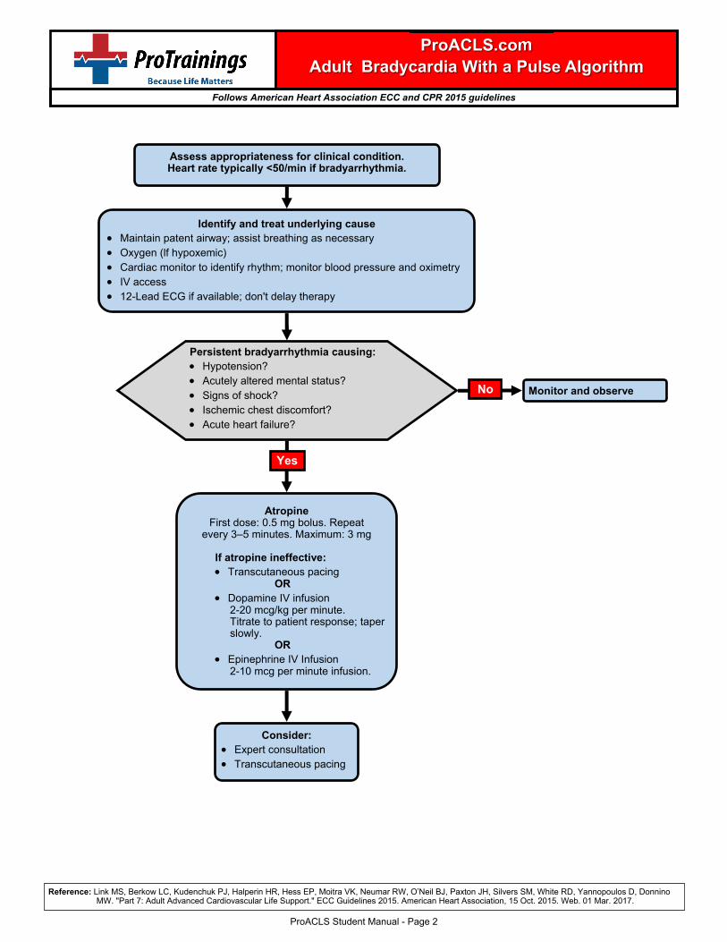
Assess appropriateness for clinical condition. Heart rate typically <50/min if bradyarrhythmia.
Identify and treat underlying cause Maintain patent airway; assist breathing as necessary Oxygen (lf hypoxemic) Cardiac monitor to identify rhythm; monitor blood pressure and oximetry IV access 12-Lead ECG if available; don't delay therapy
If atropine ineffective: Transcutaneous pacing OR Dopamine IV infusion 2-20 mcg/kg per minute. Titrate to patient response; taper slowly. OR Epinephrine IV Infusion 2-10 mcg per minute infusion.
ProACLS.com Adult Bradycardia With a Pulse Algorithm
Yes
Reference: Link MS, Berkow LC, Kudenchuk PJ, Halperin HR, Hess EP, Moitra VK, Neumar RW, O’Neil BJ, Paxton JH, Silvers SM, White RD, Yannopoulos D, Donnino MW. "Part 7: Adult Advanced Cardiovascular Life Support." ECC Guidelines 2015. American Heart Association, 15 Oct. 2015. Web. 01 Mar. 2017.
Follows American Heart Association ECC and CPR 2015 guidelines
ProACLS Student Manual - Page 2
Return of spontaneous circulation (ROSC)
Optimize ventilation and oxygenation Maintain oxygen saturation ≥94% Consider advanced airway and waveform capnography Do not hyperventilate
12-Lead ECG:STEMI or high
suspicion of AMI? Coronary reperfusion
Initiate targeted temperature management
Yes
No
Ventilation/oxygenation: Avoid excessive ventilation. Start at 10 breaths/min and titrate to tar-get PetCO2 of 35-40 mm Hg. When feasible, titrate FiO2 to minimum necessary to achieve SpO2 ≥94%.
IV Bolus Approximately 1-2 L normal saline or lactated Ringer's
Epinephrine IV infusion: 0.1-0.5 mcg/kg per minute (in 70-kg adult: 7-35 mcg per minute)
Dopamine IV infusion: 5-10 mcg/kg per minute
Norepinephrine IV infusion: 0.1-0.5 mcg/kg per minute (in 70-kg adult: 7-35 mcg per minute)
ProACLS.com Adult Immediate Post-Cardiac Arrest Care Algorithm
Follows American Heart Association ECC and CPR 2015 guidelines
Reversible Causes (H’s & T’s)
ProACLS Student Manual - Page 3
Assess appropriateness for clinical condition. Heart rate typically ≥ 150/min if tachyarrhythmia.
Identify and Treat Underlying Cause Maintain patient airway; assist breathing as necessary Oxygen (if O2 sat < 94%) Cardiac monitor to identify rhythm; monitor blood pressure and oximetry
Synchronized cardioversion: Consider sedation If regular narrow complex, consid-
Adenosine IV dose: First dose: 6 mg rapid IV push; follow with NS flush. Second dose: 12 mg if required.
Antiarrhythmic Infusions for Stable Wide-QRS Tachycardia
Procainamide IV dose: 20-50 mg/min until arrhythmia suppressed, hypotension ensues, ORSduration increases >50%, or maximum dose 17 mg/kg given. Mainte-nance infusion: 1-4 mg/min. Avoid if prolonged QT or CHF.
Amiodarone IV dose: First dose: 150 mg over 10 minutes. Repeat as needed if VT recurs. Follow by maintenance infusion of 1 mg/min for first 6 hours.
Sotalol IV dose: 100 mg (1.5 mg/kg) over 5 minutes. Avoid if prolonged QT.
Dose/Details
Reference: Link MS, Berkow LC, Kudenchuk PJ, Halperin HR, Hess EP, Moitra VK, Neumar RW, O’Neil BJ, Paxton JH, Silvers SM, White RD, Yannopoulos D, Donnino MW. "Part 7: Adult Advanced Cardiovascular Life Support." ECC Guidelines 2015. American Heart Association, 15 Oct. 2015. Web. 01 Mar. 2017.
Follows American Heart Association ECC and CPR 2015 guidelines
ProACLS Student Manual - Page 4
Assess appropriateness for clinical condition.
EMS assessment and care and hospital preparation Monitor, support ABC’s. Be prepared to provide CPR and defibrillation Administer aspirin and consider oxygen, nitroglycerin, and morphine if
needed. Obtain 12-lead ECG; if ST elevation, notify receiving hospital with trans-
mission or interpretation; note time of onset and first medical contact. Notified hospital should mobilize hospital resources to respond to
trolyte end coagulation studies Obtain portable chest x-ray (<30 minutes)
ECG Interpretation
Immediate ED general treatment: If O2 sat <90% start oxygen at 4 I/min, titrate Aspirin 160 to 325 mg (if not given by EMS) Nitroglycerin sublingual or spray Morphine IV If discomfort not relieved by
nitroglycerin
ST elevation or new or presumably new LBBB; strongly suspicious for Injury
Reference: Link MS, Berkow LC, Kudenchuk PJ, Halperin HR, Hess EP, Moitra VK, Neumar RW, O’Neil BJ, Paxton JH, Silvers SM, White RD, Yannopoulos D, Donnino MW. "Part 7: Adult Advanced Cardiovascular Life Support." ECC Guidelines 2015. American Heart Association, 15 Oct. 2015. Web. 01 Mar. 2017.
Follows American Heart Association ECC and CPR 2015 guidelines
ST depression or dynamic T-wave inversion; strongly suspicious for Ischemia
High-risk non-ST-elevation ACS (NSTE-ACS)
Normal or non-diagnostic changes in ST segment or T-wave
Low/Intermediate-risk ACS
Start adjunctive therapiesas Indicated
Do not delay reperfusion
Time from onset of symptoms ≤12 hours?
Troponin elevated or high-risk patient, consider early Invasive strategy if: Refractory ischemic chest discomfort Recurrent/persistent ST deviation Ventricular tachycardia Hemodynamic Instability Signs of heart failure
Start adjunctive therapies (eg, nitroglycerin, heparin) as Indicated See AHA/ACC NSTE-ACS Guidelines
Yes
Reperfusion goals: Therapy defined by patient and center criteria. Door to-balloon-inflation (PCI) goal
of 90 minutes Door to-needle (fibrinolysis) goal of
30 minutes
No
Consider admission to ED chest pain unit or to appropriate bed
for further monitoring and possible intervention.
ProACLS Student Manual - Page 5
Identify signs and symptoms of possible stroke Activate Emergency Response
Critical EMS assessments and actions: Support ABCs; give oxygen if needed Perform prehospital stroke assessment Establish time of symptom onset (last normal) Triage to stroke center Alert hospital; consider direct transfer to CT scan Check glucose if possible
Immediate general assessment and stabilization: Assess ABCs, vital signs Provide oxygen if hypoxemic Obtain IV access and perform laboratory assessments Check glucose; treat if indicated Perform neurologic screening assessment Activate stroke team Order emergent CT scan or MRI of brain Obtain 12-lead ECG
Does CT scan show hemorrhage?
Begin stroke orhemorrhage pathway
Admit to stroke unit orIntensive care unit
ProACLS.com Adult Suspected Stroke Algorithm
Reference: Link MS, Berkow LC, Kudenchuk PJ, Halperin HR, Hess EP, Moitra VK, Neumar RW, O’Neil BJ, Paxton JH, Silvers SM, White RD, Yannopoulos D, Donnino MW. "Part 7: Adult Advanced Cardiovascular Life Support." ECC Guidelines 2015. American Heart Association, 15 Oct. 2015. Web. 01 Mar. 2017.
Follows American Heart Association ECC and CPR 2015 guidelines
Consult neurologist or neurosurgeon; consider transfer
if not available
Probable acute ischemic stroke; consider fibrinolytic therapy: Check for fibrinolytic exclusions Repeat neurologic exam: are
deficits rapidly improving to normal?
Administer aspirin
Yes No
Review risks/benefits with patient and family. If acceptable: Give rtPA No anticoagulants or antiplatelet treatment for 24 hours Begin post-rtPA stroke pathway Aggressively monitor BP per protocol and neurologic deterioration Emergent admission to stroke unit or intensive care unit
Immediate neurologic assessment by stroke team or designee Review patient history Establish time of symptom onset or last known normal Perform neurologic examination (NIH Stroke Scale or Canadian Neurological Scale)
Yes
No Does patient remain candidate for fibrinolytic therapy?
ProACLS Student Manual - Page 6
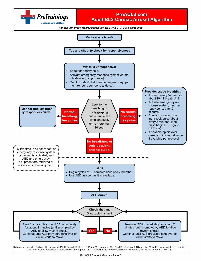
Verify scene is safe
Victim is unresponsive: Shout for nearby help. Activate emergency response system via mo-
bile device (if appropriate). Get AED, defibrillator and emergency equip-
ment (or send someone to do so).
Provide rescue breathing: 1 breath every 5-6 sec. or
about 10-12 breaths/min. Activate emergency re-
sponse system, if not al-ready done, after 2minutes.
Continue rescue breath-ing; check pulse aboutevery 2 minutes. If nopulse begin CPR (go toCPR box)
If possible opioid over-dose, administer naloxoneif available per protocol
ProACLS.com Adult BLS Cardiac Arresst Algorithm
Reference: Link MS, Berkow LC, Kudenchuk PJ, Halperin HR, Hess EP, Moitra VK, Neumar RW, O’Neil BJ, Paxton JH, Silvers SM, White RD, Yannopoulos D, Donnino MW. "Part 7: Adult Advanced Cardiovascular Life Support." ECC Guidelines 2015. American Heart Association, 15 Oct. 2015. Web. 01 Mar. 2017.
Follows American Heart Association ECC and CPR 2015 guidelines
AED Arrives
CPR Begin cycles of 30 compressions and 2 breaths. Use AED as soon as it is available.
Look for no breathing or only gasping
and check pulse (simultaneously) for no more than
10 sec.
Tap and shout to check for responsiveness
No normal breathing, has pulse.
Monitor until emergen-cy responders arrive. Normal
breathing, has pulse.
No breathing, or only gasping, and no pulse. By this time in all scenarios, an
emergency response system or backup is activated, and
AED and emergency equipment are retrieved or
someone is retrieving them.
Check rhythm. Shockable rhythm?
Give 1 shock. Resume CPR immediately for about 2 minutes (until prompted by
AED to allow rhythm check). Continue until ALS providers take over or
victim starts to move.
Resume CPR immediately for about 2 minutes (until prompted by AED to allow
rhythm check). Continue until ALS providers take over or
victim starts to move.
Yes No
ProACLS Student Manual - Page 7
ProACLS.com ECG Interpretation
1 small square = .04 sec. 1 large box = .2 sec. (5 small squares long)
5 large boxes = 1 sec. 30 large boxes = 6 sec.
1 second
6 seconds
QRS Complex- Normal is 0.04 - 0.12 sec.
(1-3 small boxes)
PR Interval- Normal is 0.12 - 0.20 sec.
(3-5 small boxes)
P wave T wave
R wave
Q
S ST Segment
ST Segment- Normally level with baseline. If higher,
possible ST elevation MI. 12 lead ECG is needed to properly evaluate ST elevation.
P Waves:. Upright and regular P-R Interval:. 0.16 sec (normal range= 0.12-0.20), one P wave for each QRS
QRS:. 0.08 sec (normal range= 0.04-0.12)
Clinical Significance: A decreased heart rate can result in decreased cardiac output, hypotension, or other serious problems depending on the cause of the bradycardia. Unless the patient has hypotension or other serious signs or symptoms, no treatment is necessary. For hypotension or other serious symptoms, Atropine 0.5mg can be given every 3-5 minutes. If atropine is ineffective, transcutaneous pacing should be done. Oth-er treatments include Dopamine 2 to 20 mcg/kg per minute or Epinephrine 2 to 10 mcg/min.
P Waves:. Upright and regular P-R Interval:. 0.16 sec (normal range= 0.12-0.20), one P wave for each QRS
QRS:. 0.04 sec (normal range= 0.04-0.12)
Clinical Significance: Typically sinus tachycardia needs no treatment. It is most often a compensatory mech-anism to an underlying cause such as fever, anxiety, hypovolemia, or shock. It is most important to identify and treat the underlying cause as needed. Rates less than 150bpm do not usually cause serious signs and symptoms. Rates over 150bpm may cause reduced cardiac output and may require treatment. Synchronized cardioversion is the first choice. If regular narrow QRS complex, consider adenosine.
ProACLS Student Manual - Page 9
Supraventricular Tachycardia
Rhythm:. Regular Rate:. 280 bpm (SVT is defined as >100bpm. Typically under 150bpm has no symptoms.)
P Waves:. Present but difficult to see on the end of the T wave because of rapid rate P-R Interval:. 0.12 sec (normal range= 0.12-0.20), one P wave for each QRS, again difficult to see
QRS:. 0.04 sec (normal range= 0.04-0.12)
Clinical Significance: SVT usually has an abrupt onset and termination in patients with high levels of stress, over exertion, high levels of caffeine, Wolff-Parkinson-White (WPW) syndrome, etc... and can usually be tol-erated for short periods of time. Runs of SVT are often felt as palpitations. Treatment is not normally needed for self-terminating SVT. If the patient is unstable, rapid treatment must be given to correct the SVT. Syn-chronized cardioversion at 50-100 joules with a monophasic or biphasic defibrillator should be given immedi-ately. For symptomatic, but stable SVT, Vagal maneuvers should be tried first. If unsuccessful, then 6mg Adenosine rapid IV push would be given. 12mg Adenosine may be tried if the first dose did not convert the rhythm.
Dysrhythmias Originating in the Atria
Atrial Flutter
Rhythm:. Regular (Can be irregular) Rate:. 110 bpm (Atrial rate is 210. Typical “sawtooth” pattern of atrial flutter.)
P Waves:. Flutter waves, or F waves, are present. P-R Interval:. F waves are consistent, 2 for every QRS (2:1 or 3:1 is typical)
QRS:. 0.12 sec (normal range= 0.04-0.12)
Clinical Significance: Treatment is not normally necessary. Rather an expert consultation is required. Pa-tients will often feel weak or dizzy. Treatment is necessary if there is a rapid ventricular rate that creates he-modynamic instability. For an unstable patient, perform synchronized cardioversion with 50 to 100 J with a monophasic or biphasic defibrillator. Pharmacologic therapy should be done only upon expert consultation or medical control direction.
ProACLS Student Manual - Page 10
Atrial Fibrillation
Rhythm:. Irregular Rate:. 90 bpm (Atrial rate is very fast and chaotic, and cannot be counted) P Waves:. Not discernible. Chaotic.
Clinical Significance: Treatment is not normally necessary. Rather an expert consultation is required. Pa-tients will often feel weak or dizzy. For an unstable patient, perform synchronized cardioversion with 200 Joules with a monophasic or 120 to 200 joules with a biphasic defibrillator. Pharmacologic therapy should be done only upon expert consultation or medical control direction.
Dysrhythmias Originating in the Ventricles
Ventricular Tachycardia (V-tach)
Rhythm:. Regular (Can be slightly irregular) Rate:. 200 bpm (Typically between 100-250)
P Waves:. Absent P-R Interval:. Absent
QRS:. Wide, 0.32 sec (usually wide and bizarre)
Clinical Significance: Ventricular tachycardia severely compromises cardiac output and coronary artery per-fusion. V-tach May be perfusing or non-perfusing. If there is a pulse and patient is stable, then Procainamide or Amiodarone may be administered. If unstable with a pulse, then synchronized cardioversion is needed. If pulseless, then defibrillate with an initial unsynchronized dose of 360 joules monophasic or 120-200 joules biphasic.
ProACLS Student Manual - Page 11
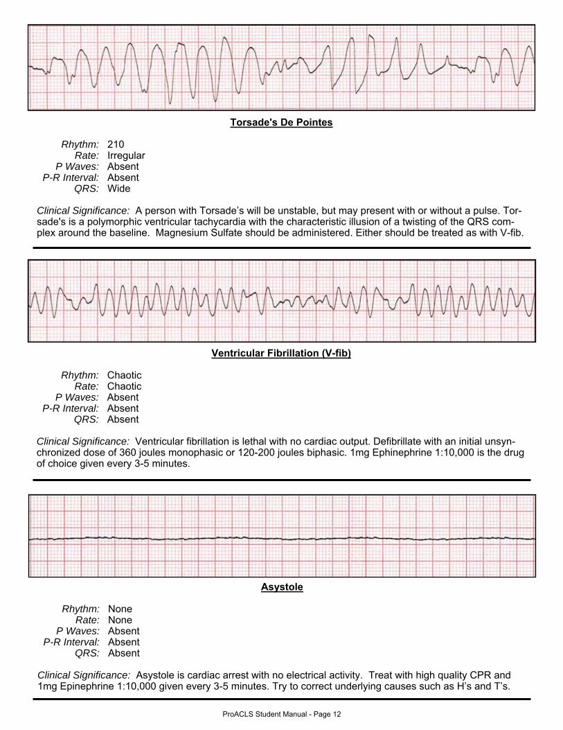
Ventricular Fibrillation (V-fib)
Rhythm:. Chaotic Rate:. Chaotic
P Waves:. Absent P-R Interval:. Absent
QRS:. Absent
Clinical Significance: Ventricular fibrillation is lethal with no cardiac output. Defibrillate with an initial unsyn-chronized dose of 360 joules monophasic or 120-200 joules biphasic. 1mg Ephinephrine 1:10,000 is the drug of choice given every 3-5 minutes.
Asystole
Rhythm:. None Rate:. None
P Waves:. Absent P-R Interval:. Absent
QRS:. Absent
Clinical Significance: Asystole is cardiac arrest with no electrical activity. Treat with high quality CPR and 1mg Epinephrine 1:10,000 given every 3-5 minutes. Try to correct underlying causes such as H’s and T’s.
Torsade's De Pointes
Rhythm:. 210 Rate:. Irregular
P Waves:. Absent P-R Interval:. Absent
QRS:. Wide
Clinical Significance: A person with Torsade’s will be unstable, but may present with or without a pulse. Tor-sade's is a polymorphic ventricular tachycardia with the characteristic illusion of a twisting of the QRS com-plex around the baseline. Magnesium Sulfate should be administered. Either should be treated as with V-fib.
ProACLS Student Manual - Page 12
Atrioventricular (AV) Heart Blocks
First-Degree AV Block
Rhythm:. Regular (can be slightly irregular) Rate:. 90
P Waves:. Normal P-R Interval:. 0.24 sec (normal range= 0.12-0.20), one P wave for each QRS
QRS:. 0.04 sec (normal range= 0.04-0.12)
Clinical Significance: The prolonged P-R interval with one P wave for each QRS is the most identifiable sign to recognize first-degree AV block. Although first-degree block is not usually serious by itself, it can be a pre-cursor to a more serious type of block. Usually treatment is not needed unless other serious signs or symp-toms are evident.
Second-Degree AV Block Mobitz Type I – Wenckebach
Rhythm:. Ventricular rhythm is irregular; Atrial rhythm is Regular Rate:. 70 (typically normal or slow)
P Waves:. Normal; Some are not followed by QRS complexes P-R Interval:. Becomes progressively longer until QRS is dropped.
QRS:. 0.04 sec (usually within normal range= 0.04-0.12)
Clinical Significance: If beats are frequently dropped, cardiac output can be compromised. This can cause syncope and angina. Usually treatment is not needed immediately unless other serious signs or symptoms are evident. If symptomatic bradycardia occurs, then 0.5mg of Atropine should be administered IV. If atropine fails, transcutaneous pacing should be administered.
ProACLS Student Manual - Page 13
Second-Degree AV Block Mobitz Type II
Rhythm:. Ventricular rhythm is irregular; Atrial rhythm is Regular Rate:. 60 (typically bradycardic)
P Waves:. Normal; Some are not followed by QRS complexes P-R Interval:. Constant for p-waves followed by a QRS
QRS:. 0.04 sec (can be longer than normal range= 0.04-0.12)
Clinical Significance: Regular P-waves with a regular P-R interval and occasional dropped QRS complexes is the most identifiable characteristic of second-degree AV block type II. If beats are frequently dropped, car-diac output can be compromised. This can cause syncope and angina. Usually treatment is not needed im-mediately unless other serious signs or symptoms are evident. If symptomatic bradycardia occurs, then transcutaneous pacing should be the first choice. Atropine is more likely to be ineffective as it can accelerate the atrial rate but worsen the AV block in a second-degree type II block. Atropine may be used, but with caution.
Third-Degree AV Block
Rhythm:. Ventricular rhythm is regular; Atrial rhythm is regular Rate:. 60 (typically bradycardic)
P Waves:. Normal; Some are hard to see and buried in QRS complexes P-R Interval:. Varies; disassociated from QRS
QRS:. 0.16 sec (normal range is= 0.04-0.12)
Clinical Significance: A third degree block is an absence of conduction between the atria and the ventricles. There is a complete electrical block between the two and they pace the heart independent of each other. Cardiac output is severely compromised. If symptomatic bradycardia is present, then transcutaneous pacing should be the first choice. Atropine is more likely to be ineffective as it can accelerate the atrial rate but wors-en the AV block in a third-degree block. Atropine may be used, but with caution.