Page 1
Bowel 1
ADULT SMALL BOWEL INTUSSUSCEPTION: A CASE REPORT AND REVIEW OF
LITERATURE
Mansour TI*1, Aboujoukh AJ 2, Bashier OH 3, Al Dalalah AL4
*Corresponding author:
1Consultant general surgery Abu-arich general hospital (Saudi Arabia) e-mail tajmansour-
[email protected] /Mobile phone 00966503532163
2 Specialist histopathology King Fahad Central hospital (Jazan-Saudi Arabia); e-mail
[email protected] .
3 Assistant prof. general surgery Rajhi University (Saudi Arabia): e-mail
[email protected] / phone 00966532426070
4 Specialist general surgery Abu-arich general hospital (Saudi Arabia); e-mail
[email protected]
Abstract
Adult small bowel intussusceptions are different from childhood with respect to etiology,
presentation and treatment and usually occurs rarely. Due to non-specific symptoms of this
disease, diagnosis of this disease can be delayed; however the reliable diagnosis of this condition
can be carried out the frequent utilization of computed tomography to evaluate patients
exhibiting abdominal pain. Treatment process in most of the cases is simple bowel resection in
most cases. Here we presents two cases one male and female, both above 60 years old present
with colicky central abdominal pain, CT revealed of enteroenteric intussusceptions, that treated
by resection and anastamosis.
Page 2
Bowel 2
Keywords: Anastomosis, Intussusceptions, Reduction, and Resection
INTRODUCTION
The term intussusceptions of bowel is described as the process associated with the
telescoping of the proximal segment of the GI tract within the adjacent segment’s lumen.
Barbette of Amsterdam was the first one who reported this issue in the year 1674 [1] and the
detailed report on this problem was presented by John Hunter in 1789 [2]. From the historical
perspective, Sir Jonathan Hutchinson in the year 1871 conducted operation of a child with
intussusception [3]. Among all cases of intussusception, only 5% are those that are considered as
adult intussusception and are considered only 1%-5% of adult intestinal obstructions [4].
Various aspects make the adult intussusception different from the pediatric
intussusception. It is frequently benign and primary in children, and for the treatment of this
condition, hydrostatic (also termed as air contrast enemas) or pneumatic reduction of the
intussusception can be used for treatment of 80% of the patients. On contrary to this, among all
cases of adult intussusception, approximately, 90% are those that occur as a secondary condition
to any pathology that acts as a prime point and examples of such secondary conditions are
polyps, carcinomas, colonic diverticulum, Meckel’s diverticulum, benign or neoplasms that are
discovered usually intraoperative [5, 6]. Associated malignancy is a risk factor that occurs in
65% people approximately [7, 8], and in adults, preoperative radiologic decompression is not
addressed. This is the reason that appropriate treatment of intussusception is required in 70 to
90% of adult and most often used treatment of choice in these cases is surgical resection [9].
Page 3
Bowel 3
Case 1
A Yamani male who was 62-years old came with a complain of central colicky
abdominal pain for 3 days, increase with oral intake, associated with vomiting, but no diarrhea,
hematemesis or melena. No anorexia, weight loss or dyspepsia, one day prior admission, pain
become more sever, with frequent vomiting. Patient diabetic on oral hypogyvemic. It was
observed that he has no family history of this or any other relevant condition. Clinical
examination, patient generally look well, his vitals were normal, abdomen moderately distended
no visible peristalsis, not tender and no palpable masses. Standard laboratory tests and
investigations consisting of hepatic function tests, renal tests, hemoglobin tests, blood sugar and
blood counts were found to be in normal limits. Plain abdominal x-y discovered multiple air-
fluid level (Fig.1), ultrasound abdomens revealed dilated bowel loops.
Figure 1:Case1 plain x-abdomen revealed air-fluid level
CT revealed diffuse mural thickening, of the distal ileum with the target and sausage like
mass (Fig.2, 3 ) respectively that suggestive of distal ileo-ileal intussusceptions.
Page 4
Bowel 4
Figure 2: Case 1 CT with contrast revealed “Target Sign (A)
Figure 3: Case 1 CT with contrast revealed “Target Sign (B)
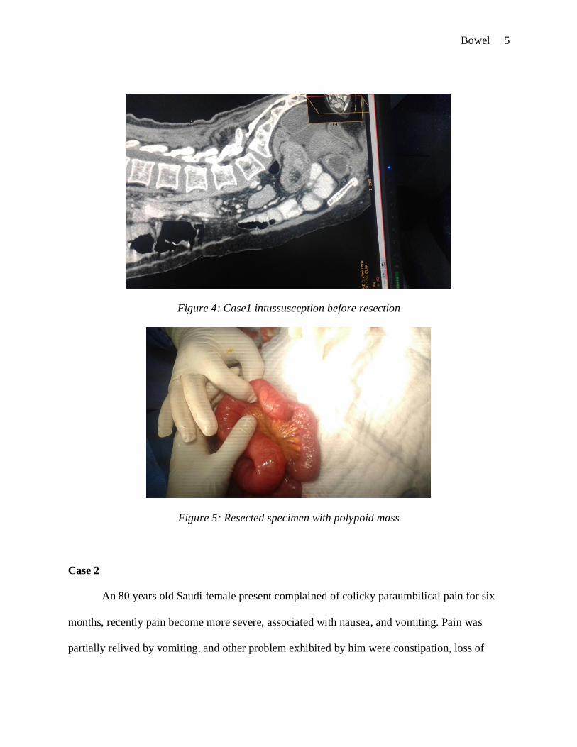
At laparotomy, there was an obvious ileo-ileal intussusceptions (Fig. 4), with a tumor that
acting as a lead point (Fig. 5), resection and anastomosis done. Patient underwent smooth
postoperative course. Histopathology revealed leimyoma and follow-up patient did well. Patient
returned back to his work after 8 weeks.
Page 5
Bowel 5
Figure 4: Case1 intussusception before resection
Figure 5: Resected specimen with polypoid mass
Case 2
An 80 years old Saudi female present complained of colicky paraumbilical pain for six
months, recently pain become more severe, associated with nausea, and vomiting. Pain was
partially relived by vomiting, and other problem exhibited by him were constipation, loss of
Page 6
Bowel 6
appetite and weight as well heartburn. He showed no cough, no neurological or urinary
symptoms. On clinical examination, patient looks un well , a febrile, pulse 84\m regular, BP
130\90, chest & CVS clear, abdomen mildly distended, there is tenderness in the RIF, but no
palpable mass, the rest of other clinical examination no abnormality detected. Routine
investigations such as CBC, RFT, and LFT are within normal limit. Plain abdomen revealed air-
fluid level (Fig. 6), ultrasound abdomen revealed dilated bowel loops.
Figure 6:Case2 plain x-abdomen revealed air-fluid level
CT with contrast revealed target sign suggestive of ileoileal intussusceptions (Fig. 7, 8).
Figure 7: Case2 CT with contrast revealed “Target Sign” (A)
Page 7
Bowel 7
Figure 8:Case2 CT with contrast revealed “Target Sign” (B)
Laporatomy confirmed ileoileal intussusception 6-7cm just proximal ileocecal junction
(Fig. 9).
Figure 9:Case2 ct intussusceptions before resection
Resection and anastomosis done. Specimen revealed polypoid mass acting as a lead point
(Fig. 10).
Page 8
Bowel 8
Figure 10:Case2 resected specimen with polypoid mass
Postoperative patient developed paralytic ileus that kept fasting six days and
histopathology revealed lymphoma.
DISCUSSION
As evident from the research studies, Barbette of Amsterdam made the first report of
intussusception in the year 1674 [1] however details was presented John Hunter in 1789 [2]. Sir
Jonathan Hutchinson is historically considered as the first person who carried out successful
operation of a child with intussusceptions in the year 1871 [3]. Moreover, it is also evident from
different studies that there are some distinct characteristics of adult intussusceptions in
comparison to adults with respect to etiology of this disease, presentation and choice of treatment
[10, 11]. In the adult patients, only 10% of the cases occur due to surgical created stoma or
involve stomach while the remaining 90% usually occur in the large or small bowel [12]. As
seen in these cases, small bowel is the most common single site [13].
Some important points that are related to the intussusceptions are attributed to the causes that are
malignant, benign, or idiopathic [11, 12]. Moreover, approximately, 6% to 30% of all cases are
related to malignant lesions that are primary or secondary in nature. Felix et al. [14] conducted a
Page 9
Bowel 9
review study and reported that 63% of cases exhibited tumor related intussusceptions. In our two
cases, one proved to be malignant, while the other is benign.
According to the locations, intussusceptions are classified into four main categories: (1)
colo-colic that only involves large bowel, (2), entero-enteric that is confined to the small bowel,
(3) ileo-cecal and (4) ileo-colic that is related to the terminal ileum’s prolapse within the
ascending colon. Our two cases were entero-enteric. Intussusceptions in adult occurs with sub-
acute, acute or chronic symptoms that are nonspecific [16]. Erkan et al. in their study
demonstrated that 61.5% of all these patients had shown acute symptoms and emergency
laporatomy was carried out in these patients [32]. Another report has presented that acute
symptoms are shown by 46% of patients [17]. According to findings of a study conducted by
Ghaferi et al. chronic non-specific symptoms are reported in 53% of patients while sub-acute or
acute symptoms are shown by 47% of cases [18].
In our cases, one had acute presentation, while the other showed chronic onset. Some of
classical symptoms that are related to presentation of acute intussusception in pediatrics such as
bloody diarrhea, triad of cramping abdominal pain, and palpable tender mass not occurs in adults
except some rare case [19]. Most of the studies have presented pain as the commonest symptom
that is also revealed by our two cases and this symptoms is seen in almost 71% to 90% of
patients, and the other common symptom that are associated with the condition are bleeding
from the rectum and bloody diarrhea that are reported in most of patients [11]. An important
characteristic that is associated with this type of pain is that it is periodic and intermittent in
nature that leads to the elusive diagnosis of the disease and can lead to some delays in the
process of diagnosis of the disease and before operation, only half cases are diagnosed [11]. In
24% to 42% of all cases, abdominal; mass is reported as a symptom [10, 12].
Page 10
Bowel 10
Azar et al. conducted a review study [10] and finding of this study demonstrated that the
mean duration between the onset and presentation of the symptoms was 37.4 days (it may range
from 1–365 days). The patients with enteric and lesions presented longer duration of symptoms
as compared to the patients with colonic or malignant lesions. However, in our two cases the
benign case showed acute presentation, while the malignant intussusception had chronic onset.
The variance in the imaging features and clinical presentation of the disease has made it a
difficult and challenging task to do the preoperative diagnosis of intussusception. Reijnen et al.
[20] in their research study reported the 50% preoperative diagnostic rate, while considerably
lower rate of 40.7% was reported by Eisen et al. [21]. In our two cases with help CT scan, both
cases were diagnosis confidently preoperatively.
The first diagnostic tool in most of cases is plain abdominal films, however, the clinical
picture is dominated by the obstructive symptoms in most of the cases. The signs of the intestinal
obstruction are usually demonstrated by such films [21, 22]. In our two cases plain x-y abdomen
revealed air-fluid level suggestive of small bowel obstruction without pointing to the primary
reasons of obstruction. The upper gastrointestinal contrast series can present appearance like
“stacked coin” or “coil-spring” [21, 23, 24].
Another useful diagnostic tool for intussusception is ultrasonography that is effective for
diagnosis in adults and children [25]. “Doughnut” or “Target” signs on the transverse view are
the classical imaging characteristics and the other characteristic features are “pseudo-kidney”
sign or “hay-fork” sign exhibited in the longitudinal view [25]. In our two cases, ultrasound
abdomen was not diagnostic and the classical futures such as target and "doughnut" signs were
not described. However, ultrasound is an operator dependent.
Page 11
Bowel 11
In order to make preoperative diagnosis, the most reliable diagnostic information can be
provided by the computed tomography and this method is most effective for those patients that
presents non-specific abdominal pain or require elusive diagnosis [26]. The appearance of
intussusceptions on the CT scan is a "target" mass and "sausage shaped" mass in the transverse
axis and longitudinal axis respectively [18]. In our two cases CT revealed target signs and was
diagnostic.
The frequency of underlying abnormality are associated with the attempts related to
hydrostatic reduction and therefore laparotomy is permitted in case of adult intussusception [10,
27]. Some controversies are still associated with the decision of reducing the intussuscepting
lesion at the time of operation. The previous reports have mentioned that before resection,
intussusception should be reduced [28, 29]. The disadvantages associated with this condition is
that dissemination of the malignant cells may occur; however, regarding this issue, no clear
evidences are provided. On contrary to this, reduction in the intussusceptions is associated with
some benefits, for instance, it is possible to preserve the bowel length considerably in case, if
small bowel is affected, that can lead to prevention of development of a syndrome termed as
short bowel syndrome.
In a study conducted by Begos et al [27], in case when bowel is friable, inflamed or
ischaemic, the suggested method is resection that is carried out without considering reduction
and this condition is very obvious in case of the colo-colic intussusception that provides the high
chances of malignancy. However, in case of other conditions, initial attempt should be
reduction. On the other hand, Azar et al. [30] presented the view that in case of adults, surgical
resection carried out without reduction is preferable treatment and malignancy is related to
approximately 50% of both enteric and colonic intussusceptions. In case when bowel presents no
Page 12
Bowel 12
pathological symptoms and if the idiopathic intussusceptions and post-traumatic condition occur,
the acceptable method is simple reduction is however acceptable in and where no pathological
cause is usually present in the bowel [31]. In our two cases reduction attempted followed by
resection and anastomosis.
CONCLUSION
Entero-enteric intussusception is found to be a rare problem in adults. Due to episodic
and non-specific symptoms related to this condition, diagnosis become difficult. The diagnosis of
this condition can be difficult as symptoms are often non-specific and episodic. Abdominal
computed tomography is considered as the most appropriate method in this condition. Treatment
requires reduction when possible followed by resection and anastomosis.
CONSENT
Before the publication of these cases, the informed consent was taken from the patients
along with accompanying images. Editor-in-Chief has a copy of written consent obtained from
patient before using the information of these patients.
COMPETING INTERESTS
The authors of this article have declared competing or conflicting interests.
AUTHORS’ CONTRIBUTIONS
Mansour TI; perceived the study and did the major part of research i.e. literature search
and also provided full coordination regarding editing, write-up and article submission. Aboujokh
Page 13
Bowel 13
AJ, Bashier OH, and Al-Dalaleh AM all actively participated in the editing and write-up of this
research. The final manuscript is read, edited, proofread by all researchers before submission.
AUTHORS’ INFORMATION
Dr.Tajeddinn Ibrahim Mansour; MD general surgery, Ms.HepatoPancreaticoBiliary surgery,
formerly assistant prof.general surgery Kassala University (Sudan), currently consultant surgeon
Abu-arich general hospital (Saudi Arabia)
Ayman Jaafar Aboujoukh MD histopathology, formerly assistant prof. histopathology Kassala
University (Sudan), currently specialist histopathology King Fahad Central hospital ( Jazan-
Saudi Arabia).
Osman Habeeb Bshier MD general surgery, formerly assistant prof.general surgery Kassala
University (Sudan), currently assistant prof. general surgery Rajhi University (Saudi Arabia); e-
mail [email protected]
Ali Mohammed Al-Dalaleh Ms, PHD, Romanian Board of general surgery, specialist general
surgery Abu-arich general hospital (SaudiArabia)
Page 14
Bowel 14
ACKNOWLEDGEMENTS
I would like to pay a special gratitude to those who assisted in the preparation of this
manuscript and I would like to pay special thanks to patients that provided consent to consider
their cases for this research and accompanying images of patients.
Page 15
Bowel 15
References
1. de Moulin D. Paul Barbette, M.D.: a seventeenth-century Amsterdam author of best-selling
textbooks. Bull Hist Med. 1985;59:506–514
2. Noble I. Master surgeon: John Hunter. J. Messner: New York; 1971. p. 185
3. Hutchinson H, Hutchinson J. Jonathan Hutchinson, life and letters. 1st ed. Wm Heinemann
Medical Books: London; 1946
4. Azar T, Berger DL. Adult intussusception Ann Surg. 1997;226:134–138
5.Weilbaecher D, Bolin JA, Hearn D, Ogden W 2nd. Intussu-sception in adults. Review of 160
cases. Am J Surg. 1971;121:531–535
6. Akcay MN, Polat M, Cadirci M, Gencer B. Tumor-induced ileo-ileal invagination in adults.
Am Surg. 1994; 60:980–981
7. Nagorney DM, Sarr MG, McIlrath DC. Surgical management of intussusception in the adult.
Ann Surg. 1981;193:230–236
8. Haas EM, Etter EL, Ellis S, Taylor TV. Adult intussusception. Am J Surg. 2003;186:75–76
9. Begos DG, Sandor A, Modlin IM. The diagnosis and management of adult intussusception.
Am J Surg. 1997;173:88–94
10 .Azar T, Berger DL. Adult intussusception. Ann Surg 1997;226:134–8
11. Reijnen HA, Joosten HJ, De Boer HH. Diagnosis and treatment of adult intussusception. Am
J Surg 1989;158:25–8
12. Stubenord WT, Thorblamarson B. Intussusception in adults. Ann Surg 1970;172:306–10
13. S Yalamarthi, R C Smith. Adult intussusception: case reports and review of literature.
Postgrad Med J 2005;81:174–177. doi: 10.1136/pgmj.2004.022749
Page 16
Bowel 16
14. Felix EL, Cohen MH, Bernstein AD, et al. Adult intussusception: case report of
recurrent intussusception and review of the literature. Am J Surg 1976;131:758–61
15. Haas EM, Etter EL ,Ellis S, Taylor TV. Adult intussusception. Am J Surg
2003;186:75e6
16.BarussaudM,RegenetN,BriennonX,deKervilerB,PessauxP, Kohneh-Sharhi N, et al. Clinical
spectrum and surgical approach of adult intussusceptions: a multicentric study. Int J Colorectal
Dis 2006;21: 834e9
17. Hamid Ghaderi, Ali J afarian, Ali Aminian, Seyedeh Adeleh, Mirjafari Daryasari. Clinical
presentations,diagnosisandtreatmentofadultintussusception,a20yearssurvey. International
JournalofSurgery8(2010)318-320
18. Begos DG, Sandor A, Modlin IM. The diagnosis and management of adult intussusception.
Am J Surg 1997;173:88e94
19. Reijnen HA, Joosten HJ, de Boer HH. Diagnosis and treatment of adult intussusception. Am
J Surg. 1989;158:25–28
20. Eisen LK, Cunningham JD, Aufses AH Jr. Intussusception in adults: institutional review. J
Am Coll Surg. 1999;188:390–395
21. Cerro P, Magrini L, Porcari P, De Angelis O. Sonographic diagnosis of intussusceptions in
adults. Abdom Imaging. 2000;25:45–47
22. Zubaidi A, Al-Saif F, Silverman R. Adult intussusception: a retrospective review. Dis Colon
Rectum. 2006;49:1546–1551
23. Wiot JF, Spitz HB. Small bowel intussusception demonstrated by oral barium. Radiology.
1970;97:361–366
Page 17
Bowel 17
24. Boyle MJ, Arkell LJ, Williams JT. Ultrasonic diagnosis of adult intussusception. Am J
Gastroenterol. 1993;88:617–618
25. Weissberg DL, Scheible W, Leopold GR. Ultrasonographic appearance of adult
intussusception. Radiology 1977;124:791e2 [15.
26. Eisen LK , Cunningham JD, Aufses AH Jr. Intussusception in adults: institutional review Am
Coll Surg 1999; 188:390e5
27. Begos DG, Sandor A, Modlin IM. The diagnosis and management of adult intussusception.
Am J Surg 1997;73:88–94
28. Donhauser DL, Kelly EC. Intussusception in the adult. Am J Surg 1950;79:673–7
29. Brayton D, Norris WJ. Intussusception in adults. Am J Surg 1954;88:32–43
30. Hutchinson J. A successful case of abdominal section for intussusception. Proc R Med Chir
Soc 1873;7:195–8
31. Kitamura K, Kitagawa S, Mori M, et al. Endoscopic correction of intussusception and
removal of a colonic lipoma. Gastrointest Endosc 1990;36:509–11
32. Erkan N, Haciyanli M, Yildirim M, Sayhan H, Vardar E, Polat AF. Intussusception in adults:
an unusual and challenging condition for surgeons. Int J Colorectal Dis 2005;20:452e6.


![CASE REPORT Open Access Recurrent adult jejuno-jejunal ... · bowel intussusception can occur without a demonstrable pathological cause [4]. Transient intussusception is more common](https://static.documents.pub/doc/80x56/5fa751f2df687c45ad43cf2f/case-report-open-access-recurrent-adult-jejuno-jejunal-bowel-intussusception.jpg)


