Multiple isoforms and differential allelic expression of CHRNA5 in lung tissue and lung adenocarcinoma
Felicia S.Falvella1, Tiziana Alberio2, Sara Noci1, Luigi Santambrogio3, Mario Nosotti3, Matteo Incarbone4, Ugo Pastorino5, Mauro Fasano2 and Tommaso A.Dragani1,* 1Department of Predictive and Preventive Medicine, Fondazione IRCCS Istituto Nazionale dei Tumori, Milan 20133, Italy, 2Department of Theoretical and Applied Sciences - Biomedical Research Division, and Centre of Neuroscience, University of Insubria, Busto Arsizio 21052, Italy, 3Fondazione IRCCS Ospedale Maggiore Policlinico, Università degli Studi di Milano, Milan 20122, Italy, 4Thoracic Surgery, Ospedale San Giuseppe, Multimedica, Milan 20123, Italy and 5Thoracic Surgery, Fondazione IRCCS Istituto Nazionale dei Tumori, Milan 20133, Italy
*To whom correspondence should be addressed. Tel: +39-0223902642; Fax: +39-0223902764; Email: [email protected]
CHRNA5 gene expression variation may play a role in individual susceptibility to lung cancer. Analysis of CHRNA5 transcripts expressed in normal lung tissue detected the full-length tran-script (isoform-1) and four splicing transcripts (isoform-2 to iso-form-5), derived from the recognition of other splice sites in exon 5. Isoforms-2, -3 and -4 were found by protein modeling to form a completely folded, potentially functional extracellular domain and were observed at the protein level, whereas isoform-5 lacked a consistent part of the distorted β sandwich and was not seen at the protein level. Only isoform-1 appeared to encode a complete, functional subunit able to fulfill the ion channel function. We previously reported that CHRNA5 expression is associated with genetic polymorphisms at this locus and that three haplotypes in its promoter region show functional regulation in vitro. Analysis of differential allelic expression (DAE) of three single nucleotide polymorphisms (rs503464, rs55853698 and rs55781567) tagging the expression haplotypes of the CHRNA5 promoter indicated statistically significant DAE at rs55853698 and rs55781567, in both normal lung and lung adenocarcinoma. Overall, our find-ings provide evidence for the presence of multiple CHRNA5 mes-senger RNA (mRNA) isoforms that may modulate the multimeric nicotine receptor and cis-regulatory variations in the CHRNA5 locus that act in vivo in the control of CHRNA5 mRNA expres-sion, in normal lung tissue and in lung adenocarcinoma.
Introduction
Genetic polymorphisms on chromosome 15q25 are associated with the risk of lung cancer (1–4), nicotine addiction (3,5) and chronic obstructive pulmonary disease (6). Association of the same locus with the risk of dependence on multiple substances (alcohol, cocaine and opioids) has also been reported (7). The tight linkage disequilibrium of tagging single nucleotide polymorphisms (SNPs) of this region precluded the identification of the functional variant(s) (1,8). This locus encodes six genes, three of which are nicotinic receptor subunits (CHRNA5, CHRNA3 and CHRNB4). These genes encode the α5, α3 and β4 subunits, respectively, of the neuronal nicotinic acetylcho-line receptors (nAChRs). These receptors are expressed in the brain and their complex functions in nervous signaling and interaction with nicotine are, at least partially, characterized (9). Neuronal nAChRs are
also expressed in lung and in other tissues, but their role outside the central nervous system is not known (9).
In a previous study, we used quantitative messenger RNA (mRNA) expression analysis to investigate the association of 15q25 locus genes with lung cancer. We found that CHRNA5 mRNA was upreg-ulated 30-fold in lung adenocarcinoma as compared with normal lung tissue, whereas CHRNA3 mRNA was downregulated 2-fold, CHRNB4 mRNA was absent and mRNA levels of the other genes were unchanged (10). Moreover, we found that CHRNA5 expression in normal lung tissue was modulated by genetic variations located within this gene (10,11). In particular, we identified three CHRNA5 promoter haplotypes associated with lung mRNA levels and, using luciferase reporter assays in lung cancer cell lines, documented the in vitro modulation of CHRNA5 transcriptional activity by these pro-moter variations (11).
A further complexity in the in vivo regulation of the function of the CHRNA5 gene product, namely the α5 subunit, may be added by the possible existence of mRNA isoforms. Currently, it is not known if CHRNA5 is transcribed as a single mRNA or as multiple isoforms. As it has been observed for other complex receptors, for example, ino-sitol 1,4,5-trisphosphate receptor (12), mRNA isoforms can encode protein variants that interact with the full-length protein or with the other receptor subunits. Therefore, defining the pattern of CHRNA5 isoform expression in lung could advance our understanding of this gene’s role in lung tumorigenesis.
In this study, we characterized novel mRNA isoforms of CHRNA5 in normal lung tissue and in lung adenocarcinoma, and also examined their expression at the protein level in whole cells and on the cell sur-face. Using the deduced protein sequences, we modeled their tertiary structures. Finally, we examined the impact of three SNPs located in the CHRNA5 promoter region on transcription activity using differen-tial allelic expression (DAE), a method that allows the identification of cis-acting variants affecting gene expression.
Materials and methods
Biological samples and nucleic acidsSurgical specimens of normal lung parenchyma were obtained from 329 patients who underwent lung lobectomy for pathologically documented lung adenocarcinoma (n = 273) or for pulmonary metastases from other primary cancers (n = 56). Specimens of normal lung parenchyma were taken as far as possible from the tumor zone; their tumor-free status was confirmed by hematoxylin and eosin staining on randomly selected specimens. From 34 lung adenocarcinoma cases, a paired tumor specimen was also taken for analysis. Information on the tumor histology was collected from pathological records. Samples were obtained from the three hospitals in Milan, Italy, where we work. The recruitment protocol had been approved by the hospitals’ ethics commit-tees and written informed consent was obtained from all patients for the use of their biological materials for research purposes. The human lung cancer cell line A549 was purchased from ATCC and cultured in Ham’s F12 medium (BE12-615F; Lonza, Verviers, Belgium) with 10% fetal bovine serum, 100 U/ml penicillin, 100 U/ml streptomycin and 1.5 g/l sodium bicarbonate in a 5% CO2 humidified atmosphere at 37°C.
Genomic DNA was extracted from lung tissue using DNeasy Blood and Tissue Kit (Qiagen). Total RNA was extracted from lung tissues and from A549 cells using the RNeasy Midi Kit (Qiagen). RNA was reverse transcribed using a 1:1 mixture of oligo(deoxythymidine)18 and random hexamer primers and the Transcriptor First Strand cDNA Synthesis Kit (Roche).
Identification of CHRNA5 transcript isoformsA pool of complementary DNA (cDNA) corresponding to 20 randomly selected samples of normal human lung tissue was used to amplify CHRNA5 transcripts (forward primer, 5′-ggcgatggcggcgcgggggtca-3′; reverse primer, 5′-tcacttatttgcatttccaatatgaac-3′). Amplicons were cloned into pCR 2.1-TOPO vector (Invitrogen). Sequences of individual clones were determined using an automated sequencer (Applied Biosystems, Foster City, CA). Sequences
were aligned and isoforms were identified using Genomatix DiAlign software (http://www.genomatix.de) and the NCBI reference sequence NM_000745. Amino acid sequences were deduced from nucleotide sequences using NCBI Open Reading Frame Finder.
Quantitative analysis of CHRNA5 mRNA isoform levelsLevels of CHRNA5 isoforms were measured in normal lung parenchyma (45 samples) and lung adenocarcinoma (n = 21) by quantitative polymerase chain reaction (qPCR) using primers specific for each isoform (Supplementary Table 1, available at Carcinogenesis Online). Amplification mixtures contained cDNA template diluted in ribonuclease-free water, 10 μl 2× Fast SYBR Green Master Mix (Applied Biosystems) and 0.3 μM PCR primers in a final volume of 20 μl. The human hypoxanthine phosphoribosyltransferase 1 gene (HPRT1; qPCR primers: 5′-gactttgctttccttggtcagg-3′, 5′-tccttttcaccagcaagcttg-3′) was used as control. Reactions were run in duplicate on the 7900HT System (Applied Biosystems). Relative changes in mRNA levels were assessed using the comparative cycle threshold method.
Protein expression analysisExpression of CHRNA5-encoded proteins was assessed in subcellular com-partments of A549 cells by western blotting. In order to detect soluble extra-cellular proteins assembled in heteromeric receptors, we analyzed extracts of surface and non-surface proteins prepared with the Cell Surface Protein Isolation Kit (Thermo Scientific) rather than with standard cell fractionation methods. Briefly, proteins exposed on the cell membrane were labeled with EZ-Link Sulfo-NHS-SS-Biotin, a thiol-cleavable amine-reactive biotinyla-tion reagent. Cells were lysed with a mild detergent solution and the labeled proteins were isolated with NeutrAvidin Agarose. Non-surface proteins were collected in the non-retained fraction (flow-through), whereas bound proteins were released from the resin by incubating in sodium dodecyl sul-fate–polyacrylamide gel electrophoresis sample buffer containing 50 mM dithiothreitol.
Surface and non-surface proteins were resolved by sodium dodecyl sulfate–polyacrylamide gel electrophoresis on 10% or 14% polyacrylamide gels and then transferred to polyvinylidene difluoride membranes (Millipore) at 1 mA/cm2 for 1.5 h. Membranes were incubated (4°C, overnight) with three polyclonal antisera directed against different epitopes of the human protein: mouse anti-CHRNA5 (Abnova; H00001138-A01); rabbit anti-CHRNA5 (Acris; BP2140) and goat anti-CHRNA5 (Abnova; PAB19688). Primary antibodies were diluted 1:1000, 1:1600 and 1:1000, respectively, in 5% non-fat dry milk in 10 mM Tris–HCl pH 8, 150 mM NaCl and 0.05% Tween 20. As a positive control for intracel-lular proteins, goat anti-VDAC-2 (Abcam; ab37985) was used. Protein bands were visualized using peroxidase-conjugated antimouse (Thermo Scientific), antirabbit (Thermo Scientific) and antigoat (Millipore) IgG secondary antibod-ies and the ECL Plus Western Blotting Detection System (Millipore).
Homology modelingPrimary protein sequences of CHRNA5 isoforms were progressively aligned to the extracellular domain (ECD; residues 1–250) of the full-length protein (isoform-1) using ClustalW2 (13). Homology models were built using the SwissModel server (14) in alignment mode using the Torpedo CHRNA5 X-ray structure (PDB ID 2BG9) as the template. Structural models were rendered using SwissPdbViewer software (14).
CHRNA5 haplotype analysis and DAEDAE is based on the fact that, in the presence of cis-regulatory elements, heterozygous individuals preferentially express one of their two alleles; this allelic imbalance can be observed in cDNA and is context specific (15). As DAE can only be performed on heterozygous samples at any given SNP, we first genotyped the 329 samples of genomic DNA from normal lung tissue for three SNPs (rs503464, rs55853698 and rs55781567) already known to be localized in the CHRNA5 promoter. To this end, we first PCR amplified the SNP-containing fragment using a two-step process to eliminate any possible contamination from genomic DNA in later analyses. First, cDNA was ampli-fied using a forward primer mapping in exon 1 and a reverse primer map-ping in exon 2 (Supplementary Table 1, available at Carcinogenesis Online). Then, 25% of the PCR product was amplified again using the same forward primer and a different reverse primer mapping in exon 1. The resulting PCR products were then genotyped by pyrosequencing using a PSQ96MA pyrosequencing system (Qiagen) and allele-specific primers (Supplementary Table 1, available at Carcinogenesis Online). Finally, the proportions of indi-vidual alleles for each SNP were obtained using the PyroMark MD software package (Qiagen).
Samples determined to be heterozygous for each SNP were used in DAE. For this purpose, pairs of genomic DNA and cDNA from the same subjects were analyzed and for each cDNA, its allelic ratio (log10 values) was compared with that of the corresponding genomic DNA.
Statistical analysisDifferences in quantitative levels were analyzed by one-way analysis of vari-ance or Welch t-test. All statistical tests were two-sided and were carried out using the Rcmdr package in R (16).
Results
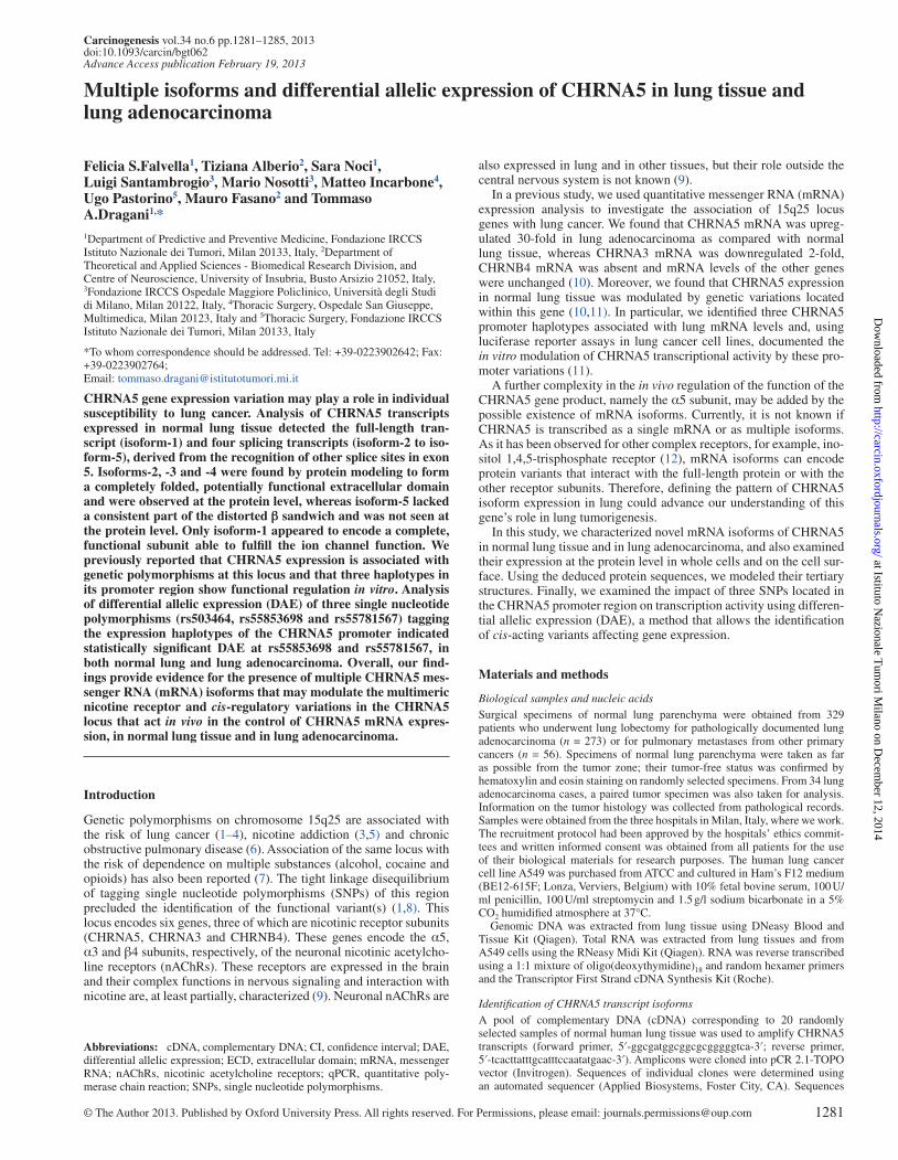
Multiple CHRNA5 lung transcriptsSequence analysis of CHRNA5 transcripts expressed in a pool of cDNA from 20 samples of normal lung tissue led to the identification of one full-length transcript (isoform-1; NM_000745) and four novel isoforms (isoform-2 to isoform-5). These isoforms were the result of alternate splicing at other splice sites in exon 5 and produced trun-cated mRNAs (Figure 1).
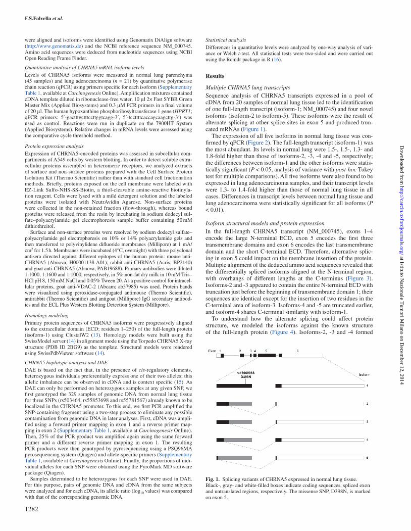
The expression of all five isoforms in normal lung tissue was con-firmed by qPCR (Figure 2). The full-length transcript (isoform-1) was the most abundant. Its levels in normal lung were 1.5-, 1.5-, 1.3- and 1.8-fold higher than those of isoforms-2, -3, -4 and -5, respectively; the differences between isoform-1 and the other isoforms were statis-tically significant (P < 0.05, analysis of variance with post-hoc Tukey test for multiple comparisons). All five isoforms were also found to be expressed in lung adenocarcinoma samples, and their transcript levels were 1.3- to 1.4-fold higher than those of normal lung tissue in all cases. Differences in transcript levels between normal lung tissue and lung adenocarcinoma were statistically significant for all isoforms (P < 0.01).
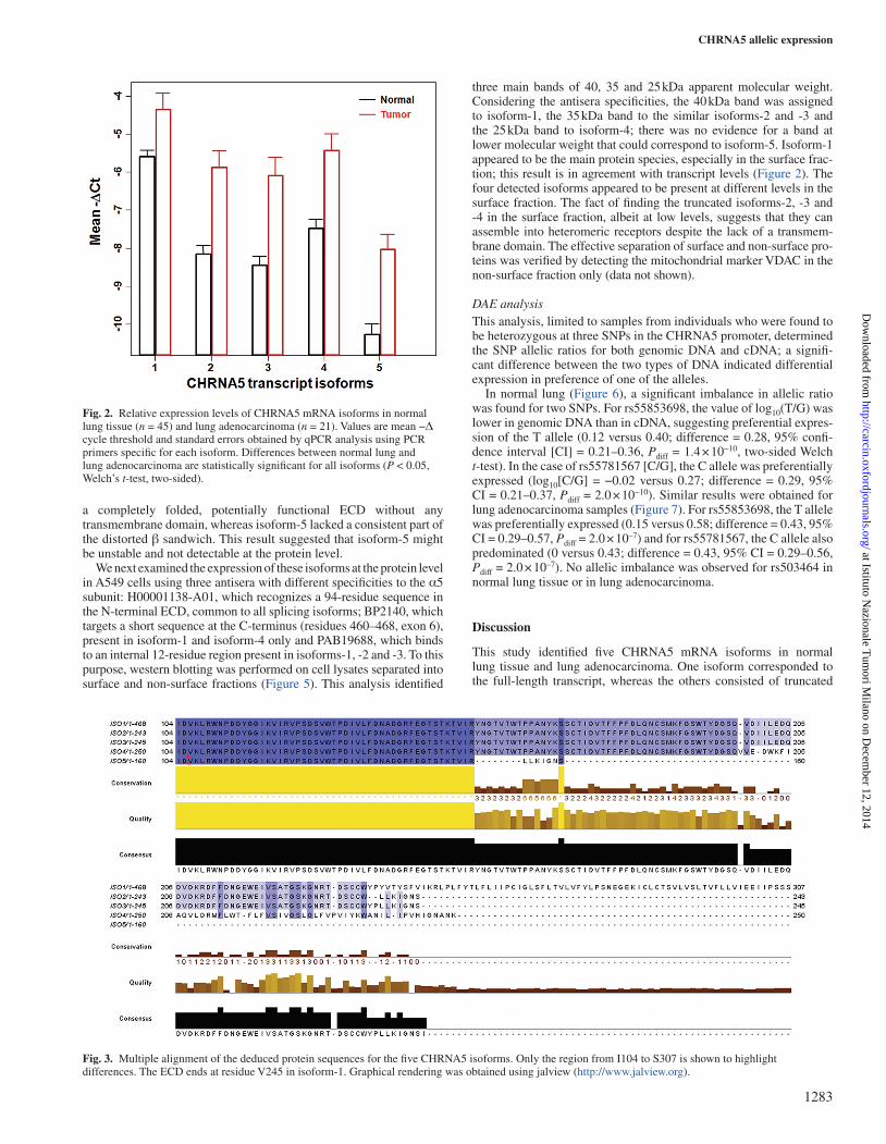
Isoform structural models and protein expressionIn the full-length CHRNA5 transcript (NM_000745), exons 1–4 encode the large N-terminal ECD, exon 5 encodes the first three transmembrane domains and exon 6 encodes the last transmembrane domain and the short C-terminal ECD. Therefore, alternative splic-ing in exon 5 could impact on the membrane insertion of the protein. Multiple alignment of the deduced amino acid sequences revealed that the differentially spliced isoforms aligned at the N-terminal region, with overhangs of different lengths at the C-terminus (Figure 3). Isoforms-2 and -3 appeared to contain the entire N-terminal ECD with truncation just before the beginning of transmembrane domain 1; their sequences are identical except for the insertion of two residues in the C-terminal area of isoform-3. Isoforms-4 and -5 are truncated earlier, and isoform-4 shares C-terminal similarity with isoform-1.
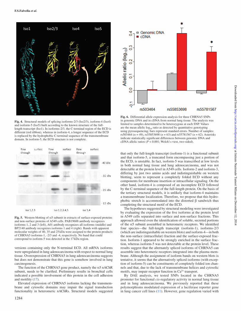
To understand how the alternate splicing could affect protein structure, we modeled the isoforms against the known structure of the full-length protein (Figure 4). Isoforms-2, -3 and -4 formed
Fig. 1. Splicing variants of CHRNA5 expressed in normal lung tissue. Black-, gray- and white-filled boxes indicate coding sequences, spliced exon and untranslated regions, respectively. The missense SNP, D398N, is marked on exon 5.
a completely folded, potentially functional ECD without any transmembrane domain, whereas isoform-5 lacked a consistent part of the distorted β sandwich. This result suggested that isoform-5 might be unstable and not detectable at the protein level.
We next examined the expression of these isoforms at the protein level in A549 cells using three antisera with different specificities to the α5 subunit: H00001138-A01, which recognizes a 94-residue sequence in the N-terminal ECD, common to all splicing isoforms; BP2140, which targets a short sequence at the C-terminus (residues 460–468, exon 6), present in isoform-1 and isoform-4 only and PAB19688, which binds to an internal 12-residue region present in isoforms-1, -2 and -3. To this purpose, western blotting was performed on cell lysates separated into surface and non-surface fractions (Figure 5). This analysis identified
three main bands of 40, 35 and 25 kDa apparent molecular weight. Considering the antisera specificities, the 40 kDa band was assigned to isoform-1, the 35 kDa band to the similar isoforms-2 and -3 and the 25 kDa band to isoform-4; there was no evidence for a band at lower molecular weight that could correspond to isoform-5. Isoform-1 appeared to be the main protein species, especially in the surface frac-tion; this result is in agreement with transcript levels (Figure 2). The four detected isoforms appeared to be present at different levels in the surface fraction. The fact of finding the truncated isoforms-2, -3 and -4 in the surface fraction, albeit at low levels, suggests that they can assemble into heteromeric receptors despite the lack of a transmem-brane domain. The effective separation of surface and non-surface pro-teins was verified by detecting the mitochondrial marker VDAC in the non-surface fraction only (data not shown).
DAE analysisThis analysis, limited to samples from individuals who were found to be heterozygous at three SNPs in the CHRNA5 promoter, determined the SNP allelic ratios for both genomic DNA and cDNA; a signifi-cant difference between the two types of DNA indicated differential expression in preference of one of the alleles.
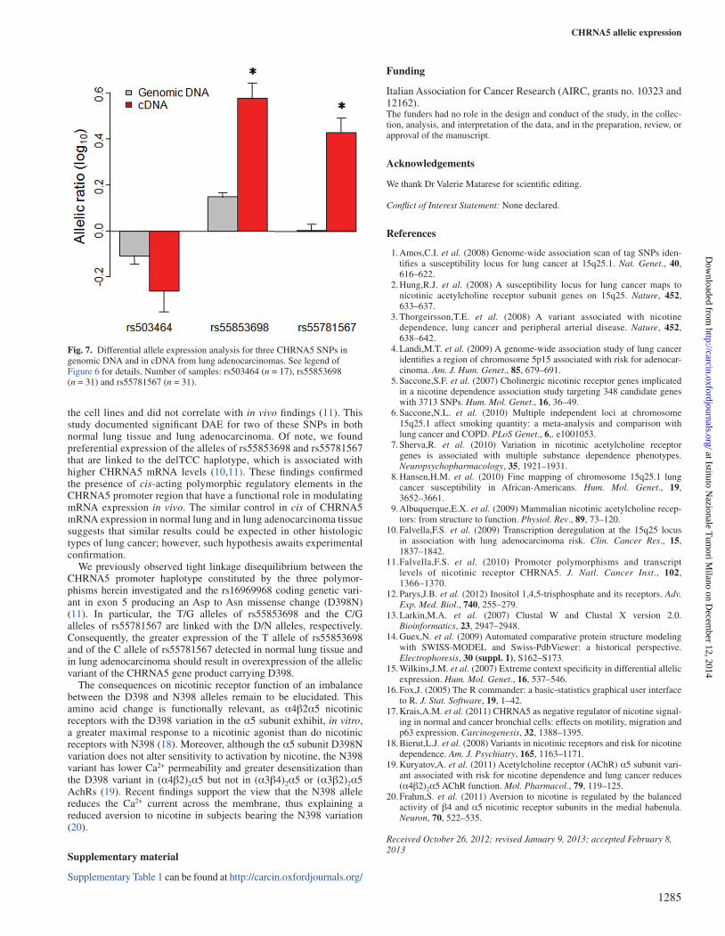
In normal lung (Figure 6), a significant imbalance in allelic ratio was found for two SNPs. For rs55853698, the value of log10(T/G) was lower in genomic DNA than in cDNA, suggesting preferential expres-sion of the T allele (0.12 versus 0.40; difference = 0.28, 95% confi-dence interval [CI] = 0.21–0.36, Pdiff = 1.4 × 10⁻¹⁰, two-sided Welch t-test). In the case of rs55781567 [C/G], the C allele was preferentially expressed (log10[C/G] = −0.02 versus 0.27; difference = 0.29, 95% CI = 0.21–0.37, Pdiff = 2.0 × 10⁻¹⁰). Similar results were obtained for lung adenocarcinoma samples (Figure 7). For rs55853698, the T allele was preferentially expressed (0.15 versus 0.58; difference = 0.43, 95% CI = 0.29–0.57, Pdiff = 2.0 × 10⁻⁷) and for rs55781567, the C allele also predominated (0 versus 0.43; difference = 0.43, 95% CI = 0.29–0.56, Pdiff = 2.0 × 10⁻⁷). No allelic imbalance was observed for rs503464 in normal lung tissue or in lung adenocarcinoma.
Discussion
This study identified five CHRNA5 mRNA isoforms in normal lung tissue and lung adenocarcinoma. One isoform corresponded to the full-length transcript, whereas the others consisted of truncated
Fig. 2. Relative expression levels of CHRNA5 mRNA isoforms in normal lung tissue (n = 45) and lung adenocarcinoma (n = 21). Values are mean −Δ cycle threshold and standard errors obtained by qPCR analysis using PCR primers specific for each isoform. Differences between normal lung and lung adenocarcinoma are statistically significant for all isoforms (P < 0.05, Welch’s t-test, two-sided).
Fig. 3. Multiple alignment of the deduced protein sequences for the five CHRNA5 isoforms. Only the region from I104 to S307 is shown to highlight differences. The ECD ends at residue V245 in isoform-1. Graphical rendering was obtained using jalview (http://www.jalview.org).
versions containing only the N-terminal ECD. All mRNA isoforms were upregulated in lung adenocarcinoma with respect to normal lung tissue. Overexpression of CHRNA5 in lung adenocarcinoma suggests but does not demonstrate that this gene is somehow involved in lung carcinogenesis.
The function of the CHRNA5 gene product, namely the α5 nAChR subunit, needs to be clarified. Preliminary results in bronchial cells indicated a possible involvement of this protein in the cell adhesion and motility (17).
Elevated expression of CHRNA5 isoforms lacking the transmem-brane and cytosolic domains may impair the signal transduction functionality in heteromeric nAChRs. Structural models suggested
that only the full-length transcript (isoform-1) is a functional subunit and that isoform-5, a truncated form encompassing just a portion of the ECD, is unstable. In fact, isoform-5 was transcribed at low levels in both normal lung tissue and lung adenocarcinoma, and was not detectable at the protein level in A549 cells. Isoform-2 and isoform-3, differing by just two amino acids and indistinguishable on western blotting, seem to represent a completely folded ECD without any components for membrane insertion or intracellular signaling. On the other hand, isoform-4 is composed of an incomplete ECD followed by the C-terminal sequence of the full-length protein. On the basis of the tertiary structural models, it is unlikely that isoform-4 maintains a transmembrane localization. Therefore, we propose that this hydro-phobic stretch is accommodated into the distorted β sandwich thus completing the structural motif of the ECD.
The hypotheses suggested by structural modeling were investigated by evaluating the expression of the five isoforms at the protein level in A549 cells separated into surface and non-surface fractions. This approach allowed even the identification of soluble, secreted portions of the α5 subunit assembled in heteromeric receptors. We identified four species—the full-length transcript (isoform-1), isoforms-2/3 (which are indistinguishable on western blots) and isoform-4—in both the non-surface (intracellular) fraction and the surface-exposed frac-tion. Isoform-1 appeared to be strongly enriched in the surface frac-tion, whereas isoform-5 was not detectable at the protein level. These results suggest that the alternately spliced isoforms of CHRNA5 can assemble into heteromeric receptors integrated into the plasma mem-brane. Although the assignment of isoform bands on western blots is tentative, it seems that the alternatively spliced isoforms (with excep-tion of isoform-5) can be constituents of completely folded ion chan-nels and that, due to the lack of transmembrane helices and cytosolic motifs, may impair receptor function in Ca2+ transport.
By DAE analysis, we tested SNPs located in the CHRNA5 promoter for functional cis-regulatory activity in normal lung tissue and in lung adenocarcinoma. We previously reported that these polymorphisms modulated expression of a luciferase reporter gene in lung cancer cell lines (11). However, gene regulation varied with
Fig. 4. Structural models of splicing isoforms-2/3 (Iso2/3), isoform-4 (Iso4) and isoform-5 (Iso5) built according to the known structure of the full-length transcript (Iso1). In isoforms-2/3, the C-terminal region of the ECD is different (red ribbon), whereas in isoform-4, a longer sequence of the ECD is replaced by the hydrophobic C-terminal sequence of the transmembrane domain. In isoform-5, the ECD structure is not complete.
Fig. 5. Western blotting of α5 subunit in extracts of surface-exposed proteins and non-surface proteins of A549 cells. PAB19688 antibody recognizes isoforms 1, 2 and 3 (left), A01 antibody recognizes all isoforms (middle) and BP2140 antibody recognizes isoforms 1 and 4 (right). Bands with apparent molecular weights of 40, 35 and 25 kDa were assigned to the protein products of CHRNA5 isoforms-1, -2/3 and -4, respectively. No band that could correspond to isoform-5 was detected in the 17 kDa region.
Fig. 6. Differential allele expression analysis for three CHRNA5 SNPs in genomic DNA and in cDNA from normal lung tissue. The analysis was limited to samples determined to be heterozygous at each SNP. Values are the mean allelic log10 ratio as detected by quantitative genotyping using pyrosequencing; bars represent standard errors. Number of samples: rs503464 (n = 49), rs55853698 (n = 63) and rs55781567 (n = 62). Asterisks indicate statistically significant differences between genomic DNA and cDNA allelic ratios (P < 0.001; Welch’s t-test, two-sided).
the cell lines and did not correlate with in vivo findings (11). This study documented significant DAE for two of these SNPs in both normal lung tissue and lung adenocarcinoma. Of note, we found preferential expression of the alleles of rs55853698 and rs55781567 that are linked to the delTCC haplotype, which is associated with higher CHRNA5 mRNA levels (10,11). These findings confirmed the presence of cis-acting polymorphic regulatory elements in the CHRNA5 promoter region that have a functional role in modulating mRNA expression in vivo. The similar control in cis of CHRNA5 mRNA expression in normal lung and in lung adenocarcinoma tissue suggests that similar results could be expected in other histologic types of lung cancer; however, such hypothesis awaits experimental confirmation.
We previously observed tight linkage disequilibrium between the CHRNA5 promoter haplotype constituted by the three polymor-phisms herein investigated and the rs16969968 coding genetic vari-ant in exon 5 producing an Asp to Asn missense change (D398N) (11). In particular, the T/G alleles of rs55853698 and the C/G alleles of rs55781567 are linked with the D/N alleles, respectively. Consequently, the greater expression of the T allele of rs55853698 and of the C allele of rs55781567 detected in normal lung tissue and in lung adenocarcinoma should result in overexpression of the allelic variant of the CHRNA5 gene product carrying D398.
The consequences on nicotinic receptor function of an imbalance between the D398 and N398 alleles remain to be elucidated. This amino acid change is functionally relevant, as α4β2α5 nicotinic receptors with the D398 variation in the α5 subunit exhibit, in vitro, a greater maximal response to a nicotinic agonist than do nicotinic receptors with N398 (18). Moreover, although the α5 subunit D398N variation does not alter sensitivity to activation by nicotine, the N398 variant has lower Ca2+ permeability and greater desensitization than the D398 variant in (α4β2)2α5 but not in (α3β4)2α5 or (α3β2)2α5 AchRs (19). Recent findings support the view that the N398 allele reduces the Ca2+ current across the membrane, thus explaining a reduced aversion to nicotine in subjects bearing the N398 variation (20).
Supplementary material
Supplementary Table 1 can be found at http://carcin.oxfordjournals.org/
Funding
Italian Association for Cancer Research (AIRC, grants no. 10323 and 12162).The funders had no role in the design and conduct of the study, in the collec-tion, analysis, and interpretation of the data, and in the preparation, review, or approval of the manuscript.
Acknowledgements
We thank Dr Valerie Matarese for scientific editing.
Conflict of Interest Statement: None declared.
References
1. Amos,C.I. et al. (2008) Genome-wide association scan of tag SNPs iden-tifies a susceptibility locus for lung cancer at 15q25.1. Nat. Genet., 40, 616–622.
2. Hung,R.J. et al. (2008) A susceptibility locus for lung cancer maps to nicotinic acetylcholine receptor subunit genes on 15q25. Nature, 452, 633–637.
3. Thorgeirsson,T.E. et al. (2008) A variant associated with nicotine dependence, lung cancer and peripheral arterial disease. Nature, 452, 638–642.
4. Landi,M.T. et al. (2009) A genome-wide association study of lung cancer identifies a region of chromosome 5p15 associated with risk for adenocar-cinoma. Am. J. Hum. Genet., 85, 679–691.
5. Saccone,S.F. et al. (2007) Cholinergic nicotinic receptor genes implicated in a nicotine dependence association study targeting 348 candidate genes with 3713 SNPs. Hum. Mol. Genet., 16, 36–49.
6. Saccone,N.L. et al. (2010) Multiple independent loci at chromosome 15q25.1 affect smoking quantity: a meta-analysis and comparison with lung cancer and COPD. PLoS Genet., 6,. e1001053.
7. Sherva,R. et al. (2010) Variation in nicotinic acetylcholine receptor genes is associated with multiple substance dependence phenotypes. Neuropsychopharmacology, 35, 1921–1931.
8. Hansen,H.M. et al. (2010) Fine mapping of chromosome 15q25.1 lung cancer susceptibility in African-Americans. Hum. Mol. Genet., 19, 3652–3661.
9. Albuquerque,E.X. et al. (2009) Mammalian nicotinic acetylcholine recep-tors: from structure to function. Physiol. Rev., 89, 73–120.
10. Falvella,F.S. et al. (2009) Transcription deregulation at the 15q25 locus in association with lung adenocarcinoma risk. Clin. Cancer Res., 15, 1837–1842.
11. Falvella,F.S. et al. (2010) Promoter polymorphisms and transcript levels of nicotinic receptor CHRNA5. J. Natl. Cancer Inst., 102, 1366–1370.
12. Parys,J.B. et al. (2012) Inositol 1,4,5-trisphosphate and its receptors. Adv. Exp. Med. Biol., 740, 255–279.
13. Larkin,M.A. et al. (2007) Clustal W and Clustal X version 2.0. Bioinformatics, 23, 2947–2948.
14. Guex,N. et al. (2009) Automated comparative protein structure modeling with SWISS-MODEL and Swiss-PdbViewer: a historical perspective. Electrophoresis, 30 (suppl. 1), S162–S173.
15. Wilkins,J.M. et al. (2007) Extreme context specificity in differential allelic expression. Hum. Mol. Genet., 16, 537–546.
16. Fox,J. (2005) The R commander: a basic-statistics graphical user interface to R. J. Stat. Software, 19, 1–42.
17. Krais,A.M. et al. (2011) CHRNA5 as negative regulator of nicotine signal-ing in normal and cancer bronchial cells: effects on motility, migration and p63 expression. Carcinogenesis, 32, 1388–1395.
18. Bierut,L.J. et al. (2008) Variants in nicotinic receptors and risk for nicotine dependence. Am. J. Psychiatry, 165, 1163–1171.
19. Kuryatov,A. et al. (2011) Acetylcholine receptor (AChR) α5 subunit vari-ant associated with risk for nicotine dependence and lung cancer reduces (α4β2)2α5 AChR function. Mol. Pharmacol., 79, 119–125.
20. Frahm,S. et al. (2011) Aversion to nicotine is regulated by the balanced activity of β4 and α5 nicotinic receptor subunits in the medial habenula. Neuron, 70, 522–535.
Received October 26, 2012; revised January 9, 2013; accepted February 8, 2013
Fig. 7. Differential allele expression analysis for three CHRNA5 SNPs in genomic DNA and in cDNA from lung adenocarcinomas. See legend of Figure 6 for details. Number of samples: rs503464 (n = 17), rs55853698 (n = 31) and rs55781567 (n = 31).