Advance photodynamic inactivation of dental pathogenic microorganisms with water-soluble and cationic phthalocyanines
Vanya Mantareva1,*, Veselin Kussovski2, Ivan Angelov1 and Slavcho Dimitrov3 1 Institute of Organic Chemistry with Centre of Phytochemistry, Bulgarian Academy of Sciences, 1113 Sofia, Bulgaria 2 The Stephan Angeloff Institute of Microbiology, Bulgarian Academy of Sciences, 1113 Sofia, Bulgaria 3 Faculty of Dental Medicine, Medical University - Sofia, 1 Georgi Sofijski str., 1431 Sofia, Bulgaria
Photodynamic therapy (PDT) is well accepted as complementary curative treatment of tumors and other pathologic conditions. Recent promising application includes the acute infections in humans. PDT appears not in competition but in advance to the conventional treatments. Moreover, this non-invasive and gentle procedure has not alternatives intended for multi-resistant pathogenic microorganisms, especially in cases of infections which cannot be influenced by the conventional antibiotics associated therapy. The present study explores the PDT method for treatment of pathogenic bacteria and fungi related to dental practice. A number of water-soluble and cationic phthalocyanine complexes of biocompatible metals (zinc, gallium and silicon) were synthesized to be studied as PDT agents towards oral pathogens. The Gram-positive cariogenic bacteria, Streptococcus mutans, Actinomyces israelii and Enterococcus faecalis, and the Gram-negative periodontal pathogens Prevotella intermedia and the pathogenic fungus Candida albicans were investigated for their susceptibility to photodynamic inactivation with metal phthalocyanines.
Keywords Photodynamic therapy, phthalocyanine, fluorescence, singlet oxygen, dental pathogenic microorganisms
1. Introduction
The extensive multi-drug resistance of human-spreaded pathogenic microorganisms has forced the research on the development of new techniques for cure of acute infections. The photodynamic action was first observed on microorganisms in the year 1900, but totally forgot during the Golden age of antibiotics [1]. The resistance of pathogenic microorganisms forces the research in the well-accepted for diagnosis and treatment of tumours technology namely Photodynamic therapy (PDT). The last two decades experimental studies are indicative for the valuable future of PDT for treatment of local infections [2, 3]. The method is still on the research phase for clinical applications such as healing wounds, nasal disinfection and treatment of pathogens-associated oral diseases [4]. The oral cavity is the major reservoir of a variety of pathogens including bacteria, fungi and viruses. Most of the dental pathogenic microorganisms are highly resistant to the known and newly developed antibiotics and appear responsible to the expansion of dental complications. Obviously, the Dental medicine, including all aspects of the pathogens associated with oral cavity, features as the field of human medicine which is the most promising for wide application of antimicrobial PDT. In many cases of infection diseases, the only solution in fight with pathogens could be PDT [5]. For example, the repeated infections, where the pathogens become more and more resistant to the conventional treatment with antibiotics the combination of the known treatment and in addition the PDT can significantly improve the outcome for the immuno compromise patiences [6]. Moreover the aggressive actions of the aerobic and especially anaerobic bacterial and fungal cells, which grow in the periodontal pockets leading to caries, periodontal diseases and tooth loss, can be reduced by the so called PDT disinfection [7]. The high risk of the oral pathogenic microorganisms is a consequence of the produced destruction products during metabolic processes of pathogens which get into the blood stream and further lead to several complications in human health. The photodynamic process can be described as a combined action of three components such as a photoactive compound (photosensitizer), the proper light from the visible and near infra-red spectra, and the presence of molecular oxygen atmosphere [8]. Upon irradiation, the photoactive compound becomes excited and the absorbed energy is transferred to the ground-state triplet of molecular oxygen to undergo electron alterations to highly reactive and toxic to the cells singlet oxygen state (type II mechanism of photosensitization). The produced singlet oxygen and other reactive oxygen species (ROSs) can undergo photochemical reactions with the surrounded bioorganic macromolecules, which finally resulted in irreversible cell damage and cell death. In parallel, the type I mechanism of photosensitization can be involved. It includes an electron or hydrogen transfer from the excited triplet state of the photosensitizer to the cellular-associated biomolecules of proteins, lipids and DNA. The both mechanisms lead to formation of highly reactive and toxic to the cells ROSs. The great number of studies in PDT suggested that the most of the photosensitizers need a surrounding rich of molecular oxygen to proceed their cytotoxic action [9-11]. Dental caries is the destruction of the enamel, dentin or cementum of teeth due to bacterial activities. Caries are initiated by direct demineralization of the enamel of teeth due to lactic acid and other organic acids which accumulate in dental plaque. Dental plaque, which is material adhering to the teeth, consists of bacterial cells, salivary polymers, and bacterial extracellular products. Plaque is a naturally-constructed biofilm, in which the consortia of bacteria may reach a thickness of 300-500 cells on the surfaces of the teeth. These accumulations subject the teeth and gingival tissues to
Science against microbial pathogens: communicating current research and technological advances A. Méndez-Vilas (Ed.)______________________________________________________________________________
high concentrations of bacterial metabolites, which result in dental disease [12]. The dominant bacterial species in dental plaque are Streptococcus sanguis and Streptococcus mutans, both of which are considered responsible for plaque. Plaque formation is initiated by a weak attachment of the streptococcal cells to salivary glycoproteins forming a pellicle on the surface of the teeth. This is followed by a stronger attachment by means of extracellular sticky polymers of glucose (glucans) which are synthesized by the bacteria from dietary sugars, principally sucrose. An enzyme on the cell surface of Streptococcus mutans, glycosyl transferase, is involved in initial attachment of the bacterial cells to the tooth surface and in the conversion of sucrose to dextran polymers (glucans) which form plaque. Streptococcus mutans appears to be important in the initiation of dental caries because its activities lead to colonization of the tooth surfaces, plaque formation, and localized demineralization of tooth enamel. It is not however, the only cause of dental decay. After initial weakening of the enamel, various oral bacteria gain access to interior regions of the tooth. Lactobacilli, Actinomyces, and various proteolytic bacteria are commonly found in human carious dentin and cementum, which suggests that they are secondary invaders that contribute to the progression of the lesions. Periodontal diseases are bacterial infections that affect the supporting structures of the teeth (gingiva, cementum, periodontal membrane and alveolar bone). The most common form, gingivitis, is an inflammatory condition of the gums. It is associated with accumulations of bacterial plaque in the area. Diseases that are confined to the gum usually do not lead to loss of teeth, but there are other more serious forms of periodontal disease that affect periodontal membrane and alveolar bone resulting in tooth loss. The microbiota of periodontal disease is predominantly anaerobic from the three groups of microorganism’s species. Bacteria in these lesions are very complex populations consisting of the Gram-positive organisms (including Actinomyces) and the Gram-negative organisms (primarily Porphyromonas gingivalis, Aggregatibacter actinomycetemcomitans, Bacteroides forsythus, Prevotella intermedia, Fusobacterium nucleatum and spirochaetes) [13]. The mechanisms of tissue destruction in periodontal disease are not clearly defined but hydrolytic enzymes, endotoxins, and other toxic bacterial metabolites seem to be involved. Actinomyces israelii (non-acid, fast, non-motile, Gram-positive organism revealing characteristic branching filaments that end in clubs or hyphae) is normally present in the mouth but can cause disease if it enters tissues following an injury. A. israelii is an anaerobic bacterium which grows very well in deep tissues where oxygen levels are low. Tooth extraction, tooth disease, root canal treatment, jaw surgery as well as a poor dental hygiene can allow A. israelii to cause an infection. Prevotella intermedia is Gram-negative, rod shaped, black pigmented, obligate anaerobic pathogen [14]. Apical periodontitis is a sequel to endodontic infection and manifests itself as the host defence response to microbial challenge emanating from the root canal system. Only a few species have been found in the root canals of teeth that have undergone proper endodontic treatment (predominantly Gram-positive cocci, rods, and filaments). Bacteria belonging to the genera Actinomyces, Enterococcus, and Propionibacterium are frequently isolated from such root canals and characterized [15, 16]. Enterococcus faecalis (Gram-positive, facultatively anaerobic cocci, which occurs singly, in pairs or short chains) is resistant to most of the intracanal medicaments probably due to its ability to regulate internal pH with an efficient proton pump [17]. Microbiological and electron microscopic studies have shown the presence of yeasts in canals of root-filled teeth with unresolved apical periodontitis [18]. Candida albicans (diploid fungus/a form of yeast) is the most frequently isolated fungus from root-filled teeth with apical periodontitis [19].
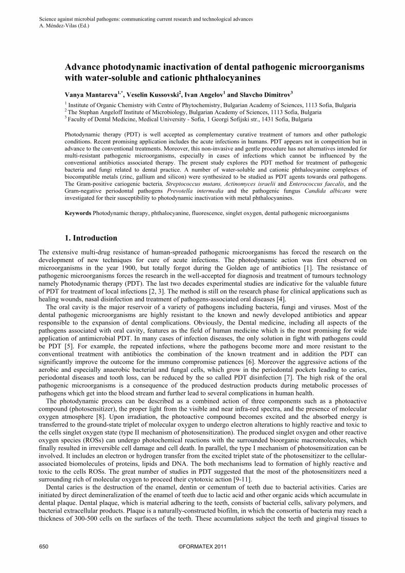
Scheme 1. Chemical structure of phthalocyanine complexes peripherally substituted with methylpyridyloxy functional groups. The present study focuses on PDT study of water-soluble and cationic phthalocyanine complexes {MPcs, M = Zn(II), Ga(III) and Si(IV)} (Scheme 1) towards some typical dental pathogens. The Gram-positive bacteria Enterococcus faecalis (E. faecalis), methicillin-resistant Staphylococcus aureus (MRSA), Actinomyces israelii (A. israelii), Streptococcus mutans (S. mutans) and fungus Candida albicans (C. albicans) were cultured as planktonic and as biofilms grown on acrylic resin used in dental practice.
Science against microbial pathogens: communicating current research and technological advances A. Méndez-Vilas (Ed.)_______________________________________________________________________________
2. Experimental
The metal complexes of Zn(II)-, Ga(III)- and Si(IV)- phthalocyanine were studied against selected dental pathogenic strains. The syntheses were carried out as were described in our recent publications [20-23].
2.1 Chemicals
All chemicals for spectrophotometric studies and photodynamic inactivation experiments were products of Sigma-Aldrich and Fluka (Germany). The measurements were carried out with dimethylsulphoxide (DMSO) and dimethylformamide (DMF) of spectroscopic grade. Sodium dodecyl sulfate (SDS) and Cremophor EL (CEL) were purchased from Sigma. The photochemical study was carried out with 1,3-diphenylisobenzofuran (DPBF) which was diluted before measurements.
2.2 Bacterial strains
The Gram-positive, anaerobic bacterial strain Actinomyces israelii 104 875 and facultative anaerobic strain Streptococcus mutans 103220 T from the collection of “Institute Pasteur” (France) were used in the investigation. The strains of MRSA and Enterococcus faecalis are purchased from the Bulgarian National bank for industrial microorganisms. They are the representative for the Gram-positive and aerobic microorganisms in the experiments. The bacteria were grown overnight at 37 °C and after harvesting by centrifugation were resuspended in sterile PBS. Before the experiments they were diluted to the required cell densities.
2.3 Fungal strain
The fungus Candida albicans 74 from the Bulgarian National bank for industrial microorganisms and cell cultures (NBIMCC) was used. The fungal strain was grown aerobically on Yeast Mold (YM) agar (Difco) overnight at 37 °C. Cells were harvested by centrifugation and were resuspended in sterile PBS. Before the experiments they were diluted to the required cell densities from 109 to 105 cells mL-1.
2.4 Steady state and time-resolved fluorimetry
The fluorescence emission spectra were recorded at excitation wavelength 610 nm for the spectral region of 625-750 nm by using 1 cm quartz cuvette at RT by Jasco 6600 fluorimeter. The fluorescence quantum yields (ΦF) were calculated on the basis of absorption and fluorescence spectra of SiPc1, GaPc1 and ZnPc by using a comparative method, where the equation (1) was employed for calculations:
ΦF = ΦF(R) (I A(R) η
2 )/ (I(R) A η(R)2) (1)
where ΦF(R) is the fluorescence yield of the reference standard (ZnPc, ΦF(R) = 0.18 in DMSO [24]), I and I(R) are the integrated fluorescence of the sample (SiPc1 or GePc1) and the reference standard (ZnPc), A and A(R) are the absorbance of the sample and the reference, and η and η(R) are the refractive indices of the solvents employed in calculating of fluorescence quantum yields. The absorbances of the sample and the reference were kept under 0.05 at λexc in order to avoid the reabsorption effect. The fluorescence lifetimes (τF) of ZnPcMe, SiPc1 and GaPc1 and the used as a standard ZnPc were measured at 20 °C in 1 cm quartz cuvette with a stopper by the means of a SPEX Fluorolog -3 fluorometer (Horiba Jobin Yvon). The time correlated single photon counting (TCSPC) mode of the apparatus equipped with the Fluorolog fitting software IBH program was used. Briefly, the set-up consists of the excitation source (light emitting diode NanoLED, 365 nm) with 1 MHz repetition rate with a linear polarizer. The fluorescence was detected by a peltier cooled photomultiplier tube (PMT) and the integrated electronics. A monochromator with a spectral width of 2 nm was used to select the required emission wavelength band. The response function of the system, which was measured with a scattering Ludox solution (DuPont), had a full width at half-maximum (FWHM) of about 500 ps for NanoLED. All decay curves were measured at the emission maximum of PSs and the lifetimes were obtained by deconvolution of the decay curves with the Fluorolog fitting software IBH program. The measured decays were fitted to the convolution of single exponential functions and the low values of factor χ2 and random distribution of residuals were used as criteria for well fit.
Science against microbial pathogens: communicating current research and technological advances A. Méndez-Vilas (Ed.)______________________________________________________________________________
2.5 Uptake study
The cellular suspensions with different cell densities (105 - 108 CFU mL-1) were incubated with the studied MPcs in concentration of 1.0 μM for 15 min at 22 0C by gentle stirring and in the dark place. After incubation the supernatants were collected and stored for fluorescent measurements. The cells were washed with PBS in triplicate and three supernatants from one sample were collected and stored on the dark. The sediment of cells was resuspended in the mixture of THF: aqueous 2% SDS (1:9). The extraction was continued for 30 min by gentle shaking. Then the samples were centrifuged, and the collected solutions were examined by fluorescence measurements. The results were calculated as a number of dye molecules per one bacterial or fungal cell by processing the obtained values for fluorescence intensities and referring to the recorded calibration curves for each sensitiser in the extraction mixture. The assessment of the sensitizer concentration prior the experimental work was performed by spectrophotometric measurements (Jasco) by using the Lambert-Beer equation.
2.6 In vitro PDT
Samples of 1 mL microbial suspensions were incubated for 10 min in the dark with 5-10 μL MPcs stock solution to final concentrations between 0.5-6 μM MPcs. The incubation was carried out at room temperature and by gentle stirrer. Bacterial and fungal suspensions were with cell densities from 105 to 108 CFU mL-1 and all prepared in PBS before the treatments. After incubation time was passed an aliquot (200 µL) from the suspension was placed in a standard palette where the irradiation was performed. The lights from two red LEDs with wavelength at 635 nm and 645 nm were applied with irradiance of 60 mW cm-2 controlled during the experiment by photometer (Spectra Physics, USA). The different exposure times were applied so that the light doses of 12 to 60 J cm-2 were achieved. Four samples groups with microbial cells were collected: (1) light control (LC) - without photosensitizer, but illuminated; (2) dark control (DC) - with MPc, but no light (dark toxicity); (3) only cells (C) - only bacterial or fungal suspension (no photosensitizer, no light) and (4) PDT treated group. Followed the irradiation 0.1 mL samples were taken off and serially diluted (10-fold) with PBS. Aliquots (0.1 ml) were spread over Trypticase® Soy agar. The number of colonies (CFU) formed on each plate was counted following 48 h incubation at 37 0C.
2.7 Statistics
Each experiment was carried out in triplicate and the data are presented as a mean ± standard deviation (SD). The difference between two means was compared by a two-tailed unpaired Student’s test. The values of P<0.05 were considered as significant.
3. Results and discussion
3.1 Water-soluble and cationic metal phthalocyanines
In the present study four water-soluble and photodynamically effective phthalocyanine metal complexes were investigated for inactivation of dental pathogenic microorganisms (Scheme 1). The chemical structure of the synthesized phthalocyanines was designed to have four or eight (GaPc2) positive charges in order to allow better membrane transport into the pathogenic microorganisms and due to water solubility of the hydrophobic phthalocyanine sensitizers which favour the application without need of carrier system. The synthetical approach for synthesis of quaternized phthalocyanines is known since twenty years and recently was introduced for the large atoms such as gallium(III) and indium(III) [25]. The strong reaction conditions are needed to prepare the metal phthalocyanines coordinated with large atoms than these for synthesis of zinc(II) and silicon(IV) complexes (ZnPcMe and SiPc1).
3.2 Photophysical properties of the singlet state
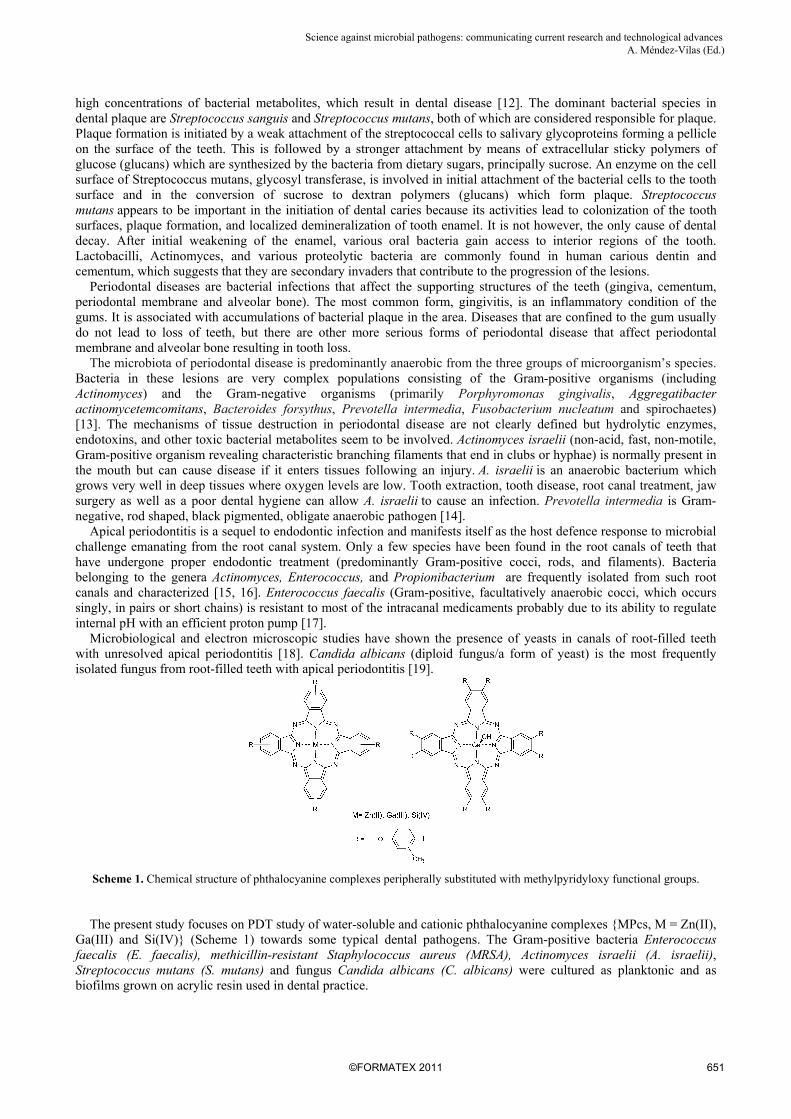
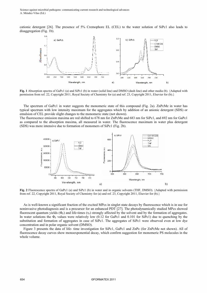
The studied metal phthalocyanines ZnPcMe, GaPc1 and SiPc1 exhibit an intensive Q-band in the range 673-681 nm. The quaternized complexes are soluble in water, but only GaPc1 and only partly SiPc1 existed as monomers in aqua solutions with slight aggregation (Fig. 1a, b). GaPc1 spectrum shows monomeric state of the sensitizer which can be attributed to the large atom of gallium which lay out of the phthalocyanine macrocycle and prevents formation of aggregates. The spectrum of SiPc1 in water shows the Q-band at 675 nm, which is accompanied by a band around 643 nm attributed to the aggregated form of phthalocyanine (Fig. 1b). The disruption of aggregates was achieved by changing the dielectric constant of the medium as was well investigated for sulfonated phthalocyanines by addition of
Science against microbial pathogens: communicating current research and technological advances A. Méndez-Vilas (Ed.)_______________________________________________________________________________
cationic detergent [26]. The presence of 5% Cremophore EL (CEL) to the water solution of SiPc1 also leads to disaggregation (Fig. 1b).
Fig. 1 Absorption spectra of GaPc1 (a) and SiPc1 (b) in water (solid line) and DMSO (dash line) and other media (b). {Adapted with permission from ref. 22, Copyright 2011, Royal Society of Chemistry for (a) and ref. 23, Copyright 2011, Elsevier for (b).}
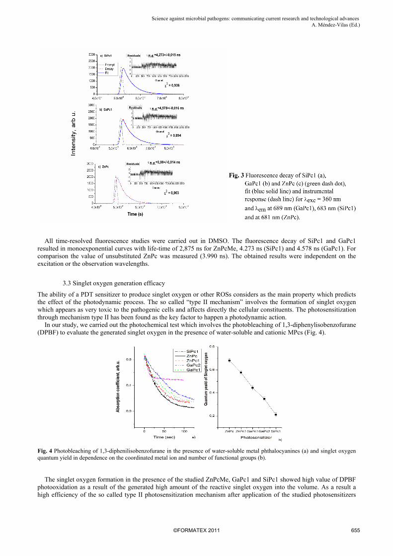
The spectrum of GaPc1 in water suggests the monomeric state of this compound (Fig. 2a). ZnPcMe in water has typical spectrum with low intensity maximum for the aggregates which by addition of an anionic detergent (SDS) or emulsion of CEL provide slight changes to the monomeric state (not shown). The fluorescence emission maxima are red shifted to 678 nm for ZnPcMe and 683 nm for SiPc1, and 692 nm for GaPc1 as compared to the absorption maxima, all measured in water. The fluorescence maximum in water plus detergent (SDS) was more intensive due to formation of monomers of SiPc1 (Fig. 2b).
a) b) b
Fig. 2 Fluorescence spectra of GaPc1 (a) and SiPc1 (b) in water and in organic solvents (THF, DMSO). {Adapted with permission from ref. 22, Copyright 2011, Royal Society of Chemistry for (a) and ref. 23, Copyright 2011, Elsevier for (b).}
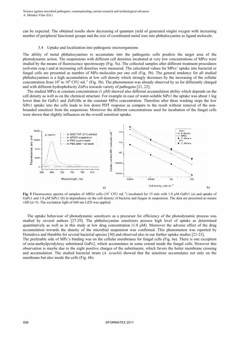
As is well-known a significant fraction of the excited MPcs in singlet state decays by fluorescence which is in use for noninvasive photodiagnosis and is a precursor for an enhanced PDT [27]. The photodynamically studied MPcs showed fluorescent quantum yields (ΦF) and life-times (τF) strongly affected by the solvent and by the formation of aggregates. In water solutions the ΦF values were relatively low (0.12 for GaPc1 and 0.101 for SiPc1) due to quenching by the substitution and formation of aggregates in case of SiPc1. The aggregates of SiPc1 were observed even at low dye concentration and in polar organic solvent (DMSO). Figure 3 presents the data of life- time investigation for SiPc1, GaPc1 and ZnPc (for ZnPcMe not shown). All of fluorescence decay curves show monoexponential decay, which confirm suggestion for monomeric PS molecules in the whole volume.
Science against microbial pathogens: communicating current research and technological advances A. Méndez-Vilas (Ed.)______________________________________________________________________________
All time-resolved fluorescence studies were carried out in DMSO. The fluorescence decay of SiPc1 and GaPc1 resulted in monoexponential curves with life-time of 2,875 ns for ZnPcMe, 4.273 ns (SiPc1) and 4.578 ns (GaPc1). For comparison the value of unsubstituted ZnPc was measured (3.990 ns). The obtained results were independent on the excitation or the observation wavelengths.
3.3 Singlet oxygen generation efficacy
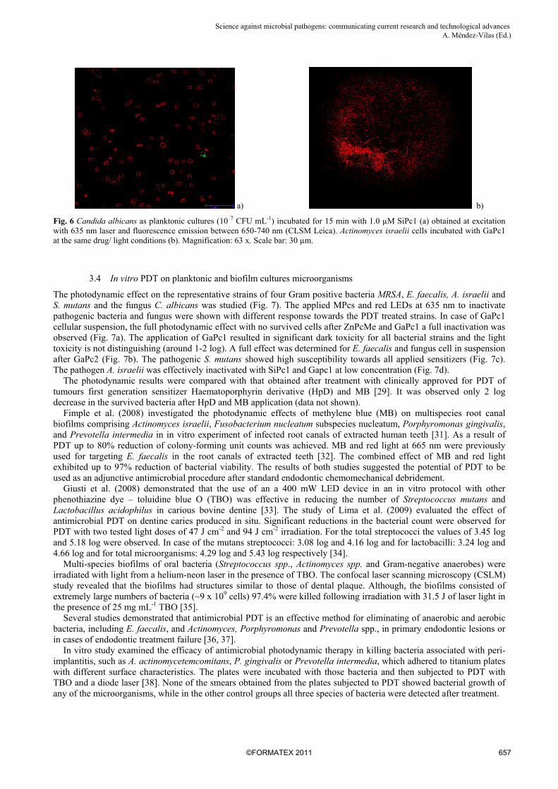
The ability of a PDT sensitizer to produce singlet oxygen or other ROSs considers as the main property which predicts the effect of the photodynamic process. The so called “type II mechanism” involves the formation of singlet oxygen which appears as very toxic to the pathogenic cells and affects directly the cellular constituents. The photosensitization through mechanism type II has been found as the key factor to happen a photodynamic action. In our study, we carried out the photochemical test which involves the photobleaching of 1,3-diphenylisobenzofurane (DPBF) to evaluate the generated singlet oxygen in the presence of water-soluble and cationic MPcs (Fig. 4).
Fig. 4 Photobleaching of 1,3-diphenilisobenzofurane in the presence of water-soluble metal phthalocyanines (a) and singlet oxygen quantum yield in dependence on the coordinated metal ion and number of functional groups (b).
The singlet oxygen formation in the presence of the studied ZnPcMe, GaPc1 and SiPc1 showed high value of DPBF photooxidation as a result of the generated high amount of the reactive singlet oxygen into the volume. As a result a high efficiency of the so called type II photosensitization mechanism after application of the studied photosensitizers
Science against microbial pathogens: communicating current research and technological advances A. Méndez-Vilas (Ed.)_______________________________________________________________________________
can be expected. The obtained results show decreasing of quantum yield of generated singlet oxygen with increasing number of peripheral functional groups and the size of coordinated metal ions into phthalocyanine as ligand molecule.
3.4 Uptake and localization into pathogenic microorganisms
The ability of metal phthalocyanines to accumulate into the pathogenic cells predicts the target area of the photodynamic action. The suspensions with different cell densities incubated at very low concentrations of MPcs were studied by the means of fluorescence spectroscopy (Fig. 5a). The collected samples after different treatment procedures (solvents resp.) and at increasing cell densities were measured. The calculated values for MPcs’ uptake into bacterial or fungal cells are presented as number of MPc-molecules per one cell (Fig. 5b). The general tendency for all studied phthalocyanines is a high accumulation at low cell density which strongly decreases by the increasing of the cellular concentration from 105 to 108 CFU mL-1 (Fig. 5b). The phenomenon was already observed by us for differently charged and with different hydrophobicity ZnPcs towards variety of pathogens [21, 22]. The studied MPcs at constant concentration (1 μM) showed also different accumulation ability which depends on the cell density as well as on the chemical structure. For example in case of water-soluble SiPc1 the uptake was about 1 log lower than for GaPc1 and ZnPcMe at the constant MPcs concentration. Therefore after three washing steps the low SiPc1 uptake into the cells leads to low down PDT response as compare to the result without removal of the non-bounded sensitizer from the suspension. Moreover the different concentrations used for incubation of the fungal cells were shown that slightly influences on the overall sensitizer uptake.
a) b)
Fig. 5 Fluorescence spectra of samples of MRSA cells (107 CFU mL-1) incubated for 15 min with 1.0 µM GaPc1 (a) and uptake of GaPc1 and 1.0 µM SiPc1 (b) in dependence on the cell density of bacteria and fungus in suspension. The data are presented as means ±SD (n=5). The excitation light of 660 nm LED was applied. The uptake behaviour of photodynamic sensitizers as a precursor for efficiency of the photodynamic process was studied by several authors [27-29]. The phthalocyanine sensitizers possess high level of uptake as determined quantitatively as well as in this study at low drug concentration (1.0 μM). Moreover the adverse effect of the drug accumulation towards the density of the microbial suspension was confirmed. This phenomenon was reported by Demidova and Hamblin for several bacterial species [30] and observed also in our further uptake studies [21-23]. The preferable side of MPc’s binding was on the cellular membranes for fungal cells (Fig. 6a). There is one exception of octa-methylpyridyloxy substituted GaPc2, which accumulates in some extend inside the fungal cells. However this observation is maybe due to the eight positive charges of the substituents, which favors the better membrane crossing and accumulation. The studied bacterial strain (A. israelii) showed that the sensitiser accumulates not only on the membrane but also inside the cells (Fig. 6b).
Science against microbial pathogens: communicating current research and technological advances A. Méndez-Vilas (Ed.)______________________________________________________________________________
a) b)
Fig. 6 Candida albicans as planktonic cultures (10 7 CFU mL-1) incubated for 15 min with 1.0 µM SiPc1 (a) obtained at excitation with 635 nm laser and fluorescence emission between 650-740 nm (CLSM Leica). Actinomyces israelii cells incubated with GaPc1 at the same drug/ light conditions (b). Magnification: 63 x. Scale bar: 30 µm.
3.4 In vitro PDT on planktonic and biofilm cultures microorganisms
The photodynamic effect on the representative strains of four Gram positive bacteria MRSA, E. faecalis, A. israelii and S. mutans and the fungus C. albicans was studied (Fig. 7). The applied MPcs and red LEDs at 635 nm to inactivate pathogenic bacteria and fungus were shown with different response towards the PDT treated strains. In case of GaPc1 cellular suspension, the full photodynamic effect with no survived cells after ZnPcMe and GaPc1 a full inactivation was observed (Fig. 7a). The application of GaPc1 resulted in significant dark toxicity for all bacterial strains and the light toxicity is not distinguishing (around 1-2 log). A full effect was determined for E. faecalis and fungus cell in suspension after GaPc2 (Fig. 7b). The pathogenic S. mutans showed high susceptibility towards all applied sensitizers (Fig. 7c). The pathogen A. israelii was effectively inactivated with SiPc1 and Gapc1 at low concentration (Fig. 7d). The photodynamic results were compared with that obtained after treatment with clinically approved for PDT of tumours first generation sensitizer Haematoporphyrin derivative (HpD) and MB [29]. It was observed only 2 log decrease in the survived bacteria after HpD and MB application (data not shown). Fimple et al. (2008) investigated the photodynamic effects of methylene blue (MB) on multispecies root canal biofilms comprising Actinomyces israelii, Fusobacterium nucleatum subspecies nucleatum, Porphyromonas gingivalis, and Prevotella intermedia in in vitro experiment of infected root canals of extracted human teeth [31]. As a result of PDT up to 80% reduction of colony-forming unit counts was achieved. MB and red light at 665 nm were previously used for targeting E. faecalis in the root canals of extracted teeth [32]. The combined effect of MB and red light exhibited up to 97% reduction of bacterial viability. The results of both studies suggested the potential of PDT to be used as an adjunctive antimicrobial procedure after standard endodontic chemomechanical debridement. Giusti et al. (2008) demonstrated that the use of an a 400 mW LED device in an in vitro protocol with other phenothiazine dye – toluidine blue O (TBO) was effective in reducing the number of Streptococcus mutans and Lactobacillus acidophilus in carious bovine dentine [33]. The study of Lima et al. (2009) evaluated the effect of antimicrobial PDT on dentine caries produced in situ. Significant reductions in the bacterial count were observed for PDT with two tested light doses of 47 J cm-2 and 94 J cm-2 irradiation. For the total streptococci the values of 3.45 log and 5.18 log were observed. In case of the mutans streptococci: 3.08 log and 4.16 log and for lactobacilli: 3.24 log and 4.66 log and for total microorganisms: 4.29 log and 5.43 log respectively [34]. Multi-species biofilms of oral bacteria (Streptococcus spp., Actinomyces spp. and Gram-negative anaerobes) were irradiated with light from a helium-neon laser in the presence of TBO. The confocal laser scanning microscopy (CSLM) study revealed that the biofilms had structures similar to those of dental plaque. Although, the biofilms consisted of extremely large numbers of bacteria (~9 x 10⁹ cells) 97.4% were killed following irradiation with 31.5 J of laser light in the presence of 25 mg mL-1 TBO [35]. Several studies demonstrated that antimicrobial PDT is an effective method for eliminating of anaerobic and aerobic bacteria, including E. faecalis, and Actinomyces, Porphyromonas and Prevotella spp., in primary endodontic lesions or in cases of endodontic treatment failure [36, 37]. In vitro study examined the efficacy of antimicrobial photodynamic therapy in killing bacteria associated with peri-implantitis, such as A. actinomycetemcomitans, P. gingivalis or Prevotella intermedia, which adhered to titanium plates with different surface characteristics. The plates were incubated with those bacteria and then subjected to PDT with TBO and a diode laser [38]. None of the smears obtained from the plates subjected to PDT showed bacterial growth of any of the microorganisms, while in the other control groups all three species of bacteria were detected after treatment.
Science against microbial pathogens: communicating current research and technological advances A. Méndez-Vilas (Ed.)_______________________________________________________________________________
Fig. 7 PDT effect on pathogenic bacteria (MRSA, E. faecalis, S. mutans, P. aeruginosa and A. israelii) and fungus C. albicans as planktonic cultures treated with GaPc1, GaPc2, SiPc1 and ZnPcMe. The data present the survived cells ± SD (n=5) after PDT at soft irradiation with 635 nm LED, irradiance of 60 mW cm-2 and dose of 50 J cm-2. {Adapted with permission from ref. 22, Copyright 2011, Royal Society of Chemistry for (a) and (b).} Rovaldi et al. [39] have designed a porphyrin derivative conjugated to a pentalysine moeity that endows the molecule with activity against the Gram-positive and the Gram-negative bacteria. Whereas the porphyrin and chlorin e6 were evaluated with in vitro activity against a limited spectrum of Gram-positive bacteria, chlorin e6 conjugated to pentalysine was shown with in vitro activity against all oral microorganisms tested, including Porphyromonas gingivalis, Actinobacillus actinomycetemcomitans, Bacteroides forsythus, Campylobacter rectus, Eikenella corrodens, Fusobacterium nucleatum, Actinomyces viscosus, and several streptococci (S. mutans, S. sobrinus, S. mitis, S. oralis). Potent antimicrobial activity (>5-log-unit reduction in the numbers of CFU per milliliter) was retained in the presence of up to 25% whole sheep blood. Peri-implantitis is a multifactorial process involving bacterial contamination of the implant surface and the formation of biofilms. Bacterial plaque on implants leads to inflammatory changes in the adjacent soft tissues. An application of TBO on implant surfaces in patients with peri-implantitis, followed by illumination with a diode soft laser (690 nm), significantly reduced the numbers of dental pathogens A. actinomycetemcomitans, P. gingivalis, and P. intermedia [40]. Dentures are commonly colonized by microorganisms derived from saliva, oral mucosa and skin, and the denture plaque formation is associated with inflammation and acute infections. The synthesized phthalocyanines ZnPcMe, GaPc1 and SiPc1 were evaluated as photodynamic drugs for treatment of bacterial biofilms of selected bacteria MRSA and E. faecalis and the fungus C. albicans, which were grown on denture acrylic resin polymethylmethacrylate (PMMC). The studies were carried out in comparison to the known as photodynamic sensitizes in dental practice such as phenothiazine dye MB (Fig. 8). As can be seen an increasing resistance of microbial species in biofilms occurred when compared to the same microbial cells in cell suspensions (Figs. 7 and 8).
Science against microbial pathogens: communicating current research and technological advances A. Méndez-Vilas (Ed.)______________________________________________________________________________
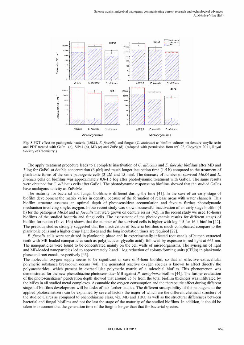
Fig. 8 PDT effect on pathogenic bacteria (MRSA, E. faecalis) and fungus (C. albicans) as biofilm cultures on denture acrylic resin and PDT treated with GaPc1 (a), SiPc1 (b), MB (c) and ZnPc (d). (Adapted with permission from ref. 22, Copyright 2011, Royal Society of Chemistry.) The apply treatment procedure leads to a complete inactivation of C. albicans and E. faecalis biofilms after MB and 3 log for GaPc1 at double concentration (6 µM) and much longer incubation time (1.5 h) compared to the treatment of planktonic forms of the same pathogenic cells (3 µM and 15 min). The decrease of number of survived MRSA and E. faecalis cells on biofilms was approximately 0.8-1.5 log after photodynamic treatment with GaPc1. The same results were obtained for C. albicans cells after GaPc1. The photodynamic response on biofilms showed that the studied GaPcs have analogous activity as ZnPcMe. The maturity for bacterial and fungal biofilms is different during the time [41]. In the case of an early stage of biofilm development the matrix varies in density, because of the formation of release areas with water channels. This biofilm structure assumes an optimal depth of photosensitizer accumulation and favours further photodynamic mechanism involving singlet oxygen. In our recent study was shown successful inactivation of an early stage biofilm (4 h) for the pathogens MRSA and E. faecalis that were grown on denture resins [42]. In the recent study we used 16-hours biofilms of the studied bacteria and fungi cells. The assessment of the photodynamic results for different stages of biofilm formation (4h vs 16h) shows that the number of the survived cells is higher with log 4-5 for 16 h biofilm [42]. The previous studies strongly suggested that the inactivation of bacteria biofilms is much complicated compare to the planktonic cells and a higher drug/ light doses and the long incubation times are required [22]. E. faecalis cells were sensitized in planktonic phase and in experimentally infected root canals of human extracted teeth with MB-loaded nanoparticles such as poly(lacticco-glycolic acid), followed by exposure to red light at 665 nm. The nanoparticles were found to be concentrated mainly on the cell walls of microorganisms. The synergism of light and MB-loaded nanoparticles led to approximately 2 and 1 log reduction of colony-forming units (CFUs) in planktonic phase and root canals, respectively [43]. The molecular oxygen supply seems to be significant in case of 4-hour biofilm, so that an effective extracellular polymeric substance breakdown occurs [44]. The generated reactive oxygen species is known to affect directly the polysaccharides, which present in extracellular polymeric matrix of a microbial biofilm. This phenomenon was demonstrated for the new phenothiazine photosensitizer MB against P. aeruginosa biofilm [44]. The further evaluation of the photosensitizers’ penetration depth showed that around 75 % from the total biofilm thickness was infiltrated by the MPcs in all studied metal complexes. Assumable the oxygen consumption and the therapeutic effect during different stages of biofilms development will be tasks of our further studies. The different susceptibility of the pathogens to the applied photosensitizers can be explained by several factors the major of which are the different chemical structure of the studied GaPcs as compared to phenothiazine class, viz. MB and TBO, as well as the structural differences between bacterial and fungal biofilms and not the last the stage of the maturity of the studied biofilms. In addition, it should be taken into account that the generation time of the fungi is longer than that for bacterial species.
Science against microbial pathogens: communicating current research and technological advances A. Méndez-Vilas (Ed.)_______________________________________________________________________________
The penetration of the photosensitizer into the biofilms during different stages of biofilm growth is also very important part for the success of the treatment procedure. Recently, the combination of PDT and hydrogen peroxide was reported as a solution to the penetration problem with limited transfer of drugs throughout the biofilm channels [45]. The photodynamically associated dual activity is not observed in the conventional antibacterial treatment. This fact represents a significant advantage of the photodynamic approach towards the antibiotic treatment, especially for resistant pathogenic microorganisms, that are growing as persistent microbial biofilms.
Acknowledgements The National Science Fund, Bulgaria for financial support (Grants DO-02-177 and DO-02-112) and the EC Project FR7-REGPOT-2009-1(ReProForce) for confocal fluorescence microscopy study in collaboration with Dr. Rumen Dimitrov (IBIR-BAS), we gratefully acknowledge.
References
[1] Hamblin M, Hasan T. Photodynamic therapy: a new antimicrobial approach to infection disease? Photochem. Photobiol. Sci. 2004;3(5):436-450.
[2] Tianhong D, Huang Y-Y, Hamblin M. Photodynamic therapy for localized infections – State of the art. Photodiagnosis and Photodynamic therapy. 2009;6:170-188.
[3] Jori G, Fabris C, Soncin M, Ferro S, Coppellotti O, Dei D, Fantetti L, Chiti G, Roncucci G. Photodynamic therapy in the treatment of microbial infections: Basic principles and perspective applications. Las. in Surg. & Med. 2006;38(6):468–481.
[4] Konopka K, Goslinski T. Photodynamic therapy in dentistry. J. Dent. Res. 2007; 86(8):694-707. [5] Di Poto A, Sbarra S. M, Provenza G, Visai L, Speziale P, The effect of photodynamic treatment combined with antibiotic action
or host defence mechanisms on Staphylococcus aureus biofilms. Biomaterials, 2009;30:3158-3166. [6] Teichert MC, Jones JW, Usacheva MN, Biel MA. Treatment of oral candidiasis with methylene blue-mediated photodynamic
therapy in an immunodeficient murine model. Oral Surg Oral Med Oral Pathol Oral Radiol Endod. 2002;93:155-160. [7] Ewerton, GDM, Pavarina, AC , Dovigo, LN, Vergani, CE, Souza, CA, Kurachi, C, Bagnato, VS. Susceptibility of Candida
albicans to photodynamic therapy in a murine model of oral candidosis. Oral Surg Oral Med Oral Pathol Oral Radiol Endod. 2010;109(3):392-401.
[8] Sharman MW, Allen MC, van Lier EJ, Photodynamic therapeutics: basic principles and clinical applications, Drug Discovery Today. 1999;4:507-517.
[9] Maisch T, Baier J, Franz B, Maier M, Landthaler M, Szeimies R-M, Baeumler W. The role of singlet oxygen inactivation of bacteria. Proceed. Nat. Acad. Sci. U.S.A. 2007;104(17):7223-7228.
[10] Spiller M, Kliesch H, Wöhrle D, Hackbarth S, Roeder B, Singlet oxygen quantum yield of different photosensitizers in polar solvents and micellar solutions. J. Porphyrins Phthalocyanines. 1998;2:145-158.
[11] Mantareva V, Angelov I, Kussovski V, Wöhrle D, Dimitrov S. Metallophthalocyanines as photodynamic sensitizers for treatment of pathogenic bacteria. Synthesis and singlet oxygen formation, Compt. rend. Acad. bulg. Sci. 2009;61(12):1521-1526.
[12] van Houte J. Role of micro-organisms in caries etiology. J. Dent. Res. 1994;73(3): 672-681. [13] Kömerick N, Nakanishi H, MacRobert AJ, Henderson B, Speight P, Wilson M. In vivo killing of porphyromonas gingivalis by
toluidine blue-mediated photosensitization in an animal model. Antimicrob Agents Chemother. 2003;47:932–940. [14] Sigusch BW, Pfitzner A, Albrecht V, Glockmann E. Efficacy of photodynamic therapy on inflammatory signs and two selected
periodontopathogenic species in a beagle dog model. J. Periodontol. 2005;76:1100–1105. [15] Hancock H, Sigurdsson A, Trope M, Moiseiwitsch J. Bacteria isolated after unsuccessful endodontic treatment in a North
American population. Oral Surg Oral Med Oral Pathol. 2001;91:579-586. [16] Pinheiro ET, Gomes BPFA, Ferraz CCR, Sousa ELR, Teixeira FB, Souza-Filho FJ. Microorganisms from canals of root filled
teeth with periapical lesions. Int Endod. 2003;36:1-11. [17] Evans M, Davies JK, Sundqvist G, Figdor D. Mechanisms involved in the resistance of Enterococcus faecalis to calcium
hydroxide. Int Endod. 2002;35:221-228. [18] Nair PNR. Pathogenesis of Apical Periodontitis and the Causes of Endodontic Failures. Critical Reviews in Oral Biology &
Medicine. 2004;15:348-381. [19] Sundqvist G, Figdor D, Persson S, Sjögren U. Microbiologic analysis of teeth with failed endodontic treatment and the outcome
of conservative re-treatment. Oral Surg Oral Med Oral Pathol. 1998;85:86-93. [20] Wöhrle D, Iskander D, Graschew G, Sinn H, Friedrich EF, Maier-Borst E, Stern J, Schlag P. Synthesis of positively charged
phthalocyanines and their activity in the photodynamic therapy of cancer cells. Photochem. Photobiol. 1990; 51:351-356. [21] Mantareva V, Kussovski V, Angelov I, Borisova E, Avramov L, Schnurpfeil G, Wöhrle D. Photodynamic activity of water-
soluble phthalocyanine zinc(II) complexes against pathogenic microorganisms, Bioorg. & Med. Chem. 2007;15:4829-4835. [22] Mantareva V, Kussovski V, Angelov I, Wöhrle D, Dimitrov R, Popova E, Dimitrov S. Non-aggregated Ga(III) phthalocyanines
for photodynamic inactivation of planktonic and biofilm cultured microorganisms. Photochem. Photobiol. Sci. 2011;10:91-102. [23] Mantareva V, Angelov I, Kussovski V, Dimitrov R, Lapok L, Wöhrle D. Photodynamic efficacy of water-soluble Si(IV) and
Ge(IV) phthalocyanines towards Candida albicans planktonic and biofilm cultures, European Journal of Medicinal Chemistry, 2011;46:4430-4440.
[24] Jacques P, Braun AM. Laser flash photolysis of phthalocyanines in solution and microemulsion. Helv. Chim. Acta, 1981;64:1800-1806.
[25] Durmus M, Nyokong T. Synthesis, photophysical and photochemical properties of tetra- and octa-substituted gallium and indium phthalocyanines. Polyhedron. 2007;26:3323-3335.
[26] Schneider G, Wöhrle D, Spiller W, Stark J, Schulz-Ekloff G. Photochem. Photobiol. 1994;60:333-342.
Science against microbial pathogens: communicating current research and technological advances A. Méndez-Vilas (Ed.)______________________________________________________________________________
[27] Minnock A, Vernon ID, Schofield J, Griffiths J, Parish HJ, Brown BS. Mechanism of uptake of a cationic water-soluble pyridinium zinc phthalocyanine across the outer membrane of Escherichia coli. Antimicrob. Agents Chemother. 2000;44:522–527.
[28] Costa L, Alves E, Carvalho CMB, Tome JPC, Faustino MAF, Neves MJPS, Tome AC, Cavaleiro JAS, Cunha A, Almeida A, Sewage bacteriophage photoinactivation by cationic porphyrins: a study of charge effect. Photochem. Photobiol. Sci. 2008;7:415-422.
[29] Angelov I., Mantareva V, Kussovski V, Wöhrle D, Kisov H, Belcheva M, Georgieva T, Dimitrov S, Susceptibility of representative Dental Pathogens to inactivation by the PDT with water-soluble photosensitizers. 2011, Proceedings of SPIE - The International Society for Optical Engineering 7994, art. no. 79941A
[30] Demidova NT, Hamblin RM. Effect of cell-photosensitizer binding and cell-density on microbial photoinactivation, Antimicrob. Agents Chem. 2005;49:2329-2335.
[31] Fimple JL, Fontana CR, Foschi F, Ruggiero K, Song XQ, Pagonis TC, Tanner ACR , Kent R, Doukas AG, Stashenko PP , Soukos NS. Photodynamic treatment of endodontic polymicrobial infection in vitro. J. Endod. 2008;34(6):728-734.
[32] Foschi F, Fontana CR, Ruggiero K, Riahi R, Vera A, Doukas AG, Pagonis TC, Kent R., Stashenko PP, Soukos NS. Photodynamic inactivation of Enterococcus faecalis in dental root canals in vitro. Lasers Surg. Med. 2007;39:782-787.
[33] Giusti JSM, Santos-Pinto L, Pizzolito AC, Helmerson K, Carvalho-Filho E, Kurachi C, Bagnato VS. Antimicrobial photodynamic action on dentin using a light-emitting diode light source. Photomed. Laser Surg. 2008;26:279-285.
[34] Lima JPM, Sampaio de Melo MA, Borges FMC, Teixeira AH, Steiner-Oliveira C, Nobre dos Santos M, Rodrigues LKA, Zanin ICJ. Evaluation of the antimicrobial effect of photodynamic antimicrobial therapy in an in situ model of dentine caries. Eur J Oral Sci 2009;117:568–574.
[35] O’Neill JF, CK. Hope, M. Wilson. Oral bacteria in multi-species biofilms can be killed by red light in the presence of Toluidine Blue. Lasers in Surgery and Medicine 2002;31:86-90.
[36] Garcez AS, Nunez SC, Hamblin MR, Ribeiro MS. Antimicrobial effects of photodynamic therapy on patients with necrotic pulps and periapical lesion. J Endod. 2008;34:138-142.
[38] Haas R, Dortbudak O, Mensdorff-Pouilly N, Mailath G. Elimination of bacteria on different implant surfaces through photosensitization and soft laser. An in vitro study. Clin Oral Implants Res. 1997;8: 49-254.
[39] Rovaldi CR, Pievsky A, Sole NA, Friden PM, Rothstein DM, Spacciapoli P. Photoactive porphyrin derivative with broad-spectrum activity against oral pathogens in vitro. Antimicrob. Agents Chemother. 2000; 44(12):3364-3367.
[40] Dortbudak O, Haas R, Bernhart T, Mailath-Pokorny G. Lethal photosensitization for decontamination of implant surfaces in the treatment of peri-implantitis. Clin Oral Implants Res. 2001;12:104-108.
[41] Thein Z, Samaranayake Y, Samaranayake L. Characteristics of dual species Candida biofilms on denture acrylic surfaces, Arch. of Oral Biol.2007; 52:1200-1208.
[42] Mantareva V, Angelov I, Kussovski V, Woehrle D, Dimitrov S. Metallophthalocyanines as photodynamic sensitizers for treatment of pathogenic bacteria. Uptake and photodynamic effect. Compt. rend. Acad. bulg. Sci. 2009;63(1):77-84.
[43] Pagonis TC, Chen J, Fontana CR, Devalapally H, Ruggiero K, Song X, Foschi F, Dunham J, Skobe Z, Yamazaki H, Kent R, Tanner ACR, Amiji MM, Soukos NS. Nanoparticle-based endodontic antimicrobial photodynamic therapy. J. Endod. 2010;36(2):322-328.
[44] Wainwright M, Crossley BK, Photosensitising agents–circumventing resistance and breaking down biofilms: a review. Intern. Biodeter. & Biodegrad. 2004;53:119-126.
[45] Garcez AS, Nunez SC, Baptista MS, Dahastanli NA, Itri R, Hamblin MR, Ribeiro MS. Antimicrobial mechanisms behind photodynamic effect in the presence of hydrogen peroxide. Photochem. Photobiol. Sci. 2011;10:483-490.
Science against microbial pathogens: communicating current research and technological advances A. Méndez-Vilas (Ed.)_______________________________________________________________________________