129


Advanced
Interpretation of Adult
Vital Signs in Trauma William D. Hampton, DO Emergency Physician 26 March 2015

1. Better understand vital signs for what they can tell you (and what they can’t) in the assessment of a trauma patient.
2. Appreciate best practices in obtaining accurate vital signs in trauma patients.
3. Learn what teaching about vital signs is evidence-based and what is not.
4. Explain the importance of vital signs to more accurately triage, diagnose, and confidently disposition our trauma patients.
5. Apply the monitoring (and manipulation of) vital signs to better resuscitate trauma patients.
Learning Objectives

• Faculty/Presenters/Authors/Content Reviewers/Planners disclose no conflict of interest relative to this educational activity.
Disclosure Statement

• To successfully complete this course, participants must attend the entire event and complete/submit the evaluation at the end of the session.
• Society of Trauma Nurses is accredited as a provider of continuing nursing education by the American Nurses Credentialing Center's Commission on Accreditation.
Successful Completion

Vital Signs

Vital Signs
Philosophy:
“View vital signs as compensatory to the illness/complaint as opposed to primary.”
Crowe, Donald MD. “Vital Sign Rant.” EMRAP: Emergency Medicine Reviews and Perspectives. February, 2010.

Vital Signs
Truth over Accuracy:
• Document the true status of the patient: sick or not?
• Complete vital signs on every patient, every time, regardless of the chief complaint.
• If vital signs seem misleading or inaccurate, repeat them!
• Beware sending a patient home with abnormal vitals (especially tachycardia)!
•Treat vital signs the same as any other diagnostics—review them carefully prior to disposition.

The Mother’s Vital Sign: Temperature

Case #1 - 76-y/o homeless ♂
CC: 76-y/o homeless ♂ brought to the ED by police for eval. Nighttime temps have been in the 50s-60s. He has been turned away from all shelters due to his chronic etOH use.
VS: T 94°F (oral), HR 96, RR 22, BP 107/97, SaO2 92%
PE: Disheveled, malnourished, and intoxicated. No visible injuries. GCS = E1 V4 M5.
“toxic sock syndrome.”

T 94°F, HR 96, RR 22, BP 107/97, SaO2 92%
Does a GCS of 10 correlate with a core temperature
of 94 F (34.4°C)?
No.
Any patient with a
temperature ≥ 90° F (32C) with ongoing AMS needs further workup as
to why: intoxication, trauma, infection, etc.
Marx J, Hockberger R, Walls R. Rosen’s Emergency Medicine: Concepts and Clinical Practice. 7th ed. Chap 138, “Accidental Hypothermia,” New York, NY: Elsevier Health Sciences; 2009: 2236-2253.
Case #1 - 76-y/o homeless ♂

T 94°F, HR 96, RR 22, BP 107/97, SaO2 92%
Could a temporal thermometer be used to give a true “core temperature” reading in this patient?
No. Temporal
thermometry “should not be used…in the
setting of suspected hypothermia.” Rectal, Esophageal, or Foley catheter thermister is
recommended.
Marx J, Hockberger R, Walls R. Rosen’s Emergency Medicine: Concepts and Clinical Practice. 7th ed. Chap 138, “Accidental Hypothermia,” New York, NY: Elsevier Health Sciences; 2009: 2236-2253.
Case #1 - 76-y/o homeless ♂

Walker GA, Runde D, Rolston DM, Wiener D, Lee J, “Emergency department rectal temperatures in over 10 years: A retrospective observational study,” World J Emerg Med, Vol 4, No 2, 2013: 107-112.
World Journal of Emergency Medicine, 2013
Retrospective chart review of 27,130 adult patients in a high-volume ED over 8 years.
The average difference between the initial temporal artery thermometry (n = 988) and
the rectal temp was 1.2° F (0.7°C) (p<0.001).
In almost 1 in 5 patients (18.8%), fever was missed by the initial triage temp.
Temporal thermometry in adults?
Temperature

Temporal thermometry in Trauma?
Marable K, Shaffer LE, Dizon V, Opalek JM. Temporal artery scanning falls short as a secondary, noninvasive thermometry method for trauma patients. J Trauma Nurs, 2009;16(1):41-7.
Temperature

Temporal thermometry in the ICU?
Dybwik K, [Infrared temporal thermometry] - Tidsskr Nor Laegeforen 6-NOV-2003; 123(21): 3025-6
Tidsskr Nor Laegeforen, 2003
Comparison study of 164 ICU patients between rectal and TAT measurement.
Fever was a rectal temp ≥ 100.4° F (38°C); TAT detected fever in 33 of 70
febrile patients. Sn = 53%
Conclusion: TAT’s sensitivity for detecting rectally measured fever is too low to
recommend its use in adult ICU patients.
Temperature

Temperature
Temporal thermometry in adults?
Temporal thermometry in Trauma?
Temporal thermometry in the ICU?

Case #2 - Two ♂s from winter MVC
EMS calls for medical control after responding to a single-car rollover off the side of the road. The driver was found up the road about a mile. The passenger is trapped inside due to significant compartment intrusion, his chest pinned between the dash and the seat. Wind
chill is -40°F & C.
The passenger is much warmer than the driver due to being inside the car, but has absent vital signs. The driver has no signs of injury, but as he is dressed in a T-shirt, jeans, and tennis shoes with no socks, and also has no vital signs.
EMS has a transport time of 40 minutes and as they are a two-person crew, can only resuscitate one person.

Case #2 - Two ♂s from winter MVC
Which patient should EMS transfer and attempt to resuscitate: the driver with apparent hypothermia, the passenger still trapped inside the relatively warm car, both, or neither?
The driver.
Successful resuscitation following blunt trauma is
approximately 1-2%.
Primary hypothermia (even in the face of cardiopulmonary
arrest) has a resuscitation chance of approximately 50%.
Hopson LR, Hirsh E, et. al., “Guidelines For Withholding Or Termination Of Resuscitation In Prehospital Traumatic Cardiopulmonary Arrest: a joint position paper from the national association of EMS physicians standards and clinical practice committee and the American College of Surgeons Committee on Trauma,” Prehospital Emergency Care, January / March 2003 Vol 7(1):141-146.

Case #2 - 21-y/o ♂ found outside
EMS: The driver is placed on a cardiac monitor.
PE: Bradycardia rate of 24 bpm is seen with a widened QRS, but no pulse can be palpated. The patient is apneic. They have no thermometer on their rig. SaO2 cannot capture a waveform.

Case #2 - 21-y/o ♂ found outside
Marx J, Hockberger R, Walls R. Rosen’s Emergency Medicine: Concepts and Clinical Practice. 7th ed. Chap 138, “Accidental Hypothermia,” New York, NY: Elsevier Health Sciences; 2009: 2236-2253.
EMS asks how should the patient’s airway be managed as they don’t want to do something that might change the pulseless bradycardia to V-fib.
Intubate the patient.
Hypothermic patients are at d risk for aspiration. Intubation
facilitates warmed humidified air administration and has
NOT been shown to cause dysrhythmias in hypothermia.
Temp ??, HR 24, RR , BP , SaO2

Case #2 - 21-y/o ♂ found outside
Marx J, Hockberger R, Walls R. Rosen’s Emergency Medicine: Concepts and Clinical Practice. 7th ed. Chap 138, “Accidental Hypothermia,” New York, NY: Elsevier Health Sciences; 2009: 2236-2253.
EMS asks, “Should they start CPR on this patient en route to your facility?”
YES! He has no palpable
pulse! CPR will perfuse the brain until definitive
resuscitation is available.
Temp ??, HR 24, RR , BP , SaO2

Case #2 - 21-y/o ♂ found outside
Marx J, Hockberger R, Walls R. Rosen’s Emergency Medicine: Concepts and Clinical Practice. 7th ed. Chap 138, “Accidental Hypothermia,” New York, NY: Elsevier Health Sciences; 2009: 2236-2253.
What treatment should be initiated to treat the patient’s bradycardia: atropine, external pacing, dopamine, epinephrine, or something else?
Temp ??, HR 24, RR , BP , SaO2
Treat the hypothermia.
Bradycardia is due to d spontaneous depolarization of pacemaker cells. Atropine will
be ineffective. Cold myocardium is irritable, and
external pacing may convert an organized rhythm into V-fib.

Case #2 - 21-y/o ♂ found outside
Marx J, Hockberger R, Walls R. Rosen’s Emergency Medicine: Concepts and Clinical Practice. 7th ed. Chap 138, “Accidental Hypothermia,” New York, NY: Elsevier Health Sciences; 2009: 2236-2253.
EMS asks how should they treat the hypothermia while en route?
Temp ??, HR 24, RR , BP , SaO2
Prevent further heat loss.
Dry and cover the patient. Turn up the heat and
administer warm IVF and warmed/humidified O2 if
possible.

Case #2 - 21-y/o ♂ found outside
ED: The patient arrives and is placed on a cardiac monitor.
PE: Asystole is seen. The patient is apneic. Pupils are fixed and dilated. Nursing staff
reports to you a rectal temperature of 82 F. No blood pressure (obviously) and no signs of trauma on your secondary survey.
Temp ??, HR , RR , BP , SaO2

Case #2 - 21-y/o ♂ found outside
Brown DJA, Brugger H, “Accidental Hypothermia,” New England Journal of Medicine 2012;367:1930-8. Brown, DJA, Accidental Hypothermia, EM-RAP: Emergency Medicine Reviews and Perspectives, January 2014:14(1).
What should be done now for your asystolic, apneic,
blood pressure, fixed/dilated pupil patient with a core
temperature of 82° F (28°C)?
T 82° F (28°C), HR , RR , BP , SaO2
Initiate ECMO / CPB. Hypothermia patients with
cardiac arrest who are treated at an ECMO/CPB Center have a
50% survival rate. Hypothermia patients with
cardiac arrest who are treated without ECMO / CPB have a
10% survival rate.

How do you know if your
patient is truly dead?

Warm and Dead?
“They’re not dead until they’re warm and dead.”
Warm adults to 32°C (90° F) before pronouncing.
Warm children to 35°C (95° F) before pronouncing.

A potassium (K+) level > 12 mEq/L Ammonia (NH3) > 250 mmol/L are both reasons to stop resuscitation
regardless of core temperature.
Nose or mouth occluded with ice (preventing ventilation) or a
core temp < 15°C (59° F).
Obvious lethal injuries are a reason to cease resuscitative efforts.
A rigid, non-compressible thorax is also a reason to stop a hypothermic resuscitation.
Frozen or clotted blood is a sign that the resuscitation should be stopped.
Signs of Irreversible Death

Dead or not?

Dead or not?

Dead or not?

Hypothermia: Children vs. Adults
• Infant BSA:mass is 3x adult
• Child BSA:mass is 2x adult
• The large surface-area to body-mass ratio results in quicker heat loss for infants and children
• Faster cooling cerebral protection from hypoxia-- even in submersion injury
Blackburn ST, Maternal, Fetal, & Neonatal Physiology: A Clinical Perspective, 3e (Maternal Fetal and Neonatal Physiology), Saunders; 3rd edition (March 14, 2007): 800 pgs.

Avalanche Burial
< 35 min. burial NOT hypothermia
> 35 min. burial possibly hypothermia
Dead or Not?

In hypothermia, these signs death!

Dead or not?

Key Facts: Temperature A hypothermic patient warmed to 90° F (32C ) with ongoing AMS needs further workup as to why.
Temporal thermometry has been shown to be unreliable in the setting of suspected hypothermia, the ICU, in adults, and in trauma.
At temperatures < 86° F (30°C) anticipate cardiac arrest and initiate ECMO/CPB in appropriate patients (got cold…then died). Duration of CPR is not a predictor of survival in hypothermia.
Drowning (especially children), lightning strike, and hypothermia are all special circumstances that suggest a resuscitation attempt even in the face of apparent death.
SIRS Criteria is T ≥ 38°C (100.4°F) or ≤ 36°C (96.8°F), a HR > 90 bpm, a RR > 20 bpm, and a WBC > 12 or < 4 or > 10% bands.
Sepsis - 2 out of 4 SIRS criteria + Infection
Septic Shock - 2/4 SIRS criteria + Infection + hypoTN after IVF
Severe Sepsis - 2/4 SIRS criteria + Infection + Lactate > 4

The Paramedic’s Vital Sign: Pulse

Case #3 - 28-y/o ♀ bike vs. auto
CC: 28-y/o ♀ with bike vs. auto. Car passed her on left and clipped her handlebars with the side mirror
causing her to crash. Helmet. Estimated speed was ~28mph.
Meds: MVI, OCPs, and Ibuprofen.
VS: HR 54, RR 18, BP 105/89, SaO2 100%
PE: Appears pale but in no acute distress. Pelvis is grossly unstable.

Case #3 - 28-y/o ♀ bike vs. auto Afebrile, HR 54, RR 18, BP 105/89, SaO2 100%
Does this patient’s relative bradycardia effectively rule-out significant blood loss?
No. Paradoxical bradycardia has been reported in the
literature in cases of massive blood loss.
Secher NH, Sander JK, Werner C, Warberg J, Bie P, “Bradycardia during severe but reversible hypovolemic shock in man.” Circulatory Shock, 1984;14(4): 267-74.

Case #3 - 28-y/o ♀ bike vs. auto Afebrile, HR 54, RR 18, BP 105/89, SaO2 100%
What is this patient’s pulse pressure?
What is a normal value?
What does an abnormal value imply?
16mm Hg
Secher NH, Sander JK, Werner C, Warberg J, Bie P, “Bradycardia during severe but reversible hypovolemic shock in man.” Circulatory Shock, 1984;14(4): 267-74.
30 to 40mm Hg
Narrow pulse pressure in the setting trauma is
suggestive of blood loss ( preload CO).

Case #3 - 28-y/o ♀ bike vs. auto Afebrile, HR 54, RR 18, BP 105/89, SaO2 100%
Secher NH, Sander JK, Werner C, Warberg J, Bie P, “Bradycardia during severe but reversible hypovolemic shock in man.” Circulatory Shock, 1984;14(4): 267-74.
What does this patient’s SaO2 of 100% suggest to you about her hemoglobin level?
It’s probably LOW. A low hemoglobin will a
falsely increased SaO2. The degree of oxygen saturation is inversely
related to the amount of hemoglobin present.

Pulse-oximetry & Hb
• Hemoglobin levels can affect pulse oximetry.
• Hb (anemia) the easier it is to saturate Hb. The SaO2 may be high, but total oxygen content will be low.
• Hb (polycythemia) difficult to saturate the extra molecules of Hb with oxygen pseudo-hypoxemia (when the patient’s oxygen content may well be normal).
Patricia Carroll, RRT, RN, “Pitfalls, Perils, and Pearls of Pulse Oximetry,” RT: For Decision Makers in Respiratory Care (online journal), April/May, 2003. Accessed 8/3/14.

Case #3 - 28-y/o ♀ bike vs. auto Afebrile, HR 54, RR 18, BP ???/??, SaO2 100%
On arrival in the ED, the patient has normal femoral & carotid pulses bilaterally with diminished radial pulses. What does this tell you about her systolic BP?
Nothing. The presence or
absence of pulses does not consistently correlate
with any given SBP.
Guly HR, Bouamra O, Spiers M, et al. Vital signs and estimated blood loss in patients with major trauma: testing the validity of the ATLS classification of hypovolaemic shock. Resuscitation. 2011;82(5):556-559.
Heffernan DS, Thakkar RK, Monaghan SF, et al. Normal presenting vital signs are unreliable in geriatric blunt trauma victims. J Trauma. 2010;69(4):813-820.

ATLS Classes of Hemorrhagic Shock
Guly HR, Bouamra O, Spiers M, et al. Vital signs and estimated blood loss in patients with major trauma: testing the validity of the ATLS classification of hypovolaemic shock. Resuscitation. 2011;82(5):556-559.
Heffernan DS, Thakkar RK, Monaghan SF, et al. Normal presenting vital signs are unreliable in geriatric blunt trauma victims. J Trauma. 2010;69(4):813-820.

ATLS: Palpable pulses correlate with SBP
Radial pulse: SBP > 80 mmHg
Carotid & Femoral pulse only: SBP 70 - 80 mmHg
Carotid pulse only: SBP 60 - 70 mmHg
T.J. Poulton, "ATLS paradigm fails.", Annals of emergency medicine, 1988. C.D. Deakin, and J.L. Low, "Accuracy of the advanced trauma life support guidelines for predicting systolic blood
pressure using carotid, femoral, and radial pulses: observational study.", BMJ (Clinical research ed.), 2000.

Guly HR, Bouamra O, Spiers M, et al. Vital signs and estimated blood loss in patients with major trauma: testing the validity of the ATLS classification of hypovolaemic shock. Resuscitation. 2011;82(5):556-559.
Heffernan DS, Thakkar RK, Monaghan SF, et al. Normal presenting vital signs are unreliable in geriatric blunt trauma victims. J Trauma. 2010;69(4):813-820.
Resuscitation, May, 2011
Retrospective chart review of 107,649 adult blunt trauma patients from 1989 – 2007.
In trauma patients there is an inter-relationship between derangements of HR, SBP, RR and GCS but not to the same degree as that suggested by the ATLS classification of shock.
Vital signs and estimated blood loss in patients with major trauma: Testing the validity of the
ATLS classification of hypovolemic shock

BMJ, September, 2000
Observer blinded assessment of 20 patients with hypovolemic shock and arterial lines.
The disappearance of pulse always occurred in the
following order radial → femoral carotid pulse.
Accuracy of ATLS guidelines in predicting SBP using carotid, femoral, and radial pulses.
5/20 (25%) patients were correctly predicted by ATLS guidelines
10/20 (50%) patients had false over-estimation of BP by ATLS guidelines
False over-estimation of BP was greatest in patients with lowest BPs
Mean difference of actual and estimated BP using ATLS was 34 mmHg
Conclusion: ATLS guidelines that correlate pulse presence/absence with SBP in hypovolemic shock tend to overestimate the patient’s SBP.
C.D. Deakin, and J.L. Low, "Accuracy of the advanced trauma life support guidelines for predicting systolic blood pressure using carotid, femoral, and radial pulses: observational study.", BMJ (Clinical research ed.), 2000.
Mutschler M, Nienaber U, et. a.l., “A critical reappraisal of the ATLS classification of hypovolaemic shock: does it really reflect clinical reality?” Resuscitation 2012, 84:309–313.

Key Facts: Pulse
Paradoxical bradycardia has been reported in the literature in cases of massive GI bleed as well as hypovolemic trauma.
A low hemoglobin will a fasely increased SaO2. The degree of oxygen saturation is inversely related to the amount of hemoglobin present.
Maximal heart rate = (220 - patient’s age).
HR < 150 are usually due to disease / injury.
HR > 150 imply a primary cardiac dysrhythmia.

Key Facts: Pulse
A heart range of 50-90 bpm has been proposed as the new normal for a healthy population.
Sinus tachycardia demonstrates variability. A constant tachycardia that does not vary suggests a cardiac dysrhythmia.
In trauma patients there is an inter-relationship between derangements of HR, SBP, RR and GCS but not to the same degree as that suggested by the ATLS classification of shock.

The Nurse’s Vital Sign: Blood Pressure

Case #4 – 32-y/o ♀ c/o “headache” CC: 32-year-old ♀ presents
to ED c/o “migraine headache.” She gets these 3 to 4 times / year. Trauma service is consulted b/c of a car accident three weeks ago. Denies any change in pattern, location, or intensity of HA.
PMHx: HAs. BTL.
VS: Afebrile, HR 86, RR 18, BP 182/104, SaO2 99%
PE: Normal neuro exam.

Case #4 – 32-y/o ♀ c/o “headache” Afebrile, HR 86, RR 18, BP 182/104, SaO2 99%
How will lowering this patient’s blood pressure help her headache?
It won’t. Hypertension is likely not
the cause of a headache in a patient with an otherwise
normal neurologic exam.

Blood pressure
• Two large epidemiologic studies, the Nord Trøndelag Health Survey 1984-86 (HUNT-1) and 1995-97 (HUNT-2), to evaluate the association between BP (SBP, DP, MAP, PP) and migraine & nonmigrainous headache.
• SBP and PP were associated with prevalence of both types of headache for both sexes.
Hypertension & Headache
Tronvik E, Stovner LJ, et. al., “High pulse pressure protects against headache: Prospective and cross-sectional data (HUNT study),” Neurology, April 15, 2008 70:1329-1336.
Tronvik E, Zwart JA, “Association between blood pressure measures and recurrent headache in adolescents: cross-sectional data from the HUNT-Youth study,” Journal of Headache Pain (2011) 12:347–353.

Case #5 – 56-y/o ♀ c/o “dizzy” CC: 56-year-old ♀ c/o syncope. She was at home when she fainted and fell down 5 stairs. As she meets criteria for “dangerous mechanism of injury,” the trauma service is consulted. Feels worse with standing and better when seated or supine.
PMHx: HTN on HCTZ.
VS: Afebrile, HR 94, RR 19, BP 168/97, SaO2 97%.
PE: Normal, ambulatory.

Case #5 – 56-y/o ♀ c/o “dizzy” Afebrile, HR 94, RR 19, BP 168/97, SaO2 97%
What would orthostatic vital signs
add to the work-up and management of
this patient?

• Orthostatic vital signs are a non-invasive way to look for occult hypovolemia in a patient in whom you wouldn’t otherwise expect to find it. • For example, a patient presenting with syncope.
• Positive orthostats = hypovolemia is a likely cause.
• Negative orthostats = something else syncope.
• And now that you understand how they are supposed to work…
Blood pressure
Orthostatic Vital Signs

Orthostatic vital signs are neither sensitive nor specific for hypovolemia, and their presence or absence should
not be used to explain this patient’s syncope.
• Orthostats may be seen in 23% of those younger than 60 years.
• Orthostats are present in up to 40% of asymptomatic patients older than 70 years.
• In frail elderly individuals living in nursing homes, the prevalence of orthostatic hypotension is 50% or higher.
Blood pressure
Orthostatic Vital Signs
Atkins D, Hanusa B, Sefcik T, et al. Syncope and orthostatic hypotension. Am J Med. 1990;91:179-185.
Harris T, Lipsitz LA, Kleinman JC, Cornoni-Huntley J. Postural change in blood pressure associated with age and systolic blood pressure: the National Health and Nutrition Examination Survey II. J Gerontol. 1991;46:M159-M163.

Case #5 – 56-y/o ♀ c/o “dizzy” 98.6° F (37°C), HR 94, RR 19, BP 168/97, SaO2 97%
Bottom line?
Treat the patient to their symptoms.
Oral rehydration is substantially under-
utilized in most EDs in the United States.

CC: 23-y/o ♂ s/p motorcycle vs. auto. Helmet. LOC.
Significant B/L hip pain. Arrives EMS with C-spine & L-board.
PE: HR 140, RR 20, BP 80/50. AAO x 3. GCS 15. Obvious Rt
femur deformity. Pelvis clearly unstable.
CC: 21-y/o ♂ s/p MVC. Unrestrained driver. LOC. Windshield spidering. Multiple facial lacerations. EMS fully immobilized.
PE: HR 140, BP 80/50. RR 20.
SaO2 94% etOH. GCS 11. Combative.
Cases #6 & 7 - 21- & 23-y/o ♂s MVC trauma

Cases #6 & 7 - 21- & 23-y/o ♂s MVC trauma
Excluding blood products, the best resuscitation fluid (0.9NS or LR) is…
Irrelevant. Initial fluid choice
has not been shown to affect outcomes
(morbidity or mortality) in trauma
resuscitation.
Bulger EM, May S, Kerby JD, et al. Out-of-hospital hypertonic resuscitation after traumatic hypovolemic shock: a randomized, placebo controlled trial. Ann Surg. 2011;253(3):431-441.
Cherkas D, "Traumatic hemorrhagic shock: advances in fluid management." Emerg Med Pract 13.11 (2011): 1-20.
Cases #6 & 7 - 21- & 23-y/o ♂s MVC trauma
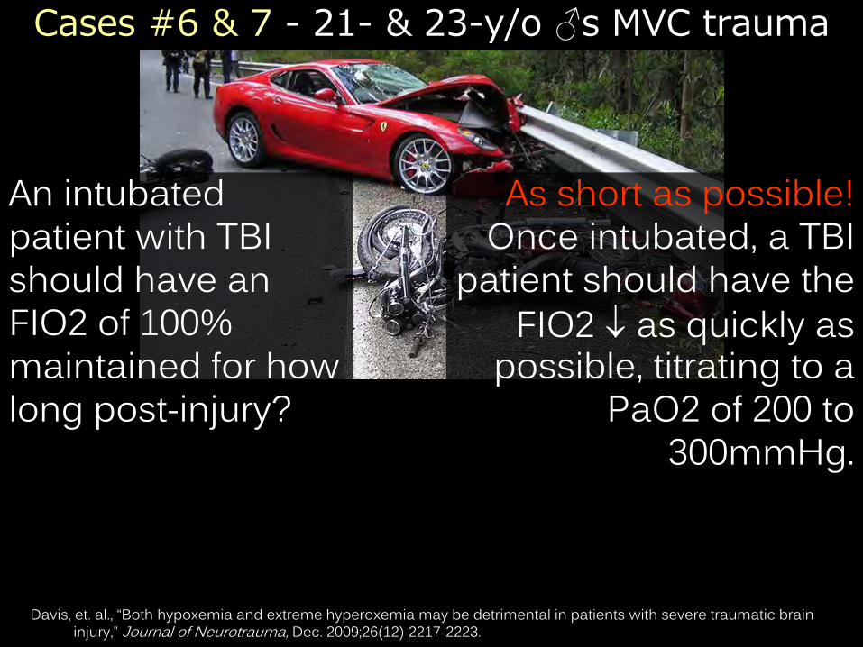
An intubated patient with TBI should have an FIO2 of 100% maintained for how long post-injury?
As short as possible! Once intubated, a TBI
patient should have the
FIO2 as quickly as possible, titrating to a
PaO2 of 200 to 300mmHg.
Davis, et. al., “Both hypoxemia and extreme hyperoxemia may be detrimental in patients with severe traumatic brain injury,” Journal of Neurotrauma, Dec. 2009;26(12) 2217-2223.

Even normal oxygenation in the body can have hypoxia in the brain in TBI.
Oxygenation in TBI
Pulse oximetry
• Optimal care for the serious TBI patient is to keep SaO2 around 99 to 100% (PaO2 of 200 - 300mmHg).
• An FIO2 of 50% will have a maximum PaO2 of around 350mmHg.
• Hyperventilation appears to be even more harmful than hyperoxia--use the ETCO2 monitor to titrate between 35 and 40 mmHg.
Davis, et. al., “Both hypoxemia and extreme hyperoxemia may be detrimental in patients with severe traumatic brain injury,” Journal of Neurotrauma, Dec. 2009;26(12) 2217-2223.

Cases #6 & 7 - 21- & 23-y/o ♂s MVC trauma
What is the minimum target SBP for the motorcyclist (the patient with no evidence of TBI)?
70mm Hg. For uncontrolled
hemorrhage (in the absence of TBI), target resuscitation to a SBP
between 70 and 90 mm Hg or normal mentation and palpable peripheral
pulses
Cherkas D, "Traumatic hemorrhagic shock: advances in fluid management." Emerg Med Pract 13.11 (2011): 1-20.

Blood pressure
DCR: Damage Control Resuscitation
Cherkas D, "Traumatic hemorrhagic shock: advances in fluid management." Emerg Med Pract 13.11 (2011): 1-20.
Hemorrhage
Fluid replacement
Inflammation
Tissue leakage
Hypotension

Cases #6 & 7 - 21- & 23-y/o ♂s MVC trauma
T/F – In patients with TBI, it has been shown that even a single episode of hypotension causes a doubling of mortality in this patient population.
TRUE. Any treatment strategies that permit hypotension in patients with TBI are
absolutely contraindicated.
Cherkas D, "Traumatic hemorrhagic shock: advances in fluid management." Emerg Med Pract 13.11 (2011): 1-20.

Permissive hypotension...Damage control resuscitation...Delayed resuscitation...
are all only for trauma patients without TBI!
None of these principles are applicable to hypotensive medical patients!

CC: 77-y/o ♀ c/o fatigue and malaise. Symptoms present “for months.” Denies pain. No travel history.
PMHx: Denies. tobacco. alcohol. drugs.
VS: Temp 96.8° F (36°C), HR 52, RR 8, BP 86/51, SaO2 93%.
Case #8 - 77-y/o ♀ c/o fatigue & malaise

Case #8 - 77-y/o ♀ c/o fatigue & malaise
Temp 96.8° F (36°C), HR 52, RR 8, BP 86/51, SaO2 93%
Assuming a normal cardiac exam (EKG), peripheral pulses, circulating volume (CBC), and no underlying infection (UA & CXR), could these vitals be considered otherwise normal for this patient?
No. This patient has
classic vital signs and physical
appearance for hypothyroidism with likely progression to myxedema coma if
left untreated.

Blood pressure
Arterial BP reflects the dynamic balance between cardiac output and
peripheral vascular resistance.
Definition

Blood pressure
What number of physiologic components determine a patient’s blood pressure?

Blood pressure
Cardiac output (rate & contractility)
Circulating volume (hemorrhage, dehydration)
Vascular tone (neurogenic or inflammatory)
Vascular permeability (anaphylaxis, sepsis)
Endocrine system (hypothyroidism, adrenal tumor)
Five Components

Blood pressure
Adults
SBP < 90 mm Hg
SBP by ≥ 40mm Hg from baseline
Hypotension: Definition
Pediatrics
SBP < 70mm Hg + [2 x age] from 0 - 10 yrs

Pediatric Vital Signs
Do we really need a blood pressure in kids less than 3 years of age?
• Blood pressure is a vital sign no matter the age of the child.
• Most pediatric cases “under review” are missing blood pressure on their medical record.
Cantor R, “The Top 5 Peds Signs Not to Miss,” Emergency Medicine Reviews and Perspectives, July 2012; 12(7). Eisenhart AW, “Balancing Pediatric Emergency Medicine Practice: Evidence Based Emergency Medicine with
Community Hospital Systems,” The Pulse, July 2007, p 11-19.

The day you’ve bantered about for years finally happens. A busload of hemophiliacs crashes on their way to their
annual meeting of the Organisation de Hemophlia et Société Hémoglobine de International Transfusion.
How can vital signs guide you as to who will need blood?
Case #9 – A bus crash...

Early recognition and management of hypovolemic shock remains one of the most challenging tasks in the initial assessment of trauma patients.
Case #9 – A busload of hemophiliacs...
What do we already know?
Isolated vital signs (SBP & HR) are limited in their ability to
identify life-threatening hypovolemic shock.
The Shock Index (SI) correlates with the extent of hypovolemia in severely injured patients, as reflected by increased transfusion requirement, higher rates of
massive transfusion, morbidity & mortality. Mutschler et al.: The Shock Index revisited – a fast guide to transfusion requirement? A retrospective analysis on
21,853 patients derived from the TraumaRegister DGUW. Critical Care 2013 17:R172.
What do we need to know?

Blood pressure
The Shock Index (SI)
SI Heart Rate
Mutschler et al.: The Shock Index revisited – a fast guide to transfusion requirement? A retrospective analysis on 21,853 patients derived from the Trauma Register DGUW. Critical Care 2013 17:R172.
= SBP
SI of 80 120 = 0.6 SI of 100 120 = 0.83 SI of 120 100 = 1.2 SI of 140 80 = 1.75

Blood pressure
The Shock Index (SI)
SI Heart Rate
Mutschler et al.: The Shock Index revisited – a fast guide to transfusion requirement? A retrospective analysis on 21,853 patients derived from the Trauma Register DGUW. Critical Care 2013 17:R172.
= SBP
• Between 2002 and 2011, 21,853 adult trauma patients from the Trauma-Register database were divided into 4 groups.
• Units of transfused blood increased from 1.0 (± 4.8) in Group I to 21.4 (± 26.2) in Group IV.
• The Shock Index at ED presentation can be used as a clinical indicator of hypovolemic shock.
• The four SI groups also parallel the recently published Base Deficit-based classification.
• In daily clinical practice, SI may be used to assess for hypovolemic shock if point-of-care testing / technology is not readily available.

Blood pressure
Shock:
Class I Class II Class III Class IV
SI:
Need blood?
- min. mild moderate severe
< 0.6 ≥0.6 to <1 ≥1 to <1.4 ≥ 1.4
watch consider act MT!
Mutschler et al.: The Shock Index revisited – a fast guide to transfusion requirement? A retrospective analysis on 21,853 patients derived from the Trauma Register DGUW. Critical Care 2013 17:R172.
SI Heart Rate
= SBP

Blood pressure
Base Deficit-based Classification of Hypovolemic Shock
Base Deficit (BD):
Shock:
Class I Class II Class III Class IV
SI:
Blood?
- min. mild moderate severe
< 0.6 ≥0.6 to <1 ≥1 to <1.4 ≥ 1.4
watch consider act MT!
Mutschler et al.: The Shock Index revisited – a fast guide to transfusion requirement? A retrospective analysis on 21,853 patients derived from the Trauma Register DGUW. Critical Care 2013 17:R172.
Admit BD (mmol/L):
≤ 2 > 2 to 6 > 6 to 10 > 10

Key Facts: Blood Pressure Hypertension is not the cause of headache in a patient with an otherwise normal neurologic exam.
Orthostatic vital signs are neither sensitive nor specific for hypovolemia, and their presence or absence should not be used to diagnose or disposition patients.
Initial fluid choice (excluding blood products!) has not been shown to affect outcomes (morbidity or mortality) in trauma resuscitation.
For uncontrolled hemorrhage (in the absence of TBI), target resuscitation to a SBP between 70 and 90 mm Hg or normal mentation and palpable peripheral pulses.
Hypotension in adults is a SBP < 90. Hypotension in pediatrics is a SBP < 70 + [2 x age in years]

Key Facts: Blood Pressure BP is a vital sign no matter the age of the child; most pediatric cases “under review” are missing BP on their medical record.
The Shock Index is HR SBP and (at ED presentation) can be used as a clinical indicator of hypovolemic shock.
Arterial BP reflects the dynamic balance between cardiac output and peripheral vascular resistance and is determined by five components:
1. Cardiac output (rate & contractility)
2. Circulating volume (hemorrhage, dehydration)
3. Vascular tone (neurogenic or inflammatory)
4. Vascular permeability (anaphylaxis, sepsis)
5. Endocrine regulation (hypothyroid, adrenal tumor)

The Physician’s Vital Sign: Respiratory Rate

Case #10 - 17-y/o ♀ “not acting right” CC: 17-y/o ♀ brought to ED
following a minor MVC for “not acting right.” Similar symptoms intermittently over the last few weeks. Patient without c/o.
PMHx: Denied. OCPs.
THC “once”. VS: Temp 97.8° F (36.6°C)
HR 74, RR 8, BP 114/73, SaO2 97%
PE: Benign exam. Poor eye contact. Hypoactive bowel sounds noted.

Case #10 - 17-y/o ♀ “not acting right” 97.8° F (36.6°C) HR 74, RR 8, BP 114/73, SaO2 97%
Given a patient who is otherwise healthy and appears unremarkable (and is awake), what is the most likely explanation for the bradypnea?
A respiratory rate
≤ 12 in a patient who is not asleep strongly suggests
opioid intoxication.
Boyer EW, “Drug Therapy: Management of opioid analgesic overdose,” New England Journal of Medicine, July 2012; 367(2): 146-55.

Key Points: Respiratory Rate Among elderly patients, the respiratory rate is the most sensitive in detecting early systemic infection, sepsis, or a progressive metabolic acidosis.
Respiratory rate is a highly sensitive indicator of acute illness in elderly patients.
Abnormal respiratory patterns may be a primary
respiratory insult or metabolic or CNS disease.
Subtle tachypnea can be the only sign of serious illness.
A respiratory rate ≤ 12 in a patient who is not asleep strongly suggests opioid intoxication.

The Student’s Vital Sign: Pulse Oximetry

CC: 44-y/o ♂ firefighter c/o SOB immediately after recent fire. Tank ran out during blaze, but he continued to rescue / work without it.
PE: Temp 99.5° F (37.5°C), HR 118, RR 26, BP 147/96, SaO2 99%. Lungs CTA B/L. No rhonchi, rales, or wheeze.
CC: A 22-y/o ♀ c/o SOB. Onset < 1 hour PTA. Recent hx includes UTI tx and dental procedure. Denies previous SOB.
PE: Temp 98.6° F (37°C), HR 112, RR 28, BP 110/56, SaO2 85%. Lungs CTA B/L. No rhonchi, rales, or wheezing heard.
Cases #11-12, 22-y/o ♀ & 44-y/o ♂ c/o SOB

Case #11 - 22-y/o ♀ c/o SOB
98.6° F (37°C), HR 112, RR 28, BP 110/56, SaO2 85%
Additional information:
Recent medications include: TMP-SMX, phenazopyridine, benzocaine, and bupivicaine.
Supplemental O2 has no effect on her pulse oximetry.
Given this clinical history and the SaO2 of 85%, what do you suspect is the reason for this patient’s dyspnea?

The oxidation of iron Fe++ from
Fe++ (ferrous) Fe+++ (ferric)
Methemoglobinemia
Pulse oximetry
• Methemoglobin does not bind & carry oxygen the way normal Hb does.
• Pulse oximetry will be inaccurate in a dose-dependent fashion, typically 85% to 90%.
• Co-oximetry will show meth-Hb; a value > 20% generally threshold for treatment.

Case #12 - 44-y/o ♂ firefighter c/o SOB 99.5° F (37.5°C), HR 118, RR 26, BP 147/96, SaO2 99%
Given a clinical history of smoke inhalation and the seemingly normal SaO2 of 99%, what do you suspect is the reason for this patient’s dyspnea?

CO binds to Hb with an affinity 250 x that of O2 preventing loading & unloading of O2 to lungs & cells
Pulse oximetry
• Pulse oximetry will be normal b/c at the two wavelengths of light the pulse oximeter reads, oxyHb absorbs identical to carboxyHb.
• Treament is 100% oxygen by NRBR (or hyperbaric O2)
the half-life of carboxyHb from 300 min. 75 min.
Carboxyhemoglobinemia

Case #13 - 80 y/o ♂ c/o weak & AMS
CC: 2-m/o ♂ with “fussy” & d PO.
VS: 99.6° F, RR 42, HR 225, BP 63/36, SaO2 100%
PE: Crying vigorously, but consolable.
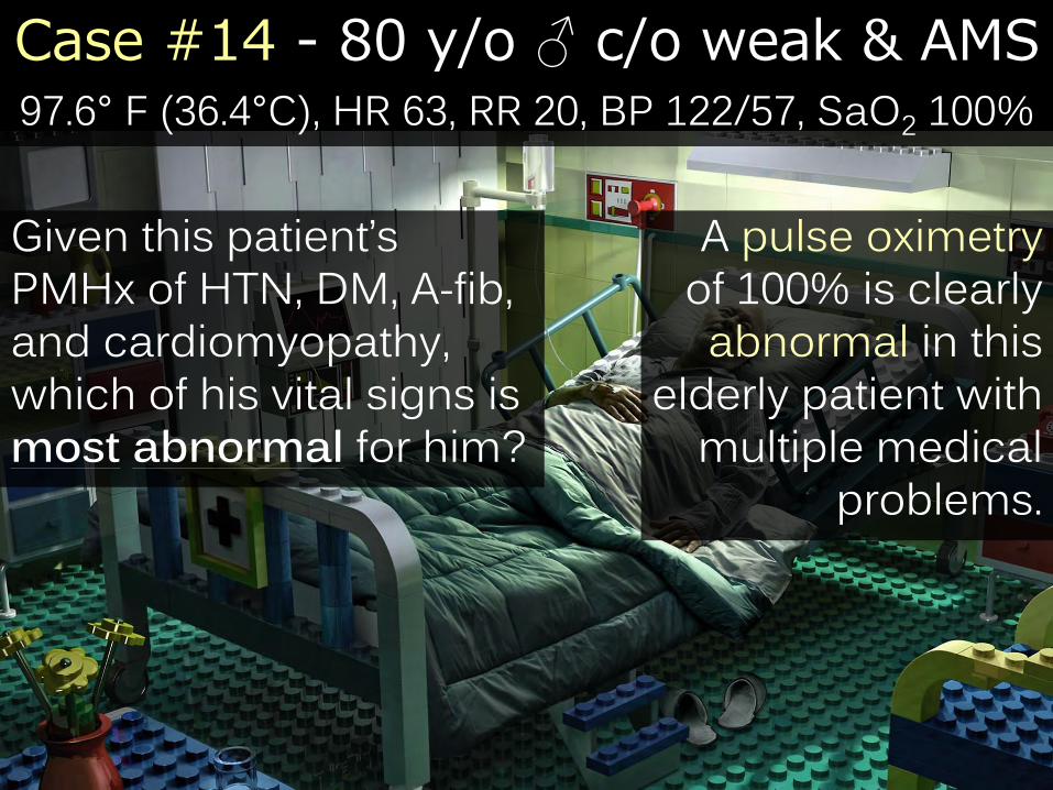
CC: 80-y/o ♂ c/o several day hx of progressive weakness and confusion.
PMHx: A-fib, cardiomyopathy (EF ~30%), HTN, DM
VS: 97.6° F (36.4°C), HR 63, RR 20, BP 122/57, SaO2 100%
PE: Confused. No focal neurologic findings. Unable to ambulate.
Case #14 - 80 y/o ♂ c/o weak & AMS
CC: 2-m/o ♂ with “fussy” & d PO.
VS: 99.6° F, RR 42, HR 225, BP 63/36, SaO2 100%
PE: Crying vigorously, but consolable.
97.6° F (36.4°C), HR 63, RR 20, BP 122/57, SaO2 100%
Given this patient’s PMHx of HTN, DM, A-fib, and cardiomyopathy, which of his vital signs is most abnormal for him?
A pulse oximetry of 100% is clearly abnormal in this
elderly patient with multiple medical
problems.

Only patients with fully functional CV/Pulm systems should have an SaO2 of 100% on RA.
Pulse oximetry
• Elderly with chronic medical problems have fluid retention, alveolar interstitial edema, scarring, protein
deposition gas exchange in the lungs.
• Severe dehydration removes that alveolar fluid falsely reassuring SaO2 of 100%
• 80-y/o ♂’s labs ultimately showed renal failure (BUN 160, Cr 6.4) due to severe intravascular hypovolemia.
SaO2 of 100% = Abnormal

Pulse oximetry
In carboxyhemoglobinemia, the pulse oximetry will be falsely normal (b/c at two wavelengths of light that pulse oximeter reads, oxyHb absorbs identical to COHb).
Confounders
In methemoglobinemia, the pulse oximetry will be 85% (usually)
because MetHb large pulsatile absorbance signal at the red & IR
wavelengths absorbance ratio to unity (read as 85%).

The Procedural Vital Sign: Capnography (ETCO2)

“Breath by breath” measure of respiratory rate and CO2 exchange
• ETCO2 closely approximates arterial CO2 levels
• Capnography gives an early warning (~1 min.) device to identify Subclinical Respiratory Depression (respiratory depression without hypoxia) before hypoxia occurs.
• This physician’s opinion is that it should be standard care in procedural sedation & analgesia.
Capnography (ETCO2)
Meta-Analysis of capnography during procedural sedation, Journal of Clinical Anesthesiology, 2011;23:189. Miner, Annals of Emergency Medicine, 2001-2003.

Cases #14-16 – Three ♂s with mult. c/os
CC: 5-y/o ♂ c/o unresponsive. “Little sick” yesterday, now unable to be awakened by Mom.
VS: 97.3° F, RR 36, HR 134, BP 83/62, SaO2 98%
PE: Moans to pain. No visible trauma.
CC: 21-y/o ♂ c/o seizure & confused. EMS transport from cross-country race.
VS: 100.1° F, RR 24, HR 119, BP 113/56, SaO2 99%
PE: Sweaty and ill appearing.
CC: 75-y/o ♂ c/o stroke. Left-sided
hemiplegia 1° PTA. Speech is garbled.
VS: 97.4°F, RR 18, HR 98, BP 173/107, SaO2 96%
PMHx: HTN, DM.

“I’ve just been informed that your
CT scanner is down indefinitely. Per
unwritten hospital policy, no one will tell you when it is
back up and running, and you will
be criticized mercilessly for
calling three times in the next 12 hours to ask for an update.”

Cases #14-16 – Three ♂s with mult. c/os
In these clinical scenarios, what vital sign abnormality is common to all three patients?
CC: 5-y/o ♂ c/o unresponsive.
VS: 97.3° F, RR 36, HR 134, BP 83/62, SaO2 98%
CC: 21-y/o ♂ c/o seizure & confused.
VS: 100.1° F, RR 24, HR 119, BP 113/56, SaO2 99%
CC: 75-y/o ♂ c/o Left-sided stroke.
VS: 97.4°F, RR 18, HR 98, BP 173/107, SaO2 96%

The Forgotten Vital Sign: Blood Glucose

• The only fuel the brain can use is glucose.
• If you don’t have enough glucose to feed your brain, you (and your friends) will notice the difference.
• Glucose—there simply is no substitute.
DONT – Dextrose

Cases #14-16 – Three ♂s with mult. c/os
CC: 5-y/o ♂ c/o unresponsive.
VS: 97.3° F, RR 36, HR 134, BP 83/62, SaO2 98%
CC: 21-y/o ♂ c/o seizure & confused.
VS: 100.1° F, RR 24, HR 119, BP 113/56, SaO2 99%
CC: 75-y/o ♂ c/o Left-sided stroke.
VS: 97.4°F, RR 18, HR 98, BP 173/107, SaO2 96%
New-onset Type I Diabetes Mellitus
Hypoglycemia (didn’t eat before
his race)
Hypoglycemia (took insulin and
forgot to eat lunch)

Accucheck
Hypoglycemia is defined according to the following serum glucose levels:
< 50 mg/dL in men
< 45 mg/dL in women
< 40 mg/dL in infants and children
Or any decrease in the blood glucose level (or its
utilization) that demonstrable signs or symptoms
Decreased
Smeeks, Frank C, MD, “Hypoglycemia,” www.emedicine.com, updated December 8, 2009, last accessed on 8/3/10.

The Gestalt of all Vital Signs
The Unspoken Vital Sign: Gestalt

Fever in the elderly (>65 y/o) is frequently associated with serious illness.
Of 470 elderly patients with “serious illness”, 76% had associated clinical features:
Temp > 103°F, RR > 30, or pulse > 120 bpm.
“Effect of Aging on the Clinical Significance of Fever in Ambulatory Adult Patients,” Keating, HJ, et. al. Journal of American Geriatric Society. 1 April 1984: 282-7.
“Fever in Geriatric Emergency Patients: Clinical Features Associated with Serious Illness,” Catherine A Marco, MD, et. al . Annals of Emergency Medicine, July 1995.
Gestalt: The Elderly

The Rule of 60s: In an infant…
…a heart rate ~ 60 bpm
…a respiratory rate ~ 60 bpm
…or a systolic BP ~ 60 mmHg
Are all signs that the baby in front of you is dying--get busy.
Gestalt: Pediatrics

The Vital Sign of Pregnancy: Fetal Heart Tones

Fetal Heart Tones
Darren Farley, MD, Donald J. Dudley, MD, Fetal Assessment During Pregnancy, Pediatric Clin North America, 56 (2009) 489–504.
• Fetal heart tones and maternal perception of fetal movement are the single best indicators of fetal well being.
• First able to auscultate FHTs around 10 weeks gestational age.
• Normal FHT range is 110 – 160 bpm.
• Trauma in pregnancy is the most common cause of non-obstetrical maternal death.
• Focus all resuscitative efforts on mom—if she dies, the baby dies.

Trauma in Pregnancy
Brown, Carlos, “Trauma In Pregnancy: A Surgeon’s Perspective,” Emergency Medicine Reviews and Perspectives, Written Summary: January 2013: Volume 13: Issue: 1.
• Supine Hypotensive Syndrome. Place the patient in a left lateral decubitus position. Hypotension due to position is a diagnosis of exclusion--think of bleeding first!
• Increased heart rate.
• Decreased blood pressure starting in the 2nd trimester.
• Pregnant patients have increased plasma volume which can mask hemorrhagic shock until collapse.
• Uterine blood flow is 20% of cardiac output; in hemorrhage, blood is shunted away from the fetus.
• Increased minute ventilation and tidal volume.

The Eye’s Vital Sign: Visual Acuity

Visual acuity
Visual acuity should be assessed for all
eye-related complaints.

The Bane of Vital Signs: Pain

Pain
“A Pain-Drug Champion Has Second Thoughts” Thomas Catan and Evan Perez
Wall Street Journal December 17, 2012


Pain
In 1986 at age 31, Dr. Russell Portenoy, co-wrote a landmark paper arguing that opioids could be used in non-cancer patients with chronic pain.
His paper was based on 38 cases.
Previous to the 1990s, opioids were characterized as highly addictive, potentially dangerous, and were largely reserved only for cancer-related pain.
Charming and articulate, Portenoy became a sought-after public speaker and rose to Chairman of Pain
Medicine & Palliative Care at Beth Israel Medical Center in New York.

Pain
Dr. Portenoy sought to “de-stigmatize these drugs.” Steven Passik, a psychologist and colleague of Dr.
Portenoy admits their message wasn't based on scientific evidence so much as a zeal to improve
patients' lives. "It had all the makings of a religious movement at the time," he said.
Drug companies noticed. In 1996, Purdue Pharma LP released OxyContin (a form of oxycodone) in a patented, time-release form, and other drug manufacturers began to compete. Today, sales of opioid painkillers total more than $9 billion a year.

Pain
Dr. Portenoy's ideas caught momentum.
In a 1998 talk in Houston, Alan Spanos, a South Carolina pain specialist, said patients with chronic non-
cancer pain could be trusted to decide themselves how many pills to take without risk of overdose.
Dr. Spanos said his understanding was that a patient would simply "go to sleep" before s/he stopped breathing. While asleep, the patient "can't take a dangerous dose. It sounds scary, but as far as I know, nobody anywhere is getting burned…doing it this way.” Dr. Spanos declined to say whether he still agreed with
this previous statement.

Pain In the late 1990s, groups such as the American Pain Foundation, of which Dr. Portenoy was a director, urged tackling what they called an epidemic of untreated pain.
The American Pain Society, of which he was president, campaigned to make pain referred to as
the "fifth vital sign.“ Dr. Portenoy helped compose a landmark 1996 consensus statement by two professional pain societies that said there was little risk of addiction or overdose among pain patients. In lectures he quoted a statistic that < 1% of opioid users became addicted.

Pain Today, opioid supporters say that figure was incorrect.
"It's obviously crazy to think that only 1% of the population is at risk for opioid addiction," said Lynn
Webster, president-elect of the American Academy of Pain Medicine (one of the publishers of the 1996
statement). "It's just not true."
The 1% figure comes from a single-paragraph in a 1980 NEJM article describing hospitalized patients briefly given opioids.
Dr. Portenoy now admits that information was irrelevant for patients with chronic non-cancer pain as there is little evidence that opioids are safe & effective
for long-term use in those patients.

Pain
In 1998, the Federation of State Medical Boards released a recommended policy reassuring doctors that they wouldn't face regulatory action for prescribing even large amounts of narcotics, as long as it was in the course of medical treatment.
In 2004 the group called on state medical boards to make undertreatment of pain punishable for the first time.
That policy was drawn up with the help of several people with links to opioid makers, including David Haddox, a senior Purdue Pharma executive then and now.
The FSMB said it has received nearly $2 million from opioid makers since 1997.

Pain
In 2001, the Joint Commission, which accredits U.S. hospitals, issued new standards telling hospitals to regularly ask patients about pain and to make treating it a priority. The now-familiar pain scale was introduced in many hospitals, with patients being asked to rate their pain from 1 to 10 and circle a smiling or frowning face.
The Joint Commission published a guide sponsored by Purdue Pharma. "Some clinicians have inaccurate and exaggerated concerns" about addiction, tolerance and
risk of death, the guide said. "This attitude prevails despite the fact there is no evidence that addiction is a
significant issue when persons are given opioids for pain control."
Pain Over his career, Dr. Portenoy has disclosed relationships with more than a dozen companies, most of which produce opioid painkillers. "My viewpoint is that I can have those relationships, they would benefit my educational mission, they benefit in my research mission, and to some extent, they can benefit my own pocketbook, without producing in me any tendency to engage in undue influence or misinformation" he said.
Dr. Portenoy and Beth Israel declined to give details of their funding by drug companies. A 2007 fundraising prospectus
shows that Dr. Portenoy's program received millions of dollars over the preceding decade in funding from opioid
makers including Endo, Abbott Laboratories, Cephalon, Purdue Pharma, and Johnson & Johnson.


Pain “A Pain-Drug Champion Has Second Thoughts,” Thomas Catan and Evan Perez, Wall Street Journal, December 17, 2012.
“Advocates for Opioid Chronic Pain Treatment Reexamine Stance,” Stratus EMR, December 20, 2012.
Doctor who championed use of opioids for chronic pain now says “it was the wrong thing to do,” The Rehab Center, Inc., 2012.
The "King of Pain" Recants - Pharmaceutical Paid Key Opinion Leader Admits it was all “Misinformation” Health
Care Renewal, hcrenewal.blogspot.com, 12/2012

Summary –Vital Sign Cases

Summary – Adult Vital Signs in Trauma

References
“Fever in the Adult Patient”, Chapter 11, Rosen’s Emergency Medicine: Concepts and Clinical Practice, 6th ed., 2006. Fleming, Susannah, et. al., “Normal ranges of heart rate and respiratory rate in children from birth to 18 years of age: a systematic review of observational studies,”
The Lancet, March 2011, 377(9770):1011-8. Gallagher EJ, “Identification of serious illness in febrile adults,” Am J Emerg Med - 01-MAR-1994; 12(2): 129-33 Graneto JW, Soglin DF, “Maternal screening of childhood fever by palpation,” Pediatric Emergency Care, 1996, 12(3):183-184. Karras DJ, et al., “Utility of routine testing for patients with asymptomatic severe blood pressure elevation in the emergency department.,” Annals of Emergency
Medicine, March 2008;51(3):231-239. Keane AM, Kasten MJ, “39-Year-Old Woman With Fever and Weight Loss,” Mayo Clinical Proceedings, March 2008, pgs. 351-354. Keating HJ, “Effect of aging on the clinical significance of fever in ambulatory adult patients,” J Am Geriatr Soc 01-APR-1984; 32(4): 282-7 Lynch, Gerald, M.D., “Post-operative Fever,” Common Surgical Diseases, 1998, pp 449-452. Marco Catherine A, MD, et. al.“Fever in Geriatric Emergency Patients: Clinical Features Associated with Serious Illness,” Annals of Emergency Medicine, July 1995. Mauck KF, Litin SC, “Clinical Pearls in Perioperative Medicine,” Mayo Clinical Proceedings, June 2009, pgs. 546-50. McFadden, JP; Price, RC; Eastwood, HD; et. al. “Raised Respiratory Rate in Elderly Patients: A Valuable Physical Sign.” British Medical Journal, 27 February 1982: 626-
627. Nishijima, et. al., “Routine testing in patients with asymptomatic elevated blood pressure in the ED,” American Journal of Emergency Medicine, 2010 Feb;28(2) 235-
242. Rogers, Robert MD, “Asymptomatic Hypertension in the ED: A Rational Approach,” EMRAP: Emergency Medicine Reviews and Perspectives, November, 2009. The Seventh Report of the Joint National Committee on Prevention, Detection, Evaluation, and Treatment of High Blood Pressure. US Department of Health & Human
Services. August 2004: 1-87. Smyth RL. “Lessons from normal heart and respiratory rates in children,” Lancet 2011; 377: 974-975. Spodick, DH, et. al. “Operation Definition of Normal Sinus Heart Rate,” American Journal of Cardiology, 1992; 69: 1245. Spodick, DH, et. al. “Survey of Selected Cardiologists for an Operational Definition of Normal Sinus Heart Rate,” American Journal of Cardiology, 1993; 72: 487. Spodick, DH, et. al. “Normal sinus heart rate: appropriate thresholds for sinus tachycardia and bradycardia,” Southern Medical Journal, 1996; 89: 666. Tolia J, Smith LG. Fever of Unkown Origin: Historical and Physical Clues to Making the Diagnosis. Infectious Disease Clinics of North America, December 1 2007;
21(4): 917-936. Walker GA, Runde D, Rolston DM, Wiener D, Lee J, “Emergency department rectal temperatures in over 10 years: A retrospective observational study,” World J Emerg
Med, Vol 4, No 2, 2013: 107-112. Weingart S, Levitan R, “Preoxygenation and Prevention of Desaturation During Emergency Airway Management,” Annals of Emergency Medicine, November 2011. Witting MD, “Unique cutpoints for sitting-to-standing orthostatic vital signs,”Am J Emerg Med. January 2003; 21(1): 45-7.
Boyer EW, “Drug Therapy: Management of opioid analgesic overdose,” New England Journal of Medicine, July 2012; 367(2): 146-55.
http://www.circadian.org (accessed 3-20-2007) Cantor R, “The Top 5 Peds Signs Not to Miss,” Emergency Medicine Reviews and Perspectives, July 2012; 12(7). Crislip, Mark, MD, “The Girl With Faget's Sign,” Medscape Emergency Medicine, www.medscape.com, Posted online
09/28/2010. Cunha, Burke A., Fever of Unknown Origin: Focused Diagnostic Approach Based on Clinical Clues from the History,
Physical Examination, and Laboratory Tests, Infectious Disease Clinics of North America, Vol 21(4) December 2007: 1137–1187.
Davis, et. al., “Both hypoxemia and extreme hyperoxemia may be detrimental in patients with severe traumatic brain injury,” Journal of Neurotrauma, Dec. 2009;26(12) 2217-2223.
Edmonds ZV, Mower WR, Lovato LM, Lomeli R, “The reliability of vital sign measurements.” Annals of Emergency Medicine, 2002; 39:233.
Eisenhart AW, “Balancing Pediatric Emergency Medicine Practice: Evidence Based Emergency Medicine with Community Hospital Systems,” The Pulse, July 2007, p 11-19.

D. O.


