30


Advanced Surfaces for Stem Cell
Research

Scrivener Publishing
100 Cummings Center, Suite 541J
Beverly, MA 01915-6106
Advanced Materials Series
The Advanced Materials Series provides recent advancements of the fascinating
field of advanced materials science and technology, particularly in the area of
structure, synthesis and processing, characterization, advanced-state properties,
and applications. The volumes will cover theoretical and experimental
approaches of molecular device materials, biomimetic materials, hybrid-type
composite materials, functionalized polymers, supramolecular systems,
information- and energy-transfer materials, biobased and biodegradable or
environmental friendly materials. Each volume will be devoted to one broad
subject and the multidisciplinary aspects will be drawn out in full.
Series Editor: Ashutosh Tiwari
Biosensors and Bioelectronics Centre
Linköping University
SE-581 83 Linköping
Sweden
E-mail: [email protected]
Managing Editors: Sachin Mishra and Sophie Thompson
Publishers at Scrivener
Martin Scrivener ([email protected])
Phillip Carmical ([email protected])

Advanced Surfaces for Stem
Cell Research
Edited by
Ashutosh Tiwari, Bora Garipcan and Lokman Uzun

Copyright © 2017 by Scrivener Publishing LLC. All rights reserved.
Co-published by John Wiley & Sons, Inc. Hoboken, New Jersey, and Scrivener Publishing LLC, Beverly,
Massachusetts.
Published simultaneously in Canada.
No part of this publication may be reproduced, stored in a retrieval system, or transmitted in any form or
by any means, electronic, mechanical, photocopying, recording, scanning, or other wise, except as permit-
ted under Section 107 or 108 of the 1976 United States Copyright Act, without either the prior writ-
ten permission of the Publisher, or authorization through payment of the appropriate per-copy fee to
the Copyright Clearance Center, Inc., 222 Rosewood Drive, Danvers, MA 01923, (978) 750-8400, fax
(978) 750-4470, or on the web at www.copyright.com. Requests to the Publisher for permission should be
addressed to the Permissions Department, John Wiley & Sons, Inc., 111 River Street, Hoboken, NJ 07030,
(201) 748-6011, fax (201) 748-6008, or online at http://www.wiley.com/go/permission.
Limit of Liability/Disclaimer of Warranty: While the publisher and author have used their best efforts
in preparing this book, they make no representations or warranties with respect to the accuracy or
completeness of the contents of this book and specifically disclaim any implied warranties of merchant-
ability or fitness for a particular purpose. No warranty may be created or extended by sales representa-
tives or written sales materials. The advice and strategies contained herein may not be suitable for your
situation. You should consult with a professional where appropriate. Neither the publisher nor author
shall be liable for any loss of profit or any other commercial damages, including but not limited to spe-
cial, incidental, consequential, or other damages.
For general information on our other products and services or for technical support, please contact
our Customer Care Department within the United States at (800) 762-2974, outside the United States at
(317) 572-3993 or fax (317) 572-4002.
Wiley also publishes its books in a variety of electronic formats. Some content that appears in print may
not be available in electronic formats. For more information about Wiley products, visit our web site
at www.wiley.com.
For more information about Scrivener products please visit www.scrivenerpublishing.com.
Cover design by Russell Richardson
Library of Congr ess Cataloging-in-Publication Data:
ISBN 978-1-119-24250-5
Printed in the United States of America
10 9 8 7 6 5 4 3 2 1
v
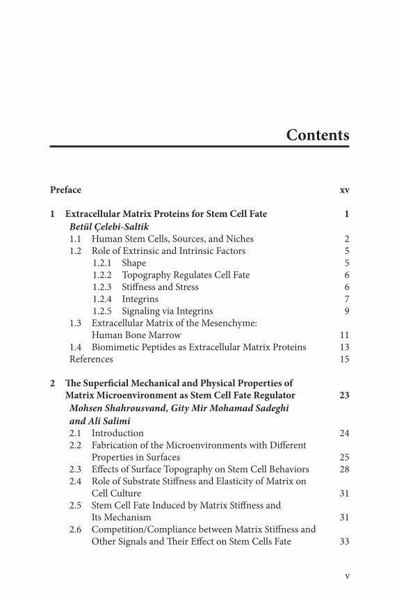
Contents
Preface xv
1 Extracellular Matrix Proteins for Stem Cell Fate 1
Betül Çelebi-Saltik1.1 Human Stem Cells, Sources, and Niches 21.2 Role of Extrinsic and Intrinsic Factors 5
1.2.1 Shape 51.2.2 Topography Regulates Cell Fate 61.2.3 Stiffness and Stress 61.2.4 Integrins 71.2.5 Signaling via Integrins 9
1.3 Extracellular Matrix of the Mesenchyme: Human Bone Marrow 11
1.4 Biomimetic Peptides as Extracellular Matrix Proteins 13References 15
2 The Superficial Mechanical and Physical Properties of Matrix Microenvironment as Stem Cell Fate Regulator 23
Mohsen Shahrousvand, Gity Mir Mohamad Sadeghi
and Ali Salimi2.1 Introduction 242.2 Fabrication of the Microenvironments with Different
Properties in Surfaces 252.3 Effects of Surface Topography on Stem Cell Behaviors 282.4 Role of Substrate Stiffness and Elasticity of Matrix on
Cell Culture 312.5 Stem Cell Fate Induced by Matrix Stiffness and
Its Mechanism 312.6 Competition/Compliance between Matrix Stiffness and
Other Signals and Their Effect on Stem Cells Fate 33

vi Contents
2.7 Effects of Matrix Stiffness on Stem Cells in Two Dimensions versus Three Dimensions 33
2.8 Effects of External Mechanical Cues on Stem Cell Fate from Surface Interactions Perspective 35
2.9 Conclusions 36Acknowledgments 36References 37
3 Effects of Mechanotransduction on Stem Cell Behavior 45
Bahar Bilgen and Sedat Odabas3.1 Introduction 453.2 The Concept of Mechanotransduction 473.3 The Mechanical Cues of Cell Differentiation and Tissue
Formation on the Basis of Mechanotransduction 483.4 Mechanotransduction via External Forces 49
3.4.1 Mechanotransduction via Bioreactors 503.4.2 Mechanotransduction via Particle-based Systems 533.4.3 Mechanotransduction via Other External Forces 55
3.5 Mechanotransduction via Bioinspired Materials 563.6 Future Remarks and Conclusion 56Declaration of Interest 57References 57
4 Modulation of Stem Cells Behavior Through Bioactive Surfaces 67
Eduardo D. Gomes, Rita C. Assunção-Silva, Nuno Sousa,
Nuno A. Silva and António J. Salgado4.1 Lithography 684.2 Micro and Nanopatterning 724.3 Microfluidics 734.4 Electrospinning 734.5 Bottom-up/Top-down Approaches 764.6 Substrates Chemical Modifications 77
4.6.1 Biomolecules Coatings 784.6.2 Peptide Grafting 79
4.7 Conclusion 80Acknowledgements 81References 81

Contents vii
5 Influence of Controlled Micro- and Nanoengineered Environments on Stem Cell Fate 87
Anna Lagunas, David Caballero and Josep Samitier5.1 Introduction to Engineered Environments for the
Control of Stem Cell Differentiation 885.1.1 Stem Cells Niche In Vivo: A Highly Dynamic
and Complex Environment 885.1.2 Mimicking the Stem Cells Niche In Vitro:
Engineered Biomaterials 905.2 Mechanoregulation of Stem Cell Fate 91
5.2.1 From In Vivo to In Vitro: Influence of the Mechanical Environment on Stem Cell Fate 91
5.2.2 Regulation of Stem Cell Fate by Surface Roughness 92
5.2.3 Control of Stem Cell Differentiation by Micro- and Nanotopographic Surfaces 94
5.2.4 Physical Gradients for Regulating Stem Cell Fate 985.3 Controlled Surface Immobilization of Biochemical
Stimuli for Stem Cell Differentiation 1025.3.1 Micro- and Nanopatterned Surfaces: Effect of
Geometrical Constraint and Ligand Presentation at the Nanoscale 102
5.3.2 Biochemical Gradients for Stem Cell Differentiation 109
5.4 Three-dimensional Micro- and Nanoengineered Environments for Stem Cell Differentiation 1145.4.1 Three-dimensional Mechanoregulation of
Stem Cell Fate 1155.4.2 Three-dimensional Biochemical Patterns
for Stem Cell Differentiation 1215.5 Conclusions and Future Perspectives 124References 124
6 Recent Advances in Nanostructured Polymeric Surface: Challenges and Frontiers in Stem Cells 143
Ilaria Armentano, Samantha Mattioli, Francesco Morena,
Chiara Argentati, Sabata Martino, Luigi Torre and
Josè Maria Kenny6.1 Introduction 1446.2 Nanostructured Surface 1466.3 Stem Cell 148

viii Contents
6.4 Stem Cell/Surface Interaction 1496.5 Microscopic Techniques Used in Estimating
Stem Cell/Surface 1506.5.1 Fluorescence Microscopy 1506.5.2 Electron Microscopy 1516.5.3 Atomic Force Microscopy 155
6.5.3.1 Instrument 1566.5.3.2 Cell Nanomechanical Motion 1586.5.3.3 Mechanical Properties 158
6.6 Conclusions and Future Perspectives 160References 160
7 Laser Surface Modification Techniques and Stem Cells Applications 167
Çağrı Kaan Akkan7.1 Introduction 1687.2 Fundamental Laser Optics for Surface
Structuring 1687.2.1 Definitive Facts for Laser Surface Structuring 169
7.2.1.1 Absorptivity and Reflectivity of the Laser Beam by the Material Surface 169
7.2.1.2 Effect of the Incoming Laser Light Polarization 170
7.2.1.3 Operation Mode of the Laser 1717.2.1.4 Beam Quality Factor 1727.2.1.5 Laser Pulse Energy/Power 173
7.2.2 Ablation by Laser Pulses 1747.2.2.1 Focusing the Laser Beam 1747.2.2.2 Ablation Regime 175
7.3 Methods for Laser Surface Structuring 1767.3.1 Physical Surface Modifications by Lasers 176
7.3.1.1 Direct Structuring 1777.3.1.2 Beam Shaping Optics 1797.3.1.3 Direct Laser Interference Patterning 182
7.3.2 Chemical Surface Modification by Lasers 1837.3.2.1 Pulsed Laser Deposition 1837.3.2.2 Laser Surface Alloying 1867.3.2.3 Laser Surface Oxidation and Nitriding 188
7.4 Stem Cells and Laser-modified Surfaces 1897.5 Conclusions 193References 194

Contents ix
8 Plasma Polymer Deposition: A Versatile Tool for Stem Cell Research 199
M. N. Macgregor-Ramiasa and K. Vasilev8.1 Introduction 1998.2 The Principle and Physics of Plasma Methods for
Surface Modification 2018.2.1 Plasma Sputtering, Etching an Implantation 2028.2.2 Plasma Polymer Deposition 203
8.3 Surface Properties Influencing Stem Cell Fate 2048.3.1 Plasma Methods for Tailored Surface Chemistry 205
8.3.1.1 Oxygen-rich Surfaces 2068.3.1.2 Nitrogen-rich Surfaces 2108.3.1.3 Systematic Studies and Copolymers 212
8.3.2 Plasma for Surface Topography 2138.3.3 Plasma for Surface Stiffness 2168.3.4 Plasma for Gradient Substrata 2178.3.5 Plasma and 3D Scaffolds 220
8.4 New Trends and Outlook 2218.5 Conclusions 221References 222
9 Three-dimensional Printing Approaches for the Treatment of Critical-sized Bone Defects 233
Sara Salehi, Bilal A. Naved and Warren L. Grayson9.1 Background 234
9.1.1 Treatment Approaches for Critical-sized Bone Defects 234
9.1.2 History of the Application of 3D Printing to Medicine and Biology 235
9.2 Overview of 3D Printing Technologies 2369.2.1 Laser-based Technologies 237
9.2.1.1 Stereolithography 2379.2.1.2 Selective Laser Sintering 2389.2.1.3 Selective Laser Melting 2389.2.1.4 Electron Beam Melting 2399.2.1.5 Two-photon Polymerization 239
9.2.2 Extrusion-based Technologies 2409.2.2.1 Fused Deposition Modeling 2409.2.2.2 Material Jetting 240

x Contents
9.2.3 Ink-based Technologies 2419.2.3.1 Inkjet 3D Printing 2419.2.3.2 Aerosol Jet Printing 241
9.3 Surgical Guides and Models for Bone Reconstruction 2429.3.1 Laser-based Surgical Guides 2429.3.2 Extrusion-based Surgical Guides 2429.3.3 Ink-based Surgical Guides 244
9.4 Three-dimensionally Printed Implants for Bone Substitution 2449.4.1 Laser-based Technologies for Metallic
Bone Implants 2469.4.2 Extrusion-based Technologies for Bone Implants 2479.4.3 Ink-based Technologies for Bone Implants 248
9.5 Scaffolds for Bone Regeneration 2489.5.1 Laser-based Printing for Regenerative Scaffolds 2499.5.2 Extrusion-based Printing for
Regenerative Scaffolds 2499.5.3 Ink-based Printing for Regenerative Scaffolds 2529.5.4 Pre- and Post-processing Techniques 253
9.5.4.1 Pre-processing 2539.5.4.2 Post-processing: Sintering 2599.5.4.3 Post-processing: Functionalization 259
9.6 Bioprinting 2609.7 Conclusion 264List of Abbreviation 265References 266
10 Application of Bioreactor Concept and Modeling Techniques to Bone Regeneration and Augmentation Treatments 279
Oscar A. Deccó and Jésica I. Zuchuat10.1 Bone Tissue Regeneration 280
10.1.1 Proinflammatory Cytokines 281 10.1.2 Transforming Growth Factor Beta 281 10.1.3 Angiogenesis in Regeneration 282
10.2 Actual Therapeutic Strategies and Concepts to Obtain an Optimal Bone Quality and Quantity 283
10.2.1 Guided Bone Regeneration Based on Cells 284 10.2.1.1 Embryonic Stem Cells 284 10.2.1.2 Adult Stem Cells 284 10.2.1.3 Mesenchymal Stem Cells 285

Contents xi
10.2.2 Guided Bone Regeneration Based on Platelet-Rich Plasma (PRP) and Growth Factors 286
10.2.2.1 Bone Morphogenetic Proteins 289 10.2.3 Guided Bone Regeneration Based on Barrier
Membranes 290 10.2.4 Guided Bone Regeneration Based on Scaffolds 292
10.3 Bioreactors Employed for Tissue Engineering in Guided Bone Regeneration 293
10.3.1 Spinner Flask Bioreactors 294 10.3.2 Rotating Wall Bioreactors 295 10.3.3 Perfusion Bioreactors 295
10.4 Bioreactor Concept in Guided Bone Regeneration and Tissue Engineering: In Vivo Application 296
10.4.1 Sand Blasting 298 10.4.2 Chemical Treatment 299 10.4.3 Heat Treatment 300
10.5 New Multidisciplinary Approaches Intended to Improve and Accelerate the Treatment of Injured and/or Diseased Bone 305
10.5.1 Application of Bioreactor in Dentistry: Therapies for the Treatment of Maxillary Bone Defects 306
10.5.2 Application of Bioreactor in Cases of Osteoporosis 309
10.6 Computational Modeling: An Effective Tool to Predict Bone Ingrowth 312
References 313
11 Stem Cell-based Medicinal Products: Regulatory Perspectives 323
Deniz Ozdil and Halil Murat Aydin11.1 Introduction 32311.2 Defining Stem Cell-based Medicinal Products 32511.3 Regional Regulatory Issues for Stem Cell Products 32811.4 Regulatory Systems for Stem Cell-based Technologies 329
11.4.1 The US Regulatory System 33011.5 Stem Cell Technologies: The European
Regulatory System 338References 342

12 Substrates and Surfaces for Control of Pluripotent Stem Cell Fate and Function 343
Akshaya Srinivasan, Yi-Chin Toh, Xian Jun Loh
and Wei Seong Toh12.1 Introduction 34412.2 Pluripotent Stem Cells 34412.3 Substrates for Maintenance of Self-renewal
and Pluripotency of PSCs 346 12.3.1 Cellular Substrates 346 12.3.2 Acellular Substrates 347
12.3.2.1 Biological Matrices 347 12.3.2.2 ECM Components 350 12.3.2.3 Decellularized Matrices 352 12.3.2.4 Cell Adhesion Molecules 353 12.3.2.5 Synthetic Substrates 354
12.4 Substrates for Promoting Differentiation of PSCs 357 12.4.1 Cellular Substrates 357 12.4.2 Acellular Substrates 358
12.4.2.1 Biological Matrices 358 12.4.2.2 ECM Components 360 12.4.2.3 Decellularized Matrices 364 12.4.2.4 Cell Adhesion Molecules 365 12.4.2.5 Synthetic Substrates 365
12.5 Conclusions 368Acknowledgments 369References 369
13 Silk as a Natural Biopolymer for Tissue Engineering 381
Ayşe Ak Can and Gamze Bölükbaşi Ateş13.1 Introduction 382
13.1.1 Mechanical Properties 383 13.1.2 Biodegradation 384 13.1.3 Biocompatibility 385
13.2 SF as a Biomaterial 385 13.2.1 Fibroin Hydrogels and Sponges 386 13.2.2 Fibroin Films and Membranes 388 13.2.3 Nonwoven and Woven Silk Scaffolds 388 13.2.4 Silk Fibroin as a Bioactive Molecule Delivery 388
13.3 Biomedical Applications of Silk-based Biomaterials 389 13.3.1 Bone Tissue Engineering 389 13.3.2 Cartilage Tissue Engineering 391
xii Contents

Contents xiii
13.3.3 Ligament and Tendon Tissue Engineering 393 13.3.4 Cardiovascular Tissue Engineering 393 13.3.5 Skin Tissue Engineering 395 13.3.6 Other Applications of Silk Fibroin 395
13.4 Conclusion and Future Directions 395References 396
14 Applications of Biopolymer-based, Surface-modified Devices in Transplant Medicine and Tissue Engineering 401
Ashim Malhotra, Gulnaz Javan and Shivani Soni14.1 Introduction to Cardiovascular Disease 40214.2 Need Assessment for Biopolymer-based Devices in
Cardiovascular Therapeutics 40214.3 Emergence of Surface Modification Applications in
Cardiovascular Sciences: A Historical Perspective 40314.4 Nitric Oxide Producing Biosurface Modification 40514.5 Surface Modification by Extracellular Matrix Protein
Adherence 40614.6 The Role of Surface Modification in the Construction of
Cardiac Prostheses 40714.7 Biopolymer-based Surface Modification of Materials
Used in Bone Reconstruction 40814.8 The Use of Biopolymers in Nanotechnology 411
14.8.1 Protein Nanoparticles 412 14.8.1.1 Albumin-based Nanoparticles and
Surface Modification 413 14.8.1.2 Collagen-based Nanoparticles and
Surface Modification 414 14.8.1.3 Gelatin-based Nanoparticle Systems 415
14.8.2 Polysaccharide-based Nanoparticle Systems 415 14.8.2.1 The Use of Alginate for Surface
Modifications 415 14.8.2.2 The Use of Chitosan-based
Nanoparticles and Chitosan-based Surface Modification 416
14.8.2.3 The Use of Chitin-based Nanoparticles and Chitin-based Surface Modification 418
14.8.2.4 The Use of Cellulose-based Nanoparticles and Cellulose-based Surface Modification 419
References 420

xiv Contents
15 Stem Cell Behavior on Microenvironment Mimicked Surfaces 425
M. Özgen Öztürk Öncel and Bora Garipcan15.1 Introduction 42615.2 Stem Cells 427
15.2.1 Definition and Types 427 15.2.1.1 Embryonic Stem Cells 428 15.2.1.2 Adult Stem Cells 428 15.2.1.3 Reprogramming and Induced
Pluripotent Stem Cells 429 15.2.2 Stem Cell Niche 429
15.3 Stem Cells: Microenvironment Interactions 430 15.3.1 Extracellular Matrix 431 15.3.2 Signaling Factors 431 15.3.3 Physicochemical Composition 432 15.3.4 Mechanical Properties 432 15.3.5 Cell–Cell Interactions 433
15.4 Biomaterials as Stem Cell Microenvironments 433 15.4.1 Surface Chemistry 433 15.4.2 Surface Hydrophilicity and Hydrophobicity 436 15.4.3 Substrate Stiffness 437 15.4.4 Surface Topography 437
15.5 Biomimicked and Bioinspired Approaches 438 15.5.1 Bone Tissue Regeneration 441 15.5.2 Cartilage Tissue Regeneration 442 15.5.3 Cardiac Tissue Regeneration 443
15.6 Conclusion 444References 444
Index 453

Preface
Stem cells have attracted much attention in the fields of regenerative medi-cine and tissue engineering for their important role in the treatment of several diseases. This is due to their unique properties such as their self-renewal capability and ability to differentiate into specific cell types. New research and therapies in these fields are mainly focused on a better under-standing of the natural mechanisms of stem cells and the control and regu-lation of their behavior under in-vivo or in-vitro conditions. Since a natural and/or synthetic surface is an important physical structure for most of the cells, the effect of surface properties, such as chemistry, charge, energy, hydropathy, pattern, topography, and stiffness, with or without differentia-tion media, influences stem cell behavior as well as controls and directs stem cell differentiation. Biomaterials that are developed by altering sur-face properties are a promising challenge for regenerative medicine and tissue engineering fields, drug investigation/toxicity studies and stem cell-based therapies.
This book, Advanced Surfaces in Stem Cell Research, part of the Advanced Materials Series, first outlines the importance of extra cellular matrix (ECM), which is a natural surface for most cells, and is discussed in the first chapter entitled “Extracellular Matrix Proteins for Stem Cell Fate.” Chapters 2 through 6 discuss the influence of biological, chemical, mecha-nical, and physical properties on stem cell behavior and fate. The mecha-nical and physical properties of matrix microenvironment as stem cell fate regulator are reviewed in Chapter 2, followed by a discussion on the effect of mechanotransduction on stem cell behavior in chapter 3. In chapter 4, stem cell modulation on bioactive surfaces is disputed. Since micro- and nanoscale structure and surfaces have an influence on stem cell behavior and fate, these properties are discussed in chapters 5 and 6, respectively entitled “Influence of Controlled Micro- and Nano-Engineered Surfaces on Stem Cell Fate” and “Recent Advances in Nanostructured Polymeric Surface: Challenges and Frontiers in Stem Cells.” Chapters 7 through 10 deliberate 2D and 3D surface fabrication and modification using different techniques on stem cell fate. Laser surface modification techniques and
xv

xvi Preface
stem cell applications and plasma polymer deposition as a versatile tool for stem cell research are discussed in chapters 7 and 8, respectively. The effect of 3D structures and dynamic cell environment, such as bioreactors, on stem cell fate are presented in detail in chapters 9 and 10, respectively entitled “3D Printing Approaches for the Treatment of Critical-Sized Bone Defect” and “Application of Bioreactor Concept and Modeling Techniques in Bone Regeneration and Augmentation Treatments.” Chapter 11 is an important and interesting chapter which will inform readers from a diffe-rent point of view, with regulatory perspectives on medical products as stem cell-based medicinal products. One of the recent stem cell sources, pluri-potent stem cells, are discussed in chapter 12, “Substrates and Surfaces for Control of Pluripotent Stem Cell Fate and Function.” Surface engineering applications are discussed in tissue engineering, regenerative medicine and different types of biomaterials in chapters 13 and 14, respectively entitled “Silk as a Natural Biopolymer for Tissue Engineering” and “Application of Biopolymer-Based, Surface Modified Devices in Transplant Medicine and Tissue Engineering.” Biomimetic and bioinspired approaches are also indi-cated for developing microenvironment of several tissues in chapter 15, “Stem Cell Behavior on Microenvironment Mimicked Surfaces.”
We would like to thank the authors that have contributed to the chapters of this book, including all scientists who have contributed to this topic. We hope and believe that this book will be very useful to those in the bioma-terials, tissue engineering, regenerative medicine, stem cell research and material science communities.
EditorsAshutosh Tiwari, PhD, DSc
Bora Garipcan, PhDLokman Uzun, PhD
September 2016

1
Ashutosh Tiwari, Bora Garipcan and Lokman Uzun (eds.) Advanced Surfaces for Stem Cell Research,
(1–22) © 2017 Scrivener Publishing LLC
1
Extracellular Matrix Proteins for Stem Cell Fate
Betül Çelebi-Saltik
Graduate School of Health Sciences, Department of Stem Cell Sciences,
Hacettepe University, Ankara, Turkey
Center for Stem Cell Research and Development, Hacettepe University,
Ankara, Turkey
AbstractStem cell-based regenerative medicine aims to repair and regenerate injured and/
or diseased tissues by implanting a combination of cells, biomaterials, and soluble
factors. Unfortunately, due to an incomplete understanding and knowledge of the
interactions between biomaterials and specific stem cell types, and the inability to
control the complex signaling pathways ensured by these interactions, the abil-
ity to design functional tissue and organ substitutes has been limited. The great-
est challenge remains the ability to control stem cells’ fate outside of the cell’s
natural microenvironment or “niche”. Stem cell fate is known to be regulated by
signals from the microenvironment, such as extracellular matrix (ECM) including
glycosaminoglycans and proteoglycans to which stem cells adhere. They represent
an essential player in stem cell microenvironment because they can directly or
indirectly modulate the maintenance, proliferation, self-renewal, and differentia-
tion of stem cells. The interactions between stem cells and the ECM play a critical
role in living tissue development, repair, and regeneration as well. The design of
artificial ECM and/or binding site is important in tissue engineering because arti-
ficial ECM and/or binding regulates cellular behaviors. Identification of binding
sites and key motifs in ECM proteins that interact with cellular receptors can allow
researchers to generate small peptides that can mimic the function of large ECM
proteins.
Keywords: Extracellular matrix proteins, stem cells, niche, integrin, signaling
Corresponding author: [email protected]
2 Advanced Surfaces for Stem Cell Research
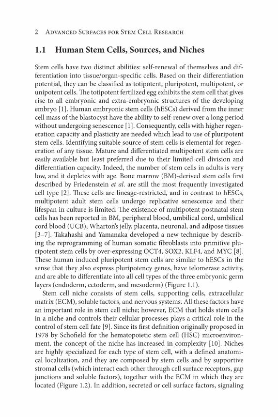
1.1 Human Stem Cells, Sources, and Niches
Stem cells have two distinct abilities: self-renewal of themselves and dif-ferentiation into tissue/organ-specific cells. Based on their differentiation potential, they can be classified as totipotent, pluripotent, multipotent, or unipotent cells. The totipotent fertilized egg exhibits the stem cell that gives rise to all embryonic and extra-embryonic structures of the developing embryo [1]. Human embryonic stem cells (hESCs) derived from the inner cell mass of the blastocyst have the ability to self-renew over a long period without undergoing senescence [1]. Consequently, cells with higher regen-eration capacity and plasticity are needed which lead to use of pluripotent stem cells. Identifying suitable source of stem cells is elemental for regen-eration of any tissue. Mature and differentiated multipotent stem cells are easily available but least preferred due to their limited cell division and differentiation capacity. Indeed, the number of stem cells in adults is very low, and it depletes with age. Bone marrow (BM)-derived stem cells first described by Friedenstein et al. are still the most frequently investigated cell type [2]. These cells are lineage-restricted, and in contrast to hESCs, multipotent adult stem cells undergo replicative senescence and their lifespan in culture is limited. The existence of multipotent postnatal stem cells has been reported in BM, peripheral blood, umbilical cord, umbilical cord blood (UCB), Wharton’s jelly, placenta, neuronal, and adipose tissues [3–7]. Takahashi and Yamanaka developed a new technique by describ-ing the reprogramming of human somatic fibroblasts into primitive plu-ripotent stem cells by over-expressing OCT4, SOX2, KLF4, and MYC [8]. These human induced pluripotent stem cells are similar to hESCs in the sense that they also express pluripotency genes, have telomerase activity, and are able to differentiate into all cell types of the three embryonic germ layers (endoderm, ectoderm, and mesoderm) (Figure 1.1).
Stem cell niche consists of stem cells, supporting cells, extracellular matrix (ECM), soluble factors, and nervous systems. All these factors have an important role in stem cell niche; however, ECM that holds stem cells in a niche and controls their cellular processes plays a critical role in the control of stem cell fate [9]. Since its first definition originally proposed in 1978 by Schofield for the hematopoietic stem cell (HSC) microenviron-ment, the concept of the niche has increased in complexity [10]. Niches are highly specialized for each type of stem cell, with a defined anatomi-cal localization, and they are composed by stem cells and by supportive stromal cells (which interact each other through cell surface receptors, gap junctions and soluble factors), together with the ECM in which they are located (Figure 1.2). In addition, secreted or cell surface factors, signaling

Extracellular Matrix Proteins for Stem Cell Fate 3
cascades and gradients, as well as physical factors such as shear stress, oxy-gen tension, and temperature, promote to control stem cell behavior [11].
Stem cell niche cells mesenchymal and HSCs play an important role in many regeneration processes in human body. HSCs are blood forming cells which were recognized more than 50 years ago [12]. They are pro-duced during embryogenesis in a complex developmental process in sev-eral anatomical sites (the yolk sac, the aorta-gonad-mesonephros region, the placenta, and the fetal liver), after which HSCs colonize the BM at birth [13]. These cells are divided into two major progenitor lineages; com-mon myeloid (CMP) and common lymphoid progenitors (CLP). CMPs give rise to megakaryocyte (MK)/erythroid and granulocyte/macrophage
Embriyonicstem cells
Cardiacstem cells
Neuralstem cells
Mesenchymalstem cells
Hematopoieticstem cells
Adultstem cells
Stem cells
Induced pluripotentstem cells
EndodermEctoderm
Mesoderm
EndodermEctoderm
Mesoderm
Figure 1.1 Embryonic stem cells, adult stem cells, and induced pluripotent stem cells.
Gap junction
Niche cells
Solublefactors
Neurons
Extracellular matrix (ECM)
Stem cell-ECMinteraction (integrin)
Stem cells
Stem cell-niche cellinteraction
Stem cell-soluble factorinteraction
Figure 1.2 Stem cell niche. Adapted from Jhala et al. [9].

4 Advanced Surfaces for Stem Cell Research
progenitors developing into platelet producing MKs, erythrocytes, mast cells, neutrophils, eosinophils, monocytes, and macrophages. CLPs will mature into B-lymphocytes, T-lymphocytes, and natural killer cells. They are positive to cell surface markers such as CD34, CD45, and CD117 [14]. HSCs have the ability to leave their tissue of origin, enter circulation, iden-tify, and relocate to an available niche elsewhere during early development [15]. In the adult, they can leave the BM and return back to it through homing mechanisms [16].
Since the identification of mesenchymal stem cells (MSCs) as colony-forming unit fibroblasts (CFU-Fs) by Friedenstein et al. in 1970 and the first detailed description of the tri-lineage capacity by Pittenger et al., our understanding of these cells has taken great strides forward [17]. These plastic adherent multipotent cells are able to differentiate into bone, carti-lage, and fat cells and can be isolated from many adult tissue types. They have been obtained from a wide variety of fetal and adult tissues: adipose tissue, placenta, umbilical cord, Wharton’s Jelly, synovial membrane, and dental pulp [18]. Specifically, a recent study reported plastic-adherent MSC-like colonies derived from the non-mesodermal tissues such as brain, spleen, liver, kidney, lung, BM, muscle, thymus, and pancreas [19]. They are positive to cell surface markers such as CD29, CD44, CD105, CD90, and CD73 [20]. MSCs secrete cytokines, chemokines, growth fac-tors, and ECM either spontaneously or after induction by other cytokines [21]. The mesenchymal ECM consists of various proteins such as colla-gens types I, III, IV, V, and VI, laminin, fibronectin, proteoglycans, such as syndecan, Pln, decorin, and the glycosaminoglycan (GAG) hyaluronan (Figure 1.3) [22]. Proteoglycans are macromolecules consisting of a core protein attached to several polysaccharides (GAGs). They are able to bind cells to the matrix and bind growth factors to GAGs, thus preventing extra-cellular protease degradation of growth factors [23].
The ECM is a highly dynamic structure that is constantly being remod-eled, either enzymatically or non-enzymatically, and its molecular compo-nents are subjected to many post-translational modifications [24]. ECM proteins vary not only in composition but also in physical parameters including elasticity and topography. Such physical properties are known to affect the micro- to nanotopography of integrin receptors (alpha and beta subunits) and to influence a range of cellular processes through changes in cell shape and the actin cytoskeleton [25–27]. Cell adhesion to the ECM is mediated by ECM receptors, such as integrins, discoidin domain receptors, and syndecans [24]. ECM properties including stiff-ness, fiber orientation, and ligand presentation dimensionality provoke specific cellular behaviors [24]. It is clear that ECM-based control of the

Extracellular Matrix Proteins for Stem Cell Fate 5
cell may also occur through multiple physical mechanisms, such as ECM geometry at the micro- and nanoscale (shape, porosity, and topography), ECM elasticity or mechanical signals (stiffness and stress) transmit-ted from the ECM to the stem cells that turn into biochemical response (Figure 1.4). An understanding of the interaction of these mediators with signaling pathways may provide new insights into the regulation of self-renewal and differentiation of stem cells [28].
1.2 Role of Extrinsic and Intrinsic Factors
1.2.1 Shape
The interactions between many extrinsic and intrinsic factors that govern cell shape are varied and may involve relatively long-term interactions with the cellular niche, as well as more acute changes due to physical factors such as mechanical or osmotic stress. In 1911, Carrel and Burrows showed that cells were responsive to shape cues [29] and over the last decade, the
Collagen type IV
Collagen type I
Laminin
Fibronectin
Figure 1.3 The mesenchymal ECM proteins network.
ECM composition
Biochemical Geometrical Mechanical
Extracellular matrix properties
Shapeporosity
topographyStiffness
stress
Figure 1.4 ECM-based control of the cell.

6 Advanced Surfaces for Stem Cell Research
effects of surface shape have been well documented. Chen et al. performed a study to clarify how cells can differentiate on different surface shape via molecular signaling pathways. Based on their research, human MSCs were induced to take on either round or spread out shapes by fibronectin- patterned surfaces. The cells on the round islands underwent adipogenesis but those on spread out islands underwent osteogenesis. This phenomenon was reported due to the activation of RhoA which is induced by surface shape [30]. In other research, MSCs that cultured on small 30 nm nano-tubes showed no differentiation whereas MSCs on 70–100 nm nanotubes showed cytoskeletal stress and osteoblastic lineage differentiation. This change was also reported due to the activation of RhoA and its effector ROCK kinase [31].
1.2.2 Topography Regulates Cell Fate
Cells are encountering different topographies sized clues in vivo from macro- (bone and ligament) to micro- (other cells) and nanotopography (proteins and ligands), all of which influence cell behavior and cell fate. It is known clear that topographical clues alone can produce the same effect as chemical induction including growth factors, chemokines and cyto-kines [32]. Topographical (physical) cues in the sub-micrometer range such as increasing the roughness or displaying specific geometrical shapes elicit specific cell responses. Pattering techniques such as soft lithogra-phy, photolithography, electrospinning, layer-by-layer microfluidic pat-terning, three-dimensional (3D) printing, ion milling, and reactive ion etching create scaffolds with precise controlled geometry, texture, poros-ity, and rigidity [32]. Micro-topographies that include micropits, micro-grooves, and micropillars induce the cell body by physical confinement or alignment [33]. Focal adhesions (FAs), the sites of cell attachment to the underlying surfaces, play a pivotal role in all cell actions in response to nano- and microtopography. These dynamic adhesions are subject to complex regulation involving integrin binding to ECM components and the reinforcement of the adhesion plaque by recruitment of additional proteins [33].
1.2.3 Stiffness and Stress
The mechanical properties of the ECM not only allow such tissues to cope with stresses but also regulate various cellular functions such as spread-ing, migration, proliferation, stem cell differentiation, and apoptosis [28, 34]. Gels based on natural ECM, such as collagen type I, Matrigel,

Extracellular Matrix Proteins for Stem Cell Fate 7
and fibrin, were the first materials used to suggest an impact of stiff-ness on cell fate [34]. Increasing the cross-linking of these matrixes that modulates matrix stiffness impacts integrin signaling and acto-myosin-mediated cellular tension [35]. For example, when a low concentration of cross-linker was used to attach collagen to stiff hydrogels, epidermal stem cells were stimulated to terminally differentiate, and MSCs differentiated into adipocytes rather than osteoblasts [36]. In the case of MSCs, osteo-genic differentiation is favored by stiff substrates, whereas adipogenic differentiation is promoted by soft substrates [25]. Increases in microen-vironment stiffness led to greater ECM and adhesion protein expression in cardiac stem cells [37]. Substrate stiffness also directs skeletal muscle stem cells and neuronal stem cells to either self-renew or differentiate. It has been also reported that cell spreading, self-renewal, and differentia-tion were inhibited on soft substrates (10 Pa), whereas with moduli of 100 Pa or greater, cells exhibited peak levels of a neuronal marker, beta-tubulin III, on substrates that had the approximate stiffness of brain tis-sue. Softer substrates (100–500 Pa) promoted neuronal differentiation (neural stem cells cultured on fibronectin peptide containing hydrogels in serum-free neuronal differentiation medium exhibit maximal differen-tiation at 500 Pa), whereas stiffer substrates (1,000–10,000 Pa) led to glial differentiation [38].
1.2.4 Integrins
Stem cells interact with ECM proteins via a family of surface receptors called integrins. Their types and quantities on each stem cell are specific to the cell and tissue type. Integrins facilitate the interactions between stem cells and ECM proteins and transduce chemical and physical signals from the matrix to the cells. They are involved in many cellular functions, including quiescence, cell cycle progression, cell adhesion, migration, and survival. Integrins are heterodimeric transmembrane receptors. An inte-grin molecule is mainly composed of two glycoprotein subunits, alpha (α) and beta (β). In vertebrates, 18 α subunits and 8 β subunits have been found, and they can form 24 different heterodimeric structures [39]. α Subunit has four Ca2+-binding domain on its extracellular part of poly-peptide chain, and β subunit bears a number of cysteine-rich domains on its extracellular part of polypeptide chain. The extracellular domain of integrins binds to ECM ligands, such as laminin, fibronectin, collagen, and vitronectin, while the intracellular domain connects with cytoskel-etal proteins, such as α-actinin and talin, as well as regulatory proteins, such as calreticulin and cytohesin. Integrins interact with ligands through

8 Advanced Surfaces for Stem Cell Research
weak interactions, but the ligand-binding affinity may be modulated by intracellular signals. After the ligand binds between the two subunits, the induced conformational change physically pushes the two subunits apart and initiates downstream signaling [e.g. activation of cytoplasmic TPK (FAK) and serine/threonine kinases, activation of small GTPases, induction of calcium transport, or changes of phospholipid synthesis]. This transmembrane linker makes integrins important in cell–cell and cell–ECM adhesion, signal transduction, and growth factor receptor responses [40]. Basement membranes are typically rich in laminins and non-fibrillar collagen type IV, whereas soft connective tissue is dominated by the presence of fibrillar collagens types I and III [41]. Cells respond to the mechanical and biochemical changes in ECM through the cross-talk between integrins and the actin cytoskeleton [42].
Integrins have been recognized as a key regulator in embryogenesis. Endoderm is one of the three primary germ layers. The expression of integrin subunits α3, α6, and β4 decreased in definitive endoderm (DE) compared to undifferentiated cells, but αV and β5 are highly expressed in undifferentiated hESCs, and this expression is increased after DE forma-tion. When ESCs are directed to differentiate toward endoderm lineages, laminin substrates are useful during the differentiation toward DE. This response is mediated by integrins αV and β5. Besides, when cells are dif-ferentiated toward pancreatic or hepatic lineages, integrin β1 becomes important [40]. Mesoderm derived from ESC has potential to regener-ate multiple tissues and organs. Integrins such as α5β1 and α6β1 may be useful in mesoderm induction. The activation of α5β1 modulated bone morphogenetic protein (BMP)-4 expression and BMP-4 induction during differentiation activates BMP, Wnt, fibroblast growth factor (FGF), and transforming growth factor (TGF)-β/nodal/activin signaling [43]. The generation of neuroectoderm lineages from ESC becomes the main focus of present ectoderm differentiation study. Deficiency of the α3 integrin subunit has been observed in mice with defective neuron migration. On the other hand, the interaction of laminin and fibronectin with β1 integrins promotes the maintenance and migration of neural precursor cells [44]. It has been reported that α1β1, α6β1, and α5β1 are crucial in the differentiation of specific neural crest lineages [40]. Skin substitutes derived from ESC differentiation may serve as a continual source for the treatment of wound healing. β1 Integrin is responsible for melano-cyte proliferation, α2β1-collagen IV, α3β1-laminin, and α6β4-laminin are responsible for keratinocytes differentiation [45], α6β1-laminin and α2, α5, αvβ3-collagen type IV are responsible for melanocytes differentiation (Table 1.1) [46].

Extracellular Matrix Proteins for Stem Cell Fate 9
1.2.5 Signaling via Integrins
Expression of integrins is regulated by ECM to cell signaling. It has been reported that TGF-β1 induced elevated expression of α2β1 integrin in fibroblasts. Therefore, cytokines regulate the expression of integrins [47]. FGFs and vascular endothelial growth factors (VEGFs) have been shown to bind to heap and can be cleaved off from the GAG components of hepa-ran sulfate proteoglycans (HSPG) by the enzyme heparanase and released as soluble ligand [48]. Fibronectin and tenascin-C bind to VEGF, which potentiates the VEGF-mediated signaling through its receptor VEGFR2 (Table 1.2) [49].
Several tyrosine kinases and phosphatases are necessary for the regula-tion of mechano-sensory activity of FAs; such as FA kinase (FAK), Src, receptor type tyrosine protein phosphatase α (RPTP α) and SH2 domain containing protein tyrosine phosphatase 2 (SHP2) [50]. FAK localizes at FAs on integrin clustering to regulate cell adhesion, migration and mecha-notransduction [51]. FAK is a non-receptor tyrosine kinase which is acti-vated upon integrin binding to autophosphorylate Y397—this induces subsequent binding of Src by the SH2 domain, leading to stable and increased activation of Src–FAK complex [42]. Src is a non-receptor tyro-sine kinase associated with the cytoplasmic tail of β3 integrins via its SH3 domains. Overexpression and phosphorylation of FAK correlate with the increase in cell motility and invasion. Adhesion and spreading of cells on a variety of ECM proteins, including collagen type IV, lead to an increase
Table 1.1 Adhesive sequences in matrix proteins and their integrin receptor.
ECM proteins Sequence Integrins
Collagens GFOGER α1β1, α2β1, α10β1, α11β1
Fibronectin RGD α5β1, αVβ3, α8β1, αVβ1, αVβ6, αIIbβ3
LDV α4β1, α4β7
REDV α4β1
Laminins E1 fragment α1β1, α2β1, α10β1
E8 fragment α3β1, α6β1, α7β1, α6β4
Vitronectin RGD αVβ3, αIIbβ3, αVβ5, αVβ1, αVβ8
Fibrinogen RGD αVβ3
KQAGV αIIbβ3
Von Willebrand
factorRGD αVβ3, αIIbβ3
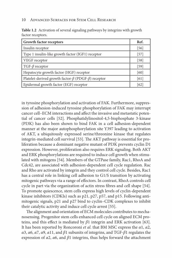
10 Advanced Surfaces for Stem Cell Research
in tyrosine phosphorylation and activation of FAK. Furthermore, suppres-sion of adhesion-induced tyrosine phosphorylation of FAK may interrupt cancer cell–ECM interactions and affect the invasive and metastatic poten-tial of cancer cells [52]. Phosphatidylinositol-4,5-bisphosphate 3-kinase (PI3K) has also been shown to bind FAK in a cell adhesion-dependent manner at the major autophosphorylation site Y397 leading to activation of AKT, a ubiquitously expressed serine/threonine kinase that regulates integrin-mediated cell survival [53]. The AKT pathway is essential for pro-liferation because a dominant negative mutant of PI3K prevents cyclin D1 expression. However, proliferation also requires ERK signaling. Both AKT and ERK phosphorylations are required to induce cell growth when stimu-lated with mitogens [54]. Members of the GTPase family, Rac1, RhoA and Cdc42, are associated with adhesion-dependent cell cycle regulation. Rac and Rho are activated by integrin and they control cell cycle. Besides, Rac1 has a central role in linking cell adhesion to G1/S transition by activating mitogenic pathways via a range of effectors. In contrast, RhoA controls cell cycle in part via the organization of actin stress fibres and cell shape [54]. To promote quiescence, stem cells express high levels of cyclin-dependent kinase inhibitors (CDKIs) such as p21, p27, p57, and p15. Following anti-mitogenic signals, p21 and p27 bind to cyclin–CDK complexes to inhibit their catalytic activity and induce cell cycle arrest [55].
The alignment and orientation of ECM molecules contributes to mecha-nosensing. Progenitor stem cells enhanced cell cycle on aligned ECM pro-teins, and this effect is mediated by β1 integrin and ERK activation [63]. It has been reported by Roncoroni et al. that BM MSC express the α1, α2, α3, α6, α7, α9, α11, and β1 subunits of integrins, and TGF-β1 regulates the expression of α2, α6, and β1 integrins, thus helps forward the attachment
Table 1.2 Activation of several signaling pathways by integrins with growth
factor receptors.
Growth factor receptors Ref.
Insulin receptor [56]
Type 1 insulin-like growth factor (IGF1) receptor [57]
VEGF receptor [58]
TGF-β receptor [59]
Hepatocyte growth factor (HGF) receptor [60]
Platelet-derived growth factor-β (PDGF-β) receptor [61]
Epidermal growth factor (EGF) receptor [62]

Extracellular Matrix Proteins for Stem Cell Fate 11
of MSC to ECM proteins [64]. Therefore, cytokines regulate the expression of integrins too. ECM remodeling takes place by means of proteases like matrix metalloproteases, serine and cysteine proteases. These interactions stimulate or inhibit various signaling pathways in the stem cell niche.
1.3 Extracellular Matrix of the Mesenchyme: Human Bone Marrow
In adult, hematopoiesis is restricted to the extravascular compartment where HSCs are in contact or close proximity with a heterogeneous popu-lation of stromal cells in the niche. Cellular interactions between HSCs and stromal cells involve various cell surface molecules, including integrins, selectins, sialomucins, and the immunoglobulin gene superfamily, that are subsequently translated into cell signaling regulating the localization and function of the cells within the niche [65].
In BM, integrin receptors interact with ligands that include ECM pro-teins, immunoglobulin superfamily members, and vascular cell adhesion protein 1 (VCAM-1) and, therefore, they play an important role in cell adhesion and signaling. Several integrins have been identified in the BM microenvironment and more specifically on HSCs [66, 67]. Functional integrins depend on the cytokine medium in which they reside, making adhesive events regulatable by cytokines. Among the different integrin subtypes, the β1 integrins have shown to play an important role in HSC migration and homing to the BM [67]. Although β1 integrin is generally known to promote proliferation, it can also be inhibitory, constituting a dominant negative signal over the stimulatory effects [54]. In the HSCs, the α4, α5, α6, α7, and α9 integrin subunits are expressed [68]. A study on the spatial location of ECM proteins including fibronectin, collagen types; I, III, and IV, and laminin in murine femoral BM by immunofluorescence has revealed distinct locations for each protein, supporting the notion that they have an important role in the homing and lodgment of transplanted cells (Table 1.3, [69]). Collagen type IV, laminin, nidogen/entactin, and perlecan are the major components of this network [70]. Minor compo-nents bind to the major components in a tissue-specific manner via their chains, and they contribute to BM’s heterogeneity [70]. Many reports have documented the importance of α4β1 and α5β1, in modulating adhesive interactions between HPCs and ECM components that comprise the stem cell niche [71, 72]. A study conducted by Van Der Loo et al. demon-strated that α5β1 is expressed on mouse and human long-term repopu-lating hematopoietic cells and binds to fibronectin in the ECM and that

12 Advanced Surfaces for Stem Cell Research
disruption of this binding can lead to decreased engraftment in the BM [73]. Also, expression of the laminin receptor has been found on erythroid progenitors and its inhibitions blocks BM homing of Burst forming unit-erythroid (BFU-E) [74]. MK maturation and platelet generation are conse-quent to MK migration from the osteoblastic to the vascular niche, where MKs extend proplatelets, and newly generated platelets are released into the blood [75].
It has been demonstrated that interactions of MKs with fibrinogen or von Willebrand factor in vascular space, are able to sustain MK maturation and proplatelets, whereas collagen type I suppresses these events and pre-vents premature platelet release in the osteoblastic niche [76]. The negative regulation of proplatelets by collagen type I is mediated by the interac-tion with the integrin α2β1. Hence, recent studies have demonstrated that fibronectin may represent a new regulator of MK maturation and platelet release [77]. Collagen fibers stimulate platelet activation, leading to inside out regulation of the integrin GP IIb–IIIa, secretion from dense and alpha granules, generation of thromboxanes, and expression of procoagulant activity, all of which support the hemostatic process. ECM proteins such as laminin, collagen type IV self-assemble in the BM into a honeycomb-like polymer. The role of collagen in supporting platelet adhesion to the endo-thelium is mediated through indirect and direct interactions [78]. Another ECM protein, osteopontin, is a phosphorylated glycoprotein that can be produced by a variety of BM cells, especially bone surface. Osteopontin
Table 1.3 ECM proteins and their localization within the BM.
Bone
compact
and/or
trabeculae
Endosteal
surface/
marrow
Periosteal
surface
Central
marrow
Marrow
vessel:
sinuses and
arteries
Fibronectin +++ +++ +++ ++ –
Collagen
type I
+++ +++ – – –
Collagen
type II
– – ++ – –
Collagen
type IV
+ ++ ++ – ++
Laminin + – +++ + +++
+++, bright expression; ++, moderate expression; +, faint expression; –, absent. Adapted
from [69].


