63
Journal of Thyroid Research Guest Editors: Juan C. Galofré, Leonidas H. Duntas, L. D. Premawardhana, and Terry F. Davies Advances in Graves’ Disease

Journal of Thyroid Research
Guest Editors: Juan C. Galofré, Leonidas H. Duntas, L. D. Premawardhana, and Terry F. Davies
Advances in Graves’ Disease

Advances in Graves’ Disease

Journal of Thyroid Research
Advances in Graves’ Disease
Guest Editors:Juan C. Galofre, Leonidas H. Duntas,L. D. Premawardhana, and Terry F. Davies

Copyright © 2012 Hindawi Publishing Corporation. All rights reserved.
This is a special issue published in “Journal of Thyroid Research.” All articles are open access articles distributed under the CreativeCommons Attribution License, which permits unrestricted use, distribution, and reproduction in any medium, provided the originalwork is properly cited.

Editorial Board
P. Beck-Peccoz, ItalyFausto Bogazzi, ItalyGlenn D. Braunstein, USAS. Y. Cheng, USAOrlo H. Clark, USAG. L. Clayman, USAAlbert A. Driedger, CanadaThomas J. Fahey, UKNadir Rashad Farid, TunisiaDouglas L. Fraker, USAGary L. Francis, USAJeremy L. Freeman, CanadaEric M. Genden, USA
Ronald A. Ghossein, USADavid Goldenberg, USAM. Gross, USAK. Kaserer, AustriaChristian Koch, USANoriyuki Koibuchi, JapanMarian Ludgate, UKC. Marcocci, ItalyAidan McElduff, AustraliaFiemu Nwariaku, USAA. Pinchera, ItalyMelanie Richards, USAJoanne Rovet, Canada
A. R. Shaha, USACarmen C. Solorzano, USAJulie A. Sosa, USAB. Stack, USAHiroshi E. Takami, JapanGiovanni Tallini, ItalyMassimo Tonacchera, ItalyDuncan Topliss, AustraliaJack R. Wall, AustraliaMalcolm H. Wheeler, UKMingzhao M. Xing, USAMasanobu Yamada, Japan

Contents
Advances in Graves’ Disease, Juan C. Galofre, Leonidas H. Duntas, L. D. Premawardhana,and Terry F. DaviesVolume 2012, Article ID 809231, 2 pages
Changes of TSH-Stimulation Blocking Antibody (TSBAb) and Thyroid Stimulating Antibody (TSAb)Over 10 Years in 34 TSBAb-Positive Patients with Hypothyroidism and in 98 TSAb-Positive GravesPatients with Hyperthyroidism: Reevaluation of TSBAb and TSAb in TSH-Receptor-Antibody(TRAb)-Positive Patients, Nobuyuki Takasu and Mina MatsushitaVolume 2012, Article ID 182176, 11 pages
The Role of Thyrotrophin Receptor Antibody Assays in Graves Disease, C. Kamath, M. A. Adlan,and L. D. PremawardhanaVolume 2012, Article ID 525936, 8 pages
Microchimerism in Graves’ Disease, Juan C. GalofreVolume 2012, Article ID 724382, 7 pages
New Genetic Insights from Autoimmune Thyroid Disease, Terry F. Davies, Rauf Latif, and Xiaoming YinVolume 2012, Article ID 623852, 6 pages
Determinants of Extraocular Muscle Volume in Patients with Graves’ Disease,Samer El-Kaissi and Jack R. WallVolume 2012, Article ID 368536, 4 pages
The Evolving Role of Selenium in the Treatment of Graves’ Disease and Ophthalmopathy,Leonidas H. DuntasVolume 2012, Article ID 736161, 6 pages
The Role of Oxidative Stress on the Pathogenesis of Graves’ Disease, Milos ZarkovicVolume 2012, Article ID 302537, 5 pages
Atypical Clinical Manifestations of Graves’ Disease: An Analysis in Depth,Mohamed Osama Hegazi and Sherif AhmedVolume 2012, Article ID 768019, 8 pages

Hindawi Publishing CorporationJournal of Thyroid ResearchVolume 2012, Article ID 809231, 2 pagesdoi:10.1155/2012/809231
Editorial
Advances in Graves’ Disease
Juan C. Galofre,1 Leonidas H. Duntas,2 L. D. Premawardhana,3 and Terry F. Davies4
1 Department of Endocrinology and Nutrition, Clinica Universidad de Navarra, 36 Pio XII Avenue, 31080 Pamplona, Spain2 Endocrine Unit, Evgenidion-Hospital, University of Athens, 11528 Athens, Greece3 Department of Medicine, C2 Link, University Hospital of Wales, Cardiff CF14 4XN, UK4 Division of Endocrinology and Metabolism, Mount Sinai School of Medicine, James J. Peters VA Medical Center, New York,NY 10468, USA
Correspondence should be addressed to Juan C. Galofre, [email protected]
Received 13 March 2012; Accepted 13 March 2012
Copyright © 2012 Juan C. Galofre et al. This is an open access article distributed under the Creative Commons AttributionLicense, which permits unrestricted use, distribution, and reproduction in any medium, provided the original work is properlycited.
“From the very commencement the student should set out towitness the progress and effects of sickness and ought to perse-vere in the daily observation of disease during the whole periodof his studies.”
It was Dr. Robert J. Graves who used to pronounce thisstatement at the inauguration of his yearly university lecturesin Dublin. This was in the nineteen century and he hadonly just described Graves’ disease, the most common hyper-thyroid condition that is so widely recognized today. Therecould be at least two complementary ways of interpretingDr. Graves’ perennial advice. The first way is from a practicalviewpoint. It emphasizes the importance of observation andmonitoring clinical evolution. This is an important good-practice guide for doctors (and students) that are at apatient’s bedside. This practical approach promotes a deepscrutiny of the disease, taking into consideration that it isnot an abstract concept, but an ailment embodied in a givenpatient. The second interpretation of Graves’ statement couldbe more theoretical. Thus the significance of the statementsupports the concept of clinical and laboratory research.Physicians must participate in research at all levels: basic,translational, and clinical. Dr. Graves’ counsel encouragesefforts to achieve a deep knowledge of disease as individualentities. Therefore, both objectives, practical and theoretical,are closely entwined—laboratory advances connected tothe bedside—what we now call translational medicine.Unfortunately, this link is often weak.
Since 1835, when Graves described his disease, dramaticprogress has been made in our knowledge of the illness. Dur-ing these nearly two centuries, we have come to understand avariety of molecular, genetic, and autoimmune mechanisms
that give rise to and maintain the disease. However, it is alsotrue that despite recent advances, the clinical managementof Graves’ disease has changed very little over the lastfew decades. Nevertheless, thanks to a group of outstand-ing physician-investigators able to integrate the laboratorywith the bedside, we sense that exciting changes in themanagement of Graves’ disease are at hand. Currently,for instance, there are several molecular target therapiesunder development that will significantly alter the clinicalmanagement of the disease within the next few years. Thisspecial issue is intended to highlight some of the most recentbreakthroughs in this area. The issue includes a completeoverview: from basic reviews to clinical papers throughtranslational studies.
T. F. Davies et al. summarizes the new genetic insightsinto autoimmune thyroid diseases (AITDs), a complex topicthat is actively being investigated. At present, more thantwenty genes have been associated with AITD that canbe categorized into two groups: immune regulatory genes(which are common to other autoimmune diseases) andthyroid-specific genes. Despite the described gene-AITDassociation, the individual gene contribution to AITD devel-opment is complex. Furthermore, no single polymorphismseems to contribute substantially to the development ofthe autoimmune reaction in thyroid diseases. The emergingevidence indicates that some environmental and/or epige-netic modifications over a predisposing genetic backgroundcould change individual gene expression, which subsequentlyelicits AITD manifestation. Although new genetic findingshave emphasized the identification of the environmentalcomponents that interact with host genetic factors in other

2 Journal of Thyroid Research
autoimmune diseases, this approach has been elusive so farfor AITD. Unfortunately for the clinician, the genetic profil-ing of AITD patients is unlikely to be productive in the nearfuture, with the corresponding limitation in the developmentof new strategies in prevention and predictive treatment.
The role of microchimerism in Graves’ disease is the sub-ject of J. C. Galofre’s review article. In this paper the authorupdates and reviews the main evidence that suggests a closerelationship linking fetal microchimerism and the devel-opment of AITD. Certainly, the presence of intrathyroidalfetal cells within the maternal thyroid is an attractive can-didate mechanism for the modulation of Graves’ disease inpregnancy and the postpartum period. At present, however,microchimerism responsibility in the generation of AITDremains a hypothesis.
In their review articles, M. Zarkovic and L. H. Duntasaddress an important and emerging matter: the role ofoxidative stress on the pathogenesis of Graves’ disease andits specific treatment, respectively. M. Zarkovic describeshow oxidative stress is indeed an environmental factorthat induces and maintains the development of Graves’ophthalmopathy. Subsequently L. H. Duntas reviews theemerging role of selenium in the treatment of Graves’disease and ophthalmopathy. Both contributors tackle thequestion of the inflammatory process in AITD. The imbal-ance of the antioxidant-oxidant mechanism is describedin detail. The authors illustrate how there is an increasedproduction of radical oxygen species and cytokines, whichsustain the autoimmune process and perpetuate the dis-ease. It is stressed that selenium, a potent antioxi-dant, has been recently applied in patients with mildGraves’ ophthalmopathy, slowing the progression of dis-ease, decreasing the clinical activity score, and appreciablyimproving the quality of life. Questions remain open to fur-ther research such as whether enforced selenium nutritionalsupplementation has the same results on Graves’ disease andwhether prolonging selenium administration may have animpact on the prevention of disease.
S. El-Kaissi and J. R. Wall contribute with an originalresearch article. The authors study the determinants of extra-ocular muscle volume (assessed by MRI) in 39 patients withGraves’ disease. The study shows that patients with recentlydiagnosed Graves’ disease and extraocular muscle volumeenlargement have higher serum TSH and more severehyperthyroidism at baseline than patients without extraoc-ular muscle enlargement, with no difference in anti-TSH-Rantibody positivity when comparing both groups.
C. Kamath et al. summarize the role of thyrotrophin re-ceptor antibody (TR-Ab) assays in Graves’ disease. TR-Abassays commonly used and widely available to clinicians,measure thyroid-binding inhibiting immunoglobulins (TBIIor receptor assays), and do not differentiate between stimu-lating (TRS-Ab), neutral, and blocking antibodies (TRB-Ab).This limitation can induce confusion in managing Graves’disease patients although the patient may be the best bio-assay. The current 2nd-3rd generation receptor assays arehighly sensitive and specific when used to differentiate be-tween the functional types of TR-Ab. The authors alsoencourage measuring TR-Ab in pregnant women under
appropriate circumstances. Unfortunately, current data arenot conclusive about its use in predicting the outcome ofGraves’ disease after antithyroid drug therapy, as there isa significant variability in assay methodology, populationcharacteristics (e.g., their iodine intake), and study design inpublished data.
An example of the inherent difficulties in interpretationof positive TR-Ab significance, as postulated in the C.Kamath et al. review article, is illustrated by N. Takasuand M. Matsushita original research article. The authorsstudy the changes in serum TRB-Ab and TRS-Ab levels in34 TRB-Ab-positive patients with hypothyroidism and in98 TRS-Ab-positive Graves’ patients with hyperthyroidism.The study covers a ten-year period. Serum TRB-Ab levelsremained elevated during the entire study period in halfof the patients with initial hypothyroidism. Interestingly,hypothyroid patients were divided according to the presenceof atrophic or goitrous autoimmune thyroiditis. Despitethe presence of positive TRB-Ab, all the patients withthe goitrous form recovered from hypothyroidism whereasonly 21% of the atrophic patients evolved to euthyroidism.Around 10% of the positive TRS-Ab patients remained withelevated circulating TRS-Ab levels at the end of the follow-up and these patients continued to have hyperthyroidismdue to Graves’ disease. On the other hand, remission ofGraves’ disease occurs in 82% of patients in whom TRS-Ab disappeared from the serum. The switch from TRB-Ab to TRS-Ab or vice versa took place in 5.8% and 2.0%,respectively, always inducing a change in the gland function.The authors’ main conclusion is that positive TR-Ab maybe associated with two manifestations: hyperthyroidism andhypothyroidism.
M. O. Hegazi and S. Ahmed review article focuses onatypical clinical manifestations of Graves’ disease. Some ofthe atypical features are specifically related to Graves’ disease(including anemia, vomiting, jaundice, and right heartfailure), while others are also similarly found in patients withother forms of hyperthyroidism. Pulmonary hypertension isreported to be associated with Graves’ disease and reportedlyresponds to its treatment. Such atypical signs and symptomsshould be considered suspect and should not be allowed todelay diagnosis or unnecessary investigation.
We sincerely hope that the present volume will helpclinicians who work in the stimulating field of thyroidologyto persevere in the daily observation of disease during the wholeperiod of their studies for the benefit of their patients.
Juan C. GalofreLeonidas H. Duntas
L. D. PremawardhanaTerry F. Davies

Hindawi Publishing CorporationJournal of Thyroid ResearchVolume 2012, Article ID 182176, 11 pagesdoi:10.1155/2012/182176
Clinical Study
Changes of TSH-Stimulation Blocking Antibody(TSBAb) and Thyroid Stimulating Antibody (TSAb)Over 10 Years in 34 TSBAb-Positive Patients withHypothyroidism and in 98 TSAb-Positive Graves’ Patientswith Hyperthyroidism: Reevaluation of TSBAb and TSAb inTSH-Receptor-Antibody (TRAb)-Positive Patients
Nobuyuki Takasu and Mina Matsushita
Department of Endocrinology and Metabolism, Aizawa Hospital, 2-5-1 Honjo, Mtasumoto 390-8521, Japan
Correspondence should be addressed to Nobuyuki Takasu, [email protected]
Received 31 August 2011; Revised 22 January 2012; Accepted 24 January 2012
Academic Editor: Terry F. Davies
Copyright © 2012 N. Takasu and M. Matsushita. This is an open access article distributed under the Creative CommonsAttribution License, which permits unrestricted use, distribution, and reproduction in any medium, provided the original work isproperly cited.
Two TRAbs: TSBAb and TSAb. TSBAb causes hypothyroidism. TSAb causes Graves’ hyperthyroidism. TSBAb and TSAb blockTSH-binding to cells as TRAb, measured as TSH-binding inhibitory immunoglobulin (TBII). We reevaluate TSBAb and TSAb.We studied TSBAb, TSAb, and TBII over 10 years in 34 TSBAb-positives with hypothyroidism and in 98 TSAb-positives withhyperthyroidism. Half of the 34 TSBAb-positives with hypothyroidism continued to have persistently positive TSBAb, continuedto have hypothyroidism, and did not recover from hypothyroidism. Ten of the 98 TSAb-positives with hyperthyroidism continuedto have positive TSAb and continued to have hyperthyroidism. TSBAb had disappeared in 15 of the 34 TSBAb-positives withhypothyroidism. With the disappearance of TSBAb, recovery from hypothyroidism was noted in 13 (87%) of the 15 patients.TSAb had disappeared in 73 of the 98 TSAb-positives with hyperthyroidism. With the disappearance of TSAb, remissions ofhyperthyroidism were noted in 60 (82%) of the 73. Two of the 34 TSBAb-positives with hypothyroidism developed TSAb-positive Graves’ hyperthyroidism. Two of the 98 TSAb-positive Graves’ patients with hyperthyroidism developed TSBAb-positivehypothyroidism. TSBAb and TSAb are TRAbs. TSBAb-hypothyroidism and TSAb-hyperthyroidism may be two aspects of onedisease (TRAb disease). Two forms of autoimmune thyroiditis: atrophic and goitrous. We followed 34 TSBAb-positive patientswith hypothyroidism (24 atrophic and 10 goitrous) over 10 years. All of the 10 TSBAb-positive goitrous patients recovered fromhypothyroidism and 19 (79%) of the 24 TSBAb-positive atrophic patients continued to have hypothyroidism.
1. Introduction
There are two types of TSH receptor antibodies (TRAbs):thyroid stimulating antibody (TSAb) [1, 2] and TSH-stimu-lation blocking antibody (TSBAb) [3]. TSAb stimulates thethyroid and causes Graves’ hyperthyroidism. TSBAb blocksTSH-stimulation of the thyroid and causes hypothyroidism.Both TSAb and TSBAb block TSH-binding to thyroid cells asTSH-receptor antibody (TRAb), which has been measured asTSH-binding inhibitory immunoglobulin (TBII) [1–3]. TBII
indicates the inhibition of TSH-binding to TSH receptorbut does not indicate the function of TRAb. TRAb can bestimulatory or inhibitory. To know whether TRAb is stimu-latory or inhibitory, TSAb and TSBAb have been measured[1–3]. TRAb has been measured by different assay methodsand given various names. Among them, TBII [1, 4, 5] andTSAb [1, 2, 6–9] have been measured as TRAb to diagnoseGraves’ disease and to follow the patients. TBII is measuredas a receptor assay. TSAb is measured as a stimulator assay,using porcine thyroid cells. TSAb indicates the stimulation

2 Journal of Thyroid Research
activity of TRAb. TSBAb [3, 10–13] and TBII [3, 4, 10–13]have been measured as TRAb to diagnose TSBAb-positivehypothyroidism and to follow the patients. TSBAb has beenmeasured as a TSH-stimulation blocking assay, using porcinethyroid cells [3, 10–13]. TSBAb indicates the inhibitory activ-ity of TRAb. TSAb and TSBAb are TSH-receptor antibodies(TRAb). The former TRAb (TSAb) is a stimulating antibody[1, 2, 6–9], and the latter TRAb (TSBAb) is a blockingantibody [3, 10–13]. TSBAb blocks TSH-stimulation of thethyroid and causes hypothyroidism. TSBAb blocks TSH-binding to thyroid cells and is TRAb. TSBAb blocks TSH-stimulation of the thyroid and is measured as inhibition ofTSH-stimulated cAMP synthesis of thyroid cells. TSBAb andTSAb are TRAb. TBII reflects TSBAb- and TSAb-activities.
TSAb stimulates the thyroid and causes Graves’ hyperthy-roidism. Treatment with antithyroid drugs (ATDs) decreasesserum TSAb [14]. With the disappearance of TSAb, remis-sions of Graves’ hyperthyroidism have been seen [14].TSBAb blocks TSH-stimulation of the thyroid and causeshypothyroidism [3]. With the disappearance of TSBAb,recovery from hypothyroidism occurs [3].
It has been generally believed that Graves’ patients haveTSAb but do not have TSBAb, and that blocking antibody-(TSBAb-) positive patients with hypothyroidism have TSBAbbut do not have TSAb. However, TSBAb-positive patientswith hypothyroidism and TSAb-positive Graves’ patientswith hyperthyroidism could have both TSBAb and TSAb[13]. Some patients may have TSBAb and TSAb simulta-neously or sequentially [13]. The balance of TSBAb andTSAb determines whether a patient has hypothyroidism orhyperthyroidism [13]. We have encountered TSBAb-positivepatients with hypothyroidism, who developed TSAb-positiveGraves’ hyperthyroidism, and also TSAb-positive Graves’patients with hyperthyroidism, who developed TSBAb-positive hypothyroidism. Thyroid function can oscillatebetween hypothyroidism and hyperthyroidism as TSBAb orTSAb becomes dominant.
There are two forms of autoimmune thyroiditis: atrophicautoimmune thyroiditis and goitrous autoimmune thyroidi-tis [3]. It has become evident that hypothyroidism may occuras a result of the production of TSBAb. TSBAb has beensaid to cause hypothyroidism in the patients with atrophicautoimmune thyroiditis [3]. However, TSBAb has beenfound in patients with atrophic autoimmune thyroiditis, andalso in patients with goitrous autoimmune thyroiditis [11].TSBAb was detected in 25% of the patients with atrophicautoimmune thyroiditis and in 9% of those with goitrousautoimmune thyroiditis [3]. TSBAb causes hypothyroidism.With the disappearance of TSBAb, recovery from hypothy-roidism has been reported [3]. Here, we followed 24 TSBAb-positive hypothyroid patients with atrophic autoimmunethyroiditis and 10 TSBAb-positive hypothyroid patientswith goitrous autoimmune thyroiditis over 10 years. All ofthe 10 TSBAb-positive patients with goitrous autoimmunethyroiditis recovered from hypothyroidism and 19 (79%) ofthe 24 TSBAb-positive patients with atrophic autoimmunethyroiditis continued to have hypothyroidism.
We reevaluated TSBAb and TSAb in TRAb-positivepatients. We studied serial changes of TSBAb and TSAb
over 10 years in 34 TSBAb-positive patients with hypothy-roidism and in 98 TSAb-positive Graves’ patients withhyperthyroidism. With persistently positive TSBAb, recoveryfrom hypothyroidism was not observed. With persistentlypositive TSAb, remissions of Graves’ hyperthyroidism werenot obtained. With the disappearance of TSBAb, recoveryfrom hypothyroidism was seen. With the disappearance ofTSAb, remissions of Graves’ hyperthyroidism were also seen.Two of the 34 TSBAb-positive patients with hypothyroidismdeveloped TSAb-positive Graves’ hyperthyroidism. Two ofthe 98 TSAb-positive Graves’ patients with hyperthyroidismdeveloped TSBAb-positive hypothyroidism. TSBAb-positivehypothyroidism and TSAb-positive hyperthyroidism may betwo aspects of one disease (TRAb disease).
2. Subjects and Method
2.1. Subjects. We studied 34 TSBAb-positive patients withhypothyroidism and 98 TSAb-positive Graves’ patients withhyperthyroidism (Table 1). The 34 TSBAb-positive patientswith hypothyroidism were treated with thyroxine (T4) andthe 98 TSAb-positive Graves’ patients with hyperthyroidismwere treated with antithyroid drugs (ATDs). Serial changesof TSBAb and TSAb over 10 years were studied in 34 TSBAb-positive patients with hypothyroidism (I) and in 98 TSAb-positive Graves’ patients with hyperthyroidism (II). TSBAb-positive patients with hypothyroidism were diagnosed onthe basis of the history, signs of hypothyroidism, and thelaboratory findings, including positive TSBAb (>+40%) anddecreased serum-free thyroxine (fT4) and free triiodothyro-nine (fT3) with high TSH [3, 13]. The diagnosis of goitrousautoimmune thyroiditis was based on the finding of palpablegoiter and that of atrophic autoimmune thyroiditis on theabsence of goiter [3]. The 34 TSBAb-positive patients withhypothyroidism were treated with thyroxine (T4). Thyroxinewas discontinued at 3 months after the disappearance ofTSBAb. After the discontinuation of T4, the patients hadbeen seen every 1–3 months. When the patients continuedto be in euthyroid states and to have negative TSBAband negative TBII for more than 1 year after the T4-discontinuation, they were considered to have recoveryfrom hypothyroidism; otherwise, they had recurrence [3].When serum TSH became higher than 10 mIU/L, T4-administration was restarted [3]. TSAb-positive Graves’patients with hyperthyroidism were diagnosed on the basisof the history, signs of hyperthyroidism with diffuse goiter,and the laboratory findings, including positive TRAb (TSAband/or TBII) and elevated fT4 and fT3 with low TSH [1,2]. The 98 Graves’ patients were treated with antithyroiddrugs (ATDs). They had been treated with ATD overseveral years. ATD was discontinued at 6 months after theTSAb-disappearance. After the discontinuation of ATD, thepatients had been seen every 1–3 months. When the patientscontinued to be in euthyroid states and to have negativeTSAb and negative TBII for more than 1 year after theATD-discontinuation, they were considered to be in remis-sion; otherwise, they had recurrence [14]. When they hadrecurrence, ATD-treatment was restarted. We had followedthese 34 TSBAb-positive patients with hypothyroidism and

Journal of Thyroid Research 3
Table 1: Changes of TSBAb (TSH-stimulation blocking antibody) and TSAb (thyroid stimulating antibody) over 10 years in 34 TSBAb-positive patients with hypothyroidism and in 98 TSAb-positive Graves’ patients with hyperthyroidism.
(I) 34 TSBAb-positive patients with hypothyroidism 34
Ia: Positive TSBAb persisted Continued to have hypothyroidism 17 17
Ib: TSBAb disappearedIb1: Recovered from hypothyroidism 13
15Ib2: Continued to have hypothyroidism 2
Ic: TSBAb→ TSAb TSBAb-positive hypo → Graves’ hyper 2 2
(II) 98 TSAb-positive Graves’ patients with hyperthyroidism 98
IIa: Positive TSAb persisted Continued to have Graves’ hyperthyroidism 10 10
IIb: Complex changes of TSAbIIb1: Remission 1
13IIb2: Recurrence 12
IIc: TSAb disappearedIIc1: Remission 60
73IIc2: Recurrence 13
IId: TSAb → TSBAb Graves’ hyper → TSBAb-positive hypo 2 2
Numbers of the patients are shown.Serial changes of TSBAb and TSAb over 10 years were studied in 34 TSBAb-positive patients with hypothyroidism (I) and in 98 TSAb-positive Graves’ patientswith hyperthyroidism (II). The 34 TSBAb-positive patients with hypothyroidism were treated with thyroxine (T4) and the 98 TSAb-positive Graves’ patientswith hyperthyroidism were treated with antithyroid drugs (ATDs). Half (17) (Ia) of the 34 TSBAb-positive patients with hypothyroidism (I) continued to havepositive TSBAb and continued to have hypothyroidism. Ten (IIa) of the 98 TSAb-positive Graves’ patients with hyperthyroidism (II) continued to have positiveTSAb and continued to have Graves’ hyperthyroidism. With the disappearance of TSBAb, recovery from hypothyroidism was noted in 13 (Ib1) (87%) of the15 patients, in whom TSBAb had disappeared (Ib). With the disappearance of TSAb, remissions of Graves’ hyperthyroidism were noted in 60 (IIc1) (82%) ofthe 73, in whom TSAb had disappeared (IIc). Two of the 34 TSBAb-positive patients with hypothyroidism developed TSAb-positive Graves’ hyperthyroidism(Ic), and two of the 98 TSAb-positive Graves’ patients with hyperthyroidism developed TSBAb-positive hypothyroidism (IId).
98 TSAb-positive Graves’ patients with hyperthyroidism over10 years.
2.2. Porcine Thyroid Cell Cyclic AMP Production: TSBAb andTSAb. TSBAb and TSAb were measured as before [13, 14].Cyclic AMP (cAMP) production was determined accordingto the instruction in commercial assay kit (Yamasa, Chosi,Chiba, Japan). Crude IgG, obtained as PEG (6000) 12.5%precipitated fraction- (final concentration) from 0.2 mLaliquot of test serum, was dissolved in modified Hanks’solution without NaCl. Porcine thyroid cells were incubatedwith test IgG in 0.25 mL Hanks’ solution without NaCl, pH7.5, containing 1.5% bovine serum albumin, 20 mM Hepes,and 0.5 mM 3-isobutyl-l-methylxanthine. Cyclic AMP pro-duction during 5 h incubation at 37◦C was measured byradioimmunoassay (RIA), using a commercial kit (Yamasa).To measure TSBAb-activities, crude IgG was incubatedwith porcine thyroid cells in the presence of 25 μU bTSH(100 mU/L, final concentration), as before [3, 10–13, 15].Cyclic AMP production during 5 h incubation was measured.TSBAb-activity was expressed as percentage inhibition ofbTSH-stimulated cAMP production by test IgG. TSBAb-activity was calculated as follows:TSBAb (%) = [1 − (c −b)/(a − b)] × 100 [3, 10–13, 15], where a: cAMP generatedin the presence of normal IgG and bTSH, b: cAMP generatedin the presence of normal IgG, and c: cAMP generated in thepresence of test IgG and bTSH. Test IgG and normal IgG werethe 12.5% PEG-precipitated fraction from test serum andnormal human serum, respectively. TSBAb, described in thisreport, corresponds to TSBAb-A in the previous report [13].TSBAb activities were studied in 95 normal subjects (normalvalues were less than +40%) [13]. TSBAb activities weremore than +40% in all of the TSBAb-positive patients withhypothyroidism. TSAb activity was expressed as percentage
cAMP production compared with the mean values for 125normal subjects (normal values were less than 180%) [1, 2,14]: TSAb (%) = [d/b] × 100, where b: cAMP generated inthe presence of normal IgG, and d: cAMP generated in thepresence of test IgG.
2.3. TSH-Binding Inhibitory Immunoglobulin (TBII). TBIIwas measured by radioreceptor assay with a commercial kit(R. S. R. Limited, Cardiff, UK). Assay results were expressedas the percentage inhibition of I125-TSH-binding to thyroidplasma membrane as before [1, 2, 5, 14]. Normal values wereobtained from 128 normal control subjects and were lessthan l0% [1, 2, 14].
2.4. Statistical Analysis and Others. All samples were testedin duplicate or triplicate. Statistical analysis was performedusing Student’s t-test or χ2-test. P values less than 0.05 wereconsidered to be statistically significant. Serum-free T3, -freeT4, and TSH were determined by electrochemiluminescenceimmunoassays (ECLIAs) (Roche Diagnostics, Tokyo, Japan).Normal reference ranges are as follows: fT3 3.5–6.6 nmol/L,fT4 11.6–21.9 pmol/L, and TSH 0.4–4.20 mIU/L. The studyplan was reviewed and approved by our institutional reviewcommittee. Written informed consent was obtained from thepatient prior to publication of this paper.
3. Resuls
Serial changes of TSBAb and TSAb over 10 years werestudied in 34 TSBAb-positive patients with hypothyroidismand in 98 Graves’ patients with hyperthyroidism (Table 1).The 34 TSBAb-positive patients with hypothyroidism (I)were treated with thyroxine (T4) and the 98 TSAb-positive

4 Journal of Thyroid Research
Table 2: Characteristics of the 34 TSBAb-positive patients with hypothyroidism and the 98 TSAb-positive Graves’ patients with hyperthy-roidism.
Number of patientsGender
Age (years)Before treatment
Men/Women TSBAb (%) TSAb (%) TBII (%)
(I) 34 TSBAb-positive patients with hypothyroidism
Ia 17 5/12 42 ± 17 94± 6 146 ± 10 95± 5
Ib 15 4/11 45 ± 16 90± 9 136 ± 8 92± 7
Ic 2 1/1 38, 45 98, 97 100, 98 96, 95
Ia+Ib+Ic 34 10/24 43 ± 18 92± 7 140 ± 9 94± 7
(II) 98 TSAb-positive Graves’ patients with hyperthyroidism
IIa 10 3/7 40 ± 16 9 ± 8 839± 421 76± 15
IIb 13 3/10 42 ± 17 10 ± 11 846± 195 68± 16
IIc 73 18/55 44 ± 16 10 ± 10 746± 390 56± 18
IId 2 0/2 40, 48 2, 5 1625, 852 76, 58
IIa+IIb+IIc+IId 98 24/74 43 ± 17 10 ± 9 775± 396 57± 17
Values are means± SD. I, Ia, Ib, Ic, II, IIa, IIb, IIc, and IId correspond to those in Table 1. No differences of gender and ages were noted among I, Ia, Ib, Ic, II,IIa, IIb, IIc, and IId. No differences of TSAb-, TSBAb-, and TBII-activities were noted among Ia, Ib, and Ic and among IIa, IIb, IIc, and IId.All of the 34 TSBAb-positive patients with hypothyroidism had strongly positive TSBAb (85–103%, mean ± SD = 92 ± 7%) (Ia+Ib+Ic). Some of them hadweakly positive TSAb. Their TSAb activity ranged from 92% to 240%. The TSAb activities were 180–240% in 7 (21%) of the 34 TSBAb-positive patients withhypothyroidism and were less than 180% in the other 27 patients (79%). Seven (21%) of the 34 TSBAb-positive patients with hypothyroidism had positiveTSAb. TSBAb-positive patients with hypothyroidism had narrow distribution of TSBAb (82–104%, 92 ± 7%) and TSAb (92–240%, 140 ± 9%). All of the 98Graves’ patients with hyperthyroidism had positive TSAb (250–1795%, 775± 396%) (IIa+IIb+IIc+IId). Some of them had TSBAb. The TSBAb activities were+40–+52% in 11 (11%) and were less than +40% in the other 87 patients (89%). Graves’ patients with hyperthyroidism had wide distributions of TSAb (250–1795%, 775 ± 396%) and TSBAb (−28–+52%, 10 ± 9%).
Graves’ patients with hyperthyroidism (II) were treated withantithyroid drugs (ATDs). Among the 34 TSBAb-positivepatients with hypothyroidism (I), 17 patients (Ia) continuedto have persistently positive TSBAb and continued to havehypothyroidism. Half (17) (Ia) of the 34 TSBAb-positivepatients continued to have persistently positive TSBAb,continued to have hypothyroidism, and did not recoverfrom hypothyroidism. TSBAb disappeared in 15 (Ib) ofthe 34 TSBAb-positive patients with hypothyroidism. Withthe disappearance of TSBAb, recovery from hypothyroidismwas seen in 13 (Ib1) (87%) of the 15 patients, in whomTSBAb had disappeared (Ib). Among the 98 TSAb-positiveGraves’ patients with hyperthyroidism (II), 10 patients (IIa)continued to have persistently positive TSAb and continuedto have hyperthyroidism. Ten of the 98 TSAb-positive Graves’patients with hyperthyroidism continued to have persistentlypositive TSAb. They continued to have hyperthyroidism anddid not get remissions of Graves’ hyperthyroidism. Theycontinued to take ATD. Complex changes of TSAb werenoted in 13 TSAb-positive patients (IIb). One (IIb1) of the13 patients with complex changes of TSAb got remissions,but the other 12 patients (IIb2) did not. TSAb disappearedin 73 (IIc) (74%) of the 98 TSAb-positive Graves’ patientswith hyperthyroidism. With the disappearance of TSAb, 60(IIc1) (82%) of the 73 patients, in whom TSAb had disap-peared (IIc), got remissions of Graves’ hyperthyroidism. TwoTSBAb-positive patients with hypothyroidism developedTSAb-positive Graves’ hyperthyroidism (Ic). Two TSAb-positive Graves’ patients with hyperthyroidism developedTSBAb-positive hypothyroidism (IId).
Table 2 shows characteristics of the 34 TSBAb-positivepatients with hypothyroidism (I) and the 98 TSAb-positive
Graves’ patients with hyperthyroidism (II). I, Ia, Ib, Ic, II,IIa, IIb, IIc, and IId correspond to those in Table 1. Nodifferences of gender and ages were noted among I, Ia,Ib, Ic, II, IIa, IIb, IIc, and IId. No differences of TSAb-,TSBAb-, and TBII-activities were noted among Ia, Ib, andIc and among IIa, IIb, IIc, and IId. All of the 34 TSBAb-positive patients with hypothyroidism had strongly positiveTSBAb (85–103%, mean ± SD = 92 ± 7%) (Table 2,Ia+Ib+Ic). Some of them had weakly positive TSAb. TheirTSAb activity ranged from 92% to 240%. The TSAb activitieswere 180–240% in 7 (21%) of the 34 TSBAb-positivepatients with hypothyroidism and were less than 180% inthe other 27 patients (79%). Seven (21%) of the 34 TSBAb-positive patients with hypothyroidism had positive TSAb.TSBAb-positive patients with hypothyroidism had narrowdistribution of TSBAb (82–104%, 92 ± 7%) and TSAb(92–240%, 140 ± 9%). All of the 98 Graves’ patients withhyperthyroidism had positive TSAb (250–1795%, 775 ±396%) (Table 2, IIa+IIb+IIc+IId). Some of them had TSBAb.The TSBAb activities were +40–+ 52% in 11 (11%) andwere less than +40% in the other 87 patients (89%). Graves’patients with hyperthyroidism had wide distributions ofTSAb (250–1795%, 775 ± 396%) and TSBAb (−28–+52%,10 ± 9%).
3.1. 34 TSBAb-Positive Patients with Hypothyroidism (I)(Tables 1 and 2, I). All of the 34 TSBAb-positive patientswith hypothyroidism had strongly positive TSBAb. Someof them had weakly positive TSAb. TSBAb-positive patientswith hypothyroidism had narrow distributions of TSBAb(82–104%, 92 ± 7%) and TSAb (92–240%, 140 ± 9%)(Table 2, Ia+b+c). Figure 1 shows the changes of TSBAb

Journal of Thyroid Research 5
0
20
40
60
80
100
120
10 2 3 4 5 6 7 8 9 10
(years)
10 2 3 4 5 6 7 8 9 10
(years)
10 2 3 4 5 6 7 8 9 10
(years)
10 2 3 4 5 6 7 8 9 10
(years)
TSBAb
TSBAb
TSB
Ab
(%)
TSB
Ab
(%)
0
20
40
60
80
100
120
TSBAb
TSB
Ab
(%)
0
20
40
60
80
100
120
0
200
400
600
800
1000
1200
TSA
b (%
)
TSAb
MMI
T4
T4
MMI
(b)
(a) (c1)
(c2)
Figure 1: The changes of TSBAb in 34 TSBAb-positive patients with hypothyroidism (Table 1, I). Among the 34 TSBAb-positive patients withhypothyroidism, 17 patients continued to have persistently positive TSBAb and continued to have hypothyroidism (Table 1, Ia) (a). Half ofthe 34 TSBAb-positive patients continued to have persistently positive TSBAb, continued to have hypothyroidism, and did not recover fromhypothyroidism. They continued to take thyroxine (T4). TSBAb disappeared in 15 of the 34 TSBAb-positive patients with hypothyroidism(Table 1, Ib) (b). Recovery from hypothyroidism was noted with the disappearance of TSBAb in 13 (87%) of the 15 patients, in whomTSBAb had disappeared. (c1, c2) show the changes of TSBAb and TSAb, respectively, in the 2 patients with TSBAb-positive hypothyroidism,who developed TSAb-positive Graves’ hyperthyroidism (Table 1, Ic). In these 2 patients, TSBAb was dominant initially (c1), and then TSAbbecame dominant (c2); 2 patients with TSBAb-positive hypothyroidism developed TSAb-positive Graves’ hyperthyroidism. Hypothyroidismwas treated with thyroxine (T4). Graves’ hyperthyroidism was treated with 1-methyl 2-mercapto imidazole (MMI). TSBAb: TSH-stimulationblocking antibody; TSAb: thyroid stimulating antibody.
in the 34 TSBAb-positive patients with hypothyroidism(Table 1, I). Among the 34 TSBAb-positive patients withhypothyroidism (I), 17 (Ia) (Table 1, Ia, Figure 1(a)) con-tinued to have persistently positive TSBAb and continuedto have hypothyroidism. Half (17) (Ia) of the 34 TSBAb-positive patients (I) continued to have persistently positiveTSBAb, continued to have hypothyroidism, and did notrecover from hypothyroidism. They continued to take T4.TSBAb disappeared in 15 (Ib) (Table 1, Ib, Figure 1(b)) of the34 TSBAb-positive patients (I) with hypothyroidism. Withthe disappearance of TSBAb, recovery from hypothyroidismwas noted in 13 (Ib1) (87%) of the 15 patients, in whomTSBAb had disappeared (Ib).
Figures 1(c1) and 1(c2) show the changes ofTSBAb and TSAb, respectively, in the 2 patients with
TSBAb-positive hypothyroidism, who developed TSAb-positive Graves’ hyperthyroidism (Table 1, Ic). In these2 patients, TSBAb was dominant initially (Figure 1(c1)),and then TSAb became dominant (Figure 1(c2)). These2 TSBAb-positive patients had hypothyroidism and thendeveloped TSAb-positive Graves’ hyperthyroidism. Theywere treated with T4 and then treated with 1-methyl2-mercapto imidazole (MMI). Figure 2 demonstrates theclinical course of one of these 2 patients with TSBAb-positive hypothyroidism, who developed TSAb-positiveGraves’ hyperthyroidism (Table 1, Ic). A 45-year-old womanwith TSBAb-positive hypothyroidism developed TSAb-positive Graves’ hyperthyroidism. TSBAb was dominantinitially (Figure 2(a)), and then TSAb became dominant(Figure 2(b)). She had TSBAb-positive hypothyroidism with

6 Journal of Thyroid Research
0
20
40
60
80
100
120
0 10 20 30 40 50
TSBAb (%)
TSB
Ab
(%),
TB
II (
%)
TBII
(months)
(a)
(months)
1000
900
800
700
600
500
400
300
200
100
0
MMIT4
TSAb (%)
TSA
b (%
), T
SH (
mIU
/L)
TSH (mIU/L)
0 10 20 30 40 50
(b)
Figure 2: The clinical course of one of the 2 patients, who initially had TSBAb-positive hypothyroidism and then developed TSAb-positive Graves’ hyperthyroidism (Table 1, Ic). A 45-year-old woman with TSBAb-positive hypothyroidism developed TSAb-positive Graves’hyperthyroidism. She had TSBAb-positive hypothyroidism ((a), �) with high serum TSH ((b), ◦) and then developed TSAb-positiveGraves’ hyperthyroidism ((b), �) with undetectable serum TSH ((b), ◦). TSBAb was dominant initially ((a), �), and then TSAb becamedominant ((b), �). TBII (TSH-binding inhibitory immunoglobulin) ((a), ◦) reflects TSBAb- and TSAb-activity. A patient with TSBAb-positive hypothyroidism developed TSAb-positive Graves’ hyperthyroidism. She was treated with T4 and then with MMI.
high serum TSH and then developed TSAb-positive Graves’hyperthyroidism with undetectable serum TSH. She wastreated with T4 and then treated with MMI. She had a goiterinitially and had goitrous autoimmune thyroiditis.
Among the 34 TSBAb-positive patients with hypothy-roidism (Table 1, I), 24 had atrophic autoimmune thyroiditisand 10 had goitrous autoimmune thyroiditis (Table 3(a)).The 34 TSBAb-positive patients with hypothyroidism con-sisted of 17 patients (a: positive TSBAb persisited), 15patients (b: TSBAb disappeared), and 2 patients (c:TSBAb → TSAb) (Table 3(a)). All of the 17 (a) patientscontinued to have positive TSBAb and continued to havehypothyroidism. All of the 17 (a) patients had atrophicautoimmune thyroiditis and none of them had goitrousautoimmune thyroiditis. TSBAb disappeared in the 15 (b)patients: 13 (b1) (87%) of the 15 (b) patients recovered fromhypothyroidism and 2 (b2) (13%) of the 15 (b) patients con-tinued to have hypothyroidism. Of the 13 (b1) patients, whorecovered from hypothyroidism, 5 had atrophic autoimmunethyroiditis and 8 had goitrous autoimmune thyroiditis. The2 (b2) patients, who continued to have hypothyroidism, hadatrophic autoimmune thyroiditis. Of the 15 (b) patients, inwhom TSBAb had disappeared, 7 [5 (b1) + 2 (b2)] hadatrophic autoimmune thyroiditis and 8 [8 (b1)] had goitrousautoimmune thyroiditis. Two (c) patients of the 34 TSBAb-positive patients with hypothyroidism developed TSAb-positive Graves’ hyperthyroidism had goitrous autoimmunethyroiditis.
Table 3(b) demonstrates recovery from hypothyroidismin the 34 TSBAb-positive patients with hypothyroidism(24 patients with atrophic autoimmune thyroiditis and 10patients with goitrous autoimmune thyroiditis). Among
the 34 TSBAb-positive patients with hypothyroidism, 19[(17 (a) + 2 (b2)) in Table 3(a)] continued to havehypothyroidism over 10 years and 15 [13 (b1) + 2 (c)]recovered from hypothyroidism (13 (b1) recovered fromhypothyroidism and had remissions and 2 (c) recoveredfrom hypothyroidism and developed hyperthyroidism). Allof the 19 TSBAb-positive patients with hypothyroidism,who continued to have hypothyroidism [17 (a) + 2 (b2)],had atrophic autoimmune thyroiditis, and none of themhad goitrous autoimmune thyroiditis. Fifteen [13 (b1) + 2(c)] of the 34 TSBAb-positive patients with hypothyroidismrecovered from hypothyroidism. Five [5 (b1)] of the 15patients, who recovered from hypothyroidism, had atrophicautoimmune thyroiditis and the other 10 [8 (b1) + 2 (c)]had goitrous autoimmune thyroiditis. Nineteen (79%) ofthe 24 TSBAb-positive hypothyroid patients with atrophicautoimmune thyroiditis continued to have hypothyroidismand the other 5 (21%) recovered from hypothyroidism. All(100%) of the 10 TSBAb-positive hypothyroid patients withgoitrous autoimmune thyroiditis [8 (b1) + 2 (c)] recoveredfrom hypothyroidism. Significant differences of recoveryfrom hypothyroidism were noted between the patients withgoitrous autoimmune thyroiditis and those with atrophicautoimmune thyroiditis (χ2 = 17.9, P value < 0.05). All ofthe 10 TSBAb-positive patients with goitrous autoimmunethyroiditis recovered from hypothyroidism and 19 (79%)of the 24 patients with atrophic autoimmune thyroiditiscontinued to have hypothyroidism.
3.2. 98 TSAb-Positive Graves’ Patients with Hyperthyroidism(II) (Tables 1 and 2, II). All of the 98 Graves’ patients

Journal of Thyroid Research 7
0
200
400
600
800
1000
1200
1400
1600
1800
2000
TSA
b (%
)T
SAb
(%)
TSA
b (%
)
TSAb
TSAb
0
200
400
600
800
1000
1200
1400
1600
TSAb
0
200
400
600
800
1000
1200
1400
1600
1800
(b)
(a)
(d1)
10 2 3 4 5 6 7 8 9 10
(years)
TSA
b (%
)
TSAb
0
500
1000
1500
2000
2500
3000
(c)
10 2 3 4 5 6 7 8 9 10
(years)
10 2 3 4 5 6 7 8 9 10
(years)
10 2 3 4 5 6 7 8 9 10
(years)
TSB
Ab
(%)
TSBAb
0
20
40
60
80
100
120
(d2)
10 2 3 4 5 6 7 8 9 10
(years)
Figure 3: The changes of TSAb in 98 Graves’ patients with hyperthyroidism (II) (Table 1, II). Among the 98 Graves’ patients withhyperthyroidism, 10 patients continued to have persistently positive TSAb and continued to have hyperthyroidism (Table 1, IIa) (a). Tenof the 98 TSAb-positive Graves’ patients with hyperthyroidism continued to have persistently positive TSAb. They continued to havehyperthyroidism and did not get remissions of Graves’ hyperthyroidism. They continued to take MMI. Complex changes of TSAb werenoted in 13 TSAb-positive patients (Table 1, IIb) (b). One of the 13 patients with complex changes of TSAb got remissions, but the other 12patients did not get remissions. TSAb disappeared in 73 (74%) of the 98 TSAb-positive Graves’ patients with hyperthyroidism (Table 1, IIc)(c). With the disappearance of TSAb, 60 (82%) of the 73 patients, in whom TSAb had disappeared, got remissions of Graves’ hyperthyroidism.(d1, d2) show the changes of TSAb and TSBAb, respectively, in the 2 patients with TSAb-positive Graves’ hyperthyroidism, who developedTSBAb-positive hypothyroidism (Table 1, IId). In these 2 patients, TSAb was dominant initially (d1), and then TSBAb became dominant(d2). Two patients with TSAb-positive Graves’ hyperthyroidism developed TSBAb-positive hypothyroidism. Graves’ hyperthyroidism wastreated with MMI, and hypothyroidism was treated with T4.

8 Journal of Thyroid Research
Table 3: Atrophic autoimmune thyroiditis or goitrous autoimmune thyroiditis in the 34 TSBAb (TSH-stimulation-blocking-antibody)-positive patients with hypothyroidism (a) and recovery from hypothyroidism (b).
(a) Atrophic autoimmune thyroiditis (atrophic) or goitrous autoimmune thyroiditis (goitrous) in the 34 TSBAb-positive patients with hypothyroidism
34 TSBAb-positive patients with hypothyroidism (I in Table 1)† 34
Atrophic (24) Goitrous (10)
a: Positive TSBAb persisted(Ia)
17 17 0
b: TSBAb disappeared (Ib) 15b1: recovered (Ib1) 13 5 8
b2: hypothyroid (Ib2) 2 2 0
c: TSBAb → TSAb (Ic) 2 0 2
Numbers of the patients are shown. (I in Table 1)† correspond to those in Table 1Among the 34 TSBAb-positive patients with hypothyroidism (Table 1, I), 24 had atrophic autoimmune thyroiditis and 10 had goitrous autoimmunethyroiditis. The 34 TSBAb-positive patients with hypothyroidism consisted of 17 patients (a: positive TSBAb persisited), 15 patients (b: TSBAbdisappeared), and 2 patients (c: TSBAb → TSAb). All of the 17 (a) patients continued to have positive TSBAb and continued to have hypothyroidism.All of the 17 (a) patients had atrophic autoimmune thyroiditis and none of them had goitrous autoimmune thyroiditis. TSBAb disappeared in the 15 (b)patients: 13 (b1) (87%) of the 15 (b) patients recovered from hypothyroidism and 2 (b2) (13%) of the 15 (b) patients continued to have hypothyroidism.Of the 13 (b1) patients, who recovered from hypothyroidism, 5 had atrophic autoimmune thyroiditis and 8 had goitrous autoimmune thyroiditis. The 2(b2) patients, who continued to have hypothyroidism, had atrophic autoimmune thyroiditis. Of the 15 (b) patients, in whom TSBAb had disappeared,7 [5 (b1) + 2 (b2)] had atrophic autoimmune thyroiditis and 8 [8 (b1)] had goitrous autoimmune thyroiditis. Two (c) patients of the 34 TSBAb-positivepatients with hypothyroidism who developed TSAb-positive Graves’ hyperthyroidism who had goitrous autoimmune thyroiditis.
(b) Recovery from hypothyroidism in the patients with atrophic autoimmune thyroiditis (atrophic) and in those with goitrous autoimmune thyroiditis(goitrous)
Atrophic (24) Goitrous (10)
Continued to have hypothyroidism19 (79%)[17 (a) + 2 (b2)]∗
0 (0%) 19χ2 = 17.9 P value < 0.05
Recovered from hypothyroidism5 (21%)[5 (b1)]∗
10 (100%)[8 (b1) + 2 (c)]∗
15
24 (100%) 10 (100%) 34
Numbers (%) of the patients are shown. [ ]∗ corresponds to Table 3(a).Among the 34 TSBAb-positive patients with hypothyroidism (Table 1, I), 24 had atrophic autoimmune thyroiditis and 10 had goitrous autoimmunethyroiditis. Among the 34 TSBAb-positive patients with hypothyroidism, 19 [(17 (a) + 2 (b2)] (Table 3(a)) continued to have hypothyroidism over 10years and 15 [13 (b1) + 2 (c)] recovered from hypothyroidism [13 (b1) recovered from hypothyroidism and had remissions and 2 (c) recovered fromhypothyroidism and developed hyperthyroidism]. All of the 19 TSBAb-positive patients with hypothyroidism, who continued to have hypothyroidism[17 (a) + 2 (b2)], had atrophic autoimmune thyroiditis, and none of them had goitrous autoimmune thyroiditis. Fifteen [13 (b1) + 2 (c)] of the 34TSBAb-positive patients with hypothyroidism recovered from hypothyroidism. Five [5 (b1)] of the 15 patients, who recovered from hypothyroidism,had atrophic autoimmune thyroiditis and the other 10 [8 (b1) + 2 (c)] had goitrous autoimmune thyroiditis. Nineteen (79%) of the 24 TSBAb-positive hypothyroid patients with atrophic autoimmune thyroiditis continued to have hypothyroidism and the other 5 (21%) of them recoveredfrom hypothyroidism. All of the 10 TSBAb-positive hypothyroid patients with goitrous autoimmune thyroiditis [8 (b1) + 2 (c)] recovered fromhypothyroidism. Significant differences of recovery from hypothyroidism were noted between the patients with goitrous autoimmune thyroiditis andthose with atrophic autoimmune thyroiditis (χ2 = 17.9, P value < 0.05). All (100%) of the 10 TSBAb-positive patients with goitrous autoimmunethyroiditis recovered from hypothyroidism and 19 (79%) of the 24 patients with atrophic autoimmune thyroiditis continued to have hypothyroidism.
with hyperthyroidism had positive TSAb. Some of them hadpositive TSBAb. Graves’ patients with hyperthyroidism hadwide distributions of TSAb and TSBAb. Some of the Graves’patients had both positive TSAb and TSBAb. Figure 3 showsthe changes of TSAb in 98 Graves’ patients with hyperthy-roidism (II) (Table 1, II). Among the 98 Graves’ patients withhyperthyroidism, 10 patients continued to have persistentlypositive TSAb and continued to have hyperthyroidism (IIa)(Figure 3(a)). Ten of the 98 TSAb-positive Graves’ patientswith hyperthyroidism continued to have positive TSAb andcontinued to have Graves’ hyperthyroidism. They did not getremissions of Graves’ hyperthyroidism and continued to takeATD. Complex changes of TSAb were noted in 13 TSAb-positive patients (IIb) (Figure 3(b)). One (IIb1) of the 13patients with complex changes of TSAb got remissions, butthe other 12 patients (IIb2) did not get remissions. TSAbdisappeared in 73 (IIc) (74%) of the 98 TSAb-positive
Graves’ patients with hyperthyroidism (IIc) (Figure 3(c)).With the disappearance of TSAb, 60 (IIc1) (82%) of the73 patients, in whom TSAb had disappeared (IIc), gotremissions of Graves’ hyperthyroidism. Figures 3d1 and3d2 show the changes of TSAb and TSBAb, respectively,in the 2 patients with TSAb-positive Graves’ hyperthy-roidism, who developed TSBAb-positive hypothyroidism(IId) (Table 1, IId). In these 2 patients, TSAb was dom-inant initially (Figure 3(d1)), and then TSBAb becamedominant (Figure 3(d2)). The 2 patients had TSAb-positiveGraves’ hyperthyroidism and then developed TSBAb-positive hypothyroidism. They were treated with MMI, andthen treated with T4. Figure 4 demonstrates the clinicalcourse of one of these 2 patients with TSAb-positive Graves’hyperthyroidism, who developed TSBAb-positive hypothy-roidism (Table 1, IId). A 40-year-old woman with TSAb-positive Graves’ hyperthyroidism developed TSBAb-positive

Journal of Thyroid Research 9
1800
1600
1400
1200
1000
800
600
400
200
0
MMI T4
TSAb
TSA
b (%
)
(months)0 10 20 30 40 50 60
(a)
TSBAb
TSB
Ab
(%),
TB
II (
%),
TSH
(m
IU/L
)
TBIITSH (mIU/L)
120
100
80
60
40
20
0
(months)0 10 20 30 40 50 60
(b)
Figure 4: Clinical course of one of the 2 patients, who had TSAb-positive Graves’ hyperthyroidism and then developed TSBAb-positive hypothyroidism (Table 1, IId). A 40-year-old woman with TSAb-positive Graves’ hyperthyroidism developed TSBAb-positivehypothyroidism. She had TSAb-positive Graves’ hyperthyroidism ((a), �) with undetectable serum TSH ((b), •) and then developed TSBAb-positive hypothyroidism ((b), �) with high serum TSH ((b), •). TSAb was dominant initially ((a), �), and then TSBAb became dominant((b),�). TBII ((b), ◦) reflects TSBAb- and TSAb-activity. A patient with TSAb-positive Graves’ hyperthyroidism developed TSBAb-positivehypothyroidism. She was treated with MMI and then with T4.
hypothyroidism. TSAb was dominant initially (Figure 4(a)),and then TSBAb became dominant (Figure 4(b)). She hadTSAb-positive Graves’ hyperthyroidism with undetectableserum TSH and then developed TSBAb-positive hypothy-roidism with high TSH. She was treated with MMI and thentreated with T4. She had a goiter over 10 years.
4. Discussion
We have reevaluated TSBAb and TSAb in 34 TSBAb-positive patients with hypothyroidism and in 98 TSAb-positive Graves’ patients with hyperthyroidism. Half of the34 TSBAb-positive patients continued to have persistentlypositive TSBAb, continued to have hypothyroidism and didnot recover from hypothyroidism. Ten of the 98 Graves’patients continued to have positive TSAb. They continuedto have hyperthyroidism, and did not get remissions ofGraves’ hyperthyroidism. TSBAb had disappeared in 15 ofthe 34 TSBAb-positive patients with hypothyroidism. Withthe disappearance of TSBAb, recovery from hypothyroidismwas noted in 13 (87%) of the 15 TSBAb-positive patients.TSAb had disappeared in 73 of the 98 TSAb-positive Graves’patients with hyperthyroidism. With the disappearance ofTSAb, 60 (82%) of the 73 TSAb-positive patients gotremissions. Two of the 34 TSBAb-positive patients withhypothyroidism developed TSAb-positive Graves’ hyper-thyroidism. Two of the 98 TSAb-positive Graves’ patientswith hyperthyroidism developed TSBAb-positive hypothy-roidism. TSBAb causes hypothyroidism. TSAb causes Graves’hyperthyroidism. TSBAb and TSAb are TRAb. TSBAb-
positive hypothyroidism and TSAb-positive hyperthyroidismmay be two aspects of one disease (TRAb disease).
TSBAb blocks TSH-stimulation of the thyroid and causeshypothyroidism. TSAb stimulates the thyroid and causesGraves’ hyperthyroidism. Both TSBAb and TSAb block TSH-binding to thyroid cells as TSH receptor antibodies (TRAbs),which have been measured as TSH-binding inhibitoryimmunoglobulin (TBII) [1–3, 13]. TBII reflects TSBAb- andTSAb-activities. TBII measures the binding of antibody toTSH receptor by competition with radiolabeled TSH anddoes not distinguish between TSBAb and TSAb. TSBAb ismeasured as a TSH-stimulation blocking assay and TSAb asa stimulator assay. TSBAb is a blocking antibody [3, 13] andTSAb is a stimulating antibody [1, 2, 13].
TSBAb-activities were expressed as percentage inhibitionof TSH-stimulated cAMP production by test IgG [3, 10–13, 15–20]. Two formulas (TSBAb-A and TSBAb-B) havebeen proposed to calculate TSBAb [3, 10–13]. TSBAb-A wasused in the earlier reports [3, 10–13], and TSBAb-B in thelater report [13]. TSBAb-A ignores TSAb activity in serumand might give low TSBAb activity. TSBAb-B considers TSAbactivity in serum and might give high TSBAb activity. Allof the TSBAb-positive patients with hypothyroidism hadstrongly positive TSBAb-A and TSBAb-B. Both TSBAb-Aand TSBAb-B could be used to estimate TSBAb activities[13]. The details were discussed in the previous paper [13].TSBAb, described in this paper, corresponds to TSBAb-A inthe previous paper [13]. TSBAb-A [13] is used as TSBAb inthis report.

10 Journal of Thyroid Research
All of the 34 TSBAb-positive patients with hypothy-roidism and all of the 98 TSAb-positive Graves’ patients hadpositive TBII (TRAb). TSBAb and TSAb are TSH-receptorantibodies (TRAbs), which have been measured as TBII.TBII does not distinguish between TSBAb and TSAb. TBIIreflects TSBAb- and TSAb-activities [1–3, 13]. All of the 34TSBAb-positive patients with hypothyroidism had stronglypositive TSBAb. Some of them had positive TSAb [13].All of the 98 Graves’ patients had positive TSAb. Someof them had positive TSBAb [13]. TSBAb-positve patientswith hypothyroidism had narrow distributions of TSBAband TSAb, and Graves’ patients with hyperthyroidism hadwide distributions of TSBAb and TSAb [13]. TSBAb-positivepatients with hypothyroidism have strongly positive TSBAb.
TBII reflects TSBAb- and TSAb-activities [1–3, 13].Some of the TBII-positive patients have hypothyroidism,and the other TBII-positive patients have hyperthyroidism.The former TBII is TSBAb, and the latter TBII is TSAb.The numbers of the former TSBAb-positive patients withhypothyroidism are less than those of the latter TSAb-positive Graves’ patients with hyperthyroidism. All of theTSBAb-positive patients with hypothyroidism have hightiters of TBII, which is TSBAb [3]. Almost all of the untreatedGraves’ patients with hyperthyroidism have TBII, which isTSAb [1, 2]. TSBAb- (TRAb-) positive hypothyroidism andTSAb- (TRAb-) positive Graves’ hyperthyroidism may betwo aspects of one disease (TRAb disease).
Hypothyroidism may result from the production ofTSBAb [3]. In 1992, we followed 21 TSBAb-positive patientswith hypothyroidism over 10 years and found that withthe disappearance of TSBAb, recovery from hypothyroidismwas noted in 6 (40%) of the 15 TSBAb-positive patients[3]. Here, we followed 34 TSBAb-positive patients withhypothyroidism over 10 years and found that with thedisappearance of TSBAb, recovery from hypothyroidism wasnoted in 13 (87%) of the 15 patients. The frequency ofrecovery from hypothyroidism with the disappearance ofTSBAb in this paper is much higher than that in the previousone [3]. With the disappearance of TSBAb, recovery fromhypothyroidism is observed. The production of TSBAb maysubside, producing remissions of hypothyroidism.
It is important to know whether a patient with Graves’disease gets remission or not during ATD treatment. Dis-appearance of TSAb predicted the remissions of Graves’hyperthyroidism [14]. With the disappearance of TSAb, 36(82%) of the 44 patients were reported to get remissions inthe previous paper [14] and 60 (82%) of the 73 patients arereported to get remissions in this paper. Disappearance ofTSAb predicts the remissions of Graves’ hyperthyroidism.
Two of the 34 TSBAb-positive patients with hypothy-roidism developed TSAb-positive Graves’ hyperthyroidism(Ic). Two of the 98 TSAb-positive Graves’ patients withhyperthyroidism developed TSBAb-positive hypothyroidism(IId). In the former, TSBAb was dominant initially and thenTSAb became dominant. In the latter, TSAb was dominantinitially and then TSBAb became dominant. Thyroid func-tion can oscillate between hypothyroidism and hyperthy-roidism as TSBAb or TSAb becomes dominant. TSAb andTSBAb can be used to document the functions of TRAb
[13]. TBII-positive patients with strongly positive TSBAbhave hypothyroidism. TBII-positive patients with posi-tive TSAb have hyperthyroidism. TSBAb-positive patientswith hypothyroidism and TSAb-positive Graves’ patientswith hyperthyroidism may have both TSBAb and TSAb[1, 2, 13, 21–26]. TSBAb-positive patients with hypothy-roidism may develop TSAb-positive hyperthyroidism. TSAb-positive Graves’ patients with hyperthyroidism may developTSBAb-positive hypothyroidism. TSBAb and TSAb areTRAb. TSBAb- (TRAb-) positive hypothyroidism and TSAb-(TRAb-) positive hyperthyroidism may be two aspects of onedisease (TRAb disease).
In Japan, TRAb has been measured as TBII and TSAb[14]. TSAb is a bioassay, using porcine thyroid cells. Weusually measure TSAb, using a commercially available kit[14]. In Japan, TSAb-assay kit is available, but TSBAb-assay kit is not. When a patient has hypothyroidism withelevated TSH and positive TBII, this TBII is thought to beTSBAb. We usually do not measure TSBAb. Practically, whena patient with hypothyroidism has positive TBII, this TBIImay be TSBAb. When a patient with hyperthyroidism haspositive TBII, this TBII may be TSAb. TSAb and TSBAb canbe used to document TRAb-function. TBII, measuring theantibody-binding to the receptor by competition with radio-labeled TSH, does not distinguish between TSAb and TSBAb.A positive TBII result in a patient with hypothyroidismis evidence for the presence of TSBAb. A positive TBIIresult in a patient with hyperthyroidism is evidence forthe presence of TSAb. These bioassays (TSAb and TSBAb)are useful to detect transient neonatal hyperthyroidism andhypothyroidism [10] and are also important to confirm thecauses of hyperthyroidism and hypothyroidism [13]. TBII-positive patients may have TSBAb or TSAb. Thyroid functioncan oscillate between hypothyroidism and hyperthyroidismas TSBAb or TSAb becomes dominant. TSAb and TSBAb canbe used to document TRAb-function [13].
There are two forms of autoimmune thyroiditis: atrophicautoimmune thyroiditis and goitrous autoimmune thy-roiditis [3]. We followed 34 TSBAb-positive patients withhypothyroidism (24 patients with atrophic autoimmunethyroiditis and 10 with goitrous autoimmune thyroiditis)over 10 years. TSBAb has been found in patients withatrophic autoimmune thyroiditis, and also in patients withgoitrous autoimmune thyroiditis [11]. All of the 10 TSBAb-positive patients with goitrous autoimmune thyroiditisrecovered from hypothyroidism and 19 (79%) of the 24 withatrophic autoimmune thyroiditis continued to have hypothy-roidism. With the disappearance of TSBAb, recovery fromhypothyroidism has been seen. TSBAb-positive hypothyroidpatients with goitrous autoimmune thyroiditis may recoverfrom hypothyroidism, and those with atrophic autoimmunethyroiditis may continue to have hypothyroidism.
Conflict of Interests
The authors have accepted no funding or support froman organization that may gain or lose financially from theresults of their study. They have not been employed by anyorganization that may gain or lose financially from the resultof their study.

Journal of Thyroid Research 11
References
[1] N. Takasu, C. Oshiro, H. Akamine et al., “Thyroid-stimulatingantibody and TSH-binding inhibitor immunoglobulin in 277Graves’ patients and in 686 normal subjects,” Journal ofEndocrinological Investigation, vol. 20, no. 8, pp. 452–461,1997.
[2] N. Takasu, K. Kamijo, Y. Sato et al., “Sensitive thyroid-stimulating antibody assay with high concentrations ofpolyethylene glycol for the diagnosis of Graves’ disease,”Clinical and Experimental Pharmacology and Physiology, vol.31, no. 5-6, pp. 314–319, 2004.
[3] N. Takasu, T. Yamada, M. Takasu et al., “Disappearance ofthyrotropin-blocking antibodies and spontaneous recoveryfrom hypothyroidism in autoimmune thyroiditis,” The NewEngland Journal of Medicine, vol. 326, no. 8, pp. 513–518, 1992.
[4] K. Endo, K. Kasagi, J. Konishi et al., “Detection and propertiesof TSH-binding inhibitor immunoglobulins in patients withGraves’ disease and Hashimoto’s thyroiditis,” Journal of Clini-cal Endocrinology and Metabolism, vol. 46, no. 5, pp. 734–739,1978.
[5] B. R. Smith and R. Hall, “Measurement of thyrotropinreceptor antibodies,” Methods in Enzymology, vol. 74, pp. 405–420, 1981.
[6] K. Kasagi, J. Konishi, and Y. Iida, “A new in vitro assayfor human thyroid stimulator using cultured thyroid cells:effect of sodium chloride on adenosine 3’,5’-monophosphateincrease,” Journal of Clinical Endocrinology and Metabolism,vol. 54, no. 1, pp. 108–114, 1982.
[7] T. F. Davies, M. Platzer, A. Schwartz et al., “Functionalityof thyroid-stimulating antibodies assessed by cryopreservedhuman thyroid cell bioassay,” Journal of Clinical Endocrinologyand Metabolism, vol. 57, no. 5, pp. 1021–1027, 1983.
[8] B. Rapoport, F. S. Greenspan, S. Filetti et al., “Clinicalexperience with a human thyroid cell bioassay for thyroid-stimulating immunoglobulin,” Journal of Clinical Endocrinol-ogy and Metabolism, vol. 58, no. 2, pp. 332–338, 1984.
[9] M. Ludgate, J. Perret, M. Parmentier et al., “Use of therecombinant human thyrotropin receptor (TSH-R) expressedin mammalian cell lines to assay TSH-R autoantibodies,”Molecular and Cellular Endocrinology, vol. 73, no. 1, pp. R13–R18, 1990.
[10] N. Takasu, T. Mori, Y. Koizumi et al., “Transient neona-tal hypothyroidism due to maternal immunoglobulins thatinhibit thyrotropin-binding and post-receptor processes,”Journal of Clinical Endocrinology and Metabolism, vol. 59, no.1, pp. 142–146, 1984.
[11] N. Takasu, T. Yamada, M. Katakura et al., “Evidence forthyrotropin (TSH)-blocking activity in goitrous Hashimoto’sthyroiditis with assays measuring inhibition of TSH recep-tor binding and TSH-stimulated thyroid adenosine 3’,5’-monophosphate responses/cell growth by immunoglobulins,”Journal of Clinical Endocrinology and Metabolism, vol. 64, no.2, pp. 239–245, 1987.
[12] N. Takasu, T. Yamada, A. Sato et al., “Graves’ disease followinghypothyroidism due to Hashimoto’s disease: studies of eightcases,” Clinical Endocrinology, vol. 33, no. 6, pp. 687–698, 1990.
[13] N. Takasu, K. Yamashiro, Y. Ochi et al., “TSBAb (TSH-stimulation blocking antibody) and TSAb (Thyroid stimu-lating antibody) in TSBAb-positive patients with hypothy-roidism and graves’ patients with hyperthyroidism,” Hormoneand Metabolic Research, vol. 33, no. 4, pp. 232–237, 2001.
[14] N. Takasu, K. Yamashiro, I. Komiya et al., “Remissionof graves’ hyperthyroidism predicted by smooth decreases
of thyroid-stimulating antibody and thyrotropin-bindinginhibitor immunoglobulin during antithyroid drug treat-ment,” Thyroid, vol. 10, no. 10, pp. 891–896, 2000.
[15] T. Kouki, T. Inui, K. Yamashiro et al., “Demonstration offragments with thyroid stimulating activity from Thyroidstimulation blocking antibodies-IgG molecules by papaindigestion,” Clinical Endocrinology, vol. 47, no. 6, pp. 693–698,1997.
[16] J. Konishi, Y. Iida, K. Endo et al., “Inhibition of thyrotropin-induced adenosine 3’5’-monophosphate increase byimmunoglobulins from patients with primary myxedema,”Journal of Clinical Endocrinology and Metabolism, vol. 57, no.3, pp. 544–549, 1983.
[17] H. Inomata, N. Sasaki, and K. Tamaru, “Relationship betweenpotency of blocking type thyrotropin-binding inhibitorimmunoglobulin in three women with primary myxedemaand thyroid function of their neonates,” Endocrinologia Japon-ica, vol. 33, no. 3, pp. 353–359, 1986.
[18] N. Yokoyama, M. Izumi, S. Katamine et al., “Heterogeneityof Graves immunoglobulin G: comparison of thyrotropinreceptor antibodies in serum and in culture supernatants oflymphocytes transformed by Epstein-Barr virus infection,”Journal of Clinical Endocrinology and Metabolism, vol. 64, no.2, pp. 215–218, 1987.
[19] I. Matsui, S. Sakata, T. Ogawa et al., “Biological activitiesof rat antisera raised against synthetic peptides of humanthyrotropin receptor,” Endocrine Journal, vol. 40, no. 5, pp.607–612, 1993.
[20] Y. Hidaka, V. Guimaraes, M. Soliman et al., “Production ofthyroid-stimulating antibodies in mice by immunization withT-cell epitopes of human thyrotropin receptor,” Endocrinology,vol. 136, no. 4, pp. 1642–1647, 1995.
[21] E. Macchia, R. Concetti, G. Carone et al., “Demonstrationof blocking immunoglobulins G, having a heterogeneousbehaviour, in sera of patients with Graves’ disease: possiblecoexistence of different autoantibodies directed to the TSHreceptor,” Clinical Endocrinology, vol. 28, no. 2, pp. 147–156,1988.
[22] A. Miyauchi, N. Amino, H. Tamaki et al., “Coexistenceof thyroid-stimulating and thyroid-blocking antibodies ina patient with Graves’ disease who had transient hypothy-roidism,” The American Journal of Medicine, vol. 85, no. 3, pp.418–420, 1988.
[23] H. Tamai, K. Kasagi, Y. Takaichi et al., “Development ofspontaneous hypothyroidism in patients with Graves’ diseasetreated with antithyroidal drugs: clinical, immunological,and histological findings in 26 patients,” Journal of ClinicalEndocrinology and Metabolism, vol. 69, no. 1, pp. 49–53, 1989.
[24] K. Kasagi, A. Hidaka, K. Endo et al., “Fluctuating thyroidfunction depending on the balance between stimulating andblocking types of TSH receptor antibodies: a case report,”Thyroid, vol. 3, no. 4, pp. 315–318, 1993.
[25] K. Takeda, J. Takamatsu, K. Kasagi et al., “Development ofhyperthyroidism following primary hypothyroidism: a casereport with changes in thyroid-related antibodies,” ClinicalEndocrinology, vol. 28, no. 4, pp. 341–344, 1988.
[26] V. P. Michelangeli, C. Poon, D. J. Topliss et al., “Specific effectsof radioiodine treatment on TSAb and TBAb levels in patientswith Graves’ disease,” Thyroid, vol. 5, no. 3, pp. 171–176, 1995.

Hindawi Publishing CorporationJournal of Thyroid ResearchVolume 2012, Article ID 525936, 8 pagesdoi:10.1155/2012/525936
Review Article
The Role of Thyrotrophin Receptor Antibody Assays inGraves’ Disease
C. Kamath,1 M. A. Adlan,1 and L. D. Premawardhana1, 2
1 Department of Medicine, Caerphilly Miners’ Hospital, St. Martin’s Road, Caerphilly CF83 2WW, UK2 Centre for Endocrine and Diabetes Sciences, University Hospital of Wales, Heath Park, Cardiff CF14 4XN, UK
Correspondence should be addressed to L. D. Premawardhana, [email protected]
Received 31 October 2011; Accepted 27 January 2012
Academic Editor: Juan Carlos Galofre
Copyright © 2012 C. Kamath et al. This is an open access article distributed under the Creative Commons Attribution License,which permits unrestricted use, distribution, and reproduction in any medium, provided the original work is properly cited.
Thyrotrophin receptor antibodies (TRAb) exist as stimulating or blocking antibodies in the serum (neutral TRAb have beenidentified recently). The clinical features of GD occur when stimulating TRAb predominate. But the relationship of TRAb toclinical phenotype and outcome is not clear when current assay methods are used. Therefore no consensus exists about itsutility in diagnosing and predicting outcome in GD. The most commonly used TRAb assays, measure thyroid binding inhibitingimmunoglobulins (TBII or “receptor assays”) and don’t differentiate between stimulating and blocking antibodies. However, themore expensive, technically demanding and less freely available “biological assays” differentiate between them by their abilityto stimulate cyclic AMP or failure to do so. Failure to differentiate between TRAb types and its heterogeneous molecular andfunctional properties has limited TBII use to GD diagnosis and differentiating from other forms of thyrotoxicosis. The current2nd-3rd generation receptor assays are highly sensitive and specific when used for this purpose. TRAb assays should also be donein appropriate pregnant women. Current data do not support its use in outcome prediction as there is a significant variability ofassay methodology, population characteristics and study design in published data, resulting in a lack of consensus.
1. Introduction
The immunopathogenesis of Graves’ disease (GD) is a storythat continues to evolve. GD is unique amongst autoimmuneendocrine diseases as the underlying immune perturbationresults in thyroid stimulation rather than its functionalor structural inhibition. The contribution of genetic (MHC,CTLA-4, and PTPN22) and environmental influences(smoking, stress, drugs, micronutrients) to the aetiologyof GD has been described extensively [1–6]. This complexgenetic/environmental interaction results in the productionof Thyrotrophin Receptor Antibodies (TRAb) which stimu-late the TSH receptor (TSHR) and are the proximate cause ofGD. Their precise role in the extrathyroidal manifestations ofGD is currently being investigated [7].
The earliest description of a thyroid stimulator in GDwas by Adams and Purves in 1956 [8]. The discovery of this“long-acting thyroid stimulator (LATS)” led to further at-tempts to characterize it [9]. The target antigen for LATSwas the TSHR [10], and research showed these “thyroid
stimulators” in GD were in fact autoantibodies to the TSHR;that is, TRAb. The complex nature of the interaction betweenTSHR and TRAb has been elegantly demonstrated usingadvanced techniques, and the molecular and crystallinestructure of TRAb has been described in detail [11–14]. Itwould seem intuitive therefore that measurement of TRAb,the proximate cause of GD and so intimately involved in itspathogenesis, would assist in its diagnosis and management.However, neither contention is consistently borne out inclinical practice. The relationship between TRAb measuredusing currently available assays and GD is complex and needsto be understood by clinicians if they are to be correctlyinterpreted in clinical practice.
Current assays detect TRAb in 95-96% of subjects withGD although only some can demonstrate their functionalcharacteristics [15]. However, there is no consensus aboutits role in diagnosing and managing GD, and its utility inpredicting outcome. The inherent functional properties ofTRAb, the variability in study design, and assay methodologyhave contributed to this uncertainty.

2 Journal of Thyroid Research
Table 1: A comparison of TBII and biological assays.
TBII assays Biological assays
Advantages
Freely available commercially
Differentiate between stimulating and blocking activities of TRAbRelatively cheap
Easy to perform
Sensitive 2nd-3rd generationassays available
Disadvantages
Do not differentiate betweenstimulating and blockingactivities of TRAb
Most are technically complex and time consuming
Lack absolute correlation withclinical phenotype
Relatively expensive
No correlation with severity ofillness
Lack predictive value for GDoutcome
TBII are easy to perform, cheap and are highly sensitive. They remain the preferred assay method of choice in clinical practice. Bioassays have the abilityto differentiate between stimulating and blocking TRAb, but the utility of this property in day-to-day clinical practice is unclear. Furthermore, they requiregreater technical expertise to perform and currently are more expensive.
2. The Structure of TRAb and Their Interactionwith TSHR in GD
TRAb are heterogeneous in both molecular structure andbiological activity with a propensity to change during thecourse of the disease. They may stimulate the TSHR (thyroidstimulating antibodies-TSAb) or block its activity (thyroidblocking antibodies-TBAb) [16]. The clinical phenotype isthus determined by the balance between their opposingactions-thyrotoxicosis when TSAb predominate, and hypo-thyroidism when TBAb predominate. Neutral TRAb havealso been isolated recently and their role in GD is yet to bedefined [17]. TSAbs probably undergo affinity maturationand bind TSHR with high affinity, although details are notaccurately known [18, 19]. A new classification has been pro-posed for TRAb based on their ability to stimulate or blockboth classical cyclic AMP (cAMP) and nonclassical non-cAMP signalling pathways. This classification is functionallymore accurate and intellectually more attractive [16].
The TSHR is a G protein-coupled receptor and has amolecular structure consistent with this. The extracellularcomponent consists of a Leucine-rich repeat domain (LRD)and a hinge region (HR), which links to the 7 domaintransmembrane and intracellular components. The increas-ingly important role and the structure and function ofthe HR are currently being defined [20, 21]. There havebeen major recent studies of the synthesis, post translationalmodification, shedding of the α-subunit and the effect of theunbound α-subunit on the TSHR [22–25]. The α-subunitappears to be the primary autoantigen for TRAb formation[23, 26].
TRAb, in common with TSH, bind to the concave surfaceof the LRD. Recent crystallization studies using the TSHRstimulating human monoclonal antibody M-22 have shownthe importance of several residues on this concave surface
to the binding process [27] which seemed to be specificto this antibody [13]. These residues may not be specificfor native TSH signalling. After binding to the TSHR, TRAbstimulate cAMP-dependent signal transduction (and alsonon-cAMP-dependent signalling pathways) resulting ulti-mately in increased thyroid hormone secretion [28]. Theclinical features of GD are thus produced when TSAb pre-dominate. Predominant TBAb have the opposite effect.
3. Measuring TRAb
3.1. Assay Methodology and Sensitivity. There are two cur-rently available methods for measuring TRAb [29].
(1) “Receptor assays” using I125 labelled TSH are freelyavailable commercially for clinical use.
(2) “Bioassays” using cultured cells, which measurecAMP production as an indicator of TSHR stimu-lation or inhibition, are still most often used in aresearch setting (Table 1).
3.1.1. Receptor Assays. Receptor assays measure “thyroid-binding inhibiting immunoglobulins” (TBIIs); that is, anti-bodies that block binding of TSH to an in vitro TSHRpreparation and do not therefore differentiate between TSAband TBAb in serum samples. Some who do not advocateroutine testing of TRAb in GD insist that this is of minorconsequence as clinical and biochemical features will identifyfunctional characteristics of the predominant TRAb in apatient with GD. The lack of correlation between TRAbin these assays and the clinical and biochemical severity ofGD and its outcome may indeed be related to this inabilityto differentiate between the functional properties of TRAb.They therefore do not accurately predict GD phenotype in

Journal of Thyroid Research 3
every patient. These assays also have wide intermethod vari-ability. It has been estimated that the interassay coefficientof variation between various commercially available assays is15.2–21.6% [30]. They are commercially freely available andare easy to perform (Table 1).
While first-generation TBII assays using porcine cellsand bovine labelled TSH had a sensitivity of only 50–80%[31], second-generation assays using recombinant humanTSHR are said to be 90–99% sensitive and 95–100% specific[32–34]. Third-generation assays using human monoclonalTSHR stimulating antibodies are said to be even better[35] with improved sensitivities (97%) compared to secondgeneration assays (94%) in one study [36].
There are still a minority of individuals who have GDwho remain TRAb negative even when modern TBII assaysare used. They usually have mild disease, smaller goitres,and minimal RAI uptake on scintigraphy [37]. In a recentstudy only 1.4% of an untreated group of thyrotoxic patientswere in this group when a third-generation assay was used[38]. It is speculated that they have intrathyroidal TRAb pro-duction which does not spill over to the circulation, or thateven third-generation TBII assays are too insensitive. Fullyautomated TBII assays are now available and should improvetheir use [39].
3.1.2. Biological Assays. Biological assays in contrast mea-sure the ability of TRAb to stimulate or inhibit TSHRactivity. They measure the production of cAMP when sera-containing TRAb are exposed to TSHR on cell preparationssuch as FRTL-5 or CHO [40, 41]. Therefore, they are able todifferentiate between TSAb and TBAb. However, their sensi-tivity at predicting GD recurrence is still surprisingly pooras some studies indicate [42]. This may relate to inherentproperties of TRAb (e.g., antibodies with both blocking andstimulating activities, very similar receptor-binding charac-teristics and affinity for the TSHR) or to antibodies thatinterfere with these assays that make results difficult to inter-pret. More recent bioassays using a luciferase reporter geneon cell lines expressing the TSHR are technically less de-manding and more rapidly done [43, 44].
Assays utilising modified TSHR, substituting someamino acid residues from the luteinizing hormone receptor(LHR), have produced encouraging results. These chimaericTSHR-containing assay systems, for instance using the Mc4TSHR where amino acid residues 262–368 of the humanwild type receptor have been replaced by residues 262–334of the rat LHR, seem to perform well under experimentalconditions [45, 46].
Biological assays are currently limited to research in manycentres. Although they provide information about the func-tional status of TRAb, their use has been restricted becauseof expense, and technical expertise and time required toperform them. Furthermore, the current utility of TBII as-says in association with clinical and biochemical features topredict the functional status of TRAb in GD confers on theman advantage over biological assays. However, with advancingtechnology some of the above disadvantages should beovercome [26].
3.2. TRAb Assays and Specificity. Current TRAb assays lackspecificity and may be positive in other thyroid disease.Recent studies have shown that a significant minority withpainless thyroiditis (9.2%) and subacute thyroiditis (6.7%),hypothyroidism (9%) and multinodular goitre (17.2%) isTRAb positive using receptor assays [36, 52]. The inability ofcurrent assays to functionally define TRAb may account forthis lack of specificity.
4. TRAb in the Diagnosis of Thyrotoxic States
4.1. Establishing a Diagnosis of GD and Differentiating fromOther Causes of Thyrotoxicosis. Some argue that TRAb assaysare not necessary to diagnose GD and for its differentialdiagnosis from other causes of thyrotoxicosis. If clinicalsymptoms and signs are nonspecific, they advocate the useof radioiodine (RAI) scintigraphy to differentiate GD fromother thyrotoxic states [53]. In some centres about 20%remained of “indeterminate origin” even after RAI scintig-raphy [54, 55], despite a retrospective cost effectivenessanalysis comparing ultrasound to radioiodine scintigraphyin GD, which found a high sensitivity (97.4%) and specificity(98.8%) for RAI with equally good positive and negative pre-dictive values [56]. Some argue that assays for other antibod-ies such as thyroid peroxidase antibodies (TPOAbs), presentonly in about 80% of GD but which are easier to performand freely and more cheaply available, could be used insteadof TRAb. TPOAb has a low sensitivity and specificity in thiscontext and therefore is not very helpful in our opinion.Thus RAI uptake scans and TPOAb assays are inadequate forroutine clinical use for the differential diagnosis of thyrotoxicstates.
GD is difficult to diagnose in the minority of patientswhere goitre, overt clinical features, and GO are absent. Theproponents of TRAb agree that the availability of sensitiveand easy to perform, comparatively cheap assays shouldmake TRAb an essential tool in the diagnostic work-up. Itshigh sensitivity ensures that virtually all subjects with GD arepicked up. This is important from a practical point of view incentres where first line therapy for GD and other thyrotoxicstates differs. Most clinicians treat GD initially with thion-amides, before giving RAI therapy for a recurrence [57]. Theyalso treat toxic nodular disease (almost all TRAb negative)with RAI as first line therapy (usually after making themeuthyroid with thionamides) [58]. The use of TRAb wouldtherefore help this decision-making process at an early stage.There is also an economic argument for using relativelycheap TRAb assays without using more expensive and cum-bersome thyroid scintigraphy. In centres where TRAb assayshave been established as routine and are cheaper to do, thisdifferential in expense is even greater. The current use ofTRAb in diagnosing GD seems to be governed by tradition,expense, and the availability of suitable assays.
5. Special Situations
5.1. Pregnancy. GD is responsible for nearly 85% of the0.1–0.4% of pregnancies that are complicated by hyperthy-roidism [59, 60]. Transplacental passage of TRAb causes

4 Journal of Thyroid Research
foetal or neonatal thyrotoxicosis in 1–5% of pregnancies inwomen with current or past GD [61]. In the majority ofpregnant women, TRAb levels begin to decline at around 20weeks of gestation because of gestational immune modula-tion; the immune milieu is consistent with the Th2 paradigmduring pregnancy and the important roles of hormones andregulatory T cells in this process are not within the scope ofthis paper [62]. The persistence of high levels of TRAb in thethird trimester (measured between 22–26 weeks) increasesrisk to the foetus and indicates the need for close monitoringin association with obstetricians and neonatal specialists.Some would limit third trimester TRAb testing only to thosemothers who had high titres in the first trimester [63].Although investigators have attempted to correlate TRAbactivity in the mother and neonate with foetal and neonatalGD, there has been no consensus. Some investigators foundmaternal TRAb of >40 U/L (using human recombinantreceptor assays) predicted neonatal GD [64]. Japanese inves-tigators also found that in mothers who had RAI for GD,TRAb levels at delivery were significantly higher in those whodelivered infants with neonatal hyperthyroidism comparedto those who did not [65].
The current indications for TRAb testing in pregnancyare as follows [66].
(a) Current GD that is, those on thionamide therapy.
(b) Previous radioiodine treatment or surgery for GDeven if euthyroid—2–10% risk of foetal and neonatalhyperthyroidism.
(c) Previous history of delivering an infant with neonatalhyperthyroidism.
Subjects who have had previous GD who are in remission(i.e., on no drug therapy), do not need TRAb testing as theireuthyroid state implies the absence of significant levels ofTRAb and therefore no risk to the foetus.
5.2. Immune Reconstitution Syndromes. Modern lymphocytedepleting agents such as Alemtuzumab (CAMPATH), ananti-CD52 monoclonal antibody, cause thyroid dysfunctionin a significant minority of patients, as many as 30% whenused to treat multiple sclerosis. This immune reconstitutionsyndrome may also occur in highly active antiretroviral ther-apy (HAART) for HIV infection, and bone marrow trans-plantation from a GD patient [67]. These subjects developGD and have detectable TRAb. The mechanisms in Alem-tuzumab and HAART induced GD seem to be naive CD4 T-cell expansion, while a graft versus host disease may accountfor it in bone marrow recipients [67].
5.3. Orbitopathy. A significant proportion of subjects withGD have clinically evident Graves’ orbitopathy (GO), esti-mated to be between 30–50% in various studies. Sight threat-ening disease occurs in about 5% [68]. The coexistence ofsymptoms and signs of GD in the majority of them helpsestablish an accurate diagnosis.
However, TRAb assays are mandatory in two circum-stances: (a) to diagnose the minority where GO occurs as an
isolated disorder without symptoms or signs of GD and (b)rarely when GO occurs in a hypothyroid patient.
6. What Happens to TRAb When GD Is Treated?
Both thionamide therapy and thyroid surgery reduce TRAbin GD. Thionamides, reduce TRAb primarily by their im-munomodulatory effects [69, 70]. Surgery does so byremoving the antigen, TSHR [71], and possibly by T and Blymphocytes apoptosis following high level antigen releaseduring surgery [25]. The effects of RAI therapy on TRAbare different. An initial rise in TRAb after RAI is followedby a gradual fall [72]. This initial rise is probably a resultof the release of TSHR antigen following tissue destructionby RAI. RAI-induced inhibition of T regulatory cells (TReg)may also contribute [73]. The modulation of TRAb levelsafter the three modalities of treatment described above wouldsuggest that the persistence of TRAb at significant levelswould predict further recurrences. However, the story is farfrom clear.
7. TRAb in Predicting Recurrences of GD
The inability of currently available TRAb assays to predictremission and recurrences of GD remains a great shortcom-ing in this area. A prediction tool such as TRAb could sparepatients from long and sometimes complicated drug regimeswith potentially serious side effects. The ability to predict thecourse of GD would also facilitate early definitive therapywith RAI or surgery. Clinical utility at predicting recurrenceswas inadequate when using clinical data (goitre volume,family history of GD, age, gender, smoking, etc.) and bio-chemical/immunological data (thyroid hormone levels,TRAb levels, rate of TRAb decline during treatment, etc.)either singly or in combination.
Early attempts at using TRAb to predict remission of GDfollowed a meta-analysis which suggested that the absenceof TRAb after antithyroid drug therapy predicted remission[47]. But the practical value of this analysis was questionableand limited as nearly 25% of subjects were misclassified[29]. Although large scale, well-powered prospective studiesaddressing this question are lacking, a brief examination ofthe data from the last decade for the use of TRAb as apredictor of GD outcome is warranted (Table 2).
The predictive value for TRAb at the assay diagnosticcutoff value of 1.5 U/L, was low and not of high clinicalutility in an early study published in 2002 [74]. Subsequentstudies attempted to use TRAb thresholds that were higher,and measured at various points during the course of thedisease to improve its predictive value. A cutoff above 10 IU/Lat 6 months increased the positive predictive value (PPV) to97% in one study. But its negative predictive value (NPV) wastoo low for clinical utility [48]. In a subsequent multicentreprospective study it was found that within 2 years of stoppingantithyroid drugs (ATD) 49% of 96 patients relapsed. In thisstudy TRAb at a level of 10 U/L measured at 4 weeks afterstopping ATD had a PPV 0f 83% and NPV of 62% (specificity92%). But TSH also measured at 4 weeks after stopping ATD

Journal of Thyroid Research 5
Table 2: Recent clinical studies examining the utility of TRAb assays in predicting GD outcome.
Author (year,(ref))
Assay (n) Study designTRAb cutoffvalue
% Relapse PPV %
Zimmermann-Belsing et al.(2002, [47])
TBII (129)TRAb assays at diagnosis (122)and at withdrawal of drugs (129):median followup 18 months
1.5 U/L 45 49
Quadbeck et al.(2005, [48]) TBII (96) TRAb assays done 4 weeks after
withdrawal of drugs: followupfor 2 years
1.5 U/L 49 49
10 U/L 83
Quadbeck et al.(2005, [48]) Bioassay (96) As above 1.5 U/L
49
TSAb-51
Schott et al.(2007, [49]) TBII (131)
TRAb and TPOAb assays done4.3 months (mean) after GDdiagnosis
>2 and <6 U/L 71.8 66.7–90
>6 + >5000 100
>6 + >500 93.7–96
Cappelli et al.[2007, [50]] TBII (216)
TRAb assays done at diagnosisand 6 monthly for 120 months
>46.5 U/L atdiagnosis or
67.1 52%
>30.7 U/L at6 months
53.2
Massart et al.(2009, [51])
TBII (128)TRAb assays compared after 18months of treatment: 3-yearfollowup
0.94–3.2 IU/L 48 53–66%
Most recent studies are small and retrospective. They were variable in their study design (e.g., timing of TRAb measurement), assay methodology and TRAbcutoff values used for analysis, and population characteristics (i.e., geographically disparate). Although there was a high relapse rate (45–71.8%), TRAb assayby itself had a poor PPV and was a poor predictor of relapse even when different cutoff values were used.
had a PPV of 70% and a negative predictive (NPV) valueof 62% for a relapse [49]. Another study made use of thefact that thyroid peroxidase antibodies (TPOAb) which aredetectable in GD may be used to advantage in combinationwith TRAb to increase the predictive value of a relapse.71.8% of 131 patients with GD relapsed during followupfor between 10–77 months [49]. The PPV for relapse was100% when a cutoff of >5000 U/mL was used for TPOAb andof >6 U/L for TRAb (Table 2). Cappelli and his colleaguesstudied 216 patients with GD prospectively for 120 months.They measured TRAb at diagnosis and every 6 monthsthereafter for the duration of the study. TRAb at >46.5 U/Lat diagnosis had a PPV of 52% and NPV 0f 77% andat >30.7 U/L at 6 months had a PPV of 53.2% and NPV of79% for a relapse [50]. A study comparing human mon-oclonal antibody M22-based TRAb assays and second gen-eration TRAb assays by Massart and colleagues was notconclusive either [51]. They measured TRAb after 18 monthsof antithyroid drug treatment and found that the newerM22-based assays did not improve the predictive value ofrelapse. They also commented on high intermethod variabil-ity.
Defining a consensus is therefore difficult and relates toseveral pertinent issues. The above studies were variable bothin relation to TRAb assay methodology and study design.Some studies were retrospective (with all their associatedproblems) and some prospective. In the retrospective studiesattempts were made to find the most sensitive and specificcutoff values for TRAb and in one its use in combination withTPOAb was examined. They were also variable in the timingof TRAb assay, being measured at diagnosis or at different
points in the course of their disease. Furthermore, popula-tion genetics and iodine status may also have influenced thesestudies as they were done in geographically disparate areas.Therefore, it is our view that till further good quality evi-dence is forthcoming, TRAb assays seem a rather blunt toolto predict remission or relapse of GD using current method-ology.
8. Conclusions and Indications forTRAb Testing
The clinical utility of TRAb as an important tool in thedifferential diagnosis of thyrotoxic states is established inour opinion. Although some experts doubt its value insubjects with typical features of GD, we believe that TRAbassays should be done in all patients to positively establish adiagnosis and to help in differentiating between the variouscauses of thyrotoxicosis. Most such experts base their argu-ment for selective TRAb testing, on the basis of cost, avail-ability of assays, and traditional practice. However, TRAbmeasurements using modern 2nd-3rd generation receptorassays are increasingly more freely available, quickly doneand cheap (certainly in high volume laboratories). They offera greater advantage over TPOAb and thyroid scintigraphy, interms of higher sensitivity and specificity, logistical consider-ations and cost savings. Furthermore, newer automated 3rd-generation assays provide excellent sensitivity and specificitywith high PPV and NPV in subjects with biochemical hyper-thyroidism [75]. Table 3 illustrates the current indications forperforming TRAb tests.

6 Journal of Thyroid Research
Table 3: Current indications for TRAb testing.
Indications for TRAb testing
Establishing diagnosis of GD and differentiating from other thyro-toxic states
Thyrotoxicosis complicating the Immune reconstitution syndrome(CAMPATH and HAART)
Euthyroid or unilateral orbitopathy
Orbitopathy with hypothyroidism
Pregnancy in women:(a) currently on ATD therapy(b) who have had previous ablative therapy (RAI or surgery)(c) with previous children who had neonatal thyrotoxicosis
In the first trimester and at 22–26 weeks gestation
The current indications for TRAb testing are detailed above. Its use is limitedto diagnostic indications. There is no clinical utility of TRAb in predictingoutcome at present.
However, its utility in predicting GD remission/relapseis still unproven. An ideal prediction tool would be easyand cheap to measure, sensitive with high PPV and NPV,when measured early in the course of the disease. The lackof large, reproducible, well-designed, prospective studies isa shortcoming in this area of thyroidology. Furthermore,the variability of study design, TRAb assay methodology,and target study populations in currently published studies,added to the variability of intrinsic molecular and functionalcharacteristics of the TRAb molecule, make this aspect of GDmanagement frustrating and lacking in consensus.
References
[1] L. Bartalena, “The dilemma of how to manage Graves’ hyper-thyroidism in patients with associated orbitopathy,” Journal ofClinical Endocrinology and Metabolism, vol. 96, no. 3, pp. 592–599, 2011.
[2] T. J. Smith, “Pathogenesis of Graves’ orbitopathy: a 2010 up-date,” Journal of Endocrinological Investigation, vol. 33, no. 6,pp. 414–421, 2010.
[3] Y. Tomer, “Genetic susceptibility to autoimmune thyroid dis-ease: past, present, and future,” Thyroid, vol. 20, no. 7, pp. 715–725, 2010.
[4] Y. Tomer and A. Huber, “The etiology of autoimmune thyroiddisease: a story of genes and environment,” Journal of Autoim-munity, vol. 32, no. 3-4, pp. 231–239, 2009.
[5] O. J. Brand and S. C. L. Gough, “Genetics of thyroid autoim-munity and the role of the TSHR,” Molecular and CellularEndocrinology, vol. 322, no. 1-2, pp. 135–143, 2010.
[6] M. L. Tanda, E. Piantanida, A. Lai et al., “Thyroid autoimmu-nity and environment,” Hormone and Metabolic Research, vol.41, no. 6, pp. 436–442, 2009.
[7] A. K. Eckstein, K. T. Johnson, M. Thanos, J. Esser, and M.Ludgate, “Current insights into the pathogenesis of Graves’orbitopathy,” Hormone and Metabolic Research, vol. 41, no. 6,pp. 456–464, 2009.
[8] D. D. Adams and H. D. Purves, “Abnormal responses in theassay of thyrotopin,” Proceedings of the University of OtagoMedical School, vol. 34, pp. 11–20, 1956.
[9] J. P. Kriss, V. Pleshakov, and J. R. Chien, “Isolation and iden-tification of the long acting thyroid stimulator and its relation
to hyperthyroidism and circumscribed pretibial myxoedema,”The Journal of clinical endocrinology and metabolism, vol. 24,pp. 1005–1028, 1964.
[10] B. R. Smith and R. Hall, “The thyroid stimulating hormonereceptor and thyroid stimulating antibodies,” Journal of Endo-crinology, vol. 63, no. 2, pp. 59–60, 1974.
[11] J. Sanders, D. Y. Chirgadze, P. Sanders et al., “Crystal structureof the TSH receptor in complex with a thyroid-stimulatingautoantibody,” Thyroid, vol. 17, no. 5, pp. 395–410, 2007.
[12] B. Rees Smith, J. Sanders, M. Evans, T. Tagami, and J. Furma-niak, “TSH receptor—autoantibody interactions,” Hormoneand Metabolic Research, vol. 41, no. 6, pp. 448–455, 2009.
[13] J. Sanders, R. N. Miguel, J. Bolton et al., “Molecular inter-actions between the TSH receptor and a thyroid-stimulatingmonoclonal autoantibody,” Thyroid, vol. 17, no. 8, pp. 699–706, 2007.
[14] T. Ando, R. Latif, and T. F. Davies, “Antibody-induced modu-lation of TSH receptor post-translational processing,” Journalof Endocrinology, vol. 195, no. 1, pp. 179–186, 2007.
[15] K. Zophel, D. Roggenbuck, and M. Schott, “Clinical reviewabout TRAb assay’s history,” Autoimmunity Reviews, vol. 9, no.10, pp. 695–700, 2010.
[16] K. Michalek, S. A. Morshed, R. Latif, and T. F. Davies, “TSHreceptor autoantibodies,” Autoimmunity Reviews, vol. 9, no. 2,pp. 113–116, 2009.
[17] S. A. Morshed, T. Ando, R. Latif, and T. F. Davies, “Neutralantibodies to the TSH receptor are present in Graves’ diseaseand regulate selective signaling cascades,” Endocrinology, vol.151, no. 11, pp. 5537–5549, 2010.
[18] C. D. C. Allen, T. Okada, H. L. Tang, and J. G. Cyster, “Imagingof germinal center selection events during affinity matura-tion,” Science, vol. 315, no. 5811, pp. 528–531, 2007.
[19] C. D. C. Allen, T. Okada, and J. G. Cyster, “Germinal-centralcell organization and cellular dynamics,” Immunity, vol. 27,no. 2, pp. 190–202, 2007.
[20] S. Mueller, H. Jaeschke, R. Gunther, and R. Paschke, “Thehinge region: an important receptor component for GPHRfunction,” Trends in Endocrinology and Metabolism, vol. 21, no.2, pp. 111–122, 2010.
[21] S. Mueller, M. W. Szkudlinski, J. Schaarschmidt, R. Gunther,R. Paschke, and H. Jaeschke, “Identification of novel TSHinteraction sites by systematic binding analysis of the TSHRhinge region,” Endocrinology, vol. 152, no. 8, pp. 3268–3278,2011.
[22] G. Vassart, L. Pardo, and S. Costagliola, “A molecular dis-section of the glycoprotein hormone receptors,” Trends inBiochemical Sciences, vol. 29, no. 3, pp. 119–126, 2004.
[23] J. Sanders, J. Bolton, P. Sanders et al., “Effects of TSH receptormutations on binding and biological activity of monoclonalantibodies and TSH,” Thyroid, vol. 16, no. 12, pp. 1195–1206,2006.
[24] C. R. Chen, P. Pichurin, Y. Nagayama, F. Latrofa, B. Rapoport,and S. M. McLachlan, “The thyrotropin receptor autoantigenin Graves disease is the culprit as well as the victim,” Journal ofClinical Investigation, vol. 111, no. 12, pp. 1897–1904, 2003.
[25] T. F. Davies, T. Ando, R. Y. Lin, Y. Tomer, and R. Latif, “Thy-rotropin receptor-associated diseases: from adenomata toGraves disease,” Journal of Clinical Investigation, vol. 115, no.8, pp. 1972–1983, 2005.
[26] M. Atger, M. Misrahi, J. Young et al., “Autoantibodies inter-acting with purified native thyrotropin receptor,” EuropeanJournal of Biochemistry, vol. 265, no. 3, pp. 1022–1031, 1999.

Journal of Thyroid Research 7
[27] J. Sanders, M. Evans, L. D. K. E. Premawardhana et al., “Hu-man monoclonal thyroid stimulating autoantibody,” Lancet,vol. 362, no. 9378, pp. 126–128, 2003.
[28] S. D. Lytton and G. J. Kahaly, “Bioassays for TSH-receptor au-toantibodies: an update,” Autoimmunity Reviews, vol. 10, no.2, pp. 116–122, 2010.
[29] R. A. Ajjan and A. P. Weetman, “Techniques to quantify TSHreceptor antibodies,” Nature Clinical Practice Endocrinologyand Metabolism, vol. 4, no. 8, pp. 461–468, 2008.
[30] U. Feldt-Rasmussen, “Analytical and clinical performancegoals for testing autoantibodies to thyroperoxidase, thyroglob-ulin, and thyrotropin receptor,” Clinical Chemistry, vol. 42, no.1, pp. 160–163, 1996.
[31] B. R. Smith and R. Hall, “The thyroid stimulating hormonereceptor and thyroid stimulating antibodies,” Journal of Endo-crinology, vol. 63, no. 2, pp. 59–60, 1974.
[32] S. Costagliola, N. G. Morgenthaler, R. Hoermann et al., “Sec-ond generation assay for thyrotropin receptor antibodies hassuperior diagnostic sensitivity for Graves’ disease,” Journal ofClinical Endocrinology and Metabolism, vol. 84, no. 1, pp. 90–97, 1999.
[33] D. Villalta, E. Orunesu, R. Tozzoli et al., “Analytical and diag-nostic accuracy of “second generation” assays for thyrotrophinreceptor antibodies with radioactive and chemiluminescenttracers,” Journal of Clinical Pathology, vol. 57, no. 4, pp. 378–382, 2004.
[34] J. Paunkovic and N. Paunkovic, “Does autoantibody-negativeGraves’ disease exist? A second evaluation of the clinical diag-nosis,” Hormone and Metabolic Research, vol. 38, no. 1, pp. 53–56, 2006.
[35] B. R. Smith, J. Bolton, S. Young et al., “A new assay for thy-rotropin receptor autoantibodies,” Thyroid, vol. 14, no. 10, pp.830–835, 2004.
[36] K. Kamijo, K. Ishikawa, and M. Tanaka, “Clinical evaluation of3rd generation assay for thyrotropin receptor antibodies: theM22-biotin-based ELISA initiated by Smith,” Endocrine Jour-nal, vol. 52, no. 5, pp. 525–529, 2005.
[37] T. Mukuta, H. Tamai, A. Oshima et al., “Immunological find-ings and thyroid function of untreated Graves’ disease patientswith undetectable TSH-binding inhibitor immunoglobulin,”Clinical Endocrinology, vol. 40, no. 2, pp. 215–219, 1994.
[38] A. Theodoraki, G. Jones, J. Parker et al., “Performance of athird-generation TSH-receptor antibody in a UK clinic,” Clin-ical Endocrinology, vol. 75, no. 1, pp. 127–133, 2011.
[39] M. Schott, D. Hermsen, M. Broecker-Preuss et al., “Clinicalvalue of the first automated TSH receptor autoantibody assayfor the diagnosis of Graves’ disease (GD): an internationalmulticentre trial,” Clinical Endocrinology, vol. 71, no. 4, pp.566–573, 2009.
[40] N. G. Morgenthaler, I. Pompel, G. Aust, J. Seissler, and W. A.Scherbaum, “Application of a bioassay with CHO cells for theroutine detection of stimulating and blocking autoantibodiesto the TSH-receptor,” Hormone and Metabolic Research, vol.30, no. 3, pp. 162–168, 1998.
[41] K. Kamijo, A. Nagata, and Y. Sato, “Clinical significance of asensitive assay for thyroid-stimulating antibodies in Graves’disease using polyethylene glycol at high concentrations andporcine thyroid cells,” Endocrine Journal, vol. 46, no. 3, pp.397–403, 1999.
[42] M. K. Gupta, “Thyrotropin-receptor antibodies in thyroiddiseases: advances in detection techniques and clinical applica-tions,” Clinica Chimica Acta, vol. 293, no. 1-2, pp. 1–29, 2000.
[43] G. C. J. Hovens, A. M. J. Buiting, M. Karperien et al., “A biolu-minescence assay for thyrotropin receptor antibodies predicts
serum thyroid hormone levels in patients with de novo Graves’disease,” Clinical Endocrinology, vol. 64, no. 4, pp. 429–435,2006.
[44] C. Evans, N. G. Morgenthaler, S. Lee et al., “Development ofa luminescent bioassay for thyroid stimulating antibodies,”Journal of Clinical Endocrinology and Metabolism, vol. 84, no.1, pp. 374–377, 1999.
[45] S. D. Lytton, K. A. Ponto, M. Kanitz, N. Matheis, L. D. Kohn,and G. J. Kahaly, “A novel thyroid stimulating immunoglob-ulin bioassay is a functional indicator of activity and severityof graves’ orbitopathy,” Journal of Clinical Endocrinology andMetabolism, vol. 95, no. 5, pp. 2123–2131, 2010.
[46] K. Kamijo, H. Murayama, T. Uzu, K. Togashi, and G. J. Kahaly,“A novel bioreporter assay for thyrotropin receptor antibodiesusing a chimeric thyrotropin receptor (Mc4) is more useful indifferentiation of graves’ disease from painless thyroiditis thanconventional thyrotropin-stimulating antibody assay usingporcine thyroid cells,” Thyroid, vol. 20, no. 8, pp. 851–856,2010.
[47] T. Zimmermann-Belsing, B. Nygaard, A. K. Rasmussen, and U.Feldt-Rasmussen, “Use of the 2nd generation TRAK humanassay did not improve prediction of relapse after antithyroidmedical therapy of Graves’ disease,” European Journal ofEndocrinology, vol. 146, no. 2, pp. 173–177, 2002.
[48] B. Quadbeck, R. Hoermann, U. Roggenbuck, S. Hahn, K.Mann, and O. E. Janssen, “Sensitive thyrotropin and thy-rotropin-receptor antibody determinations one month afterdiscontinuation of antithyroid drug treatment as predictors ofrelapse in Graves’ disease,” Thyroid, vol. 15, no. 9, pp. 1047–1054, 2005.
[49] M. Schott, A. Eckstein, H. S. Willenberg, T. B. T. Nguyen, N. G.Morgenthaler, and W. A. Scherbaum, “Improved prediction ofrelapse of Graves’ thyrotoxicosis by combined determinationof TSH receptor and thyroperoxidase antibodies,” Hormoneand Metabolic Research, vol. 39, no. 1, pp. 56–61, 2007.
[50] C. Cappelli, E. Gandossi, M. Castellano et al., “Prognostic val-ue of thyrotropin receptor antibodies (TRAb) in Graves’ dis-ease: a 120 months prospective study,” Endocrine Journal, vol.54, no. 5, pp. 713–720, 2007.
[51] C. Massart, J. Gibassier, and M. d’Herbomez, “Clinical value ofM22-based assays for TSH-receptor antibody (TRAb) in thefollow-up of antithyroid drug treated Graves’ disease: com-parison with the second generation human TRAb assay,” Clin-ica Chimica Acta, vol. 407, no. 1-2, pp. 62–66, 2009.
[52] I. B. Pedersen, A. Handberg, N. Knudsen, L. Heickendorff, andP. Laurberg, “Assays for thyroid-stimulating hormone receptorantibodies employing different ligands and ligand partnersmay have similar sensitivity and specificity but are not inter-changeable,” Thyroid, vol. 20, no. 2, pp. 127–133, 2010.
[53] R. S. Bahn, H. B. Burch, D. S. Cooper et al., “Hyperthyroidismand other causes of thyrotoxicosis: management guidelines ofthe American Thyroid Association and American Associationof Clinical Endocrinologists,” Thyroid, vol. 21, no. 6, pp. 593–646, 2011.
[54] O. E. Okosieme, D. Chan, S. A. Price, J. H. Lazarus, and L.D. K. E. Premawardhana, “The utility of radioiodine uptakeand thyroid scintigraphy in the diagnosis and management ofhyperthyroidism,” Clinical Endocrinology, vol. 72, no. 1, pp.122–127, 2010.
[55] J. A. Franklyn, “What is the role of radioiodine uptake mea-surement and thyroid scintigraphy in the diagnosis and man-agement of hyperthyroidism?” Clinical Endocrinology, vol. 72,no. 1, pp. 11–12, 2010.

8 Journal of Thyroid Research
[56] C. Cappelli, I. Pirola, E. De Martino et al., “The role of imagingin Graves’ disease: a cost-effectiveness analysis,” European Jour-nal of Radiology, vol. 65, no. 1, pp. 99–103, 2008.
[57] B. Vaidya, G. R. Williams, P. Abraham, and S. H. S. Pearce,“Radioiodine treatment for benign thyroid disorders: resultsof a nationwide survey of UK endocrinologists,” Clinical Endo-crinology, vol. 68, no. 5, pp. 814–820, 2008.
[58] G. J. Kahaly, L. Bartalena, and L. Hegedus, “The AmericanThyroid Association/American Association of Clinical Endo-crinologists guidelines for hyperthyroidism and other causesof thyrotoxicosis: a European perspective,” Thyroid, vol. 21, no.6, pp. 585–591, 2011.
[59] J. H. Mestman, “Hyperthyroidism in pregnancy,” Clinical Ob-stetrics and Gynecology, vol. 40, no. 1, pp. 45–64, 1997.
[60] G. N. Burrow, “Thyroid function and hyperfunction duringgestation,” Endocrine Reviews, vol. 14, no. 2, pp. 194–202,1993.
[61] D. Zimmerman, “Fetal and neonatal hyperthyroidism,” Thy-roid, vol. 9, no. 7, pp. 727–733, 1999.
[62] A. P. Weetman, “Immunity, thyroid function and pregnancy:molecular mechanisms,” Nature Reviews Endocrinology, vol. 6,no. 6, pp. 311–318, 2010.
[63] D. Luton, I. Le Gac, E. Vuillard et al., “Management of Graves’disease during pregnancy: the key role of fetal thyroid glandmonitoring,” Journal of Clinical Endocrinology and Metabo-lism, vol. 90, no. 11, pp. 6093–6098, 2005.
[64] S. Clavel, A. M. Madec, H. Bornet, P. Deviller, A. Stefanutti,and J. Orgiazzi, “Anti TSH-receptor antibodies in pregnantpatients with autoimmune thyroid disorder,” British Journalof Obstetrics and Gynaecology, vol. 97, no. 11, pp. 1003–1008,1990.
[65] N. Hamada, N. Momotani, N. Ishikawa et al., “Persistent highTRAb values during pregnancy predict increased risk of ne-onatal hyperthyroidism following radioiodine therapy forrefractory hyperthyroidism,” Endocrine Journal, vol. 58, no. 1,pp. 55–58, 2011.
[66] A. Stagnaro-Green, M. Abalovich, E. Alexander et al., “Guide-lines of the American Thyroid Association for the diagnosisand management of thyroid disease during pregnancy andpostpartum,” Thyroid, vol. 21, no. 10, pp. 1081–1125, 2011.
[67] A. Weetman, “Immune reconstitution syndrome and the thy-roid,” Best Practice and Research: Clinical Endocrinology andMetabolism, vol. 23, no. 6, pp. 693–702, 2009.
[68] K. A. Ponto, M. Kanitz, P. D. Olivo, S. Pitz, N. Pfeiffer, andG. J. Kahaly, “Clinical relevance of thyroid-stimulating im-munoglobulins in Graves’ ophthalmopathy,” Ophthalmology,vol. 118, no. 11, pp. 2279–2285, 2011.
[69] A. P. Weetman, “The immunomodulatory effects of antithy-roid drugs,” Thyroid, vol. 4, no. 2, pp. 145–146, 1994.
[70] C. Carella, G. Mazziotti, F. Sorvillo et al., “Serum thyrotropinreceptor antibodies concentrations in patients with Graves’disease before, at the end of methimazole treatment, and afterdrug withdrawal: Evidence that the activity of thyrotropinreceptor antibody and/or thyroid response modify during theobservation period,” Thyroid, vol. 16, no. 3, pp. 295–302, 2006.
[71] E. D. Mukhtar, B. R. Smith, and G. A. Pyle, “Relation of thyroidstimulating immunoglobulins to thyroid function and effectsof surgery, radioiodine, and antithyroid drugs,” Lancet, vol. I,no. 7909, pp. 713–715, 1975.
[72] P. Laurberg, G. Wallin, L. Tallstedt, M. Abraham-Nordling,G. Lundell, and O. Torring, “TSH-receptor autoimmunity inGraves’ disease after therapy with anti-thyroid drugs, surgery,or radioiodine: a 5-year prospective randomized study,” Euro-pean Journal of Endocrinology, vol. 158, no. 1, pp. 69–75, 2008.
[73] A. M. McGregor, S. M. McLachlan, B. R. Smith, and R. Hall,“Effect of irradiation on thyroid-autoantibody production,”Lancet, vol. 2, no. 8140, pp. 442–444, 1979.
[74] M. Schott, N. G. Morgenthaler, R. Fritzen et al., “Levels ofautoantibodies against human TSH receptor predict relapse ofhyperthyroidism in Graves’ disease,” Hormone and MetabolicResearch, vol. 36, no. 2, pp. 92–96, 2004.
[75] N. R. Syme, A. D. Toft, M. Stoddart, and G. J. Beckett, “Clinicalperformance of the Roche cobas e411 automated assay sys-tem for thyrotropin-receptor antibodies for the diagnosis ofGraves’ disease,” Annals of Clinical Biochemistry, vol. 48, no. 5,pp. 471–473, 2011.

Hindawi Publishing CorporationJournal of Thyroid ResearchVolume 2012, Article ID 724382, 7 pagesdoi:10.1155/2012/724382
Review Article
Microchimerism in Graves’ Disease
Juan C. Galofre
Department of Endocrinology and Nutrition, University Clinic of Navarra, University of Navarra, Pio XII 36,31080 Pamplona, Spain
Correspondence should be addressed to Juan C. Galofre, [email protected]
Received 31 October 2011; Accepted 16 January 2012
Academic Editor: Terry F. Davies
Copyright © 2012 Juan C. Galofre. This is an open access article distributed under the Creative Commons Attribution License,which permits unrestricted use, distribution, and reproduction in any medium, provided the original work is properly cited.
Microchimerism is the presence of cells from one individual in another genetically distinct individual. Pregnancy is the main causeof natural microchimerism through transplacental bidirectional cell trafficking between mother and fetus. The consequences ofpregnancy-related microchimerism are under active investigation. However, many authors have suggested a close relationshiplinking fetal microchimerism and the development of autoimmune diseases. It has been more than ten years now since thedemonstration of the presence of a significant high number of fetal microchimeric cells residing in thyroid glands from operatedpatients with Graves’ disease. This intrathyroidal fetal microchimerism is an attractive candidate mechanism for the modulationof Graves’ disease in pregnancy and the postpartum period.
1. Introduction
Microchimerism is defined by the presence of alien cellswithin an individual tissue with genetically different back-ground [1]. Microchimeric cells have two possible origins:natural and artificial. Examples of the former are pregnancy,miscarriage, and twinning or sexual intercourse, whereas themost common cases for the later are tissue transplant orblood transfusion. Pregnancy is the major source of naturalmicrochimerism.
Contrary to previous expectations, the placental tropho-blastic physical barrier effect is not a perfect cutoff system.A certain cell leakage is present between mother and fetusduring gestation, and this transplacental cell trafficking is atwo-way process. Fetal cells movement into maternal circu-lation starts very early during pregnancy. Circulating fetalcells have been found in maternal blood as soon as the fourthweek of gestation [2], whereas maternal microchimerism hasbeen detected in a newborn thyroid autopsied at day 2, butso far has not been reported in thyroid diseases [3]. Earlierevidences concluded that fetal cells transfer into maternalcirculation was more intense than maternal cells into fetalblood [4]. However, subsequent investigations estimated thatfetus-to-maternal transfer should be as frequent as maternal-to-fetus trafficking because maternal DNA has been detected
in 40–100% of cord blood samples when polymerase chainreaction (PCR) techniques were used [5, 6]. The extent ofthis phenomenon is universal since fetal cells can be foundin the peripheral blood of almost 100% of women duringpregnancy [7]. Although the level of circulating cells has beenreported to be very low (1 : 500,000 fetal : maternal cells) [8],such fetal cells can remain after delivery for more than 38years postpartum [9]. The most plausible explanation forthis long cell persistence is that fetal microchimeric cells canengraft into maternal bone marrow and provide a renewingsource of fetal cells in maternal blood for decades after de-livery [10].
So far, only the human leukocyte antigen (HLA) compat-ibility between mother and fetus has been identified as afactor influencing the persistence of microchimeric cells.Microchimerism and chronic graft versus host disease resem-ble each other. Both diseases share many clinical andpathological features with autoimmune diseases. Since auto-immune disease are more frequent in childbearing agefemales, it has been hypothesized that fetal microchimerismmaybe involved in their etiology. For a successful pregnancy,the maternal immune system must not overreact to the fetus.Dramatic changes throughout gestation make possible ma-ternal tolerance of the fetus and permit fetal cells to moveinto maternal circulation and settle in maternal tissues. As a

2 Journal of Thyroid Research
Figure 1: Pregnancy is the major source of natural microchim-erism. Pregnancy related microchimerism results from deficienciesin the natural placental physical barrier tissue that divide thematernal circulation from the fetal circulation. As a consequence,there is mutual and bidirectional maternal and fetal cell trafficduring pregnancy. Microchimeric cells enter the circulation andpersist for many years in the host tissues. These cells are toleratedwhile acquire specific and diverse biological actions. (Copy ofFigure 5 in [1]).
result, maternal tolerance allows the persistence of fetal mi-crochimerism (Figure 1).
Intrathyroidal fetal microchimerism has been reportedusing different techniques. PCR-based analysis identifies Y-chromosomal and merely demonstrates the presence of malecells in maternal tissues [11, 12]. Other methodologies, suchas immunohistochemistry, fluorescence in situ hybridization(FISH) and HLA typing identify location, cellular progeny,and immunogenic properties of microchimerism in differenttissues [13, 14].
2. Effects of Microchimerism
According to cellular characterization, fetal microchimericcells could cover a wide spectrum of action. Experimentaldata support a variety of important hypotheses concerningtheir biological implications. The types of cells crossing theplacenta into the mother include both immune cells andcytokeratin-positive epithelial cells. Such cells have beenidentified as hematopoietic progenitor cells, nucleated ery-throcytes, trophoblast cells, and leukocytes [15]. Therefore,microchimeric cells could potentially operate as effectors’cells or as targets of an immune response. Other possibilitiesinclude reactivity of microchimeric T-cell clones to the non-shared maternal human leukocyte antigen (HLA) antigensand presentation of microchimeric peptides by one host cellto another host cell [16].
We have previously proposed a three-role division forfetal microchimerism, which covers pathogenic, beneficial,and neutral microchimerism [1]. The concept of pathogenicmicrochimerism initially suggested by Nelson [17], hypoth-esizes that fetal cells following gestation may lead to a graftversus host-like reaction in women. Accordingly, maternalimmune response to these foreign cells may support an auto-immune reaction. It is also plausible the existence of a bene-ficial microchimerism, where persistent fetal cells may have abeneficial effect as a new source of progenitor cells potentiallycapable to contribute to maternal tissue repair processes. Thethird possibility could be neutral microchimerism, where fetalcells may act as innocent bystanders playing no role in biol-ogy at all.
In line with our proposed microchimeric fetal cells variedeffects, Fugazzola et al. [10] have recently speculated aboutthree new possible roles of these foreign cells in relation withcancer, which are cell cancer destruction, tissue repair, andpromotion of cell cancer progression. Apparently, a givenfetal cell could act differently according to the particulartissue environment or depending on the type of malignancy.
3. Immune Changes during Pregnancy
For a successful pregnancy, the maternal immune systemmust not overreact to the fetus. The mechanisms throughwhich the immune systems between mother and fetus inter-act to induce and maintain this immune tolerance are notfully understood, but several changes have been described.Trophoblast cells serve both as physical barrier as well as im-mune modulator by expressing several molecules and secret-ing specific cytokines. This involves the expression of Fas-ligand, cytokines, indoleamine 2,3-dioxygenase, immune-modulating sex steroids, and HLA-G. Fas-ligand is involvedin the removal of maternal T-cell clones that react to fetalantigens. In animal models, indoleamine 2,3-dioxygenasecatabolizes tryptophan from maternal immune cells in theplacental area, which is essential for a successful pregnancyin murine models, whereas HLA-G, exclusively expressedon trophoblast cells, inhibits natural killer cell-mediatedcellular immunity [11].
These changes also include a reduction of T-regulatorycells activity and affect maternal T-helper (Th) cell dif-ferentiation. Placental immune modulation promotes sup-pression of Th1 (cellular), whereas relative enhancement ofTh2 (humoral) immunity occur. Thus placental immunesuppression helps establish fetal microchimerism. Immunetolerance to fetal implant allows pregnant woman to acceptfetal circulating cells [14]. Consequently, once fetal cellmigrate and take up residence in maternal tissues, they maysurvive. This immune suppression may remain some monthsafter delivery [18], allowing fetal cells to establish themselvesand to survive the postpartum period [19]. Such dramaticchanges throughout gestation make possible maternal toler-ance of the fetus and permit fetal cells to move into mater-nal circulation and settle in maternal tissues. As a result,maternal tolerance allows the persistence of fetal microchim-erism.
Journal of Thyroid Research 3
This complex modulation of the maternal immunesystem by pregnancy is also reflected by the variation of cir-culating levels of autoantibodies. Furthermore, there is com-pelling evidences that circulating thyroid autoantibodies arepredictors for the increased risk of pregnancy complicationslike miscarriage, breech presentation, or prematurity [20].Probably thyroid autoimmunity represents a more profoundabnormal immune state, which induces an unstable implant[21]. For instance, a recent Italian study showed that 37.2%of thyroid peroxidase (TPO) antibody-positive women hadpostpartum thyroiditis, versus 1.7% of the TPO antibody-negative women. Furthermore, 20% of the TPO antibody-positive women remained hypothyroid at the end of thefirst postpartum year versus 1% of TPO antibody-negativewomen [22]. Another classic study described that up to 60%of reproductive Graves’ disease women reported the onset ofthe disease within one year after delivery [23].
At the same time, it has been hypothesized that preg-nancy-related immune changes have important positiveeffects for the fetal immune system. There is evidence sug-gesting that maternal immune cells instruct fetal cells howto balance the requirement for self-defense on one handand the need for immunologic tolerance on the other. Thereis still much to understand about tolerogenic versus immu-nogenic forms of microchimerism, both of which have beenreported [24].
Taken together, these different observations a questionmay be raised whether the sequence of events in autoimmu-nity is at least partly due to alloimmunity rather than auto-immunity.
4. Pregnancy and Autoimmune Diseases
It is well recognized the highest prevalence of autoimmunediseases in childbearing-age women than in men. It is alsowell known that autoimmune diseases have a profoundinfluence on pregnancy outcome [19, 25–27]. Hormonal andgenetic factors are probably involved, but a clear explanationfor this type of preponderance is currently lacking. However,the role of fetal microchimerism as a contributing factor forthe starting or maintenance of the autoimmune reaction inwomen is an attractive hypothesis [1].
Fetal cells have been found not just in peripheral bloodbut also within a variety of damaged tissues, where theautoimmune reaction is taking place [11, 28–31]. The pres-ence of activated immune fetal cells within the maternal tis-sues may trigger susceptible women to develop autoimmunedisorders. As the placental-induced immune suppression isvanished, the fetal immune cells may indeed become acti-vated and initiate the autoimmune reaction [19]. The causeof this relationship has been based on the degree of HLA dis-crepancy between host and alien cells which may determinethe status of any potential graft versus host reaction.
On the other hand, it should be emphasized that apositive effect of pregnancy on autoimmune disease is gen-erally observed. As previously mentioned, the placentainduces immune suppression and so lessens autoimmuneactivity. Actually, the amelioration of autoimmune clinical
manifestations along gestation is a usual clinical observation.Experimental data suggest that, despite the relative enhance-ment of Th2 reaction, both arms of the immune response(Th1 and Th2) are globally reduced during pregnancy. Theobservation is supported by the increase in T-regulatory cellsobserved in pregnancy and because autoantibodies greatlydecrease during pregnancy [32].
Assuming that part of microchimeric fetal cells could beprogenitor cells of the fetal immune system, the pathogeniceffect of fetal microchimeric cells in autoimmune diseasescan be perpetuated. These cells could survive in bone marrowor move to maternal different organs, where they couldproliferate, differentiate, and activate. The activation of fetalimmature T cells, monocytes, macrophages, and naturalkiller cells and the production of inflammatory cytokines andchemokines are believed to initiate then autoimmune dis-eases [33, 34]. Alternatively, these cells could be recognizedas partially alloimmune and in consequence give rise to theautoimmune reaction [10].
Finally, this potential association is also supported by theobservation that, albeit with few exceptions, the resetting tonormal immune status in the postpartum period is usuallycoincidental with a clinical exacerbation of many autoim-mune diseases.
5. Microchimerism and AutoimmuneThyroid Disease
Several evidences have shown that female subjects withautoimmune thyroid diseases frequently have microchimericfetal cells residing within their thyroid glands [3, 11, 12].This has been described both in animals [27, 35] andin humans [11, 12, 14, 36, 37]. Whether the presenceof fetal cells increases maternal thyroiditis or a previousepisode of thyroiditis increases the recruitment of these fetalcells remains unknown. A number of studies revealed thisassociation demonstrating that the prevalence of male cellsis higher in women with autoimmune thyroid diseases whopreviously had given birth to a son than in women withoutautoimmune thyroid diseases who previously had givenbirth to a son [10, 14]. These results generate the attractivehypothesis of a causal relationship between microchimerismand autoimmune thyroid diseases.
Davies’ laboratory has extensively investigated the influ-ence of pregnancy in autoimmune thyroid diseases, includ-ing the relationship between fetal microchimerism andautoimmune thyroid diseases [27, 35]. The group initiallyfound that experimental autoimmune thyroiditis in miceenhanced the accumulation of intrathyroidal fetal cellsduring pregnancy [35]. The murine model of experimentalautoimmune thyroiditis was established in female miceusing murine thyroglobulin (Tg) as antigen. Tg-treated micedeveloped a florid lymphocytic infiltration by 4–6 weeks afterimmunization [27]. In addition, the results showed that therewere no significant differences in thyroid function betweennonimmunized and Tg-immunized pregnant mice.
Davies’ group, going forward in the study of fetalmicrochimerism as a cause involved in the development

4 Journal of Thyroid Research
of autoimmune thyroid disease, used their experimen-tal autoimmune thyroiditis model using sex-determiningregion Y (SRY) gene as the marker of presence of malemicrochimeric cells within maternal thyroids. The resultsdemonstrated the presence of fetal cells in 46% of Tg-immunized pregnant mice, whereas few male fetal cells weredetected in only 20% of controls or nonimmunized pregnantmice. Subsequent studies of cell characterization revealedthe immune origin of the cells that accumulate within thethyroid of mice with experimental autoimmune thyroiditisduring pregnancy and early postpartum [35]. These fetalcells were identified as regulatory and cytotoxic CD4+(in a significant proportion), CD11c+, and weakly CD8+,but not from B220/CD45R+, CD11b+, or Sca-1+, whichindicates that intra-thyroidal fetal cells included T-cell anddendritic cell lineage. This was accompanied by high titers ofantibodies to Tg. Furthermore, mice postpartum followuprevealed that intra-thyroidal fetal cells were most easilyseen in experimental autoimmune thyroiditis animals duringpregnancy, whereas the presence of these cells decreased inthe postpartum period. So, a fetal cell-induced modulationduring pregnancy and postpartum was highly feasible.
As aforementioned, several clinical studies have foundmale cells in thyroid samples of women previously diagnosedof Hashimoto’s thyroiditis [12–14, 37]. The presence ofalien cells in autoimmune involved thyroids ranged between38 and 83%. This wide spectrum of percentages probablyreflects the discrepancies in study design. Interestingly, thepercentage differences between the Hashimoto’s thyroiditisand control groups in individual studies are constant.Although the presence of fetal microchimerism was notidentified in normal thyroids or patients with nodular goitersby all authors [12, 13], nowadays it is considered thatmicrochimeric cells are present in normal glands and around20% of follicular adenomas [10]. A recent Italian reporthas revealed fetal microchimeric cells in normal maternalthyroid tissue [38]. The authors explain the discrepancydue to the different origin of normal samples, from thenormal tissue contralateral, to a neoplastic lesion. Thisfinding further supports the idea that, in the presence ofa neoplastic process, microchimeric cells could migrate tothe thyroid and participate in the repair process [38]. Allin all, these results indicate a higher number of fetal micro-chimeric cells in autoimmune thyroid diseases than in nor-mal thyroids or benign proliferative disorders [11, 14, 19, 37].Therefore taken together, these findings strongly support apossible pathogenic role for fetal microchimeric cells in thedevelopment autoimmune thyroid disease. However, thereis still an important missing link that is the presence ofmaternal microchimerism in male patients with Graves’disease, which has been poorly studied.
6. Fetal Microchimerism and Graves’ Disease
6.1. Graves’ Disease in Pregnancy. Pregnancy-related factorshave a strong influence in Graves’ disease [25]. Normally,the clinical course of Graves’ disease improves as pregnan-cy progresses, paralleling the reduction in serum TSH
receptor (TSH-R) autoantibodies level. The reflection ofthis clinical improvement could be not only the quantityof serum anti-body concentration but also the quality (orthe biological action) of these antibodies. Some authorshave hypothesized that the equilibrium between TSH-Rantibodies blocking and stimulating activities may shiftin favor of blocking antibodies [39], though not all theexperts share this opinion [40]. In any event, a fluctuantGraves’ disease clinical evolution is a common finding duringpregnancy with exacerbation during approximately the firstthree months and improvement in the last trimester [41].
In the postpartum period as the pregnancy-associatedimmune-privileged state disappears, a relapse, exacerbation,or new onset of Graves’ disease may occur. This outbreaknormally happens between 4–12 months after delivery [42].In fact, epidemiological studies show that around 60% ofchildbearing age women develop Graves’ disease during thefirst year after delivery [29, 43], whereas the frequency ofrelapses varies from 30% to 70% of cases [44]. Likewise, anincreased risk of developing Graves’ disease after pregnancymay be greater in older patients (>35 years), and this risk lastsfor several years after delivery [21, 45].
6.2. Experimental Evidence. It was also Davies’ group whoin 2002 first demonstrated that intrathyroidal fetal micro-chimerism was common and profound in female patientswith Graves’ disease [11]. Renne et al. confirmed thesefindings two years later [14]. Since then, more informationhas arisen in the scientific arena [10].
Davies’ group research was conducted in a sample of 27thyroid glands from patients with a past medical history ofGraves’ disease [11]. The investigators analyze the presenceof male-specific SRY gene in maternal thyroid glands byELISA-PCR technique for the detection of DNA. This wasa two-step designed study. Initially, male cell assay wasapplied to screen for circulating peripheral blood fetalmicro-chimerism in 20 females and lastly in stored thyroidtissue. The preliminary results showed that none of 16never-pregnant females had male cells detected. However, inprevious pregnant women, male cells were easily detected in28.6% of blood samples. Furthermore, 47% of female Graves’blood specimens contained significant male cells. Theseresults were similar to prior reports indicating that periph-eral blood fetal microchimerism is a common finding inchildbearing-age women.
A subsequent analysis was focused on thyroid samplesfrom females previously diagnosed of Graves’ and in a groupof thyroid adenomas as control. The storage of the thyroidsamples had been diversed. Twenty glands had been preparedin paraffin blocks, whereas the remaining seven were frozen.Interestingly, SRY gene was found in only 20% of paraffinembedded tissues, while 86% of the frozen samples werepositive for the Y chromosome gene. In the former group,the ratio of male to female cells ranges from 14 to 295 by105, with a median of 37/105 male/female cells. SRY genewas searched in 10 thyroid adenoma specimens (6 in paraffinand 4 frozen). None of the 6 paraffin-embedded thyroid ad-enoma was positive for the SRY gene analysis, whereas 1 out

Journal of Thyroid Research 5
Table 1: Summary of the main experimental and clinical findings of Graves’ disease and microchimerism.
Thyroid tissue origin Storage N Gene and technique Presence of microchimeric cells (%) Reference
Graves’ disease
Paraffin-embedded 20 SRY gene by ELISA-PCR 20% Ando et al. [11]
Frozen 7 SRY gene by ELISA-PCR 86% Ando et al. [11]
Paraffin-embedded 15 X and Y chromosomes by FISH 40% Renne et al. [14]
Thyroid adenoma
Paraffin-embedded 6 SRY gene by ELISA-PCR 0% Ando et al. [11]
Frozen 4 SRY gene by ELISA-PCR 25% Ando et al. [11]
Paraffin-embedded 9 X and Y chromosomes by FISH 22% Renne et al. [14]
Hashimoto’s thyroiditis Paraffin-embedded 25 X and Y chromosomes by FISH 60% Renne et al. [14]
SRY: sex-determining region Y; PCR: polymerase chain reaction; FISH: fluorescence in situ hybridization.
of 4 frozen samples with thyroid nodules showed male cells.Authors speculate that the greater detection of the SRY genein frozen female thyroid tissues was probably due to DNAfragmentation in the paraffin-derived samples. Unexpect-edly, many of the patients with male cell-positive thyroidshad no history of earlier male pregnancies at the time of sur-gery. However, as the authors stated in the discussion, this didnot necessarily exclude the possibility of undetected firsttrimester pregnancies because it has been demonstrated thatfetal microchimerism can be established in the first month ofpregnancy [11, 46].
Renne et al. study compared three entities: Graves’disease, Hashimoto’s thyroiditis, and nodular or diffusefollicular adenomas from women whose childbirth historywas positive for sons [14]. These investigators screened byfluorescence in situ hybridization for X chromosome andY chromosome specific staining from paraffin-embeddedthyroid specimen taken at surgery. The results showed that 23out of 49 thyroids (47%) were positive for Y-chromosome-specific staining. These authors found no Y-chromosomes-positive thyrocytes. The proportion of Y-chromosome-positive thyroid section was highest in Hashimoto’s thy-roiditis (15/25; 60%), lower in Graves’ disease (6/15; 40%),and infrequent in follicular adenomas (2/9; 22%) [14].The results indicate a higher degree of microchimerism inautoimmune thyroid disease than in benign proliferativedisorder. The results were consistent with previous reportsalso showing that microchimeric cells are found in moresubjects with autoimmune thyroid diseases than in any otherthyroid diseases [11, 13].
A summary of these findings is presented in Table 1.
7. Controversial Studies about the Relationshipbetween Microchimerism and AITD
Those dissenting from the appraisal of the relation betweenautoimmune thyroid disease and microchimerism mainlybase on data from epidemiological studies. An Australianstudy over a large community-based female population(1,045 participants) showed no association between parityand presence of thyroid autoantibodies or thyroid dys-function. Hence, the authors suggested a lesser role offetal microchimerism in autoimmune thyroid diseases [47].
A year later, a Danish group also investigated the associationbetween the presence of circulating thyroid autoantibodiesand previous pregnancy, parity, and the use of estrogens inan even larger population cohort of 3,712 women. Theseauthors reinforce the Australian conclusion, as they also didnot find any association between thyroid antibodies andpregnancy. No association was observed between hormonalreplacement therapy and serum TPO antibodies levels either.In line with the Australian authors, the Danish investigatorsconcluded that there was no association between previouspregnancy and serum thyroid Abs level, which argues, intheir opinion, against the role of microchimerism as a triggerfactor of thyroid autoimmunity [48]. These two studies seemto indicate that the risk of having TPO antibodies or Tgantibodies was similar in nulliparous women compared withwomen with one or more previous pregnancies. However,recently in 2011, an American study was published with aconfronting conclusion [49]. The authors analyze the rela-tionship between TPO antibodies and increasing parity in apopulation of 17,298 women, larger than the two previoussamples. The authors analyze the relationship of serumTPO antibody levels and increasing parity. Despite thatthe incidence of abnormally elevated TPO antibody levelsincreased with advancing parity, this trend was not signifi-cant after adjustment for maternal characteristics. Howeverat higher TPO antibody levels, a significant relationshipwith advancing parity persisted after adjustments. Therefore,it was concluded that advancing parity is associated withan increased risk for high serum concentrations of TPOantibodies, suggesting that fetal microchimerism may playa role in development of autoimmune thyroid disorders[49]. The two main results from this large American studycould help to explain why Australian and Danish authorscould not find any relationship between parity and presenceof TPO antibody levels. Nevertheless, it remains to bedemonstrated that the establishment of microchimeric cellsopenly influences the natural history of autoimmune thyroiddiseases.
8. Conclusions
Currently, there is a large body of evidence showing thatmaternal thyroid gland is a significant organ where a varietyof fetal cells settle and persist for decades. These foreign

6 Journal of Thyroid Research
immune cells may be activated after delivery once placentalimmune suppression is over. This remains an attractivehypothesis for the postpartum increase of prevalence ofGraves’ disease.
Acknowledgment
The author thanks Professor E. Santiago for reviewing thepaper and for his valuable suggestions.
References
[1] J. C. Galofre and T. F. Davies, “Microchimerism and thyroiddisease,” 2007, http://www.hotthyroidology.com/.
[2] M. R. Thomas, R. Williamson, I. Craft, N. Yazdani, and C.H. Rodeck, “Y chromosome sequence DNA amplified fromperipheral blood of women in early pregnancy,” The Lancet,vol. 343, no. 8894, pp. 413–414, 1994.
[3] B. Srivatsa, S. Srivatsa, K. L. Johnson, and D. W. Bianchi,“Maternal cell microchimerism in newborn tissues,” Journalof Pediatrics, vol. 142, no. 1, pp. 31–35, 2003.
[4] J. Schroder, “Transplacental passage of blood cells,” Journal ofMedical Genetics, vol. 12, no. 3, pp. 230–242, 1975.
[5] Y. M. D. Lo, E. S. F. Lo, N. Watson et al., “Two-way cell trafficbetween mother and fetus: biologic and clinical implications,”Blood, vol. 88, no. 11, pp. 4390–4395, 1996.
[6] T. Petit, E. Gluckman, E. Carosella, Y. Brossard, O. Brison,and G. Socie, “A highly sensitive polymerase chain reactionmethod reveals the ubiquitous presence of maternal cells inhuman umbilical cord blood,” Experimental Hematology, vol.23, no. 14, pp. 1601–1605, 1995.
[7] C. Danae Steele, R. J. Wapner, J. Bruce Smith, M. K. Haynes,and L. G. Jackson, “Prenatal diagnosis using fetal cells isolatedfrom maternal peripheral blood: a review,” Clinical Obstetricsand Gynecology, vol. 39, no. 4, pp. 801–813, 1996.
[8] S. Aractingi, N. Berkane, P. Bertheau et al., “Fetal DNA in skinof polymorphic eruptions of pregnancy,” The Lancet, vol. 352,no. 9144, pp. 1898–1901, 1998.
[9] P. C. Evans, N. Lambert, S. Maloney, D. E. Furst, J. M.Moore, and J. L. Nelson, “Long-term fetal microchimerism inperipheral blood mononuclear cell subsets in healthy womenand women with scleroderma,” Blood, vol. 93, no. 6, pp. 2033–2037, 1999.
[10] L. Fugazzola, V. Cirello, and P. Beck-Peccoz, “Fetal microchim-erism as an explanation of disease,” Nature Reviews Endocri-nology, vol. 7, pp. 89–97, 2011.
[11] T. Ando, M. Imaizumi, P. N. Graves, P. Unger, and T. F. Davies,“Intrathyroidal fetal microchimerism in Graves’ disease,” Jour-nal of Clinical Endocrinology and Metabolism, vol. 87, no. 7, pp.3315–3320, 2002.
[12] M. Klintschar, P. Schwaiger, S. Mannweiler, S. Regauer, andM. Kleiber, “Evidence of fetal microchimerism in Hashimoto’sthyroiditis,” Journal of Clinical Endocrinology and Metabolism,vol. 86, no. 6, pp. 2494–2498, 2001.
[13] B. Srivatsa, S. Srivatsa, K. L. Johnson, O. Samura, S. L. Lee,and D. W. Bianchi, “Microchimerism of presumed fetal originin thyroid specimens from women: a case-control study,” TheLancet, vol. 358, no. 9298, pp. 2034–2038, 2001.
[14] C. Renne, E. R. Lopez, S. A. Steimle-Grauer et al., “Thy-roid fetal male microchimerisms in mothers with thyroiddisorders: presence of Y-chromosomal immunofluorescence
in thyroid-infiltrating lymphocytes is more prevalent inHashimoto’s thyroiditis and graves’ disease than in follicularadenomas,” Journal of Clinical Endocrinology and Metabolism,vol. 89, no. 11, pp. 5810–5814, 2004.
[15] T. Klonisch and R. Drouin, “Fetal-maternal exchange of mul-tipotent stem/progenitor cells: microchimerism in diagnosisand disease,” Trends in Molecular Medicine, vol. 15, no. 11, pp.510–518, 2009.
[16] K. M. Adams Waldorf and J. L. Nelson, “Autoimmune dis-easeduring pregnancy and the microchimerism legacy of preg-nancy,” Immunological Investigations, vol. 37, no. 5-6, pp. 631–644, 2008.
[17] J. L. Nelson, “Maternal-fetal immunology and autoimmunedisease: is some autoimmune disease auto-alloimmune orallo-autoimmune?” Arthritis and Rheumatism, vol. 39, no. 2,pp. 191–194, 1996.
[18] A. Stagnaro-Green, S. H. Roman, R. H. Cobin, E. El-Harazy,S. Wallenstein, and T. F. Davies, “A prospective study oflymphocyte-initiated immunosuppression in normal preg-nancy: evidence of a T-cell etiology for postpartumthyroid dysfunction,” Journal of Clinical Endocrinology andMetabolism, vol. 74, no. 3, pp. 645–653, 1992.
[19] T. Ando and T. F. Davies, “Self-recognition and the role of fetalmicrochimerism,” Best Practice and Research, vol. 18, no. 2, pp.197–211, 2004.
[20] A. Stagnaro-Green, M. Abalovich, E. Alexander et al., “Guide-lines of the American Thyroid Association for the diagnosisand management of thyroid disease during pregnancy andpostpartum,” Thyroid, vol. 21, no. 10, pp. 1081–1125, 2011.
[21] T. Ando and T. F. Davies, “Postpartum autoimmune thyroiddisease: the potential role of fetal microchimerism,” Journalof Clinical Endocrinology and Metabolism, vol. 88, no. 7, pp.2965–2971, 2003.
[22] A. Stagnaro-Green, A. Schwartz, R. Gismondi, A. Tinelli, T.Mangieri, and R. Negro, “High rate of persistent hypothy-roidism in a large-scale prospective study of postpartumthyroiditis in Southern Italy,” Journal of Clinical Endocrinologyand Metabolism, vol. 96, no. 3, pp. 652–657, 2011.
[23] R. Jansson, P. A. Dahlberg, B. Winsa, O. Meirik, J. Safwenberg,and A. Karlsson, “The postpartum period constitutes animportant risk for the development of clinical Graves’ diseasein young women,” Acta Endocrinologica, vol. 116, no. 3, pp.321–325, 1987.
[24] W. J. Burlingham, “A lesson in tolerance—maternal instruc-tion to fetal cells,” New England Journal of Medicine, vol. 360,no. 13, pp. 1282–1357, 2009.
[25] J. C. Galofre and T. F. Davies, “Autoimmune thyroid disease inpregnancy: a review,” Journal of Women’s Health, vol. 18, no.11, pp. 1847–1856, 2009.
[26] T. F. Davies, “The thyroid immunology of the postpartumperiod,” Thyroid, vol. 9, no. 7, pp. 675–684, 1999.
[27] M. Imaizumi, A. Pritsker, M. Kita, L. Ahmad, P. Unger, andT. F. Davies, “Pregnancy and murine thyroiditis: thyroglobulinimmunization leads to fetal loss in specific allogeneic pregnan-cies,” Endocrinology, vol. 142, no. 2, pp. 823–829, 2001.
[28] J. L. Nelson, D. E. Furst, S. Maloney et al., “Microchimerismand HLA-compatible relationships of pregnancy in sclero-derma,” The Lancet, vol. 351, no. 9102, pp. 559–562, 1998.
[29] C. M. Artlett, J. B. Smith, and S. A. Jimenez, “Identification offetal DNA and cells in skin lesions from women with systemicsclerosis,” New England Journal of Medicine, vol. 338, no. 17,pp. 1186–1191, 1998.
[30] C. M. Artlett, “Pathophysiology of fetal microchimeric cells,”Clinica Chimica Acta, vol. 360, no. 1-2, pp. 1–8, 2005.

Journal of Thyroid Research 7
[31] J. L. Nelson, K. M. Gillespie, N. C. Lambert et al., “Maternalmicrochimerism in peripheral blood in type 1 diabetes andpancreatic islet β cell microchimerism,” Proceedings of theNational Academy of Sciences of the United States of America,vol. 104, no. 5, pp. 1637–1642, 2007.
[32] A. Stagnaro-Green, S. H. Roman, R. H. Cobin, E. El-Harazy,A. Alvarez-Marfany, and T. F. Davies, “Detection of at-riskpregnancy by means of highly sensitive assays for thyroidautoantibodies,” Journal of the American Medical Association,vol. 264, no. 11, pp. 1422–1425, 1990.
[33] T. Klonisch and R. Drouin, “Fetal-maternal exchange of mul-tipotent stem/progenitor cells: microchimerism in diagnosisand disease,” Trends in Molecular Medicine, vol. 15, no. 11, pp.510–518, 2009.
[34] R. P. Miech, “The role of fetal microchimerism in autoimmunedisease,” International Journal of Clinical and ExperimentalMedicine, vol. 3, no. 2, pp. 164–168, 2010.
[35] M. Imaizumi, A. Pritsker, P. Unger, and T. F. Davies, “Intrathy-roidal fetal microchimerism in pregnancy and postpartum,”Endocrinology, vol. 143, no. 1, pp. 247–253, 2002.
[36] K. Badenhoop, “Intrathyroidal microchimerism in Graves’disease or Hashimoto’s thyroiditis: regulation of toleranceor alloimmunity by fetal-maternal immune interactions?”European Journal of Endocrinology, vol. 150, no. 4, pp. 421–423, 2004.
[37] M. Klintschar, U. D. Immel, A. Kehlen et al., “Fetal microchim-erism in Hashimoto’s thyroiditis: a quantitative approach,”European Journal of Endocrinology, vol. 154, no. 2, pp. 237–241, 2006.
[38] V. Cirello, M. P. Recalcati, M. Muzza et al., “Fetal cell micro-chimerism in papillary thyroid cancer: a possible role in tumordamage and tissue repair,” Cancer Research, vol. 68, no. 20, pp.8482–8488, 2008.
[39] A. W. C. Kung and B. M. Jones, “A change from stimulatoryto blocking antibody activity in Graves’ disease during preg-nancy,” Journal of Clinical Endocrinology and Metabolism, vol.83, no. 2, pp. 514–518, 1998.
[40] N. Amino, Y. Izumi, Y. Hidaka et al., “No increase of blockingtype anti-thyrotropin receptor antibodies during pregnancy inpatients with graves’ disease,” Journal of Clinical Endocrinologyand Metabolism, vol. 88, no. 12, pp. 5871–5874, 2003.
[41] S. J. Mandel, C. A. Spencer, and J. G. Hollowell, “Are detectionand treatment of thyroid insufficiency in pregnancy feasible?”Thyroid, vol. 15, no. 1, pp. 44–53, 2005.
[42] J. H. Lazarus, “Epidemiology and prevention of thyroid dis-ease in pregnancy,” Thyroid, vol. 12, no. 10, pp. 861–865, 2002.
[43] A. P. Weetman, “Immunity, thyroid function and pregnancy:molecular mechanisms,” Nature Reviews Endocrinology, vol. 6,no. 6, pp. 311–318, 2010.
[44] Y. Nakagawa, K. Mori, S. Hoshikawa, M. Yamamoto, S. Ito,and K. Yoshida, “Postpartum recurrence of Graves’ hyperthy-roidism can be prevented by the continuation of antithyroiddrugs during pregnancy,” Clinical Endocrinology, vol. 57, no.4, pp. 467–471, 2002.
[45] D. B. Rochester and T. F. Davies, “Increased risk of Graves’ dis-ease after pregnancy,” Thyroid, vol. 15, no. 11, pp. 1287–1290,2005.
[46] M. R. Thomas, R. Williamson, I. Craft, N. Yazdani, and C.H. Rodeck, “Y chromosome sequence DNA amplified fromperipheral blood of women in early pregnancy,” The Lancet,vol. 343, no. 8894, pp. 413–414, 1994.
[47] J. P. Walsh, A. P. Bremner, M. K. Bulsara et al., “Parity and therisk of autoimmune thyroid disease: a community-based
study,” Journal of Clinical Endocrinology and Metabolism,vol. 90, no. 9, pp. 5309–5312, 2005.
[48] I. B. Pedersen, P. Laurberg, N. Knudsen et al., “Lack of as-sociation between thyroid autoantibodies and parity in a pop-ulation study argues against microchimerism as a trigger ofthyroid autoimmunity,” European Journal of Endocrinology,vol. 154, no. 1, pp. 39–45, 2006.
[49] L. G. Greer, B. M. Casey, L. M. Halvorson, C. Y. Spong, D. D.McIntire, and F. G. Cunningham, “Antithyroid antibodies andparity: further evidence for microchimerism in autoimmunethyroid disease,” American Journal of Obstetrics and Gynecol-ogy, vol. 205, no. 5, pp. 471.e1–471.e4, 2011.

Hindawi Publishing CorporationJournal of Thyroid ResearchVolume 2012, Article ID 623852, 6 pagesdoi:10.1155/2012/623852
Review Article
New Genetic Insights from Autoimmune Thyroid Disease
Terry F. Davies, Rauf Latif, and Xiaoming Yin
Thyroid Research Unit, Mount Sinai School of Medicine, James J. Peters VA Medical Center, New York, NY 10468, USA
Correspondence should be addressed to Terry F. Davies, [email protected]
Received 21 September 2011; Accepted 12 December 2011
Academic Editor: Juan Carlos Galofre
Copyright © 2012 Terry F. Davies et al. This is an open access article distributed under the Creative Commons Attribution License,which permits unrestricted use, distribution, and reproduction in any medium, provided the original work is properly cited.
The autoimmune thyroid diseases (AITDs) (Graves’ disease and Hashimoto’s thyroiditis) are complex genetic diseases which mostlikely have more than 20 genes contributing to the clinical phenotypes. To date, the genes known to be contributing fall into twocategories: immune regulatory genes (including HLA, CTLA4, PTPN22, CD40, CD25, and FCRL3) and thyroid-specific genes (TGand TSHR). However, none of these genes contribute more than a 4-fold increase in risk of developing one of these diseases,and none of the polymorphisms discovered is essential for disease development. Hence, it appears that a variety of different geneinteractions can combine to cause the same clinical disease pattern, but the contributing genes may differ from patient to patientand from population to population. Furthermore, this possible mechanism leaves open the powerful influence of the environmentand epigenetic modifications of gene expression. For the clinician, this means that genetic profiling of such patients is unlikely tobe fruitful in the near future.
1. Introduction
Many diseases have a tendency to run in families, andwe know that this may be due to either environmentalinfluences, or family genetics, or both. The autoimmunethyroid diseases (AITDs), Graves’ disease and Hashimoto’sthyroiditis, are typical examples of such complex diseasesand have been recognized for many years as having animportant genetic component. In the last 10 years we havelearned many new insights into the way genetic influencescan enhance thyroid autoimmunity, but there remain largegaps in our knowledge which are unlikely to be filled withoutmajor theoretical and technical advances. This brief reviewexamines the current state of knowledge and what newinsights we have gained from exploring the genetics of theAITDs, and in particular Graves’ disease.
2. Thyroid Autoantibodies
Autoantibodies to thyroid peroxidase (TPO) and thyroglob-ulin (Tg) are reflections of thyroid disease rather thancausative agents [1]. Hence, such thyroid autoantibodiesmay develop before the onset of clinical AITD and havebeen long known to increase the risk of developing clinicalAITD [2]. The recognition of a familial association for the
production of thyroid antibodies [3] led to studies of first-degree relatives of probands with AITD and indicated adominant pattern of inheritance. Indeed, up to 50% of thesiblings of patients with AITD are thyroid antibody positive[4, 5] in contrast to ∼15% in the general population [6].Several segregation analyses have also shown a Mendeliandominant pattern of inheritance for the expression ofthyroid autoantibodies [7, 8], and genetic transmission ofTPO antibody subclass “fingerprints” has suggested that thepattern of autoantibody recognition of the TPO antigen wasalso genetically transmitted [9].
3. Genetic Susceptibility to AITD
The recognition of an association between AITD andcertain human leukocyte antigens (HLA) first provided amechanism for the genetic contribution to Graves’ diseaseand Hashimoto’s thyroiditis [10]. This association has beenespecially well seen in identical twins [11]. The HLA antigensprovide a means for the immune system to recognize thyroidantigenic peptides, and recent data have demonstratedthis enhanced association as secondary to the presence ofparticular residues in the HLA class II binding pocket such asArg 74 [12]. In addition, as the pathological and molecular

2 Journal of Thyroid Research
Table 1: Methods of genetic analysis.
(A) Linkage analysis
This is based on the principle that the chance for arecombination event between 2 loci (i.e., a marker, such as thecandidate gene, and the true disease gene) is proportional to thechromosomal distance between them. Therefore, if a marker isclose to a disease susceptibility gene, this marker willcosegregate with the disease in families.
The logarithm of odds (LOD) score is a measure of the evidencefor or against linkage between a marker and a trait or disease[13]. LOD score analysis has had important advantages for thestudy of AITD because it has allowed a way to test for thepresence of heterogeneity within the data set and alloweddeduction of the mode of inheritance and the degree ofpenetrance from the linkage data.
Linkage studies are highly specific but have been clearly shownnot to be highly sensitive.
(B) Association studies
These studies simply compare the presence of a disease marker(such as the candidate gene) in the disease population with thepresence of the marker in a control population without thedisease.
Here, the difficulty may lie in the appropriate controlpopulation, which needs to be comparable and large.
If this difficulty is overcome, association studies can reveal agenetic influence, and with large patient groups, this type ofstudy can be highly sensitive.
mechanisms involved in AITD became known, many ofwhich were not only common to all autoimmune diseasesbut also highly variable between individuals; this allowedthe recognition of candidate genes responsible for diseasesusceptibility. Such genes could then be assessed by eitherlinkage analysis or association studies (see Table 1).
4. Detecting Susceptibility Genes in AITD
The candidate HLA gene complex was first associated withAITD in association studies but then failed to show linkagewith AITD [14]. This showed that the genetic contributionof HLA to AITD was not strong enough to be seen inlinkage analyses [13]. This indicated that association studieswere more likely to detect genes contributing small effectson disease susceptibility. As a consequence of the HumanGenome Project, it became possible to identify genes fordiseases that had a complex genetic basis without resorting tothe candidate gene approach. This was achieved by “typing”individuals using a genome screen of genetic markers, at firstwith microsatellites (1 microsatellite per 10 cM DNA) andlater single-nucleotide polymorphisms (SNPs) (∼1 SNP per< 1 cM DNA), which covered the entire genome (Table 2)[15]. Then investigators observed which markers segregatedwith the disease. However, the reduced sensitivity of linkageanalyses, compared to association studies, made it moredifficult to perform these analyses for the complex traitscharacteristic of a non-Mendelian pattern of inheritanceand with variable clinical phenotypes. However, using large
Table 2: Methods for whole-genome screening.
(A) Microsatellites
These are regions in the genome that are composed of repetitivesequences. The most common microsatellites are the CA(dC-dA)n repeats. Microsatellite loci are highly polymorphicbecause of variation in the number of repeats (usually there are5 to 15 alleles per locus), and they are uniformly distributedthroughout the genome at distances of fewer than 1 million basepairs [15]. Therefore, microsatellites served as useful markers inlinkage studies designed to search for unknown diseasesusceptibility genes. Investigators then further narrowed thesuspected gene region with more dense markers, and the genecould be identified.
(B) Single-nucleotide polymorphisms (SNPs)
Without having to enlist families, it is now possible to usegenome-wide association studies involving up to 106 SNPs (on amicrochip), each of which is in linkage disequilibrium withlarge segments of the genome, and then analyze theirassociation with any disease.
numbers of SNPs, developed as a result of the HapMapproject [16, 17], and which had a much greater degree ofcoverage of the whole genome, it was easier to decipherwhich markers segregated with the disease using associationanalyses. These SNP markers occur more frequently thanmicrosatellite markers and are easy to detect, allowing forgreater genetic sensitivity. The suspected gene region canthen be further narrowed with more dense SNPs and thegene can be identified. Results are now available for a varietyof autoimmune diseases including rheumatoid arthritis andtype 1 diabetes mellitus [18] and most recently for AITD[19].
It is obviously essential that whole-genome associationstudy results must be reliably and repeatedly reproduced,but the complexity of this type of analysis and the highcost have raised problems [20, 21]. If common diseasesare associated with common risks, then replication acrosspopulations can be expected. But common diseases maybe related to population-specific risks, and, therefore, suchdata can only be reproduced in the same population as thatwhich was studied in the original report. Reproducibility hadbeen a problem for studies that used microsatellite screening,including the studies in patients with AITD, and this problemhas persisted in the much larger studies using whole-genomeassociation studies such as in those analyzing Parkinson’sdisease and also obesity. Hence, all reports of genetic linkageand association require confirmation by independent studiesbefore they can be accepted.
5. Genes for AITD
The HLA and CTLA4 genes were the first genes identified bythe candidate approach [22, 23] (Table 3).
As discussed earlier, the HLA genes make up the majorhistocompatibility complex (MHC) which contains manygenes related to immune system function in humans. Theseinclude HLA class I (A, B, and C), HLA class II (DP, DM,DOA, DOB, DQ, and DR), and HLA class III (coding for
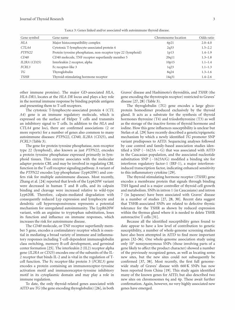
Journal of Thyroid Research 3
Table 3: Genes linked and/or associated with autoimmune thyroid disease.
Gene symbol Gene name Chromosome location Odds ratio
HLA Major histocompatibility complex 6p21 2.0–4.0
CTLA4 Cytotoxic T-lymphocyte-associated protein 4 2q33 1.5–2.2
PTPN22 Protein tyrosine phosphatase, non-receptor type 22 (lymphoid) 1p13 1.4–1.9
CD40 CD40 molecule, TNF receptor superfamily member 5 20q11 1.3–1.8
IL2RA (CD25) Interleukin 2 receptor, alpha 10p15 1.1–1.4
FCRL3 Fc receptor-like 3 1q23 1.1–1.3
TG Thyroglobulin 8q24 1.3–1.6
TSHR Thyroid-stimulating hormone receptor 14q31 1.4–2.6
other immune proteins). The major GD-associated HLA,HLA-DR3, locates at the HLA DR locus and plays a key rolein the normal immune response by binding peptide antigensand presenting them to T-cell receptors.
The cytotoxic T-lymphocyte-associated protein 4 (CTLA4) gene is an immune regulatory molecule, which isexpressed on the surface of Helper T cells and transmitsan inhibitory signal to T cells. In addition to the HLA andCTLA4 gene loci, there are confirmed associations (2 ormore reports) for a number of genes also common to manyautoimmune diseases: PTPN22, CD40, IL2RA (CD25), andFCRL3 (Table 3).
The gene for protein tyrosine phosphatase, non-receptortype 22 (lymphoid), also known as just PTPN22, encodesa protein tyrosine phosphatase expressed primarily in lym-phoid tissues. This enzyme associates with the molecularadapter protein CBL and may be involved in regulating CBLfunction in the T-cell receptor signaling pathway. A variant ofthe PTPN22 encodes Lyp phosphatase (Lyp620W) and con-fers risk for multiple autoimmune diseases. Most recently,Zhang et al. [24] reported that levels of the Lyp620W variantwere decreased in human T and B cells, and its calpainbinding and cleavage were increased relative to wild-typeLyp620R. Therefore, calpain-mediated degradation withconsequently reduced Lyp expression and lymphocyte anddendritic cell hyperresponsiveness represents a potentialmechanism for unregulated autoimmunity. The LypR620Wvariant, with an arginine to tryptophan substitution, losesits function and influence on immune responses, whichincreases the risk for autoimmune disease.
The CD40 molecule, or TNF receptor superfamily mem-ber 5 gene, encodes a costimulatory receptor which is essen-tial in mediating a broad variety of immune and inflamma-tory responses including T-cell-dependent immunoglobulinclass switching, memory B-cell development, and germinalcenter formation [25]. The interleukin 2 (IL2) receptor alphagene (IL2RA or CD25) encodes one of the subunits of the IL-2 receptor that binds IL-2 and is vital in the regulation of T-cell function. The Fc receptor-like protein 3 (FCRL3) geneencodes a protein containing an immunoreceptor-tyrosineactivation motif and immunoreceptor-tyrosine inhibitorymotif in its cytoplasmic domain and may play a role inimmune regulation.
To date, the only thyroid-related genes associated withAITD are TG (the gene encoding thyroglobulin) [26], in both
Graves’ disease and Hashimoto’s thyroiditis, and TSHR (thegene encoding the thyrotropin receptor) restricted to Graves’disease [27, 28] (Table 3).
The thyroglobulin (TG) gene encodes a large glyco-protein homodimer produced exclusively by the thyroidgland. It acts as a substrate for the synthesis of thyroidhormones thyroxine (T4) and triiodothyronine (T3) as wellas the storage of the inactive forms of thyroid hormone andiodine. How this gene influences susceptibility is unclear butStefan et al. [29] have recently described a genetic/epigeneticmechanism by which a newly identified TG promoter SNPvariant predisposes to AITD. Sequencing analyses followedby case control and family-based association studies iden-tified a SNP (−1623A→G) that was associated with AITDin the Caucasian population, and the associated nucleotidesubstitution SNP (−1623A/G) modified a binding site forinterferon regulatory factor-1 (IRF-1), a major interferon-induced transcription factor, indicating enhanced sensitivityto this inflammatory cytokine [29].
The thyroid stimulating hormone receptor (TSHR) geneencodes a membrane protein that signals through bindingTSH ligand and is a major controller of thyroid cell growthand metabolism. SNPs in intron 1 (in Caucasians) and intron7 (in Japanese) have been associated with Graves’ diseasein a number of studies [27, 28, 30]. Recent data suggestthat TSHR-associated SNPs are related to defective thymictolerance for the TSHR as shown by reduced expressionwithin the thymus gland where it is needed to delete TSHRautoreactive T cells [31].
Because all the identified susceptibility genes found todate appear to have a low level of contribution to geneticsusceptibility, a number of whole-genome screening studieshave also been attempted in AITD to find more importantgenes [32–36]. One whole-genome association study usingonly 104 nonsynonymous SNPs (those involving parts of agene likely to affect the product character) showed a numberof the previously recognized genes, as well as locating somenew sites, but the new sites could not subsequently beconfirmed [37, 38]. Most recently, the first full genome-wide study of Graves’ disease with 660 K SNPs has nowbeen reported from China [19]. This study again identifiedmany of the known genes for AITD, but also described twonew sites on chromosomes 6q and 4p. These await furtherconfirmation. Again, however, no very highly associated newgenes have emerged.

4 Journal of Thyroid Research
6. The Degree of Enhanced SusceptibilityRemains Low
All the genes associated with AITD are individually ableto confer only modest degrees of disease susceptibility(expressed as odds ratios, see Table 3). Hence, these dataonly allow us to conclude that the AITDs, both Graves’ dis-ease (including Graves’ ophthalmopathy) and Hashimoto’sthyroiditis, are complex genetic disorders involving multiplegenes that may interact to provide a susceptible backgroundfor disease development. Furthermore, there appear to bedisease-specific genes, such as the gene encoding the TSHRin Graves’ disease and a larger group of susceptibility genes,such as CTLA4, which are common to many autoimmunediseases. This combination of gene polymorphisms likelyallows epigenetic phenomena, subsequent to a variety ofinfluences such as infection and the environment, to initiatedisease.
7. The Controversy over Major Genes in AITD
After the clarification that multiple genes are at work inAITD, it is likely that more than 20 potential genes contributeto the AITD phenotypes. But major genes, those essentialto disease development, have not been found [39]. A majorgene should be involved in the majority of patients withthe disease, and the risk ratios, even for HLA, do notreveal such a gene (Table 3). This most likely means thatdifferent combinations of genes may produce similar clinicalphenotypes or that epigenetic phenomena are dominant.So far, in the whole-genome screening of families, siblings,and populations with AITD, a number of sites have beenestablished for Graves’ disease and Hashimoto’s thyroiditissusceptibility, but none of them have had very high statisticalvalues (LOD scores) [32, 33, 35]. This finding has been truenot just for AITD, but also for other autoimmune diseasesincluding type 1 diabetes mellitus. This is best understood bythinking of HLA once again. Not every patient with Graves’disease has the associated HLA-DR3 subtype and not eventhe associated Arg74 in its binding pocket, irrespective of theHLA-DR subtype [12]. Hence, the disease can occur in theabsence of the expected HLA association.
8. A Note on Epigenetics
One mechanism by which environmental factors may com-bine with genetic risk to promote AITD is by altering theepigenetic control of gene expression as seen, for example,in the pancreas [40] and as shown for a virus interactingwith a susceptibility gene in Crohn’s disease [41]. Whilelittle is known about such interactions with AITD, therehas been wide confirmation of a role for X chromosomeinactivation (XCI) [42, 43]. Patients with AITD more oftenthan expected showed a biased expression of a maternalor paternal X chromosome leading to the hypothesis thatthe poorly expressed chromosome could become active incertain tissues such as the thyroid and express new antigenicsequences not previously recognized by the immune system.
These potential mechanisms for enhanced susceptibility toAITD require further exploration.
9. Conclusions
How environmental factors combine with genetic risk at themolecular level to promote complex genetic diseases suchas AITD is largely unknown. The genes that are linked toand/or associated with AITD are each small contributors togenetic risk. Multiple-gene polymorphisms (combinationsof haplotypes) appear to be needed to develop AITD andmay differ between geographic populations secondary toepigenetic influences. Much remains to be learned.
Abbreviations
AITD: Autoimmune thyroid diseaseLOD: Logarithm of oddsSNP: Single-nucleotide polymorphism.
Disclosure
TFD is a Board Member of Kronus Inc., Star, Idaho (adistributor of thyroid antibody test kits). The other authorshave no conflict of interests to disclose.
Acknowledgments
This work is supported in part by DK052464 and DK069713from the National Institute of Diabetes and Digestive andKidney Diseases, the VA Merit Award program, and theDavid Owen Segal Endowment.
References
[1] T. Davies, “Hypothyroidism and thyroiditis,” in WilliamsTextbook of Endocrinology, J. Wilson, D. W. Foster, P. R.Larsen, and H. Kronenberg, Eds., pp. 423–456, W.B. Saunders,Orlando, Fla, USA, 2002.
[2] M. P. J. Vanderpump, W. M. G. Tunbridge, J. M. French etal., “The incidence of thyroid disorders in the community:a twenty-year follow-up of the Whickham Survey,” ClinicalEndocrinology, vol. 43, no. 1, pp. 55–68, 1995.
[3] R. Hall and J. B. Stanbury, “Familial studies of autoimmunethyroiditis,” Clinical and Experimental Immunology, vol. 2, pp.719–725, 1967.
[4] I. J. Chopra, D. H. Solomon, and U. Chopra, “Abnormalitiesin thyroid function in relatives of patients with Graves’disease and Hashimoto’s thyroiditis: lack of correlation withinheritance of HLA B8,” Journal of Clinical Endocrinology andMetabolism, vol. 45, no. 1, pp. 45–54, 1977.
[5] C. L. Burek, W. H. Hoffman, and N. R. Rose, “The presenceof thyroid autoantibodies in children and adolescents withautoimmune thyroid disease and in their siblings and parents,”Clinical Immunology and Immunopathology, vol. 25, no. 3, pp.395–404, 1982.
[6] W. M. G. Tunbridge, D. C. Evered, and R. Hall, “The spectrumof thyroid disease in a community: the Whickham survey,”Clinical Endocrinology, vol. 7, no. 6, pp. 481–493, 1977.

Journal of Thyroid Research 5
[7] D. L. Pauls, M. Zakarija, J. M. McKenzie, and J. A. Egeland,“Complex segregation analysis of antibodies to thyroid per-oxidase in Old Order Amish families,” American Journal ofMedical Genetics, vol. 47, no. 3, pp. 375–379, 1993.
[8] D. Phillips, L. Prentice, M. Upadhyaya et al., “Autosomal dom-inant inheritance of autoantibodies to thyroid peroxidase andthyroglobulin—studies in families not selected for autoim-mune thyroid disease,” Journal of Clinical Endocrinology andMetabolism, vol. 72, no. 5, pp. 973–975, 1991.
[9] J. C. Jaume, J. Guo, D. L. Pauls et al., “Evidence for genetictransmission of thyroid peroxidase autoantibody epitopic ’fin-gerprints’,” Journal of Clinical Endocrinology and Metabolism,vol. 84, no. 4, pp. 1424–1431, 1999.
[10] N. R. Farid and J. C. Bear, “The human major histocompati-bility complex and endocrine disease,” Endocrine Reviews, vol.2, no. 1, pp. 50–86, 1981.
[11] T. H. Brix, K. Christensen, N. V. Holm, B. Harvald, andL. Hegedus, “A population-based study of graves’ disease indanish twins,” Clinical Endocrinology, vol. 48, no. 4, pp. 397–400, 1998.
[12] Y. Ban, T. F. Davies, D. A. Greenberg et al., “Arginine atposition 74 of the HLA-DR β1 chain is associated with Graves’disease,” Genes and Immunity, vol. 5, no. 3, pp. 203–208, 2004.
[13] J. Ott, “Complex traits on the map,” Nature, vol. 379, no. 6568,pp. 772–773, 1996.
[14] S. H. Roman, D. Greenberg, P. Rubinstein, S. Wallenstein, andT. F. Davies, “Genetics of autoimmune thyroid disease: lackof evidence for linkage to HLA within families,” Journal ofClinical Endocrinology and Metabolism, vol. 74, no. 3, pp. 496–503, 1992.
[15] J. L. Weber, “Human DNA polymorphisms and methods ofanalysis,” Current Opinion in Biotechnology, vol. 1, no. 2, pp.166–171, 1990.
[16] K. A. Frazer, D. G. Ballinger, D. R. Cox et al., “A secondgeneration human haplotype map of over 3.1 million SNPs,”Nature, vol. 449, no. 7164, pp. 851–861, 2007.
[17] K. Christensen and J. C. Murray, “Focus on research: whatgenome-wide association studies can do for medicine,” NewEngland Journal of Medicine, vol. 356, no. 11, pp. 1094–1097,2007.
[18] T. A. Manolio, L. D. Brooks, and F. S. Collins, “A HapMapharvest of insights into the genetics of common disease,”Journal of Clinical Investigation, vol. 118, no. 5, pp. 1590–1605,2008.
[19] X. Chu, C.-M. Pan, S.-X. Zhao et al., “A genome-wideassociation study identifies two new risk loci for Graves’disease,” Nature Genetics, vol. 43, no. 9, pp. 897–901, 2011.
[20] D. J. Hunter and P. Kraft, “Drinking from the fire hose—statistical issues in genomewide association studies,” NewEngland Journal of Medicine, vol. 357, no. 5, pp. 436–439, 2007.
[21] D. Shriner, L. K. Vaughan, M. A. Padilla, and H. K. Tiwari,“Problems with genome-wide association studies,” Science,vol. 316, no. 5833, pp. 1840–1841, 2007.
[22] T. Yanagawa, Y. Hidaka, V. Guimaraes, M. Soliman, and L.J. DeGroot, “CTLA-4 gene polymorphism associated withGraves’ disease in a Caucasian population,” Journal of ClinicalEndocrinology and Metabolism, vol. 80, no. 1, pp. 41–45, 1995.
[23] N. R. Farid, “Immunogenetics of autoimmune thyroid disor-ders,” Endocrinology and Metabolism Clinics of North America,vol. 16, no. 2, pp. 229–245, 1987.
[24] J. Zhang, N. Zahir, Q. Jiang et al., “The autoimmune disease-associated PTPN22 variant promotes calpain-mediated Lyp/Pep degradation associated with lymphocyte and dendritic cell
hyperresponsiveness,” Nature Genetics, vol. 43, no. 9, pp. 902–907, 2011.
[25] Y. Tomer, “Genetic susceptibility to autoimmune thyroid dis-ease: past, present, and future,” Thyroid, vol. 20, no. 7, pp. 715–725, 2010.
[26] Y. Tomer, D. A. Greenberg, E. Concepcion, Y. Ban, and T.F. Davies, “Thyroglobulin is a thyroid specific gene for thefamilial autoimmune thyroid diseases,” Journal of ClinicalEndocrinology and Metabolism, vol. 87, no. 1, pp. 404–407,2002.
[27] X. Yin, R. Latif, R. Bahn, Y. Tomer, and T. F. Davies, “Influenceof the TSH receptor gene on susceptibility to Graves’ diseaseand Graves’ ophthalmopathy,” Thyroid, vol. 18, no. 11, pp.1201–1206, 2008.
[28] B. M. Dechairo, D. Zabaneh, J. Collins et al., “Association ofthe TSHR gene with Graves’ disease: the first disease specificlocus,” European Journal of Human Genetics, vol. 13, no. 11,pp. 1223–1230, 2005.
[29] M. Stefan, E. M. Jacobson, A. K. Huber et al., “Novel variantof thyroglobulin promoter triggers thyroid autoimmunitythrough an epigenetic interferon α-modulated mechanism,”Journal of Biological Chemistry, vol. 286, no. 36, pp. 31168–31179, 2011.
[30] H. Hiratani, D. W. Bowden, S. Ikegami et al., “Multiple SNPsin intron 7 of thyrotropin receptor are associated with Graves’disease,” Journal of Clinical Endocrinology and Metabolism, vol.90, no. 5, pp. 2898–2903, 2005.
[31] R. Colobran, M. d. P. Armengol, R. Faner et al., “Associationof an SNP with intrathymic transcription of TSHR andgraves’ disease: a role for defective thymic tolerance,” HumanMolecular Genetics, vol. 20, no. 17, pp. 3415–3423, 2011.
[32] Y. Tomer, G. Barbesino, D. A. Greenberg, E. Concepcion,and T. F. Davies, “Mapping the major susceptibility loci forfamilial Graves’ and Hashimoto’s diseases: evidence for geneticheterogeneity and gene interactions,” Journal of ClinicalEndocrinology and Metabolism, vol. 84, no. 12, pp. 4656–4664,1999.
[33] Y. Tomer, Y. Ban, E. Conception et al., “Common and uniquesusceptibility loci in Graves and Hashimoto diseases: resultsof whole-genome screening in a data set of 102 multiplexfamilies,” American Journal of Human Genetics, vol. 73, no. 4,pp. 736–747, 2003.
[34] Y. Tomer, F. Menconi, T. F. Davies et al., “Dissecting geneticheterogeneity in autoimmune thyroid diseases by subsetanalysis,” Journal of Autoimmunity, vol. 29, no. 2-3, pp. 69–77,2007.
[35] J. C. Taylor, S. C. Gough, P. J. Hunt et al., “A genome-wide screen in 1119 relative pairs with autoimmune thyroiddisease,” Journal of Clinical Endocrinology and Metabolism, vol.91, no. 2, pp. 646–653, 2006.
[36] K. Sakai, S. Shirasawa, N. Ishikawa et al., “Identification ofsusceptibility loci for autoimmune thyroid disease to 5q31-q33 and Hashimoto’s thyroiditis to 8q23-q24 by multipointaffected sib-pair linkage analysis in Japanese,” Human Molecu-lar Genetics, vol. 10, no. 13, pp. 1379–1386, 2001.
[37] P. R. Burton, D. G. Clayton, L. R. Cardon et al., “Associationscan of 14,500 nonsynonymous SNPs in four diseases identi-fies autoimmunity variants,” Nature Genetics, vol. 39, no. 11,pp. 1329–1337, 2007.
[38] P. R. Newby, O. J. Pickles, S. Mazumdar et al., “Follow-up ofpotential novel graves’ disease susceptibility loci, identified inthe UK WTCCC genome-wide nonsynonymous SNP study,”

6 Journal of Thyroid Research
European Journal of Human Genetics, vol. 18, no. 9, pp. 1021–1026, 2010.
[39] T. F. Davies, “Really significant genes for autoimmune thyroiddisease do not exist—so how can we predict disease?” Thyroid,vol. 17, no. 11, pp. 1027–1029, 2007.
[40] I. Sandovici, N. H. Smith, M. D. Nitert et al., “Maternal dietand aging alter the epigenetic control of a promoter-enhan-cer interaction at the Hnf4a gene in rat pancreatic islets,”Proceedings of the National Academy of Sciences of the UnitedStates of America, vol. 108, no. 13, pp. 5449–5454, 2011.
[41] K. Cadwell, K. K. Patel, N. S. Maloney et al., “Virus-plus-susceptibility gene interaction determines Crohn’s diseasegene Atg16L1 phenotypes in intestine,” Cell, vol. 141, no. 7,pp. 1135–1145, 2010.
[42] T. H. Brix, G. P. S. Knudsen, M. Kristiansen, K. O. Kyvik,K. H. Ørstavik, and L. Hegedus, “High frequency of skewedx-chromosome inactivation in females with autoimmunethyroid disease: a possible explanation for the female predis-position to thyroid autoimmunity,” Journal of Clinical Endo-crinology and Metabolism, vol. 90, no. 11, pp. 5949–5953, 2005.
[43] X. Yin, R. Latif, Y. Tomer, and T. F. Davies, “Thyroidepigenetics: X chromosome inactivation in patients with au-toimmune thyroid disease,” Annals of the New York Academy ofSciences, vol. 1110, pp. 193–200, 2007.

Hindawi Publishing CorporationJournal of Thyroid ResearchVolume 2012, Article ID 368536, 4 pagesdoi:10.1155/2012/368536
Research Article
Determinants of Extraocular Muscle Volume inPatients with Graves’ Disease
Samer El-Kaissi1 and Jack R. Wall2
1 Specialized Diabetes and Endocrine Centre, King Fahad Medical City, Dabab Street, P.O. Box 59046, Riyadh 11525, Saudi Arabia2 Department of Medicine, Nepean Clinical School, Nepean Hospital, The University of Sydney, Derby Street, P.O. Box 63, Penrith 2751,New South Wales, Australia
Correspondence should be addressed to Samer El-Kaissi, [email protected]
Received 26 September 2011; Accepted 30 November 2011
Academic Editor: Leonidas H. Duntas
Copyright © 2012 S. El-Kaissi and J. R. Wall. This is an open access article distributed under the Creative Commons AttributionLicense, which permits unrestricted use, distribution, and reproduction in any medium, provided the original work is properlycited.
Background. To examine factors contributing to extraocular muscle (EOM) volume enlargement in patients with Graves’ hyper-thyroidism. Methods. EOM volumes were measured with orbital magnetic resonance imaging (MRI) in 39 patients with recentlydiagnosed Graves’ disease, and compared to EOM volumes of 13 normal volunteers. Thyroid function tests, uptake on thyroid scin-tigraphy, anti-TSH-receptor antibody positivity and other parameters were then evaluated in patients with EOM enlargement.Results. 31/39 patients had one or more enlarged EOM, of whom only 2 patients had clinical EOM dysfunction. Compared toGraves’ disease patients with normal EOM volumes, those with EOM enlargement had significantly higher mean serum TSH (0.020± 0.005 versus 0.007 ± 0.002 mIU/L; P value 0.012), free-T4 (52.9± 3.3 versus 41.2± 1.7 pmol/L; P value 0.003) and technetiumuptake on thyroid scintigraphy (13.51± 1.7% versus 8.55± 1.6%; P value 0.045). There were no differences between the 2 groupsin anti-TSH-receptor antibody positivity, the proportion of males, tobacco smokers, or those with active ophthalmopathy. Con-clusions. Patients with recently diagnosed Graves’ disease and EOM volume enlargement have higher serum TSH and more severehyperthyroidism than patients with normal EOM volumes, with no difference in anti-TSH-receptor antibody positivity betweenthe two groups.
1. Introduction
Thyroid-associated ophthalmopathy (TAO) is an autoim-mune disorder of uncertain aetiology. While the involvementof extraocular muscles (EOMs) in patients with Graves’ dis-ease may seem infrequent on clinical examination, orbitalmagnetic resonance imaging (MRI) studies suggest that themajority of such patients have EOM enlargement [1].
Along with the orbital fibroblast, the EOM is likely to bea primary target in TAO. This is supported by evidence of T-cell reactivity against both orbital fibroblast and EOM cellsin vitro [2] and muscle fibre damage in electron microscopicstudies of EOM from patients with recent onset TAO [3, 4].In addition, expression of the thyrotropin-receptor (TSH-R)in EOM [5, 6], as opposed to the widespread distribution ofTSH-R in adipose tissues throughout the body [7], may indi-cate that EOMs have a more direct and specific role in TAOthan previously thought [8].
In this study, we investigated potential factors affectingEOM volume enlargement as measured by orbital MRI in pa-tients with recently diagnosed Graves’ disease.
2. Materials and Methods
A total of 39 patients diagnosed with Graves’ hyperthyroid-ism within the preceding 3 months were selected for thisstudy. The patients were involved in a larger study lookingat potential risk factors for TAO [9].
The diagnosis of Graves’ disease was based on the pres-ence of biochemical hyperthyroidism, a symmetrical goitreand positive thyroid autoantibodies, and/or diffuse uptakeon 99mTechnetium thyroid nuclear scan. The abbreviatedclinical activity score (CAS) model was employed for the dia-gnosis of active ophthalmopathy. This model assigns onepoint for each of the following: spontaneous retrobulbar

2 Journal of Thyroid Research
pain, pain on eye movement, eyelid erythema, eyelid oe-dema, chemosis, conjunctival injection, and swelling of thecaruncle [10]. A total score ≥4 out of 7 was defined as activeophthalmopathy [11]. EOM function was evaluated by ask-ing the patient to move their eyes in an H-shaped pattern,and proptosis was assessed using a Hertel exophthalmometer.The ophthalmic examination was performed by a trainedclinical nurse.
Patients who were pregnant, less than 18 years of age,and those with a history of radioactive iodine therapy, orbitalsurgery, orbital irradiation, or significant loss of vision wereexcluded. The study was conducted at an outpatient endo-crine practice in Victoria, Australia. Written, informed con-sent was obtained, and the study was approved by the Bar-won Health Research and Ethics Advisory Committee.
EOM volumes were measured by a single investigator(SEK) from T1-weighted, 2 mm slice orbital MRI scans usingthe digital software MRIcro (Version 1.38 Beta; Chris Ror-den) as previously described [9]. Briefly, the volumes of themedial, inferior, and lateral recti were measured manuallyby circling the muscle perimeter on each slice. The superiorrectus muscle, the superior ophthalmic vein, and the levatorpalpebrae superioris were measured together as the superiormuscle group (SMG) because of difficulties in delineatingthese structures from each other. Orbital measurements wereexpressed as a percentage of the mean globe volume for eachpatient in order to adjust for interindividual variation inEOM volumes.
3. Statistical Analysis
Statistical analysis was performed with the software pro-grams Minitab 14.12 and SPSS 13.0. Proportions were com-pared with Fisher’s exact test while the sample means wereevaluated with the 2-sample t-test. The effect of TSH-R anti-body positivity on EOM volume was examined with binarylogistic regression. Significance was set at P value less than0.05.
The cutoff values of MRI-measured EOM volumes weredetermined with receiver-operating-characteristic analysisby comparing patient EOM volumes to those of 13 normalvolunteers with no history of thyroid or eye disease. The cut-off values, their sensitivities and specificities, and the coeffi-cients of variation of each measurement were detailed in anearlier publication [9].
4. Results
Based on the EOM volume cutoff value, only 8 patients hadnormal volumes in all 4 EOM groups. Of the 31/39 patientswith at least one enlarged EOM volume, 3 patients had oneenlarged EOM, 3 patients had 2 enlarged EOM, 7 patientshad 3 enlarged EOM, and all 4 muscles were enlarged in 18patients. The most frequently affected EOMs were the medialand lateral (n = 27 each) followed by the inferior recti andSMG (n = 24 each).
Assessment of baseline characteristics in patients withand without EOM volume enlargement on MRI showed nosignificant differences in the proportion of males, tobacco
smokers, those with active ophthalmopathy (CAS ≥ 4), orelevations in anti-TSH-R, antithyroid peroxidase (TPO), orantithyroglobulin autoantibodies (Table 1). Only two pa-tients, both with EOM volume enlargement, had clinicallyevident EOM dysfunction.
However, patients with enlarged EOM volumes had sig-nificantly higher 99mtechnetium uptake on thyroid scintigra-phy and greater serum free-T4 and thyrotropin (TSH) levels(Table 2). Importantly, there were no significant differencesbetween the two groups in the proportion of patients whoreceived anti-thyroid medications prior to recruitment intothe study. Overall, 8/31 patients with enlarged and 2/8 pa-tients with normal EOM volumes received anti-thyroid med-ications for a mean 1.06± 0.45 weeks and 1.13± 0.79 weeks,respectively. Exclusion of those patients from the analysisresulted in persistent elevations of free-T4 and TSH levels inpatients with EOM enlargement, although the differences inmean TSH became of borderline significance (mean free-T442.4±1.9 versus 52.3±3.9 pmol/L (P value 0.034); mean TSH0.021± 0.006 versus 0.008± 0.003 mIU/L (P value 0.054)).
In further analysis using binary logistic regression, EOMvolumes were not associated with elevated anti-TSH-R, anti-TPO, or antithyroglobulin autoantibodies, smoking, or thepresence of active ophthalmopathy.
5. Discussion
This study shows that patients with newly diagnosed Graves’disease and EOM enlargement have higher serum TSH andmore severe hyperthyroidism, as suggested by the higher ser-um free-T4 and greater uptake on thyroid scintigraphy, thanpatients without EOM enlargement.
While more severe hyperthyroidism has not been identi-fied as an independent risk factor for TAO [12], a greater ser-um free-T3 at baseline is associated with an increased risk ofTAO after radioiodine therapy for Graves’ hyperthyroidism[13]. In this study, EOM volume enlargement was associatedwith higher free-T4 levels (P value 0.003; Table 2) and greateruptake on thyroid scintigraphy (P value 0.045; Table 2). Theserum free-T3 level was greater in patients with EOM volumeenlargement without reaching statistical significance (P value0.062; Table 2), perhaps due to the small sample size. Themechanism whereby more severe hyperthyroidism leads togreater EOM volumes is uncertain, but we speculate that itmay be related to higher levels of the shared thyroid-orbitalantigen(s).
In this study, the mean serum TSH was significantly high-er in patients with EOM volume enlargement. The role ofserum TSH in the initiation and propagation of TAO is welldocumented after RAI therapy [13–16], and empirical thy-roid hormone replacement after RAI ablation, but before theonset of biochemical hypothyroidism, has been shown toreduce the incidence of TAO after RAI [17]. It is possible thatEOMs, which express TSH-R [5, 6], are sensitive to seeminglyminor elevations in serum TSH in patients with Graves’ hy-perthyroidism, leading to greater EOM volumes. The higherTSH levels in patients with enlarged EOM volumes occurreddespite higher free-T4 and free-T3 levels in this group. Whilethe serum TSH usually changes in a reciprocal fashion to

Journal of Thyroid Research 3
Table 1: Comparison between patients with and without EOM volume enlargement. The number of patients is shown, and the proportionsP value was calculated using Fisher’s exact test.
Enlarged EOM Normal EOMP value
volume (n = 31) volume (n = 8)
Males 3 1 1.0
Smokers 9 3 0.7
Active ophthalmopathy 15 3 0.7
Clinical EOM dysfunction 2 0 1.0
Elevated TSH-R antibodies 23 7 0.6
Elevated thyroglobulin antibodies 14 5 0.4
Elevated TPO antibodies 19 7 0.2
Table 2: Means ± SEM of measurements for patients with and without EOM volume enlargement. Where the measurement was not per-formed on all patients, the number of patients is shown in square brackets. P values calculated using 2-sample t-test.
Enlarged EOM Normal EOMP value
volume (n = 31) volume (n = 8)
Age (yrs) 44± 2.1 40.5± 4.1 0.599mTechnetium uptake (%) 13.51± 1.7, (n = 28) 8.55± 1.6, (n = 6) 0.045
TSH (mIU/L) 0.020± 0.005 0.007± 0.002 0.012Free-T4 (pmol/L) 52.9± 3.3 41.2± 1.7 0.003
Free-T3 (pmol/L) 22.8± 1.9, (n = 29) 18.3± 1.3 0.062
CAS 2.97± 0.53 3.25± 1.2 0.8
Proptosis (mm) 17.46± 0.46 16.00± 0.71 0.097
the serum free-T4 and free-T3 levels, it is worth noting thatthis relationship is attenuated or “flattened” in hyperthyroidpatients with a suppressed serum TSH below 0.01 mIU/L[18]. Upon starting treatment with anti-thyroid medications,the serum free-T4 and free-T3 fall rapidly whereas the serumTSH typically “lags” behind and remains undetectable for upto 3 months [18]. It is therefore unlikely that the greater ser-um TSH in patients with EOM volume enlargement was rela-ted to treatment with anti-thyroid medications prior to re-cruitment into the study, especially because the mean dura-tion of treatment with anti-thyroid medications was 1.08 ±0.39 weeks, and did not exceed 3 months in any patient. Inaddition, exclusion of patients who received anti-thyroidmedications from the analysis did not abolish the differencesin serum TSH between the two groups, although the dif-ferences became of borderline significance (P value 0.054),possibly due to the smaller sample size.
While the majority of patients in this study had elevatedanti-TSH-R antibody levels, there were no significant differ-ences in the prevalence of anti-TSH-R antibody positivitybetween patients with and without EOM volume enlarge-ment, and in binary logistic regression analysis there was noassociation between antibody positivity and EOM volumes.TAO is thought to occur following sensitization of T-lym-phocytes to a common thyroid and orbital antigen. The iden-tity and location of this antigen remains unknown, but theTSH-R is the most likely candidate [7, 19]. Autoimmunityagainst other antigens particularly the skeletal muscle proteincalsequestrin [20] is of potential importance, but is not wellunderstood. The role of TSH-R in the initiation and propaga-tion of TAO is supported by the close temporal relationship
between the onset of ophthalmopathy and Graves’ diseasewhich is caused by stimulating anti-TSH-R antibodies [12],and the positive correlation between these antibodies and theprevalence of TAO in untreated Graves’ disease [21]. In addi-tion, TSH-R antibody levels are closely associated with CASreadings, the severity of the eye disease [22], and to a lesserextent with proptosis [23]. Therefore, the lack of an associ-ation between TSH-R antibody positivity and EOM volumeenlargement in this study should be interpreted with caution,especially because of the small sample size and the increasedrisk of a type 2 error.
Similarly, the small sample size may account for non-significant differences in measures of proptosis, which wasgreater in patients with enlarged EOM volumes withoutreaching statistical significance (P value 0.097; Table 2). Incontrast, the lack of an association between active ophthal-mopathy and EOM enlargement may be related to the useof the CAS model which measures soft tissue and periorbitalinflammation rather than EOM involvement [10, 24].
6. Conclusions
In patients with newly diagnosed Graves’ disease, EOM vol-ume enlargement is associated with greater serum TSH levelsand more severe hyperthyroidism, as suggested by greaterserum free-T4 levels and more avid uptake on thyroid scin-tigraphy. There was no association between EOM volumesand anti-TSH-R antibody positivity, although the small sam-ple size may have contributed to this negative finding. Largerstudies are needed to examine the relationship between ser-um TSH, anti-TSH-R antibodies, and EOM enlargement.

4 Journal of Thyroid Research
Conflict of Interests
The authors declare that there is no conflict of interests.
Acknowledgments
This study was undertaken at the University of Melbourne,Department of Clinical and Biomedical Sciences at BarwonHealth in Victoria, Australia. It was funded by the NationalHealth and Medical Research Council of Australia. The au-thors are thankful to J. Bowden for performing the clinicalophthalmological assessments.
References
[1] M. C. Villadolid, N. Yokoyama, M. Izumi et al., “UntreatedGraves’ disease patients without clinical ophthalmopathydemonstrate a high frequency of extraocular muscle (EOM)enlargement by magnetic resonance,” The Journal of ClinicalEndocrinology and Metabolism, vol. 80, no. 9, pp. 2830–2833,1995.
[2] E. A. Otto, K. Ochs, C. Hansen, J. R. Wall, and G. J. Kahaly,“Orbital tissue-derived T lymphocytes from patients withGraves’ ophthalmopathy recognize autologous orbital anti-gens,” The Journal of Clinical Endocrinology and Metabolism,vol. 81, no. 8, pp. 3045–3050, 1996.
[3] J. R. Wall, N. Bernard, A. Boucher et al., “Pathogenesis of thy-roid-associated ophthalmopathy: an autoimmune disorder ofthe eye muscle associated with Graves’ hyperthyroidism andHashimoto’s thyroiditis,” Clinical Immunology and Immuno-pathology, vol. 68, no. 1, pp. 1–8, 1993.
[4] J. R. Wall, I. Stachura, and J. H. Kennerdell, “Mitochondrialabnormalities in eye muscle fiber from three cases of thyroid-associated ophthalmopathy,” Thyroid, vol. 16, no. 11, pp.1181–1183, 2006.
[5] B. E. Busuttil and A. G. Frauman, “Extrathyroidal manifesta-tions of Graves’ disease: the thyrotropin receptor is expressedin extraocular, but not cardiac, muscle tissues,” The Journalof Clinical Endocrinology and Metabolism, vol. 86, no. 5, pp.2315–2319, 2001.
[6] Y. Hiromatsu, M. Sato, Y. Inoue et al., “Localization and Clin-ical significance of thyrotropin receptor mRNA expression inorbital fat and eye muscle tissues from patients with thyroid-associated ophthalmopathy,” Thyroid, vol. 6, no. 6, pp. 553–562, 1996.
[7] R. S. Bahn, “Clinical review 157—pathophysiology of Graves’ophthalmopathy: the cycle of disease,” The Journal of ClinicalEndocrinology and Metabolism, vol. 88, no. 5, pp. 1939–1946,2003.
[8] S. J. Kloprogge, B. E. Busuttil, and A. G. Frauman, “TSH recep-tor protein is selectively expressed in normal human extraoc-ular muscle,” Muscle and Nerve, vol. 32, no. 1, pp. 95–98, 2005.
[9] S. El-Kaissi, J. Bowden, M. J. Henry et al., “Association betweenradioiodine therapy for Graves’ hyperthyroidism and thyroid-associated ophthalmopathy,” International Ophthalmology,vol. 30, no. 4, pp. 397–405, 2010.
[10] A. Pinchera, W. Wiersinga, D. Glinoer et al., “Classification ofeye changes of Graves’ disease,” Thyroid, vol. 2, pp. 235–236,1992.
[11] M. F. Prummel, A. Bakker, W. M. Wiersinga et al., “Multi-center study on the characteristics and treatment strategies ofpatients with Graves’ orbitopathy: the first European Group
on Graves’ Orbitopathy experience,” European Journal of En-docrinology, vol. 148, no. 5, pp. 491–495, 2003.
[12] H. B. Burch and L. Wartofsky, “Graves’ ophthalmopathy: cur-rent concepts regarding pathogenesis and management,” En-docrine Reviews, vol. 14, no. 6, pp. 747–793, 1993.
[13] L. Tallstedt, G. Lundell, O. Torring et al., “Occurrence of oph-thalmopathy after treatment for Graves’ hyperthyroidism,”The New England Journal of Medicine, vol. 326, no. 26, pp.1733–1738, 1992.
[14] R. D. Hamilton, W. E. Mayberry, W. M. McConahey, and K. C.Hanson, “Ophthalmopathy of Graves’ disease: a comparisonbetween patients treated surgically and patients treated withradioiodide,” Mayo Clinic Proceedings, vol. 42, no. 12, pp. 812–818, 1967.
[15] F. A. Karlsson, P. A. Dahlberg, R. Jansson, K. Westermark, andP. Enoksson, “Importance of TSH receptor activation in thedevelopment of severe endocrine ophthalmopathy,” Acta En-docrinologica, Supplement, vol. 121, no. 2, pp. 132–141, 1989.
[16] A. W. C. Kung, C. C. Yau, and A. Cheng, “The incidence ofophthalmopathy after radioiodine therapy for Graves’ disease:prognostic factors and the role of methimazole,” The Journalof Clinical Endocrinology and Metabolism, vol. 79, no. 2, pp.542–546, 1994.
[17] L. Tallstedt, G. Lundell, H. Blomgren, and J. Bring, “Does earlyadministration of thyroxine reduce the development ofGraves’ ophthalmopathy after radioiodine treatment?” Euro-pean Journal of Endocrinology, vol. 130, no. 5, pp. 494–497,1994.
[18] L. M. Demers and C. A. Spencer, “Laboratory medicine prac-tice guidelines: laboratory support for the diagnosis and mon-itoring of thyroid disease,” Clinical Endocrinology, vol. 58, no.2, pp. 138–140, 2003.
[19] S. El-Kaissi, A. G. Frauman, and J. R. Wall, “Thyroid-associat-ed ophthalmopathy: a practical guide to classification, naturalhistory and management,” Internal Medicine Journal, vol. 34,no. 8, pp. 482–491, 2004.
[20] H. Lahooti, K. R. Parmar, and J. R. Wall, “Pathogenesis of thy-roid-associated Ophthalmopathy: does autoimmunity againstcalsequestrin and collagen XIII play a role?” Clinical Ophthal-mology, vol. 4, no. 1, pp. 417–425, 2010.
[21] D. H. C. Khoo, S. C. Ho, L. L. Seah et al., “The combination ofabsent thyroid peroxidase antibodies and high thyroid-stim-ulating immunoglobulin levels in Graves disease identifies agroup at markedly increased risk of ophthalmopathy,” Thy-roid, vol. 9, no. 12, pp. 1175–1180, 1999.
[22] A. K. Eckstein, M. Plicht, H. Lax et al., “Thyrotropin receptorautoantibodies are independent risk factors for Graves’ oph-thalmopathy and help to predict severity and outcome of thedisease,” The Journal of Clinical Endocrinology and Metabolism,vol. 91, no. 9, pp. 3464–3470, 2006.
[23] M. N. Gerding, J. W. C. Van Der Meer, M. Broenink, O.Bakker, W. M. Wiersinga, and M. F. Prummel, “Association ofthyrotrophin receptor antibodies with the clinical features ofGraves’ ophthalmopathy,” Clinical Endocrinology, vol. 52, no.3, pp. 267–271, 2000.
[24] W. M. Wiersinga, P. Perros, G. J. Kahaly et al., “Clinical as-sessment of patients with Graves’ orbitopathy: the EuropeanGroup on Graves’ orbitopathy recommendations to general-ists, specialists and clinical researchers,” European Journal ofEndocrinology, vol. 155, no. 3, pp. 387–389, 2006.

Hindawi Publishing CorporationJournal of Thyroid ResearchVolume 2012, Article ID 736161, 6 pagesdoi:10.1155/2012/736161
Review Article
The Evolving Role of Selenium in the Treatment ofGraves’ Disease and Ophthalmopathy
Leonidas H. Duntas
Endocrine Unit, Evgenidion Hospital, University of Athens, 20 Papadiamantopoulou Street, 11528 Athens, Greece
Correspondence should be addressed to Leonidas H. Duntas, [email protected]
Received 5 September 2011; Accepted 17 October 2011
Academic Editor: Juan Carlos Galofre
Copyright © 2012 Leonidas H. Duntas. This is an open access article distributed under the Creative Commons Attribution License,which permits unrestricted use, distribution, and reproduction in any medium, provided the original work is properly cited.
Graves’ disease (GD) and ophthalmopathy (GO) are organ-specific autoimmune-inflammatory disorders characterized by acomplex pathogenesis. The inflammatory process is dominated by an imbalance of the antioxidant-oxidant mechanism, increasedproduction of radical oxygen species (ROS), and cytokines which sustain the autoimmune process and perpetuate the disease.Recently, selenium, which is a powerful antioxidant, has been successfully applied in patients with mild GO, slowing theprogression of disease, decreasing the clinical activity score, and appreciably improving the quality of life. The mechanismsof selenium action are variable. The aim of this review is to summarize the actions of selenium in GD and GO. Selenium asselenocysteine is incorporated in selenoproteins, such as glutathione peroxidase which catalyzes the degradation of hydrogenperoxide and lipid hydroperoxide that are increasingly produced in hyperthyroidism. Moreover, selenium decreases the formationof proinflammatory cytokines, while it contributes, in synergy with antithyroid drugs, to stabilization of the autoimmune processin GD and alleviation of GO. It is now to be clarified whether enforced nutritional supplementation has the same results andwhether prolonging selenium administration may have an impact on the prevention of disease.
1. Introduction
Observed and briefly described, though not published, byParry in the late 1700s, Graves’ disease (GD) was definitivelyidentified and documented by Robert Graves in 1835 andclassically described by von Basedow in 1840 [1–3]. GD isan autoimmune disease characterized by the activation ofautoantibodies against the TSH receptor (TRAB), leading toexcessive thyroid hormone production [4]. GD manifests,interalia, via thyrotoxicosis and extrathyroid involvementoften entailing orbitopathy (GO) and, rarely, dermopathy(pretibial myxedema) and acropathy. Moreover, the TRAB,by stimulating cyclic adenosine monophosphate (AMP),cause proliferation and hyperplasia of the thyroid follicularcells resulting in enlargement of the gland, frequently thefirst sign of the disease, the swelling ranging from slightto marked [5]. Clinically, the thyroid is firm in consistencyand tender in patients with a greatly enlarged goiter, whilepalpation lobulations are also commonly detected which canbe mistaken for nodules.
No single gene has been pinpointed as causing GD, adisease which is most prevalent in women between the ages
of 20 and 50 years. However, it has been associated withcertain MHC Class II HLA alleles depending on the racialgroup, for example, HLA-DR3 in whites [4]. An associationof GD with polymorphisms of the cytotoxic T-lymphocyteantigen 4 (CTLA-4) gene has also been established, suggest-ing a functional role of CTLA-4 in autoreactive T cells [4, 5].
A combination of genetic and environmental factors isresponsible for the initiation of autoimmunity. Interactionsbetween genetic and environmental factors are underscoredby the existing associations linking age at diagnosis, goiter,disease severity, smoking, and family history [6]. In addi-tion, iodine repletion in iodine-deficient areas is usuallyaccompanied by an increased incidence of GD due to theJod-Basedow phenomenon. Stress is also thought to be asignificant factor precipitating GD in susceptible individuals[7], while smoking is well established as being linked to GObut not to GD [8].
Treatment modalities of GD consist of administration ofantithyroid drugs, radioiodine therapy, or surgery. Radioio-dine therapy, is favored only in USA, whereas antithyroiddrugs, including methimazole, carbimazole, and propylth-iouracil, comprise first choice treatment in the rest of the

2 Journal of Thyroid Research
world. Nevertheless, according to a recent study examiningthe frequency of antithyroid drug prescription in USA,methimazole (MMI) has lately become the most frequentlyprescribed antithyroid drug, indicating a clear shift towardspharmacological treatment as the primary treatment optionin GD [9]. Treatment should be planned for a period of atleast 12 months, and patients are usually becoming euthyroidwithin this timeframe; nevertheless, the duration of theremission period is unpredictable, since the disease is markedby cycles of remission and relapse of variable duration [4].
Recently, evidence has emerged indicating that seleniumadministration could be effective and safe in patients withGD and with mild forms of GO [10].
The aim of this paper is to briefly evaluate the currentknowledge concerning the pathogenesis of GD and GO anddiscuss the evolving role of selenium within the context ofits potential as a therapeutic means of intervention in thesedisorders.
2. Pathogenesis of GD and GO
Hyperthyroidism is caused by the binding of TSH-stimu-lating antibodies to the TSH receptor, a G-protein-coupledreceptor. However, the first step in this process is consideredto be precipitation by environmental factors of an HLA-related organ-specific defect in suppressor T-lymphocytefunction [5]. This leads to decreased suppression of thyroid-directed helper T-lymphocytes which, in the presence ofdendritic cells and macrophages, produce the cytokines γ-interferon (IFNγ) and interleukin-1 (IL-1), subsequentlydifferentiating B cells to plasma cells and generating TRAB.Concomitantly, IFNγ enhances the expression of HLA-DRantigens on the surface of thyroid cells (Figure 1). Thus, IFNγmodulates the autoimmune process and, by stimulatingchemokine production by thyroid follicular cells, contributesto the maintenance of the autoimmune process [10]. Thecontribution of dendritic cells and B cells is apparentlycrucial for the initiation of disease since they express thecostimulatory molecules, CD80 and CD86, that are keytriggers for the reaction of T lymphocytes to thyroid cellpresenting antigens [4]. TRAB stimulate the TSHR on thethyroid follicular cells, resulting in increased thyroid hor-mone production, which may further reduce the number andfunction of suppressor T lymphocytes and stimulate helper Tlymphocyte, thus, perpetuating the cyclicity of disease [4, 5].
GO is a complex autoimmune disease. Whereas thecycle of GD consists of two components, immunologicaland hormonal, that perpetuate the process, the progressionof GD to GO, and rarely to dermopathy, is likely to bea positive feedback cycle composed of three interrelatedcomponents: mechanical, immunological, and cellular [5].Comprehensive reviews on the pathophysiology of GOhave recently been published [11–14]. Briefly, the loss oftolerance of T cells to the TSHR, via as yet unknownmechanisms, ignites the autoimmune process. The TSHRis internalized and presented by antigen-presenting cells tohelper T cells. Subsequently, the TRAB, which are secretedby activated B cells, recognize the TSHR on the fibroblasts
of the orbita, where they initiate the ocular changes [12,13]. The fibroblasts have been recognized as target cells inGO. Orbital fibroblasts stimulated by IFNγ, tumor necrosisfactor-α (TNF-α), growth factors and oxygen reactive species(ROS), secrete hyaluronic acid, and prostaglandin E2, knownmediators of inflammation, while a subgroup may differ-entiate into mature adipocytes presenting TSHR [13, 14].The subsequent proliferation of adipocytes and fibroblastsresults in increased synthesis of glycosaminoglycans (GAG),which causes edema of orbital structures, extraocular muscleenlargement, and adipose tissue expansion; these events areconstituting the signs of disease [15].
Concerning the recent enquiry as to whether autoimmu-nity against IGF-1R is primarily involved in the pathogenesisof GO, it is likely that it is not specific but instead constitutesa secondary reaction of the autoimmune process [16]
The mechanisms promoting oxidative stress have alsobeen implicated in the pathogenesis of GO. Hyperthyroidismincreases oxidants and decreases antioxidants leading tooxidative stress, this process is dominated by the productionof ROS which have long been recognized as intermediatesof various essential biological redox reactions [17, 18]. Theadverse effects induced by ROS have been suggested as beingpartly responsible for the tissue injury. Mitochondria area major source of superoxide anion (O2
−) and hydrogenperoxides (H2O2), while a number of intracellular enzymes,xanthine oxidase being the best known, are involved inoxidation reactions in which molecular oxygen (O2) isreduced to O2
− [19].Ongoing autoimmunity may contribute to increased
oxidative stress even in euthyroid GD patients, while patientswho have relapsed present increased markers of oxidativestress [20]. Moreover, the content of 8-hydroxy 2′-deoxy-guanosine (8-OHdG), an important biomarker of oxidativeDNA damage, was found significantly higher in orbitalfibroblasts together with O2
− and H2O2, underscoring themajor role that ROS play in the pathogenesis of GO [21].
Recently, increased 11β-hydroxysteroid dehydrogenase(11β-HSD1) expression, induced by cytokines, was describedin orbital adipose cells, a condition leading to elevated localgeneration of cortisol by 11β-HSD1, which may suppresscytokine synthesis and resolve the inflammation [22]. 11β-HSD1 activates cortisone to cortisol in peripheral andvisceral adipose tissues. According to the authors, sincefailure to produce adequate levels of local glucocorticoids inthe orbita may signify persistence of the disease, 11β-HSD1could provide a new therapeutic target of disease [22].
3. Presentation and TreatmentNovelties of GD and GO
TRAB levels in serum are pathognomonic for GD, predictingthe course of disease and response to antithyroid treatment;they do not, on the other hand, predict the development ofGO [23]. In conjunction with the high levels of TRAB, therisk of relapse is related to young age, male gender, and largegoiter [24]. Tobacco smoking has been consistently linkedto development or deterioration of GO [8, 25]. Since RAI

Journal of Thyroid Research 3
Helper T cell
HLA defect
TSH2
Fibroblast
Eye
Thyroid
Expandedorbital tissues
B cells
IFNγIl-1
TRAB
TNFαIFNγ
TRABAPC
T4, T3
(a)
Helper T cell
HLA defect
Selenium
Selenium
Fibroblast
Eye
Thyroid
Expandedorbital tissues
B cells
APC
TSH2
TRAB
TRAB
GAG
IFNγIl-1
TNFαIFNγ
(b)
Figure 1: (a) Schematic presentation of the cascade of events in the pathogenesis of Graves’ ophthalmopathy. Secretion of cytokines, suchas IFN-γ and IL-2, by activated helper cells result in activation of B cells and secretion of TSH receptor antibodies. These bind to the TSHreceptor in the orbital fibroblast and on the thyroid follicular cells, thereby, extending muscle enlargement resulting in oedema. (b) Seleniumby suppressing cytokines production considerably attenuates the inflammation leading to alleviation of symptoms and signs. Abbreviations:HLA: human leukocyte antigen; APC: antigen presenting cell; IFN-γ: interferon-γ; IL-1: interleukin-1; TRAB: TSH-receptor antibodies;GAG: glycosaminoglycans.
treatment for GD is associated with a worsening of GO,patients, and particularly those who are smokers, shouldbe administered oral steroids [26]. Interestingly, a recentstudy from Varese has suggested that steroid prophylaxiscan be achieved by applying lower prednisone doses, thatis, 0.2 mg/kg BW, than had previously been reported [27].Moreover, RAI when applied for treatment for GD resultsmore frequently in aggravation or appearance of GO thanafter antithyroid treatment [28]. Nevertheless, choice of thebest treatment for hyperthyroidism in patients with activeGO remains a dilemma [29]. In a recent prospective analysisof the data of 108 patients with Graves’ hyperthyroidism andsevere orbitopathy, it was reported that prolonged treatmentapplying partial block therapy with low-dose thionamidesplus LT4, over a median duration of 80 months, led toeuthyroidism and stabilized the orbitopathy [30]. Within thiscontext, a retrospective study proposes block-replacementtreatment of GD patients with GO as a feasible treatmentoption until the orbitopathy becomes inactive, and nofurther treatment is required [31].
Neither antithyroid drug treatment nor thyroidectomyhas any impact on the course of GO, and treatment in theactive phase is based on the clinical activity score (CAS) [32,33]; introduced by Mourits et al. in 1989, the CAS remainsa reliable and easily applied scoring system enabling theclassification of patients into those with active or inactivedisease [33].
Recently, rituximab, a CD-20 antibody which blocks thedifferentiation of B cells and potentially inhibits B-cells-mediated immunity, was applied with encouraging results
in patients with GO [34]. The compound was shown toimprove GO without, however, affecting the TRAB levels[35]. Serum cytokine IL-6 levels did not change, whilechemokine ligand 10 (CXCL10) increased at B-cell depletion.
Based on the knowledge of the crucial role of the oxidantsin the pathogenesis of GD as well as in the developmentof GO, several studies have been conducted administratingantioxidants as the treatment modality in patients with GDand GO.
In a nonrandomized study, 82% of the 11 patientswith active GO responded to antioxidant treatment withnicotinamide and allopurinol as compared to only 27%of the control group. Soft tissue inflammation parametersresponded better than any other component of disease [36].
Supplementation with a mixture of antioxidants, includ-ing selenium, beta-carotene, and vitamins C and E, inaddition to methimazole, in 29 patients with GD led toeuthyroidism faster than in 28 patients taking only methi-mazole and who served as the control group [37]. Serumselenium levels as well as glutathione peroxidase activitywere statistically significantly elevated in the supplementedpatients, validating treatment with antioxidants, especiallywhen this incorporated selenium.
In a more recent, randomized, double-blind, and place-bo-controlled study recruiting 159 patients with mild GO,the effects of selenium administration for 6 months in theform of selenite were assessed versus an anti-inflammatoryagent [10]. Selenium improved quality of life and sig-nificantly slowed the progression of GO, while it greatlydecreased the CAS when compared with the pentoxifylline or

4 Journal of Thyroid Research
placebo group. A 6-month followup confirmed the results ofthe 6-month treatment. The authors hypothesized a reversalof the disturbed antioxidant-oxidant balance in GD and GOalthough the exact mechanisms of selenium action are notelucidated.
In another study assessing the selenium levels in patientsgoing into remission (n = 24) and relapses (n = 59),no statistically significant differences were detected betweenthe two groups. However, patients in remission of GD hadthe highest (>120 μg/L) serum selenium levels, while it isof interest that TRAB levels and selenium were negativelycorrelated [38].
4. Mechanisms of SeleniumAction in GD and GO
Selenium is vital for a wide range of biological processes;hence, the state of “selenostasis” is essential for wellbeing andhuman health [39]. The many biological and clinical benefitsconferred by selenium are achieved by virtue of its remark-able antioxidative effects mediated mainly by the selenopro-teins GPx and TRx reductase. TRx is a stress- and iodine-induced protein, possessing strong redox activities, and it hasbeen postulated that it may be implicated in the regulationof T3 production in GD. It has been reported highlyproduced in GD and expressed in the thyroid follicular cells.Nevertheless, its precise role, though of considerable interestdue to its characteristics, remains as yet unraveled [40].
The hypermetabolic state in acute GD, the intracellularATP, and increased oxygen consumption lead to mitochon-dria dysfunction, which generates ROS and disrupts theoxidant and antioxidant balance, thereby, causing oxidativestress and tissue injury [41]. By activating GPxs, seleniumignites the “second line” of antioxidant defense, behindthe enzymatic “first line” defense system composed ofthe superoxide dismutase (SOD) and catalase (CAT) [42].Thus, SOD and CAT synthesize an efficient antioxidativemechanism capable of neutralizing the biologic effects of freeradicals; when this mechanism is saturated, the “second line,”regulated by selenium availability, is activated. Experimentalstudies in hyperthyroidism have documented an enhancedactivity of the TRx and GPx systems, stimulated by thecalcium phosphatidylinositol cascade which is usually acti-vated in hyperthyroidism, as well as increased levels of SODand of glutathione in erythrocytes [43, 44]. These findingsprovide evidence of an upregulation of the antioxidativeand protective systems in acute GD, depending, however,on the duration and severity of the disease; these system(s)might become saturated, following which supplementationor nutritional intervention is required.
The induced oxidative stress enflames lipid peroxidationand activates various inflammatory pathways. ROS maystimulate the NF-κB pathway, a cornerstone of immuneand inflammatory response, which has been associated withincreased production of TNF-α and IL-6 cytokines [45].Selenium inhibits NF-κB from binding to its gene promotersand consequently diminishes cytokine production and atten-uates the inflammation; by contrast, selenium is likely not to
interfere with the translocation of NF-κB and its subunits tothe nucleus [46]. This could be one of the most importantanti-inflammatory effects of selenium supplementation andthus be of potential benefit for patients suffering from GDand, especially, GO.
In GO, the balance of T helper (Th) 1/Th2 lymphocytesshifts to a prevalence of Th1 type CD4+, which plays a pivotalrole in the development of disease [47]. Consequently,the ratio Th1/Th2 has been proposed as a biomarker ofdisease activity and as a target for specific immune therapyof GO. The subsequent overproduction of cytokines, suchas TNF-α and IFNγ, sustains the inflammatory process.It is of interest that treatment with a mixture containingselenium-suppressed Th1 while upregulating Th2 [48]. Th1predominate in eye muscles (EM) and IFN-γ, TNF-α, IL-1β, and IL-6 mRNA have been abundantly detected in EMin contrast to orbit fat where IL-4 and IL-10 mRNA, withsignificant variations within patients, were more frequentlydetected [49]. Thus, mediated by the suppression of Th1-likecytokines, selenium alleviates the soft tissue inflammationand improves eye motility.
ROS, such as H2O2, may also activate p38 mitogen-activated protein kinase (p38MAPK) and induce expressionof high levels of cyclooxygenase (COX)-2; this reaction isdepending on the severity of GO, in orbital fibroadiposetissues [50]. Recently, it has been shown that seleniumwas able to reduce H2O2-mediated expression of COX-2in vascular endothelial cells by inhibiting the p38 MAPKpathway [51].
In summary, selenium influences the inflammatory pro-cess in GD and GO by inhibiting various pathways thoughits mechanism of action is not completely clarified. It isnonetheless possible that, in synergy with antithyroid drugsor immune modulators, selenium might offer an alternativetherapeutic approach in patients with severe disease. Italso remains to be established whether enforced nutritionalsupplementation has the same effects and whether long-termselenium administration in the form of selenomethionineor as nutritional intervention may have an impact on theincidence of relapse of GD and GO.
Conflict of Interests
The author declares that there is no conflict of interests.
References
[1] C. H. Parry, Collections from the Unpublished Medical Writingsof the Late Caleb Hillier Parry, vol. 2, Underwood, London,UK, 1982.
[2] R. Graves, “Newly observed affection of the thyroid: clinicallectures,” The London Medical and Surgical Journal, vol. 7, pp.516–517, 1835.
[3] K. von Basedow, “Exophthalmos durch Hypertrophie desZellgewebes in der Augenhohle,” Wochenschr Heilkunde, vol.6, pp. 197–204, 1840.
[4] A. P. Weetman, “Graves’ disease,” The New England Journal ofMedicine, vol. 343, no. 17, pp. 1236–1248, 2000.
[5] R. Volpe, “Grave’s disease. Pathogenesis,” in Werner andIngbar’s The Thyroid: A Fundamental and Clinical Text,

Journal of Thyroid Research 5
L. E. Braverman and R. D. Utiger, Eds., pp. 648–657, J. B.Lippincott, Philadelphia, Pa, USA, 6th edition, 1986.
[6] N. Manji, J. D. Carr-Smith, K. Boelaert et al., “Influences ofage, gender, smoking, and family history on autoimmune thy-roid disease phenotype,” The Journal of Clinical Endocrinology& Metabolism, vol. 91, no. 12, pp. 4873–4880, 2006.
[7] L. Chiovato and A. Pinchera, “Stressful life events and Graves’disease,” European Journal of Endocrinology, vol. 134, no. 6, pp.680–682, 1996.
[8] L. Bartalena, F. Bogazzi, M. L. Tanda, L. Manetti, E. Dell’Unto,and E. Martino, “Cigarette smoking and the thyroid,” Euro-pean Journal of Endocrinology, vol. 133, no. 5, pp. 507–512,1995.
[9] A. B. Emiliano, L. Governale, M. Parks, and D. S. Cooper,“Shifts in propylthiouracil and methimazole prescribing prac-tices: antithyroid drug use in the United States from 1991 to2008,” The Journal of Clinical Endocrinology & Metabolism, vol.95, no. 5, pp. 2227–2233, 2010.
[10] C. Marcocci, G. J. Kahaly, G. E. Krassas et al., “Selenium andthe course of mild Graves’ orbitopathy,” The New EnglandJournal of Medicine, vol. 364, no. 20, pp. 1920–1931, 2011.
[11] M. Rotondi, E. Lazzeri, P. Romagnani, and M. Serio, “Rolefor interferon-γ inducible chemokines in endocrine autoim-munity: an expanding field,” Journal of EndocrinologicalInvestigation, vol. 26, no. 2, pp. 177–180, 2003.
[12] B. S. Prabhakar, R. S. Bahn, and T. J. Smith, “Currentperspective on the pathogenesis of Graves’ disease and oph-thalmopathy,” Endocrine Reviews, vol. 24, no. 6, pp. 802–835,2003.
[13] R. S. Bahn, “Pathophysiology of Graves’ ophthalmopathy:the cycle of disease,” The Journal of Clinical Endocrinology &Metabolism, vol. 88, no. 5, pp. 1939–1946, 2003.
[14] T. J. Smith, “Pathogenesis of Graves’ orbitopathy: a 2010update,” Journal of Endocrinological Investigation, vol. 33, no.6, pp. 414–421, 2010.
[15] R.S. Bahn, “Mechanisms of disease-Graves’ ophthalmopathy,”The New England Journal of Medicine, vol. 362, pp. 726–738,2010.
[16] W. M. Wiersinga, “Autoimmunity in Graves’ ophthalmopathy:the result of an unfortunate marriage between TSH receptorsand IGF-1 receptors?” The Journal of Clinical Endocrinology &Metabolism, vol. 96, no. 8, pp. 2386–2394, 2011.
[17] L. Bartalena, M. L. Tanda, E. Piantanida, and A. Lai, “Oxida-tive stress and Graves’ ophthalmopathy: in vitro studies andtherapeutic implications,” BioFactors, vol. 19, no. 3-4, pp. 155–163, 2003.
[18] M. Aslan, N. Cosar, H. Celik et al., “Evaluation of oxidativestatus in patients with hyperthyroidism,” Endocrine, vol. 40,no. 2, pp. 285–289, 2011.
[19] H. Kohler and H. Jenzer, “Interaction of lactoperoxidase withhydrogen peroxide. Formation of enzyme intermediates andgeneration of free radicals,” Free Radical Biology & Medicine,vol. 6, no. 3, pp. 323–339, 1989.
[20] E. Ademoglu, N. Ozbey, Y. Erbil et al., “Determination ofoxidative stress in thyroid tissue and plasma of patients withGraves’ disease,” European Journal of Internal Medicine, vol. 17,no. 8, pp. 545–550, 2006.
[21] C. C. Tsai, S. B. Wu, C. Y. Cheng et al., “Increased oxidativeDNA damage, lipid peroxidation, and reactive oxygen speciesin cultured orbital fibroblasts from patients with Gravesophthalmopathy: evidence that oxidative stress has a role inthis disorder,” Eye, vol. 24, no. 9, pp. 1520–1525, 2010.
[22] J. W. Tomlinson, O. M. Durrani, I. J. Bujalska et al., “Therole of 11β-hydroxysteroid dehydrogenase 1 in adipogenesis
in thyroid-associated ophthalmopathy,” The Journal of ClinicalEndocrinology & Metabolism, vol. 95, no. 1, pp. 398–406, 2010.
[23] A. K. Eckstein, M. Plicht, H. Lax et al., “Thyrotropin receptorautoantibodies are independent risk factors for graves’ oph-thalmopathy and help to predict severity and outcome of thedisease,” The Journal of Clinical Endocrinology & Metabolism,vol. 91, no. 9, pp. 3464–3470, 2006.
[24] J. Orgiazzi and A. M. Madec, “Reduction of the risk of relapseafter withdrawal of medical therapy for Graves’ disease,”Thyroid, vol. 12, no. 10, pp. 849–853, 2002.
[25] M. N. Stan and R. S. Bahn, “Risk factors for developmentor deterioration of Graves’ ophthalmopathy,” Thyroid, vol. 20,no. 7, pp. 777–783, 2010.
[26] L. Bartalena, C. Marcocci, F. Bogazzi, M. Panicucci, A. Lepri,and A. Pinchera, “Use of corticosteroids to prevent progres-sion of Graves’ ophthalmopathy after radioiodine therapy forhyperthyroidism,” The New England Journal of Medicine, vol.321, no. 20, pp. 1349–1352, 1989.
[27] A. Lai, L. Sassi, E. Compri et al., “Lower dose prednisoneprevents radioiodine-associated exacerbation of initially mildor absent Graves’ orbitopathy: a retrospective cohort study,”The Journal of Clinical Endocrinology & Metabolism, vol. 95,no. 3, pp. 1333–1337, 2010.
[28] L. Bartalena, C. Marcocci, F. Bogazzi et al., “Relation betweentherapy for hyperthyroidism and the course of Graves’ oph-thalmopathy,” The New England Journal of Medicine, vol. 338,no. 2, pp. 73–78, 1998.
[29] L. Bartalena, “The dilemma of how to manage Graves’hyperthyroidism in patients with associated orbitopathy,” TheJournal of Clinical Endocrinology & Metabolism, vol. 96, no. 3,pp. 592–599, 2011.
[30] P. Laurberg, D. C. Berman, S. Andersen, and I. B. Pedersen,“Sustained control of graves’ hyperthyroidism during long-term low-dose antithyroid drug therapy of patients with severegraves’ orbitopathy,” Thyroid, vol. 21, no. 9, pp. 951–956, 2011.
[31] L. Elbers, M. Mourits, and W. Wiersinga, “Outcome of verylong-term treatment with antithyroid drugs in Graves’ hyper-thyroidism associated with Graves’ orbitopathy,” Thyroid, vol.21, no. 3, pp. 279–283, 2011.
[32] L. Bartalena, A. Pinchera, and C. Marcocci, “Management ofgraves’ ophthalmopathy: reality and perspectives,” EndocrineReviews, vol. 21, no. 2, pp. 168–199, 2000.
[33] M. P. Mourits, L. Koornneef, W. M. Wiersinga, M. F. Prummel,A. Berghout, and R. van der Gaag, “Clinical criteria for theassessment of disease activity in Graves’ ophthalmology: anovel approach,” British Journal of Ophthalmology, vol. 73, no.8, pp. 639–644, 1989.
[34] G. Vannucchi, I. Campi, M. Bonomi et al., “Rituximab treat-ment in patients with active Graves’ orbitopathy: effects onproinflammatory and humoral immune reactions,” Clinical &Experimental Immunology, vol. 161, no. 3, pp. 436–443, 2010.
[35] M. Salvi, G. Vannucchi, I. Campi, and P. Beck-Peccoz,“Rituximab in the treatment of thyroid eye disease: sciencefiction?” Orbit, vol. 28, no. 4, pp. 251–255, 2009.
[36] E. A. Bouzas, P. Karadimas, G. Mastorakos, and D. A. Koutras,“Antioxidant agents in the treatment of Graves’ ophthalmop-athy,” American Journal of Ophthalmology, vol. 129, no. 5, pp.618–622, 2000.
[37] V. B. Vrca, F. Skreb, I. Cepelak, Z. Romic, and L. Mayer,“Supplementation with antioxidants in the treatment ofGraves’ disease; the effect on glutathione peroxidase activityand concentration of selenium,” Clinica Chimica Acta, vol.341, no. 1-2, pp. 55–63, 2004.

6 Journal of Thyroid Research
[38] T. Wertenbruch, H. S. Willenberg, C. Sagert et al., “Serumselenium levels in patients with remission and relapse ofGraves’ disease,” Medicinal Chemistry, vol. 3, no. 3, pp. 281–284, 2007.
[39] L. H. Duntas, “Selenium and the thyroid: a close-knit connec-tion,” The Journal of Clinical Endocrinology & Metabolism, vol.95, no. 12, pp. 5180–5188, 2010.
[40] M. Kihara, K. Kontani, A. Yamauchi et al., “Expression ofthioredoxin in patients with Graves’ disease,” InternationalJournal of Molecular Medicine, vol. 15, no. 5, pp. 795–799,2005.
[41] P. Venditti, S. Di Meo, and T. De Leo, “Effect of thyroid stateon characteristics determining the susceptibility to oxidativestress of mitochondrial fractions from rat liver,” CellularPhysiology and Biochemistry, vol. 6, no. 5, pp. 283–295, 1996.
[42] M. Abalovich, S. Llesuy, S. Gutierrez, and M. Repetto,“Peripheral parameters of oxidative stress in Graves’ disease:the effects of methimazole and 131 iodine treatments,” ClinicalEndocrinology, vol. 59, no. 3, pp. 321–327, 2003.
[43] L. H. Duntas, “The role of selenium in thyroid autoimmunityand cancer,” Thyroid, vol. 16, no. 5, pp. 455–460, 2006.
[44] A. Seven, O. Seymen, S. Hatemi, H. Hatemi, G. Yigit, andG. Candan, “Antioxidant status in experimental hyperthy-roidism: effect of vitamin E supplementation,” Clinica ChimicaActa, vol. 256, no. 1, pp. 65–74, 1996.
[45] P. J. Barnes and M. Karin, “Nuclear factor-κB—a pivotaltranscription factor in chronic inflammatory diseases,” TheNew England Journal of Medicine, vol. 336, no. 15, pp. 1066–1071, 1997.
[46] F. Zhang, W. Yu, J. L. Hargrove et al., “Inhibition of TNF-α induced ICAM-1, VCAM-1 and E-selectin expression byselenium,” Atherosclerosis, vol. 161, no. 2, pp. 381–386, 2002.
[47] N. Xia, S. Zhou, Y. Liang et al., “CD4+ T cells and theTh1/Th2 imbalance are implicated in the pathogenesis ofGraves’ ophthalmopathy,” International Journal of MolecularMedicine, vol. 17, no. 5, pp. 911–916, 2006.
[48] Y. Chang, S. L. Piao, S. Gao, and D. M. Zheng, “Regulatoryeffects of micronutrient complex on the expression of Th1 andTh2 cytokines in diabetic C57BL mice,” Wei Sheng Yan Jiu, vol.34, no. 1, pp. 64–66, 2005.
[49] Y. Hiromatsu, D. Yang, T. Bednarczuk, I. Miyake, K. Non-aka, and Y. Inoue, “Cytokine profiles in eye muscle tissueand orbital fat tissue from patients with thyroid-associatedophthalmopathy,” The Journal of Clinical Endocrinology &Metabolism, vol. 85, no. 3, pp. 1194–1199, 2000.
[50] Y. B. Li, J. Y. Han, W. Jiang, and J. Wang, “Seleniuminhibits high glucose-induced cyclooxygenase-2 and P-selectinexpression in vascular endothelial cells,” Molecular BiologyReports, vol. 38, no. 4, pp. 2301–2306, 2011.
[51] E. B. Y. Konuk, O. Konuk, M. Misirlioglu, A. Menevse, andM. Unal, “Expression of cyclooxygenase-2 in orbital fibroad-ipose connective tissues of Graves’ ophthalmopathy patients,”European Journal of Endocrinology, vol. 155, no. 5, pp. 681–685, 2006.

Hindawi Publishing CorporationJournal of Thyroid ResearchVolume 2012, Article ID 302537, 5 pagesdoi:10.1155/2012/302537
Review Article
The Role of Oxidative Stress on the Pathogenesis ofGraves’ Disease
Milos Zarkovic1, 2
1 School of Medicine, University of Belgrade, 11000 Belgrade, Serbia2 Clinic of Endocrinology, Clinical Center of Serbia, Dr Subotica 13, 11000 Belgrade, Serbia
Correspondence should be addressed to Milos Zarkovic, [email protected]
Received 4 September 2011; Accepted 1 November 2011
Academic Editor: Leonidas H. Duntas
Copyright © 2012 Milos Zarkovic. This is an open access article distributed under the Creative Commons Attribution License,which permits unrestricted use, distribution, and reproduction in any medium, provided the original work is properly cited.
Graves’ disease is a most common cause of hyperthyroidism. It is an autoimmune disease, and autoimmune process inducesan inflammatory reaction, and reactive oxygen species (ROSs) are among its products. When balance between oxidants andantioxidants is disturbed, in favour of the oxidants it is termed “oxidative stress” (OS). Increased OS characterizes Graves’ disease.It seems that the level of OS is increased in subjects with Graves’ ophthalmopathy compared to the other subjects with Graves’disease. Among the other factors, OS is involved in proliferation of orbital fibroblasts. Polymorphism of the 8-oxoG DNA N-glycosylase 1 (hOGG1) involved in repair of the oxidative damaged DNA increases in the risk for developing Grave’s disease.Treatment with glucocorticoids reduces levels of OS markers. A recent large clinical trial evaluated effect of selenium on mildGraves’ ophthalmopathy. Selenium treatment was associated with an improved quality of life and less eye involvement and slowedthe progression of Graves’ orbitopathy, compared to placebo.
1. Introduction
Graves’ disease is a most common cause of hyperthyroidismin iodine sufficient areas [1]. It is characterized by diffusegoitre and hyperthyroidism. Graves’ orbitopathy representsorbit involvement and is clinically relevant in about halfof the patients with the Graves’ disease. In 3 to 5% of thepatients, orbitopathy is severe [2]. Graves’ disease is an auto-immune disease characterized by the presence of the serumautoantibodies. TSH receptor antibody represents the majorautoantibody in Graves’ disease [3].
Autoimmune process induces an inflammatory reactionand reactive oxygen species (ROSs) are among its products.ROSs are formed as normal metabolic products and areimportant in normal cellular functioning, but their produc-tion can be increased under pathological conditions andcause damage [4, 5]. Therefore, a large number of antioxi-dant systems act as protective mechanism. Among them aresuperoxide dismutase which catalyses dismutation of super-oxide to peroxide, catalase which catalyses the decompositionof hydrogen peroxide to water and oxygen, while glutathioneperoxidise which reduces lipid hidroperoxides while simulta-neously oxidizing glutathione [6]. Situation in which balance
between oxidants and antioxidants is disturbed in favour ofthe oxidants is termed “oxidative stress” (OS) [4].
2. Oxidative Stress and the Thyroid Gland
Synthesis of thyroid hormones requires formation of thehydrogen peroxide, a highly reactive oxidant. Hydrogen per-oxide and oxidized iodine are immediately used in per-oxidation reaction that is catalysed by thyroid peroxidase[7]. To protect thyroid cells from reactive oxygen species(ROSs) a potent antioxidant system exists in thyroid. Per-oxiredoxin, glutathione peroxidase, thioredoxin, and catalaseare involved in this antioxidant system [8]. Peroxiredoxinsbelong to a family of antioxidant proteins that are wellconserved during evolution. Peroxiredoxin 5 (PRDX5) is ex-pressed in the thyroid, mostly in the cytoplasm. The levelof expression is correlated with the functional status ofthyroid cells, being higher in multinodular goitres, and evenhigher in hyperthyroid tissues [9]. Catalase and glutathioneperoxidases are also increased in hyperthyroid tissues [10].
Some level of oxidative load is necessary for thyroidfunction and proliferation. In a healthy thyroid, ROSs are

2 Journal of Thyroid Research
produced in an area that is located at the apical pole of thecell in microvilli, where H2O2 is consumed either during thehormone synthesis or by antioxidant systems. However, Th1-induced ROS production causes ROS accumulation both inthe cytoplasm and in nuclei, where it can become toxic.Interestingly, in vivo, both the antioxidant N-acetylcysteine(NAC) and the anti-inflammatory prostaglandin 15deoxy-12,14-prostaglandin J2 (15dPGJ2) protect the thyroid againsttoxic effects of the OS. It seems that NAC and 15dPGJ2mainly act on infiltrating inflammatory cells, reducing theextrafollicular ROS load [11]. As hydrogen peroxide andiodine are cosubstrates in thyroid hormone production,iodine inhibits hydrogen peroxide production [12]. Tobaccosmoke contains thyocyanate that blocks iodine transportinto thyrocite. This could increase H2O2 production andoxidative load, especially when associated with other envi-ronmental factors [13, 14].
Poncin and coworkers suggested that thyroid interstitialinflammation depends on the balance of the OS and theantioxidative defences (AODs). In basal, healthy conditions,both OS and AOD are low, and there is no inflammation.Increase in OS balanced by the increase in AOD would lead tominimal inflammation, but unopposed increase in OS wouldlead to strong inflammation and cell necrosis. ReducingOS would lead to inflammation reduction and vice versa[11, 15].
3. Oxidative Stress in Graves’ Disease andPeripheral Tissues
Graves’ disease is characterized by increased oxidative stress.Abalovich at all found increased markers of OS and decreasein markers of AOD in erythrocytes of patients with Graves’disease. All analysed markers normalized when euthyroidismwas achieved after treatment with methimazole. However,after treatment with radioactive iodine, levels of tert-butylhydroperoxide initiated chemiluminiscence and superoxidedismutase levels did not normalize [16]. Increased markersof OS were found in plasma of Graves’ disease patients, evenwhen they are rendered euthyroid. Levels of OS and AODmarkers were higher, both in plasma and in thyroid tissue inpatients whose treatment was shorter than 6 months [17].However, thyroid hormones, per se, induce OS, which istissue and species specific [18]. Even in subclinical hyperthy-roidism, oxidative stress and antioxidative response seem tobe increased [19]. It seems that the oxidative stress-inducedactivation of the NF-kappaB pathway might play a rolein the autoimmune response in hyperthyroidism [20, 21].Therefore, when antioxidant supplementation is added tomethimazole, euthyroidism is more rapidly achieved [22].However, it seems that the level of OS is increased in subjectswith Graves’ ophthalmopathy compared to the other subjectswith the Graves’ disease. Methimazole treatment normalizesmarkers of oxidative stress in plasma in subject with Graves’disease, but not in subjects with Graves’ ophthalmopathy[23].
Hyperthyroidism is associated with increased lipid per-oxidation products in rat liver and with increased activities of
glutathione peroxidase, superoxide dismutase, and catalasein the liver [24]. Liver oxidative stress increases quickly afterincrease of thyroid hormones [25]. In rat kidney and testis,hyperthyroidism is associated with increased oxidative stressand lipid peroxidation [26–28].
Hyperthyroidism is also associated with increased oxida-tive stress and oxidative damage to lipids and genomic DNAin the aortic wall [29]. During hyperthyroidism, there is anincrease in myocardial oxidative stress that is associated withlipid peroxidation and protein oxidation. Myocardial antiox-idant enzyme activities elevation accompanied by proteinexpression induction occurs after four weeks of hyperthy-roidism [30]. It seems that oxidative stress plays an importantrole in cardiac hypertrophy, by the redox activation of AKT1and JUN/FOS signaling pathways [31]. Redox imbalancedue to hyperthyroidism induces adaptation of antioxidantsystems, also inducing ERK1/2 activation and leading todevelopment of cardiac hypertrophy [32]. It is interesting tonote that although long-term thyroxin administration causescardiac hypertrophy, it is also associated with enhancedtolerance of the myocardium to ischemia and reperfusion.This response may involve the thyroid hormone-inducedupregulation of HSP70 [33]. In skeletal muscle, hyperthy-roidism causes increased oxidative stress associated withoxidative modification in myosin heavy chain causing thedecrease in force production [34].
4. Oxidative Stress in Graves’ Disease andRetroorbital Tissues
Graves’ orbitopathy is caused by inflammation in the orbitalconnective tissue. Enhanced adipogenesis and overproduc-tion of glycosaminoglycans causes an increase in orbitalvolume and fibrosis of the extraocular muscles [35]. Amongthe other factors, OS is involved in proliferation of orbitalfibroblasts. In orbital fibroblasts, obtained from subjects withsevere grave orbitopathy, superoxide radicals induce a dose-dependent cellular proliferation. This effect is not observedin fibroblast cultures obtained from control subjects [36].However, superoxide-induced fibroblast proliferation couldbe prevented by methimazole, the xanthine oxidase inhibitorallopurinol, and nicotinamide [36, 37]. In orbital tissuesamples, there is increased level of lipid hydroxyperox-ide, superoxide dismutase, glutathione peroxidise, and glu-tathione reductase in Graves’ orbitopathy patients, comparedto controls. Furthermore, there is strong negative correlationbetween the ophthalmopathy index and glutathione level[38].
IL-1β is produced by activated macrophages and is animportant mediator of the inflammatory response. Add-ing IL-1β to cultures of retroorbital fibroblasts causes anincreased oxygen-free radical production in a dose-depend-ent manner. This is observed both in Graves’ and in controlcultures. Total intracellular superoxide dismutase (SOD)activity was stimulated by IL-1β, both in control and inGraves’ cultures. However, in Graves’ cultures SOD activitywas increased at rest and less responsive to IL-1β stimula-tion. IL-1β was a potent stimulator of glycosaminoglycan

Journal of Thyroid Research 3
(GAG) accumulation in both normal and GO retroocularfibroblasts. IL-1β significantly stimulated the GAG synthesisin both normal and Graves’ fibroblasts cells in a dose-de-pendent manner. Adding SOD and catalase partially blockedaccumulation of the GAG induced by IL-1β [39].
HSP72 is a stress inducible form of cytosolic HSP70. Itsexpression is induced by the environmental stress, such asheat shock, anoxia, and ischemia. HSP72 has cytoprotectiveeffects and functions as a molecular chaperone in proteinfolding, transport, and degradation. Moreover, HSP72 caninhibit apoptosis by several different mechanisms. In addi-tion, HSPs are potent activators of the innate immune systemand they stimulate the production of proinflammatorycytokines. In retroorbital fibroblasts obtained from GOpatients, both H2O2 and heat stress significantly increasedHSP72 expression. Antioxidants, methimazole, and PTUreduced H2O2-induced HSP72 expression, and to a lesserdegree heat-induced HSP72 expression [40–42].
Oxidative DNA damage was found to be significantlyelevated in cultured orbital fibroblasts, but only slightlyincreased in fibroadipose tissues of patients with Graves’ or-bitopathy. In patients with Graves’ orbitopathy, there wassignificant correlation between TSH receptor antibody levelsand 8-hydroxy-2′-deoxyguanosine (a biomarker of DNAdamage) content [43]. The presence of oxidative stressparameters in cultured orbital fibroblasts and its correlationwith TSH receptor antibody levels represents a good indica-tion that oxidative stress exerts action in GO.
Urinary 8-hydroxy-2′-deoxyguanosine (8-OhdG) is alsoa marker of oxidative DNA damage. The study by Tsai et al.found that the urinary level of 8-OHdG was significantlyincreased in GO patients (1.9-fold compared with normalsubjects). This increase was pronounced in patients withactive GO (2.4-fold compared with normal subjects). More-over, urinary 8-OhdG level significantly correlated with bothclinical activity score and ophthalmopathy index. However,this association becomes nonsignificant after adjustment forother parameters, particularly the smoking status. It shouldbe noted that smoker had higher urinary 8-OhdG level thannever-smokers, and that smoking was significant factor inmultivariate analysis [44]. It is well known, from epidemi-ological studies, that strong evidence for a causal associationbetween smoking and development of Graves’ orbitopathyexists [45]. Study by Tsai et al. implies that smoking-inducedoxidative stress contributes to the pathogenesis of Graves’orbitopathy [44].
One of the major forms of DNA damage induced byOS is 7, 8-dihydro-8-oxoguanine, referred in an abbreviatedway as 8-oxoguanine (8-oxoG). This type of DNA damage isrepaired by the base excision repair pathway. This pathway isinitiated by the recognition and excision of the oxidized gua-nine by a DNA glycosylase. In humans, the major glycosylaseis 8-oxoG DNA N-glycosylase 1 (hOGG1). The hOGG1 islocated on chromosome 3p25/26 and is highly polymorphic.The C to G substitution at position 1245 in exon 7 resultsin substitution of serine with cysteine in codon 326 hasbeen associated with a reduced capacity to repair oxidativeDNA damage. Tanrikulu et al. assessed hOGG1 Ser326Cyspolymorphism (rs1052133) as a candidate risk factor for GD.
They found that Cys/Cys genotype had a 3.5-fold (95% CI:2.10–6.01, P < 0.001) and the Cys allele had 1.83-fold (95%CI: 1.43–2.34, P < 0.001) increase in the risk for developingGrave’s disease in their population [46]. The Ser326Cyspolymorphism in hOGG1 gene was shown to reduce thehOGG1 activity in both in vitro and in vivo studies [46]. Asthe production of 8-oxoG is increased both in retroorbitalfibroblasts and in urine of patients with GD and correlateswith the disease activity, it could be argued that reducedhOGG1 activity causes increased DNA damage and increasedOS making subject more susceptible to development ofGraves’ orbitopathy [43, 44].
5. Antioxidants as Treatment for Graves’ Disease
Treatment of the Graves’ disease reduces OS both byrendering patients euthyroid and by the direct effect ofantithyroid drugs, particularly methimazole, on OS. Methi-mazole completely normalized parameters of OS in periph-eral erythrocytes, while radioactive iodine did not [16].In cultured fibroblasts methimazole prevented superoxide-induced fibroblast proliferation, while propylthiouracil hadlittle effect [36]. Other forms of treatment for Graves’ diseasealso influence parameters of OS. In euthyroid patients treat-ment of Graves’ ophthalmopathy with oral glucocorticoidssignificantly reduced urinary level of 8-OhdG (a markerof oxidative DNA damage). It was noted that in patientswho had recurrence of GO urinary level of 8-OhdG washigh [47]. In the study by Akarsu et al. serum levels of serumlevel malondialdehyde (MDA, a product of ROS degra-dation of degrade polyunsaturated lipids) were higher inpatients with GO, compared to controls and Graves’ diseasepatients without GO. On the other hand, level of glutathione(GSH, a nonenzymatic antioxidant) was decreased in GOpatients. Treatment with intravenous or oral methylpred-nisolone reduced MDA level. However, intravenous methyl-prednisolone induced more rapid therapeutic response andmore rapid reduction in MDA level (in 4 weeks). Twelveweeks after the end of the treatment, clinical activity scoreand serum level of MDA were the same in both methyl-prednisolone-treated groups [48].
Treatment of Graves’ disease with antioxidants is basedon a premise of role of the OS in its’ pathogenesis. A smalltrial using allopurinol and nicotinamide showed effective-ness of antioxidant treatment of mild and moderately severeGraves’ ophthalmopathy [49].
Selenium is a trace element and is essential for seleno-proteins synthesis where selenium functions as a redox cen-tre. Some of selenoproteins like thioredoxin reductase andglutathione peroxidases play the key role in antioxidativedefences. Most of the European countries are selenium defi-cient [50]. Previous clinical trials showed some effect of sele-nium on thyroid autoimmunity [51]. A recent large clinicaltrial evaluated effect of selenium on mild Graves’ ophthal-mopathy. Patients from several European countries weretreated with sodium selenite in a dose of 100 μg twice daily.Selenium treatment was associated with an improved quality

4 Journal of Thyroid Research
of life and less eye involvement and slowed the progressionof Graves’ orbitopathy, compared to placebo [52].
6. Concluding Remarks
Evidence from previous studies suggests that oxidative stressplays a role in the pathogenesis of Graves’ disease. In vitroand in vivo studies showed that antithyroid drugs andantioxidants influence parameters of oxidative stress both inretroorbital tissue and in the whole organism. However, untilrecently, all studies were small, nonrandomized, or uncon-trolled, and large, controlled study was asked for [53]. Now,a large, randomized, controlled study proved that seleniumsupplementation significantly improves quality of life andreduces ocular involvement in patients with mild Graves’orbitopathy. Although it seems that antioxidative therapywill not play a major role in the treatment of Graves’ disease,further trials are necessary to define its place as adjunctivetherapy, or as the therapy for mild and moderate Graves’ophthalmopathy.
References
[1] P. Laurberg, K. M. Pedersen, H. Vestergaard, and G. Sig-urdsson, “High incidence of multinodular toxic goitre inthe elderly population in a low iodine intake area vs. highincidence of Graves’ disease in the young in a high iodineintake area: comparative surveys of thyrotoxicosis epidemiol-ogy in East-Jutland Denmark and Iceland,” Journal of InternalMedicine, vol. 229, no. 5, pp. 415–420, 1991.
[2] W. M. Wiersinga and L. Bartalena, “Epidemiology and pre-vention of Graves’ ophthalmopathy,” Thyroid, vol. 12, no. 10,pp. 855–860, 2002.
[3] T. J. Smith, “Pathogenesis of Graves’ orbitopathy: a 2010update,” Journal of Endocrinological Investigation, vol. 33, no.6, pp. 414–421, 2010.
[4] H. Sies, “Oxidative stress: oxidants and antioxidants,” Experi-mental Physiology, vol. 82, no. 2, pp. 291–295, 1997.
[5] M. Valko, D. Leibfritz, J. Moncol, M. T. D. Cronin, M. Mazur,and J. Telser, “Free radicals and antioxidants in normal phys-iological functions and human disease,” International Journalof Biochemistry and Cell Biology, vol. 39, no. 1, pp. 44–84, 2007.
[6] J. M. Mates, C. Perez-Gomez, and I. N. de Castro, “Antioxidantenzymes and human diseases,” Clinical Biochemistry, vol. 32,no. 8, pp. 595–603, 1999.
[7] P. R. Larsen, T. F. Davies, M. Schlumberger, and I. D. Hay, “10,”in Williams Textbook of Endocrinology, H. M. Kronenberg, S.Melmed, K. S. Polonsky, and P. R. Larsen, Eds., pp. 299–332,Saunders Elsevier, 2008.
[8] Y. Song, N. Driessens, M. Costa et al., “Roles of hydrogen per-oxide in thyroid physiology and disease,” The Journal of Clin-ical Endocrinology and Metabolism, vol. 92, no. 10, pp. 3764–3773, 2007.
[9] A. C. Gerard, M. C. Many, C. Daumerie, B. Knoops, and I.M. Colin, “Peroxiredoxin 5 expression in the human thyroidgland,” Thyroid, vol. 15, no. 3, pp. 205–209, 2005.
[10] T. Mono, R. Shinohara, K. Iwase et al., “Changes in free radicalscavengers and lipid peroxide in thyroid glands of variousthyroid disorders,” Hormone and Metabolic Research, vol. 29,no. 7, pp. 351–354, 1997.
[11] S. Poncin, I. M. Colin, B. Decallonne et al., “N-acetylcysteineand 15 deoxy-Δ12,14-prostaglandin J2 exert a protective effectagainst autoimmune thyroid destruction in vivo but notagainst interleukin-1α/interferon γ-induced inhibitory effectsin thyrocytes in vitro,” The American Journal of Pathology, vol.177, no. 1, pp. 219–228, 2010.
[12] L. C. Cardoso, D. C. L. Martins, M. D. L. Figueiredo etal., “Ca2+/nicotinamide adenine dinucleotide phosphate-de-pendent H2O2 generation is inhibited by iodide in human thy-roids,” The Journal of Clinical Endocrinology and Metabolism,vol. 86, no. 9, pp. 4339–4343, 2001.
[13] N. Knudsen, I. Bulow, P. Laurberg, L. Ovesen, H. Perrild, andT. Jørgensen, “Association of tobacco smoking with goiter in alow-iodine-intake area,” Archives of Internal Medicine, vol. 162,no. 4, pp. 439–443, 2002.
[14] C. Steinmaus, M. D. Miller, and R. Howd, “Impact of smokingand thiocyanate on perchlorate and thyroid hormone associa-tions in the 2001-2002 National Health and Nutrition Exam-ination Survey,” Environmental Health Perspectives, vol. 115,no. 9, pp. 1333–1338, 2007.
[15] S. Poncin, A. C. Gerard, M. Boucquey et al., “Oxidative stressin the thyroid gland: from harmlessness to hazard dependingon the iodine content,” Endocrinology, vol. 149, no. 1, pp. 424–433, 2008.
[16] M. Abalovich, S. Llesuy, S. Gutierrez, and M. Repetto, “Periph-eral parameters of oxidative stress in Graves’ disease: theeffects of methimazole and 131 iodine treatments,” ClinicalEndocrinology, vol. 59, no. 3, pp. 321–327, 2003.
[17] E. Ademoglu, N. Ozbey, Y. Erbil et al., “Determination ofoxidative stress in thyroid tissue and plasma of patients withGraves’ disease,” European Journal of Internal Medicine, vol. 17,no. 8, pp. 545–550, 2006.
[18] P. Venditti and S. Di Meo, “Thyroid hormone-induced oxida-tive stress,” Cellular and Molecular Life Sciences, vol. 63, no.4, pp. 414–434, 2006.
[19] A. Cetinkaya, E. B. Kurutas, M. A. Buyukbese, B. Kantarceken,and E. Bulbuloglu, “Levels of malondialdehyde and superox-ide dismutase in subclinical hyperthyroidism,” Mediators ofInflammation, vol. 2005, no. 1, pp. 57–59, 2005.
[20] D. N. Nandakumar, B. C. Koner, R. Vinayagamoorthi et al.,“Activation of NF-κB in lymphocytes and increase in serumimmunoglobulin in hyperthyroidism: possible role of oxida-tive stress,” Immunobiology, vol. 213, no. 5, pp. 409–415, 2008.
[21] B. Makay, O. Makay, C. Yenisey et al., “The interaction ofoxidative stress response with cytokines in the thyrotoxic rat-pIs there a link?” Mediators of Inflammation, vol. 2009, ArticleID 391682, 7 pages, 2009.
[22] V. B. Vrca, F. Skreb, I. Cepelak, Z. Romic, and L. Mayer, “Sup-plementation with antioxidants in the treatment of Graves’disease; the effect on glutathione peroxidase activity and con-centration of selenium,” Clinica Chimica Acta, vol. 341, no. 1-2, pp. 55–63, 2004.
[23] J. Bednarek, H. Wysocki, and J. Sowinski, “Oxidative stressperipheral parameters in Graves’ disease: the effect of methi-mazole treatment in patients with and without infiltrativeophthalmopathy,” Clinical Biochemistry, vol. 38, no. 1, pp. 13–18, 2005.
[24] M. Messarah, A. Boumendjel, A. Chouabia et al., “Influenceof thyroid dysfunction on liver lipid peroxidation and antioxi-dant status in experimental rats,” Experimental and ToxicologicPathology, vol. 62, no. 3, pp. 301–310, 2010.
[25] S. Chattopadhyay, D. K. Sahoo, A. Roy, L. Samanta, and G. B.N. Chainy, “Thiol redox status critically influences mitochon-drial response to thyroid hormone-induced hepatic oxidative

Journal of Thyroid Research 5
injury: a temporal analysis,” Cell Biochemistry and Function,vol. 28, no. 2, pp. 126–134, 2010.
[26] D. K. Sahoo, A. Roy, S. Bhanja, and G. B. N. Chainy, “Ex-perimental hyperthyroidism-induced oxidative stress andimpairment of antioxidant defence system in rat testis,” IndianJournal of Experimental Biology, vol. 43, no. 11, pp. 1058–1067,2005.
[27] A. Zamoner, K. P. Barreto, D. W. Filho et al., “Hyperthyroidismin the developing rat testis is associated with oxidative stressand hyperphosphorylated vimentin accumulation,” Molecularand Cellular Endocrinology, vol. 267, no. 1-2, pp. 116–126,2007.
[28] R. Mogulkoc, A. K. Baltaci, E. Oztekin, A. Ozturk, and A.Sivrikaya, “Short-term thyroxine administration leads to lipidperoxidation in renal and testicular tissues of rats with hy-pothyroidism,” Acta Biologica Hungarica, vol. 56, no. 3-4, pp.225–232, 2005.
[29] K. G. Moulakakis, M. V. Poulakou, K. I. Paraskevas et al.,“Hyperthyroidism is associated with increased aortic oxidativeDNA damage in a rat model,” In Vivo, vol. 21, no. 6, pp. 1021–1026, 2007.
[30] A. S. R. Araujo, M. F. M. Ribeiro, A. Enzveiler et al., “Myocar-dial antioxidant enzyme activities and concentration andglutathione metabolism in experimental hyperthyroidism,”Molecular and Cellular Endocrinology, vol. 249, no. 1-2, pp.133–139, 2006.
[31] A. S. R. Araujo, P. Schenkel, A. T. Enzveiler et al., “The role ofredox signaling in cardiac hypertrophy induced by experimen-tal hyperthyroidism,” Journal of Molecular Endocrinology, vol.41, no. 6, pp. 423–430, 2008.
[32] A. S. Araujo, T. Fernandes, M. F. Ribeiro, N. Khaper, and A.Bello-Klein, “Redox regulation of myocardial Erk 1/2 phos-phorylation in experimental hyperthyroidism: role of thi-oredoxin-peroxiredoxin system,” Journal of CardiovascularPharmacology, vol. 56, no. 5, pp. 513–517, 2010.
[33] C. Pantos, V. Malliopoulou, I. Mourouzis et al., “Hyperthy-roid hearts display a phenotype of cardioprotection againstischemic stress: a possible involvement of heat shock protein70,” Hormone and Metabolic Research, vol. 38, no. 5, pp. 308–313, 2006.
[34] T. Yamada, T. Mishima, M. Sakamoto, M. Sugiyama, S.Matsunaga, and M. Wada, “Oxidation of myosin heavy chainand reduction in force production in hyperthyroid rat soleus,”Journal of Applied Physiology, vol. 100, no. 5, pp. 1520–1526,2006.
[35] A. K. Eckstein, K. T. Johnson, M. Thanos, J. Esser, and M.Ludgate, “Current insights into the pathogenesis of Graves’orbitopathy,” Hormone and Metabolic Research, vol. 41, no. 6,pp. 456–464, 2009.
[36] H. B. Burch, S. Lahiri, R. S. Bahn, and S. Barnes, “Superoxideradical production stimulates retroocular fibroblast prolifera-tion in Graves’ ophthalmopathy,” Experimental Eye Research,vol. 65, no. 2, pp. 311–316, 1997.
[37] Y. Hiromatsu, D. Yang, I. Miyake et al., “Nicotinamidedecreases cytokine-induced activation of orbital fibroblastsfrom patients with thyroid-associated ophthalmopathy,” TheJournal of Clinical Endocrinology and Metabolism, vol. 83, no.1, pp. 121–124, 1998.
[38] A. Hondur, O. Konuk, A. S. Dincel, A. Bilgihan, M. Unal, andB. Hasanreisoglu, “Oxidative stress and antioxidant activityin orbital fibroadipose tissue in Graves’ ophthalmopathy,”Current Eye Research, vol. 33, no. 5-6, pp. 421–427, 2008.
[39] R. Lu, P. Wang, L. Wartofsky et al., “Oxygen free radicalsin interleukin-1β-induced glycosaminoglycan production by
retro-ocular fibroblasts from normal subjects and Graves’ophthalmopathy patients,” Thyroid, vol. 9, no. 3, pp. 297–303,1999.
[40] M. F. Tsan and B. Gao, “Heat shock proteins and immunesystem,” Journal of Leukocyte Biology, vol. 85, no. 6, pp. 905–910, 2009.
[41] J. H. H. Williams and H. E. Ireland, “Sensing danger—hsp72and hmgb1 as candidate signals,” Journal of Leukocyte Biology,vol. 83, no. 3, pp. 489–492, 2008.
[42] A. E. Heufelder, B. E. Wenzel, and R. S. Bahn, “Methimazoleand propylthiouracil inhibit the oxygen free radical-inducedexpression of a 72 kilodalton heat shock protein in Graves’retroocular fibroblasts,” The Journal of Clinical Endocrinologyand Metabolism, vol. 74, no. 4, pp. 737–742, 1992.
[43] C. C. Tsai, S. B. Wu, C. Y. Cheng et al., “Increased oxidativeDNA damage, lipid peroxidation, and reactive oxygen speciesin cultured orbital fibroblasts from patients with Gravesophthalmopathy: evidence that oxidative stress has a role inthis disorder,” Eye, vol. 24, no. 9, pp. 1520–1525, 2010.
[44] C. C. Tsai, C. Y. Cheng, C. Y. Liu et al., “Oxidative stress inpatients with Graves’ ophthalmopathy: relationship betweenoxidative DNA damage and clinical evolution,” Eye, vol. 23,no. 8, pp. 1725–1730, 2009.
[45] J. Thornton, S. P. Kelly, R. A. Harrison, and R. Edwards,“Cigarette smoking and thyroid eye disease: a systematicreview,” Eye, vol. 21, no. 9, pp. 1135–1145, 2007.
[46] S. Tanrikulu, S. Dogru-Abbasoglu, A. Ozderya et al., “The 8-oxoguanine DNA N-glycosylase 1 (hOGG1) Ser326Cys variantaffects the susceptibility to Graves’ disease,” Cell Biochemistryand Function, vol. 29, no. 3, pp. 244–248, 2011.
[47] C. C. Tsai, S. C. Kao, C. Y. Cheng et al., “Oxidative stresschange by systemic corticosteroid treatment among patientshaving active graves ophthalmopathy,” Archives of Ophthal-mology, vol. 125, no. 12, pp. 1652–1656, 2007.
[48] E. Akarsu, H. Buyukhatipoglu, S. Aktaran, and N. Kurtul,“Effects of pulse methylprednisolone and oral methylpred-nisolone treatments on serum levels of oxidative stress mark-ers in Graves’ ophthalmopathy,” Clinical Endocrinology, vol.74, no. 1, pp. 118–124, 2011.
[49] E. A. Bouzas, P. Karadimas, G. Mastorakos, and D. A. Koutras,“Antioxidant agents in the treatment of Graves’ ophthal-mopathy,” American Journal of Ophthalmology, vol. 129, no.5, pp. 618–622, 2000.
[50] M. P. Rayman, “The importance of selenium to humanhealth,” The Lancet, vol. 356, no. 9225, pp. 233–241, 2000.
[51] L. H. Duntas, “Selenium and the thyroid: a close-knit connec-tion,” The Journal of Clinical Endocrinology and Metabolism,vol. 95, no. 12, pp. 5180–5188, 2010.
[52] C. Marcocci, G. J. Kahaly, G. E. Krassas et al., “Selenium andthe course of mild Graves’ orbitopathy,” The New EnglandJournal of Medicine, vol. 364, no. 20, pp. 1920–1931, 2011.
[53] L. Bartalena, M. L. Tanda, E. Piantanida, and A. Lai, “Oxida-tive stress and Graves’ ophthalmopathy: in vitro studies andtherapeutic implications,” BioFactors, vol. 19, no. 3-4, pp. 155–163, 2003.

Hindawi Publishing CorporationJournal of Thyroid ResearchVolume 2012, Article ID 768019, 8 pagesdoi:10.1155/2012/768019
Review Article
Atypical Clinical Manifestations of Graves’ Disease:An Analysis in Depth
Mohamed Osama Hegazi and Sherif Ahmed
Medical Department, Al Adan Hospital, P.O. Box 262, Hadiya 52853, Kuwait
Correspondence should be addressed to Mohamed Osama Hegazi, [email protected]
Received 25 June 2011; Revised 30 July 2011; Accepted 1 September 2011
Academic Editor: Juan C. Galofre
Copyright © 2012 M. O. Hegazi and S. Ahmed. This is an open access article distributed under the Creative Commons AttributionLicense, which permits unrestricted use, distribution, and reproduction in any medium, provided the original work is properlycited.
Over the past few decades, there has been an increase in the number of reports about newly recognized (atypical or unusual)manifestations of Graves’ disease (GD), that are related to various body systems. One of these manifestations is sometimes themain presenting feature of GD. Some of the atypical manifestations are specifically related to GD, while others are also similarlyseen in patients with other forms of hyperthyroidism. Lack of knowledge of the association between these findings and GD maylead to delay in diagnosis, misdiagnosis, or unnecessary investigations. The atypical clinical presentations of GD include anemia,vomiting, jaundice, and right heart failure. There is one type of anemia that is not explained by any of the known etiological factorsand responds well to hyperthyroidism treatment. This type of anemia resembles anemia of chronic disease and may be termed GDanemia. Other forms of anemia that are associated with GD include pernicious anemia, iron deficiency anemia of celiac disease,and autoimmune hemolytic anemia. Vomiting has been reported as a presenting feature of Graves’ disease. Some cases had thetypical findings of hyperthyroidism initially masked, and the vomiting did not improve until hyperthyroidism has been detectedand treated. Hyperthyroidism may present with jaundice, and on the other hand, deep jaundice may develop with the onset of overthyperthyroidism in previously compensated chronic liver disease patients. Pulmonary hypertension is reported to be associatedwith GD and to respond to its treatment. GD-related pulmonary hypertension may be so severe to produce isolated right-sidedheart failure that is occasionally found as the presenting manifestation of GD.
1. Introduction
Graves’ disease (GD) accounts for up to 80% of hyperthy-roidism cases and is estimated to affect 0.5% of the popula-tion [1]. It usually presents with the common well- knownsymptoms and signs (goiter, ophthalmopathy, weight loss,nervousness, tremors, palpitations, sweating, etc.) which arethe distinctive features of the disease (Table 1). We canobserve another group of manifestations, such as periodicparalysis, apathy, or psychosis, which are less common andless distinctive despite being well documented in relation toGD (Table 1). Over the past few decades, there has been anincrease in the number of reports about newly recognized(atypical or unusual) manifestations of hyperthyroidism thatare related to various body systems and may create a widerange of differential diagnosis [2, 3]. Most of these atypicalmanifestations are mainly reported in patients with GD
(Table 1), either due to a specific relation to the autoimmunethyroid disorder, or because GD accounts for the majorityof hyperthyroidism cases. Occasionally, one of the atypicalmanifestations is the main presenting feature of GD [2].Lack of knowledge of the association between these findingsand GD may lead to delay in diagnosis, misdiagnosis, orunnecessary investigations.
The atypical manifestations of GD represent a widespectrum of clinical and laboratory findings, and in thisreview we will focus on the clinical part of that spectrum. Forexample, while hematological manifestations of GD includethrombocytopenia, leucopenia, anemia, and pancytopenia;we will discuss anemia as the clinical presenting feature.Other atypical clinical presentations of GD that will bediscussed here are vomiting, jaundice, and right heart failure.These manifestations can be attributed to a wide varietyof hematological, gastrointestinal, and cardiopulmonary

2 Journal of Thyroid Research
Table 1: Manifestations of Graves’ disease (GD).
Well recognized/common Recognized/Less common Unusual/atypical (estimated prevalence in GD patients)
Weight loss Agitation/psychosis Jaundice (mild hyperbilirubinemia in up to 30%)
Anxiety/nervousness Apathy/depression Vomiting (up to 44%)
Tremors Confusion/delirium Anemia (up to 33%)
Goiter Myopathy Pancytopenia
Tachyarrhythmia Paraparesis or quadriparesis Leukopenia/thrombocytopenia
Breathlessness Abnormal liver function tests Heart block
Left ventricular failure Myocardial infarction
Increased bowelmovements
Pulmonary hypertension (up to 43%)
Sweating Right heart failure
Heat intolerance Angioedema
Staring gaze/exophthalmos Erythema annulare centrifugum
causes, and each of them represents a very common clinicalcondition.
2. Anemia
Anemia is not uncommonly found in association with GD.It has been found in 33% of GD patients [4], and wasa presenting manifestation in up to 34% of cases withhyperthyroidism [5]. It is somewhat challenging to faceanemia as the presenting manifestation of GD, especiallywhen the typical clinical features of hyperthyroidism aresubtle or overlooked. Regardless of the incidental associationof GD with other forms of anemia (e.g., iron deficiencyanemia, thalassemia, etc.), there are specific types of anemiathat are directly or indirectly related to GD (Table 2). As anautoimmune disease, GD was found to be associated withother autoimmune diseases that include pernicious anemia,celiac disease, and autoimmune hemolytic anemia [6, 7].Moreover, there is a certain type of anemia that occurs withGraves’ disease and remains unexplained after excluding allother possible causes [4, 8]. Because of its clear relation toGD, and its cure following hyperthyroidism treatment, thistype of anemia may be termed GD anemia [4].
2.1. Graves’ Disease Anemia. In the study by Gianoukakiset al., GD anemia was found in 22% of GD patients [4].In GD anemia the mean corpuscular volume (MCV) couldbe normal [8] or, probably more commonly, low [4, 9].Generally the anemia that coexists with GD is observed tobe mild and is commoner with severe disease [5] WhenGD anemia is microcytic, iron indices are normal andhereditary hemoglobinopathies are readily excluded [10].Anemia may be the sole haematological abnormality, or itmay be combined with thrombocytopenia, or leucopenia;and occasionally it may be present as a part of a GD-associated pancytopenia [9, 11, 12]. Erythropoietin levelsare within normal reference ranges [4] and bone marrow, ifexamined, is hypercellular or, less commonly, normocellular;with normal iron stores [9, 13]. The exact pathogenesisof GD anemia remains unclear [8]; however an effect of
the excess thyroid hormones has been postulated [10]. Thehypercellular marrow may indicate that erythropoiesis isenhanced due to hyperthyroidism, but in the same timeit is ineffective, hence the finding of anemia with lowMCV [10]. Hematologically, anemia in the presence ofhypercellular marrow could be related to either organ seques-tration such as observed in hypersplenism, an enhancedremoval of circulating red blood cells by an immune ortoxic mechanism, or a hemopoietic stem cell dysfunctionsuch as myelodysplasia [9]. One or both of the latter 2mechanisms could be responsible for the GD anemia, withmyelodysplasia being the most widely accepted explanation[9, 10, 13]. The finding that thyroid-stimulating hormone(TSH) receptor antibodies nonspecifically attach to thesurface of the red blood cells, may suggest an autoimmunebasis for GD anemia [14]. However, the rare occurrenceof GD anemia with hyperthyroid nodular goiter (toxicmultinodular goiter and toxic adenoma) makes the effect ofthyroid hormones on hemopoiesis a more likely explanationthan the autoimmune mechanism [12, 13]. Generally, GDanemia resembles anemia of chronic disease in many aspectsincluding red cell morphology, iron status, erythropoietinlevels, and association with markers of inflammation [4].GD anemia was observed to correct promptly with returnto the euthyroid state following hyperthyroidism treatment[4, 9, 10, 12, 13]. Correction included normalisation of thehaemoglobin concentration and also of the MCV [4, 9, 10].This improvement was observed regardless of the mode oftherapy of hyperthyroidism, with antithyroid drugs being themore commonly used agents in this regard [4, 9, 10, 13].
2.2. Pernicious Anemia. Pernicious anemia is a well-knownform of the autoimmune diseases that may occur in asso-ciation with GD [6, 7, 15]. In the study by Boelaert et al.,the prevalence of pernicious anemia among patients with GDwas 1.4% compared to 0.13% in the UK general population[7]. The finding of megaloblastic anemia (marked macrocy-tosis with hypersegmanted polymorphonuclear leukocytes)in the peripheral blood film of a GD patient should raise thesuspicion of this association. Anemia may be associated with

Journal of Thyroid Research 3
Table 2: Types of Anemia Associated with Graves’ disease (GD).
MCV� Iron status# Prevalence in GDpatients
Response to GDtreatment
GD Anemia Low or normal Normal or high 22% Y
Pernicious Anemia High Normal 1.4% N
Iron deficiency Anemia of Celiac Disease Low Low 0.9% N
Autoimmune Hemolytic Anemia Normal or high Normal Only single case reports Y∗
Y: Yes; N: No, � mean corpuscular volume, #serum iron, serum ferritin, ±bone marrow iron stores, ∗may respond to thionamide drug therapy alone.
leukopenia or thrombocytopenia; or it could form a part ofthe pancytopenia of pernicious anemia [16]. The diagnosticworkup is a straight forward one and includes checkingserum vitamin B12 concentration, red cell or serum folateconcentration (to rule out folate deficiency), anti-intrinsicfactor antibody gastric parietal cell antibody and the Schillingtest.
2.3. Iron Deficiency Anemia due to Celiac Disease. In general,the major cause of iron deficiency anemia (microcyticanemia with a low iron status) is blood loss, either overtor occult [17]. Lack of evidence of blood loss, or therefractoriness to treatment with oral iron may lead to thesuspicion of celiac disease. In GD patients, the presenceof an iron deficiency anemia may indicate an associatedceliac disease, but of course it does not mean omittingblood loss as a common possible cause. In the study byBoelaert et al., the prevalence of celiac disease was 0.9%in GD patients compared to 0.047% in the general UKpopulation [7]. Review of the literature also showed thatasymptomatic cases of celiac disease were detected whenpatients with autoimmune thyroid disease (including GD)were screened by autoantibody testing and duodenal biopsy[18]. However, Sattar et al. stated that screening for celiacdisease in patients with autoimmune thyroid disease may notbe justified without comorbidities or symptoms [19]. WhenGD and celiac disease co-exist, it is not clear whether thetreatment of one of them affects the course of the other, butit is interesting to mention that treatment with a gluten-freediet has been associated with improvement in the coexistentHashimoto’s hypothyroidism, with reduction of the requiredthyroxine doses an effect probably related to enhanced drugabsorption [18].
2.4. Autoimmune Haemolytic Anemia. The association of GDwith autoimmune haemolytic anemia has been describedin single case reports in the English and non-Englishliteratures [20–23]. It appears that autoimmune haemolyticanemia is much less commonly found in association withGD when compared with immune thrombocytopenia andpernicious anemia [24]. In some of the case reports,autoimmune haemolytic anemia was present as a partof Evans’ syndrome (autoimmune haemolytic anemia andidiopathic thrombocytopenic purpura) in association withGD [25, 26]. In the study by Rajic et al., on 362 subjectswith autoimmune haematological disorders, there was noevidence of simultaneous autoimmune thyroid disease in the
subgroup of patients with autoimmune haemolytic anemia[24]. Ikeda et al. reported a case of Evans’ syndrome in apatient with GD that was not hyperthyroid after treatmentwith radioiodine, and suggested that an underlying immuno-logical mechanism could be responsible for the association[25]. In this regard it was very interesting to get an effectivecontrol of hemolysis with the use of an antithyroid drugalone (namely, propylthiouracil) that was observed in a caseof autoimmune haemolytic anemia [20], and in another onewith Evan’s syndrome [26]. This finding might be related tothe earlier observation that microsomal antibodies and TSHreceptor antibodies decreased in parallel, while patients withGD were taking carbimazole, whereas no significant changeswere observed during treatment with placebo or propranolol[27]. The changes in autoantibody levels during carbimazoletreatment were independent of changes in serum thyroxineand could have been due to a direct effect of the drug onautoantibody synthesis [27].
3. Vomiting
Vomiting is one of the most common symptoms of gastroin-testinal disease. Patients with GD may present mainly withgastrointestinal symptoms that include diarrhea, frequentdefecation, dyspepsia, nausea, vomiting, and abdominal pain[28]. A special clinical situation arises when a thyrotoxicpatient, who lacks the typical unique features of hyper-thyroidism, presents with severe and persistent vomiting.In one of the earliest reports, Rosenthal et al. described7 patients with thyrotoxic vomiting with a delay in thedetection of hyperthyroidism of 8 & 17 months in two of thecases [29]. Lack of awareness about the association betweenvomiting and hyperthyroidism may lead to a more markeddelay in the diagnosis; that was 7 years in one case report[30]. In a review of 25 newly diagnosed thyrotoxicosis cases44% of subjects were complaining of vomiting [31]. Themechanism by which vomiting develops in hyperthyroidpatients remains uncertain [32]. Researchers have docu-mented increased levels of estrogens in patients of both sexeswith thyrotoxicosis [32]. Estrogens may act as an emeticagent with individual variation in susceptibility betweenpatients [32]. Another postulated mechanism is throughan increase in beta adrenergic activity due to an increasednumber of beta adrenergic receptors in hyperthyroid patients[32]. This mechanism has been concluded from the findingof increased adrenergic activity in hyperthyroidism [33],and from the observation that starting treatment with

4 Journal of Thyroid Research
beta blockers ameliorates the vomiting in some cases [32].However, such an explanation may be debated, as vomitingis more likely to be linked to hypo-, rather than hyper-adrenalism. In addition, the beneficial effect of beta blockerscould be due to the reduced thyroid hormone activity(reduced T3 concentration) and not due to a decrease inbeta adrenergic activity. Another possible mechanism isthrough the effect of excess thyroid hormones on gastricmotility. Thyroid hormones are thought to decrease gastricemptying secondary to a malfunction of the pyloric sphincter[32]. In a study on 23 patients with hyperthyroidism, 50%had delayed gastric emptying [34]. In another study, aslight but a statistically significant increase in the rate ofgastric emptying occurred in patients after restoration ofeuthyroidism as compared with healthy control subjects[35]. In almost all reports, thyrotoxic vomiting showed anexcellent improvement either within several days after theinitiation of antithyroid treatment, or in temporal relationwith the return to the euthyroid state [29, 30, 32].
3.1. Hyperthyroidism with Vomiting in Pregnancy. Vomitingis common in pregnancy and pregnant women are fre-quently checked for thyroid disorders [36, 37]. Hyperemesisgravidarum (HG) is known to be associated with mildtransient hyperthyroidism probably due to the thyroidstimulating effect of human chorionic gondotropin [36–39]. On the other hand, frank hyperthyroidism is notinfrequently discovered for the first time during pregnancywith GD being the most common cause [36, 40, 41].Moreover, hyperthyroidism occurs in pregnancy with clinicalpresentation similar to HG and pregnancy itself [36, 41].
A common, challenging scenario develops when a preg-nant lady gets severe vomiting together with a biochemicalevidence of hyperthyroidism. Here she could be havingeither transient hyperthyroidism that is associated with HG,or overt hyperthyroidism that manifests with vomiting. Itis important to differentiate between the two conditions(Table 3) because transient hyperthyroidism with HG isusually mild, self-limited, and requires no treatment [36,37]; while frank hyperthyroidism (due to GD in 90%of cases) confers high maternal and fetal morbidity andmortality, and needs to be early detected and treated [36,40, 41]. The presence of marked tachycardia, tremors, muscleweakness, and ophthalmopathy make the diagnosis of frankhyperthyroidism more likely (Table 3). Goiter especially ifassociated with a thyroid bruit may point to GD, butone should bear in mind that the thyroid gland mayphysiologically enlarge during normal pregnancy [41]. Thepresence of severe vomiting makes HG the likely diagnosisonly with the exception of the unusual situation whenvomiting is the main presenting symptom of thyrotoxicosis.Biochemically, transient hyperthyroidism of HG usuallyshows a picture of subclinical hyperthyroidism (Low TSHand normal free T4). The diagnosis of overt hyperthyroidismin pregnant women should be based primarily on a serumTSH value <0.01 mU/L and also a high serum-free T4value [42]. Free T3 measurements may be useful in womenwith significantly suppressed serum TSH concentrations andnormal or minimally elevated free T4 values [42]. Thyroid
Table 3: Comparison between Graves’ disease hyperthyroidism(GD) and Transient hyperthyroidism of hyperemesis Gravidarum(THHG).
GD THHG
Hyperthyroidismsymptoms1 Y N
Ophthalmopathy Y N
Goiter Y2 N3
Significant weightloss
Y N4
Severe vomiting N5 Y
TSHLow (usually<0.01 mU/L)
Low (usually not<0.01 mU/L)
free T4 High (significant rise) Normal (or mild rise)
Free T3 High Normal
Persistence >1sttrimester
Y N
Treatment required Y N
Y: Yes; N: No, 1tremors, marked tachycardia, muscle weakness. 2especiallywith a bruit. 3Thyroid gland may enlarge during normal pregnancy. 4maybe 5% or more in severe cases of HG. 5Rarely severe vomiting is ahyperthyroidism feature.
peroxidase antibodies are markers of autoimmune thyroiddisease in general and will not differentiate as they arefound in a considerable percentage of pregnant women. TSHreceptor antibodies may help to indicate that GD is the causeof the overt hyperthyroidism. Finally, if the clinical and/orthe biochemical hyperthyroidism persist beyond the firsttrimester, causes of hyperthyroidism other than HG shouldbe actively sought, putting in mind that some 10% of womenwith HG may continue to have symptoms throughoutpregnancy [40].
4. Jaundice
The spectrum of liver affection in GD extends from asymp-tomatic biochemical abnormality to frank hepatitis [3, 43].In the vast majority of cases it is only the biochemicalabnormality that attracts the physician rather than theclinically obvious liver disease [3, 43, 44]. Liver functionderangement in hyperthyroid patients can be mainly sub-divided into either transaminases elevations (hepatocellularpattern), or intrahepatic cholestasis [3, 43, 45]. In a study byGurlek et al., at least one liver function test abnormality wasfound in 60.5% of hyperthyroid patients [44]. Elevations ofalkaline phosphatase, alanine aminotransferase, and gamma-glutamyl transpeptidase levels were observed in 44%, 23%,and 14% of the patients, respectively [44]. The mechanismof hepatic injury appears to be relative hypoxia in theperivenular regions, due to an increase in hepatic oxygendemand without an appropriate increase in hepatic bloodflow [46]. One theory suggests that the liver is damagedby the systemic effects of excess thyroid hormones [47].The hypermetabolic state makes the liver more susceptibleto injury, and, in addition, thyroid hormones might alsohave a direct toxic effect on hepatic tissue [47]. In almost

Journal of Thyroid Research 5
all the reported cases, the relation of the intrahepaticcholestasis to hyperthyroidism was documented when thejaundice has resolved with hyperthyroidism treatment, andafter excluding all other possible causes of cholestasis [45–47]. Histologically, there are mild lobular inflammatorycellular infiltrates in addition to centrilobular intrahepaticcholestasis [46]. In a case series analysis by Fong et al. theliver histology changes due to hyperthyroidism were notcharacteristic and nonspecific [48].
Jaundice due to intrahepatic cholestasis may be a promi-nent symptom in GD patients, and very occasionally it isthe presenting manifestation of thyrotoxicosis [44, 48]. Veryhigh-serum bilirubin levels (up to 581 μmol/L) were occa-sionally noted in patients with hyperthyroidism [45, 47, 48].
The relation of jaundice to GD (or hyperthyroidism ingeneral) can be presented in three clinical scenarios. GD maybe the underlying cause of jaundice that develops in a pre-viously healthy subject [47, 49]. The presentation of GD forthe first time with jaundice may lead to unnecessary investi-gations and a delay in management [47]. It is prudent to lookcarefully for clinical stigmata of thyroid dysfunction, and toconsider checking thyroid hormone levels while investigatingpatients with jaundice of unknown cause. The second clinicalscenario develops when a patient with a preexisting chronicliver disease gets deterioration of his liver function tests withdeep jaundice. Numerous possibilities are usually consideredin this situation including a complicating hepatocellularcarcinoma, viral reactivation or superinfection, sepsis, anddrug side effects. In this setting, hyperthyroidism should notbe omitted as a possible cause. Hegazi et al. reported a caseof deep jaundice caused by hyperthyroidism due to a toxicadenoma in a patient with hepatitis B cirrhosis, with returnof serum bilirubin to baseline level after treatment withradio-iodine [45]. Thompson et al. reported a patient withprimary biliary cirrhosis who had dramatic deteriorationof liver functions with jaundice due to the developmentof GD [50]. The patient’s jaundice entirely reversed withtreatment of the hyperthyroidism [50]. Thirdly, when aGD patient develops jaundice, a list of possible causesshould be considered. These include, an unrelated biliary orhepatic disease [48, 51], an autoimmune liver disease thatis known to be associated with GD [46], hepatic congestiondue to concomitant congestive cardiac failure [48], hepaticmanifestations of hyperthyroidism [47, 49], and hepatotoxicside effects of antithyroid drugs [52]. In the analysis madeby Fong et al., severe liver test abnormalities, including deepjaundice occurred in patients with hyperthyroidism aloneand with hyperthyroidism with congestive cardiac failure[48]. Drug-induced hepatotoxicity should be considered inthose who present with hepatic dysfunction after initiationof thionamide therapy [46, 53].
Treatment of a hyperthyroid patient with jaundice needsto be considered and therefore, it will be discussed here.Review of the literature showed that treatment optionsother than thionamide drugs might have been preferablyused in cases of jaundice and hyperthyroidism. In manyof the cases the mode of antithyroid therapy was radio-iodine [45, 54], or thyroidectomy [51, 55]. Antithyroiddrugs have hepatotoxic side effects in 0.5% of cases with
methimazole and carbimazole mainly producing cholestasis,and propylthiouracil mainly causing hepatocellular damage[52]. These side effects are idiosyncratic rather than doserelated [46]. Methimazole therapy may deteriorate a GD-related cholestatic jaundice [53]. However, it has beenreported that carbimazole and methimazole were success-fully used in restoring euthyroidism as well as amelioratingthe hyperthyroidism-related jaundice [47, 56].
In the absence of another evidence of liver disease,and when jaundice is purely due to the hyperthyroidism,thionamide drugs may be used with monitoring of serumbilirubin and liver function tests. In patients with acute orchronic liver disease who develop GD that aggravates theirjaundice, the small probability of hepatotoxic side effects ofthionamide drugs may carry the risk of inducing fulminatinghepatic failure [51], so that alternative GD treatment optionsare preferred.
5. Right Heart Failure
Thyroid hormone effects on the cardiovascular systeminclude increased resting heart rate, left ventricular con-tractility, blood volume, and decreased systemic vascularresistance [57, 58]. Cardiac contractility is enhanced andcardiac output may be increased by 50% to-300% overthat of normal subjects [57, 58]. The well-recognizedcardiovascular manifestations of hyperthyroidism includepalpitations, tachycardia, exercise intolerance, dyspnea onexertion, widened pulse pressure, and atrial fibrillation [57,58]. In spite of the increased cardiac output and contractility,the left ventricular failure that may occur in severe andchronic cases of hyperthyroidism could be explained bya tachycardia-related left ventricular dysfunction, and/or athyrotoxic cardiomyopathy [57, 58]. The higher prevalenceof hyperthyroid heart failure in older age groups signifiesthe contribution of other cardiovascular comorbidities thatinclude hypertension and coronary artery disease [57].
In addition to the well-known presentations, a variety ofunusual cardiovascular manifestations are increasingly beingreported in association with hyperthyroidism. These includepulmonary arterial hypertension (PH) [59, 60], right heartfailure [61, 62], myocardial infarction [63], and heart block[64]. Clinically, isolated right-sided heart failure may be thepresenting feature of GD.
In an echocardiographic study by Marvisi et al., mildPH was found in 43% of the 114 hyperthyroid patientsand in none of the healthy control group [59]. In anotherstudy by Merce et al., there was a high prevalence ofPH in hyperthyroid patients [60]. Additional studies [65],case series [66], and case reports [61] have shown similarfindings. The pathophysiologic link between thyroid dis-ease and PH remains unclear [67]. Possible explanationsinclude immune-mediated endothelial damage or dysfunc-tion, increased cardiac output resulting in endothelial injury,and increased metabolism of intrinsic pulmonary vasodilatorsubstances [60]. Review of the literature reveals somesupport for the immune-mediated mechanism [68, 69]. Ina review by Biondi and kahaly, PH was more linked to GDthan to other causes of hyperthyroidism [68]; and in a study

6 Journal of Thyroid Research
by Chu et al., there was a high prevalence of autoimmunethyroid disease in patients with PH [69]. However, in astudy by Armigliato et al., the immune mechanism has beenquestioned because 52% of hyperthyroid subjects with PHdid not have evidence of autoimmune thyroid disease [70].Also in the study by Merce et al., pulmonary hypertensiondid not correlate with the cause of hyperthyroidism [60].Furthermore, Marvisi et al. found no statistical differencein thyroid antibody levels between the hyperthyroid studygroup and the euthyroid control group and stated that PHcould be due to a direct influence of thyroid hormoneson pulmonary vasculature [59]. We tend to believe that aneffect of excess thyroid hormones may be responsible for thedevelopment of PH, especially with the finding of PH also inpatients with hyperthyroid nodular goiter.
In spite of the observation that PH was mild in mostof the studied hyperthyroid patients [58], cases of severePH leading to right-sided heart failure are increasingly beingrecognized [71]. GD occasionally presents with frank isolatedright heart failure due to the severe PH [61, 72, 73]. Allother possible causes of right ventricular failure includingleft-sided systolic and/or diastolic dysfunctions have beenexcluded in reported cases [61]. PH as well as right heartfailure showed improvement after the treatment of the con-comitant hyperthyroidism [58, 61, 71, 73]. It may take severalmonths for the pulmonary artery pressure to normalizefollowing the initiation of antithyroid treatment [61, 66].In one case report, the severe pulmonary hypertension hasdropped to a near-normal value, only after 14 months frominitiation of carbimazole therapy, in spite of a long period ofclinical and biochemical euthyroidism [61].
6. Conclusions and Recommendations
The unusual manifestations of GD are diverse and affectvarious body systems. They include hematological, car-diovascular, gastrointestinal, hepatic, and dermatologicalmanifestations (Table 1). Reports of other less frequentor rare presentations like venous thromboembolism [74]and cerebral vasculitis [75] may need further support anddocumentation. One or more of the unusual manifestationsmay be the main presenting feature of GD. Awareness aboutthe relation of these presentations to GD or hyperthyroidismis essential to avoid wrong diagnosis and unnecessaryinvestigations.
The mechanism remains uncertain in the majority of theatypical manifestations. However, a good response to hyper-thyroidism treatment is almost guaranteed. The response tohyperthyroidism treatment is either rapid or quite delayed.In the case of vomiting the response occurs within days,however, in the case of right heart failure the improvementoccurs within several months from starting the treatment.The excellent recovery that occurs in response to therestoration of euthyroidism makes the effect of excess thyroidhormones the likely underlying mechanism in most of thecases. With the exception of the autoimmune conditionsthat are associated with GD, the occurrence of the atypicalmanifestations also in patients with hyperthyroid nodulargoiter stands against an autoimmune basis of pathogenesis.
Such atypical presentations appear to affect significantpercentages of GD patients; however, most of the studiesconducted in this respect were small. For instance, vomitingwas a symptom in 44% of 25 thyrotoxic patients [31], andalkaline phosphatase was raised in 44% of 43 hyperthy-roid patients [44]. Larger studies to further evaluate theprevalence of each of the atypical features in GD patientsare needed to confirm that some of these findings are notunusual, but are rather under-recognized. The widespreadhyperthyroidism manifestations that influence all body sys-tems make us believe that the thyroid hormone effects onvarious body tissues are not yet fully unveiled.
References
[1] G. A. Brent, “Clinical practice. Graves’ disease,” The NewEngland Journal of Medicine, vol. 358, no. 24, pp. 2594–2605,2008.
[2] E. A. Boxall, R. W. Lauener, and H. W. Mcintosh, “Atypicalmanifestations of hyperthyroidism,” Canadian Medical Asso-ciation Journal, vol. 91, pp. 204–211, 1964.
[3] M. O. Hegazi and M. R. El-sonbaty, “Unusual presentations ofhyperthyroidism,” in Handbook of Hyperthyroidism: Etiology,Diagnosis and Treatment, L. Mertens and J. Bogaert, Eds., pp.265–270, Nova Publishers, 2011.
[4] A. G. Gianoukakis, M. J. Leigh, P. Richards et al., “Characteri-zation of the anaemia associated with Graves’ disease,” ClinicalEndocrinology, vol. 70, no. 5, pp. 781–787, 2009.
[5] M. Klein, G. Weryha, P. Kaminsky, M. Duc, and J. Leclere,“Hematological manifestations of hyperthyroidism,” Annalesde Medecine Interne, vol. 144, no. 2, pp. 127–135, 1993.
[6] E. Biro, Z. Szekanecz, L. Czirjak et al., “Association of systemicand thyroid autoimmune diseases,” Clinical Rheumatology,vol. 25, no. 2, pp. 240–245, 2006.
[7] K. Boelaert, P. R. Newby, M. J. Simmonds et al., “Prevalenceand relative risk of other autoimmune diseases in subjects withautoimmune thyroid disease,” American Journal of Medicine,vol. 123, no. 2, pp. 183.e1–183.e9, 2010.
[8] D. Barth and J. V. Hirschmann, “Anemia of endocrine dis-orders,” in Wintrobe’s Atlas of Clinical Hematology, D. C.Tkachuk and J. V. Hirschmann, Eds., p. 6, Lippincott Williams& Wilkins, 2007.
[9] M. Hegazi, R. Kumar, Z. Bitar, and E. Ibrahim, “Pancytopeniarelated to Graves’ disease,” Annals of Saudi Medicine, vol. 28,no. 1, pp. 48–49, 2008.
[10] M. S. Akasheh, “Graves’ disease mimicking β-thalassaemiatrait,” Postgraduate Medical Journal, vol. 70, no. 822, pp. 300–301, 1994.
[11] B. Shaw and A. B. Mehta, “Pancytopenia responding to treat-ment of hyperthyroidism: a clinical case and review of theliterature,” Clinical and Laboratory Haematology, vol. 24, no.6, pp. 385–387, 2002.
[12] M. Duquenne, D. Lakomsky, J. C. Humbert, S. Hadjadj, G.Weryha, and J. Leclere, “Resolutive pancytopenia with effectivetreatment of hyperthyroidism,” Presse Medicale, vol. 24, no. 17,pp. 807–810, 1995.
[13] R. Akoum, S. Michel, T. Wafic et al., “Myelodysplastic syn-drome and pancytopenia responding to treatment of hyper-thyroidism: peripheral blood and bone marrow analysis beforeand after antihormonal treatment,” Journal of Cancer Researchand Therapeutics, vol. 3, no. 1, pp. 43–46, 2007.

Journal of Thyroid Research 7
[14] A. Sato, M. Zakarija, and J. M. McKenzie, “Characteristics ofthyrotropin binding to bovine thyroid plasma membranes andthe influence of human IgG,” Endocrine Research Communica-tions, vol. 4, no. 2, pp. 95–113, 1977.
[15] B. H. Toh, I. R. van Driel, and P. A. Gleeson, “Mechanismsof disease: pernicious anemia,” The New England Journal ofMedicine, vol. 337, no. 20, pp. 1441–1448, 1997.
[16] R. W. Burns and T. W. Burns, “Pancytopenia due to vitaminB12 deficiency associated with Graves’ disease,” MissouriMedicine, vol. 93, no. 7, pp. 368–372, 1996.
[17] J. D. Cook and B. S. Skikne, “Iron deficiency: definition anddiagnosis,” Journal of Internal Medicine, vol. 226, no. 5, pp.349–355, 1989.
[18] C. L. Ch’ng, M. K. Jones, and J. G. Kingham, “Celiac diseaseand autoimmune thyroid disease,” Clinical Medicine andResearch, vol. 5, no. 3, pp. 184–192, 2007.
[19] N. Sattar, F. Lazare, M. Kacer et al., “Celiac disease in children,adolescents, and young adults with autoimmune thyroiddisease,” Journal of Pediatrics, vol. 158, pp. 272–275, 2011.
[20] T. Ogihara, H. Katoh, H. Yoshitake, S. Iyori, and I. Saito,“Hyperthyroidism associated with autoimmune hemolyticanemia and periodic paralysis: a report of a case in which anti-hyperthyroid therapy alone was effective against hemolysis,”Japanese Journal of Medicine, vol. 26, no. 3, pp. 401–403, 1987.
[21] D. O’Brien, D. J. Lyons, and J. F. Fielding, “A case of Graves’disease associated with autoimmune haemolytic anaemia,”Irish Journal of Medical Science, vol. 158, no. 6, p. 155, 1989.
[22] M. Mukai, A. Sagawa, I. Watanabe et al., “A case of autoim-mune hemolytic anemia associated with Graves’ disease,”Nihon Naika Gakkai zasshi Journal, vol. 75, no. 5, pp. 644–649,1986.
[23] A. B. Andrusenko, T. S. Kamynina, and O. P. Bogatyrev, “Cush-ing’s syndrome associated with toxic goiter and autoimmunehemolytic anemia,” Sovetskaya Meditsina, no. 8, pp. 115–116,1990.
[24] M. Rajic, S. Djurica, D. P. Milosevic, and N. Markovic,“Autoimmune haemopoietic disturbances simultaneous withautoimmune thyroid diseases,” Srpski Arhiv za CelokupnoLekarstvo, vol. 133, pp. 52–54, 2005.
[25] K. Ikeda, Y. Maruyama, M. Yokoyama et al., “Association ofGraves’ disease with Evan’s syndrome in a patient with IgAnephropathy,” Internal Medicine, vol. 40, no. 10, pp. 1004–1010, 2001.
[26] T. Ushiki, M. Masuko, K. Nikkuni et al., “Successful remissionof Evans syndrome associated with Graves’ disease by usingpropylthiouracil monotherapy,” Internal Medicine, vol. 50, pp.621–625, 2011.
[27] A. M. McGregor, M. M. Petersen, S. M. McLachlan, P. Rooke,B. R. Smith, and R. Hall, “Carbimazole and the autoimmuneresponse in Graves’ disease,” The New England Journal ofMedicine, vol. 303, no. 6, pp. 302–307, 1980.
[28] E. C. Ebert, “The thyroid and the gut,” Journal of ClinicalGastroenterology, vol. 44, no. 6, pp. 402–406, 2010.
[29] F. D. Rosenthal, C. Jones, and S. I. Lewis, “Thyrotoxicvomiting,” British Medical Journal, vol. 2, no. 6029, pp. 209–211, 1976.
[30] L. Y. Chen, B. Zhou, Z. W. Chen, and L. Z. Fang, “Recurrentsevere vomiting due to hyperthyroidism,” Journal of ZhejiangUniversity Science B, vol. 11, no. 3, pp. 218–220, 2010.
[31] M. B. Harper, “Vomiting, nausea, and abdominal pain:unrecognized symptoms of thyrotoxicosis,” Journal of FamilyPractice, vol. 29, no. 4, pp. 382–386, 1989.
[32] S. Shim, H. S. Ryu, H. J. Oh, and Y. S. Kim, “Thyrotoxicvomiting: a case report and possible mechanisms,” Journal ofNeurogastroenterology and Motility, vol. 16, pp. 428–432, 2010.
[33] J. P. Bilezikian and J. N. Loeb, “The influence of hyperthy-roidism and hypothyroidism on α- and β-adrenergic receptorsystems and adrenergic responsiveness,” Endocrine Reviews,vol. 4, no. 4, pp. 378–388, 1983.
[34] B. Pfaffenbach, R. J. Adamek, D. Hagelmann, J. Schaffstein,and M. Wegener, “Effect of hyperthyroidism on antral myo-electrical activity, gastric emptying and dyspepsia in man,”Hepato-Gastroenterology, vol. 44, no. 17, pp. 1500–1508, 1997.
[35] K. Jonderko, G. Jonderko, C. Marcisz, and T. Golab, “Gastricemptying in hyperthyroidism,” American Journal of Gastroen-terology, vol. 92, no. 5, pp. 835–838, 1997.
[36] D. M. Neale, A. C. Cootauco, and G. Burrow, “Thyroid diseasein pregnancy,” Clinics in Perinatology, vol. 34, no. 4, pp. 543–557, 2007.
[37] T. J. Caffrey, “Transient hyperthyroidism of hyperemesis gravi-darum: a sheep in wolf ’s clothing,” Journal of the AmericanBoard of Family Practice, vol. 13, no. 1, pp. 35–38, 2000.
[38] J. E. Haddow, M. R. McClain, G. Lambert-Messerlian etal., “Variability in thyroid-stimulating hormone suppressionby human chronic gonadotropin during early pregnancy,”Journal of Clinical Endocrinology and Metabolism, vol. 93, no.9, pp. 3341–3347, 2008.
[39] T. M. Goodwin, M. Montoro, J. H. Mestman, A. E. Pekary,and J. M. Hershman, “The role of chorionic gonadotropinin transient hyperthyroidism of hyperemesis gravidarum,”Journal of Clinical Endocrinology and Metabolism, vol. 75, no.5, pp. 1333–1337, 1992.
[40] A. T. Luetic and B. Miskovic, “Is hyperthyroidism under-estimated in pregnancy and misdiagnosed as hyperemesisgravidarum?” Medical Hypotheses, vol. 75, no. 4, pp. 383–386,2010.
[41] J. C. Galofre and T. F. Davies, “Autoimmune thyroid disease inpregnancy: a review,” Journal of Women’s Health, vol. 18, no.11, pp. 1847–1856, 2009.
[42] American College of Obstetricians and Gynecologists,“ACOG practice bulletin. Clinical management guidelinesfor obstetrician-gynecologists. Number 37, August 2002.(replaces practice bulletin number 32, November 2001).Thyroid disease in pregnancy,” Obstet Gynecol, vol. 100, pp.387–396, 2002.
[43] A. Maheshwari and P. J. Thuluvath, “Endocrine diseases andthe liver,” Clinics in Liver Disease, vol. 15, no. 1, pp. 55–67,2011.
[44] A. Gurlek, V. Cobankara, and M. Bayraktar, “Liver tests inhyperthyroidism: effect of antithyroid therapy,” Journal ofClinical Gastroenterology, vol. 24, no. 3, pp. 180–183, 1997.
[45] M. O. Hegazi, A. Marafie, and M. Alajmi, “Thyrotoxicosis-associated cholestasis in a patient with hepatitis B cirrhosis,”Turkish Journal of Endocrinology and Metabolism, vol. 12, pp.99–100, 2008.
[46] R. Malik and H. Hodgson, “The relationship between thethyroid gland and the liver,” QJM, vol. 95, no. 9, pp. 559–569,2002.
[47] P. J. Owen, A. Baghomian, J. H. Lazarus, and A. J. Godkin, “Anunusual cause of jaundice,” British Medical Journal, vol. 335,no. 7623, pp. 773–774, 2007.
[48] T. L. Fong, J. G. McHutchison, and T. B. Reynolds, “Hyper-thyroidism and hepatic dysfunction: a case series analysis,”Journal of Clinical Gastroenterology, vol. 14, no. 3, pp. 240–244,1992.

8 Journal of Thyroid Research
[49] S. C. Barnes, J. M. Wicking, and J. D. Johnston, “Graves’disease presenting with cholestatic jaundice,” Annals of ClinicalBiochemistry, vol. 36, no. 5, pp. 677–679, 1999.
[50] N. P. Thompson, S. Leader, C. P. Jamieson, W. R. Burnham,and A. K. Burroughs, “Reversible jaundice in primary biliarycirrhosis due to hyperthyroidism,” Gastroenterology, vol. 106,no. 5, pp. 1342–1343, 1994.
[51] M. Enghofer, K. Badenhoop, S. Zeuzem et al., “Fulminant hep-atitis A in a patient with severe hyperthyroidism: rapid recov-ery from hepatic coma after plasmapheresis and total thy-roidectomy,” Journal of Clinical Endocrinology and Metabolism,vol. 85, no. 5, pp. 1765–1769, 2000.
[52] D. S. Cooper, “Hyperthyroidism,” The Lancet, vol. 362, no.9382, pp. 459–468, 2003.
[53] M. Majeed and A. Babu, “Cholestasis secondary to hyperthy-roidism made worse by methimazole,” American Journal of theMedical Sciences, vol. 332, no. 1, pp. 51–53, 2006.
[54] M. Chawla and C. S. Bal, “Four cases of coexistent thyrotoxi-cosis and jaundice: results of radioiodine treatment and a briefreview,” Thyroid, vol. 18, no. 3, pp. 289–292, 2008.
[55] K. Hull, R. Horenstein, R. Naglieri, K. Munir, M. Ghany, andF. S. Celi, “Two cases of thyroid storm-associated cholestaticjaundice,” Endocrine Practice, vol. 13, no. 5, pp. 476–480, 2007.
[56] H. Ichikawa, H. Ebinuma, S. Tada et al., “A case of severecholestatic jaundice with hyperthyroidism successfully treatedwith methimazole,” Clinical Journal of Gastroenterology, vol. 2,no. 4, pp. 315–319, 2009.
[57] I. Klein and S. Danzi, “Thyroid disease and the heart,”Circulation, vol. 116, no. 15, pp. 1725–1735, 2007.
[58] I. Klein and K. Ojamaa, “Thyroid hormone and the cardiovas-cular system,” The New England Journal of Medicine, vol. 344,no. 7, pp. 501–509, 2001.
[59] M. Marvisi, P. Zambrelli, M. Brianti, G. Civardi, R. Lampug-nani, and R. Delsignore, “Pulmonary hypertension is frequentin hyperthyroidism and normalizes after therapy,” EuropeanJournal of Internal Medicine, vol. 17, no. 4, pp. 267–271, 2006.
[60] J. Merce, S. Ferras, C. Oltra et al., “Cardiovascular abnormal-ities in hyperthyroidism: a prospective Doppler echocardio-graphic study,” American Journal of Medicine, vol. 118, no. 2,pp. 126–131, 2005.
[61] M. O. Hegazi, A. El Sayed, and H. El Ghoussein, “Pulmonaryhypertension responding to hyperthyroidism treatment,”Respirology, vol. 13, no. 6, pp. 923–925, 2008.
[62] Y. Paran, A. Nimrod, Y. Goldin, and D. Justo, “Pulmonaryhypertension and predominant right heart failure in thyrotox-icosis,” Resuscitation, vol. 69, no. 2, pp. 339–341, 2006.
[63] R. Patel, G. Peterson, A. Rohatgi et al., “Hyperthyroidism-associated coronary vasospasm with myocardial infarctionand subsequent euthyroid angina,” Thyroid, vol. 18, no. 2, pp.273–276, 2008.
[64] J. A. Dave and I. L. Ross, “Complete heart block in a patientwith Graves’ disease,” Thyroid, vol. 18, no. 12, pp. 1329–1331,2008.
[65] A. Yazar, O. Doven, S. Atis et al., “Systolic pulmonaryartery pressure and serum uric acid levels in patients withhyperthyroidism,” Archives of Medical Research, vol. 34, no. 1,pp. 35–40, 2003.
[66] A. Soroush-Yari, S. Burstein, G. W. Hoo, and S. M. Santiago,“Pulmonary hypertension in men with thyrotoxicosis,” Respi-ration, vol. 72, no. 1, pp. 90–94, 2005.
[67] J. H. Li, R. E. Safford, J. F. Aduen, M. G. Heckman, J. E. Crook,and C. D. Burger, “Pulmonary hypertension and thyroiddisease,” Chest, vol. 132, no. 3, pp. 793–797, 2007.
[68] B. Biondi and G. J. Kahaly, “Cardiovascular involvement inpatients with different causes of hyperthyroidism,” NatureReviews Endocrinology, vol. 6, no. 8, pp. 431–443, 2010.
[69] J. W. Chu, P. N. Kao, J. L. Faul, and R. L. Doyle, “High prev-alence of autoimmune thyroid disease in pulmonary arterialhypertension,” Chest, vol. 122, no. 5, pp. 1668–1673, 2002.
[70] M. Armigliato, R. Paolini, S. Aggio et al., “Hyperthyroidismas a cause of pulmonary arterial hypertension: a prospectivestudy,” Angiology, vol. 57, no. 5, pp. 600–606, 2006.
[71] P. Dahl, S. Danzi, and I. Klein, “Thyrotoxic cardiac disease,”Current Heart Failure Reports, vol. 5, no. 3, pp. 170–176, 2008.
[72] T. E. Whitner, C. J. Hudson, T. D. Smith, and L. Littmann,“Hyperthyroidism: presenting as isolated tricuspid regurgita-tion and right heart failure,” Texas Heart Institute Journal, vol.32, no. 2, pp. 244–245, 2005.
[73] H. F. Lozano and C. N. Sharma, “Reversible pulmonary hy-pertension, tricuspid regurgitation and right-sided heart fail-ure associated with hyperthyroidism: case report and reviewof the literature,” Cardiology in Review, vol. 12, no. 6, pp. 299–305, 2004.
[74] M. Franchini, G. Lippi, and G. Targher, “Hyperthyroidism andvenous thrombosis: a casual or causal association? A sys-tematic literature review,” Clinical and Applied Thrombo-sis/Hemostasis, vol. 17, no. 4, pp. 387–392, 2010.
[75] M. S. Rocha, S. M. Brucki, and A. C. Ferraz, “Cerebral vasculi-tis and basedow-graves disease: report of two cases,” Arquivosde Neuropsiquiatria, vol. 59, no. 4, pp. 948–953, 2001.


![Ophthalmopathy of Graves' Disease: Computerized Volume ... · Graves' disease may occur in patients without hyperthyroidism (euthyroid ophthalmic Graves' disease [EOGO]) as well as](https://static.documents.pub/doc/80x56/5cd55f0188c993f06f8c3abe/ophthalmopathy-of-graves-disease-computerized-volume-graves-disease-may.jpg)


