Neuroimaging of Epilepsy Laboratory, Department of Neurology and McConnell Brain Imaging Center, Montreal Neurological Institute and Hospital, McGill University, 3801 University Road, Montreal, QC H3A 2B4, Canada (A. Bernasconi, n. Bernasconi, B. C. Bernhardt, D. Schrader).
Advances in MrI for ‘cryptogenic’ epilepsiesAndrea Bernasconi, Neda Bernasconi, Boris C. Bernhardt and Dewi Schrader
Abstract | Nearly one-third of patients with focal epilepsy experience disabling seizures that are refractory to pharmacotherapy. Drug-resistant focal epilepsy is, however, potentially curable by surgery. Although lesions associated with the epileptic focus can often be accurately detected by MRI, in many patients conventional imaging based on visual evaluation is unable to pinpoint the surgical target. Patients with so-called cryptogenic epilepsy represent one of the greatest clinical challenges in many tertiary epilepsy centers. In recent years, it has become increasingly clear that epilepsies that are considered cryptogenic are not necessarily nonlesional, the primary histopathological substrate being subtle cortical dysplasia. This Review considers the application of new advances in brain imaging, such as MRI morphometry, computational modeling and diffusion tensor imaging. By revealing dysplastic lesions that previously eluded visual assessments, quantitative structural MRI methods such as these have clearly demonstrated an increased diagnostic yield of epileptic lesions, and have provided successful surgical options to an increasing number of patients with ‘cryptogenic’ epilepsy.
Bernasconi, A. et al. Nat. Rev. Neurol. 7, 99–108 (2011); published online 18 January 2011; doi:10.1038/nrneurol.2010.199
Introductionepilepsy affects approximately 50 million people world-wide.1,2 up to 30% of patients with focal epilepsy have persistent, disabling seizures that are refractory to pharmaco therapy.3 uncontrolled epilepsy is harmful to the brain,4 has devastating socioeconomic conse-quences,5 and is associated with increased risk of injury and sudden death.6
the two most common drug-resistant focal epilepsy syndromes are temporal lobe epilepsy, which results from mesiotemporal sclerosis, and neocortical epilepsy related to focal cortical dysplasia (FCD), which accounts for more than half of pediatric patients and a quarter of adult patients.7 in both groups, resective surgery is the most effective treatment to eliminate seizures.8–11
mri has revolutionized the evaluation and manage-ment of drug-resistant epilepsy by allowing reliable detection of the structural lesion associated with the epileptogenic zone, leading to increased rates of success-ful resective surgery.12–14 several studies have shown that the most important predictor for favorable postsurgical outcome is the complete resection of the abnormality detected by preoperative mri.7,15–18 Despite technical improvements in mri hardware and sequences, however, best-practice mri is unremarkable and thus unable to reveal the potential surgical target in up to 50% of patients with drug-resistant focal epilepsy.19,20 this limita tion is partly attributable to the anatomical complexity of the neocortex, which leads to difficulties in the localization and delineation of the epileptogenic tissue by mri.
surgery in the absence of a visible lesion is currently one of the greatest clinical challenges in many tertiary epilepsy centers, because when patients with cryptogenic
epilepsy undergo resective surgery, the outcome is gener-ally poorer than when a lesion is found on mri.7,21–24 the formulation of an a priori hypothesis about the epilepto-genic zone is demanding in these patients because of diverse patterns of seizure semiologies and spread, and nonlocalizing eeG findings in many cases. when surgery is considered, patients with cryptogenic focal epilepsy undergo prolonged eeG recordings with intracranial electrodes24—a procedure carrying complication rates and risks comparable to those of the surgical process.25 even with widespread cortical coverage, sampling errors can occur18,26–28 and the electrodes may need to be removed without a resection having been performed because the epileptic focus cannot be localized.29 these difficulties should not, however, foster skepticism towards surgery. indeed, compelling evidence is available from several studies published since 2005,28–31 and from a large meta-analysis,22 indicating that 40–60% of patients with crypto-genic extratemporal epilepsy who undergo surgery could become seizure free or attain good seizure control.
in the past few years, the fact that epilepsies ini-tially considered to be cryptogenic are not necessar-ily non lesional has become increasingly clear. indeed, in 30–50% of those patients who undergo surgery, histological examination of the resected specimens reveals the presence of epileptogenic lesions—mainly dysplasias20,28,30,31 and, rarely, subtle isolated gliosis.32 importantly, retrospective assessment of preoperative mri scans, often guided by quantitative structural image analysis, can frequently identify a lesion. these find-ings reinforce the importance of obtaining high-quality images, interpreted by mri experts, to evaluate and treat patients with crypto genic epilepsy.
this review considers the application of new advances in brain imaging, such as mri morphometry,
Competing interestsThe authors declare no competing interests.
100 | FEBRUARY 2011 | volUmE 7 www.nature.com/nrneurol
computational modeling and diffusion tensor imaging (Dti). By revealing dysplastic lesions that previously eluded visual assessments, quantitative structural mri methods such as these have clearly demonstrated an increased diagnostic yield of epileptic lesions, and have provided successful surgical options to an increasing number of patients with ‘cryptogenic’ epilepsy.
Histopathology of cortical dysplasiathe most common histopathological finding in surgi-cal specimens removed from patients with cryptogenic epilepsy is FCD,7,18,20,30,31,33,34 a highly epilepto genic malformation of cortical development caused by localized disruptions of neuronal proliferation and organization.35–37
FCD encompasses a broad spectrum of histopatho-logical abnormalities, the cardinal characteristic of which is cortical disorganization. associated features include various cytological anomalies, such as large dysmorphic neurons and balloon cells, and gliosis caused by prolifera-tion and hypertrophy of astrocytes.38 in addition to the
Key points
In drug-resistant epilepsy, the most important predictor of favorable surgical ■outcome is complete resection of the lesion detected by MRI
Epilepsies that are considered cryptogenic are not necessarily nonlesional ■
The most common histopathological finding in cryptogenic epilepsy is focal ■cortical dysplasia
Quantitative structural image analysis could reveal dysplastic lesions in ■patients considered to have cryptogenic epilepsy on the basis of visual evaluation by MRI
The continued development of new imaging modalities and computational ■methods aimed at revealing the so-called nonlesional epilepsies will enable surgery in an increasing number of patients
cortical abnormalities, neuronal density is increased in the immediate subcortical white matter in 60% of patients.39 aggregates of large neurons within this region, together with hypomyelination and gliosis, blur the gray–white matter boundary.40,41 Furthermore, balloon cells and ectopic neurons often extend radially into the deep white matter to form the so-called transmantle sign.42–44 notable variations in the degree of pathology are observed between patients, ranging from abnormali-ties that are visible to the naked eye to more-subtle ones.41 when FCD is subtle, the predominant feature of the specimen might be altered laminar organiza tion of the cortex, or anomalous cells concentrated at the bottom of a sulcus.44,45
several classification schemes have been proposed to categorize the spectrum of histopathological changes observed in FCD. the scheme by Palmini et al.35 is cur-rently the most widely used. this scheme distinguishes lesions with cortical architectural changes only (with cortical dyslamination, clustering of neurons, and hetero topic neurons in the white matter; Palmini ia) from lesions with giant or immature pyramidal cells (Palmini ib), and more-severe lesions with frankly dys-morphic cells (Palmini iia) or balloon cells (Palmini iib). in the new international league against epilepsy (ilae) classification scheme,38 the Palmini type i category, which encompasses a heterogeneous group of FCD lesions, has been subdivided into those that occur in isolation (ilae type i) and those that occur in conjunction with another epileptogenic lesion (ilae type iii), such as hippo campal sclerosis (iiia), tumors (iiib), vascular malformations (iiic), and epileptogenic lesions acquired in early life (iiid). the new classification scheme also makes the dis-tinction between isolated lesions that exhibit disruptions of radial lamination with an abundance of ‘microcolumn’ formation (ilae type ia) and lesions with tangential dys-lamination and loss of the normal distinction between layers (ilae type ib). in both classifications, lesions with cortical dyslamination and dysmorphic neurons are clas-sified as type iia, while lesions with balloon cells belong to the type iib category.
Imaging spectrum of cortical dysplasiaseveral features can identify cortical dysplasia on struc-tural mri (Figure 1). the degree of visibility of dysplastic changes on these images generally parallels the degree of histopathological derangement, with the most marked changes occurring in patients exhibiting balloon cells (Palmini type iib or ilae type iib).7,36 the main features of FCD on t1-weighted mri are thickening of the cortex (50–92% of cases)7,23,44,46–51 and blurring of the gray–white matter boundary (60–80% of cases).7,42,47,48,50–52 the t1 signal is often decreased in the subjacent white matter (42–47%),42,48,50,51 but signal changes in the gray matter are less frequent (0–23%).44,50 analysis of t2-weighted images reveals gray matter hyperintensity in 46–92% of FCD lesions,42,44,50,51 and the sensitivity of fluid- attenuated inversion recovery (Flair) images could be even higher (71–100%).44,51 in addition, t2-weighted imaging and Flair are particularly adept at detecting subcortical
a b
TI-weighted MRI FLAIR
Figure 1 | High-resolution 3T axial sections showing FCD Palmini type IIb in the frontal lobe. a | The main characteristics of FCD on T1-weighted images are thickening of the cortical gray matter and blurring of the gray–white matter transition (as a result of aggregates of large neurons, hypomyelination and gliosis). b | Hyperintense signal, best seen on FLAIR, may be more marked along the gray–white matter border. Abbreviations: FCD, focal cortical dysplasia; FLAIR, fluid-attenuated inversion recovery.
white matter pathology, with white matter hyperinten-sity being seen in 57–75% of the lesions on t242,46,48,50,51 and 68–92% of the lesions on Flair.44,51 the transmantle sign is generally visible as a funnel-shaped hyperinten-sity extending from the lesion to the superolateral margin of the lateral ventricle,43,44,53 and has been reported in 10–92% of FCD cases.7,42,44,46–48,50,51 this anomaly is likely to represent the footprint of disrupted neuronal migration along radial glial processes, which form the scaffolding of the developing brain (Figure 2).
surgical series published since 2000 indicate that up to 87% of patients with FCD type i23,47,52,54 and 33% of those with FCD type ii23,46,47,52,54 had unremarkable quali-tative mri that failed to locate the epileptic focus. the dis crepancy in detection rate could stem from the limited power of conventional mri to resolve subtle dysplastic changes. even in patients with FCD type ii, however, as the radiological spectrum on mri encompasses variable degrees and patterns of gray and white matter changes, visual identification can be challenging, particularly when inspecting the convoluted neocortex in two dimensions. strategies to overcome difficulties related to detection of subtle lesions by mri are discussed below.
Strategies for MRI visual evaluationevaluation of mri scans from patients with epilepsy is generally more challenging than brain imaging in other neurological conditions because abnormalities can be subtle and, therefore, not easily observed on routine examinations. sensitivity can be increased by use of dedicated acquisition protocols, which are reviewed by an evaluator experienced in reading mri examinations of seizure patients.55 Further improvements have been achieved at 1.5 t by the introduction of t1-weighted volumetric acquisitions with isotropic 1 mm3 voxels, which provide exquisite anatomical detail owing to increased spatial resolution and a reduction in partial volume effects.56 as an alternative visualization modality to orthogonal planes, curvilinear multiplanar reformat-ting might increase the detection yields of malformative lesions.57–59 the perpendicular orientation of the curved slices in relation to the gyri reduces artifactual cortical thickening by decreasing the obliquity of the slices in relation to the long axes of gyri and the volume averaging of different tissue types. inspection of brain areas that have a rectilinear shape, such as the parasagittal region, the insular cortex and the frontobasal areas, however, remains limited.
the advent of higher-field magnets at ≥3 t, combined with the use of phased array coils instead of a conven-tional quadrature coil, has resulted in improved image signal-to-noise and contrast-to-noise ratios.60 while such advancements are expected to radically improve the detection of subtle FCD in cryptogenic epilepsy, no large, systematic series have been published to unequivo-cally support this assertion. nevertheless, in the majority of reported cases, images obtained at 3 t allowed more detailed and complete delineation and characteriza-tion of structural changes related to FCD than those obtained at 1.5 t. in addition, encouraging data indicate
that previously unseen lesions can be revealed at higher field strengths.61,62 in a prospective study, the use of mul-tiple phased array coils at 3 t resulted in the detection of FCD in 8 of 18 patients (44%) with unremarkable 1.5 t images acquired with conventional head coils.62 as high-field scanners become increasingly available for clinical imaging, the practical implication of these observations is that surgical candidates with normal-appearing 1.5 t mri should be re-examined using established epilepsy protocols at 3 t. indeed, one of the main advantages of imaging patients at 3 t is the possibility of acquiring t2 and Flair volumetric images with isotropic resolution of ≤1 mm3 voxels (Figure 2).
in addition to t1-weighted and t2-weighted imaging, magnetic susceptibility differences between tissue samples can be used as a new type of contrast in mri. t2*-weighted and phase imaging, for example, enable mri contrast to be enhanced.63 the phase images provide sufficient contrast between gray and white matter, iron-laden tissues, venous blood vessels, and other tissues with magnetic susceptibilities that are distinct from the background tissue. in particular, at high field strengths, these techniques show superior contrast to t1-weighted
TI-weighted MRI
FLAIR
a b
c
Figure 2 | High-resolution 3 T MRI of a 16-year-old patient with drug-resistant cryptogenic frontal lobe epilepsy. Repeated 1.5 T MRI scans were reported to be normal. visual evaluation of the volumetric FLAIR sequence with a | isotropic 1 mm3 voxels revealed a subtly increased white matter signal extending from the bottom of a sulcus into the depth, towards b | the margin of the lateral ventricle (arrows in magnified panel). c | This transmantle sign was not visible on T1-weighted images. Focal cortical dysplasia Palmini type IIa was diagnosed on histological examination of the resected tissue. The patient has remained completely seizure free with a postsurgical follow-up of 2 years. Abbreviation: FLAIR, fluid-attenuated inversion recovery.
102 | FEBRUARY 2011 | volUmE 7 www.nature.com/nrneurol
images, not only between gray and white matter, but also among cortical layers.64 as these susceptibility-weighted contrasts are sensitive to variations in the iron content between cortical layers,65 this technique could potentially allow the detection of intracortical pathology related to FCD.66 whether the contrast from this new sequence will enhance the detection of dysplasias that are invisible with other techniques, however, remains unclear.
New prospects for structural MRIadvances in imaging technology, computing power and algorithm development have enhanced the area of com-putational neuroanatomy—or morphometry—research. morphometry captures and quantifies in vivo intra-individual and interindividual variability of the healthy brain and changes related to pathology through mathe-matical models of shape, cortical folding complexity, and tissue characteristics.
the overall goal of these techniques, when applied to structural mri in epilepsy, is to improve the detection of subtle abnormalities of brain tissue overlapping with the epileptogenic zone that might go unrecognized or are undetectable on visual inspection. spatial intensity varia-tions of raw mri data influence the reliability of image processing results, however, particularly when data are obtained from high-field scanners. indeed, while 3 t image techniques have increased signal-to-noise ratios and improved spatial resolution, they can have unwanted effects, such as sensitivity to magnetic field inhomo-geneities and changes in relaxation times, which lead to changes in image contrast. these problems can undermine the quality of postprocessing of the data, and could render the interpretation of results ambiguous. the tendency is to automate the analysis as far as possible, so as to enable objective analysis that is both replicable and rater indepen-dent; however, the processing steps (such as intensity non-uniformities correction, classification into tissue types, and
image registration) should remain transparent and easily accessible for quality control, particularly when used in a clinical setting. many of the new techniques can be per-formed on clinical mri data to successfully identify lesions that are undetectable by conventional means of analysis. thus, despite the advanced postprocessing involved, such techniques offer a favorable cost–benefit ratio.
in the sections that follow, several novel quantitative mri analyses are reviewed, with a particular focus on their utility in the detection of subtle cortical dysplasias. most studies to date have been performed on a mixed cohort of patients with various FCD subtypes, and histopathological confirmation was not obtained in all cases.
voxel-based morphometrythe most popular computational anatomy algorithm to date has been voxel-based morphometry (vBm), a fully automated image-processing framework that allows the identification of regional differences in brain tissue density at a voxel level without the need to predetermine the specific region to be examined. this method relies on registration to a standard stereotaxic space, correction for intensity nonuniformities, and automated classification of magnetic resonance images into discrete tissue types, such as gray matter, white matter and cerebro spinal fluid. Classified images are blurred by means of a Gaussian kernel to generate tissue density estimates at each voxel, on which statistical analyses are performed.67 the data-processing chain of vBm has been ‘optimized’ to include tissue-specific templates and a modulation step that com-pensates concentration estimates for the bias introduced by the warping applied in the registration step.68 vBm was originally developed to study differences between groups, but can be adapted to assess structural abnormalities in brains of individuals relative to a control cohort.69
several research groups have applied vBm to the detection of structural abnormalities related to mri-visible FCD on t1-weighted mri in individual patients (table 1).70–76 such studies have reported increases in gray matter concentration that co-localized with the lesion in 63–86% of cases (Figure 3). some investigators found increased sensitivity in detecting abnormalities when using vBm based on linear registration algorithms compared with optimized protocols with more-complex spatial normalization strategies.70,73 other researchers have chosen an arbitrary threshold of 2 sDs above the mean gray matter concentration in healthy controls as indicative of abnormality; therefore, the specificity of their findings was not completely evaluated. indeed, at this threshold, increases in false-positive results may be found in healthy controls. on the other hand, arbitra-rily setting the significance at ≤2 sDs may be valid when there is an a priori hypothesis based on clinical and eeG data to restrict the analysis to a given region. However, in patients who are surgical candidates and in whom scalp eeG is uninformative—a frequent scenario in crypto-genic epilepsy—the rate of false positives should be limited by opting for maximal specificity.72
about 60% of patients with FCD might present with areas of gray matter increase distant from the primary
Table 1 | vBM studies in MRI-visible focal cortical dysplasia
standard 78% (21/27) 100% 18 Palmini b,IIa,b/ILAE I,IIa,b
Bruggemann et al. (2007)71
standard 63% (5/8) NA NA
Bruggemann et al. (2009)70
standard and optimized
63% (5/8) NA NA
*Proportion of detected lesions. Numbers in brackets indicate the number of patients. ‡Percentage of controls in whom no lesion was falsely detected. §Numbers of patients with surgical specimen available for histopathology; FCD subtypes according to Palmini and ILAE classifications are shown. ||Cohort comprised 16 FCD patients and four patients with other malformations of cortical development. Abbreviations: FCD, focal cortical dysplasia; ILAE, International League Against Epilepsy; NA, not applicable; vBM, voxel-based morphometry.
lesion.72,76 in the absence of false positives in healthy controls, such abnormalities may be indicative of occult dysplastic lesions that are undetectable by conventional means, as suggested by histopathological data45,77 and a mri study by Fauser and colleagues.78 alternatively, extralesional gray matter increases could represent regions of abnormal gyration.72 indeed, as the spatial normalization of vBm does not entail exact matching of brain folding, an abnormally deep sulcus would appear as an increase in gray matter. in addition, in most studies children and adolescents were analyzed together with adults, possibly generating confounds related to template, registration and tissue classification bias.
Currently, no evidence exists to correlate the vBm parameters derived from in vivo imaging data with neu-ronal density (and glial fibrillary acidic protein stain-ing) obtained ex vivo in temporal lobe epilepsy,79 but the high rate of co-localization with histologically proven FCD suggests that this technique could identify subtle lesions.72 However, the full potential of vBm, or any other morphometric procedure that includes a tissue segmen-tation step, is hampered by the marked hyper intensity of some lesions, leading to gray matter mis classification and reducing the sensitivity of the technique to automatically detect FCD in individual patients.72
voxel-based intensity analysisthe voxel-based framework can also be used to analyze raw intensities or intensities derived from quantitative mri contrasts such as t2 relaxometry, double inver-sion recovery and magnetization transfer ratio imaging. although these approaches have shown high sensi-tivity (87–100%) in detecting obvious malformations of cortical development,80–84 their effectiveness at pin-pointing cryptogenic epilepsy has been reported as <30% (table 2).80,83,85,86 in addition, signal changes outside the lobe of the putative seizure focus were found in 6–42% of patients, depending on the type of contrast.85
owing to time constraints, contrast images have previously been limited to two-dimensional or three-dimensional sequences with anisotropic voxels.82,86 the sensitivity to detect lesions that were undetectable by conventional mri has been shown to be proportional to the number of techniques employed, which suggests that each contrast interrogates specific aspects of tissue structure. in previous voxel-based studies, each contrast was analyzed separately, and the gray matter and white matter were examined conjointly.80,83,85 as pathology in FCD may differ only slightly from normal tissue in each brain compartment and mri contrast, a multi-variate framework to assess the combined distribution of various contrasts would seem most appropriate to improve lesion detection.87
Sulcal morphometryradiological evidence indicates that FCD can be associ-ated with various degrees of sulcal anomalies, including broadening, increased depth and altered orientation.44,88,89 in the absence of obvious gray matter thickening and blurring, these signs may be the only marker of cortical
dysgenesis.90 sulcogyral anomalies are, however, difficult to discriminate visually from normal brain convolutional patterns, particularly when images are inspected on orthogonal planes.
automated labeling techniques allow interindividual shape comparisons of the major sulci and can quantify a variety of geometric characteristics.91 these methods were used to elucidate the spatial relationship between FCD and sulci, and 85% of small FCD lesions were found to be located at the bottom of an unusually deep sulcus.92 the precise mechanisms that underlie the tendency for small FCD lesions to reside in the bottom of the sulcus are not yet understood. the co-occurrence of incomplete
a b
5.5
4.5
Figure 3 | Individual voxel-based morphometry in a 20-year-old patient with cryptogenic epilepsy and a parietotemporal EEG focus. Repeated T1-weighted and T2-weighted 1.5 T MRI scans were unremarkable in this patient. a | Gray matter concentration z-score map superimposed on T1-weighted MRI scan. The map is thresholded to show regions <4.5 sDs above the mean gray matter concentration of healthy controls. b | Retrospective inspection of the MRI scan revealed a subtle region of blurring at the bottom of the intraparietal sulcus.
Table 2 | voxel-based intensity analysis in MRI-visible cortical malformations
reference Contrast group (number of patients)
Sensitivity (%)*
Histopathology‡
Rugg-Gunn et al. (2003)81
MTI MCD (15)Cryptogenic (19)
8721
Not applicable
Rugg-Gunn et al. (2005)80
FLAIR MCD (20; 5 FCD)Cryptogenic (17)
9023
Not applicable
Rugg-Gunn et al. (2006)83
DIR MCD (14; 5 FCD)Cryptogenic (14)
10028
Not applicable
salmenpera et al. (2007)85
FLAIRDIRMTI
Cryptogenic (49) 151711
1 FCD, subtype not specified; 3 gliosis; 1 hamartoma
*For cryptogenic epilepsy, sensitivity is defined as concordance with clinical and EEG findings. ‡Numbers of patients with surgical specimen available for histopathology; FCD subtypes according to Palmini and ILAE classifications are shown. § seizure semiology and focus are not provided for the entire cohort. Abbreviations: DIR, dual inversion recovery; FCD, focal cortical dysplasia; FLAIR, fluid-attenuated inversion recovery; ILAE, International League Against Epilepsy; MTI, magnetization transfer imaging; MCD, malformations of cortical development (type not specified).
104 | FEBRUARY 2011 | volUmE 7 www.nature.com/nrneurol
maturation, decreased neuronal density and disturbed connectivity in areas adjacent to and distant from the FCD could potentially create a local weakness within the developing cortical mantle, and this effect would be most pronounced at the bottom of the sulcus. indeed, a theory formulated by van essen93 suggests that weak local interconnections might lead to deep sulci owing to the predominance of traction applied by the long thalamo-cortical and corticocortical connections. experimental data reported by rakic et al., which indicate that early disruption of connectivity results in anomalous sulcation, supports this hypothesis.94 such a scenario is plausible in FCD since this condition may present with white matter disorganization and abnormal connectivity.95–97
in practical terms, evidence for preferential localiza-tion of subtle FCD at the bottoms of sulci can guide the search for developmental abnormalities in patients in whom large-scale mri features are only mildly abnor-mal or absent. moreover, such information could be useful when planning invasive eeG recordings. in the assessment of extratemporal seizures, subdural elec-trodes are often employed17,18 because they cover a large area of cortex and allow functional mapping.98 the use of implanted depth electrodes into predetermined regions usually results in more-successful detection of the discrete ictal activity of dysplastic lesions that are hidden in the depth of a sulcus.20 Finally, knowledge of the precise location of subtle FCD is likely to increase the sensitivity of novel intraoperative tools; for example, high- resolution ultrasound.99
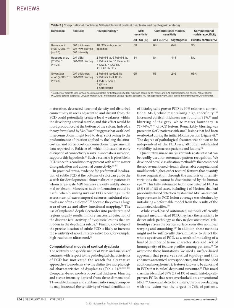
Computational models of cortical dysplasiathe relatively nonspecific nature of vBm and analysis of contrasts with respect to the pathological charac teristics of FCD has motivated the search for alternative approaches to model in vivo the distinctive morphologi-cal characteristics of dysplasias (table 3).28,100–103 Computer-based models of cortical thickness, blurring and tissue intensity derived from three-dimensional t1-weighted images and combined into a single compos-ite map increased the sensitivity of visual identification
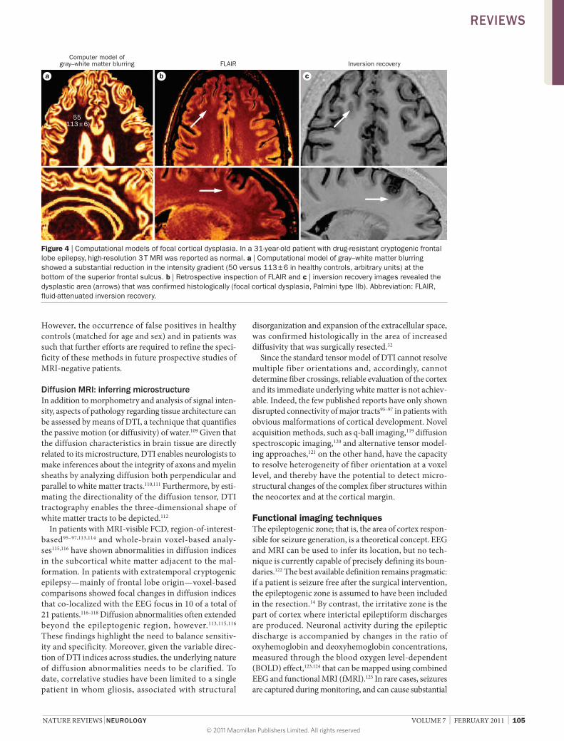
of histo logically proven FCD by 30% relative to conven-tional mri, while maintaining high specificity.100 increased cortical thickness was found in 91%,84 and blurring of the gray–white matter boundary in 72–96%,84,101 of FCD lesions. remarkably, blurring was present in 6 of 7 patients with small lesions that had been overlooked during the initial mri inspection (Figure 4).84 the degree of pathological features was shown to be independent of the FCD size, although substantial variability exists across patients and lesions.84
Quantitative image analysis provides data sets that can be readily used for automated pattern recognition. we developed novel classification methods104 that combined the above-mentioned visually discernable computational models with higher-order textural features that quantify tissue organization through the analysis of intensity variations that cannot be discriminated by the human eye.105 this fully automated technique detected FCD in 83% (15 of 18) of cases, including 4 of 7 lesions that had previously eluded detection by standard mri assessment. improvement in FCD lesion coverage was obtained by initializing a deformable model from the results of the automated classifier.106
while voxel-based automated methods efficiently segment medium-sized FCD, they lack the sensi tivity to detect subtle pathology, as they neglect ana tomical rela-tionships across the cortical surface, even after nonlinear warping and smoothing.107 in addition, these methods might not be sufficiently discriminative to detect the whole spectrum of FCD, as a result of modeling of a limited number of tissue charac teristics and lack of homogeneity of feature profiles among patients.84 to overcome these limitations, we used a surface-based approach that preserves cortical topology and thus enhances anatomical correspondence, and that included additional morphometric features known to be abnormal in FCD; that is, sulcal depth and curvature.92 this novel classifier identified 89% (17 of 19) of small, histologically proven FCDs that were overlooked on conventional mri.108 among all detected clusters, the one overlapping with the lesion was the largest in 76% of patients.
Table 3 | Computational models in MRI-visible focal cortical dysplasia and cryptogenic epilepsy
reference Features Histopathology* MrI sensitivity
Computational models: sensitivity
Computational models: specificity
All FCD (%) All FCD (%) Cryptogenic Healthy controls (%)
*Numbers of patients with surgical specimen available for histopathology; FCD subtypes according to Palmini and ILAE classifications are shown. Abbreviations: FCD, focal cortical dysplasia; GM, gray matter; ILAE, International League Against Epilepsy; NA, not applicable; vBM, voxel-based morphometry; wM, white matter.
However, the occurrence of false positives in healthy controls (matched for age and sex) and in patients was such that further efforts are required to refine the speci-ficity of these methods in future prospective studies of mri-negative patients.
Diffusion MrI: inferring microstructurein addition to morphometry and analysis of signal inten-sity, aspects of pathology regarding tissue architecture can be assessed by means of Dti, a technique that quantifies the passive motion (or diffusivity) of water.109 Given that the diffusion characteristics in brain tissue are directly related to its microstructure, Dti enables neurolo gists to make inferences about the integrity of axons and myelin sheaths by analyzing diffusion both perpen dicular and parallel to white matter tracts.110,111 Furthermore, by esti-mating the directionality of the diffusion tensor, Dti tractography enables the three-dimensional shape of white matter tracts to be depicted.112
in patients with mri-visible FCD, region-of- interest-based95–97,113,114 and whole-brain voxel-based analy-ses115,116 have shown abnormalities in diffusion indices in the subcortical white matter adjacent to the mal-formation. in patients with extratemporal cryptogenic epilepsy—mainly of frontal lobe origin—voxel-based comparisons showed focal changes in diffusion indices that co-localized with the eeG focus in 10 of a total of 21 patients.116–118 Diffusion abnormalities often extended beyond the epileptogenic region, however.113,115,116 these findings highlight the need to balance sensitiv-ity and specificity. moreover, given the variable direc-tion of Dti indices across studies, the underlying nature of diffusion abnormalities needs to be clarified. to date, corre lative studies have been limited to a single patient in whom gliosis, associated with structural
disorganization and expansion of the extracellular space, was confirmed histo logically in the area of increased diffusivity that was surgically resected.32
since the standard tensor model of Dti cannot resolve multiple fiber orientations and, accordingly, cannot determine fiber crossings, reliable evaluation of the cortex and its immediate underlying white matter is not achiev-able. indeed, the few published reports have only shown disrupted connectivity of major tracts95–97 in patients with obvious malformations of cortical development. novel acquisition methods, such as q-ball imaging,119 diffusion spectroscopic imaging,120 and alternative tensor model-ing approaches,121 on the other hand, have the capacity to resolve heterogeneity of fiber orientation at a voxel level, and thereby have the potential to detect micro-structural changes of the complex fiber structures within the neocortex and at the cortical margin.
Functional imaging techniquesthe epileptogenic zone; that is, the area of cortex respon-sible for seizure generation, is a theoretical concept. eeG and mri can be used to infer its location, but no tech-nique is currently capable of precisely defining its boun-daries.122 the best available definition remains pragmatic: if a patient is seizure free after the surgical intervention, the epileptogenic zone is assumed to have been included in the resection.14 By contrast, the irritative zone is the part of cortex where interictal epi leptiform discharges are produced. neuronal activity during the epileptic discharge is accompanied by changes in the ratio of oxyhemoglobin and deoxyhemoglobin concentrations, measured through the blood oxygen level-dependent (BolD) effect,123,124 that can be mapped using combined eeG and functional mri (fmri).125 in rare cases, seizures are captured during monitoring, and can cause substantial
a cb
Computer model ofgray–white matter blurring
55(113 ± 6)
FLAIR Inversion recovery
Figure 4 | Computational models of focal cortical dysplasia. In a 31-year-old patient with drug-resistant cryptogenic frontal lobe epilepsy, high-resolution 3 T MRI was reported as normal. a | Computational model of gray–white matter blurring showed a substantial reduction in the intensity gradient (50 versus 113 ± 6 in healthy controls, arbitrary units) at the bottom of the superior frontal sulcus. b | Retrospective inspection of FLAIR and c | inversion recovery images revealed the dysplastic area (arrows) that was confirmed histologically (focal cortical dysplasia, Palmini type IIb). Abbreviation: FLAIR, fluid-attenuated inversion recovery.
106 | FEBRUARY 2011 | volUmE 7 www.nature.com/nrneurol
movement artifacts, as well as being potentially damaging for the patient.
in focal epilepsy, attempts to compare the localization of eeG–fmri spike mapping with eeG source localization or intracranial eeG have produced mixed results.126–128 moreover, interictal epileptiform discharges generally correlate with the BolD signal in multiple brain regions. in some instances no BolD correlates of eeG spikes, or no spikes at all, are observed during the imaging session. this aspect is particularly relevant in patients with cryptogenic frontal lobe epilepsy, who often have rare or no interictal spikes.129 thus, eeG–fmri studies have mainly investigated pathophysiological mechanisms of epilepsy,130,131 and their clinical value in single patients has scarcely been addressed.
although eeG–fmri activation patterns by themselves are unlikely to form a modality that can outline the extent of a focal abnormality, they might provide evidence for putative epileptogenic zones for further investigation, such as targeted mri structural analysis or electrode implanta-tion. in one of 29 patients with partial epilepsy in whom the epileptogenic zone could not be localized, eeG–fmri data were convincing, and led to intracranial studies that resulted in subsequent successful surgery.132 in a group of nine patients with frontal lobe epilepsy,133 a link was observed between spike localization and positive BolD response in eight of the individuals. in two cases, careful review of the structural mri revealed a deep sulcus, indic-ative of an underlying dysplastic lesion.92 only one of the patients with an abnormal sulcus underwent surgery, the outcome of which was favorable.
Conclusionsa consensus exists that the primary histophological sub-strate for cryptogenic epilepsy is subtle cortical dysplasia,
but the mri signature of this entity is not yet fully defined, mainly because of the lack of a unified methodology to evaluate the structural changes related to this condition.
subtle, overlooked dysplastic lesions are likely to lie at one end of the spectrum of structural abnormalities underlying cryptogenic epilepsy. understanding the characteristics of such subtle abnormalities paves the way to further refinements aimed at capturing neo cortical abnormalities outside the range of visual detection.
By revealing FCD lesions that previously eluded visual inspection, quantitative structural image processing and analysis have clearly demonstrated increased sensitivity compared with conventional techniques. However, as the lesional tissue may differ only slightly from normal tissue, even the most advanced unimodal quantitative approach may be not be sufficiently powered to detect subtle changes. on the basis of current knowledge, successful approaches are likely to entail the design of integrated topology-preserving methods to statistically model mor-phology and signals in a multivariate framework. Future improvements in our ability to localize epileptogenic tissue should enable a more-effective clinical evaluation and make successful surgery accessible to an increasing percentage of patients with cryptogenic epilepsy.
Review criteria
we searched MEDLINE using the following terms: “epilepsy”, “MRI”, “quantitative imaging” and “cryptogenic” and their variants, synonyms, and acronyms, for articles published between January 1995 and March 2010. we also examined the bibliographies of these articles and review articles to identify any additional studies. studies were included if they examined quantitative MRI imaging methods in patients with focal neocortical epilepsy.
2. Garcia, H. H. & Del Brutto, O. H. Neurocysticercosis: updated concepts about an old disease. Lancet Neurol. 4, 653–661 (2005).
3. Kwan, P. & Brodie, M. J. Early identification of refractory epilepsy. N. Engl. J. Med. 342, 314–319 (2000).
4. Cascino, G. D. Temporal lobe epilepsy is a progressive neurologic disorder: time means neurons! Neurology 72, 1718–1719 (2009).
5. wiebe, s. Burden of intractable epilepsy. Adv. Neurol. 97, 1–4 (2006).
6. Tellez-Zenteno, J. F., Ronquillo, L. H. & wiebe, s. sudden unexpected death in epilepsy: evidence-based analysis of incidence and risk factors. Epilepsy Res. 65, 101–115 (2005).
7. Lerner, J. T. et al. Assessment and surgical outcomes for mild type I and severe type II cortical dysplasia: a critical review and the UCLA experience. Epilepsia 50, 1310–1335 (2009).
8. wiebe, s. Brain surgery for epilepsy. Lancet 362 (suppl.), s48–s49 (2003).
9. wiebe, s., Blume, w. T., Girvin, J. P. & Eliasziw, M. A randomized, controlled trial of surgery for temporal-lobe epilepsy. N. Engl. J. Med. 345, 311–318 (2001).
10. Cascino, G. D. surgical treatment for epilepsy. Epilepsy Res. 60, 179–186 (2004).
11. Fauser, s. et al. Focal cortical dysplasias: surgical outcome in 67 patients in relation to histological subtypes and dual pathology. Brain 127, 2406–2418 (2004).
12. Engel, J. Jr et al. Practice parameter: temporal lobe and localized neocortical resections for epilepsy: report of the Quality standards subcommittee of the American Academy of Neurology, in association with the American Epilepsy society and the American Association of Neurological surgeons. Neurology 60, 538–547 (2003).
13. Tellez-Zenteno, J. F., Dhar, R., Hernandez-Ronquillo, L. & wiebe, s. Long-term outcomes in epilepsy surgery: antiepileptic drugs, mortality, cognitive and psychosocial aspects. Brain 130, 334–345 (2007).
14. Bernasconi, A. in Advances in Neurology Ch. 6 (eds Blume, w. et al.) 273–278 (Lippincott-williams & wilkins, Philadelphia, 2006).
15. Cossu, M. et al. Epilepsy surgery in children: results and predictors of outcome on seizures. Epilepsia 49, 65–72 (2008).
16. Mosewich, R. K. et al. Factors predictive of the outcome of frontal lobe epilepsy surgery. Epilepsia 41, 843–849 (2000).
17. widdess-walsh, P. et al. subdural electrode analysis in focal cortical dysplasia: predictors of surgical outcome. Neurology 69, 660–667 (2007).
18. Jeha, L. E. et al. surgical outcome and prognostic factors of frontal lobe epilepsy surgery. Brain 130, 574–584 (2007).
19. Berg, A. T. et al. The multicenter study of epilepsy surgery: recruitment and selection for surgery. Epilepsia 44, 1425–1433 (2003).
20. McGonigal, A. et al. stereoelectroencephalography in presurgical assessment of MRI-negative epilepsy. Brain 130, 3169–3183 (2007).
21. Kim, D. w. et al. Predictors of surgical outcome and pathologic considerations in focal cortical dysplasia. Neurology 72, 211–216 (2009).
22. Tellez-Zenteno, J. F., Hernandez Ronquillo, L., Moien-Afshari, F. & wiebe, s. surgical outcomes in lesional and non-lesional epilepsy: a systematic review and meta-analysis. Epilepsy Res. 89, 310–318 (2010).
23. Krsek, P. et al. Incomplete resection of focal cortical dysplasia is the main predictor of poor postsurgical outcome. Neurology 72, 217–223 (2009).
24. Fauser, s. et al. Factors influencing surgical outcome in patients with focal cortical dysplasia. J. Neurol. Neurosurg. Psychiatry 79, 103–105 (2008).
25. Tanriverdi, T., Ajlan, A., Poulin, N. & Olivier, A. Morbidity in epilepsy surgery: an experience based on 2,449 epilepsy surgery procedures from a single institution. J. Neurosurg. 110, 1111–1123 (2009).
26. Duchowny, M. Clinical, functional, and neurophysiologic assessment of dysplastic cortical networks: implications for cortical functioning and surgical management. Epilepsia 50 (suppl. 9), 19–27 (2009).
27. Yun, C. H. et al. Prognostic factors in neocortical epilepsy surgery: multivariate analysis. Epilepsia 47, 574–579 (2006).
28. Bien, C. G. et al. Characteristics and surgical outcomes of patients with refractory magnetic resonance imaging-negative epilepsies. Arch. Neurol. 66, 1491–1499 (2009).
29. wetjen, N. M. et al. Intracranial electroencephalography seizure onset patterns and surgical outcomes in nonlesional extratemporal epilepsy. J. Neurosurg. 110, 1147–1152 (2009).
30. Alarcón, G. et al. Is it worth pursuing surgery for epilepsy in patients with normal neuroimaging? J. Neurol. Neurosurg. Psychiatry 77, 474–480 (2006).
31. Chapman, K. et al. seizure outcome after epilepsy surgery in patients with normal preoperative MRI. J. Neurol. Neurosurg. Psychiatry 76, 710–713 (2005).
32. Rugg-Gunn, F. J. et al. Diffusion tensor imaging in refractory epilepsy. Lancet 359, 1748–1751 (2002).
33. Frater, J. L., Prayson, R. A., Morris, H. H. III & Bingaman, w. E. surgical pathologic findings of extratemporal-based intractable epilepsy: a study of 133 consecutive resections. Arch. Pathol. Lab. Med. 124, 545–549 (2000).
34. Jayakar, P. et al. Epilepsy surgery in patients with normal or nonfocal MRI scans: integrative strategies offer long-term seizure relief. Epilepsia 49, 758–764 (2008).
35. Barkovich, A. J., Kuzniecky, R. I., Jackson, G. D., Guerrini, R. & Dobyns, w. B. A developmental and genetic classification for malformations of cortical development. Neurology 65, 1873–1887 (2005).
36. Palmini, A. et al. Terminology and classification of the cortical dysplasias. Neurology 62, s2–s8 (2004).
37. sisodiya, s. M. Malformations of cortical development: burdens and insights from important causes of human epilepsy. Lancet Neurol. 3, 29–38 (2004).
38. Blümcke, I. et al. The clinicopathologic spectrum of focal cortical dysplasias: A consensus classification proposed by an ad hoc task force of the ILAE Diagnostic Methods Commission 1. Epilepsia (in press).
39. Thom, M. et al. Cajal–Retzius cells, inhibitory interneuronal populations and neuropeptide Y expression in focal cortical dysplasia and microdysgenesis. Acta Neuropathol. 105, 561–569 (2003).
40. Andres, M. et al. Human cortical dysplasia and epilepsy: an ontogenetic hypothesis based on volumetric MRI and NeuN neuronal density and size measurements. Cereb. Cortex 15, 194–210 (2005).
41. sisodiya, s. M., Fauser, s., Cross, J. H. & Thom, M. Focal cortical dysplasia type II: biological features and clinical perspectives. Lancet Neurol. 8, 830–843 (2009).
42. Matsuda, K. et al. Neuroradiologic findings in focal cortical dysplasia: histologic correlation with surgically resected specimens. Epilepsia 42 (suppl. 6), 29–36 (2001).
43. Barkovich, A. J., Kuzniecky, R. I., Bollen, A. w. & Grant, P. E. Focal transmantle dysplasia: a specific malformation of cortical development. Neurology 49, 1148–1152 (1997).
44. Urbach, H. et al. Focal cortical dysplasia of Taylor’s balloon cell type: a clinicopathological
entity with characteristic neuroimaging and histopathological features, and favorable postsurgical outcome. Epilepsia 43, 33–40 (2002).
45. Taylor, D. C., Falconer, M. A., Bruton, C. J. & Corsellis, J. A. N. Focal dysplasia of the cerebral cortex in epilepsy. J. Neurol. Neurosurg. Psychiatry 34, 369–387 (1971).
46. Cohen-Gadol, A. A., Ozduman, K., Bronen, R. A., Kim, J. H. & spencer, D. D. Long-term outcome after epilepsy surgery for focal cortical dysplasia. J. Neurosurg. 101, 55–65 (2004).
47. Tassi, L. et al. Focal cortical dysplasia: neuropathological subtypes, EEG, neuroimaging and surgical outcome. Brain 125, 1719–1732 (2002).
48. Colombo, N. et al. Focal cortical dysplasias: MR imaging, histopathologic, and clinical correlations in surgically treated patients with epilepsy. AJNR Am. J. Neuroradiol. 24, 724–733 (2003).
49. Kloss, s., Pieper, T., Pannek, H., Holthausen, H. & Tuxhorn, I. Epilepsy surgery in children with focal cortical dysplasia (FCD): results of long-term seizure outcome. Neuropediatrics 33, 21–26 (2002).
50. Lawson, J. A. et al. Distinct clinicopathologic subtypes of cortical dysplasia of Taylor. Neurology 64, 55–61 (2005).
51. Krsek, P. et al. Different features of histopathological subtypes of pediatric focal cortical dysplasia. Ann. Neurol. 63, 758–769 (2008).
52. Krsek, P. et al. Different presurgical characteristics and seizure outcomes in children with focal cortical dysplasia type I or II. Epilepsia 50, 125–137 (2009).
53. Bronen, R. A. et al. Focal cortical dysplasia of Taylor, balloon cell subtype: MR differentiation from low-grade tumors. AJNR Am. J. Neuroradiol. 18, 1141–1151 (1997).
54. Kim, s. K. et al. Focal cortical dysplasia: comparison of MRI and FDG-PET. J. Comput. Assist. Tomogr. 24, 296–302 (2000).
55. von Oertzen, J. et al. standard magnetic resonance imaging is inadequate for patients with refractory focal epilepsy. J. Neurol. Neurosurg. Psychiatry 73, 643–647 (2002).
56. Barkovich, A. J., Rowley, H. A. & Andermann, F. MR in partial epilepsy: value of high-resolution volumetric techniques. AJNR Am. J. Neuroradiol. 16, 339–343 (1995).
57. Bastos, A. C. et al. Diagnosis of subtle focal dysplastic lesions: curvilinear reformatting from three-dimensional magnetic resonance imaging. Ann. Neurol. 46, 88–94 (1999).
58. Bastos, A. C. et al. Curvilinear reconstruction of 3D magnetic resonance imaging in patients with partial epilepsy: a pilot study. Magn. Res. Imaging 13, 1107–1112 (1995).
59. Huppertz, H. J., Kassubek, J., Altenmuller, D. M., Breyer, T. & Fauser, s. Automatic curvilinear reformatting of three-dimensional MRI data of the cerebral cortex. Neuroimage 39, 80–86 (2008).
60. wiggins, G. C. et al. 32-channel 3 Tesla receive-only phased-array head coil with soccer-ball element geometry. Magn. Reson. Med. 56, 216–223 (2006).
61. strandberg, M., Larsson, E. M., Backman, s. & Kallen, K. Pre-surgical epilepsy evaluation using 3T MRI. Do surface coils provide additional information? Epileptic Disord. 10, 83–92 (2008).
62. Knake, s. et al. 3T phased array MRI improves the presurgical evaluation in focal epilepsies: a prospective study. Neurology 65, 1026–1031 (2005).
63. Haacke, E. M., Xu, Y., Cheng, Y. C. & Reichenbach, J. R. susceptibility weighted imaging (swI). Magn. Reson. Med. 52, 612–618 (2004).
64. Duyn, J. H. et al. High-field MRI of brain cortical substructure based on signal phase. Proc. Natl Acad. Sci. USA 104, 11796–11801 (2007).
65. Marques, J. P., van der Zwaag, w., Granziera, C., Krueger, G. & Gruetter, R. Cerebellar cortical layers: in vivo visualization with structural high-field-strength MR imaging. Radiology 254, 942–948 (2010).
66. Madan, N. & Grant, P. E. New directions in clinical imaging of cortical dysplasias. Epilepsia 50 (suppl. 9), 9–18 (2009).
67. Ashburner, J. & Friston, K. J. voxel-based morphometry—the methods. Neuroimage 11, 805–821 (2000).
68. Good, C. D. et al. A voxel-based morphometric study of ageing in 465 normal adult human brains. Neuroimage 14, 21–36 (2001).
69. salmond, C. H. et al. Distributional assumptions in voxel-based morphometry. Neuroimage 17, 1027–1030 (2002).
70. Bruggemann, J. M. et al. voxel-based morphometry in the detection of dysplasia and neoplasia in childhood epilepsy: limitations of grey matter analysis. J. Clin. Neurosci. 16, 780–785 (2009).
71. Bruggemann, J. M. et al. voxel-based morphometry in the detection of dysplasia and neoplasia in childhood epilepsy: combined grey/white matter analysis augments detection. Epilepsy Res. 77, 93–101 (2007).
72. Colliot, O. et al. Individual voxel-based analysis of gray matter in focal cortical dysplasia. Neuroimage 29, 162–171 (2006).
73. wilke, M., Kassubek, J., Ziyeh, s., schulze-Bonhage, A. & Huppertz, H. J. Automated detection of gray matter malformations using optimized voxel-based morphometry: a systematic approach. Neuroimage 20, 330–343 (2003).
74. Merschhemke, M. et al. Quantitative MRI detects abnormalities in relatives of patients with epilepsy and malformations of cortical development. Neuroimage 18, 642–649 (2003).
75. Kassubek, J., Huppertz, H. J., spreer, J. & schulze-Bonhage, A. Detection and localization of focal cortical dysplasia by voxel-based 3-D MRI analysis. Epilepsia 43, 596–602 (2002).
76. Bonilha, L. et al. voxel-based morphometry reveals excess gray matter concentration in patients with focal cortical dysplasia. Epilepsia 47, 908–915 (2006).
77. Prayson, R. A., spreafico, R. & vinters, H. v. Pathologic characteristics of the cortical dysplasias. Neurosurg. Clin. N. Am. 13, 17–25 (2002).
78. Fauser, s. et al. Multi-focal occurrence of cortical dysplasia in epilepsy patients. Brain 132, 2079–2090 (2009).
79. Eriksson, s. H. et al. Quantitative grey matter histological measures do not correlate with grey matter probability values from in vivo MRI in the temporal lobe. J. Neurosci. Methods 181, 111–118 (2009).
80. Rugg-Gunn, F. J., Boulby, P. A., symms, M. R., Barker, G. J. & Duncan, J. s. whole-brain T2 mapping demonstrates occult abnormalities in focal epilepsy. Neurology 64, 318–325 (2005).
81. Rugg-Gunn, F. J. et al. Magnetization transfer imaging in focal epilepsy. Neurology 60, 1638–1645 (2003).
82. Focke, N. K., symms, M. R., Burdett, J. L. & Duncan, J. s. voxel-based analysis of whole brain FLAIR at 3T detects focal cortical dysplasia. Epilepsia 49, 786–793 (2008).
108 | FEBRUARY 2011 | volUmE 7 www.nature.com/nrneurol
83. Rugg-Gunn, F. J., Boulby, P. A., symms, M. R., Barker, G. J. & Duncan, J. s. Imaging the neocortex in epilepsy with double inversion recovery imaging. Neuroimage 31, 39–50 (2006).
84. Colliot, O., Antel, s. B., Naessens, v. B., Bernasconi, N. & Bernasconi, A. In vivo profiling of focal cortical dysplasia on high-resolution MRI with computational models. Epilepsia 47, 134–142 (2006).
85. salmenpera, T. M. et al. Evaluation of quantitative magnetic resonance imaging contrasts in MRI-negative refractory focal epilepsy. Epilepsia 48, 229–237 (2007).
86. Focke, N. K. et al. Automated normalized FLAIR imaging in MRI-negative patients with refractory focal epilepsy. Epilepsia 50, 1484–1490 (2009).
87. Pell, G. s., Briellmann, R. s., Pardoe, H., Abbott, D. F. & Jackson, G. D. Composite voxel-based analysis of volume and T2 relaxometry in temporal lobe epilepsy. Neuroimage 39, 1151–1161 (2008).
88. Barkovich, A. J. & Raybaud, C. A. Neuroimaging in disorders of cortical development. Neuroimaging Clin. N. Am. 14, 231–254 (2004).
89. Colombo, N., salamon, N., Raybaud, C., Ozkara, C. & Barkovich, A. J. Imaging of malformations of cortical development. Epileptic Disord. 11, 194–205 (2009).
90. Bronen, R. A., spencer, D. D. & Fulbright, R. K. Cerebrospinal fluid cleft with cortical dimple: MR imaging marker for focal cortical dysgenesis. Radiology 214, 657–663 (2000).
91. Riviere, D. et al. Automatic recognition of cortical sulci of the human brain using a congregation of neural networks. Med. Image Anal. 6, 77–92 (2002).
92. Besson, P., Andermann, F., Dubeau, F. & Bernasconi, A. small focal cortical dysplasia lesions are located at the bottom of a deep sulcus. Brain 131, 3246–3255 (2008).
93. van Essen, D. C. A tension-based theory of morphogenesis and compact wiring in the central nervous system. Nature 385, 313–318 (1997).
94. Rakic, P. Defects of neuronal migration and the pathogenesis of cortical malformations. Prog. Brain Res. 73, 15–37 (1988).
95. Lee, s. K. et al. Diffusion tensor MRI visualizes decreased subcortical fiber connectivity in focal cortical dysplasia. Neuroimage 22, 1826–1829 (2004).
96. widjaja, E. et al. subcortical alterations in tissue microstructure adjacent to focal cortical dysplasia: detection at diffusion-tensor MR imaging by using magnetoencephalographic dipole cluster localization. Radiology 251, 206–215 (2009).
97. widjaja, E. et al. Evaluation of subcortical white matter and deep white matter tracts in malformations of cortical development. Epilepsia 48, 1460–1469 (2007).
98. Najm, I. M., Bingaman, w. E. & Lüders, H. O. The use of subdural grids in the management of focal malformations due to abnormal cortical development. Neurosurg. Clin. N. Am. 13, 87–92 (2002).
99. Miller, D. et al. Intraoperative ultrasound to define focal cortical dysplasia in epilepsy surgery. Epilepsia 49, 156–158 (2008).
100. Bernasconi, A. et al. Texture analysis and morphological processing of magnetic resonance imaging assist detection of focal cortical dysplasia in extra-temporal partial epilepsy. Ann. Neurol. 49, 770–775 (2001).
101. Huppertz, H. J. et al. Enhanced visualization of blurred gray–white matter junctions in focal cortical dysplasia by voxel-based 3D MRI analysis. Epilepsy Res. 67, 35–50 (2005).
102. srivastava, s. et al. Feature-based statistical analysis of structural MR data for automatic detection of focal cortical dysplastic lesions. Neuroimage 27, 253–266 (2005).
103. Antel, s. B. et al. Computational models of MRI characteristics of focal cortical dysplasia improve lesion detection. Neuroimage 17, 1755–1760 (2002).
104. Antel, s. B. et al. Automated detection of focal cortical dysplasia lesions using computational models of their MRI characteristics and texture analysis. Neuroimage 19, 1748–1759 (2003).
105. Bernasconi, A. Quantitative MR imaging of the neocortex. Neuroimaging Clin. N. Am. 14, 425–436 (2004).
106. Colliot, O. et al. segmentation of focal cortical dysplasia lesions on MRI using level set evolution. Neuroimage 32, 1621–1630 (2006).
107. Bookstein, F. L. “voxel-based morphometry” should not be used with imperfectly registered images. Neuroimage 14, 1454–1462 (2001).
108. Besson, P., Bernasconi, N., Colliot, O., Evans, A. & Bernasconi, A. surface-based texture and morphological analysis detects subtle cortical dysplasia. Med. Image Comput. Comput. Assist Interv. 11, 645–652 (2008).
109. Basser, P. J., Mattiello, J. & Le Bihan, D. MR diffusion tensor spectroscopy and imaging. Biophys. J. 66, 259–267 (1994).
110. Beaulieu, C. The basis of anisotropic water diffusion in the nervous system—a technical review. NMR Biomed. 15, 435–455 (2002).
111. Concha, L., Gross, D. w., wheatley, B. M. & Beaulieu, C. Diffusion tensor imaging of time-dependent axonal and myelin degradation after corpus callosotomy in epilepsy patients. Neuroimage 32, 1090–1099 (2006).
112. Mori, s. et al. Imaging cortical association tracts in the human brain using diffusion-tensor-based axonal tracking. Magn. Reson. Med. 47, 215–223 (2002).
113. Dumas de la Roque, A. et al. Diffusion tensor imaging of partial intractable epilepsy. Eur. Radiol. 15, 279–285 (2005).
114. Gross, D. w., Bastos, A. & Beaulieu, C. Diffusion tensor imaging abnormalities in focal cortical dysplasia. Can. J. Neurol. Sci. 32, 477–482 (2005).
115. Eriksson, s. H., Rugg-Gunn, F. J., symms, M. R., Barker, G. J. & Duncan, J. s. Diffusion tensor imaging in patients with epilepsy and malformations of cortical development. Brain 124, 617–626 (2001).
116. Guye, M. et al. what is the significance of interictal water diffusion changes in frontal lobe epilepsies? Neuroimage 35, 28–37 (2007).
117. Rugg-Gunn, F. J., Eriksson, s. H., symms, M. R., Barker, G. J. & Duncan, J. s. Diffusion tensor imaging of cryptogenic and acquired partial epilepsies. Brain 124, 627–636 (2001).
118. Thivard, L. et al. Interictal diffusion MRI in partial epilepsies explored with intracerebral electrodes. Brain 129, 375–385 (2006).
119. Tuch, D. s., Reese, T. G., wiegell, M. R. & wedeen, v. J. Diffusion MRI of complex neural architecture. Neuron 40, 885–895 (2003).
120. Tuch, D. s. et al. High angular resolution diffusion imaging reveals intravoxel white matter fiber heterogeneity. Magn. Reson. Med. 48, 577–582 (2002).
121. Behrens, T. E. et al. Non-invasive mapping of connections between human thalamus and cortex using diffusion imaging. Nat. Neurosci. 6, 750–757 (2003).
122. Rosenow, F. & Luders, H. Presurgical evaluation of epilepsy. Brain 124, 1683–1700 (2001).
123. Kwong, K. K. et al. Dynamic magnetic resonance imaging of human brain activity during primary sensory stimulation. Proc. Natl Acad. Sci. USA 89, 5675–5679 (1992).
124. Ogawa, s. et al. Intrinsic signal changes accompanying sensory stimulation: functional brain mapping with magnetic resonance imaging. Proc. Natl Acad. Sci. USA 89, 5951–5955 (1992).
125. Gotman, J. Epileptic networks studied with EEG–fMRI. Epilepsia 49 (suppl. 3), 42–51 (2008).
126. Grova, C. et al. Concordance between distributed EEG source localization and simultaneous EEG-fMRI studies of epileptic spikes. Neuroimage 39, 755–774 (2008).
127. Richardson, M. Current themes in neuroimaging of epilepsy: brain networks, dynamic phenomena, and clinical relevance. Clin. Neurophysiol. 121, 1153 (2010).
128. vulliemoz, s., Lemieux, L., Daunizeau, J., Michel, C. M. & Duncan, J. s. The combination of EEG source imaging and EEG-correlated functional MRI to map epileptic networks. Epilepsia 51, 491–505 (2010).
129. Kellinghaus, C. & Luders, H. O. Frontal lobe epilepsy. Epileptic Disord. 6, 223–239 (2004).
130. Tyvaert, L. et al. Thalamic nuclei activity in idiopathic generalized epilepsy: an EEG–fMRI study. Neurology 73, 2018–2022 (2009).
131. Gotman, J. et al. Generalized epileptic discharges show thalamocortical activation and suspension of the default state of the brain. Proc. Natl Acad. Sci. USA 102, 15236–15240 (2005).
132. Zijlmans, M. et al. EEG–fMRI in the preoperative work-up for epilepsy surgery. Brain 130, 2343–2353 (2007).
133. Moeller, F. et al. EEG–fMRI: adding to standard evaluations of patients with nonlesional frontal lobe epilepsy. Neurology 73, 2023–2030 (2009).
Author contributionsA. Bernasconi and N. Bernasconi wrote the article, and contributed to the editing and reviewing of the article. All the authors provided contributions to discussions of the content, and A. Bernasconi, B. C. Bernhardt and D. schrader researched data for the article.