69
Advances In Musculoskeletal Intervention Neil Johnson, MB.BS, M.Med FRANZCR William Shiels, DO Cincinnati Children’s Hospital Nationwide Children’s Hospital

Advances In Musculoskeletal Intervention
Neil Johnson, MB.BS, M.Med FRANZCR
William Shiels, DO
Cincinnati Children’s Hospital Nationwide Children’s Hospital

Disclosures
• Dr. Johnson CCHMC is a Research Site for Philips Medical
– Research Agreement / I.R. Animal Lab
– No Personal Financial Benefits
• Dr. Shiels

• Basic MSK Intervention
• Beyond Basics – Core Biopsy
– Treating Lesions
– Screws, Bone Grafts and Hardware
• Two Important Lesions – Histiocytosis (LCH)
– Aneurysmal Bone Cyst
• Advanced Guidance and Fusion Imaging
• A Little Politics
OUTLINE

MSK Intervention: Basics
• Image Guidance
– CT / CT Fluoroscopy
– Ultrasound
– Standard Fluoroscopy
– Cone Beam CT +/- Guidance
– Combined / Fusion Imaging
• “Needle” Biopsy
– Cytology
– Small Diameter < 4mm
• Automated Gun
• True Cut (Slot) Type Devices: Fibrous Lesions
X

MSK Intervention: Basics
• Abscess Drainage
– Similar To Other Sites
• Joint Injections
– MRI Arthrography
– Steroid Injections
• Joint /Tendon Sheath
• Bursa
• Marking Deep Lesions for Surgery
• Foreign Body Removal

MSK Intervention: Beyond Basics
• Deep Large Core Bone Biopsy
– Equipment
– Guidance
• Malignant Tumor Biopsy
– Intelligent Approach Paths
– Viable Tissue – “The Edge Is The Target”
– Exceptions: When Even Good Biopsies Go Bad
• Screws, Routers and Bone Grafts
– Orthopedics Through Small Holes

Beyond The Basics NF1 Malignant Nerve Sheath Tumor: ? Mets to Sternum and T1
Ultrasound Guided Biopsy of Sternum for Diagnosis
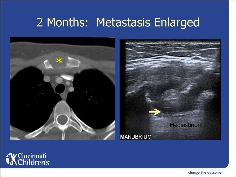
2 Months: Metastasis Enlarged
*
Mediastinum

Palliative R.F. Ablation of Sternal Met Ultrasound guidance
*
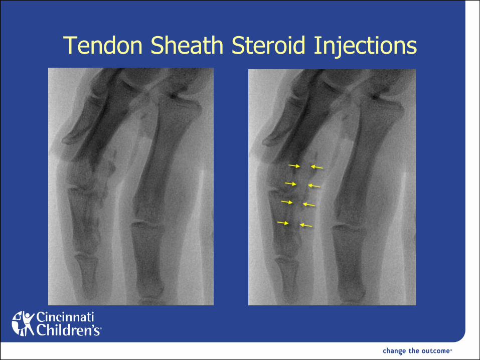
Tendon Sheath Steroid Injections

Finger Tendon Sheath Steroid

On An Awake (Smiling!) Patient

I.R. In The Operating Room
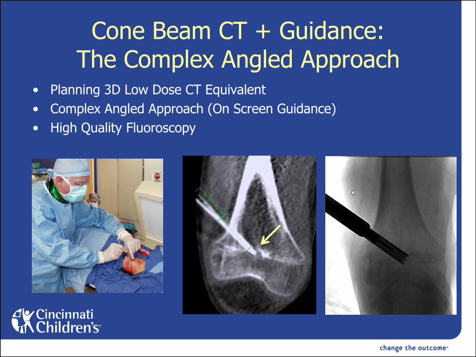
Cone Beam CT + Guidance: The Complex Angled Approach
• Planning 3D Low Dose CT Equivalent
• Complex Angled Approach (On Screen Guidance)
• High Quality Fluoroscopy

Post Traumatic Physeal Bar

Ultrasound: Avoiding Major Structures
Netter Radial Nerve?
Desmoplastic Fibroma

Ultrasound: Avoiding Major Structures
Newborn

Newborn: Forearm Biopsy
“Fibromatosis Coli”

Mediastinal Germ Cell Tumor: SVC Syndrome
*

Bone Tumor Biopsy: Co-Ordination with Surgery
Radiographics 2007; 27: 189-206

Always Co-Ordinate with Surgery
Radiographics 2007; 27: 189-206

Caution……

Posterior Component… Not Sampled (Sciatic Nerve + Surgery)
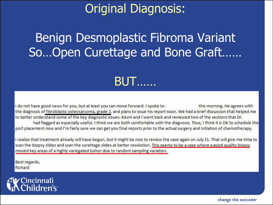
Original Diagnosis:
Benign Desmoplastic Fibroma Variant So…Open Curettage and Bone Graft……
BUT……

Two Special Lesions
• Langerhans Cell Histiocytosis
– Solitary Bone Lesion
• Aneurysmal Bone Cyst (ABC)

Langerhans Cell Histiocytosis
• Histiocytoses:
– Group of proliferative disorders arising from histiocytes, a common progenitor cell in bone marrow.
• 3 types of Histiocytes (dendritic cells)
– Langerhans cell: Epidermis
– Mononuclear Cell/Macrophage: Dermis
– Dermal dendritic cell: Dermis
• LCH and non – LCH Histiocytoses
Courtesy Dr. Joseph Palumbo, MD CCHMC

Types of Histiocytes: “Its Too Complicated for Radiologists”
CD 34+
CD 14+
DDC
MΦ
CD14- LC
Fitpatrick’s Dermatology in General Medicine, pp 106 CD1a

• Infectious? Disseminated, spontaneous remission of milder forms
– CMV, EBV, HHV-6, HHV-8 implicated; none proven
• Neoplastic ?
• Reactive Clonal Disorder ?
LCH Pathogenesis –Theories

• Congenital Self-Healing Reticulo-histiocytosis
– AKA Hashimoto-Pritzker disease
• Eosinophilic Granuloma
• Hand-Schuller-Christian disease
• Letterer-Siwe disease
Histiocytosis Clinical Types Old Classification

• Single system
– Isolated Bone Lesions (Best Prognosis***)
• Multisystem
• Disseminated
– Widespread, multi-organ disease (Poorest Prognosis)
Histiocytosis Clinical types Current Classification

LCH Isolated Bone Lesion Skull
Langerhans Cell Histiocytosis (LCH)
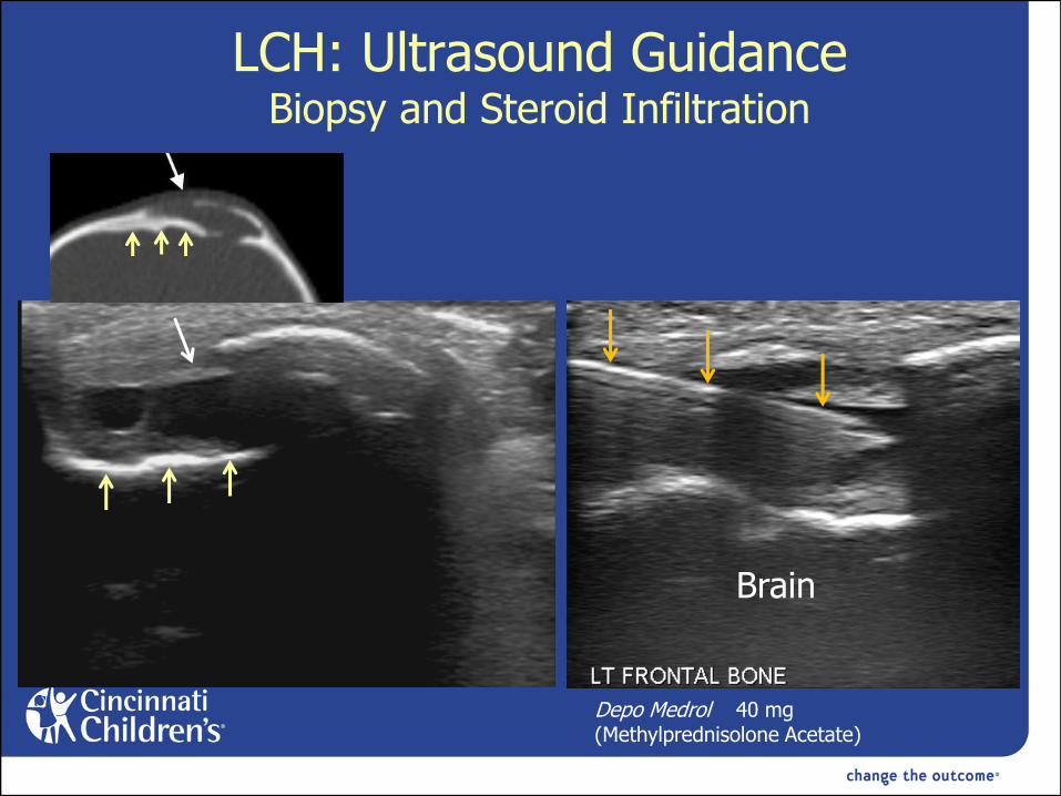
LCH: Ultrasound Guidance Biopsy and Steroid Infiltration
Depo Medrol 40 mg (Methylprednisolone Acetate)
Brain

LCH Skull

LCH Primary I.R. Treatment

Biopsy and Steroids

But….
*
*
*

2 Months Post Steroids
* *
*
At Diagnosis

LCH Rib
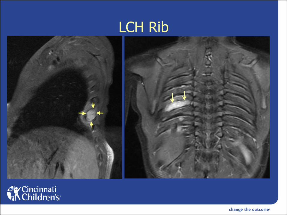
LCH Rib

LCH Rib: Ultrasound and Cone Beam CT

Post Procedure……..
But…

Histology: No Active Lesion

9 Year Old - LCH Pubis

LCH Pubis: Curettage, Steroids and Percutaneous Bone Graft

LCH Pubis: Curettage, Steroids and Percutaneous Bone Graft

20 Months Post Treatment

Incision for Pubic Ramus Surgical Approach:
(Giant Cell Tumor)
Right Leg Abdomen

LCH: Acetabulum Roof
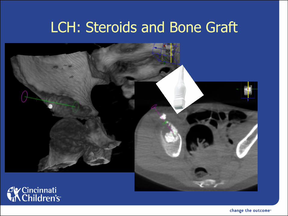
LCH: Steroids and Bone Graft

LCH: Steroids and Bone Graft Primary and Only Treatment
5 Months

Aneurysmal Bone Cyst
• Expansile Lytic Vascular Lesion of Bone
• 1.4 / Million Individuals
• Usually < 20 Years Old
• Male = Female
• Occurs In All Bones
– Most Common:
• Pelvis
• Spine ( Posterior Elements)
• Long Bones
Cottalorda, Arch Orthop Trauma Surgery (2007) 127: 105-114

Aneurysmal Bone Cyst
• 70% Primary
• 30% Secondary
– Chondroblastoma
– Osteoblastoma
– Giant Cell Tumor
– Fibrous Dysplasia
– Malignant Bone Tumors
• *** Telangiectatic OsteoSarcoma ***
Aneurysmal Bone Cyst
• Differentiation from Unicameral Bone Cyst (UBC)
– Single Cyst Vs Multiple Cysts
– Fluid Level Less Likely in UBC
– UBC Less Expansile
• BUT
– Complicated UBC (Fracture) May Be Difficult
– Biopsy Required
• UBC: Simple Cyst Lining Vs ABC
• UBC Different Histology

Aneurysmal Bone Cyst
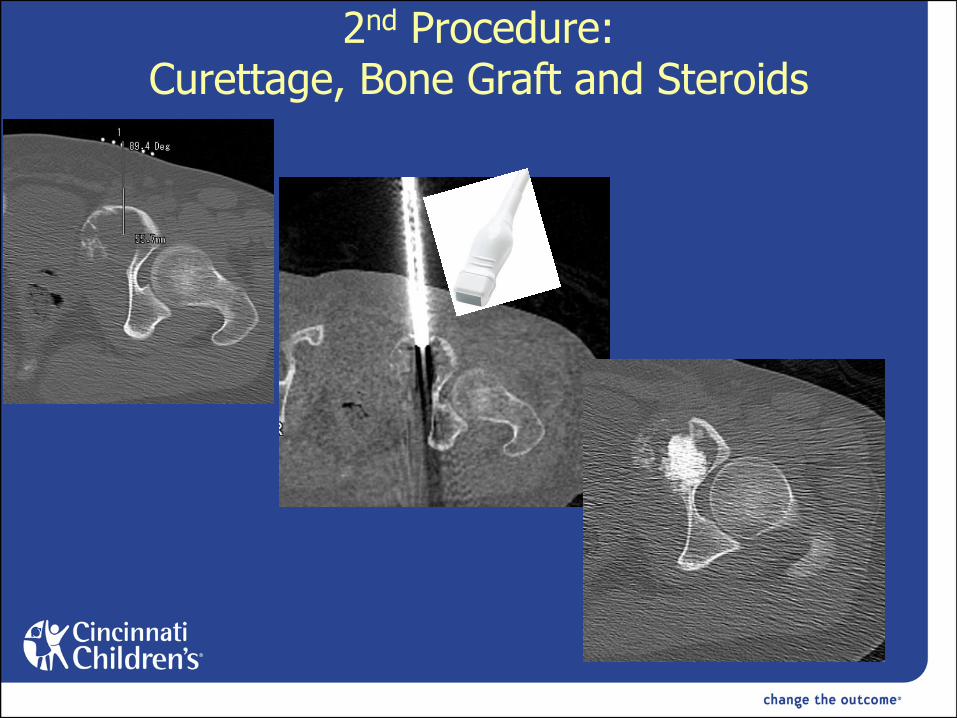
2nd Procedure: Curettage, Bone Graft and Steroids

12 Months

Percutaneous Bone Grafting: ABC
* *

Aneurysmal Bone Cyst
• Causation: Primary ABC
– Venous Obstructive Lesion
• Post Traumatic
• Post Infection
– Vascular Malformation
– Benign Neoplasm
• 16:17 q22:p13 Translocation [1]
• TRE17 / USP6 Oncogene Translocation [2]
[1] Panoutsakopoulos G, et.al. Recurrent t(16;17)(q22;p13) in aneurysmal bone cysts. Genes Chromosomes Cancer. 1999;26:265-266. [2] Ye Y, et.al. TRE17/USP6 oncogene translocated in aneurysmal bone cyst induces matrix metalloproteinase production via activation of NF-xB. Oncogene. 2010;29:3619-3629

Aneurysmal Bone Cyst
• Treatment Options
– Traditional Open Surgery
• 12-71 % “Recurrence” [1]
• Significant Complications
– Blood Loss, Loss of Function (Plates / Screws), Infection
– Radiotherapy
• Secondary Malignancy
– Percutaneous Sclerotherapy
• STS
• Ethibloc
• Doxycycline

Aneurysmal Bone Cyst
• Treatment Options
– Hybrid
• Minimally Invasive CT Guided (<1cm Incision)
• Curettage / Routing / Aspiration
• Steroid Soaked Percutaneous Bone Graft
– Image Guided Doxycycline (Dr. Shiels)
• Ultrasound or CT Guided
• Minimally Invasive
• Cysts Individually Targeted
• Doxycycline Suppresses Multiple Cellular Abnormalities
– Metalo Matrix Proteins (MMP)
– VEGF

Tumors:
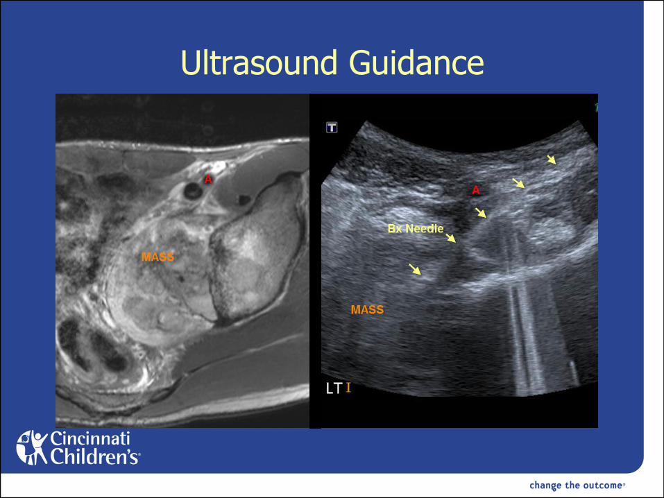
• Biopsy Guidance Ultrasound Vs CT
• Avoidance of Major Structures
• Color Doppler: Identifying Viable Tumor
Ewing’s Sarcoma

17 Year old Male 5 months Left Hip Pain

Primary Ultrasound Guidance
Ultrasound Guidance

Same Patient……. Diagnosis Please

2.5 Year Interval: Diagnosis ….

2.5 Year Interval: Diagnosis Please….

Hybrid Ultrasound Guidance ? Best of Both ?
Magnetic Field Plate Under Patient

IR / OR of the future Image Guided Orthopedics: We Need To Be There
• Surgery
– General, Ortho, Neuro
• Pulmonology /GI
• Oncology
• Led by IR ….Or Not


