Chapter 15 Immunosuppressive Properties of Mesenchymal Stromal Cells Francesco Lanza, Diana Campioni, Endri Mauro, Annalisa Pasini and Roberta Rizzo Abstract The main interest in mesenchymal stromal cells (MSCs) is correlated with their ability to suppress the proliferation of T lymphocytes induced by mitogenic agents and alloantigens which regulate the transplantation rejection. Moreover, MSCs are resistant to the CD8+ T lymphocyte cytotoxicity, they are able to inhibit the differentiation of dendritic cells responsible for the antigen presentation, the proliferation, and antibody production of B lymphocytes and they stimulate the formation of regulatory T cells. The mechanisms at the basis of MSCs activity need cell–cell interaction and the secretion of soluble molecules induced by the micro-environment. The inhibitory functions of MSCs involve several soluble molecules as hepatocyte growth factor, transforming growth factor-beta, interleukin-10 and -2, tumor necrosis factor-alpha, prostaglandin E2, indoleamine 2,3-dioxygenase (IDO), and soluble HLA-G antigens. A large consensus has been obtained on the immuno-modulatory role of IDO and HLA-G molecules. 15.1 Introduction Human multipotent mesenchymal stromal cells (hMSCs), first described by Friedenstein in 1970 [1] as nonhematopoietic cell precursors with osteogenic potential, represent stem cells for nonhematopoietic tissues [2]. The functional and F. Lanza (&) Á E. Mauro Á A. Pasini Hematology and Stem Cell Unit, Cremona, Italy e-mail: [email protected]D. Campioni Hematology Section, University of Ferrara, Ferrara, Italy R. Rizzo Microbiology Section, University of Ferrara, Ferrara, Italy H. Baharvand and N. Aghdami (eds.), Advances in Stem Cell Research, Stem Cell Biology and Regenerative Medicine, DOI: 10.1007/978-1-61779-940-2_15, Ó Springer Science + Bussines Media, LLC 2012 281
Francesco Lanza, Diana Campioni, Endri Mauro,Annalisa Pasini and Roberta Rizzo
Abstract The main interest in mesenchymal stromal cells (MSCs) is correlated withtheir ability to suppress the proliferation of T lymphocytes induced by mitogenicagents and alloantigens which regulate the transplantation rejection. Moreover,MSCs are resistant to the CD8+ T lymphocyte cytotoxicity, they are able to inhibitthe differentiation of dendritic cells responsible for the antigen presentation, theproliferation, and antibody production of B lymphocytes and they stimulate theformation of regulatory T cells. The mechanisms at the basis of MSCs activityneed cell–cell interaction and the secretion of soluble molecules induced by themicro-environment. The inhibitory functions of MSCs involve several solublemolecules as hepatocyte growth factor, transforming growth factor-beta, interleukin-10and -2, tumor necrosis factor-alpha, prostaglandin E2, indoleamine 2,3-dioxygenase(IDO), and soluble HLA-G antigens. A large consensus has been obtained on theimmuno-modulatory role of IDO and HLA-G molecules.
15.1 Introduction
Human multipotent mesenchymal stromal cells (hMSCs), first described byFriedenstein in 1970 [1] as nonhematopoietic cell precursors with osteogenicpotential, represent stem cells for nonhematopoietic tissues [2]. The functional and
F. Lanza (&) � E. Mauro � A. PasiniHematology and Stem Cell Unit, Cremona, Italye-mail: [email protected]
D. CampioniHematology Section, University of Ferrara, Ferrara, Italy
R. RizzoMicrobiology Section, University of Ferrara, Ferrara, Italy
H. Baharvand and N. Aghdami (eds.), Advances in Stem Cell Research,Stem Cell Biology and Regenerative Medicine, DOI: 10.1007/978-1-61779-940-2_15,� Springer Science + Bussines Media, LLC 2012
281
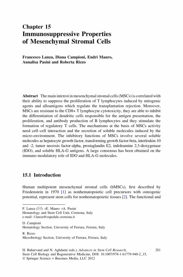
immunophenotic characteristics of stromal cells have been recently reviewed andredefined by International Society for Cellular Therapy (ISCT) [3]: they must bedefined as hMSCs, which are a plastic adherent cell population that retain in vitroclonogenic potential (defined by the presence of the fibroblast-colony forming unit,(CFU-F) [4, 5] (Fig. 15.1a, b), capable of supporting hematopoiesis [6, 7] andhaving a differentiation capacity toward a number of different cell types (osteo-blasts, chondrocytes, adipocytes, myocytes) [8–11]. Bone marrow (BM)-derivedhMSCs have been described to form in vitro stromal layers also with otheradherent nonhemopoietic cells such as endothelial cells or with the transientlyadherent macrophages (Fig. 15.1b, c). In vitro and in vivo studies also showed thathMSC could differentiate into cells of nonmesodermal origin such as neurons, skinand gut epithelial cells, hepatocytes, and pneumocytes [12].
By using multicolor flow cytometry, the existence of morphologically distincthMSCs subsets has been proposed, further allowing the identification of a func-tional subpopulation of rapidly in vitro self renewing (RS1) MSC [13]. Recent datausing carboxyfluorescein succimidyl ester (CFDA-SE) [14], that is a lipophilic dyethat irreversibly couples to cellular proteins and determine the division history ofcell populations undergoing proliferation, partially confirmed Colter findings, andsuggested the presence of different MSC subpopulations having a heterogeneousproliferative pattern. Moreover, these functionally defined hMSC subsets dis-played a distinct gene profile [15].
MSC are also capable of expressing genes of embryonic origin, cell–cellcontact molecules, different extracellular matrix proteins, collagen, and fibronectin.hMSC may also secrete chemokine ligands (including CCL2,CCL4, CCL5,CCL20, CXCL8.) and interleukins (SDF-1, IL-6, IL-7, IL-8, IL-10, IL-11, IL-12,SCF, etc.) that regulates microenvironmental homeostasis. hMSC can produceleukemia inhibitory factor, granulocyte-colony stimulating factor, granulocyte–macrophage colony stimulating factor [16, 17].
hMSC are initially isolated and characterized from the BM, but could be alsoobtained from other different sources such as amniotic membrane, skin, adiposetissue, cord blood, fetal liver, placenta, and synovium [18]. Whereas MSC havehistorically been thought to be the same general population of cells regardless ofthe tissue source, recent data suggest that hMSC gene expression profile reflectstheir tissue origin, indicating that hMSC tissue heterogeneity is biologicallyrelevant [19].
15.2 Immunophenotypic Characteristics of MSC
The isolation of hMSC from primary tissues is hampered by the limited selectivityof available markers while cultured hMSC may be defined only by a combinationof nonspecific immunophenotypic markers. The in vivo phenotype of hMSC hasbeen difficult to establish because of the exceptionally low frequency estimated onabout 0.01–0.1%. Moreover, there are no universally accepted antigenic
282 F. Lanza et al.
Fig. 15.1 A classical BM-derived CFU-F growing in culture (a) and stained with May-GrünwaldGiemsa is reported (b). Alkaline-phosphatase (ALP) positive BM-MSC (blue cells) growing inmixed culture after ex vivo expansion with BM-derived endothelial cells that resulted ALPnegative (c). BM-derived endothelial cells (red cells positive to CD31 antigen) have beenobserved in ex vivo expanded MSC monolayers (blue cells) (d)
15 Immunosuppressive Properties of Mesenchymal Stromal Cells 283
determinants for the phenotypic characterization and isolation of hMSC. Only fewmarkers have been developed and proved to be suitable for hMSC isolation.Functionally isolated MSC do not provide any information about the antigeniccomposition of the starting population. A variety of surface markers are exclu-sively found on cultured hMSC but not on their primary counterparts (such asCD271 or CD56 that are highly expressed on primary hMSC progenitor cells butrapidly downregulated in cultured hMSC). In 1990, Simmons and colleaguesidentified a stromal cell precursors in human BM by a ‘‘novel’’ monoclonalantibody, STRO-1, in a murine model [20]. Most of the monoclonal antibodiesnow available recognize also normal fibroblasts and mesenchymal derived stromalcells [21, 22]. No standardized protocols exist, nevertheless several protocols havebeen used to facilitate the prospective isolation of hMSC [23]. To identify‘‘naïve’’ hMSC, successfully positive selection with antibodies against surfacemolecules such as CD49 [24] frizzled-9 (FZD-9, Wnt receptor) CD349, CD56,CDCP1, CD73, [25, 26] and in particular CD105, D7Fib [21], were used. Isolationand characterization of CD146 positive hMSC have been also analyzed revealingtheir specific miRNA expression profile [26]. Some studies showed that hMSCmay be enriched by negative selection based on the depletion of CD45, CD14,CD34, and/or glycophorin A (CD235) markers. The CD271 seems to be one of themost specific markers [27] especially in combination with the CD56, and MSCA-1[24] to purifty nonexpanded MSC progenitor cells (Fig. 15.2). Using high sensi-tive flow cytometry, other authors have demonstrated that SSEA-1 and SSEA-4could identify BM-hMSCs [28], suggesting how these markers may be prospec-tively used to identify the most primitive progenitors with embryonic stemcell-like-features. More recently, it has been shown that mesenchymal andhemopoietic cells form a unique BM niche in which nestin positive cells are hMSC[29] and that CD146/CD271 expression on primary nonhematopoietic BM stemcells is correlated with in situ localization [30]. BM-MSC precursors express also aneural ganglioside (GD2) [31] that is not expressed by hemopoietic cells.
The immunophenotype of ex vivo expanded hMSC is currently defined byISCT that between 2005 and 2006 has established the nomenclature and theminimal criteria for defining mesenchymal multipotent stromal cells [3] as showedin Table 15.1.
So far, immunophenotypic analysis of cultured hMSC from most of the labo-ratories demonstrated that hMSC fulfilled general immunophenotypical criteria asstated by ISCT as above reported [3] hMSC are reported to be uniformly positivefor CD90, CD105, CD73, and negative for stem cell antigen CD34 and CD45 pan-leukocyte marker, and also for monocytic and lymphocytic cell antigens such asCD14, CD19, CD79a, HLA-DR by using flow cytometry. Other studies demon-strated the hMSC positivity for other antigens such as adhesion molecules (CD29,CD106, CD105, CD166, CD36), extracellular matrix protein (CD90, CD44),hemopoietic markers (CD10, CD59), histocompatibility antigens (HLA-I and IIclass) [21, 22], chemokine receptors (CD210, IL-10 receptor, CD184-SDF-1receptor) [32], neural and endothelial receptors (CD271, CD146). Only few studiesanalysed hMSC phenotype by a multiparametric cytofluorimetric approach
284 F. Lanza et al.
Fig. 15.2 A flow cytometric protocol for the study of nonexpanded MSC is shown. The putativeMSC progenitor cells in BM fresh samples were gated on viable cells (P1), CD45medlow (P2),and CD271 (P3) positive fraction (a, b). c and d show two distinct patients affected by HMhaving a different percentage of CD271 positive MSC progenitor cells. Selected CD271+ positivecells from BM fresh samples are visible after staining with May-Grünwald Giemsa (e) and duringthe in vitro culture (f)
15 Immunosuppressive Properties of Mesenchymal Stromal Cells 285
[33, 34]. Standardized protocol in functional and phenotypic hMSC analysis areneeded. A 4 colors cytofluorimetric protocol to study the immunophenotype ofhMSC has been recently proposed [35, 36]. From these studies it was stressed thatCD45+ positive cells as well as endothelial cells should be gated out from hMSCanalysis (Fig. 15.1). Furthermore, in hematological malignancies (HM) blast cellsresulted sometimes strictly adherent to in vitro expanded BM-derived MSC, thusrendering difficult the assessment of hMSC phenotype. (Fig. 15.3). Moreover, theuse of 7-AAD (7-aminoactinomycin) or Cytox 13 to identify nonviable cells isrecommended [37].
An emerging paradigm is that hMSCs could have key functional roles in thetissue in which they reside but phenotypic and functional characteristics undertheir ex vivo expansion conditions including different isolation methods, cultureprotocols: use of serum free, platelets lysate, additional cytokines, density, growthsupplements, passages, effects of cryopreservation are still not elucidated [38–41].For example the hMSC tissue source (normal vs. pathologic, and different tissues,donors variability, age in culture) may be also critically important in determiningtheir biological activity (heterogeneity of gene expression) [42]. All these vari-ables might have implications on the selection of cell type generating possiblefunctionally and phenotypically distinct hMSC subsets and altering the hMSCclonogenic and plastic potential [43]. These observations suggest that the cellprocessing protocols can be modified to enhance or repress expression of specificgene in order to optimize the cytokine profile for a given clinical indication.Standardized protocol has to be developed assuring that the manufactured cellsbehave solely in the clinical intended purpose [44, 45] and do not exert adverseeffects such as for example, uncontrolled differentiation or transformation.
The challenge for scientist is translating research into clinical scale manufac-turing of mesenchymal stromal cells [46] even though this is still completely notachieved.
Present definitions of hMSC emphasize generic functional properties of thesecells and fail to distinguish for example these cells from generic fibroblasts and todetect subsets of stromal cells with specialized niche functions [47, 48].
Emerging data focused on hMSC immunophenotype with special regards to theexpression of Thy-1 (CD90) molecule, and in a lesser extent to the LNGFR
Table 15.1 Minimal criteriato define MSC as establishedby ISCT
Adherence to plasticSpecific surface antigen (Atg) expressionMultipotent differentiation potential: osteoblasts, adipocytes,chondrocytes (demonstrated by staining of in vitro cell culture)
286 F. Lanza et al.
(CD271), CD105, CD44, CD10, CD146 molecules expression since they couldgive informations about different hMSC specialization of function. For exampledifferences related to the expression of CD105, CD106, CD44 by cultured humanBM-derived hMSCs from hematological patients (HM-MSCs) as compared to
Fig. 15.3 In a and b the same BM-derived MSC from a normal sample are cultured in twodifferent media: in a with serum and in b without serum but with different type of cytokines. After20 days of in vitro culture the percentage of CD45+ positive hematopoietic cells is reduced asshowed in the little cytometric plot. In c and d two different examples of in vitro expanded BM-derived MSC from patients with respectively a chronic lymphoprolipherative disorder and acutemyeloid leukemia where blasts are strictly adherent to the autologous MSC layer after 30 days ofculture and different changes of the medium
15 Immunosuppressive Properties of Mesenchymal Stromal Cells 287
normal samples (NS) and Skin fibroblasts (SF) seem to be correlated to a differentdifferentiation, and adhesion capacity by hMSC [49, 50].
These data may have therapeutic implication, since the use of autologoushMSCs has been proposed in a wide range of clinical applications including thosein the area of the regenerative medicine and cell therapies.
15.3 Immunological Functions of MSC
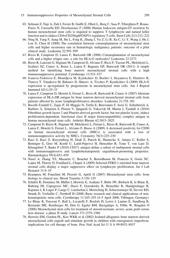
In the last years several studies have demonstrated the peculiar immunologicalcharacteristics of hMSCs, as low antigenicity and high immunomodulatory ability,suggesting their clinical use to counteract graft versus host disease (GvHD), oneof the most frequent and severe complication in allogeneic BM transplantations[51–53]. hMSCs share surface markers with thymic epithelial cells and expressadhesion molecules for T lymphocyte interaction, as vascular adhesion molecule 1and intracellular adhesion molecule 2. The low antigenicity of hMSCs is mainlycaused by the low expression of classical human leukocyte antigen (HLA) class Imolecules and the complete absence of HLA class II antigens and co-stimulatorymolecules as CD80 (B7-1), CD86 (B7-2), and CD40 [52]. The main interest inhMSCs is correlated with their ability to suppress the proliferation of T lympho-cytes induced by mitogenic agents and alloantigens which regulate the trans-plantation rejection [54]. Moreover, hMSCs are resistant to the CD8+T lymphocyte cytotoxicity, they are able to inhibit the differentiation of dendriticcells responsible for the antigen presentation, the proliferation, and antibodyproduction of B lymphocytes and they stimulate the formation of regulatoryT cells. These hematopoietic precursors are able to mediate a dose-dependenttolerogenic activity toward both innate and adaptative immunity. The mechanismsat the basis of hMSCs activity need cell–cell interaction and the secretion ofsoluble molecules induced by the micro-environment. The inhibitory functions ofhMSCs involve several soluble molecules as hepatocyte growth factor,transforming growth factor-beta (TGF-beta), interleukin-10 and -2 (IL-10, IL-2),tumor necrosis factor-alpha (TNF-alpha), prostaglandin E2 (PGE2), indoleamine2,3-dioxygenase (IDO) [55, 56], and soluble HLA-G antigens [53]. A largeconsensus has been obtained on the immuno-modulatory role of IDO and HLA-Gmolecules (Fig. 15.4) described as follows:
15.3.1 Indoleamine 2,3-dioxygenase
IDO is an immuno-modulatory enzyme that, with the epathic enzyme tryptophan2,3 dioxygenase, catalyzes the degradation of the essential amino acid L-trypto-phan to N-formylkynurenine [55]. The physiological role of IDO is not yet wellunderstood, but its ubiquitary distribution, its up-modulation by several cytokines,
288 F. Lanza et al.
as IFN-c and the ability to control the tryptophan levels, an essential amino acidfor cellular survival, increases the interest for this enzyme. In antigen presentingcells the expression of IFN-c-induced IDO converts tryptophan into formylky-nurenine with an immuno-suppressive effect toward T lymphocytes inducing theinhibition of their proliferation and activation [56]. MSCs present a costitutiveexpression of IDO. The presence of hMSCs and IDO in mixed lymphocytereactions inhibits lymphocyte proliferation, which is partially reactivated addingtryptophan to cell cultures [57]. The partial response of lymphocyte after trypto-phan treatment is due to the production of metabolites by IDO activity whichcontrasts T cell proliferation [58]. The immuno-modulatory role of IDO is con-firmed by the abrogation of rat heart transplant survival after administration of1-methyl tryptophan which inhibits IDO activity [59]. These data suggest IDO as asoluble factor implicated in the complex immuno-modulatory system of hMSCs.Spaggiari et al. [60] have demonstrated that hMSCs are able to inhibit the pro-liferation of natural killer (NK) cells, the expression of activatory NK cellreceptors (NKp44, NKp30, NKG2D) and the induction of cytotoxic activity andcytokine production. The inhibition of IFN-c receptor 1 (IFN-c R1) on hMSCs andthe consequent down-modulation of IDO production is not able to affect
Fig. 15.4 MSCs effect on immune system cells mediated by IDO and HLA-G molecules.Membrane (m) and soluble (s) HLA-G molecules from MSCs is able to inhibit the lytic activityof natural killer (NK) cells and their secretion of interferon-gamma (IFN-c), the cytotoxic activityof CD8+ T cells and their secretion of IFN-c and interleukin-5 (IL-5), to induce regulatory T cells(T reg) and their secretion of interleukin-10 (IL-10). IDO from MSCs creates an immunosup-pressive micro-environment inhibiting NK cell and CD8+ T lymphocyte cytotoxicity andcytokine secretion
15 Immunosuppressive Properties of Mesenchymal Stromal Cells 289
completely the hMSCs ability to control lympho-proliferation, suggesting thepresence of alternative ways for the hMSC immuno-modulatory functions [61].
15.3.2 HLA-G Molecule
HLA-G antigen is a nonclassical HLA class I molecule with immuno-modulatoryfunctions. It is characterized by seven mRNA splicing isoforms, four membrane-bound (HLA-G1,G2, G3, G4) and three soluble (HLA-G5, G6, G7). Both solubleand membrane-bound isoforms are able to inhibit several immune functions aslytic and cytotoxic activity of NK cells and T CD8+ lymphocytes, the maturationof dendritic cells, the alloproliferation of CD4+ T lymphocytes, and to induce theformation of regulatory T cells. HLA-G molecules differ from classical HLAantigens for their lower allelic polymorphism (47 alleles) and the limited tissuedistribution which is restricted to cytotrophoblasts and thymus in physiologicalconditions. HLA-G expression is induced in pathological conditions, as tumors,organ transplantation, viral infections [62]. Taking into consideration the tolero-genic functions of HLA-G molecules, they could be a good candidate as a solublefactor for hMSCs immuno-modulatory activity. Götherström et al. [63] havedemonstrated the presence of HLA-G mRNA in both fetal and adult hMSCs. Themodulation of HLA-G has been documented on the surface of hMSCs [64] and intheir culture supernatants [53]. Several studies have confirmed that the cell–cellcontact between hMSCs and T lymphocyte induces the secretion of HLA-Gmolecules and IL-10, a cytokine that is able to up-modulate HLA-G production.The use of neutralizing antibodies against HLA-G and IL-10 has demonstrated theimportance of soluble HLA-G, induced by IL-10, for the suppressive effect ofhMSCs toward cell proliferation [53], cytotoxic activity, and IFN-c secretion byNK cells, and the ability to induce regulatory T CD4+CD25highFoxP3+ cells for-mation [65]. These data suggest a fundamental role for HLA-G antigens in thetolerogenic function of hMSCs. Interestingly Ning et al. [66] have reported acorrelation between co-transplantation of hMSCs and the increase of hematologicdisease relapse. It is known that the modulation of HLA-G molecules is consideredas a favorable event in pregnancy and organ transplantation, where the regulationof the cell-mediated immune response is essential for a positive outcome. On thecontrary, the expression of HLA-G molecules is associated with a negative follow-up in tumors and viral infections, where the tolerogenic function of HLA-Gmolecules allows the mutated/infected cells to escape the immune system. Theassociation between hMSCs co-transplant and tumor relapse could be due to theability of HLA-G to increase the risk of cancer [67]. These considerations supportthe necessity to evaluate ‘‘a priori’’ the immuno-modulatory activity of hMSCs andthe production of HLA-G could be a good biomarker for this purpose [68].
Different factors could be responsible for the induction of HLA-G expressionby hMSCs. Progesterone, a steroid hormone involved in the female menstrualcycle, pregnancy, and embryogenesis, is able to up-regulate the HLA-G expression
290 F. Lanza et al.
by hMSCs [69]. These data could explain the ability of progesterone in regulatinguterine immune function in ways that allows for inhibition of immune responses atthe utero-placental interface without systemic immunosuppression.
hMSCs are newly detected targets for immunomodulatory activity and HLA-Gmolecules could be an important factor of this mechanism (Fig. 15.4). The availabledata suggest that there is a redundancy in the mechanisms of immunosuppression byhMSCs and only a complete comprehension of these suppressor mechanisms willoffer an insight into the clinical use of these cells in human therapy.
15.4 Immunophenotype and Immunoregulation
Since differences in molecules’ expression could give informations about differentspecialization of function and hMSC heterogeneity, some authors have focused onthe hMSC immunophenotype in relation to their immunomodulant properties. Thelow antigenicity of hMSCs is mainly caused by the low expression of classicalHLA class I molecules and the complete absence of HLA class II antigens andco-stimulatory molecules as CD80 (B7-1), CD86 (B7-2), and CD40 [52]. Never-theless the expression of class II HLA-DR molecule on BM-derived hMSCs frompatients with lymphoprolipherative disorders has been described [70]. Moreover,other studies reported up-regulation of MHC class II molecules only after pre-treatment with IFNy and TNF-a or FGF [71]. On the other hand, the down-regulation of some antigens expression seems to be associated with multilineagedifferentiation in cord blood-derived hMSC [49]. Very interesting is the findingthat (BM) derived hMSCs from HM isolated and cultured under in vitro angio-genic conditions exhibit a depressed expression of CD90 antigens that resultedassociated with high in vitro proliferative rate but with a diminished immuno-suppressive cell activity on T cell proliferation [72] suggesting the Thy-1 moleculemay be considered a marker implicated in the hMSC inhibitory ability and mightcooperate with HLA-G molecule in regulating suppressive versus stimulatoryproperties of hMSC.
The immunomodulant role of some hMSC immunophenotypic subsets has beenrecently demonstrated. For example, the CD105+, Stro-1, and CD271+ positivefraction displayed immunomodulant properties [73, 74].
15.5 MSC Clinical Application: an Overview
Based on their multipotential capabilities and immunosuppressive effects onT lymphocytes proliferation [53, 75], hMSCs could be used and expanded forclinical applications in cell therapy as well as in regenerative medicine [76–80].However, a number of issues still need to be solved before its widespread use in theclinics, namely a very low percentage of engraftment after systemic infusion, and
15 Immunosuppressive Properties of Mesenchymal Stromal Cells 291
the absence of definitive evidences about a therapeutic effect. In fact, it is stillunclear whether hMSCs contribute to tissue repair by differentiation into tissue-specific cell types, or mediate a paracrine effect [81]. Secretion of soluble mediatorsseems to be the predominant mechanism of action after systemic infusion of hMSC.Recent evidences [81] sustain the hypothesis about a paracrine role of hMSC thatare able to: (1) differentiate and populate the resident tissue, giving them a thera-peutic potential for regenerative medicine; (2) secrete cytokines or other solublemediators, (3) serve as a vehicle for delivery of proteins and gene therapy.
To date, administration of hMSC proved to be safe and also efficacious in avariety of disorders. hMSC are increasingly used in many preclinical as well asclinical settings. Many animal models have been used to assess the in vivo hMSCeffect on tissue transplantation and autoimmunity.
Ectodermal regeneration has been described for hMSC that can differentiateinto epidermal cells.
BM-derived hMSC have shown neural differentiation by different experimentalprotocols (chemical agents, growth factors, co-culture with other neuronalcells) [82]. When used in vivo hMSC can survive and differentiate into neural–glial cells. Neurological recovery has been shown in animal models of Parkinson’sdisease [83], hypoxic–ischemic neural damage. Recent data provide remarkablecues regarding the potential of hMSC in promoting endogenous reparativemechanisms that may prove applicable and promising therapeutic applications inParkinson’s disease [84, 85]. hMSC regenerative activity in humans has beendescribed in the treatment of autoimmune diseases characterized by autoimmuneaxonal demyelination associated with inflammation [multiple sclerosis andexperimental encephalomyelitis (EAE)]. Nevertheless, in these cases the beneficialeffect seems to be related to the inhibition of autoreactive cells in lymphoid organswhile the direct contribution to neuroregeneration in chronic EAE is still contro-versial [86].
The role of hMSC in mesodermal regeneration, especially bones, cartilage,tendons, and muscles has been described. hMSC in vivo differentiation towardosteoblastic and chondrogenic pathway was demonstrated by use of combinationof hMSC plus biomaterials and heterotopic implantation and directly in humanafter infusion, suggesting an important role for these cells in bone and cartilagerepair and in the treatment of osteoarthritis [76, 78].
BM-derived hMSC can differentiate in vitro in cardiomyocytes if treated with5-azacytidine and once injected in an infarcted heart, they reduce infarct size.Spontaneous differentiation of adipose tissue-derived hMSC into beating cardio-myocytes has been also observed [87]. Moreover, human cardiac and BM-derivedstromal cells exhibit distinctive properties in cardiac repair related to their origin[88] and their pre-activation demonstrated greater cytoprotection than unmodifiedhMSCs [89]. They could be also implanted in vascular prosthesis or scaffoldameliorating the restoration of vascular wall [90]. The beneficial effects of hMSCtherapy in cardiovascular disease may involve multiple mechanisms: hMSC secretea large amount of factors that could help resident cells to survive and could promoteangiogenesis. hMSC can also differentiate and/or stimulate endothelial cell
292 F. Lanza et al.
proliferation suggesting that paracrine effects are more likely responsible despiteinfrequent cellular fusion or differentiation or limited engraftment. Recently,induced pluripotent stem cell-derived cardiovascular progenitor cells represent asuitable autologous cell source for myocardial regeneration as they have thecapability to form myocardial cells and to contribute to revascularization [91].
hMSC are also involved in endodermal regeneration since they can be inducedinto hepatic differentiation and may serve as alternative for hepatocyte trans-plantation, cell-based therapy for liver injury, and preclinical drug testing [92].hMSC may help in inducing regeneration and/or proliferation of resident insulin-producing cells in diabetes disease models [93].
The use of hMSC in many kidney disorders (involving both ischemic/inflammatory and immunological injury) has been considered as a clinicallyrelevant solution as alternative to pharmacologic agents that target only a singleevent or pathway in the pathophysiology of a given disease. In contrast to con-ventional therapy hMSC use can promote cellular repair and tissue remodeling.Infusion of hMSC enhanced recovery and renal function [94, 95].
15.5.1 hMSC and Tissue Engineering
hMSCs-mediated tissue regeneration is a promising approach for tissue repair. Theuse of hMSC in three-dimensional (3D) scaffolds is promising even though it islimited by the need for an ideal scaffolds. The ideal scaffold is expected to bebiocompatible and biodegradable mimicking the structure and function of thenative extracellular matrix. The hMSC loaded on 3D scaffold are allowed tointeract with the damaged area, wherein they generate and replace the tissueespecially in the presence of specific molecules that could promote cell signaling,proliferation, and differentiation. For example, BMP-2 or bFGF growth factor areoften associated with ceramics supports, or injected in a carrier material such aspoly-lactic-co-glycolic acid, or gelatin, and collagen thus improving bone andtendon formation [96].
15.5.2 MSC and Clinical Application in Gene Therapy
The ability to transfer genes into hMSC and the ability of MSC to migrate to alesion provided evidences for a possible use of hMSC in clinical application. MSCwas successfully transfected with BMP-2 and injected in articular fractures [97].Transplantation of IL-7 gene-engineered hMSC into lethally irradiated mice led tosignificant increase in tymopoiesis and protected from GvHD [98]. For example,cell-mediated delivery of TRAIL in BM-MSC to metastatic rhabdomyosarcomatumor sites can repress the growth of tumor. Targeted delivery of TRAIL to tumors
15 Immunosuppressive Properties of Mesenchymal Stromal Cells 293
may allow systemic exposure of patients to drugs that may overcome resistance forTRAIL-induced apoptosis in RMS cells [99].
15.5.3 MSC for Improvement of Engraftment and Treatment/Prevention GVHD
Several disorders are characterized by both inflammation and tissue defects andhMSC has been shown to provide a valid alternative to pharmacological therapy totreat this clinical conditions. Nevertheless, it is sometimes unclear whether efficacyof hMSC is due to their production of trophic factors which stimulate endogenousrepair mechanism through an immunomodulatory effect, or a putative direct dif-ferentiation into various cell types.
Recent studies have demonstrated that cotransplantation of BM-derived HSC andadipose tissue-derived hMSC induced a rapid and efficient hematopoietic reconsti-tution [100, 101]. Relevant preclinical studies with hMSC have shown that hMSCexert potent immunosuppressive effect in vitro and in vivo reducing the risk of graftfailure and incidence of acute GvHD [102]. Allogeneic hMSC have been used totreat severe aplastic anemia refractory to conventional treatment and not eligible forallogeneic HSC transplantation, for inflammatory and autoimmune diseases such asChron’s disease [103, 104]. However, treating GVHD with hMSC might be a dou-ble-edged sword [67, 105] since has been demonstrated that the risk of the relapsewas increased and the survival decreased in the hMSC treated group [66]. It was alsoshown that when hMSC were infused at the onset of GVHD there was no therapeuticeffect, suggesting in this model that hMSC have a greater role in prophylaxis thantreatment [106]. These data support a previous observation that immunosuppressiveeffects are exerted more on T cell proliferation than on effector function. Moreover,not all studies give concordant or positive results and several factors are likely tocontribute to these discrepancies, including the tissue sources and method ofisolation and expansion of hMSC, the immunodepletion process, the dose andscheduling of hMSC administration. Another possible explanation for the lack of ananti-GVHD effect is that, despite the putatively immunoprivileged features of hMSCthe cells could be rejected. Furthermore, in the context of a steroid-resistant acuteGVHD where a patient could have a fatal outcome weeks or months later, theadministration of hMSC to manage this life-threatening condition resulted feasible,despite the limited and not standardized preclinical data available [107].
15.6 Potential Risks of Use of MSC
Human MSC maintained under standard culture conditions were shown to benontumorigenic, however, several reports presented their capability to modulatetumor microenvironment thus having an impact on the tumor behavior [108].
294 F. Lanza et al.
The complex relationship between hMSC and tumors should be better clarifiedand the potential risks related to the use of hMSC should be better investigated.Furthermore, hMSC may provide a support for leukemia blasts growth and relapse,and more studies are required to better understand the factors affecting malignant cellquiescence versus proliferation during hMSC-tumor cells interactions [109, 110].Recent concerns have been expressed about the potential transformation of hMSCduring the culture process before infusion. hMSC were found rarely to help tumordevelopment due to their immunosuppressive capacity [111]. However, the natureand the role of hMSC in their microenvironmental niche should be further explored.Moreover, although it has been demonstrated that ex vivo expansion processingof hMSC is feasible and should be obtained according to GMP and regulationconstraints [46], definitive standards to produce clinical grade hMSC are stilllacking.
In conclusion, the emerging functional and immunophenotypic heterogeneityof hMSCs in relation either to anatomical sites, or donor type (normal vs. path-ological) or the different culture expansion conditions, should be carefullyconsidered as an important step before using hMSC for clinical purposes beforereinfusion after ex vivo expansion.
References
1. Friedenstein AJ, Chailakhyan RK, Lalykina KS (1970) The development of fibroblastcolonies in monolayer cultures of guinea pig bone marrow and spleen colonies. Cell TissueKinet 3:393–403
2. Prockop DJ (1997) Marrow stromal cells as stem cells for non-hematopoietic tissues.Science 276:71
3. Dominici M, Le Blank K, Prockop DI, Howartz E (2006) Position paper: minimal criteriafor defining multipotent mesenchymal stromal cells. The International Society for CellularTherapy position statement. Cytotherapy 8:315–317
4. Dexter TM (1979) Cell interactions in vitro. Cin Haematol 8:453–4685. Castro-Malaspina H, Gay RE, Resnik G, Kapoor N, Meyers P, Chiarieri D, McKenzie S,
Broxmeyer HE, Moore MA (1980) Characterization of human bone marrow fibroblastcolony-forming cells (CFU-F) and their progeny. Blood 56:289–301
6. Tavassoli M, Friedenstein A (1983) Hemopoietic stromal microenvironment. Am JHaematol 15:196–203
7. Eaves AC, Eaves CJ (1988) Maintenance and proliferation control of primitive hemopoieticprogenitors in long-term cultures of human marrow cells. Blood Cells 14:355–368
8. Owen M, Friedenstein AJ (1988) Stromal stem cells: marrow-derived osteogenic precursors.Ciba Found Symp 136:42
9. Wulf GG, Jackson KA, Goodell MA (2001) Somatic stem cell plasticity: current evidenceand emerging concepts. Exp Hematol 29:1361–1370
10. Pittenger MF, Mackay AM, Beck SC, Jaiswal RK, Douglas R, Mosca JD, Moorman MA,Simonetti DW, Craig S, Marshak DR (1999) Multilineage potential of adult humanmesenchymal stem cells. Science 284:143
11. Di Girolamo CM, Stokes D, Colter D, Phinney DG, Class R, Prockop DJ (1999)Propagation and senescence of human marrow stromal cells in culture: a simple colony-forming assay identifies samples with the greatest potential to propagate and differentiate.Br J Haematol 107:275–281
15 Immunosuppressive Properties of Mesenchymal Stromal Cells 295
12. Reyes M, Lund T, Lenvik T, Aquiar D, Koodie L, Verfaillie CM (2001) Purification andex vivo expansion of postnatal human marrow mesodermal progenitor cells. Blood 98:2615–2625
13. Colter DC, Sekiya I, Prockop DJ (2001) Identification of a subpopulation of rapidly self-renewing and multipotential adult stem cells in colonies of human marrow stromal cells.PNAS 98:7841–7845
14. Urbani S, Caporale R, Lombardini L, Bosi A, Saccaridi R (2006) Use of CFDA-SE forevaluating the in vitro proliferation pattern of human MSC. Cytotherapy 8:243–253
15. Taormin A, Brune JC, Olsson E, Valcich J, Neuman U, Olofsson T, Jacobsen SE, Scheding S(2009) Characterization of bone marrow derived MSC based on gene expression profiling offunctionally defined MSC subsets. Cytotherapy 11:114–128
16. Majumdar MK, Keane-Moore M, Buyaner D et al (2003) Characterization and functionalityof cell surface molecules on human mesenchymal stem cells. J Biomed Sci 10:228–241
17. Conget PA, Minguell JJ (1999) Phenotypical and functional properties of human bonemarrow mesenchymal progenitor cells. J Cell Physiol 181:67–73
18. Beltrami AP, Cesselli D, Bergamin N, Marcon P, Rigo S, Puppato E, D’Aurizio F, Verardon R,Piazza S, Pignatelli A, Poz A, Baccaroni U, Damiani D, Fanin R, Mariuzzi L, Finato N,Masolini P, Burelli S, Belluzzi O, Schneider C, Beltrami CA (2007) Multipotent cells can begenerated in vitro from several adult human organs (heart, liver, bone marrow). Blood110:3438
19. Götherström C, West A, Liden J, Uzunel M, Lahesmaa R, Le Blanc K (2005) Differences ingene expression between human fetal liver and adult bone marrow mesenchymal stem cells.Haematologica 90:1017–1026
20. Simmons PJ, Torok-Storb B (1991) Identification of stromal precursors in human bonemarrow by a novel monoclonal antibody, STRO-1. Blood 78:55–62
21. Campioni D, Lanza F, Moretti S, Dominaci M, Punturieri M, Pauli S, Hofmann T, Horwitz E,Castaldi GL (2003) Functional and immunophenotypic characteristics of isolated CD105+and fibroblasts ? mesenchymal cells from acute myeloid leukaemia: implication for theirplasticity along endothelial lineage. Cytotherapy 5:66–79
22. Buhring HJ, Battula VL, Treml S, Schewe B, Kanz L, Vogel W (2007) Novel markers forthe prospective isolation of human MSC. Ann N Y Acad Sci 1106:262–271
23. Deschaseux F, Charbord P (2000) Human marrow stromal precursors are a1 integrinsubunit-positive. J Cell Physiol 184:319–325
24. Battula VL, Treml S, Bareiss PM, Gieseke F, Roelofs H, de Zwart P, Muller I, Schewe B,Skutella T, Fibbe WE, Kanz L, Buhring HJ (2009) Isolation of functionally distinct MSCcell subsets using antibodies aginst CD56, CD271 and mesenchymal stem cell antigen-1(MSCA-1). Haematologica 94:173–184
25. Bhuring HJ, Kuci S, Conze T, Rathke G, Scherl-Mostageer M, Brummendorf TH,Schweifer N, Lammers R (2004) CDCP1 identifies a broad spectrum of normal andmalignant stem/progenitor cell subsets of hematopoietic and non-hematopoietic origin.Stem Cells 22:334–343
26. SorrentinoA, Ferracin M, Castelli G, Biffoni M, Tomaselli G, Baiocchi M, Fatica A,Negrini M, Peschle C, Valtieri M (2008) Isolation and characterization of CD146+multipotent mesenchymal stromal cells. Exp Haematol 36:1035–1046
28. Gang EJ, Bosnakovsky D, Figueiredo CA et al (2007) SSEA-4 identifies MSC from bonemarrow. Blood 109:1743–1751
29. Mendez-Ferrer S, Michurina TV, Ferraro F, Mazloom AR, Macarthur BD, Lira SA,Scadden DT, Ma’ayan A, Enikopolov GN, Frenette PS (2010) Mesenchymal andhaematopoietic stem cells form a unique bone marrow niche. Nature 466(7308):829–834
296 F. Lanza et al.
30. Tormin A, Li O, Brune JC, Walsh S, Schtz B, Ehinger M, Ditzel N, Kassem M, Scheding S(2011) CD146 expression on primary non-hematopoietic bone marrow stem cells iscorrelated with in situ localization. Blood 117:5067–5077
31. Martinez C, Hofman TJ, Marino R, Dominaci M, Horwitz EM (2007) Human bone marrowmesenchymal cells express the neural ganglioside GD2: a novel surface markers for theidentification of MSC. Blood 109:4245–4248
32. Sordi V, Malosio ML, Marchesi F, Mercalli A, Melzi R, Giordano T, Belmonte N, Ferrari G,Leone BE, Bertuzzi F, Zerbini G, Allavena P, Bonifacio E, Piemonti L (2005) Bone marrowmesenchymal stem cells express a restricted set of functionally active chemokine receptorscapable of promoting migration to pancreatic islet. Blood 106:419–425
33. Sanchez-Guijo FM, Blanco JF, Cruz G, Muntion S, Gomez M, Carrancio S, Lopez-Villar O,Barbado MV, Sanchez-Abarca LI, Blanco B, Brinon JG, Del Canizo MC (2009)Multiparametric comparison of mesenchymal stromal cells obtained from trabecular boneby using a novel isolation method with those obtained by iliac crest aspiration from thesubjects. Cell Tissue Res 336(3):501–507
34. Landuyt KBV, Jones EA, McGonagle D, Luyten FP, Lories RJ (2010) Flow cytometriccharacterization of freshly isolated and cultured expanded human synovial cell population inpatients with chronic arthritis. Arthr Res Ther 12:R15
35. Jones E, English A, Kinsey S et al (2006) Optimization of a flow cytometry-based protocolfor detection and phenotypic characterization of multipotent mesenchymal stromal cellsfrom human bone marrow. Cytometry Part B 70B:391–399
36. Bradford JA, Clarke ST (2011) Panel development for multicolor flow cytometry testing ofproliferation and immunophenotype in h MSCs. Methods Mol Biol 698:367–385
37. Tabera S, Perez-Simon JA, Diez-Campelo M, Sanchez-Abarca LI, Blanco B, Lopez A,Benito A, Ocio E, Sanchez-Guijo FM, Canizo C, San Miguel JF (2008) The effect ofmesenchymal stem cells on the viability, proliferation and differentiation of B-Lymphocytes. Haematologica 93:1301–1309
38. Meuleman N, Tondreau T, Delforge A, Dejeneffe M, Massy M, Libertalis M, Bron D,Lagneaux L (2006) Human marrow mesenchymal stem cell culture : serum-free mediumallows better expansion than classical a-MEM medium. Eur J Haematol 76:309–316
39. Mueller I, Kordowich S, Holzwarth C, Spano C, Isensee G, Staiber A, Viebahn S, Gleseke F,Langer H, Gawaz MP, Horwitz EM, Conte P (2006) Animal serum-free culture conditionsfor isolation and expansion of multipotent mesenchymal stromal cells from human BM.Cytotherapy 8:437–444
40. Esposito MT, Di Noto R, Mirabelli P, Garrese M, Parisi S, Montanaro D, Del Vecchio L,Pastore L (2009) Culture conditions allow selection of different MSC progenitors from adultmouse bone marrow. Tissue Eng Part A 15:124
41. Sotiropoulou PA, Perez SA, Salagianni M, Baxevanis CN, Papamichail M (2006)Characterization of the optimal culture conditions for clinical scale production of humanmesenchymal stem cells. Stem Cells 24:74–85
42. Kalz N, Ringe J, Holzwarth C, Chrabord P, Niemeyer M, Jacobs VR, Peschel C (2010)Novel markers of mesenchymal stem cells defined by genome-wide gene expressionanalysis of stromal cells from different sources. Exp Cell Res 316:2609–2617
43. Lee RH, Hsu SC, Munoz J, Jung JS, Lee NR, Pochampally R, Prockop DJ (2006) A subsetof human rapidly-self renewing marrow stromal cells (MSCs) preferentially engraft in mice.Blood 107:2153–2161
44. Samuelsson H, Ringden O, Lönnies L, Le Blanc K (2009) Optimizing in vitro conditions forimmunomodulation and expansion of mesenchymal stem cells. Cytotherapy 11:129–136
45. Grisendi G, Anneren C, Cafarelli L, Sternieri R, Veronesi E, Cervo GL, Luminari S, Maur M,Frassoldati A, Palazzi G, Otsuru S, Bambi F, Paolucci P, Conte PF, Horwitz E, Dominici M(2010) GMP-manufactured density gradient media for optimized mesenchymal stromal stemcell isolation and expansion. Cytotherapy 12:466–477
46. Bieback K, Kinzebach S, Karagianni M (2010) Translating research into clinical scalemanufacturing of mesenchymal stromal cells. Stem Cells Int 2010:193519
15 Immunosuppressive Properties of Mesenchymal Stromal Cells 297
47. Haniffa MA, Collin MP, Buckley CD, Dazzi F (2009) Mesenchymal stem cells: thefibroblasts’ new clothes? Haematologica 94:258–263
48. Chang HY, Chi JT, Dudoit S, Bondre C, van de Rijn M, Botstein D, Brown PO (2002)Diversity, topographic differentiation, and positional memory in human fibroblasts. ProcNatl Acad Sci U S A 99:12877–12882
49. Jin HJ, Park SK, Oh W, Yang YS, Kim SW, Choi SJ (2009) Down-regulation of CD105 isassociated with multilineage differentiation in human umbilical cord derived-MSC.Biochem Biophys Res Comm 381:676–681
50. Kemp K, Morse R, Wexler S, Cox C, Mallam E, Hows J, Donaldson C (2010)Chemotherapy induce mesenchymal stem cell damage in patients with haematologicalmalignancy. Ann Hematol 89:701–713
51. Di Nicola M, Carlo-Stella C, Magni M, Milanesi M, Longoni PD, Matteucci P, Grisanti S,Gianni AM (2000) Human bone marrow stromal cells suppress T-lymphocyte proliferationinduced by cellular or nonspecific mitogenic stimuli. Blood 99:3838–3843
52. Le Blanc K, Tammik C, Rosendahl K, Zetterbergie E, Ringdén O (2003) HLA expressionand immunologic properties of differentiated and undifferentiated mesenchymal cells. ExpHematol 31:890–896
53. Rizzo R, Campioni D, Stignani M, Melchiorri L, Bagnara GP, Bonsi L, Alviano F, LanzoniG, Moretti S, Cuneo A, Lanza F, Baricordi OR (2008) A functional role for soluble HLA-Gantigens in immune modulation mediated by mesenchymal stromal cells. Cytotherapy10:364–375
55. Meisel R, Zibert A, Laryea M, Gobel U, Daubener W, Dilloo D (2004) Human bone marrowstromal cells inhibit allogeneic T-cell responses by indoleamine 2,3-dioxygenase-mediatedtryptophan degradation. Blood 103:4619–4621
56. Taylor MW, Feng G (1991) Relationship between interferon-gamma, indoleamine 2,3-dioxygenase, and tryptophan catabolism. FASEB J 5(11):2516–2522
57. Munn DH, Sharma MD, Lee JR, Jhaver KG, Johnson TS, Keskin DB, Marshall B, Chandler P,Antonia S, Burgess R, Slingwff CL Jr, Mellor AL (2002) Potential regulatory function ofhuman dendritic cells expressing indoleamine 2,3-dioxygenase. Science 297:1867–1870
58. Terness P, Bauer TM, Rose L, Dufter C, Watzlik A, Simon H, Opelz G (2002) Inhibition ofallogeneic T cell proliferation by indoleamine 2,3-dioxygenase-expressing dendritic cells:mediation of suppression by tryptophan metabolites. J Exp Med 196:447–457
59. Popp FC, Eggenhofer E, Renner P, Slowik P, Lang SA, Kaspar H, Geissler EK, Piso P,Schlitt HJ, Dahlke MH (2008) Mesenchymal stem cells can induce long-term acceptance ofsolid organ allografts in synergy with low-dose mycophenolate. Transpl Immunol 20(1–2):55–60
60. Spaggiari GM, Capobianco A, Abdelrazik H, Becchetti F, Mingari MC, Moretta L (2008)Mesenchymal stem cells inhibit natural killer-cell proliferation, cytotoxicity, and cytokineproduction: role of indoleamine 2,3-dioxygenase and prostaglandin E2. Blood 111(3):1327–1333
61. Spoerri PE, Caballero S, Wilson SH, Shaw LC, Grant MB (2003) Expression of IGFBP-3 byhuman retinal endothelial cell cultures: IGFBP-3 involvement in growth inhibition andapoptosis. Invest Ophthalmol Vis Sci 44(1):365–369
62. Baricordi OR, Stignani M, Melchiorri L, Rizzo R (2008) HLA-G and inflammatorydiseases. Inflamm Allergy Drug Targets 7:67–74
63. Götherström C, West A, Liden J, Uzunel M, Lahesmaa R, Le Blanc K (2005) Difference ingene expression between human fetal liver and adult bone marrow mesenchymal stem cells.Haematol 90:1017–1026
64. Nasef A, Mathieu N, Chapel A, Frick J, François S, Mazurier C, Boutarfa A, Bouchet S,Gorin NC, Thierry D, Fouillard L (2007) Immunosuppressive effects of mesenchymal stemcells: involvement of HLA-G. Transplant 84(2):231–237
298 F. Lanza et al.
65. Selmani Z, Naji A, Zidi I, Favier B, Gaiffe E, Obert L, Borg C, Saas P, Tiberghien P, Rouas-Freiss N, Carosella ED, Deschaseaux F (2008) Human leukocyte antigen-G5 secretion byhuman mesenchymal stem cells is required to suppress T lymphocyte and natural killerfunction and to induce CD4+CD25highFOXP3+ regulatory T cells. Stem Cells 2(1):212–222
66. Ning H, Yang F, Jiang M, Hu L, Feng K, Zhang J, Yu Z, Li B, Xu C, Li Y, Wang J, Hu J,Lou X, Chen H (2008) The correlation between cotransplantation of mesenchymal stemcells and higher recurrence rate in hematologic malignancy patients: outcome of a pilotclinical study. Leukemia 22:593–599
67. Rizzo R, Campioni D, Lanza F, Baricordi OR (2008) Cotransplantation of mesenchymalcells and a higher relapse rate: a role for HLA-G molecules? Leukemia 22:2273
68. Rizzo R, Lanzoni G, Stignani M, Campioni D, Alviano F, Ricci F, Tazzari PL, Melchiorri L,Scalinci SZ, Cuneo A, Bonsi L, Lanza F, Bagnara GP, Baricordi OR (2011) A simplemethod for identifying bone marrow mesenchymal stromal cells with a highimmunosuppressive potential. Cytotherapy 13:523–527
69. Ivanova-Todorova E, Mourdjeva M, Kyurkchiev D, Bochev I, Stoyanova E, Dimitrov R,Timeva T, Yunakova M, Bukarev D, Shterev A, Tivchev P, Kyurkchiev S (2009) HLA-Gexpression is up-regulated by progesterone in mesenchymal stem cells. Am J ReprodImmunol 62(1):25–33
70. Lanza F, Campioni D, Moretti S, Ferrari L, Rizzo R, Baricordi R, Cuneo A (2007) Aberrantexpression of HLA-DR antigen by bone marrow-derived mesenchymal stromal cells frompatients affected by acute lymphoproliferative disorders. Leukemia 21:378–381
71. Bocelli-Tyndall C, Zajac P, Di Maggio N, Trella E, Benvenuto F, Iezzi G, Scherberich A,Barbero A, Schaeren S, Pistoia V, Spagnoli G, Vukcevik M, Martin I, Tyndall A (2010)Fibroblast growth factor 2 and Platelet-derived growth factor, but not platelet lysete, induceproliferation-dependent, functional class II major histocompatibility complex antigen inhuman mesenchymal stem cells. Arthritis Rheum 62:3815–3825
72. Campioni D, Rizzo R, Stignani M, Melchiorri L, Ferrari L, Rizzo R, Baricordi R, Cuneo A,Lanza F, Moretti S, Ferrari L, Alviano F, Russo A (2009) A decreased positivity for CD90on human mesenchymal stromal cells (MSCs) is associated with a loss ofimmunosuppressive activity by MSCs. Cytometry 76(3):225–230
73. Kuci S, Kuci Z, Kreyemberg H, Deak E, Putsch K, Huenecke S, Amara C, Koller S,Rettinger E, Grez M, Koehl U, Latifi-Pupovci H, Henschler R, Tonn T, von Laer D,Kliengebiel T, Bader P (2010) CD271 antigen define a subset of multipotent stromal cellswith immunosuppressive and lymphohematopoietic engraftment-promoting properties.Haematologica 95(4):651–659
74. Nasef A, Zhang YZ, Mazurier C, Bouchet S, Bensidhoum M, Francois S, Gorin NC,Lopez M, Thierry D, Fouillard L, Chapel A (2009) Selected STRO-1 enriched bone marrowstromal cells display a major suppressive effect on lymphocyte proliferation. Int J LabHematol 31:9–19
75. Krampera M, Franchini M, Pizzolo G, Aprili G (2007) Mesenchymal stem cells: frombiology to clinical use. Blood Transfus 5:120–129
76. Schäfer R, Dominici M, Müller I, Horwitz E, Asahara T, Bulte JW, Bieback K, le Blanc K,Buhring HJ, Capogrossi MC, Dazzi F, Gorodetsky R, Henschler R, Handgretinger R,Kajstura J, K Luger P, Lange C, Luettichau I, Mertsching H, Schrezenmejer H, Sievert KD,Strunk D, Verfaillie C, Northoff H (2008) Basic research and clinical applications of non-hematopoietic stem cells. Cytotherapy 11:245–255 (4–5 April 2008, Tübingen, Germany)
77. Le Blanc K, Frassoni F, Ball L, Locatelli F, Roelofs H, Lewis I, Lanino E, Sundberg B,Bernardo ME, Remberger M, Dini G, Egeler RM, Bacigalupo A, Fibbe W, Ringden O(2008) Mesenchymal stem cells for treatment of steroid-resistant, severe, acute graft-versushost disease: a phase II study. Lancet 371:1579–1586
78. Horwitz EM, Gordon PL, Koo WKK et al (2002) Isolated allogeneic bone marrow-derivedmesenchymal cells engraft and stimulate growth in children with osteogenesis imperfecta:implications for cell therapy of bone. Proc Natl Acad Sci U S A 99:8932–8937
15 Immunosuppressive Properties of Mesenchymal Stromal Cells 299
79. Almeida-Porada G, Flake AW, Glimp HA, Flake AW, Glimp HA, Zanjani ED (1999)Cotransplantation of stroma results in enhancement of engraftment and early expression ofdonor hematopoietic stem cells in utero. Exp Hematol 27:1569–1575
80. Devine SM, Cobbs C, Jennings M (2003) Mesenchymal stem cells distribute to a wide rangeof tissue following systemic infusion into nonhuman primates. Blood 101:2999–3001
81. Horwitz EM, Dominici M (2008) How do mesenchymal stromal cells exert their therapeuticbenefit? Cytotherapy 10:771–774
82. Woodbury D, Schwarz EJ, Prockop DJ, Black IB (2000) Adult rat and human bone marrowstem cells differentiate into neurons. J Neurosci Res 61:223–229
83. Bouchez G, Sensebè L, Vourc’h P, Garreau L, Bodard S, Rico A, Guilloteau D, Charbord P,Besnard JC, Chalon S (2008) Partial recovery of dopaminergic pathway after graft of adultMSC in a rat model of Parkinson’s disease. Neurochem Int 52:1332–1342
85. Kingham PJ, Mantovani C, Terenghi G (2011) Stem cell and neuron co-cultures for thestudy of nerve regeneration. Methods Mol Biol 695:115–127
86. Lu P, Blesch A, Tuszynski MH (2004) Induction of bone marrow stem cells into neurons:differentiation, transdifferentiation or artefact? J Neurosci Res 77:174–191
87. Planat-Bénard V, Menard C, Andrè M, Puceat M, Perez A, Garcia-Verdugo JM, Penicaud L,Casteilla L (2004) Spontaneous cardiomyocyte differentiation from adipose tissue stromacells. Circ Res 94:223–229
88. Rossini A, Frati C, Lagrasta C, Graiani G, Scopece A,Cavalli S, Musso E, Baccarini M, DiSegni M, Fagnoni F, Germani A, Quaini E, Mayr M, Qingbo Xu, Barbuti A, Di Francesco,Pompilio G, Quaini F, Gaetano C, Capogrossi MC (2011) Human cardiac and bone marrowstromal cells exhibit distinctive properties related to their origin. Cardiovascular Res89:650–660
89. Gnecchi M, He H, Melo GM, Noiseux N, Morello F, De Boer R, Zhang L, Pratt RE,Dzau VJ, Ingwall JS (2009) Early beneficial effects of bone marrow derived mesenchymalcells overexpressing Akt on cardiac metabolism after myocardial infarction. Stem Cells27:971–979
90. Mirza A, Hyvelin JM, Rochefort GY, Lermusiaux P, Antier D, Awede B, Bonnet P,Domenech J, Eder V (2008) Undifferentiated MSC seeded on a vascular prosthesiscontribute to the restoration of vascular wall. J Vasc Surg 47:1313–1321
91. Mauritz C, Martens A, Rojas SV, Schnick T, Rathert C, Schecker N, Menke S, Glage S,Zweigerdt R, Haverich A, Martin U, Kutschaka I (2011) Induced pluripotent stem cell(iPSC)-derived Flk-1 progenitor cells engraft, differentiate, and improve heart function in amouse model of acute myocardial infarction. Eur Heart J. doi:10.1093/eurheart/ehr166
92. Lee KD, Kuo TK, Whang-Peng J, Chung YF, Lin CT, Chou SH, Chen JR, Chen YP, LeeOK (2004) In vitro hepatic differentiation of human MSC. Hepatology 40:1275–1284
93. Wang HS, Shyu JF, Shen WS, Hsu HC, Chi TC, Chen CP, Huang SW, Shyr YM, Tang KT,Chen TH (2011) Transplantation of insulin producing cells derived from umbilical cordstromal mesenchymal cells to treat NOD mice. Cell Transplant 20(3):455–466
94. Lange C, Togel F, Ittrich H, Clayton F, Nolte- Ernesting C, Zander AR, Westen Felder C(2005) Administered MSC enhance recovery from ischemia/reperfusion-induced acute renalfailure in rats. Kidney Int 68:1613–1617
95. Bussolati B (2011) Stem cells for organ repair: support or replace? Biomatter 1:9596. Kode JA, Mukherjee S, Joglekar MV, Hardikar AA (2009) Mesenchymal stem cells:
immunobiology and in immunomodulation and tissue regeneration. Cytotherapy 11:377–39197. Zachos T, Diggs A, Weisbrode S, Bartlett J, Bertone A (2007) Mesenchymal stem cells
mediated gene delivery of bone morphogenetic protein-2 in an articular fracture model. MolTher 15:1543–1550
98. Caplan AI (2007) Adult MSC for tissue engineering versus regenerative medicine. J CellPhysiol 213:341–347
99. Barti-Juhasz H, Mihalik R, Nagy K, Grisendi G, Dominici M, Petak I (2011) Bone marrowderived MSC transduced with full length human TRAIL repress the growth ofrhabdomyosarcoma cells in vitro. Haematologica 96:e21
100. Liechty KW, MacKenzie TC, Shaaban AF, Radu A, Moseley AM, Deans R, Marshak DR,Flake AW (2000) Human mesenchymal stem cells engraft and demonstrate site-specificdifferentiation after in utero transplantation in sheep. Nat Med 6:1282–1286
101. Ball LM, Bernardo ME, Roelofs H, Lankester A, Cometa A, Egeler RM, Locatelli F, FibbeWE (2007) Cotransplantation of ex vivo expanded MSC accelerates lymphocyte recoveryand may reduce risk of graft failure in haploidentical hematopoietic stem celltransplantation. Blood 110:2764–2767
102. Lazarus HM, Koc ON, Devine SM, Curtin P, Marziaz RT, Holland HK, Shpall EJ, McCarthyP, Atkinson K, Cooper BW, Gerson SL, Laughlin MJ, Loberizia FR, Moseley AB,Bacigalupo A (2005) Cotransplantation of HLA-identical sibling culture expanded MSC andHSC in hematologic malignancy patients. Biol Blood Marrow Transpl 11:389–398
103. Le Blanc K, Ringden O (2005) Immunobiology of human mesenchymal stem cells and futureuse in hematopoietic stem cell transplantation. Biol Blood Marrow Transpl 11:321–334
104. Sensebè L, Krampera M, Schrezenmeier H, Bourin P, Giordano R (2010) Mesenchymalstem cell for clinical application. Vox Sang 98:93–107
105. Vianello F, Dazzi F (2008) Mesenchymal stem cells for graft-versus host disease: a doible-egde sword? Leukemia 22:463–465
106. Tolar J, Villeneuve P, Keating A (2011) Mesenchymal stromal cells for graft-versus-hostdisease. Hum Gene Ther 22:257–262
107. Le Blanc K, Frassoni F, Ball L, Locatelli F, Roelofs H, Lewis I, lanino E, Sundberg B,Bernardo ME, Remberger M, Dini G, Egeler RM, Bacigalupo A, Fibbe W, Ringden O(2008) Mesenchymal stem cells for treatment of steroid resistant, severe, acut GVHD: aphase II study. Lancet 363:1439–1441
108. Kucerova L, Matuskova M, Hlubinova K, Altanerova V, Altaner C (2010) Tumor cellbehaviour modulation by mesenchymal stromal cells. Mol Cancer 9:129
109. Prockop D, Brenner M, Fibbe W (2010) Defining the risk of therapies with MSC.Cytotherapy 12:576–578
110. Marini FC (2009) Commentary: the complex love-hate relationship between mesenchymalstromal cells and tumors. Cytotherapy 11:375–376
111. Djouad F, Plence P, Bony C (2003) Immunosuppressive effect of mesenchymal stem cellsfavours tumor growth in allogeneic animals. Blood 102:3837–3844
15 Immunosuppressive Properties of Mesenchymal Stromal Cells 301