Page 1
1
The wood-rot ascomycetes Xylaria polymorpha produces a novel GH 78 glycoside 1
hydrolase that exhibits α-L-rhamnosidase and feruloyl esterase activity and releases 2
hydroxycinnamic acids from lignocelluloses 3
4
Do Huu Nghi1,2, Britta Bittner1, Harald Kellner1, Nico Jehmlich3, René Ullrich1, Marek J. Pecyna1, Paula 5
Nousiainen4, Jussi Sipilä4, Le Mai Huong 2, Martin Hofrichter1 and Christiane Liers1* 6
7
1Unit of Environmental Biotechnology, International Graduate School of Zittau, 8
Markt 23, 02763 Zittau, Germany 9
10 2Institute of Natural Products Chemistry, Vietnam Academy of Science and Technology, 11
18 Hoang Quoc Viet, Hanoi, Vietnam 12
13 3Interfaculty Institute for Genetics and Functional Genomics, Department of Functional Genomics, Ernst-Moritz-14
Arndt-University of Greifswald, Friedrich-Ludwig-Jahn-Strasse 15 a, 17487 Greifswald, Germany 15
16 4Department of Chemistry, Laboratory of Organic Chemistry, University of Helsinki, 17
P.O. Box 55, 00014, Finland 18
19
*Corresponding author. Unit of Bioinorganic Chemistry, Department of Bio- and Environmental Sciences, 20
International Graduate School of Zittau, Markt 23, 02763 Zittau, Germany, Tel.: +49-3583-612754; fax: +49-21
3583-612734; E-mail address: [email protected] 22
23
Running Title: Xylariaceous GH 78 glycoside hydrolase acts on glycosides and esters 24
Keywords: Xylariaceae, soft-rot, plant cell wall, lignocellulose degradation, α-L-rhamnosidase, 25
feruloyl esterase 26
27
28
Copyright © 2012, American Society for Microbiology. All Rights Reserved.Appl. Environ. Microbiol. doi:10.1128/AEM.07588-11 AEM Accepts, published online ahead of print on 27 April 2012
on June 21, 2018 by guesthttp://aem
.asm.org/
Dow
nloaded from
Page 2
2
ABSTRACT 29
Soft-rot (type II) fungi belonging to the family of Xylariaceae are known to substantially degrade 30
hardwood by means of their poorly understood lignocellulolytic system, which comprises various 31
hydrolases, including feruloyl esterases and laccase. In the present study, several members of the 32
Xylariaceae were found to exhibit high feruloyl esterase activity during growth on lignocellulosic 33
materials such as wheat-straw (up to 1,675 mU g-1) or beech-wood (up to 80 mU g-1). Following the 34
ester-cleaving activity towards methyl ferulate, a hydrolase of Xylaria polymorpha was produced in 35
solid-state culture on wheat straw and purified by different steps of anion exchange and size exclusion 36
chromatography to apparent homogeneity (specific activity of 2.2 U mg-1). The peptide sequence of 37
the purified protein deduced from the gene sequence and verified by de-novo peptide sequencing 38
shows high similarity to putative α-L-rhamnosidase sequences belonging to the glycoside hydrolase 39
family 78 (GH78, classified under EC 3.2.1.40).The purified enzyme (98 kDa– SDS-PAGE, 103 kDa– 40
SEC; pI 3.7) converted diverse glycosides (e.g. α-L-rhamnopyranoside and α-L-arabinofuranoside) but 41
also natural and synthetic esters (e.g. chlorogenic acid, hydroxycinnamic acid glycoside esters, veratric 42
acid esters or p-nitrophenyl acetate) and released free hydroxycinnamic acids (ferulic and coumaric 43
acid) from arabinoxylan and milled wheat-straw. These catalytic properties strongly suggest that X. 44
polymorpha GH78 is a multifunctional enzyme. It is the first fungal enzyme that combines glycosyl 45
hydrolase with esterase activities and may help this soft-rot fungus to degrade lignocelluloses. 46
47
on June 21, 2018 by guesthttp://aem
.asm.org/
Dow
nloaded from
Page 3
3
INTRODUCTION 48
Lignocellulose results from plant synthesis of complex cell-wall polymers, which provide rigidity and 49
mechanical stability, and are thought to protect the plant from microbial attack (32, 35). The particular 50
properties of lignocelluloses are based on the structure of their major components. In the secondary 51
cell wall of wood, cellulose (40-50%) forms the fiber backbone, hemicelluloses (mainly xylan, 20-52
40%) cover and link the cellulose fibers and lignin (20-35%) acts as molecular glue between the cell-53
wall polysaccharides (35). 54
To get access to the fermentable polysaccharides and sugars, it is necessary to remove, at least in part, 55
the persistent lignin polymer. There are different microbial strategies to accomplish this but the most 56
efficient biocatalytic systems were developed by filamentous fungi, which belong to the 57
basidiomycetes and ascomycetes. Eco-physiologically, they can be divided into three different groups: 58
white-rot, brown-rot and soft-rot fungi, which all colonize compact wood (e.g. trunks, branches and 59
stumps of trees). Whereas a lot of scientific work has been done over the last 20 years on white- and 60
brown-rot, exclusively caused by basidiomycetes, soft-rot caused by specialized ascomycetes has been 61
less well studied. On the other hand, soft-rot fungi (e.g. of the family Xylariaceae) are found in almost 62
all broad-leaved forests, indicating their substantial involvement in the recycling of woody 63
lignocelluloses and hence in the carbon cycle (23, 30, 44). Wood-dwelling ascomycetes are seemingly 64
lacking ligninolytic peroxidases, the key enzymes of lignin degradation, but possess an alternative 65
enzyme system consisting of a set of versatile hydrolases and laccases, which allows them to 66
efficiently degrade the lignocellulosic complex (30, 32). Among the hydrolases, esterases may be of 67
particular importance as they can cleave ester bonds between plant cell-wall polysaccharides and the 68
hydroxycinnamic acid units (e.g. ferulic or p-coumaric acid) of lignin and thereby releasing large 69
lignin fragments (30). Enzymes involved in the hydrolysis of these bonds are classified as feruloyl 70
esterase (ferulic acid esterases = FAE, EC 3.1.1.73) belonging to the sub-subclass of carboxylic ester 71
hydrolases (EC 3.1.1.x) and release phenolic acids and their dimers from lignocellulosic materials. 72
FAE acts on the side chains of cell-wall polysaccharide structures (e.g. arabinofuranosyl units of 73
arabinoxylan), cleaves the cross-linkages between xylan chains and between xylan and lignin parts, 74
on June 21, 2018 by guesthttp://aem
.asm.org/
Dow
nloaded from
Page 4
4
and thereby play a role in the initial degradation of lignocelluloses (45, 46). Just a few microbial 75
esterases related to lignocellulose degradation have been characterizedand among them are mostly 76
such of bacteria and microscopic ascomycetes (e.g. Clostridium spp., Pseudomonas spp., Aspergillus 77
spp., Penicillium spp., Fusarium spp., Aureobasidium pullulans) (6, 46). Isolation and characterization 78
of extracellular esterases from wood- and litter-decomposing basidiomycetes have been reported for 79
Coprinopsis cinerea, Schizophyllum commune, Volvariella volvacea and Termitomyces clypeatus (6, 80
21, 25, 31). 81
During the last years, an increasing number of glycoside hydrolases (GH) encoding genes were 82
identified in fungal genome sequencing projects. The diversity of these carbohydrate degrading 83
enzymes has been summarized and is being continuously evaluated in the Carbohydrate-Active 84
EnZymes (CAZy) database (http://www.cazy.org) (11). Within one sequence-based GH family, 85
sometimes different enzymatic activities and diverse side activities can be found, which makes it 86
difficult to deduce the actual function of a GH protein and its correct EC number. Most of the listed 87
GH sequences refer to putative enzymes whose particular catalytic properties are unknown. To fill this 88
gap of knowledge and to improve enzyme classification, more experimentally based data from 89
proteins of wild-type organisms are required, revealing their enzymatic specificities and possible 90
physiological functions. 91
We describe here the production, purification as well as catalytic and molecular characterization of the 92
first GH78 protein from a wood-dwelling xylariaceous fungus, which cleaves both glycosidic and ester 93
bonds and partly hydrolyzes native lignocellulose (milled wheat-straw). 94
95
on June 21, 2018 by guesthttp://aem
.asm.org/
Dow
nloaded from
Page 5
5
MATERIALS AND METHODS 96
Fungal organisms 97
Fungal strains used in this study were pre-selected by an agar plate screening test using ethyl ferulate 98
(EFA) as indicator substance (6). They belong to 11 different litter- and wood-decomposing basidio- 99
and ascomycetous species (Agrocybe aegerita – DSMZ22459, Coprinellus radians – DSMZ888, 100
Mycetinis (Marasmius) alliaceus – Zi06, Pleurotus ostreatus – K5, Pycnoporus cinnabarinus – 101
ATCC200478, Daldinia concentrica–A20, D. vernicosa– A31, Kretzschmaria deusta – A29, 102
Morchella elata - A30, Xylariahypoxylon – A38, X. polymorpha – A34, A35, A36) and are deposited 103
in the fungal culture collection of the International Graduate School of Zittau. 104
Media and culture conditions 105
For the FAE activity screening under conditions close to nature, solid-state cultures in 100-ml 106
Erlenmeyer flasks were prepared (30). They contained sterilized (30 min at 121 °C) beech-wood 107
shavings or chopped wheat-straw (3 g each), which were moistened with 10 and 20 ml distilled water, 108
respectively. Each flask was inoculated with three agar plugs (diameter 1cm) from a fully overgrown 109
malt-extract agar plate (1-2 weeks old).The flasks were incubated at 23°C for 3 weeks. In five-day 110
intervals, three flasks of each fungus were harvested and extracted with distilled water by shaking 111
(4 hours on a rotary shaker at 150 rpm). Aliquots (1 ml) were centrifuged and used for the 112
measurement of FAE activity and pH; enzyme activities (mU) were calculated per gram of dry mass of 113
wood or straw material. 114
Enzymatic activity 115
FAE activity was determined by following the hydrolysis of methyl ferulate (MFA,2-propenoic acid 3-116
(4-hydroxy-3-methoxyphenyl)-methyl ester, 1 mM) to ferulic acid at 37°C in 3-[N-morpholino]-117
propane sulfonic acid buffer (MOPS, 100 mM) at pH 6.0. The reaction was initiated by the addition of 118
enzyme solution and terminated after 5-30 min by an equal volume of the stop solution (11.3 vol. % 119
acetic acid/methanol) (16), which led to the complete inhibition of the enzymatic reaction (proved by a 120
control experiment). 121
on June 21, 2018 by guesthttp://aem
.asm.org/
Dow
nloaded from
Page 6
6
The reaction product (ferulic acid) was analyzed by HPLC using a reversed phase C18-column 122
(Phenomenex® Synergi Fusion-RP 80A, 4µm, 4.6 mm × 125 mm, Aschaffenburg, Germany) and an 123
Agilent 1200 Infinity liquid chromatograph (Waldbronn, Germany) operating under isocratic 124
conditions (40% vol/vol acetonitrile in 15 mM phosphoric acid, flow rate 1.0 ml min-1). FAE activities 125
were expressed in international units (U= 1,000 mU), defined as amount of enzyme that forms 1 µmol 126
of free ferulic acid per minute. To test for possible intracellular FAE activity, fungal mycelium was 127
disrupted with dry ice in a mortar and suspended in MOPS buffer. After centrifugation, the FAE 128
activity in the cell crude extract was measured as described above. 129
Production and purification of GH78 hydrolase from X. polymorpha 130
Approximately 2.0 kg wheat straw was pre-soaked with distilled water over night, then filled in large 131
autoclavable plastic bags (containing ~500 g wet straw per bag) and sterilized twice at 121°C for 132
30 min. For inoculation, the content of two overgrown agar plates of X. polymorpha (strain A35) was 133
homogenized in 160 ml of a sterile NaCl solution (0.9%) and the obtained mycelial suspension added 134
to each straw bag. After 8 weeks of incubation, the colonized wheat-straw bags were harvested and 135
extracted with distilled water. The enzyme-containing straw extract was removed from the mycelium 136
and straw particles by centrifugation (12,000 rpm) and subsequent filtration (filter GF6; Whatman, 137
Dassel, Germany), and then concentrated and dialyzed in a tangential flow ultrafiltration system at 138
11°C (10 kDa cut-off; Pall-Filtron, Dreieich, Germany). 139
Crude enzyme preparation that exhibited FAE activity was purified by three steps of fast protein liquid 140
chromatography (FPLC) using an ÄKTATM system (GE Healthcare, Freiburg, Germany). The first 141
separation step on DEAE Sepharose was carried out at pH 4.5, the final purification step on Mono Q at 142
pH 5.0 using sodium acetate buffer (10 mM) as mobile phase. Elution of the target protein was 143
performed with the same buffer and an increasing sodium chloride gradient (0 to 1.5 M for DEAE 144
Sepharose and 0 to 1.0 M for Mono Q, respectively). The second purification step was performed by 145
size exclusion chromatography (SuperdexTM 75) using sodium acetate buffer (50 mM, pH 6.5) 146
containing sodium chloride (100 mM) as eluent. In all cases, elution of protein was monitored at 280 147
on June 21, 2018 by guesthttp://aem
.asm.org/
Dow
nloaded from
Page 7
7
nm. FAE containing fractions were collected, combined, concentrated and dialyzed against 10 mM 148
sodium acetate buffer (pH 6.0) and stored at -80°C. 149
Part of the XpoGH78 fraction obtained after Mono Q separation was further purified (“polished”) by 150
semi-preparative HPLC-SEC (size exclusion chromatography using an HPLC system). For this, an 151
Agilent 1200 system fitted with a Biosep-SEC-S-2000 column (Phenomenex; 300×7.8 mm)was used 152
under isocratic conditions (solvent: 100 mM sodium acetate, 100 mM sodium chloride, pH 6.5, flow 153
rate 1 ml min-1). A total of 400 µl was repeatedly injected in form of 10-30 µl samples into the system 154
and the major peak of XpoGH78 was collected by a scale fraction collector (Agilent type). 155
Enzyme characterization 156
Molecular mass of purified XpoGH78was determined by SDS-PAGE (NovexXcellSureLock mini cell; 157
Invitrogen, Karlsruhe, Germany) and native PAGE using pre-cast gels (NuPAGE®Novex10% Bis-Tris 158
Gel; Invitrogen). Analytical isoelectric focusing was performed with the same electrophoresis system 159
but using IEF pre-cast gels (Novex IEF gel, Invitrogen) under the conditions described previously 160
(29). After SDS electrophoresis and isoelectric focusing, the protein bands were visualized in the gels 161
with Colloidal Blue Staining Kit (Invitrogen). Protein concentrations were determined by using the 162
Roti®-Nanoquant Protein Assay Kit (Roth, Karlsruhe, Germany) with bovine serum albumin as the 163
standard (29). 164
To verify the enzymatic activity of polished XpoGH78 after the HPLC-SEC step, it was applied to a 165
semi-preparative IEF analysis under the conditions described above. After electrophoresis, the gel was 166
cut into two pieces and the position of the native enzyme in one half of the gel was determined by 167
active staining using p-NPRP and sodium carbonate (1.0 M). The appearance of a distinct yellow band 168
(p-nitrophenol) in the gel marked the position of the target protein, and from the second half of the gel, 169
the respective position was cut out to obtain an electrophoretically pure fraction of XpoGH78. The gel 170
plug was transferred into sodium acetate buffer (100 mM, pH 6.0) and gently homogenized; after 171
centrifugation and concentration, this protein preparation was used to detect different enzymatic 172
activities. 173
on June 21, 2018 by guesthttp://aem
.asm.org/
Dow
nloaded from
Page 8
8
Effects of temperature and pH on enzyme activity and stability 174
The pH stability of purified XpoGH78was tested in citrate/phosphate buffer (50 mM) at pH 3.0; 7.0 175
and 10.0 at room temperature (20°C). The effect of temperature on the enzyme stability was studied at 176
25°C, 40°C and 60°C in MOPS buffer (100 mM, pH 6.0). After incubation intervals of 2, 4, 6 and 8 177
hours, aliquots of the sample were taken and the remaining FAE activity was measured as described 178
above (hydrolysis of MFA). The pH optimum of purified XpoGH78 was determined in MOPS buffer 179
(100 mM) at pH values ranging from pH 4 to 10 using MFA (0.1 mM), veratric acid methyl ester 180
(0.5 mM) and 3,4-dimethoxybenzylic acetate (0.5 mM) as substrates. The temperature optimum was 181
determined by following the hydrolysis of MFA at different temperatures (10-80°C) in MOPS buffer 182
(100 mM) at pH 6.0. All experiments were performed in triplicate. 183
Kinetic parameters and substrate specificity 184
XpoGH78 activity (U mg-1) towards numerous p-nitrophenyl substrates was tested, e.g. for p-185
nitrophenyl acetate [p-NPA; (30)], α-L-arabinofuranoside [p-NPAF; (40)], β-D-gluco- and 186
xylopyranoside [p-NPGP &p-NPXP; (40)], cellobioside [p-NPCB; (40)] as well as naringin [4',5,7-187
trihydroxyflavanon-7-rhamnoglucoside; (18)] at pH 5.0 in sodium acetate buffer (50 mM) at 405 nm. 188
Michaelis-Menten constants (Km), catalytic constants (kcat) and substrate specificity (U mg-1) of pure 189
XpoGH78 were determined for the eponymous p-nitrophenyl α-L-rhamnopyranoside as well as for 190
different aryl-alkyl esters [methyl p-coumarate (MpCA), MFA, EFA, methyl sinapate (MSA), methyl 191
caffeate (MCA), ethyl/methyl 3,4-dimethoxybenzoate = veratric acid ethyl/methyl ester], two 192
hydroxycinnamic acid glycoside esters [methyl-6-O-sinapoyl- and methyl-6-O-feruloyl-193
glucopyranoside (28)] and chlorogenic acid (CGA) under similar reaction conditions as described 194
above (100 mM MOPS buffer, pH 6.0, 37°C). After terminating the enzymatic reactions(after 5-30 195
min) by addition of the stop solution (11.3 vol. % acetic acid/methanol), the products formed (e.g. 196
hydroxycinnamic acids, 3,4-dimethoxybenzoic acid) were detected and quantified by HPLC as 197
described above either under isocratic conditions or by using an appropriate solvent gradient. 198
Dibenzoyl tartrate, the specific substrate of certain esterases from bacteria and some basidiomyceteous 199
on June 21, 2018 by guesthttp://aem
.asm.org/
Dow
nloaded from
Page 9
9
yeasts [e.g. Rhodotorula mucilaginosa; (49)], was tested with XpoGH78 as well. Lineweaver-Burk 200
plots were made from the initial rates obtained at varying substrate concentrations. 201
Enzymatic hydrolysis of esterified biopolymers 202
Insoluble wheat arabinoxylan (Megazymes, Wicklow, Ireland) and milled wheat-straw were used as 203
substrates to investigate the ability of XpoGH78 to attack ester-bond-containing cell-wall polymers via 204
the release of hydroxycinnamic acids. Arabinoxylan was suspended in distilled water (1% w/v) 205
according to the manufacture’s protocol; the wheat-straw was ground to a fine powder (particle size 206
~40×40 µm, determined microscopically) by a planetary ball-mill (Fritsch, Oberstein, Germany) and 207
then suspended in distilled water (1% w/v). Enzymatic hydrolysis was initiated by addition of Mono Q 208
purified XpoGH78 (0.05 µM) to the reaction mixture containing either arabinoxylan or wheat-straw 209
(final concentration 0.5%) in MOPS buffer (100 mM, pH 6.0). Controls contained heat-inactivated 210
enzyme (treated at 95°C for 30 min). Reaction solutions were incubated for 2, 8, 12 and 24 hours at 211
37°C. The amount of released hydroxycinnamic acids was quantified by HPLC (isocratic elution with 212
10-15% acetonitrile, flow rate 1 mlmin-1) and expressed in mg per g wheat straw or arabinoxylan used 213
(5 mg ml-1). In a control experiment, a wheat-straw sample was saponified under mild alkaline 214
conditions (1.0 M sodium hydroxide for 2 h) in order to determine the total amount of ester-linked 215
hydroxycinnamic acids in the straw (7). 216
Protein sequencing and peptide analysis 217
For peptide sequencing and N-terminal analysis, the protein was electrophoretically separated and 218
electroblotted as described previously (29). Preparation and analysis of peptide fragments were 219
performed by Protagen AG (Dortmund, Germany). 220
Tryptic peptides of the purified XpoGH78 preparation exhibiting both α-L-rhamnosidase and FAE 221
activity were analyzed using a Proxeon nano HPLC system (now Thermo Scientific) coupled with a 222
nanoelectrospray ion source (PicoTip Emitter, New Objective, Woburn, MA) and a LTQ Orbitrap 223
Velosmass spectrometer (Thermo Scientific, Waltham, MA; compare also the Supplementary Material 224
section). 225
on June 21, 2018 by guesthttp://aem
.asm.org/
Dow
nloaded from
Page 10
10
Molecular work 226
To identify the XpoGH78 encoding gene, mycelium of X. polymorpha (strain A35) grown on wheat-227
straw was harvested every third day beginning at culture day 10. Fungal biomass was used to isolate 228
total RNA (TRIzol Plus RNA Purification System, Life Technologies, Carlsbad, US), which was then 229
reverse-transcribed into cDNA(“RevertAid H Minus First Strand cDNA Synthesis Kit”, Fermentas, St. 230
Leon-Rot, Germany) using a poly(dT)-anchor primer (Table A 1), thereby adding the anchor sequence 231
to the 3’ end. Furthermore, by adding 1 μl primer TS-Short (10 μM) to the reaction mix, an anchor 232
sequence was added to the 5′ end of the cDNA using a protocol according to (33). PCR amplifications 233
were performed in a “MasterCycler EP Gradient S“ gradient cycler (Eppendorf, Hamburg, Germany). 234
All primers used were obtained from biomers.net (Ulm, Germany) and applied as 10µM stock solution 235
in case of specific primers or 100 µM stock solution in case of degenerated primers (Table S 1). The 236
PCR reaction mixtures (25 µl) contained 1 µl of cDNA (0.25 µg), 10 µl PCR Master Mix (2.5-fold 237
concentrated; 5Prime, Hamburg, Germany), 1 µl MgCl2 (25 mM) and 1 µl of each primer. 238
PCRs were started with an initial denaturation at 94°C for 2 min, followed by 40 cycles of 239
denaturation at 94 °C for 30 s, annealing for 30 s at a temperature gradient with touch-down in case of 240
degenerated primers (15 cycles temperature gradient decreases 1°C per cycle beginning with 52-72°C, 241
25 cycles temperature gradient in the range of 37-57°C) or temperatures according to the “4+2 rule” 242
(14) in case of specific primers and elongation for 2-3 min at 70°C. Final elongation was performed 243
over 10 min at 70°C. Positive PCR products were purified (QIAquick PCR Purification Kit, Qiagen, 244
Hilden, Germany) and either directly sequenced by Eurofins MWG Operon (Ebersberg, Germany) or 245
sequenced after cloning into Escherichia coli using TOPO TA Cloning Kit (Life Technologies). 246
Software BioEdit 7.0.9 was used for sequences analysis. To predict specific parameters of the deduced 247
protein sequence, the programs iPSort (4), NetNGlyc 1.0 and NetOGlyc 3.1 (22), ProtParam tool (19) 248
and SignalP (37) were used. Phylogenetic analysis of the deduced protein sequence and reference 249
sequences obtained from GenBank (http://www.ncbi.nlm.nih.gov/genbank) was performed using 250
MEGA5 (41). 251
252
on June 21, 2018 by guesthttp://aem
.asm.org/
Dow
nloaded from
Page 11
11
Sequencing strategy for the XpoGH78 encoding gene 253
By using the sequences of four internal de-novo peptide fragments of the purified XpoGH78 protein, 254
degenerated primers were designed (Table S 1; forward primers FP_R317, FP_R6_A, FP_R176_B and 255
reverse primer RP_R176_B). Furthermore, three primers (RP_R406_A, RP_R406_B, RP_R755_A) 256
were designed based on protein alignments of putative fungal α-L-rhamnosidases belonging to the 257
glycoside hydrolases (GH) family 78. 258
These primers were used with cDNA to amplify fragments of the XpoGH78 gene (several specific 259
PCR products in range of 800-1200 bp in size). A 3′ rapid amplification of cDNA end (RACE) 260
experiment was performed using the specific primer FP_SRH_002 or FP_SRH_006 and AP3 anchor 261
resulting in products of approx. 1560 and 800 bp in size, respectively, which were directly sequenced. 262
For completion of the sequence at the cDNA level, a 5´-RACE (33) was performed using specific 263
primerRP_SRH_002 and heel-carrier anchor primers. The 1:100 diluted PCR product was then used in 264
a nested PCR with heel-specific primer and degenerated primer RP_R176_B. The resulting product 265
with a size of approx. 600 bp was excised from the gel, purified and cloned. Finally, after completion 266
of the cDNA sequence, the specific primers Xpo-FAE-START-For and Xpo-FAE-STOP-Rev were 267
used in PCR to amplify complete CDS of the XpoGH78 encoding gene from genomic DNA (2,921bp). 268
The PCR product was purified, cloned and finally, five independent clones were fully sequenced. 269
Identified nucleotide sequences of XpoGH78have been deposited in GenBank nucleotide database 270
under the following accession numbers: bankit1485253 JN815084 (X. polymorpha fae1 gene), and 271
bankit1485221 JN815083 (X. polymorpha fae1 mRNA). 272
on June 21, 2018 by guesthttp://aem
.asm.org/
Dow
nloaded from
Page 12
12
RESULTS 273
FAE activity of ascomyceteous and basidiomycetous fungi during solid-state cultivation 274
An agar-plate screening using EFA as indicator substrate showed that three out of six tested 275
ascomycetous species (K. deusta, three strains of X. polymorpha, X. hypoxylon, all members of the 276
Xylariaceae) and four of five basidiomycetes were able to hydrolyze the substrate, which was proved 277
by the formation of transparent zones around the fungal mycelium (Table S 2). This result was 278
confirmed by MFA-hydrolyzing activities (later attributed to an FAE activity) detected during growth 279
on wheat-straw (13 to 1,675 mU g-1) and beech wood (40 to 80 mU g-1). Noteworthy, the three 280
ascomycetous species causing soft-rot type II produced considerably higher levels of esterase than the 281
tested basidiomycetes (Table S 2). X. polymorpha strain A35 that showed the highest FAE activity was 282
selected for more detailed investigations concerning the nature of the ester cleaving enzyme. 283
Five days after inoculation of solid-state cultures with X. polymorpha, FAE activity (100 and 114 mU 284
g-1 in straw and beech wood, respectively) was already detectable in both lignocellulosic substrates; at 285
the same time, the pH slightly decreased from 7.0 to 6.5 in wheat-straw and from 6.0 to 5.3 in beech 286
wood. Afterwards, in the wheat-straw cultures, the FAE level steadily increased up to 1,675 mU g-1on 287
day 25 of cultivation (Fig. 1A), while in beech wood, the highest activity (164 mU g-1) was observed 288
on day 10, which then decreased below 100 mU g-1 on day 25. Intracellular esterase activities were not 289
detectable in crude cell crude extracts of X. polymorpha. 290
Based on these findings, wheat-straw was chosen as growth substrate to produce larger amounts of the 291
ester-hydrolyzing enzyme for subsequent purification studies (on the basis of molecular findings, the 292
enzyme was later designated as XpoGH78 = Xylaria polymorpha glycoside hydrolase 78; compare 293
Fig. 4 and the Supplementary Material section). 294
Purification and physical characterization of XpoGH78 295
As shown in Table 1, XpoGH78 was purified 43-fold starting from the wheat-straw extract and 296
resulting in a specific activity of 2.6 and 8.0 U mg-1 for MFA and pNPRP, respectively, after a three-297
step purification procedure with a final step performed on a Mono Q column (Fig. 1B). A portion of 298
on June 21, 2018 by guesthttp://aem
.asm.org/
Dow
nloaded from
Page 13
13
the XpoGH78 protein obtained that way could be further polished by HPLC-SEC, whereby some 299
protein impurities (~0.2 mg) were removed leading to a homogeneous protein preparation that gave a 300
single protein peak with an apparent molecular mass of 103 kDa in the SEC elution profile (Fig. 1C). 301
The proteins purity was confirmed by comprehensive mass spectral analyses and label-free protein 302
quantitation method by averaging the MS signal response of the three most intense peptides, since this 303
directly correlates with the input amount of the protein (20). 304
Along with a major band at 93 kDa in the native PAGE or 98 kDa and two slight side bands appeared 305
in the SDS-PAGE gel (39 and 62 kDa) probably corresponding to XpoGH78 fragments or subunits (of 306
a heterodimer), which may originate from the rather drastic conditions of denaturing electrophoresis 307
(Fig. 1D); interestingly, the sum of both bands gives 101 kDa that is in the range of the molecular size 308
determined under native (SEC) and denaturating conditions (SDS-PAGE). This observation was 309
proved by the peptide analyses of the 39 and 62 kDa fragments that clearly show their affiliation to the 310
XpoGH78 sequence. In fact, peptide mapping data turned out that the lower mass fragment belongs 311
obviously to the first one third of the protein and the 62 kDa sized fragment seems to match with the 312
remaining two third of the identified amino acid sequence (Fig. S 2). 313
The specific activity of the polished XpoGH78 preparation slightly decreased to 2.24 and 6.70 U mg-1 314
for MFA and pNPRP, respectively, probably due to the harsh conditions during the HPLC-SEC 315
procedure. 316
XpoGH78 showed an acidic pI of 3.7 and appeared as one distinct band in the IEF-gel both after 317
Colloidal Blue (Fig. 1D) and activity staining (data not shown). The isolation of the electrophoretically 318
homogeneous XpoGH78 band from the IEF-gel resulted in a protein preparation that showed both 319
rhamnosidase and esterase activity. This finding strongly indicates that XpoGH78 is a fungal hydrolase 320
with multiple enzymatic activities. 321
The temperature and pH optima of XpoGH78were found to be 45°C and pH 6-8, respectively (Fig. S 1B). 322
XpoGH78was found to be relatively stable at neutral and alkaline pH but lost about 90% of its activity 323
within 2 hours at pH 3 (Fig S 1 D). A temperature of 60°C caused an activity loss of over 90% within 324
2 hours, and at 40°C, 50% of the initial activity was still detectable after 8 hours of incubation. (Fig. S 325
1 C). 326
on June 21, 2018 by guesthttp://aem
.asm.org/
Dow
nloaded from
Page 14
14
327
Specific activities and kinetic constants 328
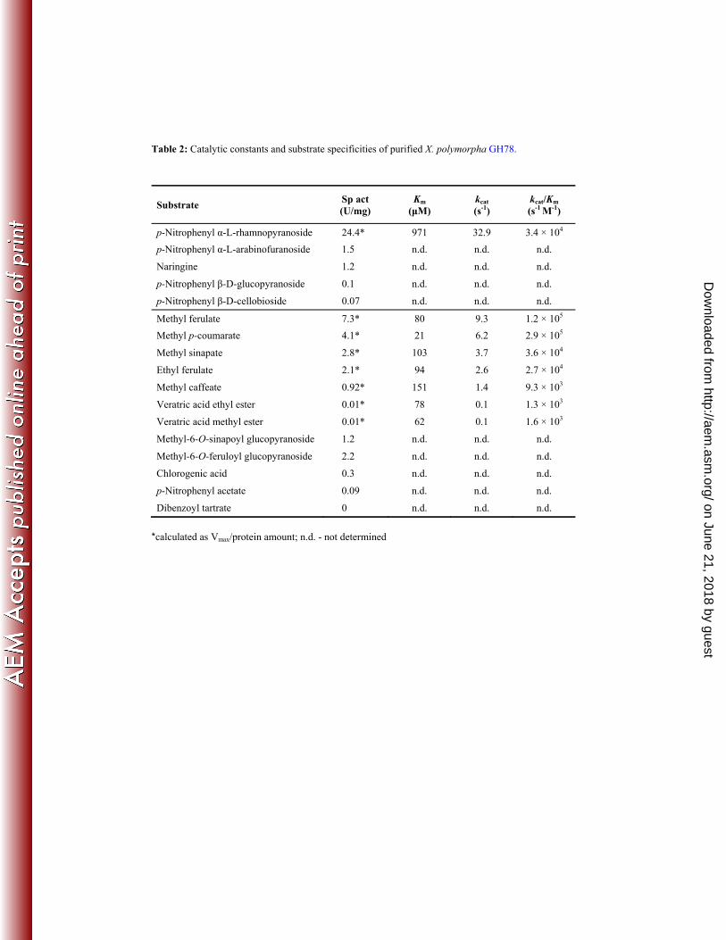
Purified XpoGH78 hydrolyzed p-NPRP with the highest specific activity (calculated as Vmax/protein 329
amount) of 24.4 U mg-1 (Table 2), which is consistent with its sequence signature as GH78. The 330
enzyme converted also the well-known rhamnosidase substrate naringin (1.2 Umg-1) into the 331
corresponding α-L-rhamnose and mono-glycosylated prunin (naringenin 7-glucoside) that was not 332
further hydrolyzed into the aglycon naringenin (data not shown). Thus the low specific β-glucosidase 333
activity observed for p-NPGP (0.1 U mg-1, Table 2) could not be confirmed for prunin. Other side 334
activities were found for p-NPAF (1.5 U mg-1) and p-NPCB (0.07 U mg-1) whilst p-NPXP was not 335
hydrolyzed by XpoGH78. 336
The broadness of the substrate spectrum of XpoGH78 became further evident by the hydrolysis of a 337
number of aryl-alkyl esters (e.g. MpCA), hydroxycinnamic acid glycoside esters (e.g. methyl-6-O-338
feruloyl-glucopyranoside) and quinic acid esters (e.g. chlorgenic acid = CGA) as well as acetylated 339
substrates (p-NPA) (Table 2). Michaelis-Menten constants (Km), turnover numbers (kcat) and catalytic 340
efficiencies (kcat/Km) were determined for typical FAE substrates, and the lowest Km of 21 µM (i.e. the 341
highest affinity) and the highst kcat/Km (2.9 × 10⁵ s-1 M-1) were observed for MpCA followed by MFA 342
(1.2 × 10⁵ s-1 M-1); the catalytic efficiencies for veratric acid esters were about two orders of magnitude 343
lower. The picture is similar when comparing the specific activities of XpoGH78: the highest values of 344
7.3 and 4.1 U mg-1 were found for MFA and respectively MpCA, while the activities for all other ester 345
substrates were lower. Relatively high activities were also observed for model compounds 346
representing glycoside-ester structures within the network of cell-wall polysaccharides (28). Thus, 347
XpoGH78 exhibited specific activities of 2.2 and 1.2 Umg-1for a feruloyl glucoside ester and a 348
sinapoyl glucoside ester, respectively. Minor activities were detected for CGA (0.3 Umg-1), an ester of 349
cyclitol quinic acid and polyphenolic caffeic acid always present in fruits, vegetables or cereals, and 350
for the artificial ester p-NPA (~0.1 Umg-1). On the other hand, dibenzoyl tartrate was not a substrate of 351
XpoGH78. All these findings indicate that the Xylaria GH78 protein is, from the catalytical point of 352
view, an α-L-rhamnosidase (EC 3.2.1.40) with strong esterase and other moderate side activities. 353
on June 21, 2018 by guesthttp://aem
.asm.org/
Dow
nloaded from
Page 15
15
354
Hydrolysis of wheat arabinoxylan and wheat straw 355
XpoGH78 partly hydrolyzed native cell-wall polymers, which was proven by the release of 356
hydroxycinnamic acids (p-coumaric and ferulic acid) from insoluble arabinoxylan or milled wheat-357
straw (Fig. 2A & B). The treatment of arabinoxylan with XpoGH78 resulted in a release of 0.37mg g-1 358
ferulic acid within 2 hours, which further increased to 0.45 mg g-1and finally 0.54 mg g-1over another 6 359
and 22 hours of incubation (Fig. 3). Remarkably, XpoGH78 acted also on native wheat-straw. The 360
major metabolites, ferulic and p-coumaric acid, were already detectable after 2 hours of incubation, 361
and their concentration of 0.24 mg g-1 and respectively 0.14 mg g-1corresponded to 14.5% and 10.1% 362
of the total amount of these acids alkaline extractable from wheat-straw. 363
Gene sequence and predicted features of XpoGH78 364
The fae1 gene (2,921bp) contains four introns in the middle of the sequence and encodes a protein of 365
866 amino acids with a calculated molecular weight of 93.4 kDa and a theoretical pI of 4.69. Peptide 366
fragments of XpoGH78 obtained by peptide mapping matched perfectly with the deduced amino acid 367
sequence according to the identified fae1 gene and cover 68.82% of the overall protein. Furthermore, 368
several conserved amino acids (e.g. catalytic residues) and GH78 specific domains (e.g. pfam08531), 369
calcium binding positions as well as six potential N-glycosylation sites were found in the translated 370
protein sequence (Fig. S 2). Surprisingly, no signal peptide sequence could be predicted with the 371
programs SignalP and iPSort. 372
DISCUSSION 373
Three ascomyceteous soft-rot fungi belonging to the family of Xylariaceae were found to produce high 374
levels of a methyl ferulate-hydrolyzing activity during growth on lignocellulosic materials. One 375
responsible enzyme with strong FAE activity was purified from X. polymorpha. Since the amino acid 376
sequence of the isolated hydrolase indicates its phylogenetic affiliation to the glycoside hydrolases 377
(EC 3.2.1.x; GH family 78, group I of α-L-rhamnosidases), it was designated as XpoGH78. The 378
on June 21, 2018 by guesthttp://aem
.asm.org/
Dow
nloaded from
Page 16
16
isolated enzyme exhibits a broad substrate specificity and was found to hydrolyze both glycosidic and 379
ester bonds. 380
The highest level of ester-cleaving activity of X. polymorpha [1,675 mU g-1, ~250 U L-1 in relation to 381
the water content of straw; (30)] was reached when the fungus was grown on wheat-straw in solid-382
state culture. Numerous microbial FAEs were found to be produced in solid-state culture when 383
complex carbon sources rich in esterified hydroxycinnamic acids, such as agricultural waste materials 384
(e.g. wheat- or maize-bran, sugar-beet pulp),were used (6, 46). Generally, the maximum FAE levels 385
reported considerably vary (2 to 180 U L-1), which depends on the particular microorganism but also 386
on the ester substrates used for enzyme detection (e.g. methyl ester, p-NPA or 4-nitrophenyl-5-O-387
feruloyl-α-L-arabinofuranoside) (5, 10). 388
Interestingly, the amino acid sequence of the isolated ester-cleaving protein does not show any 389
similarity to known fungal esterase sequences deposited in GenBank or CAZy database. Using 390
BLAST searches, strong similarities of the identified peptide sequence were found with fungal and 391
bacterial α-L-rhamnosidases of the GH family 78 (EC 3.2.1.40), which led to the designation 392
XpoGH78 for the enzyme. All of the approx. 400 homologous protein sequences of this family contain 393
highly conserved regions (Fig. 4) and amino acid residues crucial for substrate catalysis both of 394
glycosides (e.g. Asp474, Glu467) and esters (e.g. Asp741, Asp474, Glu467, Ser457, His476and 395
Cys462). Further the motif AASVA at position 631 was found that corresponds to Sm-X-Nu-X-Sm 396
(where X is any and Sm small amino acid), a characteristic sequence of the catalytic triad. 397
A phylogenetic analysis revealed a division of the GH 78 family into two large groups, I and II (Fig. 4, 398
Fig. S 3 and 4). XpoGH78 exhibits the highest sequence similarity to group I comprising 47 399
ascomycetous, one oomyceteous as well as 200 bacterial sequences. To our best knowledge, the vast 400
majority of these sequences refer to putative proteins with so far unknown catalytic properties, and all 401
fungal representatives are lacking N-terminal signal peptides. The only exception is a characterized 402
bacterial α-L-rhamnosidase from a Bacillus sp. with 38% identity and 56% similarity to XpoGH78 403
[BAB62314; (12)]. Within group I, the XpoGH78 sequence shows the highest similarities to putative 404
sequences of phylogenetically related ascomycetes (e.g. Aspergillus terreus 58% identity/72% 405
on June 21, 2018 by guesthttp://aem
.asm.org/
Dow
nloaded from
Page 17
17
similarity, Magnaporthe oryzae 54% identity/69% similarity). In contrast, the XpoGH78 sequence did 406
not match well (<20% similarity) with members of group II of the GH 78 family that includes 77 407
fungal and 42 bacterial sequences. Like group I, most members of group II are putative protein 408
sequences [only a few characterized fungal proteins have been reported; (47)], but contrary to group I, 409
many of the fungal sequences contain predicted N-terminal signal peptides (Fig. S 4). 410
XpoGH78 is a glycosylated protein (10%, data not shown) with molecular weights of 103 and 93 kDa 411
determined under native conditions (SEC & native PAGE, respectively) and 98 kDa estimated by 412
denaturing SDS-PAGE (Fig. 1D). The native protein may be a heterodimer that consists of two 413
subunits (39 and 62 kDa) or it is rather labile and may partially split under denaturing conditions. 414
Similar observations were made for different recombinant bacterial rhamnosidases which can occur as 415
monomers and dimers in dependence of native or denaturing conditions (3, 8, 50). 416
All in all, the physico-chemical and catalytic properties of XpoGH78 are comparable to that of the 417
characterized group II α-L-rhamnosidases of Aspergillus kawachii and A. terreus [90 kDa; (47)], and it 418
efficiently hydrolyzes p-NPRP but also naringin, the canonical substrates of the GH family 78 (12). 419
The specific activity of XpoGH78 for p-NPRP (24.4 U mg-1) is within the activity range reported for 420
fungal and bacterial α-L-rhamnosidases, e.g.of A. terreus [84 U mg-1; (18)], A. nidulans [9.3 U mg-1; 421
(47)] and Clostridium stercorarium [82 U mg-1; (50)]. 422
Interestingly, the enzyme was found to act on carboxylate ester bonds, a catalytic property that is 423
associated with hydrolases of the carboxyl-esterase family (CE; EC 3.1.1.x). Thus, XpoGH78 424
hydrolyzed diverse aryl esters and it turned out that an increasing number of substituents on the 425
aromatic ring of the substrate affected its affinity and the catalytic efficiency (from MpCA to MSA). 426
Thus, the additional hydroxyl group at the C3-position of MCA led to a decrease in the catalytic 427
efficiency by a factor of 30 compared to MpCA. An even more drastic activity loss was observed for a 428
FAE from A. niger, which hydrolyzed MpCA but no MCA (16). The activity towards veratric acid 429
esters increased with the decreasing number of C-atoms of the alcohol moiety (i.e. from ethyl to 430
methyl), probably due to sterical hindrance. 431
on June 21, 2018 by guesthttp://aem
.asm.org/
Dow
nloaded from
Page 18
18
The production of bifunctional and multifunctional glycosidases has been reported for several 432
filamentous fungi, mainly ascomycetes [e.g. arabinofuranosidase of Penicillium chrysogenum, 433
Aspergillus awamori; arabinofuranosidase/β-xylosidase of Fusarium graminearum; 434
arabinofuranosidase/xylobiohydrolase of Penicillium purpurogenum; (40)] and some bacteria [e.g. 435
bifunctional xylanase-deacetylase, (24); bifunctional xylanase-ferulic acid esterase, (15, 34)]. Ester 436
hydrolysis by β-glucosidases (EC 3.2.1.21) has been reported for a few bacterial proteins (e.g. from 437
Flavobacterium johnsonae and Clavibacter michiganense), which were found to cleave β-glycosyl 438
ester linkages present in phytohormone conjugates (9, 38). Overlapping substrate specificities were 439
also observed for FAE-B of A. niger, an enzyme that converted different esters including feruloylated 440
oligosaccharides, wheat arabinoxylan and alky-aryl esters; interestingly, this enzyme showed 441
significant sequence similarity to an Aspergillus tannase (EC 3.1.1.20) but without possessing the 442
respective enzymatic activity (6). A chlorogenic acid hydrolase (EC 3.1.1.42) with broad substrate 443
specificity was described for another A. niger strain and suggested to be a new esterase/protein type, 444
since it did not show any homology to known cinnamoyl esterases (2). Eventually, Koseki et al. 445
(2010) described an FAE from A. kawachii that contains the carbohydrate-binding module from the 446
GH family 54 (α-L-arabinofuranosidase, EC 3.2.1.55) (27). 447
The hydrolysis of complex esters indicates that XpoGH78 may be part of the lignocellulolytic system 448
of xylariaceous soft-rot fungi. This assumption was supported by the hydrolysis of hydroxycinnamic 449
acid glycoside esters naturally occurring in different plant tissues (28)or of quinic acid esters present 450
in bark but also by the release of coumaric and/or ferulic acids [main ester-linked hydroxycinnamic 451
acids in graminaceous plants; (6)] from polymeric wheat arabinoxylan and milled wheat-straw. Most 452
degradation studies demonstrating the release of hydroxycinnamic acids by fungal esterases were 453
carried out either in the presence of auxiliary enzymes [e.g. xylanases, arabinofuranosidases; (13, 26, 454
48)]or after pre-treatment of the complex lignocellulosic substrate e.g. steam explosion; (6, 7)]. 455
As indicated by an internal de-novo peptide located just one amino acid downstream of the 456
translational start methionine as well as by the SignalP and iPSort prediction, XpoGH78 does not 457
contain an N-terminal signal peptide that can be employed for standard protein translocation and 458
on June 21, 2018 by guesthttp://aem
.asm.org/
Dow
nloaded from
Page 19
19
secretion. Such findings,regarding obviously secreted enzymes without signal peptide, are rare but 459
have been reported for a number of extracellular bacterial proteins [e.g. lipase of Serratia marcescens; 460
(1); hemolysin of Escherichia coli; (17)] and fungal enzymes such as the β-xylosidase of the 461
phytopathogenic ascomycete Cochliobolus carbonum (43). XpoGH78 is a glycosylated (10% 462
carbohydrates, data not shown) as well as a pH- and thermostable protein that is not detectable 463
intracellular in cell lysates (data not shown). Due to these facts we assume that XpoGH78 may be 464
secreted either by the classic secretory pathway through the ER-Golgi pathway (36, 43) or by an 465
untypical protein releasing mechanism independent of the ER-Golgi pathway. Similar findings have 466
been proposed for an increasing number of proteins lacking N-terminal signal sequences (39). On the 467
other hand, this unusual structural property of XpoGH78 fits well into the picture of the α-L-468
rhamnosidase group I, all putative fungal members of which and the majority of the bacterial ones are 469
lacking N-terminal signal peptides. 470
In spite of the increasing number of glycoside hydrolase-encoding genes, which has been identified in 471
fungal genomes over the last years, the catalytic activities and physical properties of the corresponding 472
proteins are often unknown. Here, we followed the classic approach starting from esterase activities 473
detected in lignocellulosic materials and ended up with a glycoside hydrolase 78 protein (according to 474
its sequence), whose esterase activity would otherwise have been overlooked. Thus, XpoGH78 is the 475
first fungal protein of GH family 78 that exhibits aryl-alkyl esterase activity on numerous natural and 476
synthetic esters, and enables the soft-rot fungus to partially hydrolyze the lignocellulosic complex. 477
Such a catalytic versatility combined in one protein with activities both towards glycosides and esters 478
may be advantageous for the efficient degradation of the plant cell-wall complex that contains both 479
diverse sugar residues and esterified structures (42). Based on these results, it will be worthwhile to 480
test other members of the family GH 78 (as well as beyond that other glucosidases) for possible ester-481
cleaving activities. Vice versa, possible glucoside-hydrolyzing activities of known esterases (in 482
particular of FAEs), might be checked as well. 483
on June 21, 2018 by guesthttp://aem
.asm.org/
Dow
nloaded from
Page 20
20
Figure 1: (A) Timecourse of FAE activity in solid-state culture using wheat-straw (circles) and beech-484
wood (squares) as growth substrates. Data points represent mean values of three culture flasks with 485
standard deviation <5%. (B)FPLC elution profile of the 3rdpurification step of XpoGH78performed on 486
a MonoQ column. Absorbance at 280 nm (solid line), FAE activity (black circles) and NaCl gradient 487
(dashed line). (C) Elution profile of XpoGH78 before (dashed line) and after (solid line) 488
semipreparativeHPLC-SEC; the insert showsthe UV-Vis spectrumof theprotein peak exhibiting 489
rhamnosidase and FAE activity.(D) SDS-PAGE (left), native PAGE (middle) and native isoelectric 490
focusing (right) ofXpoGH78(lane 2, 4 & 5) after HPLC-SEC; protein maker (lane 1, 3 & 6). 491
Figure 2: Partial hydrolysis of arabinoxylan (A) and milled wheat-straw (B) by XpoGH78. HPLC 492
elution profiles were obtained under slightly different, isocratic separation conditions (15% and 10% 493
acetonirile, respectively) andmonitored at 323 nm after 12 h of incubation of the suspended polymeric 494
materials with active XpoGH78 (solid lines) orboiled enzyme (dashed lines). (1) ferulic acid, (2) p-495
coumaric acid. The inserts show the UV-Vis spectra of hydroxycinnamic acids. 496
Figure 3: Release of hydroxycinnamic acids from xylan and straw byMono-Q-purified 497
XpoGH78:ferulic acid from suspended arabinoxylan (closed diamonds); ferulic (open diamonds) and 498
p-coumaric (open triangles) acids from milled wheat-straw. The amount of released acids was 499
calculated in mg per g of polymeric substrate. 500
Figure 4: Phylogenetic analysis of eukaryotic and prokaryotic GH78 α-L-rhamnosidases defining two 501
major groups (I and II). The analysis was performed using the Neighbor-Joining method with Poisson 502
corrected protein distances and 500 bootstrap replicates. 371 Protein sequences were included with a 503
total of 3,939 positions. The protein alignment comprises two conserved stretches involved in 504
substrate recognition of fungal and bacterial representatives of both main groups as well as the novel 505
characterized X. polymorpha enzyme (XpoGH78). Arrows indicate structural and functional important 506
positions deduced from the 3D-structure of Bacillus sp. G1 [BAB62315;(12)]. 507
508
on June 21, 2018 by guesthttp://aem
.asm.org/
Dow
nloaded from
Page 21
21
ACKNOWLEDGMENTS 509
Financial support in the form of a doctorate scholarship by the German Academic Exchange Service 510
(DAAD; A/08/90704), the DBU project “Fungal secretoms” (13211-32), the integrated EU project 511
BIORENEW (NMP2-CT-2006-026456) and the administration of the International Graduate School 512
Zittau is gratefully acknowledged. Furthermore, we thank the colleagues of our lab for useful 513
comments and discussions as well as Ulrike Schneider and Monika Brandt for their technical 514
assistance. 515
516
517
518
519
520
521
522
523
524
525
526
527
528
on June 21, 2018 by guesthttp://aem
.asm.org/
Dow
nloaded from
Page 22
22
REFERENCES 529
1. Akatsuka, H., E. Kawai, K. Omori, S. Komatsubara, T. Shibatani, and T. Tosa. 1994. 530 The lipA gene of Serratia marcescens which encodes an extracellular lipase having no N-531 terminal signal peptide. J. Bacteriol. 176:1949-1956. 532
2. Asther, M., M. I. Estrada Alvarado, M. Haon, D. Navarro, M. Asther, L. Lesage-533 Meessen, and E. Record. 2005. Purification and characterization of a chlorogenic acid 534 hydrolase from Aspergillus niger catalysing the hydrolysis of chlorogenic acid. J. Biotechnol. 535 115:47-56. 536
3. Avila, M., M. Jaquet, D. Moine, T. Requena, C. Pelaez, F. Arigoni, and I. Jankovic. 2009. 537 Physiological and biochemical characterization of the two alpha-L-rhamnosidases of 538 Lactobacillus plantarum NCC245. Microbiol. 155:2739-2749. 539
4. Bannai, H., Y. Tamada, O. Maruyama, K. Nakai, and S. Miyano. 2002. Extensive feature 540 detection of N-terminal protein sorting signals. Bioinform. 18:298-305. 541
5. Bartolome, B., C. Gomez-Cordoves, A. I. Sancho, N. Diez, P. Ferreira, J. Solivery, and J. 542 L. Copa-Patino. 2003. Growth and release of hydroxycinnamic acids from brewer’s spent 543 grain by Streptomyces avermitilis CECT 3339. Enzyme Microb. Technol. 32:140-144. 544
6. Benoit, I., E. G. Danchin, R. J. Bleichrodt, and R. P. de Vries. 2008. Biotechnological 545 applications and potential of fungal feruloyl esterases based on prevalence, classification and 546 biochemical diversity. Biotechnol. Lett. 30:387-396. 547
7. Benoit, I., D. Navarro, N. Marnet, N. Rakotomanomana, L. Lesage-Meessen, J. C. 548 Sigoillot, M. Asther, and M. Asther. 2006. Feruloyl esterases as a tool for the release of 549 phenolic compounds from agro-industrial by-products. Carbohydr. Res. 341:1820-1827. 550
8. Birgisson, H., G. O. Hreggvidsson, O. H. Fridjonsson, A. Mort, J. K. Kristjansson, and 551 B. Mattiasson. 2004. Two new thermostable α-L-arabinofuranosidases from a novel 552 thermophilic bacterium. Enzyme Microb. Technol. 34:561-571. 553
9. Cairns, J. R. K., and A. Esen. 2010. β-glucosidases. Cell. Mol. Life Sci. 67:3389-3405. 554 10. Christov, L. P., and B. A. Prior. 1993. Esterases of xylan-degrading microorganisms: 555
production, properties, and significance. Enzyme Microb. Technol. 15:460-475. 556 11. Coutinho, P. M., and B. Henrissat. 1999. Carbohydrate-active enzymes: an integrated 557
database approach, p. 3-12. In H. J. Gilbert, G. Davies, B. Henrissat, and B. Svensson (ed.), 558 Recent advances in carbonhydrate bioengineering. The Royal Society of Chemistry, 559 Cambridge. 560
12. Cui, Z., Y. Maruyama, B. Mikami, W. Hashimoto, and K. Murata. 2007. Crystal structure 561 of glycoside hydrolase family 78 alpha-L-rhamnosidase from Bacillus sp. GL1. J. Mol. Biol. 562 374:384-98. 563
13. De Vries, R. P., H. C. M. Kester, C. H. Poulsen, J. A. E. Benen, and J. Visser. 2000. 564 Synergy between enzymes from Aspergillus involved in the degradation of plant cell wall 565 polysaccharides. Carbohydr. Res. 327:401-410. 566
14. Dieffenbach, C. W., T. M. Lowe, and G. S. Dveksler. 1993. General concepts for PCR 567 primer design. PCR Methods Appl. 3:30-37. 568
15. Dodd, D., S. A. Kocherginskaya, M. A. Spies, K. E. Beery, C. A. Abbas, R. I. Mackie, and 569 I. K. Cann. 2009. Biochemical analysis of a β-D-xylosidase and a bifunctional xylanase-570 ferulic acid esterase from a xylanolytic gene cluster in Prevotella ruminicola 23. J. Bacteriol. 571 191:3328-3338. 572
16. Faulds, C. B., and G. Williamson. 1994. Purification and characterisation of a ferulic acid 573 esterase (FAE-III) from Aspergillus niger. Specificity for the phenolic moiety and binding to 574 microcrystalline cellulose. Microbiol. 140:779-787. 575
17. Felmlee, T., S. Pellet, and R. A. Welch. 1985. Nucleotide sequence of an Escherichia coli 576 chromosomal hemolysin. J. Bacteriol. 163:94-105. 577
18. Gallego, M. V., F. Pinaga, D. Ramon, and S. Valles. 2001. Purification and characterization 578 of an α-L-rhamnosidase from Aspergillus terreus of interest in winemaking. J. Food Sci. 579 66:204-209. 580
on June 21, 2018 by guesthttp://aem
.asm.org/
Dow
nloaded from
Page 23
23
19. Gasteiger, E., C. Hoogland, A. Gattiker, S. Duvaud, M. R. Wilkins, R. D. Appel, and A. 581 Bairoch. 2005. Protein identification and analysis tools on the ExPASy server, p. 571-607. In 582 J. M. Walker (ed.), The proteomics protocols handbook. Humana Press, Totowa. 583
20. Grossmann, J., B. Roschitzki, C. Panse, C. Fortes, S. Barkow-Oesterreicher, D. 584 Rutishauser, and R. Schlapbach. 2010. Implementation and evaluation of relative and 585 absolute quantification in shotgun proteomics with label-free methods. J. Proteomics 73:1740-586 1746. 587
21. Hashimoto, K., S. Kaneko, and M. Yoshida. 2010. Extracellular carbohydrate esterase from 588 the basidiomycete Coprinopsis cinerea released ferulic and acetic acids from xylan. Biosci. 589 Biotechnol. Biochem. 74:1722-1724. 590
22. Julenius, K., A. Molgaard, R. Gupta, and S. Brunak. 2005. Prediction, conservation 591 analysis, and structural characterization of mammalian mucin-type O-glycosylation sites. 592 Glycobiol. 15:153-164. 593
23. Kendrick, B. 2000. The Fifth Kingdom, 3rd ed, vol. Focus Publishing, Newbury, 594 Massachsetts. 595
24. Khandeparker, R., and M. T. Numan. 2008. Bifunctional xylanases and their potential use 596 in biotechnology. J. Ind. Microbiol. Biotechnol. 35:635-644. 597
25. Kontkanen, H., A. Westerholm-Parvinen, M. Saloheimo, M. Bailey, M. Ratto, I. Mattila, 598 M. Mohsina, N. Kalkkinen, T. Nakari-Setala, and J. Buchert. 2009. Novel Coprinopsis 599 cinerea polyesterase that hydrolyzes cutin and suberin. Appl. Environ. Microbiol. 75:2148-600 2157. 601
26. Koseki, T., A. Hori, S. Seki, T. Murayama, and Y. Shiono. 2009. Characterization of two 602 distinct feruloyl esterases, AoFaeB and AoFaeC, from Aspergillus oryzae. Appl. Microbiol. 603 Biotechnol. 83:689-696. 604
27. Koseki, T., K. Mochizuki, H. Kisara, A. Miyanaga, S. Fushinobu, T. Murayama, and Y. 605 Shiono. 2010. Characterization of a chimeric enzyme comprising feruloyl esterase and family 606 42 carbohydrate-binding module. Appl. Microbiol. Biotechnol. 86:155-161. 607
28. Kylli, P., P. Nousiainen, P. Biely, J. Sipila, M. Tenkanen, and M. Heinonen. 2008. 608 Antioxidant potential of hydroxycinnamic acid glycoside esters. J. Agric. Food. Chem. 609 56:4797-805. 610
29. Liers, C., R. Ullrich, M. Pecyna, D. Schlosser, and M. Hofrichter. 2007. Production, 611 purification and partial enzymatic and molecular characterization of a laccase from the wood-612 rotting ascomycete Xylaria polymorpha. Enzym. Microb. Technol. 41:785-793. 613
30. Liers, C., R. Ullrich, K. T. Steffen, A. Hatakka, and M. Hofrichter. 2006. Mineralization 614 of 14C-labelled synthetic lignin and extracellular enzyme activities of the wood-colonizing 615 ascomycetes Xylaria hypoxylon and Xylaria polymorpha. Appl. Microbiol. Biotechnol. 616 69:573-579. 617
31. Liu, X., and S. Ding. 2009. Molecular characterization of a new acetyl xylan esterase 618 (AXEII) from edible straw mushroom Volvariella volvacea with both de-O-acetylation and 619 de-N-acetylation activity. FEMS Microbiol. Lett. 295:50-56. 620
32. Martínez, A. T., M. Speranza, F. J. Ruiz-Dueñas, P. Ferreira, S. Camarero, F. Guillén, 621 M. J. Martínez, A. Gutiérrez, and J. C. del Río. 2005. Biodegradation of lignocellulosics: 622 microbial, chemical, and enzymatic aspects of the fungal attack of lignin. Int. Microbiol. 623 8:195-204. 624
33. Matz, M., D. Shagin, E. Bogdanova, O. Britanova, S. Lukyanov, L. Diatchenko, and A. 625 Chenchik. 1999. Amplification of cDNA ends based on template-switching effect and step-626 out PCR. Nucleic Acids Res. 27:1558-1560. 627
34. Montanier, C., V. A. Money, V. M. Pires, J. E. Flint, B. A. Pinheiro, A. Goyal, J. A. 628 Prates, A. Izumi, H. Stalbrand, C. Morland, A. Cartmell, K. Kolenova, E. Topakas, E. J. 629 Dodson, D. N. Bolam, G. J. Davies, C. M. Fontes, and H. J. Gilbert. 2009. The active site 630 of a carbohydrate esterase displays divergent catalytic and noncatalytic binding functions. 631 PLoS Biol. 7:0687-0697. 632
35. Monties, B., and Y. Fukushima. 2001. Occurence, function and biosynthesis of lignins, p. 1-633 64. In M. Hofrichter and A. Steinbüchel (ed.), Biopolymers, vol. 1. Wiley-VCH, Weinheim. 634
36. Muesch, A., E. Hartmann, K. Rohde, A. Rubartelli, R. Sitia, and T. A. Rapoport. 1990. A 635 novel pathway for secretory proteins? Trends Biochem. Sci. 15:86-88. 636
on June 21, 2018 by guesthttp://aem
.asm.org/
Dow
nloaded from
Page 24
24
37. Nielsen, H., J. Engelbrecht, S. Brunak, and G. von Heijne. 1997. A neural network method 637 for identification of prokaryotic and eukaryotic signal peptides and prediction of their 638 cleavage sites. Int. J. Neural. Syst. 8:581-599. 639
38. Okamoto, K., H. Nakano, T. Yatake, T. Kiso, and S. Kitahata. 2000. Purification and some 640 properties of a beta-glucosidase from Flavobacterium johnsonae. Biosci. Biotechnol. 641 Biochem. 64:333-340. 642
39. Prudovsky, I., A. Mandinova, R. Soldi, C. Bagala, I. Graziani, M. Landriscina, F. 643 Tarantini, M. Duarte, S. Bellum, H. Doherty, and T. Maciag. 2003. The non-classical 644 export routes: FGF1 and IL-1alpha point the way. J. Cell Sci. 116:4871-4881. 645
40. Ravanal, M. C., E. Callegari, and J. Eyzaguirre. 2010. Novel bifunctional α-L-646 arabinofuranosidase/xylobiohydrolase (ABF3) from Penicillium purpurogenum. Appl. 647 Environ. Microbiol. 76:5247-5253. 648
41. Tamura, K., J. Dudley, M. Nei, and S. Kumar. 2007. MEGA4: Molecular evolutionary 649 genetics analysis (MEGA) software version 4.0. Mol. Biol. Evol. 24:1596-1599. 650
42. Van den Brink, J., and R. P. De Vries. 2011. Fungal enzyme sets for plant polysaccharide 651 degradation. Appl. Microbiol. Biotechnol. 91:1477-1492. 652
43. Wegener, S., R. F. Ransom, and J. D. Walton. 1999. A unique eukaryotic beta-xylosidase 653 gene from the phytopathogenic fungus Cochliobolus carbonum. Microbiol. 145:1089-1095. 654
44. Whalley, A. J. S. 1996. The xylariaceous way of life. Mycol. Res. 100:897-922. 655 45. Williamson, G., P. A. Kroon, and C. B. Faulds. 1998. Hairy plant polysaccharide: a close 656
shave with microbial esterases. Microbiol. 144:2011-2023. 657 46. Wong, D. W. 2006. Feruloyl esterase: a key enzyme in biomass degradation. Appl. Biochem. 658
Biotechnol. 133:87-112. 659 47. Yadav, V., P. K. Yadav, and K. D. S. Yadav. 2010. α-L-Rhamnosidase: A review. Pro. 660
Biochem. 45:1226-1235. 661 48. Yu, P., D. D. Maenz, J. J. McKinnon, V. J. Racz, and D. A. Christensen. 2002. Release of 662
ferulic acid from oat hulls by Aspergillus ferulic acid esterase and Trichoderma xylanase. J. 663 Agric. Food. Chem. 50:1625-1630. 664
49. Zimmer, C., T. Platz, N. Cadez, F. Giffhorn, and G. W. Kohring. 2006. A cold active 665 (2R,3R)-(-)-di-O-benzoyl-tartrate hydrolyzing esterase from Rhodotorula mucilaginosa. Appl. 666 Microbiol. Biotechnol. 73:132-140. 667
50. Zverlov, V. V., C. Hertel, K. Bronnenmeier, A. Hroch, J. Kellermann, and W. H. 668 Schwarz. 2000. The thermostable alpha-L-rhamnosidase RamA of Clostridium stercorarium: 669 biochemical characterization and primary structure of a bacterial alpha-L-rhamnoside 670 hydrolase, a new type of inverting glycoside hydrolase. Mol. Microbiol. 35:173-179. 671
on June 21, 2018 by guesthttp://aem
.asm.org/
Dow
nloaded from
Page 25
on June 21, 2018 by guesthttp://aem
.asm.org/
Dow
nloaded from
Page 26
on June 21, 2018 by guesthttp://aem
.asm.org/
Dow
nloaded from
Page 27
on June 21, 2018 by guesthttp://aem
.asm.org/
Dow
nloaded from
Page 28
on June 21, 2018 by guesthttp://aem
.asm.org/
Dow
nloaded from
Page 29
Table 1: Purification of GH78 protein from X. polymorpha (measured by demethylation of methyl
ferulate) produced during solid-state fermentation of wheat-straw. Cultures were harvested
after 25 days and extracted with water.
Purification steps Total act (U)
Total protein
(mg)
Sp act (U/mg)
Yield (%)
Purification (fold)
Wheat straw extract 57.4 910.8 0.06 100 1.0 Ultrafiltration 54.5 620.5 0.09 94.9 1.5 DEAE Sepharose 43.9 68.2 0.64 76.5 10.7 Superdex Sepharose 20.6 11.5 1.79 35.9 29.8 Mono Q 4.9 1.9 2.58 8.5 43.0 Biosep-SEC-S-2000 3.8 1.7 2.24 6.6 37.3
on June 21, 2018 by guesthttp://aem
.asm.org/
Dow
nloaded from
Page 30
Table 2: Catalytic constants and substrate specificities of purified X. polymorpha GH78.
Substrate Sp act (U/mg)
Km (μM)
kcat (s-1)
kcat/Km (s-1 M-1)
p-Nitrophenyl α-L-rhamnopyranoside 24.4* 971 32.9 3.4 × 10⁴ p-Nitrophenyl α-L-arabinofuranoside 1.5 n.d. n.d. n.d.
Naringine 1.2 n.d. n.d. n.d.
p-Nitrophenyl β-D-glucopyranoside 0.1 n.d. n.d. n.d.
p-Nitrophenyl β-D-cellobioside 0.07 n.d. n.d. n.d.
Methyl ferulate 7.3* 80 9.3 1.2 × 10⁵ Methyl p-coumarate 4.1* 21 6.2 2.9 × 10⁵
Methyl sinapate 2.8* 103 3.7 3.6 × 10⁴
Ethyl ferulate 2.1* 94 2.6 2.7 × 10⁴
Methyl caffeate 0.92* 151 1.4 9.3 × 10³
Veratric acid ethyl ester 0.01* 78 0.1 1.3 × 10³
Veratric acid methyl ester 0.01* 62 0.1 1.6 × 10³
Methyl-6-O-sinapoyl glucopyranoside 1.2 n.d. n.d. n.d.
Methyl-6-O-feruloyl glucopyranoside 2.2 n.d. n.d. n.d.
Chlorogenic acid 0.3 n.d. n.d. n.d.
p-Nitrophenyl acetate 0.09 n.d. n.d. n.d.
Dibenzoyl tartrate 0 n.d. n.d. n.d.
*calculated as Vmax/protein amount; n.d. - not determined
on June 21, 2018 by guesthttp://aem
.asm.org/
Dow
nloaded from