Page 1
UNIVERSITY OF SPLIT SCHOOL OF MEDICINE
Jasenka Kraljević, MD
Effect of aerobic interval training on pathological
remodelling and mitochondrial dysfunction in the
post-infarction failing rat heart
Doctoral Dissertation
Split, Croatia 2015
Page 2
1
UNIVERSITY OF SPLIT
SCHOOL OF MEDICINE
Jasenka Kraljević
EFFECT OF AEROBIC INTERVAL TRAINING ON
PATHOLOGICAL REMODELLING AND
MITOCHONDRIAL DYSFUNCTION IN THE
POST-INFARCTION FAILING RAT HEART
Doctoral Dissertation
Split, Croatia 2015
Page 3
2
SVEUĈILIŠTE U SPLITU
MEDICINSKI FAKULTET
Jasenka Kraljević
UĈINAK AEROBNOG INTERVALNOG TRENINGA NA
PATOLOŠKO REMODELIRANJE I MITOHONDRIJSKU
DISFUNKCIJU U ŠTAKORA S POSLIJEINFARKTNIM
ZATAJENJEM SRCA
Doktorska disertacija
Split, 2015.
Page 4
3
PREFACE
The studies included in this thesis have been carried out at the Department of
Physiology, School of Medicine, at University of Split during the years 2010-2014
under supervision of my mentor Professor Jasna Marinović – Ljubković. The working
hypothesis of the project was that aerobic interval training attenuates deterioration of
mitochondrial function in post-myocardial infarction failing rat heart. The main part
of the study included in this thesis was published in original paper below:
Kraljevic J, Marinovic J, Pravdic D, Zubin P, Dujic Z, Wisloff U, Ljubkovic M.
Aerobic interval training attenuates remodelling and mitochondrial dysfunction in the
post-infarction failing rat heart
Cardiovascular Research. 2013 Jul 1;99(1):55-64. 2013 Apr 3.
Page 5
4
ACKNOWLEDGEMENTS
First of all, very special thanks to my mentors Professor Jasna Marinović-
Ljubković and Professor Marko Ljubković for introducing me to the world of cellular
physiology, their guidance, counsel, intervention, and tremendous support throughout
this journey. I thank the head of the Department of Integrative Physiology Professor
Željko Dujić for his contribution to my scientific education, enthusiastic support and
critical advice in all parts of my work. I thank Dr. Martin Bienengraeber for helpful
discussions and critical reading of the manuscript. Thanks to Professor Ulrik Wisløff
and colleagues from Department of Circulation and Medical Imaging at Norwegian
University of Science and Technology in Trondheim where we conducted part of our
joint research.
Thanks to Ms. Ivana Banić for valuable technical assistance especially in
surgery procedures and in establishing rat-training model. I would be remiss if I did
not mention the assistance of all of my colleagues from Department of Physiology
and other departments at School of Medicine Split. The Animal Facility Unit at the
University of Split provided all animals and their help is acknowledged.
Very special thanks to my dissertation committee members Professor Janoš
Terzić, Professor Damir Fabijanić and Professor Stjepan Gamulin for valuable
advices, suggestions and comments.
This work was supported by the Unity Through the Knowledge Fund [grant
number 50/09 to M.LJ.], and the Department of Physiology, University of Split School
of Medicine; by K.G. Jebsen Foundation, The Norwegian Council on Cardiovascular
Disease, and The Research Council of Norway [U.W.]; and by the grant from the
Federal Ministry of Science and Education [D.P.].
Page 6
5
TABLE OF CONTENTS
PREFACE ...................................................................................................................... 3
ACKNOWLEDGEMENTS ........................................................................................... 4
FREQUENTLY USED ABBREVIATIONS ................................................................. 6
1. INTRODUCTION................................................................................................. 7
1.1 Heart failure in general................................................................................ 7
1.2 Etiology ....................................................................................................... 8
1.3 Ischemic heart failure .................................................................................. 8
1.4 Cardiac remodelling after myocardial infarction ........................................ 9
1.4.1 Hemodynamic and cellular changes ............................................... 10
1.4.2 Neurohormonal changes ................................................................. 12
1.4.3 Impaired energy metabolism .......................................................... 13
1.5 Mitochondria in heart failure .................................................................... 14
1.5.1 Mitochondrial morphology ............................................................. 15
1.5.2 Mitochondria-mediated apoptosis and ROS-mediated damage ..... 16
1.5.3 Mitochondrial energetics ................................................................ 17
1.6 Exercise training in chronic heart failure .................................................. 20
1.6.1 Clinical aspects ............................................................................... 20
1.6.2 Cellular and molecular effects of exercise in HF .......................... 21
2. AIMS OF THE STUDY ..................................................................................... 23
3. MATERIALS AND METHODS ....................................................................... 24
4. RESULTS ............................................................................................................ 33
5. DISCUSSION ...................................................................................................... 45
6. MAIN CONCLUSIONS ..................................................................................... 49
SUMMARY ................................................................................................................. 50
SAŽETAK ................................................................................................................... 51
REFERENCES ............................................................................................................ 55
CURRICULUM VITAE .............................................................................................. 66
Page 7
6
FREQUENTLY USED ABBREVIATIONS
HF: heart failure
CHF: chronic heart failure
MI: myocardial infarction
ROS: reactive oxygen species
ETC: electron transfer chain
MPT: mitochondrial permeability transition
MPTP: MPT pore
NYHA: The New York Heart Association
ACC/AHA: American College of Cardiology/American Heart Association
LVSD: left ventricular systolic dysfunction
HFPEF: heart failure with preserved ejection fraction
HFNEF: heart failure with normal ejection fraction
HFPSF: heart failure with preserved systolic function
EF: ejection fraction
LV: left ventricle
CAD: coronary artery disease
cAMP: cyclic adenosine monophosphate
MMPs: matrix metalloproteinases
ACE: angiotensin converting enzyme
Page 8
7
1. INTRODUCTION
1.1 Heart failure in general
Heart failure (HF) is a complex clinical syndrome defined as any structural or
functional cardiac disorder that impairs the ability of the ventricle to fill with or eject
blood. Heart failure is a leading cause of morbidity and mortality in the world as more
and more patients survive myocardial infarctions due to advances in therapeutic
strategies. The key feature of HF is the deficiency in the capability of the heart to
adequately pump blood in response to systemic demands of the metabolizing tissues
and is characterized by an altered cardiovascular, skeletal muscle, and neurohormonal
function attempting to maintain circulatory homeostasis (1). The main manifestations
of HF are dyspnea at rest or during exertion, fatigue, limited exercise tolerance, and
fluid retention, which may lead to pulmonary congestion and peripheral edema. Both
abnormalities can impair the functional capacity and quality of life of affected
individuals, but they do not necessarily dominate the clinical picture at the same time.
Because some patients present without signs or symptoms of volume overload at the
time of initial or subsequent evaluation, the term “heart failure” is preferred over the
older term “congestive heart failure.” It should be emphasized that HF is not
equivalent to cardiomyopathy or left ventricular dysfunction. These terms describe
possible structural or functional reasons for the development of HF (2). Nearly any
form of heart disease may ultimately lead to the HF syndrome. Instead, HF is defined
as a clinical syndrome that is characterized by specific symptoms in the medical
history and signs on the physical examination (3).
Heart failure is a global problem with considerable morbidity and mortality
despite improved understanding of the pathophysiology and better therapeutic
options. The prevalence of heart failure can be estimated at 1–2% in the western
world and the incidence approaches 5–10 per 1000 persons per year. The prevalence
of heart failure in 70- to 80-year-old people is between 10 and 20% (4). However, the
overall prevalence of heart failure is increasing due to ageing of the population, as
well as increasingly successful therapy and prolonged survival of patients suffering
from acute coronary events. However, despite the improved therapy, still 30–40% of
HF patients die within a year of diagnosis and 60–70% die within 5 years (5).
Page 9
8
1.2 Etiology
The causes of HF are very heterogeneous and range from disorders of
pericardium, myocardium, endocardium, heart valves, great vessels or certain
metabolic abnormalities, with the most common causes being acute or chronic
myocardial ischemia, increased vascular resistance with hypertension, and
abnormality in rhythm or conduction. In general, the etiology of HF may be divided in
two categories: 1) ischemic, resulting from coronary artery disease and myocardial
ischemia; and 2) non-ischemic (6). In developed world, heart failure most commonly
develops as a sequel of myocardial infarction or cardiac pressure overload resulting
from chronic arterial hypertension. Coronary heart disease is by far the most common
cause of HF, being the initiating cause in 70% of patients with HF (7).
Reduced coronary blood flow and oxygen delivery in CAD leads to
myocardial hypoxia and impaired cardiac function. Myocardial infarction is the final
and often fatal culmination of CAD. Degenerative valve disease is becoming more
common and accounts for about 10% of HF. The exact etiology is unknown in 20-
30% of cases of HF with depressed EF and therefore categorized in group of
idiopathic cardiomyopathies (6).
1.3 Ischemic heart failure
Acute myocardial infarction is one of the most significant causes of HF in
human population with the estimated incidence varying from 10% to 40%.
Improvements in the management and therapy together with ageing population have
contributed to a growing burden of heart failure. HF developed after myocardial
infarction is associated with a markedly elevated risk of death, with an estimated
median survival of about 4 years (8, 9). Progression to chronic heart failure after a
myocardial infarction may or may not developed which depends on multifactorial
causes involving the extent of initial myocardial damage, recurrent ischemia and the
extent of myocardial remodelling and chronic neuroendocrine stimulation (10). Post-
MI HF results from complex processes of pathological remodelling occurring in the
Page 10
9
surviving myocardium after one or more MI(s) (11). Heart failure is the most common
after anterior MI comparing to other sites (12, 13). Left ventricular systolic
dysfunction is the single most common cause of post-MI heart failure. At the cellular
level, the main substrate for the development of heart failure is a moderate amount of
myocardial necrosis with consequent ventricular remodelling (14, 15). This adverse
cardiac remodelling is a key feature of HF generally accepted as a major determinant
of clinical course and outcome of the disease, generally preceding the development of
symptoms and contributing substantially to worsening of symptoms despite treatment
(16). The process of ventricular remodelling occurs rapidly in the early period after
myocardial infarction and then more slowly thereafter. Therefore, early identification
of adverse cardiac remodelling offers the potential to modify this process and reduce
the risk of heart failure.
1.4 Cardiac remodelling after myocardial infarction
Pathological cardiac remodelling encompasses molecular, cellular and
interstitial changes after cardiac injury resulting in changes of ventricular size, shape
and function, ending in clinical manifestation of the HF syndrome. The process of
pathological remodelling is regulated by mechanical (hemodynamic load),
neurohormonal and genetic factors. Although remodelling in general is a normal
physiological process of adaptation during growth and other physiological stimuli, it
becomes pathological due to MI, hypertension or valvular heart disease. Therefore,
cardiac remodelling has been described as both an adaptive and a maladaptive
process, with the adaptive component enabling the heart to maintain function in
response to pressure or volume overloading in the acute phase of cardiac injury (17).
However, continued remodelling may not be necessary to maintain the integrity of the
circulation after cardiac injury. Once established beyond a certain phase, it may be
viewed as an adverse phenomenon that actually contributes to HF progression.
However, there is no indication when the transition from possible adaptive to
maladaptive remodelling occurs or how this might be identified in patients (18).
Progressive remodelling is always associated with a poor prognosis and patients with
major remodelling demonstrate progressive worsening of cardiac function. Cardiac
Page 11
10
remodelling is now recognized as an important aspect of disease progression and is,
therefore, emerging as a therapeutic target in HF of all etiologies (6).
In post-MI models, the process of LV remodelling begins within the first few
hours of myocyte injury and is influenced by the combination of infarct expansion,
and cardiac pressure and volume overload. Although the exact picture of all pathways
involved in LV remodelling is still unclear, intense research efforts have identified
several key factors responsible for the post-MI pathological remodelling in the remote
non-infarcted myocardium (19, 20). The progression of remodelling and deterioration
of heart failure probably occurs in two main ways. One is as a consequence of
intervening cardiac events, and the other as a consequence of the systemic
compensatory processes activated by the falling cardiac output (20).
1.4.1 Hemodynamic and cellular changes
In general, processes of ventricular remodelling encompass the entire LV in
proportion to infarct size. As a result, ejection fraction will decrease in direct relation
to the size of the infarction. The early post-MI remodelling involves infarct zone
expansion and occurs within 72 hours of MI. Late remodelling involves global
changes of LV including ventricular dilatation, distortion of shape and hypertrophy.
These changes in LV arise from the profound hemodynamic changes that occur within
the post-infarcted LV. The LV cavity enlargement is caused by the volume overload
and reflects an increase in the length of the remaining contractile tissue. The increase
LV volume augments the stroke volume by the Starling mechanism thus maintaining
the relatively normal cardiac output at increased filling volumes. Initially, the
compensatory hypertrophy in the remote myocardium makes up for the functional
loss of the infarcted myocardium (19, 21, 22). However, over the time this adaptive
hypertrophy becomes detrimental because the increased ventricular radius increases
the wall stress by Laplace law and thus increases oxygen demand. Progressive left
ventricular dilation and hypertrophy, infarct scar thinning, and alterations of the left
ventricular geometry adversely affect cardiac function (23). Such long-term
progressive remodelling of the LV with increases in the ventricular cavity size can
occur up to 2 years post-infarction with the increased risk of cardiovascular death (21,
24).
Page 12
11
At the cellular level, remodelling involves myocyte hypertrophy, necrosis,
apoptosis, interstitial fibrosis with collagen deposition and fibroblast proliferation.
Myocyte death within the infarcted and non-infarcted myocardium plays an important
role in the expansion of infarction, thinning of the ventricle wall and the cell slippage
in the remote myocardium (25). Apoptosis occurs in the infarcted, peri-infarcted area
and the remote non-infarcted myocardium, while necrosis mostly occurs acutely in the
infarction zone. Apoptosis is coordinated by multiple triggers, all of which occur in
the myocardium after MI, including neurohormonal system activation, cytokines such
as TNF alpha and interleukins, oxidative stress, mitochondrial damage, and other
extra-cellular factors (26, 27). Both apoptosis and necrosis cause further deterioration
in the composition and function of the ventricle (28).
Additionally, both the contractile apparatus and excitation-contraction
coupling are significantly altered in failing cardiac myocytes (29). For example, there
are changes in myosin heavy chain composition, down-regulation of alpha chains and
up-regulation of beta chains (30, 31). Furthermore, disturbed sarcoplasmic reticulum
function seems to play a central role for the altered systolic and diastolic performance.
Under physiological conditions calcium released from the sarcoplasmic reticulum
(SR) is the dominant source for systolic activation of contractile proteins. Diastolic
relaxation depends on calcium removal from the cytosol by the sarcoplasmic
reticulum and the sarcolemmal Na/Ca2+
- exchanger (32). In HF, there is a defect of
excitation-contraction coupling resulting from decreased capacity of the SR to
accumulate calcium and diastolic calcium accumulation in the cytosol causing the
disturbed diastolic function. Such alterations of calcium handling by the SR
significantly contribute to disturbed contractile function of individual myocytes as
well as to the development of arrhythmias in patients with heart failure (32, 33).
Although the contribution of the interstitium to the remodelling process is still
not completely clear, it is well established that alterations in the extra-cellular matrix
are critical in the process of ventricular remodelling. The extra-cellular matrix
supports the inter-cellular adhesion, coordinates cell signaling through integrins,
matrix metalloproteinases and their tissue inhibitors. It is composed of many
structural components including fibroblasts, collagen, elastin, and laminin (15, 34,
35). Between these structural components lay the zinc dependent enzymes, matrix
metalloproteinases and their respective endogenous tissue inhibitors (36-38). The
Page 13
12
balance of degradation and preservation of the structural components is controlled by
the activity of matrix metalloproteinases and their tissue inhibitors. Matrix
metalloproteinases are secreted from multiple cell types, including endothelial cells,
fibroblasts, smooth muscle cells, and cardiomyocytes into the extra-cellular space.
Over the past several years substantial evidence demonstrates that matrix
metalloproteinases are actively involved in cardiac remodelling after MI (39, 40).
In summary, at the cellular level the final common pathway for the
progression of cardiac remodelling and heart failure is the imbalance of hypertrophy
and cell death over regeneration, which, combined with the significantly altered
intracellular contractile apparatus, results in severe dysfunction at the level of whole
left ventricle (41, 42).
1.4.2 Neurohormonal changes
There is substantial evidence that the neurohormonal activation plays a crucial
role in cardiac remodelling and in the progression of HF. Neurohormonal activation in
HF is major compensatory mechanism responding to falling cardiac output, but it is
also a major component of disease progression and of the remodelling process.
Sympathetic and renin-angiotensin-aldosterone system (RAAS) respond to decreased
stroke volume and, at first, have compensatory role. They result in arterial and
venous vasoconstriction, increased blood volume, and temporary improvement in
systolic blood pressure and tissue perfusion (43). However, sympathetic and RAAS
activation leads to salt and water retention and excessive vasoconstriction that may
result in an elevation of cardiac preload and afterload, resulting in augmented cardiac
strain, energy expenditure and further progression of HF. The increase in myocardial
energy expenditure further decreases cardiac output and leads to myocardial cell death
(44). Additional reduction in cardiac output further perpetuates a cycle through
neurohormonal stimulation and adverse hemodynamic and myocardial responses. It
was shown, that higher levels of circulating plasma norepinephrine correlate with a
poorer long-term prognosis in HF patients. Moreover, neurohormonal activation also
has direct cytotoxic effects on the myocytes and interstitium that together with
stimulated myocardial fibrosis alter the structure and impair the cardiac performance
in HF. The neurohumoral factors lead to myocyte hypertrophy and interstitial fibrosis,
Page 14
13
resulting in increased myocardial volume, mass, and myocyte loss. As a result, the
cardiac architecture changes, which, in turn, leads to further increase in myocardial
volume and mass (18).
The role of renin angiotensin aldosterone system (RAAS) has been intensively
investigated in heart failure development after a myocardial infarction. It is well
established that chronic adrenergic system activation and resulting up-regulation of
the RAAS plays a major role in post-MI cardiac remodelling. In the heart, angiotensin
II has multiple direct cytotoxic effects on myocytes: inducing apoptosis, promoting
cell hypertrophy, and stimulating myocardial fibrosis by angiotensin II type 1
receptor. For example, increased levels of both angiotensin II and aldosterone have
been shown in vitro and in vivo to have cytotoxic effect and increase the rate of
myocyte apoptosis (45). Also, locally produced angiotensin II leads to increased
myocardial energy expenditure, thus having has similar actions as norepinephrine in
heart failure. Finally, aldosterone itself plays a role in left ventricular remodelling,
particularly myocardial fibrosis, by stimulating cardiac collagen synthesis, including
collagen type I and type III (46-48).
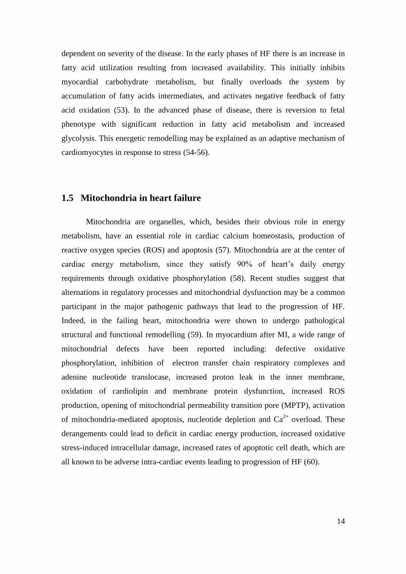
1.4.3 Impaired energy metabolism
Energy depletion, evidenced by the loss of ATP, rise in ADP, and damaged
energy transfer via creatine–phosphocreatine system, is implicated as a central factor
in the development of cardiac contractile insufficiency cardiac contractile
insufficiency (49, 50). The complex energetic state of the failing heart includes
changes in substrate utilization from fatty acid to glucose, decreased oxidative
capacity and energy production as a result of reduced mitochondrial biogenesis,
reduced energy transfer by the phosphotransfer kinases, impaired energy utilization
and efficiency of energy consumption (49, 50). Several lines of evidence support the
concept of reversion to the fetal metabolic phenotype in HF that shifts away from
fatty acids to carbohydrate utilization (51). However, decrease in fatty acids oxidation
has not been observed in all HF studies and in human studies were not consistent to
support the hypothesis of reversion to fetal pattern of metabolism (52). According to
clinical studies data both fatty acids and glucose are required for optimal function of
the failing myocardium and the observed different substrate selectivity is probably
Page 15
14
dependent on severity of the disease. In the early phases of HF there is an increase in
fatty acid utilization resulting from increased availability. This initially inhibits
myocardial carbohydrate metabolism, but finally overloads the system by
accumulation of fatty acids intermediates, and activates negative feedback of fatty
acid oxidation (53). In the advanced phase of disease, there is reversion to fetal
phenotype with significant reduction in fatty acid metabolism and increased
glycolysis. This energetic remodelling may be explained as an adaptive mechanism of
cardiomyocytes in response to stress (54-56).
1.5 Mitochondria in heart failure
Mitochondria are organelles, which, besides their obvious role in energy
metabolism, have an essential role in cardiac calcium homeostasis, production of
reactive oxygen species (ROS) and apoptosis (57). Mitochondria are at the center of
cardiac energy metabolism, since they satisfy 90% of heart’s daily energy
requirements through oxidative phosphorylation (58). Recent studies suggest that
alternations in regulatory processes and mitochondrial dysfunction may be a common
participant in the major pathogenic pathways that lead to the progression of HF.
Indeed, in the failing heart, mitochondria were shown to undergo pathological
structural and functional remodelling (59). In myocardium after MI, a wide range of
mitochondrial defects have been reported including: defective oxidative
phosphorylation, inhibition of electron transfer chain respiratory complexes and
adenine nucleotide translocase, increased proton leak in the inner membrane,
oxidation of cardiolipin and membrane protein dysfunction, increased ROS
production, opening of mitochondrial permeability transition pore (MPTP), activation
of mitochondria-mediated apoptosis, nucleotide depletion and Ca2+
overload. These
derangements could lead to deficit in cardiac energy production, increased oxidative
stress-induced intracellular damage, increased rates of apoptotic cell death, which are
all known to be adverse intra-cardiac events leading to progression of HF (60).
Page 16
15
1.5.1 Mitochondrial morphology
Observed by conventional transmission electron microscopy, mitochondria are
elliptical organelles with the inner membrane organized in characteristic folds, termed
cristae that protrude into the matrix and accommodate the respiratory chain
complexes. It is well established that mitochondrial morphology and function respond
to changes in homeostatic status of cardiomyocytes. Indeed, in various cardiac
pathologies, diverse morphological alternations of mitochondria occur such as giant
mitochondria, swelling, distortion of tree-dimensional structure, change in number,
shape and orientation of cristae, decreased matrix density, and dense rods, vacuoles
and crystalloids in both compartments (61, 62). Increased mitochondrial volume is
linked to its increased permeability known as mitochondrial permeability transition
(MPT) regulated by the MPT pore (MPTP), one of the key mediators of
cardiomyocyte death (60). Giant mitochondria have been described in animal models
of cardiac hypertrophy and in a number of human cardiomyopathies. In a HF models,
besides changes in mitochondrial size, an increased number of smaller and
fragmented mitochondria with loss of matrix density and disorganized cristae have
been noted (62, 63).
Dynamic changes in mitochondrial morphology result from processes of
mitochondrial fusion and fission, and disruption of these processes in the heart can be
an important contributor to HF development. Mitochondria continuously join in the
process of fusion that enables mixing of mitochondrial contents, protein
complementation and repair of mitochondrial DNA (64). Mitochondrial fission,
segregation into daughter organelles, requires synthesis of proteins and phospholipids
controlled by mitochondrial and nuclear DNA. Fusion and fission are regulated by
number of signaling enzymes, calcium homeostasis and the generation of ATP and
ROS (65). Calcium overload, a common feature in HF, may increase mitochondrial
fission and dysfunction by increased ROS generation. Recent studies demonstrate that
inhibition of mitochondrial fission prevents ROS generation and mitochondrial
permeability transition pore formation and subsequent cell death (65, 66). Therefore,
mitochondrial fusion and fission may serve as potential therapeutic target for a variety
of diseases associated with mitochondrial damage, including HF (67).
Page 17
16
Mitochondrial biogenesis is a regulated process of mitochondrial growth and
division relying on the coordinated synthesis of thousands of proteins encoded by
both mitochondrial and nuclear DNA (64). The main regulator of mitochondrial
biogenesis is considered to be peroxisome-proliferator-activated receptor γ coactivator
1α (PGC-1α), controlling the mitochondrial content optimal for normal
cardiomyocyte function and stimulates mitochondrial biogenesis and respiration (67).
The PGC-1α is protein that controls important metabolic functions and tissues with
high oxidative activity, like heart, are enriched with PGC-1α. Mitochondrial
biogenesis involves complicated interaction of transcription factors for target genes
encoding enzymes for fatty acid transport and oxidation, oxidative phosphorylation
and anti-oxidative defenses (65, 68). Accumulating data suggest that PGC-1α respond
to metabolic challenges and external pathological stimuli and link them to the
regulation of mitochondrial biogenesis and function. Recent studies report increased
expression of PGC-1α in cardiac hypertrophy induced by exercise training. Moreover,
it is demonstrated that PGC-1α down-regulation is linked to mitochondrial
dysfunction in both cardiac and skeletal muscle in HF. This suggests that the
decreased expression of PGC-1α probably play a significant role in the HF
pathogenesis (67).
1.5.2 Mitochondria-mediated apoptosis and ROS-mediated damage
Oxidative stress has been suggested to play an important role in the
pathogenesis of HF by inducing various aspects of mitochondrial damage. For
example, oxidative stress was shown to induce mutations in mitochondrial DNA,
which is particularly susceptible to mutations due to its limited repair mechanism.
Indeed, in HF, ROS-induced damage of mitochondrial DNA was shown to lead to the
mitochondrial dysfunction (69). On the other hand, mitochondria are the predominant
source of ROS with mitochondrially-released ROS released playing an important role
in mitochondrial bioenergetic dysfunction and triggering apoptosis. Studies suggest
that increased ROS production in mitochondria is associated with signs of oxidative
stress-related damage due to accumulation of lipid peroxidation by-products,
mutations or deletions of mitochondrial DNA, mitochondrial membrane permeability
and a consequent increase in release of cytochrome c and pro-caspases (70, 71). All of
Page 18
17
these factors indicate that the mitochondria-induced apoptotic pathway is a
requirement to cardiomyocyte death by apoptosis (28).
1.5.3 Mitochondrial energetics
Mitochondria are the major source of high energy compound adenosine
triphosphate (ATP) which is synthesized mainly by oxidative phosphorylation in the
inner mitochondrial membrane. Oxidative phosphorylation couples electron transfer
and oxygen consumption with phosphorylation of ADP to ATP (72). The main
energy substrates used by the heart are fatty acids and carbohydrates, which are
catabolized into acetyl-CoA by β-oxidation and glycolysis, respectively. Molecules
of acetyl-CoA enter the tricarboxylic (TCA) cycle and produce reduced intermediates
NADH and FADH2. These cofactors transfer electrons in the mitochondrial electron
transport chain, ultimately generating ATP (Figure 1), (50, 72).
The metabolic flexibility of myocardium allows switching between
carbohydrate and fat as fuel, in order to maintain constant rate of ATP production in
diverse physiological conditions. However, FA catabolism provides almost 90% of
ATP in normal conditions (55). Also, depending on energetic needs of the
cardiomyocytes, mitochondrial function is regulated by various factors such as cAMP,
Ca2+
and ROS (72).
In heart failure, and altered mitochondrial bioenergetics with a decreased
mitochondrial capacity for substrate oxidation was shown. Experiments using isolated
mitochondria, skinned fibers, isolated and in vivo hearts support the conclusion that
mitochondria from the failing myocardium suffer from substantial reduction of
oxygen consumption and energy production in all three components of cardiac energy
metabolism: substrate utilization, oxidative phosphorylation, and high-energy
phosphate metabolism (56). The structural support for oxidative phosphorylation is
provided by four oxidoreductase complexes (I-IV) and the ATP synthase (complex V)
(50). Three of these complexes generate inner membrane proton gradient that drives
ATP synthesis by coupling electron transport with translocation of proton from
mitochondrial matrix to the intermembrane space. Assembled in supercomplexes, the
ETC complexes form a respiratory unit or respirasome with coenzyme Q and
cytochrome c and transports electrons from NADH to reduce oxygen (72, 73). The
ETC complexes are embedded in the phospholipid bilayer of the inner mitochondrial
Page 19
18
membrane. Cardiolipin, a phospholipid exclusively present in mitochondrial inner
membrane, plays a central role in organization of ETC in supercomplexes. Defective
oxidative phosphorylation, defects in individual components of ETC and altered
supra-molecular assembly of the electron transfer chain ETC complexes, as well as
their decreased activities, have been described in CHF (74, 75). However, there is
substantial heterogeneity of experimental data on activities of specific ETC complexes
in CHF, with various studies pointing to different ETC components being primarily
affected (73). Various studies on CHF report decreased enzymatic activity of
complex I and in the combined activity of complexes I and III (76, 77). In contrast,
other studies demonstrated normal activity of the exclusively nuclear-encoded
complex II, or partially mitochondrially-encoded complexes III and IV (78, 79).
Therefore, understanding important aspects of mitochondrial dysfunction in
CHF may result in new therapeutic approaches in order to prevent cardiac energetic
failure, cardiomyocyte loss and attenuate pathological remodelling in heart failure
(57, 80).
Page 20
19
Figure 1. The basic mechanisms of the mitochondrial respiratory chain and oxidative
phosphorylation system. The electron transport chain carries both protons and
electrons, passing electrons from donors to acceptors (NADH to O2), and transporting
protons across a membrane.
Page 21
20
1.6 Exercise training in chronic heart failure
1.6.1 Clinical aspect
Beneficial effects of exercise in chronic heart failure (CHF) are well
established, with current treatment guidelines recommending exercise for patients
with stable CHF in NYHA I–III groups (81). Exercise was shown to exert a number
of beneficial effects in CHF regarding patients’ quality of life, fatigue, and mortality.
These changes, induced by exercise training, are associated with neurohormonal and
metabolic changes, anti-inflammatory effects as well as cardiovascular, skeletal
muscle, and pulmonary adaptations (82). Current evidence suggests that exercise
yields beneficial adaptations in failing myocardium regarding cardiac remodelling and
myocyte function. In patients with heart failure, exercise training was shown to
improve exercise tolerance and cardiac performance by several mechanisms such as
improved contractility, increased myocardial perfusion and angiogenesis,
normalization of sympathetic-parasympathetic balance, improvement of cardiac
energy metabolism, calcium handling, and peripheral arterial compliance. The data
from animal models is consistent with aforementioned human studies demonstrating
protective effects against pathological LV remodelling and deterioration of cardiac
function (83).
Long-term aerobic interval training was demonstrated to improve left
ventricular ejection fraction, cardiac output, and end-diastolic and end-systolic
volumes even in elderly CHF patients. In meta-analysis that included 14 trials with
measurement of cardiac performance the authors report an overall improvement in in
maximal oxygen uptake (VO2max) and ejection fraction as well as a decrease of end-
diastolic and end-systolic volumes in endurance training studies (84). The HF-
ACTION (Heart Failure–A randomized Controlled Trial Investigating Outcomes of
exercise training) study demonstrated that exercise training is safe and offers clinical
benefits in HF patients. Specifically, exercise training was associated with an 11%
reduction in all-cause mortality or hospitalization, a 9% reduction in cardiovascular
mortality or cardiovascular hospitalization, and a 15% reduction in cardiovascular
mortality or heart failure hospitalization. For most of CHF patients, especially those in
Page 22
21
advanced stages of functional impairment, the aerobic endurance training at 50–80%
of VO2max is the preferred training modality. Only one trial reported complications
associated with training, but these complications were confined to the most severe
patients with ejection fractions <30%. High-intensity interval training (HIIT) can be
recommended in relatively low-risk HF patients to achieve higher training
effectiveness (85). Other useful training programs may include inspiratory muscle
training, strength training, and relaxation therapy. However, exercise training is only
effective as long as it is maintained and continuation of regular exercise training
needs to be encouraged after the initial cardiac rehabilitation phase (83).
1.6.2 Cellular and molecular effects of exercise in HF
The specific cellular and molecular mechanisms responsible for the beneficial
effects of exercise training are not completely clear. Improvement in cardiac function
mediated by exercise, observed in patients with HF and animal models, appears to be
induced by amelioration of interstitial fibrosis, cardiomyocyte dysfunction and
apoptosis associated with HF.
At the level of cardiomyocytes, chronic exercise training was shown to
ameliorate pathological changes in Ca2+
regulation and improve the contractility of
the failing myocardial cells (86, 87). In rats with myocardial infarction, aerobic
endurance training attenuates ventricular and cellular hypertrophy and consistently
restores contractile function, intracellular Ca2+
handling, and Ca2+
-sensitivity in
cardiomyocytes (88). In animal models, studies showed that aerobic exercise training
increases glycolysis and oxidative metabolism by selectively increasing the
concentrations of regulatory enzymes of glycolysis and oxidative metabolism.
Moreover, either increase or no change in fatty acid utilization capacity was reported
(89, 90). In rabbit model of post MI-remodeled hearts, chronic exercise was shown to
modulate autophagy and fatty acid utilization (91). Also, numerous factors have been
proposed to contribute to exercise-induced improvement in cardiac function. The
cellular adaptations include maintenance of a positive inotropic state, improved
mitochondrial capacity, increased levels of mitochondrial antioxidant enzymes,
decreased ROS production and inhibition of pro-apoptotic proteins (27, 92, 93).
Exercise training decreases apoptotic processes, and protects mitochondrial function
Page 23
22
from oxidative stress (94). Exercise training seems to improve cardiac energetic
efficiency in heart failure. In studies performed on animal models of myocardial
infarction, exercise training improves expression of cytochrome oxidase subunits,
ventricular atrial natriuretic peptide, sarcoplasmic reticulum calcium ATPase and
fatty-acid binding protein (95). Whether these results extend to human heart remains
to be established. However, experimental data on the mitochondrial effects of exercise
in failing cardiac muscle are still lacking (96).
Page 24
23
2. AIMS OF THE STUDY
The main purpose of this dissertation was to gain insight into intracellular
processes occurring in the post-ischemic failing myocardium after chronic exercise.
The main hypothesis was that chronic exercise training significantly improves
function of the failing myocardium by affecting one of the main contributors of
pathological remodelling, the mitochondria. The hypothesis was tested by achieving
the following specific aims:
1. a. To establish a reliable experimental animal model for post-myocardial
infarction heart failure that shows adaptations characteristic for post-infarction
cardiac remodelling.
b. To implement a valid and reproducible aerobic interval training protocol that
will induce effective cardiovascular changes in post-myocardial infarction failing
rat heart.
2. a. To determine the effect of aerobic interval training on cardiac morphology and
functional parameters in post-infarction failing rat heart.
b. To examine the effect of aerobic interval training on mitochondrial function in
post- infarction failing rat heart.
Page 25
24
3. MATERIALS AND METHODS
Ethic statement
This study was conducted according to the Directive 2010/63/EU of the
European Parliament and was approved by the Croatian Animal Care Committee and
Ethical Committee of the University of Split, School of Medicine.
Chemicals
All chemicals used for this study, unless otherwise noted, were purchased
from Sigma-Aldrich (Saint Louis, MO, USA).
Coronary artery ligation procedure
Adult female Sprague-Dawley rats weighing between 230 to 290 g were
anesthetized with mixture of ketamine (Ketaminol 10, 90 mg/kg, Intervet
International) and xylazine (Xylapan, 8 mg/kg, Vetoquintol) injected in the right
hamstring muscles. After the rats were fully anesthetized (confirmed by the absence
of corneal reflex), the trachea was intubated for artificial ventilation. A trans-
abdominal trans-diaphragmal approach was used in order to avoid trauma to the
myocardium (besides that related to the coronary artery ligation). Myocardial
infarction (MI) was induced by permanent occlusion of the left coronary artery (LCA)
with non-absorbable 8-0 suture. The immediate discoloration of the ventricular
surface (pale appearance) was a sign of successful LCA ligation (Figure 2). Age-
matched control rats (Sham animals) underwent the same surgical procedure without
ligation of LCA.
Page 26
25
Figure 2. Photographs of the surgical procedure of the coronary artery ligation. A.
Laparotomy and exposition of the diaphragm. B. Positioning retractor for trans-
diaphragmal approach to the heart. C. Just before putting permanent occlusion of the
left coronary artery (LCA) with non-absorbable 8-0 suture. D. Permanent occlusion of
the left coronary artery and immediate pale appearance of the anterior surface of the
heart.
A B
C D
Page 27
26
Echocardiographic measurements
Transthoracic echocardiography was performed using a 12-MHz transducer
connected to Vivid 3 Expert ultrasound system (General Electric, Milwaukee, WI,
USA) under isoflurane anesthesia (1.5%) at 4 and 12 weeks after the surgery.
Parasternal two dimensional short-axis view at the level of papillary muscles was used
to measure the following parameters: left ventricular diameter in diastole and systole
(LVDd and LVDs, respectively), anterior wall thickness in diastole and systole
(AWTd and AWTs), and posterior wall thickness in diastole and systole (PWTd and
PWTs). Left ventricular fractional shortening (FS, %), was calculated according to the
following formula: FS = [(LVDd - LVDs) / LVDd] x 100. Echocardiographic
assessment at 4 weeks after surgery was used to evaluate the extent of myocardial
infarction (in MI-operated rats) and measure the cardiac contractile performance
before commencement of further experimental procedures. The animals with high
degree of myocardial damage and developed CHF, estimated at FS≤35%, were
included in the the study. This selection criterion was based on previous reports that
correlated echocardiographic FS values with invasive measurements of LV pressures
for the assessment of heart failure in this animal model (97, 98) Out of 63 MI-
operated animals that survived a 4-week postoperative period, 31 rats met the
inclusion criteria and were subsequently randomized into exercised (MI-Trained) and
sedentary (MI-Sedentary) groups. During the course of the next 8 weeks, 2 of the
trained animals were excluded from the study due to insufficient compliance for
exercise. At 12 weeks after surgery, echocardiography was again performed followed
by animal sacrifice within 3 to 6 days. All echocardiographic evaluations were
performed blinded to rats’ group allocation. Finally, three groups of rats were
assessed: MI-Trained (n=14
), MI-Sedentary (n=15) and Sham sedentary (control)
group (n=16).
Training protocol
Following echocardiographic evaluation at 4 weeks after surgery, MI-operated
rats were randomly assigned to either MI-Sedentary or MI-Trained group. No
statistical difference in any of the assessed echocardiographic parameters existed
between the two groups at this time point. MI-Trained group started an eight-week
aerobic interval training (AIT) protocol two days after the echocardiographic
Page 28
27
evaluation was performed. Animals were running on a treadmill specially designed
for small animals (Model Exer-3R, Columbus Instruments, Columbus, OH, USA),
five days a week for 70 minutes, including 10 minutes of warm-up at 40–50% of
estimated maximal oxygen consumption (VO2max) and 60 minutes of interval running.
Each interval consisted of 4 minutes of high-intensity running (estimated at
approximately 85–90% of predicted VO2max) and 2 minutes of active recovery
(estimated at approximately 50–60% of predicted VO2max). Running intensities for
each week of training were based on the previous report studying the relationship
between the running speed and VO2max in the same post-MI rat model exposed to
aerobic interval training for 8 weeks (99). Specifically, running speed was increased
gradually for the first 6 weeks of training by 0.02 m/s per week, with last two weeks
(7th
and 8th
) having the same intensity level as in the 6th
week. Treadmill inclination
during training and testing was 25°.
Page 29
28
SHAM RATS
n=16
SPRAGUE-DAWLEY RATS
(female, 230-290 g)
SHAM RATS n=16 MI RATS n=31
Coronary artery ligation procedure Surgery WITHOUT coronary artery ligation
Randomization
Animal sacrifice
Mitochondrial experiments
Inclusion criteria:
FS < 35%
SURGERY
MI-TRAINED RATS n=14
MI-SEDENTARY RATS n=15
Echocardiographic evaluation of cardiac function
BEFORE surgery
Randomization
Echocardiographic evaluation of cardiac function
12 WEEKS after surgery
MI-TRAINED RATS
n=16
aerobic interval training 8 weeks
MI-SEDENTARY RATS
n=15
normal activity 8 weeks
Echocardiographic evaluation of cardiac function
4 WEEKS after surgery
STUDY PROTOCOL
Page 30
29
Isolation of mitochondria
All animals were sacrificed for mitochondrial studies in the period of 3-6 days
following second (12-week) echocardiographic evaluation and mitochondria were
isolated from the viable part of left ventricle by differential centrifugation. After
anesthetizing the animals with intramuscular injection containing the mixture of
ketamine and xylazine (90 mg/kg and 8 mg/kg, respectively), the hearts were excised,
atria and right ventricle removed, and viable part of left ventricle was placed into an
ice-cold isolation buffer containing (in mmol/l): 50 sucrose, 200 mannitol, 5 KH2PO4,
1 EGTA, 5 MOPS, and 0.1% bovine serum albumin (BSA; pH 7.3) and minced into
small pieces. The suspension was homogenized in the presence of 5 U/ml protease
(from Bacillus licheniformis) by using Ultra Turrax T 25 homogenizer (IKA-Werke,
Staufen, Germany). The homogenate was centrifuged at 8000 g, and the obtained
pellet was resuspended in the isolation buffer using a Potter-Elvehjem homogenizer,
and centrifuged at 900 g. The resulting supernatant was centrifuged at 8000 g, and the
mitochondrial pellet was dissolved in the isolation buffer. 18
Portion of mitochondrial
suspension was stored at -80°C for later measurements of enzyme activities, while the
remaining was kept on ice and used immediately for measurements of mitochondrial
respiratory parameters and ATP synthesis. Protein concentration was determined
using a modified Lowry assay kit (DC protein assay kit, Bio-Rad, Hercules, CA,
USA), using bovine serum albumin as a standard.
Citrate synthase activity
Citrate synthase activity was determined spectrophotometrically (at 412 nm,
25 ºC) in isolated mitochondrial preparations using the kit from Sigma-Aldrich
(CS0720).
Measurement of mitochondrial oxygen consumption
Mitochondrial oxygen consumption was measured using an oxygen electrode
(Oxygraph, Hansatech Instruments, Norfolk, UK). Experiments were conducted at
30°C in a respiration buffer containing 0.5 mg/ml mitochondrial protein and (in
mmol/l): 130 KCl, 5 K2HPO4, 20 MOPS, 2.5 EGTA, 0.001 Na4P2O7, 0.1% BSA (pH
7.4). State 2 respiration was stimulated with the combination of electron transfer chain
complex I substrates pyruvate and malate (5 mmol/l each) or complex II substrate
Page 31
30
succinate (5 mmol/l) in the presence of complex I inhibitor rotenone (1 µmol/l).
Adenosine diphosphate (ADP) - stimulated (state 3) respiration was measured in the
presence of ADP (250 µmol/l), and state 4 respiration after all ADP was consumed.
Measurement of mitochondrial ATP production rate
The rate of mitochondrial ATP production was determined with a
chemiluminescence-based method utilizing firefly luciferase and luciferin (Molecular
Probes, Invitrogen, Eugene, OR, USA). Reaction solution contained respiration
buffer, dithiothreitol (0.91 mmol/l), luciferin (0.14 mg/ml), luciferase (1.14 mg/ml),
mitochondria (0.01 mg/ml) and pyruvate/malate (5 mmol/l each) or succinate (5
mmol/L) as substrates. The reaction was initiated by the addition of ADP (30 µmol/l).
Chemiluminescence was monitored in Glomax 20/20 luminometer (Promega,
Madison, WI, USA) at room temperature for 120 s.The standard curve was obtained
with defined ATP concentrations (0, 100, 1000 and 10000 nmol/l), from which the
rate of mitochondrial ATP production measured in the presence of substrates and
ADP was calculated.
Complex I (NADH: ubiquinone oxidoreductase) activity assay
Previously frozen mitochondria were thawed and solubilized on ice with 1%
cholic acid in an MSM/EDTA buffer (220 mmol/l D-mannitol, 70 mmol/l sucrose, 5
mmol/l 3-(N-morpholino) propanesulfonic acid, 2 mmol/l EDTA, pH 7.4). Complex I
enzymatic activity was determined by the rotenone-sensitive reduction of NADH
absorbance using decylubiquinone as an acceptor. The reaction mixture, containing 20
µg/ml mitochondrial protein, 50 mmol/l KH2PO4, 0.1 mmol/l EDTA, 0.2% BSA, 0.15
mg/ml asolectin, 0.02 mmol/l antimycin A, and 0.2 mmol/l NADH, was warmed at
30°C, and transferred into a pre-warmed cuvette in a spectrophotometer (DU 800,
Beckman Instruments, Fullerton, CA, USA). The reaction was initiated by adding
decylubiquinone (0.075 mmol/l), and the change in NADH absorbance was measured
at 30°C (regulated by Peltier temperature controller), at 340 nm (extinction coefficient
= 6.22 (mmol/l)-1
cm-1
).
Page 32
31
Western Blotting
Following excision, rat hearts were placed in an ice-cold PBS buffer,
weighed, and atria, right ventricle and scar tissue were removed. Left ventricles were
then snap frozen in liquid nitrogen and stored at -80°C. Left ventricles were
homogenized using Ultra-Turrax T25 in lysis buffer (1 ml of buffer per 100 mg of
tissue) containing (in mmol/l): 20 Tris HCl (pH 7.5), 150 NaCl, 1 Na2EDTA, 1
EGTA, 1% Triton X, 1% Na-deoxycholate, 1 β-glycerophosphate, 0.2
phenylmethylsulfonyl fluoride, 2.5 NaPPi, 1 Na3VO4, 1 dithiothreitol, 5 NaF and a
protease inhibitor cocktail tablet (Roche Diagnostics, Basel, Switzerland). Protein
concentration in cardiac homogenates was determined using DC protein assay kit.
Cardiac homogenate proteins were then separated by SDS-PAGE on 12% gel with 30
μg of protein loaded in each lane. In order to allow for gel-to-gel comparison, a
standard sample was loaded on each gel and all tested protein bands were normalized
to this standard control band. After electrophoresis, proteins were transferred to a
nitrocellulose membrane, blocked in 5% milk and then incubated with Mitoprofile
Total OXPHOS antibody cocktail (MS601, MitoSciences, Eugene, OR, USA)
containing mouse monoclonal antibodies against structural components of four of the
five mitochondrial respiratory complexes (the 22-kDa NDUFB8 subunit of complex I,
the 30-kDa Ip subunit of complex II, the 47-kDa core 2 subunit of complex III, and
the α-subunit of the F1F0 ATP synthase of complex V). After incubation with
secondary HRP-conjugated antibody and incubation with chemiluminescent substrate
(Supersignal West Pico, Pierce Biotechnology, Rockford, IL, USA), the blots were
imaged using Chemidoc imaging system (Bio-Rad). After stripping with 0.4 mol/l
NaOH, the blots were re-probed with anti-β-actin antibody (A5441, Sigma) that
served as loading control. The densities of bands from MI-Sedentary and MI-Trained
groups (normalized to the standard sample and loading control) were analyzed using
Image Lab 3.0 software and expressed relative to the Sham group.
Detection of protein oxidation
Protein carbonylation was measured using OxyBlot protein oxidation
detection kit (S7150, Merck Millipore). Briefly, left ventricle homogenates were
supplemented with dithiothreitol to a final concentration of 50 mM, followed by
treatment with 2,4-dinitrophenylhydrazine (DNPH) according to manufacturer’s
Page 33
32
instructions. The DNP-derivatized protein samples were separated on 10 %
polyacrylamide gel followed by transfer to the nitrocellulose membrane. DNP groups
were immunodetected with a rabbit anti-DNP primary antibody (1:150) followed by
secondary anti-rabbit HRP antibody and chemiluminescence detection using
Chemidoc imaging system (Bio-Rad) as described earlier. Ponceau S staining (after
protein transfer and before blocking) was used as a loading control.
Electron microscopy
The pieces of left ventricle were fixed in 3.5 % paraformaldehyde in 0.1 M
phosphate buffer solution (pH 7.3) during 24 h at 4 °C and then in 3% glutaraldehyde
in 0.1 M phosphate buffer solution (pH 7.2) during 2 h. The post-fixation was done in
2 % osmium tetroxide in the same buffer solution. The tissue was embedded in Epoxy
resin and cut in ultrathin sections (0.05 μm) followed by staining with uranyl-acetate
and lead citrate. The electron microscope Zeiss EM 10A was used for visualization.
The mitochondrial surface area was analyzed with Image J.
Statistical analysis
Data are presented as means ± standard deviation. Differences between MI-
Trained, MI-Sedentary and Sham sedentary rats were tested using Kruskal-Wallis test
followed by a posteriori Mann-Whitney comparisons. Differences in
echocardiographic parameters at 4 weeks post-surgery between sham-operated and
MI-operated animals were tested with Mann-Whitney test for independent samples.
Differences between 4 weeks and 12 weeks post-operative echocardiographic values
within the same group of rats were probed with Wilcoxon test for paired samples.
Statistical analysis was performed by employing commercially available software
(MedCalc, Mariakerke, Belgium), and significance was accepted at P<0.05.
Page 34
33
4. RESULTS
Echocardiography data are presented in Table 1. Four weeks after surgery,
animals that underwent coronary artery ligation and exhibited LV fractional
shortening below 35% were selected for the study (MI-operated group). As seen in the
table, and also illustrated in Figure 3A, these animals had enlarged ventricular cavity
and thinned myocardium comparing to the sham-operated animals. Twelve weeks
after surgery, the contractile function of the left ventricle deteriorated even further in
MI-Sedentary group (Figure 3B), while FS in MI-Trained group remained at the 4-
week level.
Twelve weeks after surgery, the expression of atrial natriuretic peptide (ANP)
in LV, which is often found enhanced in pathological cardiac hypertrophy, was
increased to the similar extent in MI-Trained and MI-Sedentary groups, as compared
to Sham (Figure 4). The expression of citrate synthase, a mitochondrial marker, was
not changed in any experimental group (Figure 4), nor was its specific enzymatic
activity different in isolated mitochondria (1793±191 mU/mg protein, 1925±205 and
1853±212 for Sham, MI-Sedentary and MI-Trained, respectively (data not shown)).
Furthermore, no difference was found in LV expression of PGC-1α, a marker of
mitochondrial biogenesis (Figure 4).
Respiratory function of LV mitochondria was also analyzed at 12 weeks after
surgery. As displayed in Figure 5A, in the presence of complex I ETC substrates,
ADP-supported respiration (state 3) was significantly reduced in mitochondria from
MI rats, as compared to Sham. However, it was better preserved in animals exposed
to 8 weeks of exercise training (211.5 ± 38.1 nmol O2/min/mg protein vs. 160.2 ±
45.4 in MI-Sedentary rats and 254.1 ± 38.8 in Sham). The respiratory control ratio
(RCR), calculated as the ratio of state 3 and state 4 respiration and used as an
indicator of coupling of O2 consumption and phosphorylation, was reduced only in
MI-Sedentary rats, while in the trained animals it remained at the same level as in
Sham (Figure 5B). When mitochondria were fuelled with substrate for complex II,
no difference in ADP-supported respiration or the RCR between the Sham and any of
the MI-operated animal groups was observed (Figure 6 A, B). Measurements of
mitochondrial ATP production were conducted in parallel with oxygen consumption
experiments. In the presence of pyruvate and malate, the rate of ATP generation was
Page 35
34
significantly decreased only in mitochondria from MI-Sedentary rats, as compared to
Sham (Figure 7A). When succinate was used as an electron donor, no change in ATP
production was observed in any of the tested groups.
Since data obtained from respiratory and ATP production measurements
suggested ETC damage primarily at the level of complex I, we next measured the
specific enzymatic activity of NADH: ubiquinone oxidoreductase in isolated
mitochondria. Recording the rate of complex I enzymatic turnover revealed a similar
pattern as detected in respirometry experiments. As can be inferred from Figure 7B,
relatively high rate of NADH oxidation observed in Sham mitochondria (641.2 ± 68.3
nmol NADH/min/mg mitochondria) was substantially impaired in MI-Sedentary
hearts (486.8 ± 35.3 nmol NADH/min/mg). In mitochondria isolated from MI-Trained
rats, although still depressed comparing to Sham, the complex I activity was
significantly better preserved than in MI-Sedentary group (552.3 ± 39.4 nmol
NADH/min/mg for MI-Trained).
Probing the expression of the representative subunits for mitochondrial ETC
respiratory complexes I, II, III and V revealed no differences between the three
experimental groups (Figure 8A, B). Whether a reduction in complex I activity is
accompanied with altered degree of oxidative stress was also investigated. Protein
carbonylation, an indicator of oxidative stress, was significantly increased in MI-
Sedentary hearts with respect to Sham (Figure 8C, D), while MI-Trained hearts
showed no significant difference compared either to Sham or MI-Sedentary rats.
Electron microscopy images of cardiac tissue sections revealed significantly
smaller mitochondria in hearts of sedentary MI-rats as compared to Sham and trained
MI- hearts (Figure 9 and Figure 10).
Page 36
35
Table 1. Cardiac morphological and functional parameters after surgery and exercise
training. Values presented are mean + SD. FS, left ventricular fractional shortening;
LVDd and LVDs, left ventricular diameter in diastole and systole, respectively; AWTd
and AWTs, anterior wall thickness in diastole and systole, respectively; PWTd and
PWTs, posterior wall thickness in diastole and systole, respectively; WB, body
weight; WH, heart weight. aTwo animals randomized into the MI-Trained group were
excluded from the study during the course of training.
*P <0.05 vs. Sham. #P <0.05 vs. Sham and MI-Sedentary.
†P <0.05 vs. 4-week value.
4-week post-surgery Sham MI-operated
FS (%) 53.1 ± 5.5 28.28 ± 5.7 *
LVDd (mm) 5.82 ± 0.26 7.92 ± 0.59 *
LVDs (mm) 2.73 ± 0.39 5.81 ± 0.91 *
AWTd (mm) 1.76 ± 0.05 1.4 ± 0.39
AWTs (mm) 2.78 ± 0.13 2.00 ± 0.48 *
PWTd (mm) 1.70 ± 0.18 1.59 ± 0.19
PWTs (mm) 2.88 ± 0.23 2.47 ± 0.35 *
WB (g) 257.4 ± 11.5 259.92 ± 13.10
Number of animals 16 31a
12-week post-surgery Sham MI-Sedentary MI-Trained
FS (%) 58.3 ± 8.9 21.7 ± 5.5 *†
30.4 ± 8.5 #
LVDd (mm) 5.86 ± 0.31 9.10 ± 0.98 * 7.84 ± 1.09 *
LVDs (mm) 2.46 ± 0.65 7.16 ± 1.17 * 5.50 ± 1.30 #
AWTd (mm) 1.52 ± 0.19 1.22 ± 0.61 1.49 ± 0.33
AWTs (mm) 2.78 ± 0.31 1.76 ± 0.83 2.36 ± 0.74
PWTd (mm) 1.78 ± 0.16 1.48 ± 0.40 1.81 ± 0.20
PWTs (mm) 2.84 ± 0.40 2.34 ± 0.50 2.63 ± 0.48
WB (g) 277.4 ± 18.2 278.9 ± 17.5 277.6 ± 15.56
WH (mg) 779.4 ± 57.9 1031.1 ± 139.2 * 1070.44 ± 126.1 *
WH/W
B (mg/g) 2.87 ± 0.29 3.70 ± 0.46 * 3.86 ± 0.42 *
Number of animals 16 15 14
Page 37
36
Figure 3. Echocardiographic evaluation of operated animals. A, Examples of M-
mode of two-dimensional echocardiograms taken at 4 weeks after surgery in sham-
operated and animal with coronary artery ligation (MI-operated). In animal with
induced myocardial infarction, increased left ventricular cavity dimensions and
reduced contractility and thickness of the ventricular muscle can be seen. B, Left
ventricular fractional shortening (FS) at 4 and 12 weeks after surgery. Sham, sham
operated sedentary animals (n=16); MI-Sed, sedentary animals with induced
myocardial infarction (n=15); MI-Trained, animals with myocardial infarction
exposed to 8 weeks of exercise training (n=14). *P<0.05 vs. MI-Sed and MI-Trained,
#P<0.05 vs. MI-Sed,
†P<0.05 vs. 4-weeks value in the same group.
MI-operated
Sham-operated
A
B
*
#
*
†
Page 38
37
A
B
Figure 4. Expression of ANP, citrate synthase and PGC-1α. A, Image of
representative Western blots probed with anti-ANP, anti-citrate synthase and anti-
PGC-1α primary antibodies. B, Average densities normalized to β-actin expressed
relative to the Sham group. *P<0.05 vs. MI-Sedentary (MI-Sed) and MI-Trained (MI-
Tr), n=6 animals per group. PC = PGC-1α positive control.
ST Sham MI-Sed MI-Tr PC
ST Sham MI-Sed MI-Tr
ANP
Citrate
synthase
PGC-1α
ST Sham MI-Sed MI-Tr
Page 39
38
Figure 5. Mitochondrial respiratory function assessed with substrates for complex I.
A, Oxygen consumption was recorded in isolated mitochondria in the presence of
pyruvate and malate (State 2), upon addition of ADP (State 3) and after all ADP was
consumed (State 4). B, Respiratory control ratio (RCR), an indicator of mitochondrial
coupling, was calculated as State 3/State 4 for each of the experimental groups.
*P<0.05 vs. MI-Sedentary (MI-Sed) and MI-Trained (MI-Tr), #P<0.05 vs. MI-Sed,
†P<0.05 vs. Sham and MI-Tr, n=8 animals for each experimental group.
*
A
*
#
State 2 State 3 State 4
Sham MI-Sed MI-Trained
†
B
Page 40
39
Figure 6. Mitochondrial respiratory function evaluated with substrate for complex II.
A, Mitochondrial oxygen consumption was monitored in the presence of succinate
and rotenone (State 2), after addition of ADP (State 3) and when the entire ADP was
converted into ATP (State 4). B, Calculated respiratory control ratio for the three
groups of rats. n=8 animals for each group.
State 2 State 3 State 4
Sham MI-Sed MI-Trained
A
B
Page 41
40
Figure 7. Analysis of ATP production rate and the activity of complex I of the
electron transfer chain. A, The rate of ATP generation was assessed in isolated
mitochondria in the presence of respiratory chain substrates, pyruvate and malate
(complex I) or succinate (complex II), n=8 animals for MI-Sedentary (MI-Sed) and
MI-Trained and 7 animals for Sham group. B, The enzymatic turnover of NADH:
ubiquinone oxidoreductase (complex I) was assessed in solubilized mitochondria by
recording the change of NADH absorbance in a reaction stimulated with
decylubiquinone. *P<0.05 vs. Sham, #P<0.05 vs. MI-Sed and Sham, n=8 animals for
each group.
Pyruvate/Malate Succinate
*
A
B
#
*
Sham MI-Sed MI-Trained
Page 42
41
C
D
C I
C II
C V
C III
ST ST Sham MI-Sed MI-Tr A
B
Sham
MI-Sed
MI-Trained
*
Page 43
42
Figure 8. Expression of the electron transfer chain respiratory complexes and protein
carbonylation. A, Image of the representative Western blot showing bands
corresponding to mitochondrial respiratory complexes I, II, III and V. B, Average
densities normalized to β-actin expressed relative to the Sham group. CI corresponds
to complex I; CII, complex II; CIII, complex III and CV is complex V, n=6 animals
for each group. C, Image of representative Western blot showing carbonylated
proteins after derivatization with dinitrophenyl hydrazine. D, Average protein
carbonylation normalized to Ponceau staining and expressed relative to the Sham
group. *P<0.05 vs. Sham, n=6 animals per group.
Page 44
43
Figure 9. Electron micrographs of mitochondria. A, Sham, B, MI-Sedentary and C,
MI-Trained rats.
A
B
C
Page 45
44
Figure 10. Average mitochondrial surface area measured in electron microscopy
images using Image J. *P<0.05 vs. Sham and MI-Tr
*
Page 46
45
5. DISCUSSION
The main finding of the present study is that aerobic interval training
attenuates deterioration of mitochondrial function in post-infarction heart failure in
rats. This is evidenced by better preservation of mitochondrial respiratory capacity
and activity of the complex I of the electron transfer chain in animals that underwent
8 weeks of aerobic interval training, as compared to their sedentary counterparts.
In our study, we observed impaired mitochondrial function in LV of the
failing hearts, which is primarily attributable to the reduced activity of complex I of
the electron transfer chain. This is evidenced by significantly decreased respiratory
rates observed in mitochondria energized with specific complex I substrates.
Moreover, measurement of complex I enzymatic activity revealed a diminished rate of
NADH oxidation in mitochondria from infarcted hearts of sedentary animals, as
compared to sham. Decreased complex I-dependent respiratory capacity was reported
in saponin-permeabilized cardiac fibers in a similar animal model, (100) but also in
dog (101) and human (73, 74) ischemic dilated cardiomyopathy. Furthermore, a
specific decrease in complex I enzymatic function, with preserved activity of other
ETC complexes, was found in explanted failing human hearts, as compared to healthy
donor hearts (77). Reduced complex I function may result from its diminished
expression, as reported previously in a mouse model of post-infarction heart failure,
where it was linked to LV mitochondrial DNA damage (69). In our model, this
possibility is less likely, since the expression level of complex I representative subunit
was not altered in MI-Sedentary rats. This is also in line with the study of Scheubel et
al. (77) where the mRNA levels of complex I subunits were not differentially
expressed in human failing LV, despite the enzymatic activity of the entire complex
being reduced. Alternatively, complex I activity may be depressed due to its specific
inhibition. For example, a state of prolonged inflammation that persists in chronically
failing heart was linked to enhanced activity of inducible nitric oxide synthase
(NOS2), (102). NO may act directly (S-nitrosylation) or via peroxynitrite formation
(generated through interaction with reactive oxygen species) on complex I, selectively
reducing its activity (103).
Page 47
46
Reduced mitochondrial capacity for substrate metabolization, as observed
here, may result in decline of ATP production, which would cause substantial energy
deficits in the muscle required to daily provide up to 30 kg of this energy-rich
molecule (104, 105). Indeed, in cardiac mitochondria of post-MI sedentary animals,
the rate of ATP production with complex I substrates were significantly reduced
compared to sham-operated animals. This decreased ATP-producing potential, which
may be coupled with the reported reduction in creatine kinase activity and creatine
transporter function, (106, 107) is likely to contribute to cardiac contractile
dysfunction, due to deficit in production and intracellular transfer of high-energy
phosphates. Furthermore, deficiencies in complex I of the ETC were linked to
excessive production of reactive oxygen species, (108) inflicting damage to other
cellular and mitochondrial elements (109). Indeed, we detected increased protein
carbonylation in post-infarcted myocardium, indicating increased levels of oxidative
stress. Therefore, due to mitochondrial involvement in a number of cellular processes
and a central role in myocardial energy production, their dysfunction likely causes
further damage to the cardiac muscle and elicits progression of the disease (110). In
our animal model, echocardiographic evaluation also revealed progressive
deterioration of LV contractile function in post-MI sedentary animals from 4 weeks to
12 weeks after surgery, indicating further aggravation of the disease.
In contrast to the post-MI sedentary animals, rats with high degree of
myocardial damage that underwent 8 weeks of exercise training exhibited better
preservation of LV contractile function and reduced cavity dilatation. This is in
agreement with data, both from patients and animal models, showing that physical
activity attenuates or even reverses pathological ventricular remodelling in CHF of
various etiology (111, 112). Therefore, exercise training, which was previously
considered a risk for patients with post-infarction chronic heart disease, has recently
been attributed a strong therapeutic potential for this condition. Although the
beneficial effects of physical activity in a diseased heart have been associated with
exercise training of different mode, intensity and duration, (113) aerobic interval
training, with alternating high and low intensity exercise periods, was reported
superior to other types of exercise, such as strength, endurance or moderate
continuous training (112, 114). Its protective potential has been demonstrated in
Page 48
47
animal and human studies, through the significant reduction of pathological left
ventricular remodelling (112, 115).
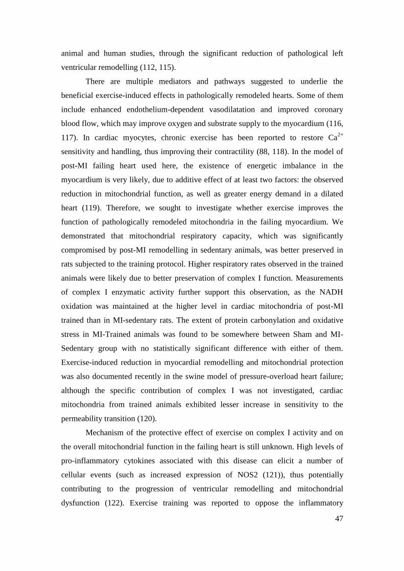
There are multiple mediators and pathways suggested to underlie the
beneficial exercise-induced effects in pathologically remodeled hearts. Some of them
include enhanced endothelium-dependent vasodilatation and improved coronary
blood flow, which may improve oxygen and substrate supply to the myocardium (116,
117). In cardiac myocytes, chronic exercise has been reported to restore Ca2+
sensitivity and handling, thus improving their contractility (88, 118). In the model of
post-MI failing heart used here, the existence of energetic imbalance in the
myocardium is very likely, due to additive effect of at least two factors: the observed
reduction in mitochondrial function, as well as greater energy demand in a dilated
heart (119). Therefore, we sought to investigate whether exercise improves the
function of pathologically remodeled mitochondria in the failing myocardium. We
demonstrated that mitochondrial respiratory capacity, which was significantly
compromised by post-MI remodelling in sedentary animals, was better preserved in
rats subjected to the training protocol. Higher respiratory rates observed in the trained
animals were likely due to better preservation of complex I function. Measurements
of complex I enzymatic activity further support this observation, as the NADH
oxidation was maintained at the higher level in cardiac mitochondria of post-MI
trained than in MI-sedentary rats. The extent of protein carbonylation and oxidative
stress in MI-Trained animals was found to be somewhere between Sham and MI-
Sedentary group with no statistically significant difference with either of them.
Exercise-induced reduction in myocardial remodelling and mitochondrial protection
was also documented recently in the swine model of pressure-overload heart failure;
although the specific contribution of complex I was not investigated, cardiac
mitochondria from trained animals exhibited lesser increase in sensitivity to the
permeability transition (120).
Mechanism of the protective effect of exercise on complex I activity and on
the overall mitochondrial function in the failing heart is still unknown. High levels of
pro-inflammatory cytokines associated with this disease can elicit a number of
cellular events (such as increased expression of NOS2 (121)), thus potentially
contributing to the progression of ventricular remodelling and mitochondrial
dysfunction (122). Exercise training was reported to oppose the inflammatory
Page 49
48
cytokine effects in experimental models of chronic heart disease, which might
counterbalance their detrimental actions, including those affecting mitochondria (123,
124). Increased myocardial expression of antioxidant enzymes, also related to
exercise training, may also confer mitochondrial protection against the proposed ETC
inhibition by reactive oxygen and nitrogen species (125). Furthermore, exercise was
shown to reduce the excessive sympathetic activity that is usually found in CHF (124)
and may act detrimentally through multiple pathways, for example by reducing the
expression of PGC-1α,(126) a master regulator of mitochondrial metabolic function
and biogenesis. However, the PGC-1α down regulation is not uniformly present in the
failing myocardium,(127) and in our model we did not observe altered PGC-1α levels
in any post-MI animal group.
In conclusion, we demonstrated that aerobic interval training in rat post-
infarction chronic heart failure model mitigated contractile deterioration of the left
ventricle. This was paralleled with better preservation of mitochondrial functional
parameters, as evidenced by preserved activity of complex I of the electron transfer
chain. Since damaged mitochondria contribute to the myocardial functional decline
through multiple pathways, their protection conferred by exercise has a significant
potential to repress this vicious cycle and alleviate the progression of contractile
dysfunction in post-infarction failing heart.
Page 50
49
6. MAIN CONCLUSIONS
1. Our experimental rat model for post-infarction heart failure shows adaptations
characteristic for post-infarction left ventricular remodelling. After myocardial
infarction animals had enlarged ventricular cavity, thinned myocardium and
deteriorated contractile function of the left ventricle comparing to the sham-
operated animals.
2. Aerobic interval training induces effective cardiovascular changes in rat heart
after myocardial infarction.
3. Aerobic interval training attenuates deterioration of mitochondrial function in
post-infarction heart failure in rats that underwent 8 weeks of aerobic interval
training:
a. better preservation of mitochondrial respiratory capacity
b. better activity of the complex I of the electron transfer chain
Page 51
50
SUMMARY
Effect of aerobic interval training on pathological remodelling and
mitochondrial dysfunction in the post-infarction failing rat heart
Aims: Following a large myocardial infarction (MI), remaining viable muscle often
undergoes pathological remodelling and progresses towards chronic heart failure
(CHF). Mitochondria may also be affected by this process and, due to their functional
importance, likely contribute to the progression of the disease. Aerobic interval
training (AIT) has been shown effective in diminishing pathological myocardial
transformation, but the effects of AIT on mitochondrial function in hearts undergoing
remodelling are not known.
Methods and Results: Adult female Sprague-Dawley rats were randomized to either 8
weeks of aerobic interval treadmill running (5 days/week), which started 4 weeks
after left coronary artery ligation (MI-Trained), or a sedentary group (MI-Sedentary).
Echocardiography was performed before and after the 8-week period, at which point
the left ventricles (LV) were also harvested. Twelve weeks after surgery, MI-
Sedentary rats had significantly lower LV fractional shortening compared to MI-
Trained. Complex I – dependent respiration assessed in isolated LV mitochondria was
decreased by ~37% in MI-Sedentary and 17% in MI-Trained (group differences
p<0.05), compared to sham-operated animals. This was paralleled with diminished
ATP-production and increased degree of protein oxidation in MI-Sedentary rats. The
enzymatic activity of complex I was also decreased to the greater extent in MI-
Sedentary than in MI-Trained animals, with no evidence of its reduced expression.
When complex II substrate was used, no differences among the three groups were
observed.
Conclusions: Exercise reduces the left ventricle contractile deterioration in post-
infarction heart failure and alleviates the extent of mitochondrial dysfunction, which
is paralleled with preserved complex I activity.
Keywords: myocardial infarction; heart failure; aerobic interval training; animal
model; mitochondria
Page 52
51
SAŢETAK
Uĉinak aerobnog intervalnog treninga na patološko remodeliranje i
mitohondrijsku disfunkciju u štakora s poslijeinfarktnim zatajenjem srca Uvod: Nakon opsežnog infarkta miokarda (IM) u dijelu srčanog mišića izvan
zahvaćenog područja dolazi do patološkog remodeliranja koje ako napreduje dovodi
do razvoja kroničnog srčanog zatajenja. Patološko remodeliranje obuhvaća
makroskopske i mikroskopske promjene na razini srčanog mišića. Remodeliranje na
razini mitohondrija vjerojatno je važni čimbenik napredovanja bolesti zbog njihove
ključne uloge u energijskom metabolizmu stanice. Aerobna intervalna tjelovježba
učinkovito ublažava patološku transformaciju miokarda iako još uvijek nisu dovoljno
istraženi mehanizmi preko kojih se to događa.
Materijali i metode: Ženke štakora Sprague-Dawley randomizirane su u dvije
skupine: jedna je skupina 4 tjedna nakon podvezivanja lijeve koronarne arterije bila
podvrgnuta aerobnoj intervalnoj tjelovježbi koja se sastojala od trčanja na pokretnoj
traci (5 dana/tjedan) dok je druga tijekom istog razdoblja živjela sedentarnim životom.
Prije i poslije perioda tjelovježbe od 8 tjedana učinjeno je ehokardiografsko testiranje.
Nakon provedenih ispitivanja životinje su bile žrtvovane, a iz srca izolirani dijelovi
lijevog ventrikula izvan područja zahvaćenog infarktom.
Rezultati: 12 tjedana nakon kirurškog zahvata sedentarne su životinje imale značajno
nižu frakciju skraćenja lijevog ventrikula u usporedbi s treniranim životinjama. U
mitohondrijima izoliranim iz lijevog ventrikula respiracija preko kompleksa I bila je
37% manja u sedentarnih i 17% manja u treniranih životinja (statistička razlika
p<0.05) u usporedbi s kontrolnom skupinom (Sham štakori). Uz ove promjene u
sedentarnih životinja postoji i smanjena proizvodnja ATP-a i povećana oksidacija
proteina. Enzimska aktivnost kompleksa I respiracijskog lanca elektrona značajno je
smanjena u sedentarnih u usporedbi s treniranim životinjama bez razlike u ekspresiji
proteina kompleksa I. Nema statistički značajne razlike među grupama kada se
respiracija odvijala uz supstrate za respiraciju preko kompleksa II.
Zaključci: Tjelovježba ublažava propadanje kontraktilne funkcije lijevog ventrikula i
ublažava mitohondrijsku disfunkciju uz očuvanu aktivnost kompleksa I u srčanom
mišiću nakon infarkta miokarda
Page 53
52
Ključne riječi: infarkt miokarda; kronično zatajenje srca; aerobni intervalni trening;
životinjski model; mitohondriji
Page 54
53
REFERENCES
1. Yancy CW, Jessup M, Bozkurt B, Butler J, Casey DE, Jr., Drazner MH, et al.
2013 ACCF/AHA guideline for the management of heart failure: a report of the
American College of Cardiology Foundation/American Heart Association Task Force
on Practice Guidelines. Journal of the American College of Cardiology.
2013;62(16):e147-239.
2. McMurray JJ, Adamopoulos S, Anker SD, Auricchio A, Bohm M, Dickstein
K, et al. ESC guidelines for the diagnosis and treatment of acute and chronic heart
failure 2012: The Task Force for the Diagnosis and Treatment of Acute and Chronic
Heart Failure 2012 of the European Society of Cardiology. Developed in
collaboration with the Heart Failure Association (HFA) of the ESC. European journal
of heart failure. 2012;14(8):803-69.
3. Fonseca C. Diagnosis of heart failure in primary care. Heart failure reviews.
2006;11(2):95-107.
4. Mosterd A, Hoes AW. Clinical epidemiology of heart failure. Heart.
2007;93(9):1137-46.
5. Morgan S, Smith H, Simpson I, Liddiard GS, Raphael H, Pickering RM, et al.
Prevalence and clinical characteristics of left ventricular dysfunction among elderly
patients in general practice setting: cross sectional survey. Bmj. 1999;318(7180):368-
72.
6. Johnson FL. Pathophysiology and etiology of heart failure. Cardiology clinics.
2014;32(1):9-19, vii.
7. Wood DA, Coats AJ, Thompson SG, Poole-Wilson PA, Suresh V, Sutton GC.
Incidence and aetiology of heart failure; a population-based study. European heart
journal. 1999;20(6):421-8.
8. Jhund PS, McMurray JJ. Heart failure after acute myocardial infarction: a lost
battle in the war on heart failure? Circulation. 2008;118(20):2019-21.
9. Hellermann JP, Jacobsen SJ, Redfield MM, Reeder GS, Weston SA, Roger
VL. Heart failure after myocardial infarction: clinical presentation and survival.
European journal of heart failure. 2005;7(1):119-25.
Page 55
54
10. Latini R, Masson S, Staszewsky L, Barlera S. Neurohormonal modulation in
heart failure of ischemic etiology: correlates with left ventricular remodeling. Current
heart failure reports. 2006;3(4):157-63.
11. Francis GS. Pathophysiology of chronic heart failure. The American journal of
medicine. 2001;110 Suppl 7A:37S-46S.
12. Lamblin N, Fertin M, de Groote P, Bauters C. Cardiac remodeling and heart
failure after a first anterior myocardial infarction in patients with diabetes mellitus.
Journal of cardiovascular medicine. 2012;13(6):353-9.
13. Meimoun P, M'Barek D, Dragomir C, Luycx-Bore A, Elmkies F, Boulanger J,
et al. [Incidence, associated factors, and follow-up of hospital heart failure
complicating acute anterior myocardial infarction successfully treated by primary
angioplasty]. Annales de cardiologie et d'angeiologie. 2013;62(5):293-300.
14. Ikeda Y, Yutani C, Huang Y, Masuda K, Yuasa T, Kawaguchi O, et al.
Histological remodeling in an ovine heart failure model resembles human ischemic
cardiomyopathy. Cardiovascular pathology : the official journal of the Society for
Cardiovascular Pathology. 2001;10(1):19-27.
15. Gallagher GL, Jackson CJ, Hunyor SN. Myocardial extracellular matrix
remodeling in ischemic heart failure. Frontiers in bioscience : a journal and virtual
library. 2007;12:1410-9.
16. Konstam MA. Patterns of ventricular remodeling after myocardial infarction:
clues toward linkage between mechanism and morbidity. JACC Cardiovascular
imaging. 2008;1(5):592-4.
17. Konstam MA, Udelson JE, Anand IS, Cohn JN. Ventricular remodeling in
heart failure: a credible surrogate endpoint. Journal of cardiac failure. 2003;9(5):350-
3.
18. Hein S, Arnon E, Kostin S, Schonburg M, Elsasser A, Polyakova V, et al.
Progression from compensated hypertrophy to failure in the pressure-overloaded
human heart: structural deterioration and compensatory mechanisms. Circulation.
2003;107(7):984-91.
19. Pfeffer MA, Braunwald E. Ventricular remodeling after myocardial infarction.
Experimental observations and clinical implications. Circulation. 1990;81(4):1161-72.
Page 56
55
20. Schaper J, Kostin S, Hein S, Elsasser A, Arnon E, Zimmermann R. Structural
remodelling in heart failure. Experimental and clinical cardiology. 2002;7(2-3):64-8.
21. Mitchell GF, Lamas GA, Pfeffer MA. Ventricular remodeling after myocardial
infarction. Advances in experimental medicine and biology. 1993;346:265-76.
22. Mitchell GF, Lamas GA, Vaughan DE, Pfeffer MA. Left ventricular
remodeling in the year after first anterior myocardial infarction: a quantitative
analysis of contractile segment lengths and ventricular shape. Journal of the American
College of Cardiology. 1992;19(6):1136-44.
23. Erlebacher JA, Richter RC, Alonso DR, Devereux RB, Gay WA, Jr. Early
infarct expansion: structural or functional? Journal of the American College of
Cardiology. 1985;6(4):839-44.
24. Erlebacher JA, Weiss JL, Weisfeldt ML, Bulkley BH. Early dilation of the
infarcted segment in acute transmural myocardial infarction: role of infarct expansion
in acute left ventricular enlargement. Journal of the American College of Cardiology.
1984;4(2):201-8.
25. Hasenfuss G, Just H. Myocardial phenotype changes in heart failure: cellular
and subcellular adaptations and their functional significance. British heart journal.
1994;72(2 Suppl):S10-7.
26. Yu ZB. [The role of cardiomyocyte apoptosis on mechanism of heart failure].
Sheng li ke xue jin zhan [Progress in physiology]. 2000;31(3):265-8.
27. Kang PM, Izumo S. Apoptosis and heart failure: A critical review of the
literature. Circulation research. 2000;86(11):1107-13.
28. Nadal-Ginard B, Kajstura J, Leri A, Anversa P. Myocyte death, growth, and
regeneration in cardiac hypertrophy and failure. Circulation research. 2003;92(2):139-
50.
29. Brixius K, Frank KF, Bolck B, Hoyer F, Schwinger RH. [Reverse remodeling
of the intracellular Ca(2+)-homeostasis: new concepts of pathophysiology and therapy
of heart failure]. Wiener medizinische Wochenschrift. 2006;156(7-8):209-15.
30. Vescovo G, Dalla Libera L, Serafini F, Leprotti C, Facchin L, Volterrani M, et
al. Improved exercise tolerance after losartan and enalapril in heart failure: correlation
with changes in skeletal muscle myosin heavy chain composition. Circulation.
1998;98(17):1742-9.
Page 57
56
31. Reiser PJ, Portman MA, Ning XH, Schomisch Moravec C. Human cardiac
myosin heavy chain isoforms in fetal and failing adult atria and ventricles. American
journal of physiology Heart and circulatory physiology. 2001;280(4):H1814-20.
32. Pott C, Eckardt L, Goldhaber JI. Triple threat: the Na+/Ca2+ exchanger in the
pathophysiology of cardiac arrhythmia, ischemia and heart failure. Current drug
targets. 2011;12(5):737-47.
33. Baartscheer A, Schumacher CA, Belterman CN, Coronel R, Fiolet JW. SR
calcium handling and calcium after-transients in a rabbit model of heart failure.
Cardiovascular research. 2003;58(1):99-108.
34. Chen YW, Pat B, Gladden JD, Zheng J, Powell P, Wei CC, et al. Dynamic
molecular and histopathological changes in the extracellular matrix and inflammation
in the transition to heart failure in isolated volume overload. American journal of
physiology Heart and circulatory physiology. 2011;300(6):H2251-60.
35. Jane-Lise S, Corda S, Chassagne C, Rappaport L. The extracellular matrix and
the cytoskeleton in heart hypertrophy and failure. Heart failure reviews.
2000;5(3):239-50.
36. Schaper J, Mollnau H, Hein S, Scholz D, Munkel B, Devaux B. [Interactions
between cardiomyocytes and extracellular matrix in the failing human heart]. Z
Kardiol. 1995;84 Suppl 4:33-8.
37. Vanhoutte D, Heymans S. TIMPs and cardiac remodeling: 'Embracing the
MMP-independent-side of the family'. Journal of molecular and cellular cardiology.
2010;48(3):445-53.
38. Vanhoutte D, Schellings M, Pinto Y, Heymans S. Relevance of matrix
metalloproteinases and their inhibitors after myocardial infarction: a temporal and
spatial window. Cardiovascular research. 2006;69(3):604-13.
39. Feldman AM, Li YY, McTiernan CF. Matrix metalloproteinases in
pathophysiology and treatment of heart failure. Lancet. 2001;357(9257):654-5.
40. Polyakova V, Hein S, Kostin S, Ziegelhoeffer T, Schaper J. Matrix
metalloproteinases and their tissue inhibitors in pressure-overloaded human
myocardium during heart failure progression. Journal of the American College of
Cardiology. 2004;44(8):1609-18.
Page 58
57
41. Figueredo VM, Camacho SA. Basic mechanisms of myocardial dysfunction:
cellular pathophysiology of heart failure. Current opinion in cardiology.
1995;10(3):246-52.
42. Minamino T, Kitakaze M. Cellular mechanisms for the treatment of chronic
heart failure: the nitric oxide- and adenosine-dependent pathways. Expert opinion on
emerging drugs. 2002;7(1):99-110.
43. Ikram H. The renin-angiotensin system and cardiac remodelling after acute
myocardial infarction. Heart. 1996;76(3 Suppl 3):68-72.
44. Orn S, Aukrust P. The prediction of adverse cardiac remodelling following
myocardial infarction: defining the need for a dynamic multimarker approach. Heart.
2012;98(15):1112-3.
45. Isidoro Tavares N, Philip-Couderc P, Baertschi AJ, Lerch R, Montessuit C.
Angiotensin II and tumour necrosis factor alpha as mediators of ATP-dependent
potassium channel remodelling in post-infarction heart failure. Cardiovascular
research. 2009;83(4):726-36.
46. Lang CC, Struthers AD. Targeting the renin-angiotensin-aldosterone system in
heart failure. Nature reviews Cardiology. 2013;10(3):125-34.
47. Sayer G, Bhat G. The renin-angiotensin-aldosterone system and heart failure.
Cardiology clinics. 2014;32(1):21-32, vii.
48. Yu Y, Wei SG, Zhang ZH, Gomez-Sanchez E, Weiss RM, Felder RB. Does
aldosterone upregulate the brain renin-angiotensin system in rats with heart failure?
Hypertension. 2008;51(3):727-33.
49. Ventura-Clapier R, Garnier A, Veksler V, Joubert F. Bioenergetics of the
failing heart. Biochimica et biophysica acta. 2011;1813(7):1360-72.
50. Liu J, Wang C, Murakami Y, Gong G, Ishibashi Y, Prody C, et al.
Mitochondrial ATPase and high-energy phosphates in failing hearts. American
journal of physiology Heart and circulatory physiology. 2001;281(3):H1319-26.
51. Ardehali H, Sabbah HN, Burke MA, Sarma S, Liu PP, Cleland JG, et al.
Targeting myocardial substrate metabolism in heart failure: potential for new
therapies. European journal of heart failure. 2012;14(2):120-9.
Page 59
58
52. Jaswal JS, Keung W, Wang W, Ussher JR, Lopaschuk GD. Targeting fatty
acid and carbohydrate oxidation--a novel therapeutic intervention in the ischemic and
failing heart. Biochimica et biophysica acta. 2011;1813(7):1333-50.
53. Abdurrachim D, Luiken JJ, Nicolay K, Glatz JF, Prompers JJ, Nabben M.
Good and bad consequences of altered fatty acid metabolism in heart failure: evidence
from mouse models. Cardiovascular research. 2015.
54. Pratt NG. Pathophysiology of heart failure: neuroendocrine response. Critical
care nursing quarterly. 1995;18(1):22-31.
55. Azevedo PS, Minicucci MF, Santos PP, Paiva SA, Zornoff LA. Energy
metabolism in cardiac remodeling and heart failure. Cardiology in review.
2013;21(3):135-40.
56. Neubauer S. The failing heart--an engine out of fuel. The New England journal
of medicine. 2007;356(11):1140-51.
57. Marin-Garcia J, Akhmedov AT, Moe GW. Mitochondria in heart failure: the
emerging role of mitochondrial dynamics. Heart failure reviews. 2013;18(4):439-56.
58. Chen L, Knowlton AA. Mitochondria and heart failure: new insights into an
energetic problem. Minerva cardioangiologica. 2010;58(2):213-29.
59. Rosca MG, Hoppel CL. Mitochondria in heart failure. Cardiovascular
research. 2010;88(1):40-50.
60. Baumgartner HK, Gerasimenko JV, Thorne C, Ferdek P, Pozzan T, Tepikin
AV, et al. Calcium elevation in mitochondria is the main Ca2+ requirement for
mitochondrial permeability transition pore (mPTP) opening. The Journal of biological
chemistry. 2009;284(31):20796-803.
61. Palmer JW, Tandler B, Hoppel CL. Biochemical differences between
subsarcolemmal and interfibrillar mitochondria from rat cardiac muscle: effects of
procedural manipulations. Archives of biochemistry and biophysics.
1985;236(2):691-702.
62. Ong SB, Hausenloy DJ. Mitochondrial morphology and cardiovascular
disease. Cardiovascular research. 2010;88(1):16-29.
63. Rosca MG, Tandler B, Hoppel CL. Mitochondria in cardiac hypertrophy and
heart failure. Journal of molecular and cellular cardiology. 2013;55:31-41.
Page 60
59
64. Tsutsui H, Kinugawa S, Matsushima S. Oxidative stress and mitochondrial
DNA damage in heart failure. Circulation journal : official journal of the Japanese
Circulation Society. 2008;72 Suppl A:A31-7.
65. Cooper MP. Interplay of mitochondrial biogenesis and oxidative stress in heart
failure. Circulation. 2013;127(19):1932-4.
66. Lu FH, Fu SB, Leng X, Zhang X, Dong S, Zhao YJ, et al. Role of the calcium-
sensing receptor in cardiomyocyte apoptosis via the sarcoplasmic reticulum and
mitochondrial death pathway in cardiac hypertrophy and heart failure. Cellular
physiology and biochemistry : international journal of experimental cellular
physiology, biochemistry, and pharmacology. 2013;31(4-5):728-43.
67. Ventura-Clapier R, Garnier A, Veksler V. Transcriptional control of
mitochondrial biogenesis: the central role of PGC-1alpha. Cardiovascular research.
2008;79(2):208-17.
68. Rimbaud S, Garnier A, Ventura-Clapier R. Mitochondrial biogenesis in
cardiac pathophysiology. Pharmacological reports : PR. 2009;61(1):131-8.
69. Ide T, Tsutsui H, Hayashidani S, Kang D, Suematsu N, Nakamura K, et al.
Mitochondrial DNA damage and dysfunction associated with oxidative stress in
failing hearts after myocardial infarction. Circulation research. 2001;88(5):529-35.
70. Merkle S, Frantz S, Schon MP, Bauersachs J, Buitrago M, Frost RJ, et al. A
role for caspase-1 in heart failure. Circulation research. 2007;100(5):645-53.
71. Narula J, Pandey P, Arbustini E, Haider N, Narula N, Kolodgie FD, et al.
Apoptosis in heart failure: release of cytochrome c from mitochondria and activation
of caspase-3 in human cardiomyopathy. Proceedings of the National Academy of
Sciences of the United States of America. 1999;96(14):8144-9.
72. Rosca MG, Vazquez EJ, Kerner J, Parland W, Chandler MP, Stanley W, et al.
Cardiac mitochondria in heart failure: decrease in respirasomes and oxidative
phosphorylation. Cardiovascular research. 2008;80(1):30-9.
73. Lemieux H, Semsroth S, Antretter H, Hofer D, Gnaiger E. Mitochondrial
respiratory control and early defects of oxidative phosphorylation in the failing human
heart. The international journal of biochemistry & cell biology. 2011;43(12):1729-38.
Page 61
60
74. Sharov VG, Todor AV, Silverman N, Goldstein S, Sabbah HN. Abnormal
mitochondrial respiration in failed human myocardium. Journal of molecular and
cellular cardiology. 2000;32(12):2361-7.
75. Nojiri H, Shimizu T, Funakoshi M, Yamaguchi O, Zhou H, Kawakami S, et al.
Oxidative stress causes heart failure with impaired mitochondrial respiration. The
Journal of biological chemistry. 2006;281(44):33789-801.
76. Ide T, Tsutsui H, Kinugawa S, Utsumi H, Kang D, Hattori N, et al.
Mitochondrial electron transport complex I is a potential source of oxygen free
radicals in the failing myocardium. Circulation research. 1999;85(4):357-63.
77. Scheubel RJ, Tostlebe M, Simm A, Rohrbach S, Prondzinsky R, Gellerich FN,
et al. Dysfunction of mitochondrial respiratory chain complex I in human failing
myocardium is not due to disturbed mitochondrial gene expression. Journal of the
American College of Cardiology. 2002;40(12):2174-81.
78. Rosca M, Minkler P, Hoppel CL. Cardiac mitochondria in heart failure:
normal cardiolipin profile and increased threonine phosphorylation of complex IV.
Biochimica et biophysica acta. 2011;1807(11):1373-82.
79. Drose S. Differential effects of complex II on mitochondrial ROS production
and their relation to cardioprotective pre- and postconditioning. Biochimica et
biophysica acta. 2013;1827(5):578-87.
80. Bayeva M, Gheorghiade M, Ardehali H. Mitochondria as a therapeutic target
in heart failure. Journal of the American College of Cardiology. 2013;61(6):599-610.
81. Dickstein K, Cohen-Solal A, Filippatos G, McMurray JJ, Ponikowski P,
Poole-Wilson PA, et al. ESC Guidelines for the diagnosis and treatment of acute and
chronic heart failure 2008: the Task Force for the Diagnosis and Treatment of Acute
and Chronic Heart Failure 2008 of the European Society of Cardiology. Developed in
collaboration with the Heart Failure Association of the ESC (HFA) and endorsed by
the European Society of Intensive Care Medicine (ESICM). European heart journal.
2008;29(19):2388-442.
82. Belardinelli R, Georgiou D, Cianci G, Purcaro A. Randomized, controlled trial
of long-term moderate exercise training in chronic heart failure: effects on functional
capacity, quality of life, and clinical outcome. Circulation. 1999;99(9):1173-82.
Page 62
61
83. Lachance D, Plante E, Bouchard-Thomassin AA, Champetier S, Roussel E,
Drolet MC, et al. Moderate exercise training improves survival and ventricular
remodeling in an animal model of left ventricular volume overload. Circulation Heart
failure. 2009;2(5):437-45.
84. Experience from controlled trials of physical training in chronic heart failure.
Protocol and patient factors in effectiveness in the improvement in exercise tolerance.
European Heart Failure Training Group. European heart journal. 1998;19(3):466-75.
85. Mentz RJ, Schulte PJ, Fleg JL, Fiuzat M, Kraus WE, Pina IL, et al. Clinical
characteristics, response to exercise training, and outcomes in patients with heart
failure and chronic obstructive pulmonary disease: findings from Heart Failure and A
Controlled Trial Investigating Outcomes of Exercise TraiNing (HF-ACTION).
American heart journal. 2013;165(2):193-9.
86. Lou Q, Janardhan A, Efimov IR. Remodeling of calcium handling in human
heart failure. Advances in experimental medicine and biology. 2012;740:1145-74.
87. Marban E. Calcium and heart failure. Cardiovascular research.
1998;37(2):277-8.
88. Wisloff U, Loennechen JP, Currie S, Smith GL, Ellingsen O. Aerobic exercise
reduces cardiomyocyte hypertrophy and increases contractility, Ca2+ sensitivity and
SERCA-2 in rat after myocardial infarction. Cardiovascular research. 2002;54(1):162-
74.
89. Terblanche SE, Gohil K, Packer L, Henderson S, Brooks GA. The effects of
endurance training and exhaustive exercise on mitochondrial enzymes in tissues of the
rat (Rattus norvegicus). Comparative biochemistry and physiology Part A, Molecular
& integrative physiology. 2001;128(4):889-96.
90. Iemitsu M, Miyauchi T, Maeda S, Sakai S, Fujii N, Miyazaki H, et al. Cardiac
hypertrophy by hypertension and exercise training exhibits different gene expression
of enzymes in energy metabolism. Hypertension research : official journal of the
Japanese Society of Hypertension. 2003;26(10):829-37.
91. Chen CY, Hsu HC, Lee BC, Lin HJ, Chen YH, Huang HC, et al. Exercise
training improves cardiac function in infarcted rabbits: involvement of autophagic
function and fatty acid utilization. European journal of heart failure. 2010;12(4):323-
30.
Page 63
62
92. Nediani C, Raimondi L, Borchi E, Cerbai E. Nitric oxide/reactive oxygen
species generation and nitroso/redox imbalance in heart failure: from molecular
mechanisms to therapeutic implications. Antioxidants & redox signaling.
2011;14(2):289-331.
93. Tsutsui H. Oxidative stress in heart failure: the role of mitochondria. Internal
medicine. 2001;40(12):1177-82.
94. Ascensao A, Ferreira R, Magalhaes J. Exercise-induced cardioprotection--
biochemical, morphological and functional evidence in whole tissue and isolated
mitochondria. International journal of cardiology. 2007;117(1):16-30.
95. Freimann S, Scheinowitz M, Yekutieli D, Feinberg MS, Eldar M, Kessler-
Icekson G. Prior exercise training improves the outcome of acute myocardial
infarction in the rat. Heart structure, function, and gene expression. Journal of the
American College of Cardiology. 2005;45(6):931-8.
96. Ventura-Clapier R. Exercise training, energy metabolism, and heart failure.
Applied physiology, nutrition, and metabolism = Physiologie appliquee, nutrition et
metabolisme. 2009;34(3):336-9.
97. Li J, Sinoway AN, Gao Z, Maile MD, Pu M, Sinoway LI. Muscle
mechanoreflex and metaboreflex responses after myocardial infarction in rats.
Circulation. 2004;110(19):3049-54.
98. Migrino RQ, Zhu X, Morker M, Brahmbhatt T, Bright M, Zhao M.
Myocardial dysfunction in the periinfarct and remote regions following anterior
infarction in rats quantified by 2D radial strain echocardiography: an observational
cohort study. Cardiovasc Ultrasound. 2008;6:17.
99. Hoydal MA, Wisloff U, Kemi OJ, Ellingsen O. Running speed and maximal
oxygen uptake in rats and mice: practical implications for exercise training. Eur J
Cardiovasc Prev Rehabil. 2007;14(6):753-60.
100. Sanbe A, Tanonaka K, Hanaoka Y, Katoh T, Takeo S. Regional energy
metabolism of failing hearts following myocardial infarction. Journal of molecular
and cellular cardiology. 1993;25(9):995-1013.
101. Sharov VG, Goussev A, Lesch M, Goldstein S, Sabbah HN. Abnormal
mitochondrial function in myocardium of dogs with chronic heart failure. J Mol Cell
Cardiol. 1998;30(9):1757-62.
Page 64
63
102. Drexler H, Kastner S, Strobel A, Studer R, Brodde OE, Hasenfuss G.
Expression, activity and functional significance of inducible nitric oxide synthase in
the failing human heart. J Am Coll Cardiol. 1998;32(4):955-63.
103. Clementi E, Brown GC, Feelisch M, Moncada S. Persistent inhibition of cell
respiration by nitric oxide: crucial role of S-nitrosylation of mitochondrial complex I
and protective action of glutathione. Proc Natl Acad Sci U S A. 1998;95(13):7631-6.
104. Neubauer S, Horn M, Cramer M, Harre K, Newell JB, Peters W, et al.
Myocardial phosphocreatine-to-ATP ratio is a predictor of mortality in patients with
dilated cardiomyopathy. Circulation. 1997;96(7):2190-6.
105. Ashrafian H, Frenneaux MP. Metabolic modulation in heart failure: the
coming of age. Cardiovasc Drugs Ther. 2007;21(1):5-7.
106. Weiss RG, Gerstenblith G, Bottomley PA. ATP flux through creatine kinase in
the normal, stressed, and failing human heart. Proc Natl Acad Sci U S A.
2005;102(3):808-13.
107. Nascimben L, Ingwall JS, Pauletto P, Friedrich J, Gwathmey JK, Saks V, et al.
Creatine kinase system in failing and nonfailing human myocardium. Circulation.
1996;94(8):1894-901.
108. Ide T, Tsutsui H, Kinugawa S, Suematsu N, Hayashidani S, Ichikawa K, et al.
Direct evidence for increased hydroxyl radicals originating from superoxide in the
failing myocardium. Circ Res. 2000;86(2):152-7.
109. Suematsu N, Tsutsui H, Wen J, Kang D, Ikeuchi M, Ide T, et al. Oxidative
stress mediates tumor necrosis factor-alpha-induced mitochondrial DNA damage and
dysfunction in cardiac myocytes. Circulation. 2003;107(10):1418-23.
110. Rosca MG, Hoppel CL. New aspects of impaired mitochondrial function in
heart failure. Journal of bioenergetics and biomembranes. 2009;41(2):107-12.
111. Giannuzzi P, Temporelli PL, Corra U, Tavazzi L. Antiremodeling effect of
long-term exercise training in patients with stable chronic heart failure: results of the
Exercise in Left Ventricular Dysfunction and Chronic Heart Failure (ELVD-CHF)
Trial. Circulation. 2003;108(5):554-9.
112. Wisloff U, Stoylen A, Loennechen JP, Bruvold M, Rognmo O, Haram PM, et
al. Superior cardiovascular effect of aerobic interval training versus moderate
Page 65
64
continuous training in heart failure patients: a randomized study. Circulation.
2007;115(24):3086-94.
113. Hambrecht R, Gielen S, Linke A, Fiehn E, Yu J, Walther C, et al. Effects of
exercise training on left ventricular function and peripheral resistance in patients with
chronic heart failure: A randomized trial. JAMA. 2000;283(23):3095-101.
114. Haykowsky MJ, Liang Y, Pechter D, Jones LW, McAlister FA, Clark AM. A
meta-analysis of the effect of exercise training on left ventricular remodeling in heart
failure patients: the benefit depends on the type of training performed. J Am Coll
Cardiol. 2007;49(24):2329-36.
115. Kemi OJ, Hoydal MA, Macquaide N, Haram PM, Koch LG, Britton SL, et al.
The effect of exercise training on transverse tubules in normal, remodeled, and
reverse remodeled hearts. J Cell Physiol. 2011;226(9):2235-43.
116. Crimi E, Ignarro LJ, Cacciatore F, Napoli C. Mechanisms by which exercise
training benefits patients with heart failure. Nature reviews Cardiology.
2009;6(4):292-300.
117. Gielen S, Schuler G, Hambrecht R. Exercise training in coronary artery
disease and coronary vasomotion. Circulation. 2001;103(1):E1-6.
118. de Waard MC, van der Velden J, Bito V, Ozdemir S, Biesmans L, Boontje
NM, et al. Early exercise training normalizes myofilament function and attenuates left
ventricular pump dysfunction in mice with a large myocardial infarction. Circ Res.
2007;100(7):1079-88.
119. Pfeffer JM, Pfeffer MA, Fletcher PJ, Braunwald E. Progressive ventricular
remodeling in rat with myocardial infarction. Am J Physiol. 1991;260(5 Pt 2):H1406-
14.
120. Emter CA, Baines CP. Low-intensity aerobic interval training attenuates
pathological left ventricular remodeling and mitochondrial dysfunction in aortic-
banded miniature swine. Am J Physiol Heart Circ Physiol. 2010;299(5):H1348-56.
121. Singh K, Balligand JL, Fischer TA, Smith TW, Kelly RA. Regulation of
cytokine-inducible nitric oxide synthase in cardiac myocytes and microvascular
endothelial cells. Role of extracellular signal-regulated kinases 1 and 2 (ERK1/ERK2)
and STAT1 alpha. J Biol Chem. 1996;271(2):1111-7.
Page 66
65
122. Lewis NP, Tsao PS, Rickenbacher PR, Xue C, Johns RA, Haywood GA, et al.
Induction of nitric oxide synthase in the human cardiac allograft is associated with
contractile dysfunction of the left ventricle. Circulation. 1996;93(4):720-9.
123. Conraads VM, Beckers P, Bosmans J, De Clerck LS, Stevens WJ, Vrints CJ,
et al. Combined endurance/resistance training reduces plasma TNF-alpha receptor
levels in patients with chronic heart failure and coronary artery disease. Eur Heart J.
2002;23(23):1854-60.
124. Roveda F, Middlekauff HR, Rondon MU, Reis SF, Souza M, Nastari L, et al.
The effects of exercise training on sympathetic neural activation in advanced heart
failure: a randomized controlled trial. J Am Coll Cardiol. 2003;42(5):854-60.
125. Atalay M, Sen CK. Physical exercise and antioxidant defenses in the heart.
Ann N Y Acad Sci. 1999;874:169-77.
126. Arany Z, Novikov M, Chin S, Ma Y, Rosenzweig A, Spiegelman BM.
Transverse aortic constriction leads to accelerated heart failure in mice lacking PPAR-
gamma coactivator 1alpha. Proc Natl Acad Sci U S A. 2006;103(26):10086-91.
127. Karamanlidis G, Nascimben L, Couper GS, Shekar PS, del Monte F, Tian R.
Defective DNA replication impairs mitochondrial biogenesis in human failing hearts.
Circ Res. 2010;106(9):1541-8.
Page 67
66
JASENKA KRALJEVIĆ
Work address: Department of Surgery, University Hospital and School of Medicine
Split
E-mail: [email protected]
PhD student Web page: http://genom.mefst.hr/physiology/cv/jkraljevic.html
Birth Date: February 10th
1984.
Birth Place: Split, Croatia
Working experience
2013 – current: Residency in Abdominal Surgery, University Hospital Split
2010 – 2013: PhD student, Department of Integrative Physiology, School of
Medicine Split
2009 – 2010: Internship at the University Hospital Split
Education
2010 – 2013: Postgraduate School „Evidence Based Medicine”
2008: M.D. (Doctor of Medicine) - University of Split, School of Medicine
Dissertation: Natural Orifice Transluminal Endoscopic Surgery (N.O.T.E.S.):
Experimental Training Models and Possibilities for Clinical Application
Awards
2014: The Best Research and Scientific Publication in Basic Medical Sciences
Annual Award 2012/2013, School of Medicine Split
2008: Dean´s Award for the best graduate student of the School of Medicine
Split
Research activities
Project participation
o 2013
Myocardial energetics as a target for treatment of ischemic heart
disease: A translational approach from patient to mitochondria,
Croatian Science Foundation Guest researcher at The Department of
Circulation and Medical Imaging, Norwegian University of Science and
Technology, Trondheim, Norway
Page 68
67
o 2010
Exercise-induced improvement of chronic heart failure: the role of
KATP channels and mitochondria, scientific project of Unity Through
Knowledge Fund, reintegration grant (3B)
Physiology of SCUBA diving, scientific project of Unity Through
Knowledge Fund, crossing borders grant (1B)
Project participation
o 2009
Natural Orifice Transluminal Endoscopic Surgery (N.O.T.E.S.),
Endoscopic Surgery, Experimental Training Models and Possibilities for
Clinical Application, The Experimental Surgery Laboratory, Department
of Surgery
Research grants
Diving with compressed air and cardiovascular system, scientific project
(216-2160133-0130) – co-investigator
Publications
Aerobic interval training attenuates remodelling and mitochondrial dysfunction in
the post-infarction failing rat heart. Kraljevic J, Marinovic J, Pravdic D, Zubin P,
Dujic Z, Wisloff U, Ljubkovic M. Cardiovasc Res. 2013 May 2.
The influence of varying inspired fractions of O₂ and CO₂ on the development of
involuntary breathing movements during maximal apnoea. Breskovic T, Lojpur M,
Maslov PZ, Cross TJ, Kraljevic J, Ljubkovic M, Marinovic J, Ivancev V, Johnson
BD, Dujic Z. Respir Physiol Neurobiol. 2012 Apr 30; 181(2):228-33.
First Croatian transvaginal laparoscopically assisted cholecystectomies. Perko Z,
Cala Z, Mimica Z, Stipić R, Bakotin T, Kraljević J, Radonić V, Strinić T, Jakus IA,
Simunić M. Hepatogastroenterology. 2012 Mar-Apr; 59(114):351-2.
Cardiovascular changes during underwater static and dynamic breath-hold dives in
trained divers. Breskovic T, Uglesic L, Zubin P, Kuch B, Kraljevic J, Zanchi J,
Ljubkovic M, Sieber A, Dujic Z. J Appl Physiol. 2011 Sep; 111(3):673-8.
Laparoscopic transabdominal preperitoneal approach for inguinal hernia repair: a
five-year experience at a single center. Perko Z, Rakić M, Pogorelić Z, Družijanić
N, Kraljević J. Surg Today. 2011 Feb; 41(2):216-21.
Page 69
68
Laparoscopic cholecystectomy in Cantonal Hospital Livno, Bosnia and Herzegovina
and University Hospital Center Split, Croatia. Perić B, Perko Z, Pogorelić Z,
Kraljević J. Coll Antropol. 2010 Mar; 34 Suppl 1:125-8.
Transvaginal laparoscopically assisted cholecystectomy: a first Croatian experience.
Perko Z, Mimica Z, Stipić R, Radonić V, Cala Z, Bakotin T, Kraljević J, Strinić T,
Jakus IA, Simunić M. Lijec Vjesn. 2009 Mar-Apr; 131(3-4):100-1.
N. O. T. E. S.--natural orifice transluminal endoscopic surgery. Perko Z, Kraljević
J. Lijec Vjesn. 2007 Oct-Nov; 129(10-11):371-2.
Book Chapters
Knjiga: Bol - uzroci i liječenje. Poglavlje: “Visceralna bol u trbuhu”; Perko Z,
Kraljevic J. Urednici: Jukić M; Majerić Kogler V; Fingler M; Izdavač:
Medicinska naklada, Zagreb, 2011. ISBN: 978-953-176-489-6
Teaching activities
2010 to 2013 - Teaching assistant, University of Split, School of Medicine
Courses:
o Physiology at Graduate Degree program in Medicine
o Physiology at Graduate Degree program in Dental Medicine
o Physiology at Undergraduate program in Physiotherapy
o Physiology at Undergraduate program in Radiology
o Physiology at Undergraduate program in Nursery
Affiliations
Croatian Medical Chamber
Croatian Physiological Society
Croatian Society for Endoscopic Surgery
The European Association for Endoscopic Surgery