1
Electronic Supplementary Information
for
Affinity-tunable dual-mode specific recognition of
glycoproteins via boronate affinity-based controllable oriented
surface imprinting
Shuangshou Wang, Jin Ye, Zijun Bie, and Zhen Liu*
*To whom correspondence should be addressed. E-mail:[email protected]
This PDF file includes:
Materials and Methods
Table S1
Figures S1 to S7
Electronic Supplementary Material (ESI) for Chemical ScienceThis journal is © The Royal Society of Chemistry 2013
2
1. MATERIALS AND METHODS
1.1. Reagents and materials
γ-Methacryloxypropyltrimethoxysiliane (γ-MAPS), glycidyl methacrylate (GMA), ribonuclease A
(RNase A), ribonuclease B (RNase B), ovalbumin (OVA), transferrin (TRF), myoglobin (Myo),
sinapinic acid (SA) were purchased from Sigma (St. Louis, MO, USA). Bovine serum albumin (BSA)
was from Shuangliu Zhenglong Chemical and Biological Research Laboratory (Sichuan, China).
Horseradish peroxidase (HRP) and ammonium persulfate (APS) were from Shanghai Lingfeng
Chemical Reagent (Shanghai, China) 3-Aminopropyltriethoxysilane (ATES), dopamine hydrochloride
and 4-formylphenylboronic acid (FPBA) were purchased from J&K Chemical (Shanghai, China).
1-dodecanol, N,N’-methylenebisacylamide (MBAA), N,N-diisopropylethylamine (DIPEA),
o-Benzotriazol-1-yl-N,N,N',N'-tetramethyluronium hexafluorophosphate (HBTU), sodium
cyanoborohydride, 1-hydroxy-7-azabenzotriazole (HOAt) and m-aminophenylboronic acid
monohydrate (APBA) were purchased from Alfa Aesar (Tianjin, China).
3,3,5’,5’-tetramethylbenzidine dihydrochloride (TMB) was from Sinopharm Chemical Reagent
(Shanghai, China). Sodium dodecyl sulfate (SDS) was obtained from Bio-Rad (Hercules, CA, USA).
3-Carboxy-benzoboroxole was in-lab synthesized according to previously reported method.1 All other
chemicals were of analytical or HPLC grade. Water used in all experiments was purified by a Milli-Q
Advantage A10 ultrapure water purification system (Millipore, Milford, MA, USA). Fused-silica
capillaries of 150 µm i.d. and 375 µm o.d. were purchased from Yongnian Optic Plant (Hebei, China).
Amino-modified sensors for binding kinetics assay were kindly provided by Pall Fortebio Analytics
(Shanghai, China).
1.2. Instruments
Scanning electron microscopy (SEM) characterization was performed on a FE-SEM S-4800
instrument (Hitachi, Tokyo, Japan). Atomic Force microscopy (AFM) characterization was performed
on a 5500 AFM instrument (Agilent Technologies, Santa Clara, CA, USA). All chromatographic
separations, except repeatability test, were carried out on a TriSep-2100 pCEC (Unimicro
Technologies, Pleasanton, CA, USA) system equipped with a UV-absorbance detector. The detection
wavelength was set at 214 nm. Repeatability test was performed on an UltiMate 3000 nanoflow LC
Electronic Supplementary Material (ESI) for Chemical ScienceThis journal is © The Royal Society of Chemistry 2013
3
system (Dionex, Sunnyvale, CA, USA) equipped with an LPG-3x00 micropump and a VWD-3400
variable-wavelength UV-vis absorbance detector with a 3 nL flow cell and a WPS-3000 automatic
sampler. The detection wavelength was set at 214 nm.
MALDI-TOF MS analyses were carried out on a 4800 TOF/TOF Analyzer (AB Sciex, Darmstadt,
Germany) equipped with a TOF/TOF ion optics, a 200-Hz Nd:YAG laser, controlled by the 4000
Series Explorer Software (V3.5.28193). Spectra were acquired in the positive linear ion mode between
10 000 and 200 000 m/z with fixed laser intensity (7000). Totally 500 laser shots per spot were
accumulated for each spectrum. The accelerating voltage was 20 kV. The matrix used was 10 mg/mL
SA in 0.1% trifluoroacetic acid:acetonitrile (70:30, v/v). Equivalent amounts (0.5 L) of the sample
and SA were sequentially dropped onto the MALDI plate for MS analysis. Data were processed using
Data Explorer Software Version 4.9 (AB Sciex, Darmstadt, Germany).
The binding properties of HRP-imprinted layers were analyzed on an Octet Red96 instrument
from FroteBio (Menlo Park, CA, USA), which is capable of reading signals from eight sensors
simultaneously. HRP-templated MIP layers were prepared onto the sensor surfaces according to the
proposed method. Binding parameters were obtained by fitting the signals for a series of template
solutions of appropriate concentrations using the software associated with the instrument.
1.3. Preparation of poly(dopamine), poly(APBA) or poly(APBA-co-dopamine) modified
substrates for property characterization
Aqueous solutions consisted of 1) 2.0 mg/mL dopamine and 0.6 mg/mL APS, 2) 1.6 mg/mL APBA
and 0.6 mg/mL APS, and 3) 2.0 mg/mL dopamine, 1.6 mg/mL APBA and 1.2 mg/mL APS, were
separately used to prepared polymeric coatings on glass slides for property characterization. The
self-polymerization reactions were sustained for 24 h at room temperature. After reaction, the glass
slides were rinsed with water to remove remaining reagents and naturally dried in air.
Electronic Supplementary Material (ESI) for Chemical ScienceThis journal is © The Royal Society of Chemistry 2013
4
1.4. Preparation and characterization of HRP-imprinted glass slides
The glass slides were first immobilized with boronic acid. The glass slides were treated with 0.1 M
NaOH and 0.1 M HCl for 1 h each, followed by rinse with water until neutralization (pH 7.0), and
then dried in a ventilated oven at 65C for 30 min. Then the glass substrates were immersed in a 1:1
(v/v) mixture of ATES and THF at 80C for 10 h, followed by rinse with methanol to remove residual
reagents. After that, the amino-modified glass slides were immersed in a methanol solution containing
1 mg/mL FPBA at 25C for 10 h, vibrated during reaction. After the reacting solution was removed,
the glass slides were immersed into 1 mg/mL sodium cyanoborohydride methanol solution at 25C for
another 10 h and vibrated during reaction. Finally, the glass slides were washed with methanol and
water to remove residual reagents, and then dried in an oven.
The boronic acid-functionalized glass slides were immersed into a solution containing 1 mg/mL
HRP and 0.1 M phosphate buffer (pH 8.5) for 10 min to form a thin template layer, followed by
rinsing with 0.1 M phosphate buffer, pH 8.5. Then the template-anchored glass slides were immersed
into an aqueous mixture containing 2.0 mg/mL dopamine, 1.6 mg/mL APBA and 1.2 mg/mL APS at
room temperature for 70 min. Finally, the glass sides were rinsed with 0.1 M HAc containing 10%
SDS (w/v) to remove the template.
To prepare non-imprinted polymer (NIP) covered slides for comparison, the processing procedure
was the same except that no template was immobilized onto the boronic acid-functionalized glass
slides.
1.5. Preparation of poly(APBA-co-dopamine)-coated glass slides for thickness controllability
characterization
Five groups of quartz glass slides were first washed with water and methanol several times to clean the
surface and dried in oven. These glass slides were then immersed into a mixture of 2.0 mg/mL
dopamine, 1.6 mg/mL APBA and 1.2 mg/mL APS, dissolved in 0.1 M phosphate buffer (pH 8.5) at
room temperature for 0.5, 1.0, 1.5, 2.0, and 2.5 h, respectively. After self-polymerization reaction, all
the glass slides were washed with water to remove the remaining reagents, and then dried at room
temperature.
Electronic Supplementary Material (ESI) for Chemical ScienceThis journal is © The Royal Society of Chemistry 2013
5
1.6. Colorimetric detection
To prevent solutions added to the detection spot from dispersing and cross-contaminating, detection
spots were defined by printing a cycle array with hydrophobic ink on the target-imprinted slides and
non-imprinted slides under investigation. In this way, solutions added to each spot were confined
within the predefined area. 10 L samples containing HRP at different concentrations were added to
the spots to incubate for 10 min. After that, each spot was rinsed with 20 L, 0.1 M phosphate buffer,
pH 8.5. Then each spot was supplemented with 10 μL TMB staining solution. After reaction for 10
min, the array was recorded with a digital camera.
1.7. Preparation of poly(APBA-co-dopamine)-coated glycoprotein-imprinted monolithic
capillaries
3-Carboxybenzoboroxole-functionalized monolithic capillaries were first prepared according a method
reported previously 1 as base columns. The base capillaries were conditioned with 0.1 M phosphate
buffer (pH 8.5) for 5 min. Then, 20 L template solution containing 1 mg/mL template dissolved in
0.1 M phosphate buffer (pH 8.5) was injected into the base monolithic capillaries to allow incubation
for 10 min at room temperature to covalently anchor the template. After that, the capillaries were filled
with 20 L imprinting solution containing 2.0 mg/mL dopamine, 1.6 mg/mL APBA and 1.2 mg/mL
APS which dissolved in 0.1 M phosphate buffer (pH 8.5) and kept at room temperature for 30, 70 and
90 min for the templates RNase B, HRP and TRF, respectively. Finally, the capillaries were rinsed
with 0.1 M HAc containing 10% SDS (w/v) to remove the templates. The procedure for non-imprinted
monolithic columns was the same except that no templates were immobilized onto the
benzoboroxole-functionalized monolithic capillaries.
1.8. Preparation of poly(APBA-co-dopamine) coated HRP-imprinted monolithic column without
template pre-immobilization
The procedure was nearly the same as the oriented imprinting approach except that HRP was not
pre-immobilized onto the benzoboroxole-functionalized monolithic capillary and the imprinting
solution contained not only dopamine, APBA and APS at the same concentrations as above but also 1
mg/ml HRP.
Electronic Supplementary Material (ESI) for Chemical ScienceThis journal is © The Royal Society of Chemistry 2013
6
1.9. Preparation of poly(dopamine) or poly(APBA)-coated HRP-imprinted monolithic capillaries
The procedure was the same as the one using poly(APBA-co-dopamine) as imprinting coating, except
that the imprinting solutions contained either dopamine or APBA.
1.10. Preparation of HRP-imprinted layers on the Fortebio sensors for binding kinetics assays
Amino-immobilized sensors were firstly immersed into a methanol solution containing 5 mg/mL
4-formylphenylboronic acid and 1 mg/mL sodium cyanoborohydride, then reacted at room
temperature for 10 h. After that, the sensors were washed with water for several times to remove the
remaining reagent, and then immersed into a 1 mg/mL HRP solution prepared with 0.1 M phosphate
buffer (pH 8.5) and incubated for 30 min at room temperature. Then, the sensors were washed with 0.1
M phosphate buffer (pH 8.5) for several times. Consequently, the sensors were immersed into an
imprinting solution containing 2.0 mg/mL dopamine, 1.6 mg/mL APBA and 1.2 mg/mL APS, which
dissolved in 0.1 M phosphate buffer (pH 8.5), and kept at room temperature for 70 min. Finally, the
sensors were washed with 0.1 M HAc solution containing 10% SDS (w/v) to remove the templates.
Non-imprinted sensors were prepared as controls using the same procedure except that no templates
were immobilized onto the sensors.
1.11. MALDI-TOF MS analysis of serum treated with TRF-imprinted monolithic capillary
The serum sample was diluted by 20 times with ultrapure water, then frozen immediately and stored at
-20 C. The samples were thawed at room temperature prior to analysis. A piece of TRF-imprinted
monolithic capillary with effective length of 33 cm was used as an extraction column. 10 l of diluted
serum sample was pumped through the column slowly and the flow-out part was collected in a 200-μL
centrifuge tube. Then, the column was washed with 20 μL ultrapure water to completely remove
uncaptured species within the column. Finally, the column was rinsed with 20 μL 0.1 M HAc, and the
flow out liquid was collected in a centrifuge tube for MALDI-TOF MS analysis.
1.12. Repeatability test of HRP-imprinted monolithic capillary
The repeatability of HRP-imprinted column was tested, results as shown in Fig. S7. RSD (n=10) of
retention time and peak height was 0.11% and 0.23%, respectively, showing excellent repeatability of
the HRP-imprinted monolithic capillary.
Electronic Supplementary Material (ESI) for Chemical ScienceThis journal is © The Royal Society of Chemistry 2013
7
Reference:
1. Li, H., Wang, H., Liu, Y. & Liu, Z. A benzoboroxole-functionalized monolithic column for the
selective enrichment and separation of cis-diol containing biomolecules. Chemical
Communications 48, 4115-4117 (2012).
Electronic Supplementary Material (ESI) for Chemical ScienceThis journal is © The Royal Society of Chemistry 2013
8
2. Supporting Data
Table S1. Relative content of C, O, B and N in different polymers measured by XPS (N as a
standard).
Polymer Relative content
C O B N
Poly(APBA) 10.11 7.13 0.98 1.00
Poly(dopamine) 8.18 8.37 0.00 1.00
Poly(APBA-co-dopamine) 10.05 1.85 0.61 1.00
1200 1000 800 600 400 200 0
0
300000
600000
900000
1200000 poly(APBA-co-dopamine) poly(APBA) poly(dopamine)
Co
un
ts /
s
Binding Energy (eV)
O1sC1s
N1s B1s
A
180 185 190 195
2000
4000
6000
8000
poly(APBA-co-dopamine) poly(APBA) poly(dopamine)
Co
un
ts /
s
Binding Energy (eV)
B
525 530 535 540
0
30000
60000
90000
120000
150000
Co
un
ts /
s
Binding Energy (eV)
poly(APBA-co-dopamine) poly(APBA) poly(dopamine)
C
279 282 285 288 291 294
0
20000
40000
60000
poly(APBA-co-dopamine) poly(APBA) poly(dopamine)
Co
un
ts /
s
Binding Energy (eV)
D
Figure S1. The X-ray photoelectron spectroscopy survey scan (A), the boron element (B), the
oxygen element (C) and the carbon element (D) of the substrates modified with different
monomers.
Electronic Supplementary Material (ESI) for Chemical ScienceThis journal is © The Royal Society of Chemistry 2013
9
0 10 20 30 40
0
20
40
60
80
UV
Ab
sorb
ance
time / min
mobile phase switching point
Blank
TRF
RNase B
OVA
HRP
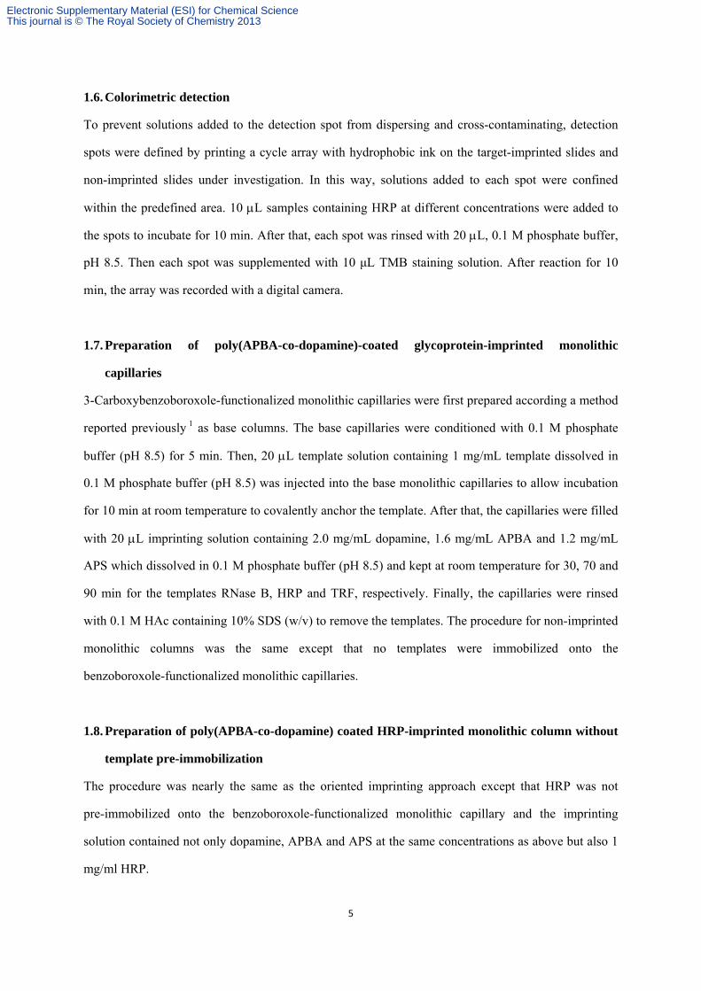
Figure S2. Chromatographic retention of glycoproteins on HRP-imprinted column without
pre-immobilization of the template. Mobile phase: 0.1 M phosphate buffer, pH 7.4, switched
to 0.1 M acetic acid at 18 min. Blank sample, 0.1 M phosphate buffer, pH 7.4. Sample: 1
mg/mL protein dissolved in 0.1 M phosphate buffer, pH 7.4.
0 10 20 30 40
0
10
20
30
40
50
60
UV
Ab
sorb
ance
time / min
poly(APBA) column
poly(dopamine) column
poly(APBA-co-dopamine) column
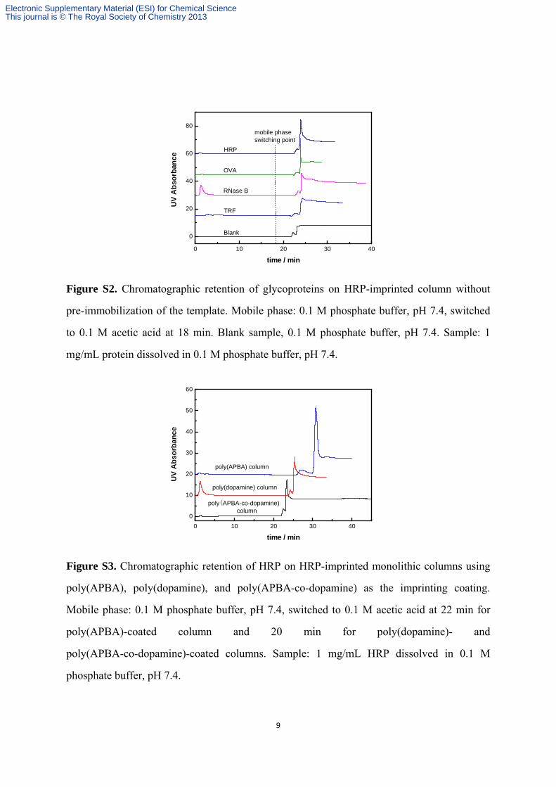
Figure S3. Chromatographic retention of HRP on HRP-imprinted monolithic columns using
poly(APBA), poly(dopamine), and poly(APBA-co-dopamine) as the imprinting coating.
Mobile phase: 0.1 M phosphate buffer, pH 7.4, switched to 0.1 M acetic acid at 22 min for
poly(APBA)-coated column and 20 min for poly(dopamine)- and
poly(APBA-co-dopamine)-coated columns. Sample: 1 mg/mL HRP dissolved in 0.1 M
phosphate buffer, pH 7.4.
Electronic Supplementary Material (ESI) for Chemical ScienceThis journal is © The Royal Society of Chemistry 2013
10
0 10 20 30 40
0
20
40
60
80
UV
Ab
sorb
ance
time / min
Blank
OVA
RNase B
TRF
HRP
mobile phaseswitching point
Figure S4. Chromatographic retention of glycoproteins on HRP-imprinted monolithic column
with poly(APBA) as the imprinting coating. Mobile phase: 0.1 M phosphate buffer, pH 7.4,
switched to 0.1 M acetic acid at 22.5 min. Blank sample, 0.1 M phosphate buffer, pH 7.4.
Sample: 1 mg/mL protein dissolved in 0.1 M phosphate buffer, pH 7.4.
0 5 10 15
0
5
10
15
20
25
UV
Ab
sorb
ance
time / min
NIP MIP
Figure S5. Breakthrough curve of HRP-imprinted monolithic column. Mobile phase: 0.1 M
phosphate buffer (pH 7.4) containing 1 mg/mL HRP and 1 mg/mL RNase A.
Electronic Supplementary Material (ESI) for Chemical ScienceThis journal is © The Royal Society of Chemistry 2013
11
0 200 400 600 800
0.00
0.05
0.10
0.15
0.20
0.25
0.30
shif
t / n
m
time / s
10 ug/mL 5 ug/mL 2.5 ug/mL 1.25 ug/mL
A
0 200 400 600 800
0.00
0.05
0.10
0.15
0.20
0.25
0.30
sh
ift
/ n
m
time / s
10 g/mL 5 g/mL 2.5 g/mL 1.25 g/mL
B
0 200 400 600 8000.0
0.1
0.2
0.3
0.4
0.5
0.6
shif
t /
nm
time / s
10 g/mL 5 g/mL 2.5 g/mL 1.25 g/mL
C
0 200 400 600 800
0.0
0.1
0.2
0.3
0.4
0.5
0.6
sh
ift
/ n
m
time / s
10 g/mL 5 g/mL 2.5 g/mL 1.25 g/mL
D
0 200 400 600 800
0.0
0.2
0.4
0.6
0.8
sh
ift
/ n
m
time / s
10 g/mL 5 g/mL 2.5 g/mL 1.25 g/mL
E
0 200 400 600 800
0.0
0.2
0.4
0.6
0.8
shif
t /
nm
10 g/mL 5 g/mL 2.5 g/mL 1.25 g/mL
time / s
F
0 200 400 600 800-0.2
0.0
0.2
0.4
0.6
0.8
1.0
shif
t /
nm
time / s
10 ug/mL 5 ug/mL 2.5 ug/mL 1.25 ug/mL
G
0 200 400 600 800-0.2
0.0
0.2
0.4
0.6
0.8
1.0
10 g/mL 5 g/mL 2.5 g/mL 1.25 g/mL
shif
t /
nm
time / s
H
Figure S6. Binding curves for HRP-imprinted layers (A, C, E and G) and non-imprinted (B,
D, F and H) layers on the sensors at binding pH. Buffer for association: 0.1 M phosphate
containing different HRP concentrations at different pH. Buffer for dissociation: 0.1 M
phosphate. pH: 3.0 for A and B; 5.5 for C and D; 7.4 for E and F; 9.0 for G and H.
Electronic Supplementary Material (ESI) for Chemical ScienceThis journal is © The Royal Society of Chemistry 2013
12
0 20 40
0
20
40
60
80
100
120
0 20 40 0 20 40 0 20 40 0 20 40 0 20 40 0 20 40 0 20 40 0 20 40 0 20 40
time / min
UV
Abso
rban
ce
Figure S7. Repeatability test of HRP-imprinted column (n = 10). Mobile phase: 0.1 M
phosphate buffer, pH 7.4, switched to 0.1 M acetic acid at 20 min. Sample: 1 mg/mL HRP
dissolved in 0.1 M phosphate buffer, pH 7.4.
Electronic Supplementary Material (ESI) for Chemical ScienceThis journal is © The Royal Society of Chemistry 2013